 Zilda Cristiani Gazim1,2*
 Juliana Silveira Valle3,2
 Isabela Carvalho dos Santos2
 Isabelle Luiz Rahal1
 Gabriela Catuzo Canonico Silva1
 Ana Daniela Lopes4
 Suelen Pereira Ruiz5
 Maria Graciela Iecher Faria5
 Ranulfo Piau Junior2
 Daniela Dib Gonçalves2
- 1Chemistry Laboratory of Natural Products, Graduate Program in Animal Science and Biotechnology Applied to Agriculture, Paranaense University, Umuarama, Brazil
- 2Preventive Veterinary Medicine and Public Health Laboratory, Postgraduate Program in Animal Science with an Emphasis on Bioactive Products, Paranaense University, Umuarama, Brazil
- 3Molecular Biology Laboratory, Graduate Program in Animal Science and Biotechnology Applied to Agriculture, Paranaense University, Umuarama, Brazil
- 4Agricultural Microbiology and Nematology Laboratory, Graduate Program in Biotechnology Applied to Agriculture, Paranaense University, Umuarama, Brazil
- 5Laboratory of Biotechnology of Plant Products and Microorganisms, Postgraduate Program in Biotechnology Applied to Agriculture, Paranaense University, Umuarama, Brazil
Baccharis dracunculifolia DC (Lamiaceae) (Asteraceae) is found in South America, mainly in Argentina, Brazil, Bolivia, Paraguay and Uruguay. Folk medicine is used as a sedative, hypotensive, bronchodilator, cardiovascular disorders, anti-flu, and also in skin wounds. Considered the main source of green propolis, which increases the pharmacological interest in this species. It is also known as a “benefactor” plant facilitating the development of other plant species around it, being indicated for the recovery of degraded areas. This species has been studied for decades in order to isolate and identify the active principles present in the aerial parts (leaves and flowers) and roots. The present study consists of a review of the scientific literature addressing the ethnobotanical, ethnomedicinal, phytochemical, pharmacological and potential cytotoxic effects of the B. dracunculifolia species. In this survey, we sought to investigate issues related to the botanical and geographic description of the species, the ethnobotanical uses, as well as the phytochemical studies of the essential oil, extracts and green propolis obtained from the aerial parts and roots of B. dracunculifolia. Using high precision analytical tools, numerous compounds have already been isolated and identified from leaves and flowers such as the flavonoids: naringenin, acacetin, dihydrokaempferol, isosakuranetin and kaempferide; phenolic acids: p-coumaric, dihydrocoumaric, ferulic (E)-cinnamic, hydroxycinnamic, gallic, caffeic, and several caffeoylquinic acids derivatives; phenolic acids prenylated: artepillin C, baccharin, drupanin; the glycosides dracuculifosides and the pentacyclic triterpenoids: Baccharis oxide and friedelanol. The predominant class in the essential oil of leaves and flowers are terpenoids comprising oxygenated monoterpenes and sesquiterpenes, highlighting the compounds nerolidol, spathulenol, germacrene D and bicyclogermacrene. These compounds give the species high antimicrobial, antioxidant, antitumor, analgesic, immunomodulatory and antiparasitic potential, making this species a promising herbal medicine. In vitro toxicity assays with B. dracunculifolia extract showed low or no cytotoxicity. However, in vivo analyses with high doses of the aqueous extract resulted in genotoxic effects, which leads us to conclude that the toxicity of this plant is dose-dependent.
1 Introduction
B. dracunculifolia is a medicinal bush widely distributed in South America. In Brazil, it is popularly known as “Vassourinha” or “Alecrim do Campo”, it is a source of a natural resinous substance produced by bees (Apis mellifera), called green propolis (Belini et al., 2016). Due to its rapid vegetative development, this species is widely used in the restoration of degraded areas, acting as a “nurse species” facilitating the development of other plant species (“beneficiaries”). This benefit occurred in areas where B. dracunculifolia were planted, indicating improvements in conditions of temperature, humidity and availability of organic matter in the soil (Siqueira et al., 2022). In folk medicine, tea made from the leaves is used for liver and stomach problems (Bonin et al., 2020). In addition, the infusion and decoction of the flowers are widely used in alternative medicine to treat inflammatory processes as well as liver disorders and stomach ulcers (Salazar et al., 2018).
Research carried out with B. dracunculifolia indicates biological activities such as anti-inflammatory (Santos et al., 2010; Brandenburg et al., 2020; França et al., 2022), antiulcerogenic (Klopell et al., 2007; Lemos et al., 2007; Massignani et al., 2009; Costa et al., 2019; Boeing et al., 2021), immunomodulatory (Burgos et al., 2022), neuroprotective (França et al., 2022), antioxidant (Veiga et al., 2017; Casagrande et al., 2018; Tomazzoli et al., 2021; Luchesi et al., 2022; Monteiro et al., 2022); antimicrobial (Veiga et al., 2017; Casagrande et al., 2018; Salazar et al., 2018; Cazella et al., 2019; Pedrotti et al., 2019; Bonin et al., 2020; Timbé et al., 2021; Barbosa et al., 2022; Bernardes et al., 2022; Monteiro et al., 2022), anti-phytopathogenic (Luchesi et al., 2022), insecticide and acaricide (Chaaban et al., 2018; Cazella et al., 2020).
The biological potential found in this species is due to the presence of phenolic and terpenoid compounds. The most common phenolic acids occur in prenylated form, highlighting drupanin, artepillin C, baccharin and baccharin-5″-aldehyde (Minteguiaga et al., 2022). The flavonoids found in aerial parts of B. dracunculifolia are naringenin, acacetin, dihydrokaempferol, isosakuranetin and kaempferide (Nagatani et al., 2002a), and are often reported as major constituents of green propolis (Bonin et al., 2020).
The essential oil present in the leaves and flowers is colorless with a characteristic odor of honey (Cazella et al., 2019), being highly valued in the perfumery industry. Trans-nerolidol as the major compound of the essential oil, is regulated by the Food and Drug Administration (FDA) and is used as a flavoring agent in the food industry (Belini et al., 2016). As for the chemical composition, it consists of terpenoids, with the majority class of oxygenated sesquiterpenes and hydrocarbons (Cazella et al., 2019; Minteguiaga et al., 2022).
Thus, this review aims to address the ethnobotanical, ethnomedicinal, phytochemical and pharmacological aspects of the essential oil, extracts and green propolis of B. dracunculifolia.
2 Asteraceae family
The Asteraceae family is one of the largest flowering plant families, including over 1,600 genera and 25,000 species worldwide. This family includes known species of medicinal importance such as wormwood, chamomile and dandelion. Most species that make up the Asteraceae family have a long history in traditional medicine; some members have been cultivated for over 3,000 years for medicinal and food purposes. They have a wide range of pharmacological activities, with emphasis on anti-inflammatory, antimicrobial, antioxidant and hepatoprotective activities (Rolnik and Olas, 2021).
The Asteraceae are distributed throughout the world, with the exception of Antarctica, with habitats ranging from forests to savanas. Regarding morphology, some species are trees reaching more than 30 m, others are shrubs such as B. dracunculifolia and most species are perennial herbs (Bohm and Stuessy, 2001).
2.1 Botanical description, geographic distribution
B. dracunculifolia is a medicinal ethnobotanical plant belonging to the flora of the Americas, mainly South America. In this continent it is known as an aromatic plant. From the aerial parts, the essential oil is extracted and used in the perfumery industry, in addition to its known therapeutic potential (Minteguiaga et al., 2022). Furthermore, this species provides many services to the ecosystem, including assisting in the formation of a rich fauna of insects and different pollinating species essential for the functioning and sustainability of ecosystems (Fernandes et al., 2018).
B. dracunculifolia is also known as a “benefactor” plant facilitating the development of other plant species (“beneficiary”) (Siqueira et al., 2022), in addition to improving the conditions of the habitat in which it is present (for example, humidity temperature, and availability of soil organic matter) (Lu et al., 2018). In that regard, Siqueira et al. (2022) evaluated the effects of B. dracunculifolia cultivation on the native plant community of an Atlantic Forest degraded area in a short period. Besides that, the study evaluated two areas of abandoned pasture colonized by grasses of the genus Urochloa spp, which originally represented riparian forest fragments in the Piranga River basin, one of the tributaries of the Rio Doce, in Minas Gerais, Brazil. In one of the areas, B. dracunculifolia was planted (restoration treatment); in the other, the pre-existing vegetation was maintained, remaining the same without intervention (degrading treatment). After 18 months of planting, the authors recorded all plant species (except Baccharis and grasses) and classified them as native, ruderal and invasive. The area cultivated with B. dracunculifolia (restored environment) showed greater species richness than the area without intervention (degraded environment). The values observed for species diversity in the areas exposed to this treatment were also higher with 17 exclusive native species, against three non-exclusive native species, in the treatment without intervention (degraded). Furthermore, fewer ruderal and invasive species were found in the areas restored with B. dracunculifolia, evidencing its importance in the recomposition of the native plant community, associated with the reduced chance of invasion by exotic species, and the possibility of use in the restoration of riparian forests, protecting mainly river springs (Siqueira et al., 2022).
2.1.1 Synonymies
Baccharis dracunculifolia DC. includes the subspecies Baccharis dracunculifolia subsp. Dracunculifolia and Baccharis dracunculifolia subsp. tandilensis (Speg.) Giuliano. Baccharis dracunculifolia subsp. Dracunculifolia has six synonyms: Baccharis bracteata Hook. & Arn, Baccharis dracunculifolia f. subviscosa Kuntze, Baccharis dracunculifolia var. integerrima Kuntze, Kuntze, Baccharis leptospermoides DC., Baccharis paucidentata Sch. Bip. ex Baker and Baccharis pulverulenta Klatt (POWO Plants of the World Online, 2022). Baccharis dracunculifolia subsp. tandilensis has one synonym: Baccharis tandilensis Speg. (POWO Plants of the World Online, 2022).
2.1.2 Popular names
B. dracunculifolia has different popular names in the various regions in which it is found. In Brazil, it is popularly known as “alecrim-do-campo”, “alecrim-bassoura”, “vassoura”, “vassourinha”, “vassoureira”, “vassourão", “bassoura-branca” and “erva-de-São-João-Maria” (Viera, 2011; Alves et al., 2018; Casagrande et al., 2018; Bonin et al., 2020; Minteguiaga et al., 2022). The Guarani indigenous people call it Ju’i vatã (Pereira et al., 2016).
2.1.3 Species description
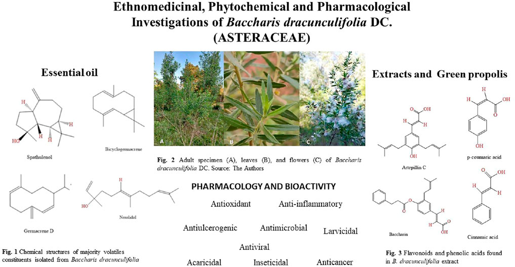
B. dracunculifolia is a bush species that can reach 2.0–3.0 m in height, with hairy branches (Figure 1A) (Vieira, 2011). The leaves are lanceolate, membranous, uninerve, measuring 1.0–2.5 cm long and 3–4 mm wide, densely punctuated with glands, with entire margins or with one to three teeth, rarely with more than three teeth (Figure 1B) (Vieira, 2011). Regarding its flowers, this plant is multi-flowered, measuring between 3 and 4 mm in height and between 3 and 4 mm in diameter. The female flower has a corolla about 2 mm long and has a toothed edge, the male flower, pentasecta, is about 2.5 mm long, and the glabrous achene is approximately 1.5 mm long (Figure 1C) (Vieira, 2011).
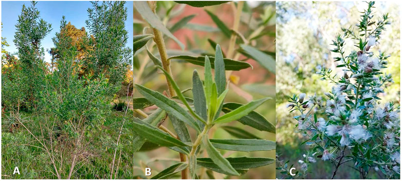
FIGURE 1. Adult specimen (A), leaves (B), and flowers (C) of Baccharis dracunculifolia DC. Source: The Authors.
2.1.4 Geographic distribution
B. dracunculifolia is distributed in South America: Argentina (Northeast and Northwest), Bolivia, Brazil (South, Southeast, and West-Central), Paraguay, Peru, and Uruguay (Powo Plants of the World Online, 2022) (Figure 2). It is present in different types of vegetation, occurring both in the fields of the plateau and in the coastal sandbanks. Currently, its greatest occurrence is in anthropic areas: capoeiras, edges of forests and paths, clearings within capoeirões, and margins of swamps, making it one of the most characteristic Brazilian Baccharis as anthropic pioneers (Vieira, 2011). However, its phytogeographic domains are the Atlantic Forest, the Cerrado, and the Pampas (Heiden, 2022; Minteguiaga et al., 2022).
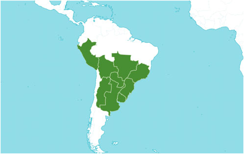
FIGURE 2. Geographical distribution of Baccharis dracunculifolia (Source: Plants of the World Online, Royal Botanic Gardens, Kew. 2022. Licensed under Creative Commons Attribution CC BY).
2.1.5 Ethnobotanical uses
In traditional communities in Latin America, mainly in Argentina, Brazil and Bolivia, species of Baccharis L. are known and widely used. Traditional literature describes the use of these species as food, fodder for livestock, tools, and dyeing plants (Ritter et al., 2021).
The ethnobotanical knowledge that we have about the species that make up the genus Baccharis is directly related to their medicinal use. For example, in South American folk medicine, several species of Baccharis are used to treat malaria and parasitic infections (Parreira et al., 2010). These species are also indicated as antiseptics for the treatment of wounds and skin ulcers, as well as for treating fever and gastrointestinal diseases; they also act as spasmolytics, diuretics, analgesics and in the treatment of diabetes and infections of bacterial and fungal origin (Abad and Bermejo, 2007).
Due to its bush size and the greater strength of its wood (Figure 1A), in several Brazilian regions B. dracunculifolia is used in the manufacture of domestic and agricultural utensils and also as free wood. It is known as “vassoura” because it is used to build rustic brooms to sweep the floor mainly from outdoor areas. Another interesting aspect of this species is its use in religious rituals, mainly in communities in the states of Bahia and São Paulo, Brazil (Ritter et al., 2021).
Teas in the form of infusion and decoction prepared with the flowering plant (Figure 1C) are used in folk medicine to treat inflammatory processes, liver disorders and stomach ulcers (Salazar et al., 2018). It is an aromatic species used by the Guarani Indians in personal hygiene (Pereira et al., 2016).
3 Chemo-profiling
B. dracunculifolia is one of the most studied species from the phytochemical and pharmacological point of view within the genus Baccharis. The presence of phenolic acids, diterpenes, flavonoids, triterpenes, different types of glycosides and essential oils was shown when chemical evaluation of both aerial and underground parts was performed (Campos et al., 2016).
3.1 B. dracunculifolia essential oil
The economic importance of the essential oil composition of B. dracunculifolia (“vassoura” oil) has been widely described in the literature. The main compound, considered an important chemomarker, present in male and female individuals, is the acyclic alcohol sesquiterpene (E)-nerolidol, a compound with antiulcerative activity (Minteguiaga et al., 2021).
3.1.1 Physical-chemical characteristics
The essential oil is found in the aerial parts that comprise the leaves and flowers is colorless (Figure 3), with a characteristic honey odor (Cazella et al., 2019). The quality of essential oils is evaluated by physicochemical analyses. Some of the parameters to detect adulterations in essential oils are the determination of rotating power density and refractive index (Gil, 2007). To the product application and commercialization are necessary carrying out biological tests such as determining the yield of essential oils within the plant. The European Pharmacopoeia standardizes that 2 ml/kg is the minimum extraction yield of essential oils for product development and application (European Pharmacopoeia, 2013). The physicochemical indices of B. dracunculifolia essential oil are shown in Table 1.
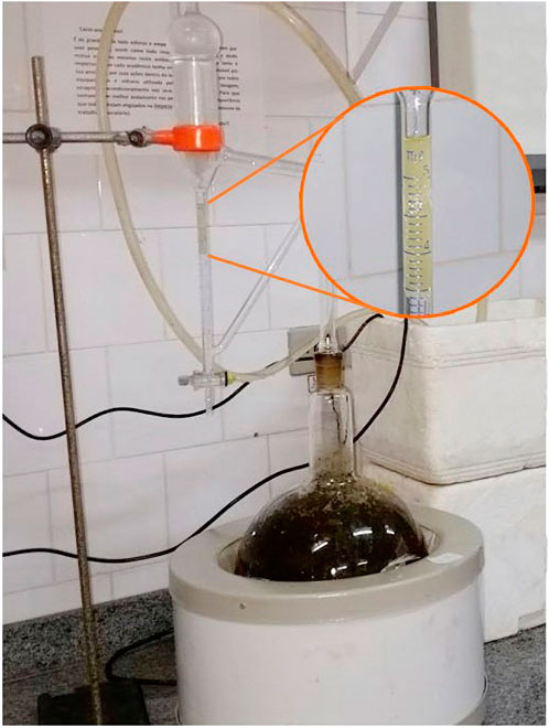
FIGURE 3. Essential oil obtained by hydrodistillation process of B. dracunculifolia leaves and flowers - Source: Chemical Laboratory of Natural Products-Paranaense University-UNIPAR, Brazil. Source: The Authors.
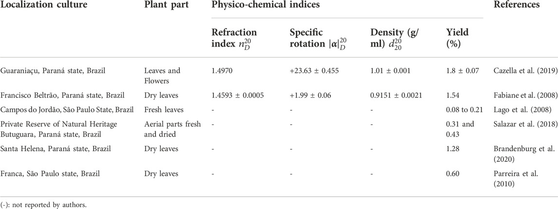
TABLE 1. Physico-chemical indexes of Baccharis dracunculifolia essential oil.
3.1.2 Chemical composition
The presence of a complex structure of terpenoids was confirmed in the essential oil of B. dracunculifolia. Oxygenated sesquiterpenes nerolidol, sphatulenol, and the hydrocarbon sesquiterpenes germacrene D and byciclogermacrene are the major compounds (Cazella et al., 2019; Minteguiaga et al., 2022). However, factors such as the location of the crop implantation, climate, altitude, soil type, and especially the collection time can alter the concentration of these terpenoids. Thus, Table 2 and Figure 4 represent the chemical composition of the essential oil of B. dracunculifolia cultivated in different locations.
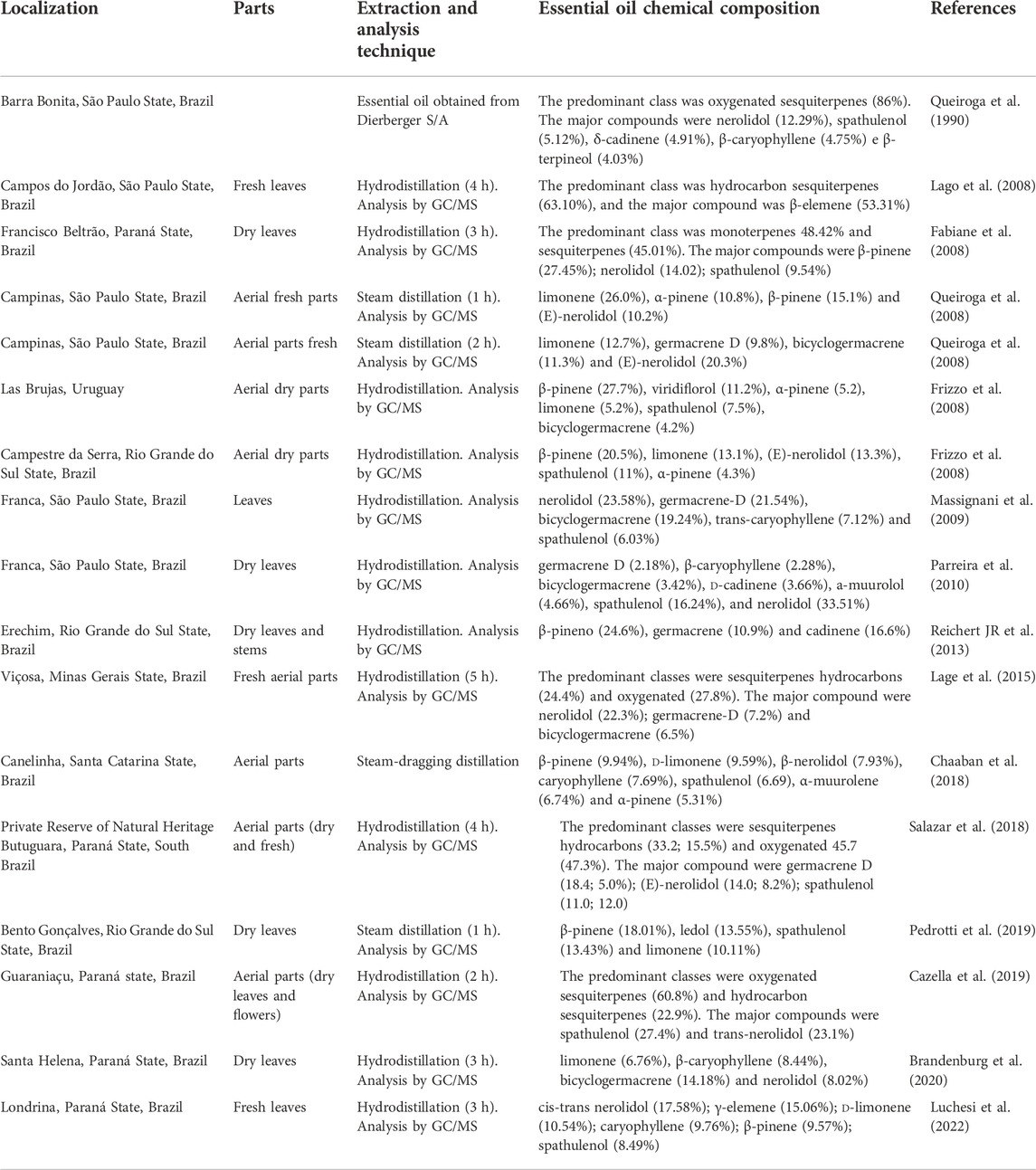
TABLE 2. Chemical Composition of Baccharis dracunculifolia essential oil.
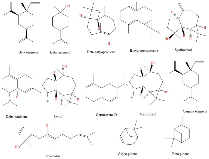
FIGURE 4. Chemical structures of majority volatiles constituents isolated from Baccharis dracunculifolia.
3.2 B. dracunculifolia leaf and flowers extracts
The main chemical constituents in plants of the genus Baccharis are phenolic and terpenoid compounds. In addition, diterpene and flavonoid compounds and phenolic acids such as derivatives of cinnamic and chlorogenic acids (Supplementary Figure S1) are also present. Diterpenes are among the most abundant compounds present in the species of this genus, highlighting those with neo-clerodane, labdane, and kaurane skeletons (Supplementary Figure S2) (Burgos et al., 2022).
In an experiment conducted by Nagatani et al. (2002a), using aerial parts of B. dracunculifolia collected in the city of São Paulo, Brazil. From this plant material, a methanolic extract was prepared and this was partitioned in water. The suspended fraction was then extracted with diethyl ether, resulting in two fractions: ethyl and aqueous. The ethyl fraction was concentrated and the residue partitioned into two solutions: the first composed of benzene-n-hexane (1:1) and the second composed of 80% methanol in water. The residue derived from the methanolic solution was separated on a silica gel column and analyzed by High Performance Liquid Chromatography (HPLC) indicating the presence of the flavonoids naringenin, acacetin, dihydrokaempferol, isosakuranetin, kaempferide, among others (Nagatani et al., 2002a) (Supplementary Figure S3).
According to Minteguiaga et al. (2022), the phenolic acids such as gallic, p-coumaric, ferulic (E)-cinnamic, hydroxycinnamic, caffeic, dihydrocoumaric, and several caffeoylquinic acids derivatives are common components when medium polar to polar organic and hydroalcoholic extracts of B. dracunculifolia are studied (Supplementary Figure S4).
The prenylated derivatives such as 3,5-diprenyl-p-coumaric (artepillin C), 3-prenyl-4-dihydrocinnamoiloxy-cinnamic (baccharin), baccharin-5″-aldehyde and 3-prenyl-p-coumaric (drupanin) are also frequents (Minteguiaga et al., 2022) (Figure 5).
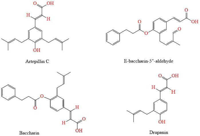
FIGURE 5. Prenylated derivatives and cinnamic derivatives found in B. dracunculifolia hydroalcoholic extracts.
Other flavonoids and phenolic acids such as baccharin, E-baccharin 5″-aldehyde, vicenin, p-coumaric acid, 7-O-methylkaempferol and E-4-(2,3-dihydrocinnamoyloxy) cinnamic acid; present in the hydroalcoholic extract of B. dracunculifolia leaves and green propolis were isoleted and analyzed by HPLC-DAD (diode array detector) by Rodrigues et al. (2020) (Figure 6).
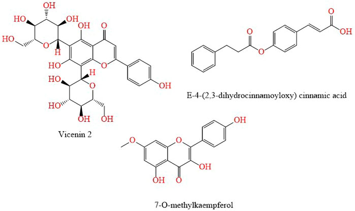
FIGURE 6. Flavonoids and phenolic acids found in the hydroalcoholic extract of B. dracunculifolia leaves and green propolis.
The hydroalcoholic extract of lyophilized B. dracunculifolia aerial parts was investigated by ultra-high-performance liquid chromatography–high-resolution mass spectrometry (UHPLC–HRMS) by Bonin et al. (2020). The flavonoids often reported as major constituents of green propolis are; naringenin, apigenin, kaempferol and kaempferide. The flavonoids pinobanksin, chrysin, apigenin, betuletol, galangin, pinocembrin, quercetin, were also identified in the hydroalcoholic extract of B. dracunculifolia (Bonin et al., 2020) (Supplementary Figure S5).
Ten new glycosides, called dracunculifosides A-J (Figure 7A and Figure 7B), were isolated from B. dracunculifolia by Nagatani et al. (2002b). Using the dried aerial parts, a methanolic extract was prepared and suspended in water. Then, with diethyl ether and partitioned into an ether-soluble fraction and a water-soluble fraction, the suspension was extracted. Through a Mitsubishi Diaion HP-20 column the water-soluble fraction was passed, then, the adsorbed material was subsequently eluted with 50% methanol in water, 70% methanol in water, and methanol 100%. The residue of 50% methanol eluate from the Diaion HP-20 column was concentrated was re-chromatographed on a silica gel column and by semi-preparative HPLC. The structures of these glycosides were determined on the basis of spectral and chemical evidence, beeing β-d-glucopyranose or β-d-apiofuranosyl-(1→6)-β-d-glucopyranose and most possess an (E)-caffeoyl group (Nagatani et al., 2002b).

FIGURE 7. (Continued). (A) Glycosides found in B. dracunculifolia dried aerial parts methanolic extract. (B) Glycosides found in B. dracunculifolia dried aerial parts methanolic extract.
The chemical composition of crude extract obtained from the aerial parts of B. dracunculifolia was investigated by Missima et al. (2007). This extract, then, was dissolved in methanol:water (7:3) and submitted to a sequential partition with hexane and dichloromethane, being this last fraction, chromatographed over silica gel under a Vacuum-liquid chromatography system, using hexane–ethyl acetate mixtures in increasing proportions as eluent, resulting in five fractions. In the first fraction the compound Baccharis oxide was identified, and in the second fraction the compound friedelanol, two pentacyclic triterpenoids were found (Missima et al., 2007) (Figure 8).
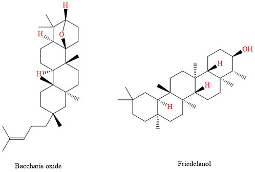
FIGURE 8. Pentacyclic triterpenoids found in B. dracunculifolia leaves and roots extract.
Fukuda et al. (2006) isolated two new monoterpenes baccharisketone and p-methoxythymol acetate (Figure 9). Fukuda et al. (2006) isolated two new monoterpenes bacarisketone and p-methoxythymol acetate (Figure 9) from the extract of dried leaves of B. dracunculifolia using ethanol as extractant liquid. The extraction technique was by ultrasound-assisted extraction, the residue obtained was chromatographed in an HP-20 column and eluted successively with pure methyl alcohol at concentrations of 40% and 70% and 100% acetone. The methanolic fraction was separated on a silica gel column and eluted with n-hexane-ethyl acetate at different graduations. then the identification was performed by HPLC.
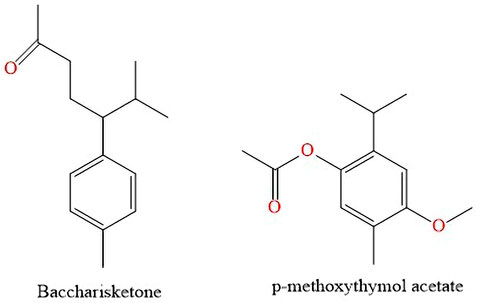
FIGURE 9. Monoterpenes found in B. dracunculifolia leaves extract.
3.3 B. dracunculifolia green propolis
Propolis is a plant resin that bees take from plants. In general, propolis is composed of essential and aromatic oils, waxes, resins and balm, pollen and other substances. Depending on the geographic origin of propolis and the vegetation from which it is extracted, its color can vary from green, to red and dark brown (Ferreira and Negri, 2018). B. dracunculifolia propolis, also known as green propolis, is produced from the vegetative apices of this species. As it is a mixture of products dependent on several external factors, the chemical analysis of propolis is challenging, this analysis is performed mainly on aqueous and ethanolic extracts as they are the most used pharmaceutical forms in different types of therapeutic applications (Nascimento et al., 2008).
Samples of Brazilian green propolis from different locations were investigated for chemical composition. The samples were extracted with methyl alcohol by ultrasound-assisted technique (360 W, 25 KHz) for 30 min. Extracts were analyzed by UHPLC coupled with UHPLC-ESI-QTOF-MS. The compounds identified were: chlorogenic acid, caffeic acid, isochlorogenic acid A, isochlorogenic acid B, isochlorogenic acid C and artepillin C (Sun et al., 2019). Compounds identified in green propolis were also found in investigations carried out by Chuang et al. (2022) (Supplementary Figure S6A,B). To complement the chemical composition of green propolis, Ferreira and Negri (2018) described in their studies the presence of triterpenoids, such as α- and β-amyrins, and their respective acetates. Chen et al. (2008) complemented the identification indicating the presence of the triterpene pentacyclic baurenyl acetate, the mono and di-caffeoylquinic acids and the flavonoids kaempferol and luteolin (Supplementary Figure S6A,B).
4 Pharmacology and bioactivity
4.1 Anti-inflammatory
4.1.1 Essential oil
The anti-inflammatory performance of B. dracunculifolia leaves essential oil was evaluated in models of skin inflammation by Brandenburg et al. (2020) (Figure 10). Essential oil doses were applied to the ear of mice where acute skin dermatitis was previously induced by 12-O-tetradecanoylphorbol-acetate (TPA) or arachidonic acid (AA). The topical application of 0.1 and 1.0 mg/ear essential oil reduced the TPA-induced edema by 67.4% and 81.8% after 6 h and by 85.6% and 95.0% after 24 h, respectively. The essential oil also reduced the leucocytes migration by 91.6% (1.0 mg/ear) and cell infiltrate by 97.1% (1.0 mg/ear). The use of 1.0 mg per ear of essential oil reduced the edema in the AA-induced dermatitis by 55%. In both models of acute inflammation, the oil showed an antiedematogenic effect similar to the positive controls dexamethasone (TPA-model) and indomethacin (AA-model). In the chronic inflammation model, multiple applications of TPA were used to induce ear skin inflammation. The essential oil (1.0 mg/ear) was topically applied twice a day from the fifth to the ninth day. At the end of the treatment, the essential oil inhibited all inflammatory parameters evaluated as ear edema (61.4%), reduced the inflammatory cell influx (74.1%), epidermis thickness (85%), and keratinocyte proliferation (74.3%), and differentiation (82%). The results of Brandenburg et al. (2020) highlight, therefore, the anti-inflammatory potential of the essential oil of B. dracunculifolia that shown to act at different points of the inflammation with good anti-inflammatory efficacy. However, in the absence of local or systemic side effects. Additionally, the authors (Brandenburg et al., 2020) suggest that the anti-inflammatory and antiproliferative activities observed might be due to the sesquiterpenes present in the essential oil (Figure 4).
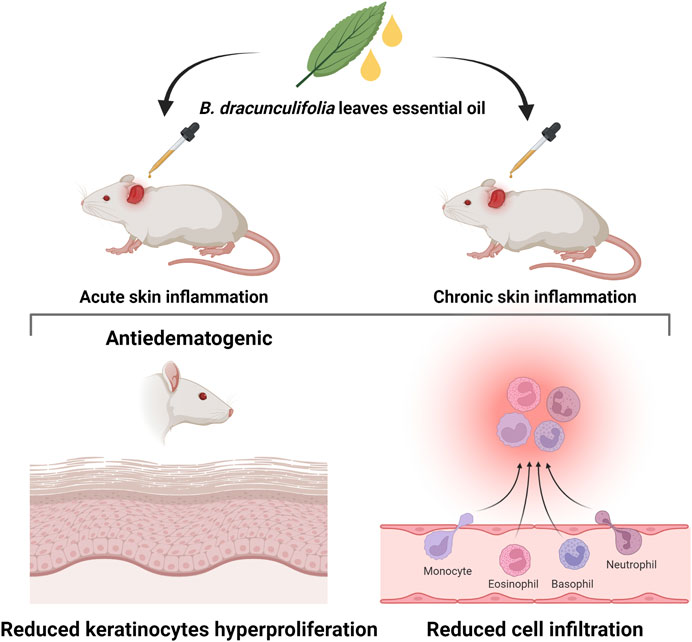
FIGURE 10. Anti-inflammatory activity of B. dracunculifolia leaves essential oil evaluated in models of skin inflammation (Brandenburg et al., 2020).
4.1.2 Extract
The anti-inflammatory potential of ethyl acetate extract from aerial parts of B. dracunculifolia was evaluated by Cestari et al. (2011) in a model of colitis in vivo. Tests of acute and chronic colitis were conducted. In the acute colitis model, rats orally received plant extract in concentrations of 5–200 mg/kg per day before colitis induction and 24 h after that. The doses of 5 and 50 mg/kg macroscopically reduced the damage score of the lesions (de 8.5 para 6.0 e 6.5, respectively, on a scale of 0–10) in the acute phase of the inflammatory process. B. dracunculifolia 5 mg/kg extract could counteract the colonic glutathione (GSH) depletion resulting from the colonic oxidative damage induced by trinitrobenzenesulfonic acid. Also, the myeloperoxidase (MPO) and alkaline phosphatase (AP) activities, commonly used as inflammatory markers, were reduced in animals treated with the extract. In the chronic colitis model, the extract reduced damage to the colon and the enzymatic activity of biochemical markers of inflammation after the first week. Cestari et al. (2011) suggest that the anti-inflammatory activity was related to the phenolic compounds as caffeic and p-coumaric acids (Figure 8), drupanin, baccharin, mainly artepillin C (Figure 5), the major component.
Figueiredo-Rinhel et al. (2019) used the ethanolic extract from B. dracunculifolia leaves and caffeic acid (Supplementary Figure S4) to examine its anti-inflammatory action in vivo. The authors tested the effects of free caffeic acid and extract and these compounds included in liposomes to modulate inflammation in vitro and in vivo. Liposomal extract and caffeic acid inhibited the neutrophil superoxide anion and total reactive oxygen species production. In the in vivo inflammation model, the free extract reduced edema, cell infiltration, and synovial levels of cytokines. The encapsulated extract also presented an anti-inflammatory effect in vivo, but with an effective dose lower. The same occurred when encapsulated caffeic acid was used, with an effective dose reduction of about sixteen times. The results demonstrate that biocompatible liposomes improve the B. dracunculifolia extract anti-inflammatory action in vivo (Figueiredo-Rinhel et al., 2019).
Lima et al. (2020) evaluated the efficacy of the ethanolic extract of B. dracunculifolia leaves in treating diversion colitis, a complication of intestinal transit diversion. Rats colostomy patients were treated for 21 days with an intrarectal infusion of B. dracunculifolia extract that reduced inflammation (from 2.7 to 2.1 on a scale ranging from 0 to 3). Still, they did not change vascular congestion in the excluded segment (Lima et al., 2020).
In a recent research conducted by França et al. (2022) the hydroalcoholic extract of B. dracunculifolia and isolated p-coumaric acid (Supplementary Figure S4) were used to treat colitis in vivo. Treatment with the extract at 300 mg/kg caused a 39.2% reduction in the disease activity index (DAI) score, prevented weight loss, preserved some histological features, and increased mucin by 318%, evidencing the pharmacological potential of the extract to treat inflammatory bowel disease. However, p-coumaric acid alone could not attenuate DAI, suggesting that the intestinal anti-inflammatory effects observed were due to the combination of extract constituents (França et al., 2022).
Due to the relationship between inflammatory bowel disease and neuropsychiatric comorbidities, França et al. (2022) also evaluated the neuroprotective action of the hydroalcoholic extract of B. dracunculifolia in mice with colitis. In different tests to measure anxious and depressive behavior, mice with colitis treated with the extract exhibited behavior similar to the healthy animals. These effects may have resulted from attenuation of central nervous system inflammation due to the preservation of intestinal barrier integrity or the inhibitory effects on inflammatory markers caused by the extract (França et al., 2022).
4.1.3 Green propolis
The effect of green propolis extract was investigated by Hori et al. (2013). In their studies, the researchers related the potential of green propolis in reducing the secretion of interleukin one beta (IL-1β) in mouse macrophages and verified that extract (30 μg/ml) did not show toxicity in the cells. This effect can be associated with the compounds found in Brazilian green propolis extract, such artepillin C, that plays a role in regulating inflammasomes (Hori et al., 2013).
In a model of dextran sulfate sodium-induced colitis in mice, Mariano et al. (2018) evaluated the anti-inflammatory potential of oral administration of hydroalcoholic extract of green propolis for 7 days (3, 30 and 300 mg/kg). The treatment with 300 mg/kg of green propolis extract reduced macroscopic and mucosal damage in the colon and reversed the decrease in mucin levels. Other effects were the recovery of the superoxide dismutase (SOD) activity and the levels of GSH in the colon. The authors suggest that the maintenance of the intestinal mucin and the activation of antioxidant system defense had a central role in the green propolis anti-inflammatory effect. However, green propolis extract did not change the patterns of gastric (emptying) and intestinal (transit rate) functioning of mice (Mariano et al., 2018).
Wang et al. (2018) compared the anti-inflammatory effects of Brazilian Baccharis green propolis and Chinese poplar-type propolis in animal models of colitis. The polyphenolic composition of the two propolis showed to be different. Of the 27 compounds identified, only five were detected in the Brazilian propolis, with artepillin C (Supplementary Figure S6A) the most abundant. On the other hand, 20 compounds were identified in Chinese propolis rich in caffeic acid phenethyl ester (CAPE), but artepillin C was not detected. The animals were orally treated with 300 mg/kg of Brazilian and Chinese propolis with an initial dose 1 week before treatment with DSS for 1 week, followed by 3 days without DSS. Both propolis significantly reduced the disease activity index, preventing damage to colonic tissue, reducing malonaldehyde levels, and increasing antioxidant capability, indicating greater resistance to DSS-induced colonic oxidative stress. The two propolis also diminished apoptosis in the colon and suppressed the inflammation markers IL-1β, IL-6, and monocyte chemoattractant protein-1 (MCP-1). However, only Brazilian propolis induced the expression of transforming growth factor β. Only Chinese propolis raised the diversity of intestinal microbiota. Wang et al. (2018) concluded that the two propolis demonstrated similar anti-inflammatory capacity despite some physiological impacts and different compositions.
Miranda et al. (2019) analyzed the anti-inflammatory potential of green propolis extract in a chronic inflammation model in mice submitted to a low-protein diet. Animals received diets with 12% (standard) or 3% (low) protein. After 28 days of starting the diet, inflammation was induced, and the treatment of daily doses of 500 mg/kg of the extract was initiated until seven or 15 days. The treatment with the extract stimulated weight restoration and conservation of serum protein levels in animals on a diet restricted in proteins. It also selectively altered the hematological parameters, increasing leukocyte recruitment. Additionally, the extract had a modulatory action in the inflammatory kinetics that was less efficient in the groups in a low-protein diet (Miranda et al., 2019).
Xu et al. (2020) tested the protective effect of green propolis extract using an in vitro model of lipopolysaccharide-stimulated mouse aortic endothelial cells (MAECs). All concentrations (5, 10, and 20 μg/ml) of extract increased cell survival, and the 20 μg/ml dose inhibited the expression of inflammatory cytokines and reduced adhesion molecules. In addition, the authors detected prenylated derivatives of p-coumaric acid, diterpenes, and flavonoids in the extract. Artepillin C (35.68%) was the major compound, followed by kaempferide (7.06%), 3-isoprenyl p-coumaric acid (6.19%), pinocembrin (5.56%), diisoprenyl-p-coumaric acid isomer (4.49%), 40-methoxy pinobanksin (4.10%) and 3-hydroxy-2,2-dimethyl-8-prenylchromane-6-propenoic (3.39%) (Xu et al., 2020) (Supplementary Figures S6A,B).
Yuan et al. (2020) also compared the anti-inflammatory action of Brazilian Baccharis propolis and Chinese poplar propolis in a murine model of systemic inflammation induced by lipopolysaccharide (LPS). Brazilian and Chinese propolis ethanol extracts (0.1 ml) were administered intragastrically in mice on the day before LPS administration, 2 hours before LPS administration, and 12 h after LPS administration. Both propolis showed similar anti-inflammatory activity, evidenced by the reduction of genic expression and reversion of the serum cytokines levels in the animals that received prophylactic doses of propolis extract. The authors unveiled differences between the propolis chemical profiles with much higher content of artepillin C, chlorogenic acid, and isochlorogenic acid A (Supplementary Figure S6A) in the Brazilian propolis and great CAPE levels and pinocembrin in Chinese propolis that could contribute to the anti-inflammatory effect (Yuan et al., 2020).
Ferreira et al. (2021) evaluated the actions of p-coumaric acid and baccharin (500 or 1,000 μg/kg) isolated from green propolis in a murine air pouch LPS-induced inflammation model. The two doses of p-coumaric acid and baccharin (Supplementary Figures S4, S5) had similar effects in reducing leukocytes and neutrophil recruitment. Additionally, histological analysis showed that the accumulation of neutrophils induced by LPS was significantly reduced by treatment with 500 μg/kg p-coumaric acid and baccharin. The author also reported significant reductions in nitric oxide (NO) production and protein extravasation in inflamed air pouches treated with 500 μg/kg p-coumaric acid or baccharin, indicating a role in vascular barrier preservation of these compounds. The evaluation of inflammatory markers showed that baccharin reduced IL-6, TNF-α, and IL-1β levels, while p-coumaric acid reduced TNF-α and IL-1β levels. However, p-coumaric acid stimulated interleukin 10 (IL-10) production. Treatment with p-coumaric acid or baccharin also significantly impacted levels of the eicosanoids prostaglandin F2α (PGF2α), 15-hydroxyeicosatetraenoic acid (15-HETE), and 12-HETE, but only p-coumaric acid suppressed 5-HETE synthesis. Apparently, p-coumaric acid and baccharin had different molecular mechanisms to modify the inflammatory response (Ferreira et al., 2021).
In a recent systematic review, Soleimani et al. (2021) analyzed eight studies on the anti-inflammatory action of green propolis on various aspects of inflammatory bowel disease in preclinical studies. Green propolis effectively improves histological inflammation aspects of the colon and clinical and morphological features in animals with colitis. The propolis benefit suggests a relationship between its protective effects against oxidative stress and endogenous antioxidant parameters. Soleimani et al. (2021) suggest that the likely mechanism of action includes impeding transcription factors and proteins.
4.2 Antiulcerogenic
4.2.1 Essential oil
Klopell et al. (2007) evaluated the antiulcerogenic potential of essential oil from leaves of B. dracunculifolia in an acute gastric injury model. Oral administration of essential oil significantly decreased the rate of ulcerative wounds (42.79–61.61%) when compared to control (vehicle) and omeprazole (30 mg/kg). Essential oil analysis showed the presence of mono- and sesquiterpenes and the major compound nerolidol (Figure 4), which was used alone at concentrations (50, 250, and 500 mg/kg) in the evaluation of antiulcerogenic activity in rats. The two highest nerolidol concentrations significantly reduced the ulcerative lesion rate in all models evaluated, emphasizing the 87.6% reduction observed when 500 mg/kg of nerolidol was used in an ethanol-induced ulcer model (Klopell et al., 2007).
Massignani et al. (2009) used the essential oil from B. dracunculifolia leaves to treat rats wich had ulcers induced by non-steroidal anti-inflammatory drugs, ethanol, and stress. They observed that the lesions index, total area and the number of wounds were lower. These results added to a decrease in gastric juice volume and total acidity, and absence of toxicity (Massignani et al., 2009).
4.2.2 Extract
Hydroalcoholic extract of B. dracunculifolia aerial parts decreased the rate of wounds in ulcer models induced by ethanol, indomethacin, and stress (Lemos et al., 2007). At the highest concentration, the extract caused inhibition from 75.6% (indometacin model) to 95% (ethanol model). Additionally, the extract reduced the stomach juice volume and increased pH in a gastric secretion evaluation model (Lemos et al., 2007).
Costa et al. (2019) stated that extract of B. dracunculifolia leaves (30, 100, and 300 mg/kg) demonstrated the ability to heal gastric ulcers in models of chronic ulcers induced by acetic acid. The extract oral administration decreased the ulcer area between 30% and 50% compared to vehicle. Additionally, the authors characterized and isolated the major compounds from the extract [ferulic acid (Figure 4), p-coumaric acid, caffeic acid (Supplementary Figure S4), artepillin C, baccharin (Figure 5), and aromadendrin-4′-O-methyl ether] to assess their gastric healing ulcer activity. p-coumaric acid reduced the ulcer area by 66%, while the other compounds showed no healing activity compared to the vehicle (Costa et al., 2019). After evaluating several physiological parameters, Costa et al. (2019) suggested that B. dracunculifolia extract recovers ulcerated gastric tissue, increasing mucus and antioxidant enzymes, and reducing gastric proton pump activity.
Recently Boeing et al. (2021) re-evaluated the effects of p-coumaric acid (10 mg/kg) extracted from B. dracunculifolia in rats with gastric ulcer. Oral p-coumaric acid reduced the extent of the ulcer base by 44.6%. It reduced the damage to the mucosa and submucosal layers compared to the vehicle, reaffirming the antiulcerogenic action of p-coumaric acid (Boeing et al., 2021).
4.2.3 Green propolis
Barros et al. (2007) evaluated the capacity of Brazilian green propolis extract to act as a gastric protector and anti-ulcer. Animals pre-treated with propolis extract exhibited lower lesion index, ulcer number, and gastric affected area, corroborating the results of Lemos et al. (2007) for B. dracunculifolia aerial parts extract. Moreover, green propolis extract decreased the volume of gastric juice, total acidity, and pH. The authors suggest good anti-ulcer activity of green propolis that can be incorporated into ulcer treatment products after pharmacological validation (Barros et al., 2007).
Costa et al. (2018) isolated the major compounds of Brazilian green propolis extract and evaluated their gastroprotective potential in murine gastric ulcers. In the ethanol/HCl-induced ulcer, the compounds artepillin C, baccharin, drupanin (Figure 5), aromadendrin-4′-O-methyl-ether, and kaempferide (Supplementary Figure S3) were orally administered. Artepillin C, drupanin, aromadendrin-4′-O-methyl-ether, and kaempferide decreased the ulcer area, reducing the necrotizing area and the damage to the gastric epithelial architecture. However, baccharin had no significant effect. When animals were treated by intraperitoneal route with a ten times lower dose, all compounds had anti-ulcer results demonstrating a systemic action. After administration of the flavonoids aromadendrin-4′-O-methyl-ether or kaempferide, there was an increase in mucin production resulting in gastroprotection. The compounds did not prevent GSH depletion, but artepillin C and drupanin reduced the lipid hydroperoxides amount at gastric tissue, indicating the prevention of oxidative stress. In addition, all compounds maintained antioxidant enzyme levels similar to those in health mucosa. The indomethacin-induced ulcer model confirmed the gastroprotective and antisecretory activities of the compounds (Costa et al., 2018).
Costa et al. (2020), continuing the research developed by the group (Costa et al., 2018), evaluated the mechanism of propolis extract related to gastric ulcer prevention and healing, and the curative effect of artepillin C. The results indicated that oral pre-treatment with extract avoided gastric damage, reduced SOD activity by about 11% (100 mg/kg) and 26% (300 mg/kg), and increased GST and CAT activity by about 20% and 80%, respectively. Furthermore, extract (300 mg/kg) reduced reactive oxygen species generation and lipid peroxidation in gastric tissue. The daily treatment with extract (300 mg/kg) in the chronic ulcer model promoted a faster healing process and normalized SOD and CAT activities but increased GST activity. The administration of the extract in the two models tested increased the PAS (Periodic Acid of Schiff’s method) staining of mucin, reduced myeloperoxidase activity at the ulcer site, and improved the immunostaining of PCNA (proliferating cell nuclear antigen). However, it did not alter collagen concentration in ulcerated tissue by acetic acid. The extract also showed a natural ability to scavenge DPPH radicals (IC50 = 0.56). Extract chemical characterization revealed artepillin C as the main compound (Figure 5) and the administration of this molecule (18 mg/kg) stimulated faster healing of gastric ulcer. The results indicate gastroprotective and gastric healing properties of extract (Costa et al., 2020). Therefore, these findings may contribute to validating their use as preventive and therapeutic approaches.
4.3 Antimicrobial
4.3.1 Essential oil
Salazar et al. (2018) studied the essential oil of fresh and dried plant material of B. dracunculifolia with the major compounds germacrene D (E)-nerolidol, spathulenol, β-pinene and bicyclogermacrene (Figure 4). The lowest MICs were observed against S. aureus strains, 102 μg/ml for ATCC 2593 and 512 μg/ml for a multidrug-resistant clinical isolate. For multidrug-resistant P. aeruginosa, the MIC was 813 μg/ml (Salazar et al., 2018). Antimicrobial activity is probably related to the presence of spathulenol (Figure 4) (Tan et al., 2016).
Cazella et al. (2019) evaluated the essential oil of dry aerial parts (leaves and flowers) against microorganisms of interest in food. The main compounds were spathulenol and trans-nerolidol (Figure 4). S. aureus, B. cereus, and P. aeruginosa were the bacteria most susceptible to the essential oil with MIC of 0.5, 1.1, and 1.05 mg/ml, respectively, and MBC of 2.1, 1, 5, and 2.1 mg/ml, respectively, showing weak activity against Enterobacteriaceae (E. coli and Salmonella). Regarding filamentous fungi, including species of the genus Penicillium, Aspergillus, and Trichoderma, the MIC ranged from 8.43 to 16.87 mg/ml (Cazella et al., 2019).
The essential oil of dried leaves with major compounds such as β-pinene, ledol, spathulenol, and limonene (Figure 4) showed antifungal activity in the control of postharvest fungal rot in grapes caused by the fungi Botrytis cinerea and Colletotrichum acutatum (Pedrotti et al. al., 2019). Debona et al. (2021) also evaluated the antifungal activity of the essential oil of the leaves of B. dracunculifolia against Alternaria alternata, a fungus capable of causing damage to dragon fruit. At concentrations above 600 μg/ml, the oil could inhibit fungal growth in vitro, a promising option in managing rot caused by this fungus in dragon fruit (Debona et al., 2021).
Monteiro et al. (2022) investigated the antimicrobial activity of essential oil from dry leaves of B. dracunculifolia, collected at three different localities in Brazil. The three essential oils showed antimicrobial action for the bacteria studied. Oil from the Midwest region of Brazil was more effective against the bacterial strains tested. For this sample, the MIC was 0.03 mg/ml for B. cereus, 0.05 mg/ml for S. aureus, Streptococcus mutans, Enterococcus faecalis, P. aeruginosa, Klebsiella pneumoniae, and Salmonella enterica, and 0.08 mg/ml for E. coli. The MBC ranged from 0.05 to 0.1 mg/ml. The major compounds in all samples were sesquiterpenes (59.88–89.29%) and monoterpenes (7.66–38.41%). In the Central-West region, the majority were (E)-nerolidol (28.15%) and spathulenol (17.68%) (Figure 4). In the Southeast region, they were limonene (19.36%) (E)-nerolidol (12.75%) and bicyclogermacrene (10.76%) (Figure 4) and in the southern region bicyclogermacrene (14.21%) (E)-nerolidol (13.95%) and limonene (10.49%) (Figure 4). Bicyclogermacrene, after enzymatic oxidation, gives rise to spathulenol (Monteiro et al., 2022).
The commercial essential oil of B. dracunculifolia from the company Harmonia Natural (Canelinha, SC, Brazil), having as main components nerolidol, beta-pinene and d-limonene (Figure 4) (Timbé et al., 2021), has antimicrobial activity against the Gram-positive bacteria studied, the largest inhibition halo was against Bacillus subtilis (23.6 mm). Not showing activity against Gram-negative bacteria and yeasts (Timbé et al., 2021). This same research group also evaluated the antimicrobial activity of nanoparticles containing B. dracunculifolia oil (Harmonia Natural - Canelinha, SC, Brazil) (Timbé et al., 2020). First, the MBC of the oil was analyzed against Listeria monocytogenes (0.56 mg/ml), S. aureus (0.28 mg/ml), B. cereus (0.14 mg/ml) and S. Enteritidis (1.13 mg/ml). Afterward, the contact test of the oil with the bacteria studied was carried out in a liquid culture medium. After 4 h of contact of the essential oil with S. aureus, B. cereus, and L. monocytogenes, and S. Enteritidis, there were no more viable cells. The reduction achieved by the nano encapsulated oil was 2 h of incubation for B. cereus, 10 h for L. monocytogenes, and 24 h for S. aureus and S. Enteritidis, suggesting a gradual release of compounds with antimicrobial activity (Timbé et al., 2020).
4.3.2 Extract
Bonin et al. (2020) studied the antimicrobial activity of the hydroalcoholic extract (70%) (dry plant material - plant parts not reported) of B. dracunculifolia against bacteria. The minimum inhibitory concentrations (MIC) and bactericidal concentrations (MBC) found, respectively, were 125 μg/ml and 250 μg/ml for S. aureus and B. subtilis, 250 μg/ml and 500 μg/ml for B. cereus. For the Salmonella enterica serovar Enteritidis, the MIC and MBC values were 1,000 μg/ml. The antimicrobial activity against Gram-positive bacteria may be associated with the compound naringenin (Supplementary Figure S3) present in the extract (Bonin et al., 2020), a compound found by Zuccolotto et al. (2016) when they analyzed the antimicrobial action of other plants belonging to the genus Baccharis against Gram-positive bacteria.
Casagrande et al. (2018) found equal antimicrobial activity of the hydroalcoholic extract (40%) and acetone extract (40%), from the aerial part of the plant, against the Gram-positive bacteria studied. For S. aureus MIC of 12.75 mg/ml and for B. cereus MIC of 3.19 mg/ml. For Gram-negative bacteria, the activity of the extracts was lower. Still, attention is drawn to the antimicrobial activity of the hydroalcoholic extract against Pseudomonas aeruginosa, with MIC and MBC of 51 mg/ml and resistance to ampicillin (Casagrande et al., 2018). This same research group evaluated the addition of the hydroalcoholic extract at different concentrations in a film based on poly (vinyl alcohol) and starch (Casagrande et al., 2021). For this, they lyophilized the extract and analyzed its antimicrobial and antioxidant characteristics before producing the film. The antimicrobial activity against Gram-positive bacteria, found by Casagrande et al. (2021), was better when compared to the liquid extract (Casagrande et al., 2018). For S. aureus and B. cereus the MIC was 1.49 mg/ml, and 0.37 mg/ml, respectively. For P. aeruginosa, the MIC was higher than the concentrations analyzed. After being added to the film, the antimicrobial activity was reduced and even inhibited for the microorganisms mentioned above (Casagrande et al., 2021).
Hydroalcoholic extract (70%) of dried leaves showed antimicrobial activity against Staphylococcus pseudintermedius, with MIC of 0.312 mg/ml and MBC of 2.5 mg/ml, in addition to having the ability to inhibit biofilm formation in vitro (Barbosa et al., 2022). This activity may be related to phenolic acid derivatives and flavonoids (Barbosa et al., 2022). The commercial hydroalcoholic extract of B. dracunculifolia was obtained from Ciclo Farma Indústria Química Ltda (Serrana, SP, Brazil), with the majority of artepillin C (Figure 5), rutin, and caffeic acid (Supplementary Figure S4) has been evaluated by Timbé et al. (2021), presenting antimicrobial activity against all tested bacteria, with the highest inhibition halo for B. cereus (20.5 mm). The material evaluated did not show activity against Gram-negative bacteria and yeasts (Timbé et al., 2021). Veiga et al. (2017) analyzed ethanolic and hexane extracts from leaf shoots. The ethanol extract presented MIC90 between 256.7 and 770.1 μg/ml for the S. aureus strains studied, while the hexane MIC90 was between 197 and 394 μg/ml. Even the ethanol extract having a higher content of phenols and flavonoids had a lower antimicrobial capacity (Veiga et al., 2017).
Bernardes et al. (2022) tested the antimicrobial activity of hydroalcoholic extracts from aerial parts and roots and trichome washing of B. dracunculifolia against bacterial and fungal strains. The best results were against S. aureus (MIC 200 μg/ml for the extract of aerial parts and trichome wash) and Trichophyton mentagrophytes (MIC 200 μg/ml for the extract of aerial parts). In addition, when the substances isolated from the extracts were evaluated, baccharin (Figure 6) showed activity against S. aureus and T. mentagrophytes, and isosakuranetin (Supplementary Figure S3) against Salmonella choleraesuis and T. mentagriphytes. According to the results found in this work, B. dracunculifolia is a potential alternative as a sanitizer against pathogenic fungi and bacteria (Bernardes et al., 2022).
Assumpção et al. (2022) evaluated the antimicrobial activity of the hydroalcoholic extract of aerial parts of B. dracunculifolia against strains of S. aureus isolated from mastitis. The MIC for two of the clinical strains was 1.25 mg/mL. As for biofilm formation, the extract showed better results for consolidated biofilms than new biofilms (Assumpção et al., 2022). The ethanolic extract (99.8%) produced from leaves, with the presence of cardiotonic heterosides, steroids, phenolic compounds, flavonoids, tannins, coumarins, and triterpenes, at a concentration of 2000 µL/100 ml, inhibited by 13.5% the growth of Rhizoctonia solani (Dilkin et al., 2022).
4.3.3 Green propolis
Barbosa et al. (2022) evaluated the hydroalcoholic extract (70%) of green propolis. They found antimicrobial activity against Staphylococcus pseudintermedius, with MIC of 0.156 mg/ml and MBC of 0.312 mg/ml, with a chemical composition similar to the extract of the leaves of B. dracunculifolia (derived from phenolic acids and flavonoids) (Barbosa et al., 2022).
Ethanolic and hexane extracts of green propolis had their antimicrobial activity studied (Veiga et al., 2017). Phenols and flavonoids were found only in the ethanol extract, in addition to the higher amount of artepillin C (Supplementary Figure S6A). Even with this difference in composition, the hexane extract showed better antimicrobial activity against the S. aureus strains analyzed (MIC90 between 78.4 and 392.0 μg/ml) (Veiga et al., 2017). Assumpção et al. (2022) studied the biofilm formation and antimicrobial action of the hydroalcoholic extract of green propolis against mastitis isolates. Propolis presented a similar result to the extract of aerial parts. The MIC for the clinical strains was between 1.25 and 2.5 mg/ml, showing better results against consolidated biofilms (Assumpção et al., 2022).
Bittencourt et al. (2015) correlated the free radical scavenging activity (IC50) and MIC to the presence of total phenolic compounds in Brazilian green propolis. They found that the IC50 and MIC values were associated with antioxidant and antibacterial activities (Bittencourt et al., 2015). Machado et al. (2016) tested ethanolic extract of green propolis against S. aureus and Escherichia coli. They identified a negative correlation between phenols concentration in the extracts and MIC. Jug et al. (2014) also evaluated the antibacterial and antifungal efficiency of green propolis extracts obtained by different extraction techniques and demonstrated that the ethanolic extract showed the best antimicrobial action. Quintino et al. (2020) verified the antimicrobial activity of the oil extracted from fresh green propolis (Figure 11), with carvacrol being the compound with the highest concentration (20.7%), finding a MIC of 6.25 μg/ml against Helicobacter pylori, 62.5 μg/ml against Mycobacterium avium and 64 μg/ml for Mycobacterium tuberculosis (Quintino et al., 2020).

FIGURE 11. Antimicrobial activity of the oil extracted from B. dracunculifolia green propolis (Quintino et al., 2020).
4.4 Antioxidant
4.4.1 Essential oil
Tomazzoli et al. (2021) analyzed the antioxidant activity of the essential oil of fresh leaves of B. dracunculifolia collected in ten locations in Paraná - Brazil. The method used was DPPH, with activity varying between 27.78% and 91.67%. The oils were predominantly composed of oxygenated sesquiterpenes, monoterpene hydrocarbons, and sesquiterpene hydrocarbons. With the majority in all ten populations, β-pinene and (E)-nerolidol (Figure 4) (Tomazzoli et al., 2021), compounds that have already been reported with antioxidant activity (Yu et al., 2017; Chen et al., 2018). Monteiro et al. (2022), analyzing oil from leaves of plants collected from three regions of Brazil, found that oil from the Center-West region of Brazil showed larger antioxidant activity by the DPPH methods (50.82% inhibition) and ABTS (113.63 mM TE/ml) concerning oils from the Southeast and South regions. Oils from the Center-West and South regions of Brazil had higher levels of phenolic compounds compared to the oil from the Southeast region of Brazil. One of the major compounds common to the three samples was (E)-nerolidol (Figure 4) (Monteiro et al., 2022).
Paroul et al. (2016) evaluated the antioxidant activity, by the DPPH method, of essential oil and aqueous extract of plant material of B. dracunculifolia. The antioxidant activity of the aqueous extract was 100 times greater than that obtained with the essential oil, which is possibly related to the greater antioxidant action of the compounds present in this type of extract, probably because they are water-soluble compounds (data not provided) (Paroul et al., 2016).
4.4.2 Extract
Casagrande et al. (2018), evaluating hydroalcoholic extract and acetone extract, verified antioxidant activity by the methods of DPPH (2,2-diphenyl-1-picrylhydrazyl), ABTS (2.2′-azinobis-(3-ethylbenzothiazoline-6-sulfonic acid), FRAP (ferric reducing/antioxidant power) which may be associated to the phenolic compounds in its composition, with ferulic acid (hydroxycinnamic acid) (Supplementary Figure S4), being the main component (Casagrande et al., 2018). Casagrande et al. (2021) evaluated the same lyophilized hydroalcoholic extract as a pre-test for addition to a film based on poly (vinyl alcohol) and starch. The antioxidant activity of the DPPH, ABTS, and FRAP methods was better at 389%, 322%, and 533%, respectively, when compared to the liquid extract (Casagrande et al., 2018). The embedded film maintained the antioxidant action when analyzed by the DPPH method (Casagrande et al., 2021).
Guimarães et al. (2012) evaluated the antioxidant activity in vitro and hepatic mitochondria, isolated from rats, of the glycolic extract from the leaves of B. dracunculifolia. The concentrations of total phenols and flavonoids were 21.18 µM (gallic acid equivalents) and 13.64 µM (quercetin equivalents), respectively. Caffeic acid, p-coumaric acid, cinnamic acid (Supplementary Figure S4), aromadendrin, isosakuranetin (Supplementary Figure S3), and artepellin C (Figure 5) were the compounds identified. By the DPPH method, the EC50 was 0.005%, by superoxide elimination, the EC50 was 0.0732%, in addition, the extract was able to chelate 77.57% of Fe2+. In mitochondria, there was a decrease in the basal generation of H2O2 and ROS production induced by Fe2+- or t-BuOOH. There was also prevention of lipid oxidation of mitochondrial membranes, thiol protein groups (Guimarães et al., 2012).
The antioxidant activity of ethanolic and hexane extracts from B. dracunculifolia sprouts (Figure 12), and green propolis was evaluated by the DPPH method. The IC50 values were 13.09 μg/ml for ethanolic extract of green propolis, 95.86 μg/ml for hexane extract of green propolis, 124.49 μg/ml for ethanolic extract of B. dracunculifolia and 141.45 µg/ml for B. dracunculifolia hexane extract. It is observed that among all samples, the ethanolic extract has the larger activity. This extract has phenols and flavonoids in its composition (Veiga et al., 2017).
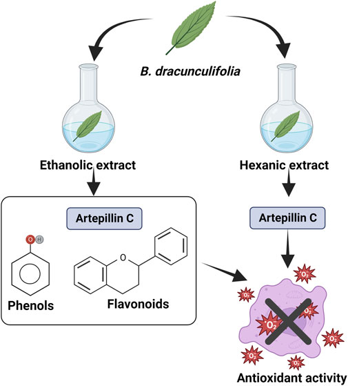
FIGURE 12. Antioxidant activity of ethanolic and hexane extracts from B. dracunculifolia leaves (Veiga et al., 2017).
4.4.3 Green propolis
The antioxidant activity of green propolis was reported by Osés et al. (2016), Cao et al. (2017), and Braakhuis (2019). According to the authors, polyphenols, one of the main propolis compounds, have a chemical structure with potential scavenging free radicals. Furthermore, the flavonoids in propolis are potent antioxidants, capable of scavenging free radicals and thus protecting cell membranes against lipid peroxidation (Osés et al., 2016; Cao et al., 2017; Braakhuis, 2019).
The antioxidant activity of the oil extracted from fresh green propolis was evaluated by the DPPH and ABTS methods, obtaining IC50 of 23.48 μg/ml and 32.18 μg/ml, respectively. In this study, the major compounds were carvacrol (20.7%), acetophenone (13.5%), spathulenol (11.0%) (E)-nerolidol (9.7%) and β-caryophyllene (6.2%) (Figure 4) (Quintino et al., 2020).
With the evidence of the antioxidant activity of B. dracunculifolia, Zanela et al. (2021) produced a biodegradable film for food based on PBAT—poly (butylene adipate co-terephthalate) and cassava starch using glycerol as a plasticizer. B. dracunculifolia leaf powder was added to the film, and the antioxidant, physical and chemical properties were found to be ideal for use as biodegradable food packaging (Zanela et al., 2021).
4.5 Antiviral
The inhibitory effects of plant extracts on the replication of various viruses have been reported in several studies. Viruses such as herpes simplex (HSV), human immunodeficiency (HIV), hepatitis B (HBV) and severe acute respiratory syndrome (SARS) were strongly inhibited by various plant extracts.
Búfalo et al. (2009) evaluated the antiviral activity of the extract, essential oil, green propolis, and isolated compounds caffeic and cinnamic acids (Supplementary Figure S4) from B. dracunculifolia against poliovirus type 1 (PV1) replication in HEp-2 cells (human laryngeal epidermoid carcinoma cells). The results indicated that the greatest inhibition of virus replication was achieved by the extract of B. dracunculifolia with 74.0%, followed by green propolis (52.2%), essential oil (33.3%), cinnamic acid (29, 8%) and caffeic acid (26.7%). The most efficient concentrations of B. dracunculifolia extract and caffeic and cinnamic acid compounds were 25 µg/100 μL, followed by green propolis (10 µg/100 µL) (Búfalo et al., 2009).
The antiviral activity of ethanol extract of green propolis obtained from B. dracunculifolia was tested by Shimizu et al. (2008) on the propagation of influenza A/PR/8/34 (H1N1) virus in Madin-Darby cells (MDCK) obtained from the kidney of dogs and female DBA/2 Cr mice. The anti-influenza efficacy of the extract at a concentration of 10 mg/kg was confirmed in a dose-dependent manner in mice. At this concentration, there was a significant reduction in the virus yield in bronchoalveolar lavage fluids from the lungs of infected mice when compared to the control group. It was observed that the reduction in virus yields by the extract at a concentration of 10 mg/kg corresponded significantly to those induced by oseltamivir at a concentration of 1 mg/kg at four post infection (Shimizu et al., 2008). In this same line of research, Shimizu et al. (2011) administered ethanolic extracts (10 mg/kg) of B. dracunculifolia orally in mice infected with Herpes virus type 1 (HSV-1). The extract significantly reduced the titers of the virus in the brain and/or in the skin from the fourth day of use, without showing toxicity. A significant increase in the production of interferon-gamma (IFN-γ) by the HSV-1 antigen was also observed in splenocytes from mice infected with HSV-1 (Shimizu et al., 2011).
4.6 Other activities
Luchesi et al. (2022) investigated the antiphytopathogenic activity of essential oil extracted from fresh leaves of B. dracunculifolia against the fungus Fusarium graminearum. The oil was tested at concentrations of 8.0 and 4.0 μL/ml, inhibiting fungal growth by 57.1% and 49.5%, respectively, after 96 h. Inhibition of the fungus may be associated with the presence of cis- and trans-nerolidol, c-elemene, d-limonene and spathulenol (Figure 4), considered by the authors as the major compounds in the essential oil (Luchesi et al., 2022).
Cazella et al. (2020) analyzed the acaricidal activity of the essential oil extracted from the leaves and flowers of B. dracunculifolia, finding the sesquiterpenes nerolidol and spathulenol as the main compounds (Figure 4). The oils (leaves and flowers) were tested at stages comprising the parasitic and free-living life cycle of the bovine tick Rhipicephalus microplus (Acari: Ixodidae). Both essential oils at a concentration of 500 mg/ml effectively controlled the hatchability of eggs, reducing the oviposition capacity and the number of adult and larval ticks. The lethal concentration (CL99,9) of essential oils (leaves and flowers) on the larvae was 35–37 mg/ml, respectively. The authors also investigated the activity of the oils in the tick free life cycle (ex situ test), indicating that at concentrations of 11–14 mg/ml there were 85 and 95% efficacy, respectively (Cazella et al., 2020).
Lage et al. (2015) also evaluated the chemical composition and acaricidal activity of the essential oil extracted from the aerial parts of B. dracunculifolia and the pure compounds nerolidol and (R)-(+)-limonene on the larvae and engorged females of Rhipicephalus microplus. For larvae, the “Larval packet test” was performed and for engorged females, the “adult immersion test”. The most efficient result was the action of nerolidol (Figure 4) on tick larvae, causing more than 90% mortality at a concentration of 10.0 mg/ml. Essential oil and nerolidol also acted in the immersion test of adults, causing a reduction in the quantity and quality of eggs produced, with inhibition of hatchability of 96.3% and 90.3% at concentrations of 60.0 and 50.0 mg/ml, respectively (Lage et al., 2015).
As an insecticide, the essential oil of this plant has already been tested against third-stage larvae of Cochliomyia macellaria by Chaaban et al. (2018), where this demonstrated as an ecological alternative against this fly because, in addition to being highly larvicidal (2.47 μL/cm2), the hatched larvae generated adults with deformities. Furthermore, from the essential oil of the leaves of B. dracunculifolia, Alves et al. (2018) verified larvicidal action on Culex quinquefasciatus. The essential oil (EO) was tested at concentrations of 25, 50, 100 and 200 mg/L, with a lethal concentration (LC50) of 34.45 mg/L for the EO. Histological changes were observed in the midgut, reduction of glucose and acetylcholinesterase levels in larvae exposed to EO, indicating increased levels of triacylglycerides and total proteins, confirming that the EO of B. dracunculifolia causes destabilization in the larva, leading to histological changes, dysregulation metabolism and, consequently, its death. The authors suggest that this effect is due to the presence of sesquiterpenes such as spathulenol and nerolidol (Figure 4), which are used to control insects and as an acaricide (Cazella et al., 2020).
Seugling et al. (2019) developed nanoemulsions containing essential oil of B. dracunculifolia in five different concentrations (5.0; 7.5; 10.0; 13.5 and 15.0%) (w/v). The tests consisted of using the nanoemulsions immediately after preparation and after 120 days of storage on Cochliomyia hominivorax larvae. The authors verified that the formulations used immediately after preparation, provided mortality of 28, 48, 70, 84 and 97%, respectively, while for the stored formulations the mortality was 17; 36; 51; 81 and 92%, respectively. The authors found no significant difference in the action of nanoemulsions in terms of storage time, suggesting that nanoemulsions of essential oil of B. dracunculifolia can be considered a promising alternative for the treatment and control of myiasis. caused by C. hominivorax (Seugling et al., 2019).
4.6.1 Green propolis
Green propolis is known for its biological properties, and in this sense, Dantas Silva et al. (2017) investigated the activity of ethanolic extracts (80%) of green propolis from different regions of Brazil on epimastigotes of Trypanosoma cruzi “Y strain”. The authors verified an inhibitory effect of the extracts on the growth of cultures at concentrations of 75 and 300 mg ml-1 on the growth of epimastigotes of the Y strain of T. cruzi. This inhibition was observed in the first 24 h of the experiment, indicating green propolis as an alternative therapeutic treatment against Chagas disease, a deadly disease considered neglected, with few therapeutic resources (Dantas Silva et al., 2017).
There is considerable evidence of anticancer properties of brazilian green propolis (Ahn et al., 2007; Szliszka et al., 2011). The use of propolis has been recommended as a complementary therapy for the treatment of various types of cancer, including bladder, blood, brain, breast, colon, head and neck, kidney, liver, pancreas, prostate and skin (Patel, 2016). Compounds isolated from propolis are patented drugs for the treatment of cancer (Chuang et al., 2016), and in this sense, Matsuno (1995) isolated a new clerodane-type diterpene (Supplementary Figure S2). These authors verified in their research that this compound inhibited the growth of hepatoma cells and stopped tumor cells in the S phase, which corresponds to the period of DNA synthesis (Matsuno, 1995).
Anticancer activities of ethanolic extracts of green propolis were also evaluated by Machado et al. (2016). Propolis extracts were obtained by supercritical (SCO2) and ethanolic (EtOH) extraction, whose samples were collected in different regions of Brazil. Assays were performed on B16F10 tumor cells, evaluating the antiproliferative effect, using two concentrations of extract (50 and 100 μg/ml). Cell proliferation was measured after 24 and 48 h, showing a significant inhibition of cell proliferation at the two evaluated times. We highlight the results obtained for the sample of green propolis collected in the state of Paraná-Brazil. This extract recorded the highest concentration of artepillin C (Supplementary Figure S6A) and p-coumaric acid (Supplementary Figure S4). Other important results in relation to artepillin C were found by Kimoto et al. (2001), who demonstrated its antileukemic effect.
Studies indicate that propolis has a complex chemical composition. For this reason, isolated compounds should be investigated in vitro and in vivo tumor assays, as well as the synergistic effects between them (Moise and Bobis, 2020). It is believed that propolis can exert a direct effect on different tumor cells in vitro assays, and its administration in animals or humans will depend on factors such as solubility and systemic bioavailability for this action to be achieved. Thus, the antitumor activity of propolis may occur mainly due to its immunomodulatory action, exerting chemopreventive or therapeutic effects (Watanabe et al., 2011).
The inhibitory effect of caffeic acid phenethyl ester, an active component of propolis, was demonstrated by Liao et al. (2003) on angiogenesis, tumor invasion, and metastatic lung capacity of colon carcinoma cell (CT26). According to the authors, the compound caffeic acid phenethyl ester prolonged the survival of mice implanted with CT26 cells, demonstrating its potential as an antimetastatic agent. Furthermore, Lee et al. (2003) reported that concentrations between 10–400 μM of CAPE provided a dose-dependent effect on the cytotoxicity of C6 glioma cells, promoting a reduction in viability to 42% when compared to the control. Lee et al. (2003) reported that concentrations between 10–400 μM of CAPE provided a dose-dependent effect on the cytotoxicity of C6 glioma cells, promoting a reduction in viability to 42% when compared to the control. Lee et al. (2005) investigated the effect of CAPE on oral cancer using a cultured cancer cell line (squamous cell carcinoma, SAS; oral squamous cell carcinoma-Meng 1, OEC-M1) and normal human oral fibroblasts (NHOF). The results demonstrated cytotoxic effects on tumor cells, demonstrating the arrest in replication of OEC-M1 cells in the G2/M phase that occurs after DNA duplication and before cell division. Based on the results, the authors suggest that these compounds may be useful in oral cancer chemotherapy (Lee et al., 2005).
Information on the pharmacology and bioactive compounds of B. dracunculifolia and green propolis are summarized in Table 3.
TABLE 3. Summary of biological activities and bioactive compounds reported in various studies, including plant parts and extracting solvent.
5 Toxicology
Rodrigues et al. (2009) evaluated the mutagenic and genotoxic effect of aqueous extract of aerial parts of B. dracunculifolia in female mice by gavage at concentrations of 0.5 g/kg, 1.0 g/kg or 2.0 g/kg for 3 days. Lethargy, decreasing locomotor activity, and exploratory behavior were the clinical signs of toxicity observed, but no animal died. In addition, the authors suggested genotoxic effects at the highest dose tested (2.0 g/kg) with a damage frequency (%) of 74.3%, which found a significant increase in DNA damage in the liver tissues and blood of the treated mice. The toxicity was confirmed by a decrease in the polychromatic erythrocytes: normochromic erythrocytes (PCE:NCE) ratio de 1.42 (the control group in saline) to 0.93 in the treated group, indicating some cytotoxicity to bone marrow. In contrast, Bonin et al. (2020) found that the hydroalcoholic extract of B. dracunculifolia showed low or null cytotoxicity against Vero cells, whose CC50 (cytotoxic concentration for 50% of the cells) after 48 h was 628.7 ± 279.2 μg/ml (Rodrigues et al., 2009).
6 Conclusion
B. dracunculifolia is a medicinal ethnobotanical plant native to South America and important in the production of green propolis. Due to its rapid vegetative development, this species is used in the restoration of degraded areas, is known as a “benefactor” plant facilitating the development of other plant species, being used in reforestation. It is traditionally used to treat inflammatory processes and liver and stomach disorders; and, as it is an aromatic species, it is used in personal hygiene. Phytochemical analysis of aerial and underground parts of B. dracunculifolia indicated the presence of essential oils, phenolic acids, flavonoids, diterpenes, triterpenes and glycosides. The essential oil extracted from aerial parts has great commercial value in the perfume industry. Sesquiterpenol (E)-nerolidol is considered a chemical marker of the oil with anti-ulcerative action. Through this review, it became clear that the presence of phenolic compounds such as phenolic acids: artepillin C, baccharin, vicenin, p-coumaric, dihydrocoumaric, ferulic, (E)-cinnamic, hydroxycinnamic, gallic acids, caffeic, and several caffeoylquinic derivatives; the flavonoids: naringenin, acacetin, dihydrokaempferol, isosakuranetin, kaempferide, pinobanksin, chrysin, apigenin, betuletol, galangin, pinocembrin, quercetin; of dracunculifosides type glycosides and pentacyclic triterpenoids: Baccharis oxide and friedelanol isolated from extracts and green propolis, guarantee this species intense anti-inflammatory, anti-ulcerogenic, antioxidant, antimicrobial activity, among others. However, research is needed to elucidate the mechanism of action and the relationship between compound structure and biological activity.
Author contributions
Conceptualization: ZG, JV, AL, SR, MF, RP, DG; Methodology: ZG, JV, IS; IR, GS, AL, SR, MF, RP, DG; Formal analysis: ZG, JV, IS, IR, GS, AL, SR, MF, RP, DG; Investigation: ZG, JV, IS, IR, GS, AL, SR, MF, RP, DG; Writing-original draft preparation: ZG, JV, IS, IR, GS, AL, SR, MF, RP, DG; Writing-review and editing: ZG, JV, AL, SR, MF, RP, DG. All authors have read and agreed to the published version of the manuscript.
Acknowledgments
The authors thank Universidade Paranaense, Coordenação de Aperfeiçoamento de Pessoal de Nível Superior–Brazil (CAPES), Conselho Nacional de Desenvolvimento Científico e Tecnológico (CNPq) and Fundação Araucária for the financial support and the fellowship.
Conflict of interest
The authors declare that the research was conducted in the absence of any commercial or financial relationships that could be construed as a potential conflict of interest.
Publisher’s note
All claims expressed in this article are solely those of the authors and do not necessarily represent those of their affiliated organizations, or those of the publisher, the editors and the reviewers. Any product that may be evaluated in this article, or claim that may be made by its manufacturer, is not guaranteed or endorsed by the publisher.
Supplementary material
The Supplementary Material for this article can be found online at: https://www.frontiersin.org/articles/10.3389/fphar.2022.1048688/full#supplementary-material
References
Abad, M. J., and Bermejo, P. (2007). Baccharis (Compositae): A review update. Arkivoc 2007 (7), 76–96. doi:10.3998/ark.5550190.0008.709
Ahn, M. R., Kunimasa, K., Ohta, T., Kumazawa, S., Kamihira, M., Kaji, K., et al. (2007). Suppression of tumor-induced angiogenesis by Brazilian propolis: Major component artepillin C inhibits in vitro tube formation and endothelial cell proliferation. Cancer Lett. 252 (2), 235–243. doi:10.1016/j.canlet.2006.12.039
Alves, K. F., Caetano, F. H., García, I. J. P., Santos, H. L., Silva, D. B., Siqueira, J. M., et al. (2018). Baccharis dracunculifolia (Asteraceae) essential oil toxicity to Culex quinquefasciatus (Culicidae). Environ. Sci. Pollut. Res. Int. 25 (31), 31718–31726. doi:10.1007/s11356-018-3149-x
Assumpção, Y., Barbosa, E., Pereira, R., Rocha, L., and Penna, B. (2022). Comparative analysis between the in vitro performances of the hydroalcoholic extracts of green propolis and Baccharis dracunculifolia against Staphylococcus aureus. J. Adv. Vet. Res. 12 (1), 68–72.
Barbosa, E. V., Assumpção, Y. M., Teixeira, I. M., Pereira, R. F., Ribeiro, V. P., Bastos, J. K., et al. (2022). In vitro comparison between antimicrobial and antibiofilm effects of green propolis and Baccharis dracunculifolia against Staphylococcus pseudintermedius isolate. An. Acad. Bras. Ciênc. 94 (3), e20211103. doi:10.1590/0001-3765202220211103
Barros, M. P., Sousa, J. P. B., Bastos, J. K., and Andrade, S. F. (2007). Effect of Brazilian green propolis on experimental gastric ulcers in rats. J. Ethnopharmacol. 110 (3), 567–571. doi:10.1016/j.jep.2006.10.022
Belini, C. M. B., Marques, M. O. M., Figueira, G. M., Bajay, M. M., Campos, J. B., Viana, J. P. G., et al. (2016). Characterization of microsatellite markers for Baccharis dracunculifolia (Asteraceae). Appl. Plant Sci. 4 (3), apps.1500093–8. doi:10.3732/apps.1500093
Bernardes, C. T. V., Ribeiro, V. P., de Carvalho, T. C., Furtado, R. A., Furtado, J. C., and Bastos, J. K. (2022). Disinfectant activities of extracts and metabolites from Baccharis dracunculifolia DC. Lett. Appl. Microbiol. 75 (2), 261–270. doi:10.1111/lam.13725
Bittencourt, M. L., Ribeiro, P. R., Franco, R. L., Hilhorst, H. W., Castro, R. D., and Fernandez, L. G. (2015). Metabolite profiling, antioxidant and antibacterial activities of Brazilian propolis: Use of correlation and multivariate analyses to identify potential bioactive compounds. Food Res. Int. 76, 449–457. doi:10.1016/j.foodres.2015.07.008
Boeing, T., Costa, P., Venzon, L., Meurer, M., Mariano, L. N. B., França, T. C. S., et al. (2021). Gastric healing effect of p-coumaric acid isolated from Baccharis dracunculifolia DC on animal model. Naunyn. Schmiedeb. Arch. Pharmacol. 94 (1), 49–57. doi:10.1007/s00210-020-01928-9
Bohm, B. A., and Stuessy, T. F. (2001). “Biology and distribution,” in Flavonoids of the sunflower family (Asteraceae) (Vienna: Springer). doi:10.1007/978-3-7091-6181-4_1
Bonin, E., Carvalho, V. M., Avila, V. D., dos Santos, N. C. A., Zanqueta, É. B., Lancheros, C. A. C., et al. (2020). Baccharis dracunculifolia: Chemical constituents, cytotoxicity and antimicrobial activity. Lwt 120, 108920. doi:10.1016/j.lwt.2019.108920
Braakhuis, A. (2019). Evidence on the health benefits of supplemental propolis. Nutrients 11 (11), 2705. doi:10.3390/nu11112705
Brandenburg, M. M., Rocha, F. G., Pawloski, P. L., da Silva Soley, B., Rockenbach, A., Scharf, D. R., et al. (2020). Baccharis dracunculifolia (Asteraceae) essential oil displays anti-inflammatory activity in models of skin inflammation. J. Ethnopharmacol. 259, 112840. doi:10.1016/j.jep.2020.112840
Búfalo, M. C., Figueiredo, A. S., De Sousa, J. P. B., Candeias, J. M. G., Bastos, J. K., and Sforcin, J. M. (2009). Anti-poliovirus activity of Baccharis dracunculifolia and propolis by cell viability determination and real-time PCR. J. Appl. Microbiol. 107 (5), 1669–1680. doi:10.1111/j.1365-2672.2009.04354.x
Burgos, C., Alfonso, L., Ferro, E., and Langjahr, P. (2022). Actividad inmunomoduladora de especies del género Baccharis. Rev. Parag. Reumatol. 8 (1), 45–50. doi:10.18004/rpr/2022.08.01.45
Campos, F. R., Bressan, J., Jasinski, V. G., Zuccolotto, T., da Silva, L. E., and Cerqueira, L. B. (2016). Baccharis (Asteraceae): Chemical constituents and biological activities. Chem. Biodivers. 13 (1), 1–17. doi:10.1002/cbdv.201400363
Cao, X. P., Chen, Y. F., Zhang, J. L., You, M. M., Wang, K., and Hu, F. L. (2017). Mechanisms underlying the wound healing potential of propolis based on its in vitro antioxidant activity. Phytomedicine 34, 76–84. doi:10.1016/j.phymed.2017.06.001
Casagrande, M., Zanela, J., Wagner Júnior, A., Busso, C., Wouk, J., Iurckevicz, G., et al. (2018). Influence of time, temperature and solvent on the extraction of bioactive compounds of Baccharis dracunculifolia: In vitro antioxidant activity, antimicrobial potential, and phenolic compound quantification. Ind. Crops Prod. 125, 207–219. doi:10.1016/j.indcrop.2018.08.088
Casagrande, M., Zanela, J., Wagner Júnior, A., Yamashita, F., Busso, C., Wouk, J., et al. (2021). Optical, mechanical, antioxidant and antimicrobial properties of starch/polyvinyl alcohol biodegradable film incorporated with Baccharis dracunculifolia lyophilized extract. Waste Biomass Valorization 12 (7), 3829–3848. doi:10.1007/s12649-020-01269-x
Cazella, L. N., de Oliveira, H. L. D. M., de Campos Bortolucci, W., Rahal, I. L., Baretta, I. P., Gonçalves, J. E., et al. (2020). Baccharis dracunculifolia DC (Asteraceae) leaf and flower essential oils to control Rhipicephalus microplus Canestrini (Arachnida: Ixodidae) in the free-living stage. Res. Soc. Dev. 9 (10), e5049108788. doi:10.33448/rsd-v9i10.8788
Cazella, L. N., Glamoclija, J., Soković, M., Gonçalves, J. E., Linde, G. A., Colauto, N. B., et al. (2019). Antimicrobial activity of essential oil of Baccharis dracunculifolia DC (Asteraceae) aerial parts at flowering period. Front. Plant Sci. 10 (27), 27–29. doi:10.3389/fpls.2019.00027
Cestari, S. H., Bastos, J. K., and Di Stasi, L. C. (2011). Intestinal anti-inflammatory activity of Baccharis dracunculifolia in the trinitrobenzenesulphonic acid model of rat colitis. Evidence-Based Complementary Altern. Med. 2011, 1–9. doi:10.1093/ecam/nep081
Chaaban, A., Martins, C. E. N., Bretanha, L. C., Micke, G. A., Carrer, A. R., Rosa, N. F., et al. (2018). Insecticide activity of Baccharis dracunculifolia essential oil against Cochliomyia macellaria (Diptera: Calliphoridae). Nat. Prod. Res. 32 (24), 2954–2958. doi:10.1080/14786419.2017.1392947
Chen, C. R., Chen, H. W., and Chang, C. I. (2008). D:C-Friedooleanane-Type triterpenoids from Lagenaria siceraria and their cytotoxic activity. Chem. Pharm. Bull. 56 (3), 385–388. doi:10.1248/cpb.56.385
Chuang, L. H., Chen, W. C., Pei, W., Chuang, M. H., Hsu, T., and Chuang, P. C. (2022). Purification of Artepillin C (ARC) with anticancer activities from Supercritical CO2-ethanol extracts of Brazilian green propolis by using adsorption column chromatography. J. Mar. Sci. Technol. 30 (3), 260–269. doi:10.51400/2709-6998.2582
Chuang, M. H., Peng, C. Y., Chi, C. Y., Chung, H. Y., and Liu, C. T., (2016). Device method of making artepillin C in propolis for anti-cancer. Washington, DC: U.S. Patent and Trademark Office. U.S. Patent 20170226042A1.
Costa, P., Almeida, M. O., Lemos, M., Arruda, C., Casoti, R., Somensi, L., et al. (2018). Artepillin C, drupanin, aromadendrin-4′-O-methyl-ether and kaempferide from Brazilian green propolis promote gastroprotective action by diversified mode of action. J. Ethnopharmacol. 226, 82–89. doi:10.1016/j.jep.2018.08.006
Costa, P., Boeing, T., Somensi, L. B., Cury, B. J., Espíndola, V. L., França, T. C. S., et al. (2019). Hydroalcoholic extract from Baccharis dracunculifolia recovers the gastric ulcerated tissue, and p-coumaric acid is a pivotal bioactive compound to this action. Biofactors 45 (3), 479–489. doi:10.1002/biof.1503
Costa, P., Somensi, L. B., da Silva, R. D. C. M. V. D., Fonseca, A., Mariano, L. N. B., Boeing, T., et al. (2020). Role of the antioxidant properties in the gastroprotective and gastric healing activity promoted by Brazilian green propolis and the healing efficacy of Artepillin C. Inflammopharmacology 28 (4), 1009–1025. doi:10.1007/s10787-019-00649-7
Dantas Silva, R. P., Machado, B. A. S., Barreto, G. D. A., Costa, S. S., Andrade, L. N., Amaral, R. G., et al. (2017). Antioxidant, antimicrobial, antiparasitic, and cytotoxic properties of various Brazilian propolis extracts. Plos one 12 (3), e0172585. doi:10.1371/journal.pone.0172585
Debona, D., Pivotto, A., Tetzlaff, A. J., Sartori, D. F., Borelli, L. A., de Oliveira, M. S., et al. (2021). Essential oil of Baccharis dracunculifolia (Asteraceae) decreases alternaria rot in Pitahaya. J. Agric. Sci. (Tor). 13 (7), 10–20. doi:10.5539/jas.v13n7p10
Dilkin, E. R. S., Matias, R., Oliveira, A. K. M., and Corrêa, B. O. (2022). Fungitoxic effect and phytochemical characteristics of Brazilian Cerrado weeds against Rhizoctonia solani and Macrophomina phaseolina fungi. Braz. J. Biol. 84, e263114. doi:10.1590/1519-6984.263114
Fabiane, K. C., Ferronatto, R., Santos, A. C. D., and Onofre, S. B. (2008). Physicochemical characteristics of the essential oils of Baccharis dracunculifolia and Baccharis uncinella DC (Asteraceae). Rev. Bras. Farmacogn. 18, 197–203. doi:10.1590/S0102-695X2008000200009
Fernandes, G. W., Oki, Y., Belmiro, M. S., Resende, F. M., Corrêa Junior, A., and de Azevedo, J. L. (2018). Multitrophic interactions among fungal endophytes, bees, and Baccharis dracunculifolia: Resin tapering for propolis production leads to endophyte infection. Arthropod. Plant. Interact. 2 (3), 329–337. doi:10.1007/s11829-018-9597-x
Ferreira, J. C., Reis, M. B., Coelho, G. D., Gastaldello, G. H., Peti, A. P. F., Rodrigues, D. M., et al. (2021). Baccharin and p-coumaric acid from green propolis mitigate inflammation by modulating the production of cytokines and eicosanoids. J. Ethnopharmacol. 278, 114255. doi:10.1016/j.jep.2021.114255
Ferreira, J. M., and Negri, G. (2018). Composição química e atividade biológica das própolis brasileiras: Verde e vermelha. Acta api. bra. 6 (1), 06–15. doi:10.18378/aab.v6i1.4962
Figueiredo-Rinhel, A. S. G., Andrade, M. F., Landi-Librandi, A. P., Azzolini, A. E. C. S., Kabeya, L. M., Bastos, J. K., et al. (2019). Incorporation of Baccharis dracunculifolia DC (Asteraceae) leaf extract into phosphatidylcholine-cholesterol liposomes improves its anti-inflammatory effect in vivo. Nat. Prod. Res. 33 (17), 2521–2525. doi:10.1080/14786419.2018.1448809
França, T. C. S., Ribeiro, A. J., Mariano, L. N. B., Dos Santos, A. C., Venzon, L., Somensi, L. B., et al. (2022). Baccharis dracunculifolia DC hydroalcoholic extract improves intestinal and hippocampal inflammation and decreases behavioral changes of colitis mice. Evid. Based. Complement. Altern. Med. 2022, 5833840. doi:10.1155/2022/5833840
Frizzo, C. D., Atti-Serafini, L., Laguna, S. E., Cassel, E., Lorenzo, D., and Dellacassa, E. (2008). Essential oil variability in Baccharis uncinella DC and Baccharis dracunculifolia DC growing wild in southern Brazil, Bolivia and Uruguay. Flavour Fragr. J. 23 (2), 99–106. doi:10.1002/ffj.1862
Fukuda, M., Ohkoshi, E., Makino, M., and Fujimoto, Y. (2006). Studies on the constituents of the leaves of Baccharis dracunculifolia (Asteraceae) and their cytotoxic activity. Chem. Pharm. Bull. 54 (10), 1465–1468. doi:10.1248/cpb.54.1465
Gil, E. S. (2007). Controle Físico-químico de qualidade de medicamentos. 2nd ed. São Paulo: Pharmabooks, 370–371.
Guimarães, N. S., Mello, J. C., Paiva, J. S., Bueno, P. C., Berretta, A. A., Torquato, R. J., et al. (2012). Baccharis dracunculifolia, the main source of green propolis, exhibits potent antioxidant activity and prevents oxidative mitochondrial damage. Food Chem. Toxicol. 50 (3-4), 1091–1097. doi:10.1016/j.fct.2011.11.014
Heiden, G. (2022). Baccharis in Flora e Funga do Brasil.. Jardim Botânico do Rio de Janeiro. Available at: https://floradobrasil.jbrj.gov.br/FB5177 (Accessed October 30, 2022)
Hori, J. I., Zamboni, D. S., Carrão, D. B., Goldman, G. H., and Berretta, A. A. (2013). The inhibition of inflammasome by Brazilian propolis (EPP-AF). Evid. Based. Complement. Altern. Med. 2013, 418508–418511. doi:10.1155/2013/418508
Jug, M., Končić, M. Z., and Kosalec, I. (2014). Modulation of antioxidant, chelating and antimicrobial activity of poplar chemo-type propolis by extraction procures. LWT - Food Sci. Technol. 57 (2), 530–537. doi:10.1016/j.lwt.2014.02.006
Kimoto, T., Aga, M., Hino, K., Koya-Miyata, S., and Yamamoto, Y., (2001). Apoptosis of human leukemia cells induced by Artepillin C, an active ingredient of Brazilian propolis. Anticancer Res. 21 (1A), 221–228.
Klopell, F. C., Lemos, M., Sousa, J. P. B., Comunello, E., Maistro, E. L., Bastos, J. K., et al. (2007). Nerolidol, an antiulcer constituent from the essential oil of Baccharis dracunculifolia DC (Asteraceae). Z. Naturforsch. C J. Biosci. 62 (7-8), 537–542. doi:10.1515/znc-2007-7-812
Lage, T. C. A., Montanari, R. M., Fernandes, S. A., Monteiro, C. M. O., Senra, T. O. S., Zeringota, V., et al. (2015). Chemical composition and acaricidal activity of the essential oil of Baccharis dracunculifolia De Candole (1836) and its constituents nerolidol and limonene on larvae and engorged females of Rhipicephalus microplus (Acari: Ixodidae). Exp. Parasitol. 148, 24–29. doi:10.1016/j.exppara.2014.10.011
Lago, J. H. G., Romoff, P., Fávero, O. A., Soares, M. G., Baraldi, P. T., Corrêa, A. G., et al. (2008). Composição química dos óleos essenciais das folhas de seis espécies do gênero Baccharis de "Campos de Altitude" da mata atlântica paulista. Quim. Nova 31, 727–730. doi:10.1590/S0100-40422008000400002
Lee, Y. J., Kuo, H. C., Chu, C. Y., Wang, C. J., Lin, W. C., and Tseng, T. H. (2003). Involvement of tumor suppressor protein p53 and p38 MAPK in caffeic acid phenethyl ester-induced apoptosis of C6 glioma cells. Biochem. Pharmacol. 66 (12), 2281–2289. doi:10.1016/j.bcp.2003.07.014
Lee, Y. T., Don, M. J., Hung, P. S., Shen, Y. C., Lo, Y. S., Chang, K. W., et al. (2005). Cytotoxicity of phenolic acid phenethyl esters on oral cancer cells. Cancer Lett. 223 (1), 19–25. doi:10.1016/j.canlet.2004.09.048
Lemos, M., De Barros, M. P., Sousa, J. P. B., Filho, A. A. D. S., Bastos, J. K., and de Andrade, S. F. (2007). Baccharis dracunculifolia, the main botanical source of Brazilian green propolis, displays antiulcer activity. J. Pharm. Pharmacol. 59 (4), 603–608. doi:10.1211/jpp.59.4.0017
Liao, H. F., Chen, Y. Y., Liu, J. J., Hsu, M. L., Shieh, H. J., Liao, H. J., et al. (2003). Inhibitory effect of caffeic acid phenethyl ester on angiogenesis, tumor invasion, and metastasis. J. Agric. Food Chem. 51 (27), 7907–7912. doi:10.1021/jf034729d
Lima, A. M., Nascimento, C. E. C., Santos, C. H. M. D., Dourado, D. M., Siqueira, G. E. C., Rigo, G. M., et al. (2020). Efficacy of Baccharis dracunculifolia in the treatment of diversion colitis in rats. J. Coloproctology 40, 061–066. doi:10.1016/j.jcol.2019.09.001
Lu, R., Zheng, J., Jia, C., Liu, Y., Huang, Z., He, H., et al. (2018). Nurse effects of patch-canopy microhabitats promote herbs community establishment in sandy land. Ecol. Eng. 118, 126–133. doi:10.1016/j.ecoleng.2018.04.019
Luchesi, L. A., Paulus, D., Busso, C., Frata, M. T., and Oliveira, J. B. (2022). Chemical composition, antifungal and antioxidant activity of essential oils from Baccharis dracunculifolia and Pogostemon cablin against Fusarium graminearum. Nat. Prod. Res. 36 (3), 849–852. doi:10.1080/14786419.2020.1802267
Machado, B. A. S., Silva, R. P. D., Barreto, G. D. A., Costa, S. S., Silva, D. F. D., Brandao, H. N., et al. (2016). Chemical composition and biological activity of extracts obtained by supercritical extraction and ethanolic extraction of Brown, green and red propolis derived from different geographic regions in Brazil. PloS One 11 (1), e0145954. doi:10.1371/journal.pone.0145954
Mariano, L. N. B., Arruda, C., Somensi, L. B., Costa, A. P. M., Perondi, E. G., Boeing, T., et al. (2018). Brazilian green propolis hydroalcoholic extract reduces colon damages caused by dextran sulfate sodium-induced colitis in mice. Inflammopharmacology 26 (5), 1283–1292. doi:10.1007/s10787-018-0467-z
Massignani, J. J., Lemos, M., Maistro, E. L., Schaphauser, H. P., Jorge, R. F., Sousa, J. P. B., et al. (2009). Antiulcerogenic activity of the essential oil of Baccharis dracunculifolia on different experimental models in rats. Phytother. Res. 23 (10), 1355–1360. doi:10.1002/ptr.2624
Matsuno, T. (1995). A new clerodane diterpenoid isolated from propolis. Z. fur Naturforsch. C 50 (1–2), 93–97. doi:10.1515/znc-1995-1-214
Minteguiaga, M., González, A., Catalán, C. A., and Dellacassa, E. (2021). Relationship between Baccharis dracunculifolia DC. and B. microdonta DC(Asteraceae) by their different seasonal volatile expression. Chem. Biodivers. 18 (6), e2100064. doi:10.1002/cbdv.202100064
Minteguiaga, M., González, H. A., Ferreira, F., and Dellacassa, E. (2022). “Baccharis dracunculifolia DC,” in Medicinal and aromatic plants of South America (Cham: Springer), 2, 85–105. doi:10.1007/978-3-030-62818-5_5
Miranda, M. B., Lanna, M. F., Nascimento, A. L. B., de Paula, C. A., de Souza, M. E., Felipetto, M., et al. (2019). Hydroalcoholic extract of Brazilian green propolis modulates inflammatory process in mice submitted to a low protein diet. Biomed. Pharmacother. 109, 610–620. doi:10.1016/j.biopha.2018.10.116
Missima, F., Filho, A. A. D. S., Nunes, G. A., Bueno, P. C. P., de Sousa, J. P. B., Bastos, J. K., et al. (2007). Effect of Baccharis dracunculifolia DC (Asteraceae) extracts and its isolated compounds on macrophage activation. J. Pharm. Pharmacol. 59 (3), 463–468. doi:10.1211/jpp.59.3.0017
Moise, A. R., and Bobiş, O. (2020). Baccharis dracunculifolia and Dalbergia ecastophyllum, main plant sources for bioactive properties in green and red Brazilian propolis. Plants 9 (11), 1619. doi:10.3390/plants9111619
Monteiro, E., Monteiro, K. S., Montes, P. S., Camara, C. A. G. D., Moraes, M. M., Fagg, C. W., et al. (2022). Chemical and antibacterial properties of Baccharis dracunculifolia DC essential oils from different regions of Brazil. J. Essent. Oil Res. 34, 1–9. doi:10.1080/10412905.2022.2103043
Nagatani, Y., Warashina, T., and Noro, T. (2002b). Studies on the constituents from the aerial part of Baccharis dracunculifolia DC. II. Chem. Pharm. Bull. 50 (5), 583–589. doi:10.1248/cpb.50.583
Nagatani, Y., Warashina, T., and Noro, T. (2002a). Studies on the constituents from the aerial part of Baccharis dracunculifolia DC. III. Nat. medicines= 生薬學雜誌 56 (5), 195–197.
Nascimento, E. A., Chang, R., Morais, S. A., Piló-Veloso, D., and Reis, D. C. (2008). Um marcador químico de fácil detecção para a própolis de Alecrim-do-Campo (Baccharis dracunculifolia). Rev. Bras. Farmacogn. 18, 379–386. doi:10.1590/S0102-695X2008000300012
Osés, S. M., Pascual-Maté, A., Fernández-Muiño, M. A., López-Díaz, T. M., Sancho, M. T., and PascuAl-Mate, A. (2016). Bioactive properties of honey with propolis. Food Chem. 196, 1215–1223. doi:10.1016/j.foodchem.2015.10.050
Paroul, N., Rosa, R. L. D., Piazza, S. P., Bertella, T., Puton, B. M. S., Falcão, L., et al. (2016). Composição química e atividade antioxidante de Baccharis trimera PERS e Baccharis dracunculifolia DC (Asteraceae). Rev. Perspect. 40, 55–64.
Parreira, N. A., Magalhães, L. G., Morais, D. R., Caixeta, S. C., de Sousa, J. P., Bastos, J. K., et al. (2010). Antiprotozoal, schistosomicidal, and antimicrobial activities of the essential oil from the leaves of Baccharis dracunculifolia. Chem. Biodivers. 7 (4), 993–1001. doi:10.1002/cbdv.200900292
Patel, S. (2016). Emerging adjuvant therapy for cancer: Propolis and its constituents. J. Diet. Suppl. 13 (3), 245–268. doi:10.3109/19390211.2015.1008614
Pedrotti, C., da Silva Ribeiro, R. T., and Schwambach, J. (2019). Control of postharvest fungal rots in grapes through the use of Baccharis trimera and Baccharis dracunculifolia essential oils. Crop Prot. 125, 104912. doi:10.1016/j.cropro.2019.104912
Pereira, G. S., Noelli, F. S., Campos, J. B., Santos, M. P., and Zocche, J. J. (2016). Ecologia histórica guarani: As plantas utilizadas no bioma mata atlântica do litoral sul de Santa catarina, brasil (parte 1). Cad. do LEPAARQ (UFPEL) 13 (26), 197–246.
POWO Plants of the World Online (2022). Facilitated by the royal botanic Gardens, Kew. Published on the Internet Available at: http://www.plantsoftheworldonline.org/(Accesssed date October 30, 2022).
Queiroga, C. L., Bastos, J. K., de Sousa, J. P., and De Magalhaes, P. M. (2008). Comparison of the chemical composition of the essential oil and the water soluble oil of Baccharis dracunculifolia DC. (Asteraceae). J. Essent. Oil Res. 20 (2), 111–114. doi:10.1080/10412905.2008.9699967
Queiroga, C. L., Fukai, A., and Marsaioli, A. (1990). Composition of the essential oil of vassoura. J. Braz. Chem. Soc. 1 (3), 105–109. doi:10.5935/0103-5053.19900023
Quintino, R. L., Reis, A. C., Fernandes, C. C., Martins, C. H. G., Colli, A. C., Crotti, A. E. M., et al. (2020). Brazilian green propolis: Chemical composition of essential oil and their in vitro antioxidant, antibacterial and antiproliferative activities. Braz. Arch. Biol. Technol. 63, e20190408. doi:10.1590/1678-4324-2020190408
Reichert, J. R. F. W., Scariot, M. A., Ecker, S. L., Campos, A. C. T. de., Meneguzzo, M., Radunz, A. L., et al. (2013). Study of the repellent, insecticide effect of essential oil of Baccharis dracunculifolia DC in controlling insects (Sitophilus zeamais) in stored corn. Cadernos de Agroecologia 8 (2), 1–5.
Ritter, M. R., Christ, A. L., Mello Zevieski, A. D., and Fülber, M. (2021). “An overview of the cultural and popular use of Baccharis,” in Baccharis (Cham: Springer), 401–416. doi:10.1007/978-3-030-83511-8_16
Rodrigues, C. R., Dias, J. H., Semedo, J. G., da Silva, J., Ferraz, A. B., and Picada, J. N. (2009). Mutagenic and genotoxic effects of Baccharis dracunculifolia (DC). J. Ethnopharmacol. 124 (2), 321–324. doi:10.1016/j.jep.2009.04.022
Rodrigues, D. M., De Souza, M. C., Arruda, C., Pereira, R. A. S., and Bastos, J. K. (2020). The role of Baccharis dracunculifolia and its chemical profile on green propolis production by. J. Chem. Ecol. 46 (2), 150–162. doi:10.1007/s10886-019-01141-w
Rolnik, A., and Olas, B. (2021). The plants of the Asteraceae family as agents in the protection of human health. Int. J. Mol. Sci. 22, 3009. doi:10.3390/ijms22063009
Salazar, G. J. T., de Sousa, J. P., Lima, C. N. F., Lemos, I. C. S., da Silva, A. R. P., de Freitas, T. S., et al. (2018). Phytochemical characterization of the Baccharis dracunculifolia DC (Asteraceae) essential oil and antibacterial activity evaluation. Ind. Crops Prod. 122, 591–595. doi:10.1016/j.indcrop.2018.06.052
Santos, D. A., Fukui, M. D. J., Nanayakkara, N. D., Khan, S. I., Sousa, J. P. B., Bastos, J. K., et al. (2010). Anti-inflammatory and antinociceptive effects of Baccharis dracunculifolia DC (Asteraceae) in different experimental models. J. Ethnopharmacol. 127 (2), 543–550. doi:10.1016/j.jep.2009.09.061
Seugling, J., Kuhnen, S., de Barros, G. P., Velerinho, M. B., Mazzarino, L., and Bricarello, P. A. (2019). Development of Baccharis dracunculifolia (Asteraceae) essential oil nanoemulsion and its biological activity on pre-pupae of Cochliomyia hominivorax (Diptera: Calliphoridae). J. Pharm. Pharmacol. 7 (6), 293–308. doi:10.17265/2328-2150/2019.06.003
Shimizu, T., Hino, A., Tsutsumi, A., Park, Y. K., Watanabe, W., and Kurokawa, M. (2008). Anti-influenza virus activity of propolis in vitro and its efficacy against influenza infection in mice. Antivir. Chem. Chemother. 19 (1), 7–13. doi:10.1177/095632020801900102
Shimizu, T., Takeshita, Y., Takamori, Y., Kai, H., Sawamura, R., Yoshida, H., et al. (2011). Efficacy of Brazilian propolis against Herpes Simplex Virus type 1 infection in mice and their modes of antiherpetic efficacies. Evid. Based. Complement. Altern. Med. 2011, 976196–976199. doi:10.1155/2011/976196
Siqueira, W. K., Moura, A. L., Perea, R., Alves, M. J. P., and Fernandes, G. W. (2022). The role of Baccharis (Asteraceae) shrubs in the short-term restoration of Atlantic rainforest. Nat. Conserv. Res. 7 (2), 42–53. doi:10.24189/ncr.2022.017
Soleimani, D., Miryan, M., Tutunchi, H., Navashenaq, J. G., Sadeghi, E., Ghayour-Mobarhan, M., et al. (2021). A systematic review of preclinical studies on the efficacy of propolis for the treatment of inflammatory bowel disease. Phytother. Res. 35 (2), 701–710. doi:10.1002/ptr.6856
Sun, S., Liu, M., He, J., Li, K., Zhang, X., and Yin, G. (2019). Identification and determination of seven phenolic acids in Brazilian green propolis by UPLC-ESI-QTOF-MS and HPLC. Molecules 24 (9), 1791. doi:10.3390/molecules.24091791
Szliszka, E., Zydowicz, G., Janoszka, B., Dobosz, C., Kowalczyk-Ziomek, G., and Krol, W. (2011). Ethanolic extract of Brazilian green propolis sensitizes prostate cancer cells to TRAIL-induced apoptosis. Int. J. Oncol. 38 (4), 941–953. doi:10.3892/ijo.2011.930
Tan, N., Satana, D., Sem, B., Tan, E., Altan, H., Demirci, B., et al. (2016). Antimycobacterial and antifungal activities of selected four Salvia species. Rec. Nat. Prod. 10, 593–603.
Timbé, P. P. R., da Motta, A. D. S., Isaía, H. A., and Brandelli, A. (2020). Polymeric nanoparticles loaded with Baccharis dracunculifolia DC essential oil: Preparation, characterization, and antibacterial activity in milk. J. Food Process. Preserv. 44 (9), e14712. doi:10.1111/jfpp.14712
Timbé, P. P. R., da Motta, A. S., Stincone, P., Pinilla, C. M. B., and Brandelli, A. (2021). Antimicrobial activity of Baccharis dracunculifolia DC and its synergistic interaction with nisin against food-related bacteria. J. Food Sci. Technol. 58 (8), 3010–3018. doi:10.1007/s13197-020-04804-9
Tomazzoli, M. M., Amaral, W. D., Cipriano, R. R., Tomasi, J. D. C., Gomes, E. N., Ferriani, A. P., et al. (2021). Chemical composition and antioxidant activity of essential oils from populations of Baccharis dracunculifolia DC. in southern Brazil. Braz. Arch. Biol. Technol. 64, e21190253. doi:10.1590/1678-4324-2021190253
Veiga, R. S., De Mendonça, S., Mendes, P. B., Paulino, N., Mimica, M. J., Lagareiro Netto, A. A., et al. (2017). Artepillin C and phenolic compounds responsible for antimicrobial and antioxidant activity of green propolis and Baccharis dracunculifolia DC. J. Appl. Microbiol. 122 (4), 911–920. doi:10.1111/jam.13400
Vieira, N. K. (2011). “Baccaris dracunculifolia, vassourinha,” in Espécies nativas da flora brasileira de valor econômico atual ou potencial: Plantas para o futuro - região sul. Editors L. Coradin, A. Siminski, and A. Reis (Brasília: DF: Ministério do Meio Ambiente), 556–560.
Wang, K., Jin, X., Li, Q., Sawaya, A. C. H. F., Le Leu, R. K., Conlon, M. A., et al. (2018). Propolis from different geographic origins decreases intestinal inflammation and Bacteroides spp. populations in a model of DSS-induced colitis. Mol. Nutr. Food Res. 62 (17), 1800080. doi:10.1002/mnfr.201800080
Watanabe, M. A. E., Amarante, M. K., Conti, B. J., and Sforcin, J. M. (2011). Cytotoxic constituents of propolis inducing anticancer effects: A review. J. Pharm. Pharmacol. 63 (11), 1378–1386. doi:10.1111/j.2042-7158.2011.01331.x
Xu, X., Yang, B., Wang, D., Zhu, Y., Miao, X., and Yang, W. (2020). The chemical composition of brazilian green propolis and its protective effects on mouse aortic endothelial cells against inflammatory injury. Molecules 25 (20), 4612. doi:10.3390/molecules25204612
Yu, L., Yan, J., and Sun, Z. (2017). D-limonene exhibits anti-inflammatory and antioxidant properties in an ulcerative colitis rat model via regulation of iNOS, COX-2, PGE2 and ERK signaling pathways. Mol. Med. Rep. 15 (4), 2339–2346. doi:10.3892/mmr.2017.6241
Yuan, M., Yuan, X. J., Pineda, M., Liang, Z. Y., He, J., Sun, S. W., et al. (2020). A comparative study between Chinese propolis and Brazilian green propolis: Metabolite profile and bioactivity. Food Funct. 11 (3), 2368–2379. doi:10.1039/C9FO02051A
Zanela, J., Casagrande, M., Radaelli, J. C., Dias, A. P., Wagner Júnior, A., Malfatti, C. R. M., et al. (2021). Active biodegradable packaging for foods containing Baccharis dracunculifolia leaf as natural antioxidant. Food bioproc. Tech. 14 (7), 1301–1310. doi:10.1007/s11947-021-02641-y
Keywords: artepillin C, baccharin, ethnopharmacology, green propolis, nerolidol, sphatulenol, traditional folk medicine
Citation: Gazim ZC, Valle JS, Carvalho dos Santos I, Rahal IL, Silva GCC, Lopes AD, Ruiz SP, Faria MGI, Piau Junior R and Gonçalves DD (2022) Ethnomedicinal, phytochemical and pharmacological investigations of Baccharis dracunculifolia DC. (ASTERACEAE). Front. Pharmacol. 13:1048688. doi: 10.3389/fphar.2022.1048688
Received: 19 September 2022; Accepted: 14 November 2022;
Published: 28 November 2022.
Edited by:
Luis Cláudio Nascimento da Silva, Universidade Ceuma, BrazilReviewed by:
Pukar Khanal, KLE College of Pharmacy, IndiaAlok Kumar Panda, KIIT University, India
Shibani Mohapatra, National Rice Research Institute (ICAR), India
Ragavendran Chinnasamy, Central Institute of Brackishwater Aquaculture (ICAR), India
Copyright © 2022 Gazim, Valle, Carvalho dos Santos, Rahal, Silva, Lopes, Ruiz, Faria, Piau Junior and Gonçalves. This is an open-access article distributed under the terms of the Creative Commons Attribution License (CC BY). The use, distribution or reproduction in other forums is permitted, provided the original author(s) and the copyright owner(s) are credited and that the original publication in this journal is cited, in accordance with accepted academic practice. No use, distribution or reproduction is permitted which does not comply with these terms.
*Correspondence: Zilda Cristiani Gazim, cristianigazim@prof.unipar.br