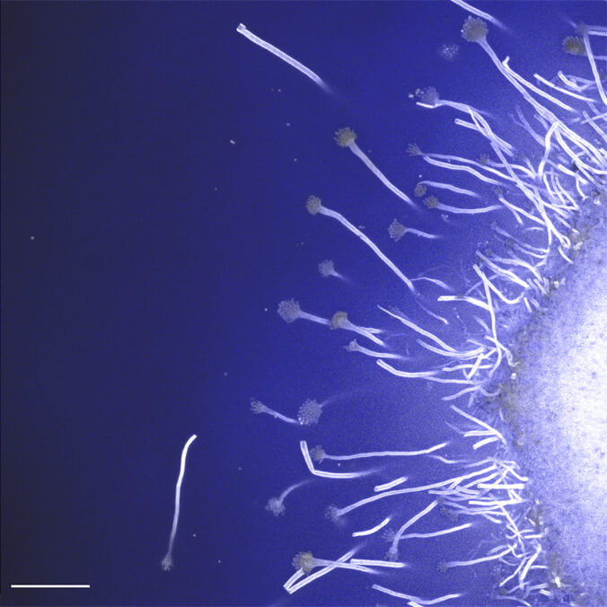
This week’s UH News Image of the Week is from University of Hawaiʻi at Mānoa’s Christine Farrar, an associate specialist in cell and molecular biology.
Farrar shared: “The image is of Aspergillus, a common type of mold, and features its spore producing structures. The attached image was created by Connor Schuller, a graduate student in the Department of Cell and Molecular Biology. It was generated from z-stack data collected on a laser scanning confocal microscope by fellow graduate students as part of a practicum assignment for Spring 2023 CMB 622: Cell Molecular Biology II, directed by Professor Peter Hoffmann. The practicum assignment was guided by Associate Specialist Christine Farrar. Class members include: Andrea Chavez, Brennan Yamamoto, Bryan Suechting, Chelsea Tanaka, Connor Schuller, Desta Rabin, Kayla Colaruotolo, Min Seok Han, and Ryan Wright.”
Previous Images
Aloha Bash
ʻŌlena
Tin Can Gong
Spring Footholds
Chick
All Images of the Week
Send us your image!
Want to get in on the action? The next UH News Image of the Week could be yours! Submit a photo, drawing, painting, digital illustration of a project you are working on, a moment from a field research outing or a beautiful and/or interesting shot of a scene on your campus. It could be a class visit during which you see an eye-catching object or scene.
Please include a brief description of the image and its connection to your campus, class assignment or other UH connection. By submitting your image, you are giving UH News permission to publish your photo on the UH News website and UH social media accounts. The image must be your original work, and anyone featured in your image needs to give consent to its publication.