
Sports Injury Education Center
As sports competition has intensified both in frequency and intensity, so have pediatric sports injuries. It is not unusual to see children practicing a sport several times a week, for hours at a time, year-round. This has led to a rise in the incidence and severity of sports injuries in children. The growing skeleton can only take so much pounding and stress before it begins to break down. Most often, sports injuries are simple fractures that can be treated successfully. Occasionally, however, the injuries can be potentially career-ending and may have long-term consequences even outside of sports participation. Appropriate management of these more sinister injuries is crucial to securing the best outcome possible.
Sports Injuries 101: Children are not little adults
Children are different from adults, among other ways, in that their bones are growing. Bones grow in areas called growth plates. Each long-bone in the arms has at least one, and most often two growth plates located near the end of the bone (Figure 1). Growth plates, also known as physes, are made up of cartilage containing multiple columns of growing and dividing cartilage cells (Figure 2). As these cells grow, they begin to ossify the cartilage matrix around them, adding layer upon layer of new bone. These new layers result in lengthening of the bone. Bones also get wider in addition to getting longer. Appositional growth (getting wider) is mediated by growth from the surface of the bone as the covering of the bone, known as periosteum, lays down new layers of bone on the surface of the established bone (Figure 3). Growth plates are not as strong as bones, and tend to break more easily than the bones on either side of them. Ligaments in children are strongest of all, even stronger than bone.
Adult sports injuries include fractures and ligament injuries. Ligaments are typically sprained (stretched) or ruptured (torn) either in the middle of the ligament or where they attach to bone. In children, because the ligaments are stronger than bone, as their ligaments are stretched, they tend not to tear, but instead pull off the piece of bone that they are attached to. This is called an avulsion fracture. Avulsion fractures are common around the knee and the elbow, as well as in the hand.
Adults also do not have growth plates. Fractures in children that occur near the joints are therefore more problematic if the injury involved the growth plate, particularly in certain locations. Injuries to the distal radial growth plate at the wrist have a 5% risk of growth arrest (bone stops growing), whereas injuries of the distal ulnar growth plate have a 50% risk of growth arrest. If the growth arrest is only partial, the bone can grow at an angle and increase the deformity. Complete growth arrest stops all growth. If the growth plate is not injured, particularly in children under 8 years of age, fractures close to the joint have some ability to correct themselves, straightening the bones as they grow.
Yet another difference between adults and children is the risk of avascular necrosis (death) of certain bones in the upper extremities due to repetitive loading. Because of the open growth plates (blood vessels cannot cross growth plates), the bone ends in children tend to have diminished blood flow. Repetitive pounding on the elbow, for example by walking on the arms common in gymnastics, can lead to avascular necrosis of the capitellum, known as Osteocondritis Dissecans (OCD). Other less common locations include the lunate and scaphoid bones in the wrist.

Ulnar positive variance
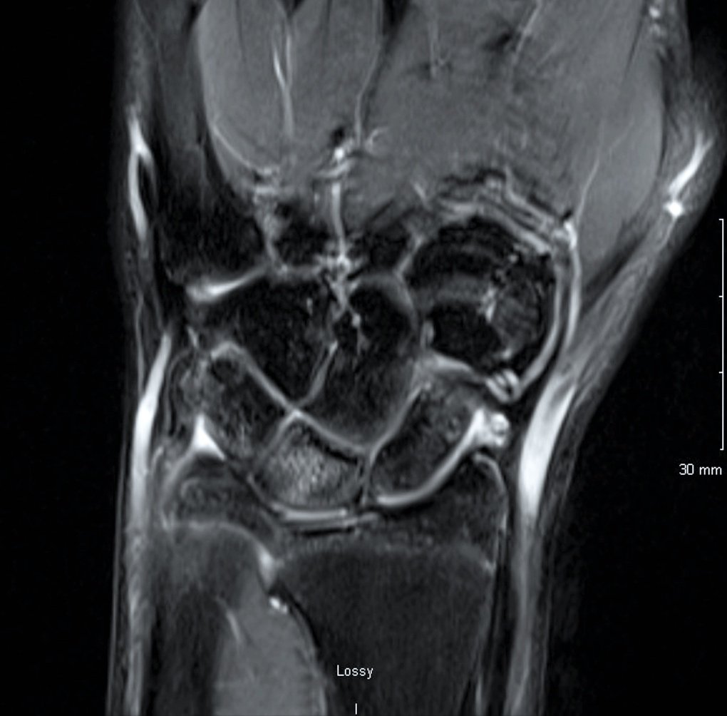
Signal changes (edema) in the lunate
Sports injuries 201: Ulno-carpal impaction
ANATOMY: The forearm is supported by 2 bone, the radius and the ulna. These bones not only both interact with the wrist and the elbow joints on either side of the forearm, but they also rotate around each other at the proximal and distal radio-ulnar joints (PRUJ and DRUJ). The ulna is the smaller of the two bones at the wrist and widens towards the elbow. The radius is wide at the wrist and narrows towards the elbow. In a normal wrist where the radius and the ulna are level, with weight bearing or grasping 80% of the load is transferred from the carpal bones of the wrist to the radius, and 20% is transferred to the ulna. If the ulna sticks out past the radius (ulnar positive variance) by just 2 mm, the load to the ulna is doubled to 40%. Because the additional load is transferred to the ulna via the lunate bone, the lunate itself also must bear more of the forces of the wrist. This often leads to a stress reaction in the lunate, leading to increased signal on the MRI (swelling inside the bone), degeneration of the lunate cartilage, and eventually fracturing, necrosis, and collapse.
ETIOLOGY: Some children may have ulnar positive variance on both sides due to normal growth. These children rarely have symptoms of ulna-carpal impaction unless the ulnar variance is more than 2 mm. The most common cause of symptomatic ulno-carpal impaction is limited growth at the radius. The growth of the radius can be stunted by injuries to the growth plate (such as Salter-Harris fractures), repetitive trauma (such as gymnast wrist), and tumors (such as osteochondromas).
PRESENTATION: Patients will present with a history of a previous fall or wrist weight-bearing sports participation (most commonly gymnastics). Pain will be localized to the ulnar side of the wrist (pinky finger side) and will be aggravated by weight bearing on the wrist and forearm rotation with ulnar deviation of the wrist. The distal ulna may be prominent as a bony mass on the ulnar side of the wrist.
IMAGING: X-rays will show an ulnar positive variance (see X-ray to the left). An MRI is not required for evaluation but will often show high signal in the lunate, indicating swelling (edema) in the bone from repetitive impacts from the distal ulna. TFCC tears are commonly seen.
TREATMENT: If pain is persistent despite 6 months of activity modification and splinting, the only surgical option is shortening of the ulna. This levels the joint, preventing the ulna from impacting on the lunate. There are multiple options for shortening the bone, all of which have a high success rate.
Sport Injuries 301: Oat procedure for ocd of the capitellum
Repetitive overload of the capitellum can result in avascular necrosis, or death of the bone, from insufficient blood supply. As the bone dies, it can no longer support the cartilage above it, leading to the formation of a bone and cartilage hole (defect) in the elbow joint. This is similar in concept to a pothole in the road. Restoring the cartilage surface has historically been impossible, but techniques such as micro fracture evolved to try to substitute cartilage scar for cartilage. The Osteochondral Autograft Transfer (OAT) procedure (1) has allowed us to replace the lost bone and cartilage with a bone and cartilage plug from a non-weight-bearing surface of the knee.
The results of the OAT procedure have been shown to be better than microfracture, with 80-90% of children returning to their pre-injury level of sports participation by 6 months after surgery.(1,2,3,4)
The OAT procedure is performed in the lateral decubitus position with the arm in an arm-holder. Prep and drape the ipsilateral arm and leg, with a non-sterile tourniquet on each limb. First, perform a diagnostic elbow arthroscopy. After confirming that the ulnar nerve does not subluxate, insufflate the joint with 15-20 cc of bupivacaine with epinephrine via a posterior portal. Exsanguinate the arm and inflate the tourniquet. Use the proximal anteromedial portal to insert the scope across the front of the joint. Using a switching stick, make an anterolateral portal. Place a cannula to prevent having to go in and out of the joint multiple times as this increases the risk of neurologic injury. Perform a synovectomy if needed and remove any loose bodies. The absence of synovitis is a sign that the lesion has healed. It is rare to be able to view the OCD lesion from the front, but it is best seen with the elbow in as much extension as achievable. Switch the viewing and working portals again using switching sticks and repeat the process for the medial side of the joint. Moving on to the posterior compartment, use a direct posterior and a proximal posterolateral portal alternatively as working and viewing portals as needed. Never debride on or near the medial gutter, as the ulnar nerve is immediately adjacent. Inspect the medial and lateral gutters for loose bodies and synovitis as was done for the anterior compartment. With the scope in the proximal posterolateral portal, establish a soft spot portal. Loose bodies and extensive synovitis are typically seen in this area. Place the scope through the soft spot portal as a viewing portal and visualize the lesion.
Challenge the lesion with a probe. If the cartilage is damaged but the subchondral bone holds firm, perform a microfracture technique. If the cartilage is soft or unstable, and the underlying bone is compromised, perform an OAT procedure. Connect the proximal posterolateral portal and the soft spot portal and split the anconeus. The lesion will be visible in deep flexion. A self-retaining retractor aids in visualization. Measure the size of the cartilage defect. Using the appropriate sized recipient harvester, remove all of the diseased bone and cartilage to a stable rim. If the diseased recipient plug cannot be removed with the harvester, core it out with a cannulated drill to a minimum depth of 10 mm.
Exsanguinate the knee and inflate the tourniquet. Holding the knee in full extension, make a 3 cm transverse incision lateral to the upper third of the patella directly over the superolateral corner of the lateral femoral condyle. The physis is just proximal to the articular surface, so make sure to leave a couple of millimeter margin and aim slightly distal. Harvest an appropriately sized plug from the superolateral corner of the articular surface to a minimum depth of 10 mm. Note the orientation of the plug via the slits on the side of the harvester. If the plug is uneven, try to match the orientation to match the contour of the articular surface of the defect in the elbow. Tap the donor plug into place but leave it proud about 1 mm. Use the 10mm sizer to tap the donor plug flush with the articular surface by maintaining the sizer halfway on the intact capitellar surface and working around the edges. Fill the defect in the knee with your choice of bone substitute. Close both wounds in layers.
The knee is maintained full weight bearing in a knee immobilizer for 2 weeks, which the patient can remove at home and begin self-directed range of motion exercises as tolerated. The elbow remains in a 60 degree elbow flexion cast for 4 weeks, followed by an unlocked hinged elbow brace for 2 more months. At 12 weeks after surgery, if the patient is completely pain free, a return to sports program is initiated with the goal of returning to sports in a further 12 weeks.

sports injuries 302: fixation of medial epicondyle fractures
Medial epicondyle fractures of the elbow result most often either from pitching or throwing injuries, or from falls on an outstretched arm. The medial epicondyle is the bony projection that can be felt on the inside portion of the elbow just above the joint. The bone serves as an attachment for the muscles that bend the wrist and fingers and that turn the palm down. The ligament that stabilizes the elbow also arises from the base of the epicondyle. When the medial epicondyle is pulled off of the humerus bone with the ligament still attached to the epicondyle, the elbow can be rendered unstable permanently. Early repair of the epicondyle is required to return a child to sport as quickly as possible but also to prevent long-term complications such as arthritis and deformity.