
Future Prospective of Radiopharmaceuticals from Natural Compounds Using Iodine Radioisotopes as Theranostic Agents

 ,
, 
Abstract
:1. Introduction
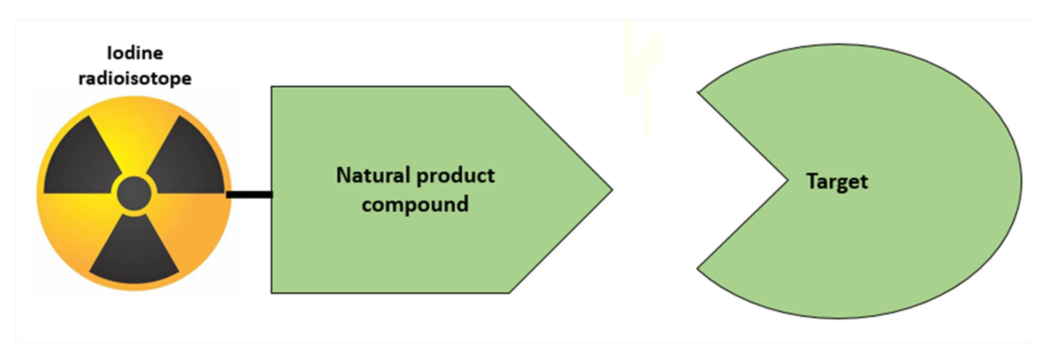
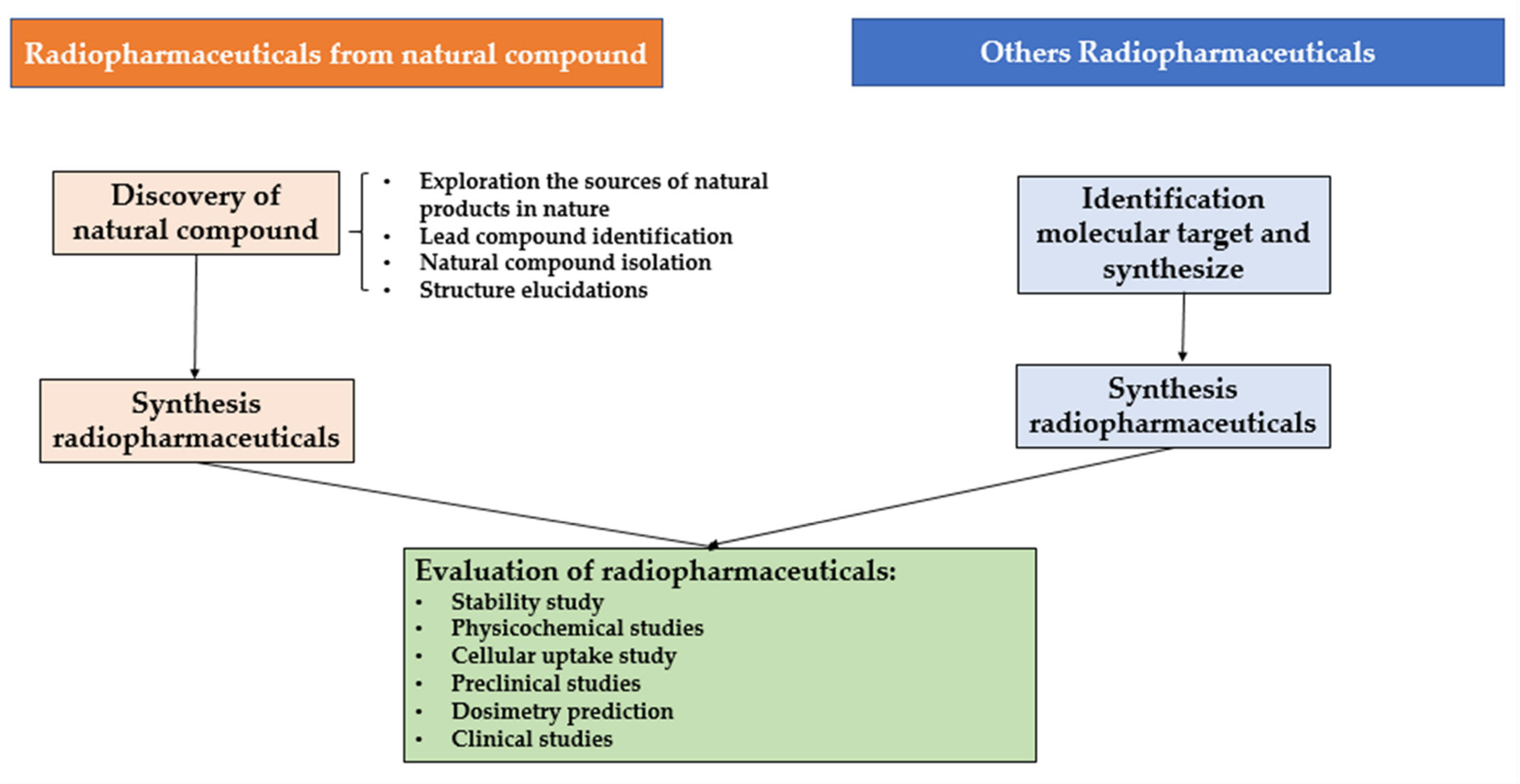
2. Differences between Radiopharmaceuticals from Natural Compounds with Other Radiopharmaceuticals
3. Available Literature on from Natural Compounds with Iodine Radioisotopes in Last 10-Year Period
4. Synthesis of Radiopharmaceuticals from Natural Compounds with Iodine Radioisotopes
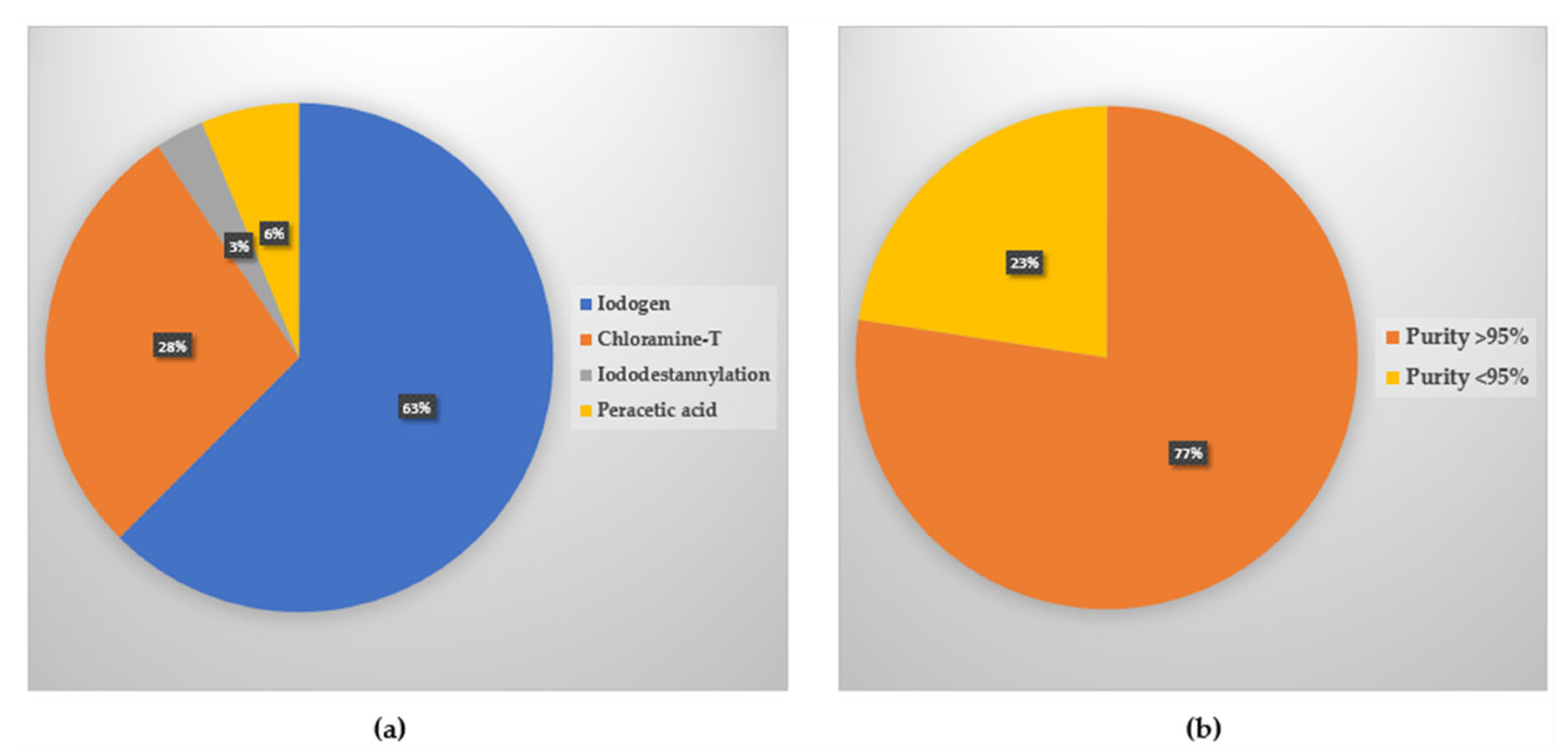
4.1. Electrophilic Substitutions
4.2. Nucleophilic Substitution
4.3. Synthesis of Radiopharmaceuticals from Natural Compound with Iodine Radioisotopes in the Last 10 Years
5. Evaluations of Radiopharmaceuticals from Natural Compounds with Iodine Radioisotopes
6. Challenge and Strategies
6.1. Problem Related to Radiochemical Purity and the Strategies
6.2. Problem Related to Biodistribution and the Strategies
7. Methods
8. Future, Prospect, and Conclusions
Author Contributions
Funding
Institutional Review Board Statement
Informed Consent Statement
Data Availability Statement
Acknowledgments
Conflicts of Interest
References
- Sgouros, G.; Bodei, L.; McDevitt, M.R.; Nedrow, J.R. Radiopharmaceutical therapy in cancer: Clinical advances and challenges. Nat. Rev. Drug Discov. 2020, 19, 589–608. [Google Scholar] [CrossRef] [PubMed]
- Lau, J.; Rousseau, E.; Kwon, D.; Lin, K.-S.; Bénard, F.; Chen, X. Insight into the Development of PET Radiopharmaceuticals for Oncology. Cancers 2020, 12, 1312. [Google Scholar] [CrossRef] [PubMed]
- Holik, H.A.; Ibrahim, F.M.; Elaine, A.A.; Putra, B.D.; Achmad, A.; Kartamihardja, A.H.S. The Chemical Scaffold of Theranostic Radiopharmaceuticals: Radionuclide, Bifunctional Chelator, and Pharmacokinetics Modifying Linker. Molecules 2022, 27, 3062. [Google Scholar] [CrossRef] [PubMed]
- Payolla, F.; Massabni, A.; Orvig, C. Radiopharmaceuticals for diagnosis in nuclear medicine: A short review. Eclét. Quím. J. 2019, 44, 11–19. [Google Scholar] [CrossRef]
- Vermeulen, K.; Vandamme, M.; Bormans, G.; Cleeren, F. Design and Challenges of Radiopharmaceuticals. Semin. Nucl. Med. 2019, 49, 339–356. [Google Scholar] [CrossRef]
- Wongso, H. Natural product-based Radiopharmaceuticals:Focus on curcumin and its analogs, flavonoids, and marine peptides. J. Pharm. Anal. 2021, 12, 380–393. [Google Scholar] [CrossRef]
- Vaidyanathan, G.; Zalutsky, M.R. The Radiopharmaceutical Chemistry of the Radioisotopes of Iodine. In Radiopharmaceutical Chemistry; Lewis, J.S., Windhorst, A.D., Zeglis, B.M., Eds.; Springer International Publishing: Berlin/Heidelberg, Germany, 2019; pp. 391–408. [Google Scholar] [CrossRef]
- Morphis, M.; van Staden, J.A.; du Raan, H.; Ljungberg, M. Validation of a SIMIND Monte Carlo modelled gamma camera for Iodine-123 and Iodine-131 imaging. Heliyon 2021, 7, e07196. [Google Scholar] [CrossRef]
- Yordanova, A.; Eppard, E.; Kürpig, S.; Bundschuh, R.A.; Schönberger, S.; Gonzalez-Carmona, M.; Feldmann, G.; Ahmadzadehfar, H.; Essler, M. Theranostics in nuclear medicine practice. OncoTargets Ther. 2017, 10, 4821–4828. [Google Scholar] [CrossRef] [Green Version]
- Silberstein, E.B. Radioiodine: The classic theranostic agent. Semin. Nucl. Med. 2012, 42, 164–170. [Google Scholar] [CrossRef]
- Treglia, G.; Muoio, B.; Giovanella, L.; Salvatori, M. The role of positron emission tomography and positron emission tomography/computed tomography in thyroid tumours: An overview. Eur. Arch. Oto-Rhino-Laryngol. 2013, 270, 1783–1787. [Google Scholar] [CrossRef]
- Braghirolli, A.M.; Waissmann, W.; da Silva, J.B.; dos Santos, G.R. Production of iodine-124 and its applications in nuclear medicine. Appl. Radiat. Isot. 2014, 90, 138–148. [Google Scholar] [CrossRef] [PubMed]
- Cascini, G.L.; Asabella, A.N.; Notaristefano, A.; Restuccia, A.; Ferrari, C.; Rubini, D.; Altini, C.; Rubini, G. 124 Iodine: A longer-life positron emitter isotope-new opportunities in molecular imaging. BioMed Res. Int. 2014, 2014, 672094. [Google Scholar] [CrossRef] [PubMed] [Green Version]
- Schwarz, S.B.; Thon, N.; Nikolajek, K.; Niyazi, M.; Tonn, J.C.; Belka, C.; Kreth, F.W. Iodine-125 brachytherapy for brain tumours—A review. Radiat. Oncol. 2012, 7, 30. [Google Scholar] [CrossRef] [Green Version]
- Takiar, V.; Voong, K.R.; Gombos, D.S.; Mourtada, F.; Rechner, L.A.; Lawyer, A.A.; Morrison, W.H.; Garden, A.S.; Beadle, B.M. A choice of radionuclide: Comparative outcomes and toxicity of ruthenium-106 and iodine-125 in the definitive treatment of uveal melanoma. Pr. Radiat. Oncol. 2015, 5, e169–e176. [Google Scholar] [CrossRef] [PubMed]
- Pelletier-Galarneau, M.; Sogbein, O.O.; Dinh, L.; Zuckier, L.S. Superiority of Digital Subtraction for Analysis of Simultaneously-Acquired Dual-Radiopharmaceutical Parathyroid Scintigraphy. Open J. Med. Imaging 2015, 5, 42–48. [Google Scholar] [CrossRef] [Green Version]
- Prashanth, R.; Roy, S.D.; Mandal, P.K.; Ghosh, S. High-Accuracy Classification of Parkinson’s Disease Through Shape Analysis and Surface Fitting in 123I-Ioflupane SPECT Imaging. IEEE J. Biomed. Health Inform. 2017, 21, 794–802. [Google Scholar] [CrossRef] [Green Version]
- Pandit-Taskar, N.; Modak, S. Norepinephrine Transporter as a Target for Imaging and Therapy. J. Nucl. Med. 2017, 58, 39S. [Google Scholar] [CrossRef] [Green Version]
- Vorobyeva, A.; Schulga, A.; Konovalova, E.; Güler, R.; Mitran, B.; Garousi, J.; Rinne, S.; Löfblom, J.; Orlova, A.; Deyev, S.; et al. Comparison of tumor-targeting properties of directly and indirectly radioiodinated designed ankyrin repeat protein (DARPin) G3 variants for molecular imaging of HER2. Int. J. Oncol. 2019, 54, 1209–1220. [Google Scholar] [CrossRef] [Green Version]
- Hanson, J. A Hundred Years in the Elucidation of the Structures of Natural Products. Sci. Prog. 2017, 100, 63–79. [Google Scholar] [CrossRef]
- Chimento, A.; Casaburi, I.; Rosano, C.; Avena, P.; De Luca, A.; Campana, C.; Martire, E.; Santolla, M.F.; Maggiolini, M.; Pezzi, V.; et al. Oleuropein and hydroxytyrosol activate GPER/ GPR30-dependent pathways leading to apoptosis of ER-negative SKBR3 breast cancer cells. Mol. Nutr. Food Res. 2014, 58, 478–489. [Google Scholar] [CrossRef]
- Sirianni, R.; Chimento, A.; De Luca, A.; Casaburi, I.; Rizza, P.; Onofrio, A.; Iacopetta, D.; Puoci, F.; Andò, S.; Maggiolini, M.; et al. Oleuropein and hydroxytyrosol inhibit MCF-7 breast cancer cell proliferation interfering with ERK1/2 activation. Mol. Nutr. Amp. Food Res. 2010, 54, 833–840. [Google Scholar] [CrossRef] [PubMed]
- Luo, C.; Li, Y.; Wang, H.; Cui, Y.; Feng, Z.; Li, H.; Li, Y.; Wang, Y.; Wurtz, K.; Weber, P.; et al. Hydroxytyrosol promotes superoxide production and defects in autophagy leading to anti-proliferation and apoptosis on human prostate cancer cells. Curr. Cancer Drug Targets 2013, 13, 625–639. [Google Scholar] [CrossRef]
- Sun, L.; Luo, C.; Liu, J. Hydroxytyrosol induces apoptosis in human colon cancer cells through ROS generation. Food Funct. 2014, 5, 1909–1914. [Google Scholar] [CrossRef] [PubMed]
- Toteda, G.; Lupinacci, S.; Vizza, D.; Bonofiglio, R.; Perri, E.; Bonofiglio, M.; Lofaro, D.; La Russa, A.; Leone, F.; Gigliotti, P.; et al. High doses of hydroxytyrosol induce apoptosis in papillary and follicular thyroid cancer cells. J. Endocrinol. Investig. 2017, 40, 153–162. [Google Scholar] [CrossRef] [PubMed]
- Ozkan, M.; Muftuler, F.; Yurt, A.; Medine, I.; Unak, P. Isolation of Hydroxytyrosol from olive leaves extract, radioiodination and investigation of bioaffinity using in vivo/in vitro methods. Radiochim. Acta 2013, 101, 585–593. [Google Scholar] [CrossRef]
- Khalil, N.; Bishr, M.; Desouky, S.; Salama, O. Ammi visnaga L., a Potential Medicinal Plant: A Review. Molecules 2020, 25, 301. [Google Scholar] [CrossRef] [Green Version]
- Khater, S.I.; Kandil, S.A.; Hussien, H. Preparation of radioiodinated khellin for the urinary tract imaging. J. Radioanal. Nucl. Chem. 2013, 295, 1939–1944. [Google Scholar] [CrossRef]
- Mullaicharam, A.R.; Nirmala, H. St John’s wort (Hypericum perforatum L.): A Review of its Chemistry, Pharmacology and Clinical properties. Int. J. Res. Phytochem. Pharmacol. Sci. 2018, 1, 5–11. [Google Scholar] [CrossRef]
- Cona, M.M.; Koole, M.; Feng, Y.; Liu, Y.; Verbruggen, A.; Oyen, R.; Ni, Y. Biodistribution and radiation dosimetry of radioiodinated hypericin as a cancer therapeutic. Int. J. Oncol. 2014, 44, 819–829. [Google Scholar] [CrossRef]
- Cona, M.M.; Alpizar, Y.A.; Li, J.; Bauwens, M.; Feng, Y.; Sun, Z.; Zhang, J.; Chen, F.; Talavera, K.; de Witte, P.; et al. Radioiodinated Hypericin: Its Biodistribution, Necrosis Avidity and Therapeutic Efficacy are Influenced by Formulation. Pharm. Res. 2014, 31, 278–290. [Google Scholar] [CrossRef]
- Pour, A.P.; Farahbakhsh, H. Lawsonia inermis L. leaves aqueous extract as a natural antioxidant and antibacterial product. Nat. Prod. Res. 2020, 34, 3399–3403. [Google Scholar] [CrossRef] [PubMed]
- Tekin, V.; Muftuler, F.Z.B.; Yurt Kilcar, A.; Unak, P. Radioiodination and biodistribution of isolated lawsone compound from Lawsonia inermis (henna) leaves extract. J. Radioanal. Nucl. Chem. 2014, 302, 225–232. [Google Scholar] [CrossRef]
- Tekin, V.; Biber Muftuler, F.Z.; Guldu, O.K.; Kilcar, A.Y.; Medine, E.I.; Yavuz, M.; Unak, P.; Timur, S. Biological affinity evaluation of Lawsonia inermis origin Lawsone compound and its radioiodinated form via in vitro methods. J. Radioanal. Nucl. Chem. 2015, 303, 701–708. [Google Scholar] [CrossRef]
- Gan, C.; Zhao, Z.; Nan, D.D.; Yin, B.; Hu, J. Homoisoflavonoids as potential imaging agents for β-amyloid plaques in Alzheimer’s disease. Eur. J. Med. Chem. 2014, 76, 125–131. [Google Scholar] [CrossRef] [PubMed]
- Aras, O.; Takan, G.; Kilcar, A.Y.; Muftuler, F.Z.B. Extraction and radioiodination of Gingko flavonoids and monitoring the cellular incorporation. J. Radioanal. Nucl. Chem. 2016, 310, 271–278. [Google Scholar] [CrossRef]
- Jiang, C.; Gao, M.; Li, Y.; Huang, D.; Yao, N.; Ji, Y.; Liu, X.; Zhang, D.; Wang, X.; Yin, Z.; et al. Exploring diagnostic potentials of radioiodinated sennidin A in rat model of reperfused myocardial infarction. Int. J. Pharm. 2015, 495, 31–40. [Google Scholar] [CrossRef]
- Liu, X.; Feng, Y.; Jiang, C.; Lou, B.; Li, Y.; Liu, W.; Yao, N.; Gao, M.; Ji, Y.; Wang, Q.; et al. Radiopharmaceutical evaluation of (131)I-protohypericin as a necrosis avid compound. J. Drug Target. 2015, 23, 417–426. [Google Scholar] [CrossRef]
- Zhang, D.; Huang, D.; Ji, Y.; Jiang, C.; Li, Y.; Gao, M.; Yao, N.; Liu, X.; Shao, H.; Jing, S.; et al. Experimental evaluation of radioiodinated sennoside B as a necrosis-avid tracer agent. J. Drug Target. 2015, 23, 180–190. [Google Scholar] [CrossRef]
- Yang, H.L.; Chen, S.C.; Kumar, K.J.S.; Yu, K.N.; Chao, P.D.L.; Tsai, S.Y.; Hou, Y.C.; Hseu, Y.C. Antioxidant and anti-inflammatory potential of hesperetin metabolites obtained from hesperetin-administered rat serum: An ex vivo approach. J. Agric. Food Chem. 2012, 60, 522–532. [Google Scholar] [CrossRef]
- Shin, K.C.; Nam, H.K.; Oh, D.K. Hydrolysis of flavanone glycosides by β-glucosidase from Pyrococcus furiosus and its application to the production of flavanone aglycones from citrus extracts. J. Agric. Food Chem. 2013, 61, 11532–11540. [Google Scholar] [CrossRef]
- Jeon, J.; Ma, S.-Y.; Choi, D.; Kang, J.; Nam, Y.; Yoon, S.; Park, S. Radiosynthesis of 123I-labeled hesperetin for biodistribution study of orally administered hesperetin. J. Radioanal. Nucl. Chem. 2015, 306, 437–443. [Google Scholar] [CrossRef]
- Chua, L.S. A review on plant-based rutin extraction methods and its pharmacological activities. J. Ethnopharmacol. 2013, 150, 805–817. [Google Scholar] [CrossRef] [PubMed]
- Choi, M.H.; Rho, J.K.; Kang, J.A.; Shim, H.E.; Nam, Y.R.; Yoon, S.; Kim, H.R.; Choi, D.S.; Park, S.H.; Jang, B.-S.; et al. Efficient radiolabeling of rutin with 125I and biodistribution study of radiolabeled rutin. J. Radioanal. Nucl. Chem. 2016, 308, 477–483. [Google Scholar] [CrossRef]
- Antonisamy, P.; Agastian, P.; Kang, C.W.; Kim, N.S.; Kim, J.H. Anti-inflammatory activity of rhein isolated from the flowers of Cassia fistula L. and possible underlying mechanisms. Saudi J. Biol. Sci. 2019, 26, 96–104. [Google Scholar] [CrossRef] [PubMed]
- Zhang, D.; Jin, Q.; Ni, Y.; Zhang, J. Discovery of necrosis avidity of rhein and its applications in necrosis imaging. J. Drug Target. 2020, 28, 904–912. [Google Scholar] [CrossRef]
- Wang, Q.; Yang, S.; Jiang, C.; Li, J.; Wang, C.; Chen, L.; Jin, Q.; Song, S.; Feng, Y.; Ni, Y.; et al. Discovery of Radioiodinated Monomeric Anthraquinones as a Novel Class of Necrosis Avid Agents for Early Imaging of Necrotic Myocardium. Sci. Rep. 2016, 6, 21341. [Google Scholar] [CrossRef] [Green Version]
- Al-Sharif, I.; Remmal, A.; Aboussekhra, A. Eugenol triggers apoptosis in breast cancer cells through E2F1/survivin down-regulation. BMC Cancer 2013, 13, 600. [Google Scholar] [CrossRef] [Green Version]
- Vidhya, N.; Devaraj, S.N. Induction of apoptosis by eugenol in human breast cancer cells. Indian J. Exp Biol. 2011, 49, 871–878. [Google Scholar]
- Ghosh, R.; Ganapathy, M.; Alworth, W.L.; Chan, D.C.; Kumar, A.P. Combination of 2-methoxyestradiol (2-ME2) and eugenol for apoptosis induction synergistically in androgen independent prostate cancer cells. J. Steroid Biochem. Mol. Biol. 2009, 113, 25–35. [Google Scholar] [CrossRef]
- Dervis, E.; Kilcar, A.Y.; Medine, E.I.; Tekin, V.; Cetkin, B.; Uygur, E.; Muftuler, F.Z.B. In Vitro Incorporation of Radioiodinated Eugenol on Adenocarcinoma Cell Lines (Caco2, MCF7, and PC3). Cancer Biother. Radiopharm. 2017, 32, 75–81. [Google Scholar] [CrossRef]
- Gibellini, L.; Pinti, M.; Nasi, M.; Montagna, J.P.; De Biasi, S.; Roat, E.; Bertoncelli, L.; Cooper, E.L.; Cossarizza, A. Quercetin and cancer chemoprevention. Evid Based Complement. Altern. Med. 2011, 2011, 591356. [Google Scholar] [CrossRef] [PubMed] [Green Version]
- Lin, C.; Yu, Y.; Zhao, H.G.; Yang, A.; Yan, H.; Cui, Y. Combination of quercetin with radiotherapy enhances tumor radiosensitivity in vitro and in vivo. Radiother. Oncol. 2012, 104, 395–400. [Google Scholar] [CrossRef] [PubMed]
- Xie, Q.; Li, X.; Wang, G.; Hou, X.; Wang, Y.; Yu, H.; Qu, C.; Luo, S.; Cui, Y.; Xia, C.; et al. Preparation and evaluation of 131I-quercetin as a novel radiotherapy agent against dedifferentiated thyroid cancer. J. Radioanal. Nucl. Chem. 2017, 311, 1697–1708. [Google Scholar] [CrossRef]
- Palensina, G.; Rosita, L.; Sagala, Z. Isolation of Arbutin from Leaves and Fruits of Buni (Antidesma Bunius L. Spreng) As Tyrosinase Enzym Inhibitor. Bioinform. Biomed. Res. J. 2021, 4, 8–18. [Google Scholar] [CrossRef]
- Ebadollahi, S.H.; Pouramir, M.; Zabihi, E.; Golpour, M.; Aghajanpour-Mir, M. The Effect of Arbutin on The Expression of Tumor Suppressor P53, BAX/BCL-2 Ratio and Oxidative Stress Induced by Tert-Butyl Hydroperoxide in Fibroblast and LNcap Cell Lines. Cell J. 2021, 22, 532–541. [Google Scholar] [CrossRef]
- Huynh, P.T.; Ha, Y.S.; Lee, W.; Yoo, J. Radio-Iodinated arbutin for tumor imaging. J. Radiopharm. Mol. Probes 2017, 3, 72–79. [Google Scholar]
- Liang, J.; Sun, Z.; Zhang, D.; Jin, Q.; Cai, L.; Ma, L.; Liu, W.; Ni, Y.; Zhang, J.; Yin, Z. First Evaluation of Radioiodinated Flavonoids as Necrosis-Avid Agents and Application in Early Assessment of Tumor Necrosis. Mol. Pharm. 2018, 15, 207–215. [Google Scholar] [CrossRef]
- Chen, G.; Pi, X.M.; Yu, C.Y. A new naphthalenone isolated from the green walnut husks of Juglans mandshurica Maxim. Nat. Prod. Res. 2015, 29, 174–179. [Google Scholar] [CrossRef]
- Su, C.; Zhang, D.; Bao, N.; Ji, A.; Feng, Y.; Chen, L.; Ni, Y.; Zhang, J.; Yin, Z.-Q. Evaluation of Radioiodinated 1,4-Naphthoquinones as Necrosis Avid Agents for Rapid Myocardium Necrosis Imaging. Mol. Imaging Biol. 2017, 20, 74–84. [Google Scholar] [CrossRef]
- Chen, C.A.; Chang, H.H.; Kao, C.Y.; Tsai, T.H.; Chen, Y.J. Plumbagin, isolated from Plumbago zeylanica, induces cell death through apoptosis in human pancreatic cancer cells. Pancreatology 2009, 9, 797–809. [Google Scholar] [CrossRef]
- Aminin, D.; Polonik, S. 1,4-Naphthoquinones: Some Biological Properties and Application. Chem. Pharm. Bull. 2020, 68, 46–57. [Google Scholar] [CrossRef] [PubMed]
- Peñalver, P.; Belmonte-Reche, E.; Adán, N.; Caro, M.; Mateos-Martín, M.L.; Delgado, M.; González-Rey, E.; Morales, J.C. Alkylated resveratrol prodrugs and metabolites as potential therapeutics for neurodegenerative diseases. Eur. J. Med. Chem. 2018, 146, 123–138. [Google Scholar] [CrossRef]
- Jin, F.; Wu, Q.; Lu, Y.F.; Gong, Q.H.; Shi, J.S. Neuroprotective effect of resveratrol on 6-OHDA-induced Parkinson’s disease in rats. Eur. J. Pharmacol. 2008, 600, 78–82. [Google Scholar] [CrossRef]
- Guimón, J.; Guimón, P. How ready-to-use therapeutic food shapes a new technological regime to treat child malnutrition. Technol. Forecast. Soc. Chang. 2012, 79, 1319–1327. [Google Scholar] [CrossRef] [Green Version]
- Leis, K.; Baska, A.; Bereźnicka, W.; Marjańska, A.; Mazur, E.; Lewandowski, B.T.; Kałużny, K.; Gałązka, P. Resveratrol in the treatment of neuroblastoma: A review. Rev. Neurosci. 2020, 31, 873–881. [Google Scholar] [CrossRef] [PubMed]
- Karatay, K.B.; Kilcar, A.Y.; Guldu, O.K.; Medine, E.I.; Muftuler, F.Z.B. Isolation of resveratrol from peanut sprouts, radioiodination and investigation of its bioactivity on neuroblastoma cell lines. J. Radioanal. Nucl. Chem. 2020, 325, 75–84. [Google Scholar] [CrossRef]
- Sakai, T.; Kogiso, M. Soy isoflavones and immunity. J. Med. Investig. 2008, 55, 167–173. [Google Scholar] [CrossRef] [Green Version]
- Yuseran, H.; Hartoyo, E.; Nurseta, T.; Kalim, H. Molecular docking of genistein on estrogen receptors, promoter region of BCLX, caspase-3, Ki-67, cyclin D1, and telomere activity. J. Taibah Univ. Med. Sci. 2019, 14, 79–87. [Google Scholar] [CrossRef]
- Rajah, T.T.; Du, N.; Drews, N.; Cohn, R. Genistein in the presence of 17beta-estradiol inhibits proliferation of ERbeta breast cancer cells. Pharmacology 2009, 84, 68–73. [Google Scholar] [CrossRef]
- Ramdhani, D.; Widyasari, E.M.; Sriyani, M.E.; Arnanda, Q.P.; Watabe, H. Iodine-131 labeled genistein as a potential radiotracer for breast cancer. Heliyon 2020, 6, e04780. [Google Scholar] [CrossRef]
- Sp, N.; Kang, D.Y.; Lee, J.M.; Bae, S.W.; Jang, K.J. Potential Antitumor Effects of 6-Gingerol in p53-Dependent Mitochondrial Apoptosis and Inhibition of Tumor Sphere Formation in Breast Cancer Cells. Int. J. Mol. Sci. 2021, 22, 4660. [Google Scholar] [CrossRef] [PubMed]
- Ray, A.; Vasudevan, S.; Sengupta, S. 6-Shogaol Inhibits Breast Cancer Cells and Stem Cell-Like Spheroids by Modulation of Notch Signaling Pathway and Induction of Autophagic Cell Death. PLoS ONE 2015, 10, e0137614. [Google Scholar] [CrossRef] [PubMed]
- Karatay, K.B.; Kılçar, A.Y.; Derviş, E.; Müftüler, F.Z.B. Radioiodinated Ginger Compounds (6-gingerol and 6-shogaol) and Incorporation Assays on Breast Cancer Cells. Anticancer Agents Med. Chem. 2020, 20, 1129–1139. [Google Scholar] [CrossRef]
- İnce, İ.; Yıldırım, Y.; Güler, G.; Medine, E.İ.; Ballıca, G.; Kuşdemir, B.C.; Göker, E. Synthesis and characterization of folic acid-chitosan nanoparticles loaded with thymoquinone to target ovarian cancer cells. J. Radioanal. Nucl. Chem. 2020, 324, 71–85. [Google Scholar] [CrossRef]
- Woo, C.C.; Kumar, A.P.; Sethi, G.; Tan, K.H. Thymoquinone: Potential cure for inflammatory disorders and cancer. Biochem. Pharm. 2012, 83, 443–451. [Google Scholar] [CrossRef] [PubMed]
- Destito, G.; Yeh, R.; Rae, C.S.; Finn, M.G.; Manchester, M. Folic Acid-Mediated Targeting of Cowpea Mosaic Virus Particles to Tumor Cells. Chem. Biol. 2007, 14, 1152–1162. [Google Scholar] [CrossRef] [Green Version]
- Sriyani, M.E.; Nuraeni, W.; Rosyidiah, E.; Widyasari, E.M.; Saraswati, A.; Shintia, M. Quality control and stability study of the [131I]I-rutin produced in acidic condition. AIP Conf. Proc. 2021, 2381, 020082. [Google Scholar] [CrossRef]
- Kumar, N.; Pruthi, V. Potential applications of ferulic acid from natural sources. Biotechnol. Rep. 2014, 4, 86–93. [Google Scholar] [CrossRef] [Green Version]
- Eroğlu, C.; Seçme, M.; Bağcı, G.; Dodurga, Y. Assessment of the anticancer mechanism of ferulic acid via cell cycle and apoptotic pathways in human prostate cancer cell lines. Tumor Biol. 2015, 36, 9437–9446. [Google Scholar] [CrossRef]
- Zhang, X.D.; Wu, Q.; Yang, S.H. Ferulic acid promoting apoptosis in human osteosarcoma cell lines. Pak. J. Med. Sci. 2017, 33, 127–131. [Google Scholar] [CrossRef]
- Sedik, G.A.; Rizq, R.S.A.; Ibrahim, I.T.; Elzanfaly, E.S.; Motaleb, M.A. Miniaturized chromatographic systems for radiochemical purity evaluation of (131)I-Ferulic acid as a new candidate in nuclear medicine applications. Appl. Radiat. Isot. 2021, 167, 109370. [Google Scholar] [CrossRef] [PubMed]
- Selim, A.A.; Essa, B.M.; Abdelmonem, I.M.; Amin, M.A.; Sarhan, M.O. Extraction, purification and radioiodination of Khellin as cancer theranostic agent. Appl. Radiat. Isot. 2021, 178, 109970. [Google Scholar] [CrossRef] [PubMed]
- Hueza, I.M.; Raspantini, P.C.; Raspantini, L.E.; Latorre, A.O.; Górniak, S.L. Zearalenone, an estrogenic mycotoxin, is an immunotoxic compound. Toxins 2014, 6, 1080–1095. [Google Scholar] [CrossRef] [PubMed] [Green Version]
- Zou, Z.; He, Z.; Li, H.; Han, P.; Meng, X.; Zhang, Y.; Zhou, F.; Ouyang, K.-P.; Chen, X.-Y.; Tang, J. In vitro removal of deoxynivalenol and T-2 toxin by lactic acid bacteria. Food Sci. Biotechnol. 2012, 21, 1677–1683. [Google Scholar] [CrossRef]
- Sanad, M.H.; Farag, A.B.; Bassem, S.A.; Marzook, F.A. Radioiodination of zearalenone and determination of Lactobacillus plantarum effect of on zearalenone organ distribution: In silico study and preclinical evaluation. Toxicol. Rep. 2022, 9, 470–479. [Google Scholar] [CrossRef]
- Subramanian, V.S.; Sabui, S.; Teafatiller, T.; Bohl, J.A.; Said, H.M. Structure/functional aspects of the human riboflavin transporter-3 (SLC52A3): Role of the predicted glycosylation and substrate-interacting sites. Am. J. Physiol.-Cell Physiol. 2017, 313, C228–C238. [Google Scholar] [CrossRef] [Green Version]
- Bulas, S.; Bedoukian, E.C.; O’Neil, E.C.; Krantz, I.D.; Yum, S.W.; Liu, G.T.; Aleman, T.S. Ocular Biomarkers of Riboflavin Transporter Deficiency. J. Neuroophthalmol. 2022. [Google Scholar] [CrossRef]
- Li, J.; Chen, Y.; Peng, C.; Hong, X.; Liu, H.; Fang, J.; Zhuang, R.; Pan, W.; Zhang, D.; Guo, Z.; et al. Micro-SPECT Imaging of Acute Ischemic Stroke with Radioiodinated Riboflavin in Rat MCAO Models via Riboflavin Transporter Targeting. ACS Chem. Neurosci. 2022, 13, 1966–1973. [Google Scholar] [CrossRef]
- Jeung, Y.J.; Kim, H.G.; Ahn, J.; Lee, H.J.; Lee, S.B.; Won, M.; Jung, C.R.; Im, J.Y.; Kim, B.K.; Park, S.K.; et al. Shikonin induces apoptosis of lung cancer cells via activation of FOXO3a/EGR1/SIRT1 signaling antagonized by p300. Biochim. Biophys. Acta 2016, 1863, 2584–2593. [Google Scholar] [CrossRef]
- Yeh, Y.-C.; Liu, T.-J.; Lai, H.-C. Shikonin Induces Apoptosis, Necrosis, and Premature Senescence of Human A549 Lung Cancer Cells through Upregulation of p53 Expression. Evid.-Based Complement. Altern. Med. 2015, 2015, 620383. [Google Scholar] [CrossRef] [Green Version]
- Selim, A.A.; Motaleb, M.A.; Fayez, H.A. Lung Cancer-Targeted [131I]-Iodoshikonin as Theranostic Agent: Radiolabeling, In Vivo Pharmacokinetics and Biodistribution. Pharm. Chem. J. 2022, 55, 1163–1168. [Google Scholar] [CrossRef]
- Mushtaq, S.; Jeon, J.; Shaheen, A.; Jang, B.S.; Park, S.H. Critical analysis of radioiodination techniques for micro and macro organic molecules. J. Radioanal. Nucl. Chem. 2016, 309, 859–889. [Google Scholar] [CrossRef]
- Takahashi, M.; Seki, K.-i.; Nishijima, K.-i.; Zhao, S.; Kuge, Y.; Tamaki, N.; Ohkura, K. Synthesis of a radioiodinated thymidine phosphorylase inhibitor and its preliminary evaluation as a potential SPECT tracer for angiogenic enzyme expression. J. Label. Compd. Radiopharm. 2008, 51, 384–387. [Google Scholar] [CrossRef]
- Zmuda, F.; Malviya, G.; Blair, A.; Boyd, M.; Chalmers, A.J.; Sutherland, A.; Pimlott, S.L. Synthesis and Evaluation of a Radioiodinated Tracer with Specificity for Poly(ADP-ribose) Polymerase-1 (PARP-1) in Vivo. J. Med. Chem. 2015, 58, 8683–8693. [Google Scholar] [CrossRef] [Green Version]
- Sadeghzadeh, M.; Daha, F.J.; Sheibani, S.; Erfani, M. Radioiodination of 4-benzyl-1-(3-iodobenzylsulfonyl)piperidine, 4-(3-iodobenzyl)-1-(benzylsulfonyl)piperazine and their derivatives via isotopic and non-isotopic exchange reactions. J. Radioanal. Nucl. Chem. 2014, 302, 1119–1125. [Google Scholar] [CrossRef]
- Hagimori, M.; Temma, T.; Kudo, S.; Sano, K.; Kondo, N.; Mukai, T. Synthesis of radioiodinated probes targeted toward matrix metalloproteinase-12. Bioorg. Med. Chem. Lett. 2018, 28, 193–195. [Google Scholar] [CrossRef]
- Lee, Y.-S. Radiopharmaceuticals for Molecular Imaging. Open Nucl. Med. J. 2010, 2, 178–185. [Google Scholar] [CrossRef]
- Waterhouse, R.N. Determination of lipophilicity and its use as a predictor of blood–brain barrier penetration of molecular imaging agents. Mol. Imaging Biol. 2003, 5, 376–389. [Google Scholar] [CrossRef]
- Arnott, J.; Lobo, S. The influence of lipophilicity in drug discovery and design. Expert Opin. Drug Discov. 2012, 7, 863–875. [Google Scholar] [CrossRef]
- Kratochwil, N.A.; Huber, W.; Müller, F.; Kansy, M.; Gerber, P.R. Predicting plasma protein binding of drugs: A new approach. Biochem. Pharm. 2002, 64, 1355–1374. [Google Scholar] [CrossRef]
- De Kruijff, R.M.; Wolterbeek, H.T.; Denkova, A.G. A Critical Review of Alpha Radionuclide Therapy—How to Deal with Recoiling Daughters? Pharmaceuticals 2015, 8, 321–336. [Google Scholar] [CrossRef] [PubMed]
- İlem-Özdemir, D.; Ekinci, M.; Gundogdu, E.; Asikoglu, M. Estimating Binding Capability of Radiopharmaceuticals by Cell Culture Studies. Int. J. Med. Nano Res. 2016, 3, 014. [Google Scholar] [CrossRef]
- Motaleb, M.A.; Ibrahim, I.T.; Sayyed, M.E.; Awad, G.A.S. (131)I-trazodone: Preparation, quality control and in vivo biodistribution study by intranasal and intravenous routes as a hopeful brain imaging radiopharmaceutical. Rev. Esp Med. Nucl. Imagen Mol. 2017, 36, 371–376. [Google Scholar] [CrossRef] [PubMed]
- Rebischung, C.; Hoffmann, D.; Stefani, L.; Desruet, M.D.; Wang, K.; Adelstein, S.J.; Artignan, X.; Vincent, F.; Gauchez, A.S.; Zhang, H.; et al. First human treatment of resistant neoplastic meningitis by intrathecal administration of MTX plus (125)IUdR. Int. J. Radiat. Biol. 2008, 84, 1123–1129. [Google Scholar] [CrossRef] [PubMed]
- Shuryak, I.; Dadachova, E. New Approaches for Modeling Radiopharmaceutical Pharmacokinetics Using Continuous Distributions of Rates. J. Nucl. Med. 2015, 56, 1622–1628. [Google Scholar] [CrossRef] [Green Version]
- Koziorowski, J.; Behe, M.; Decristoforo, C.; Ballinger, J.; Elsinga, P.; Ferrari, V.; Peitl, P.K.; Todde, S.; Mindt, T.L. Position paper on requirements for toxicological studies in the specific case of radiopharmaceuticals. EJNMMI Radiopharm. Chem. 2016, 1, 1. [Google Scholar] [CrossRef] [Green Version]
- Eberlein, U.; Bröer, J.H.; Vandevoorde, C.; Santos, P.; Bardiès, M.; Bacher, K.; Nosske, D.; Lassmann, M. Biokinetics and dosimetry of commonly used radiopharmaceuticals in diagnostic nuclear medicine—A review. Eur. J. Nucl. Med. Mol. Imaging 2011, 38, 2269–2281. [Google Scholar] [CrossRef] [Green Version]
- El-Tawoosy, M.; Ibrahim, I. Radioiodination and biological evaluation of salbutamol as a β2-adrenoceptor agonist. Radiochemistry 2012, 54, 401–406. [Google Scholar] [CrossRef]
- El-Azony, K.; El-Mohty, A.; Seddik, U.; Khater, s. Radioiodination and bioevaluation of nitrofurantoin for urinary tract imaging. J. Label. Compd. Radiopharm. 2012, 55, 315–319. [Google Scholar] [CrossRef]
- Kiess, A.P.; Minn, I.; Chen, Y.; Hobbs, R.; Sgouros, G.; Mease, R.C.; Pullambhatla, M.; Shen, C.J.; Foss, C.A.; Pomper, M.G. Auger Radiopharmaceutical Therapy Targeting Prostate-Specific Membrane Antigen. J. Nucl. Med. 2015, 56, 1401–1407. [Google Scholar] [CrossRef] [Green Version]
- Moustapha, M.E.; Motaleb, M.A.; Ibrahim, I.T.; Moustafa, M.E. Oxidative radioiodination of aripiprazole by chloramine-T as a route to a potential brain imaging agent: A mechanistic approach. Radiochemistry 2013, 55, 116–122. [Google Scholar] [CrossRef]
- Abdel-Bary, H.M.; Moustafa, K.A.; Abdel-Ghaney, I.Y.; Sallam, K.M.; Shamsel-Din, H.A. Synthesis and radioiodination of new dipeptide coupled with biologically active pyridine moiety. J. Radioanal. Nucl. Chem. 2013, 298, 9–18. [Google Scholar] [CrossRef]
- Amin, A.; Soliman, S.; El-Aziz, H. Preparation and biodistribution of [125I]Melphalan: A potential radioligand for diagnostic and therapeutic applications. J. Label. Compd. Radiopharm. 2009, 53, 1–5. [Google Scholar] [CrossRef]
- Amin, A.; Soliman, S.; El-Aziz, H.; El-Enein, S. Radioiodination of Zaleplon and Its in-vivo Biologic Behavior in Mice: An Imaging Probe for Brain. Int. J. Chem. 2013, 6, 17. [Google Scholar] [CrossRef]
- Avcıbaşı, U.; demiroğlu, H.; Unak, P.; Müftüler, F.; Ichedef, C.A.; Gumuser, F. In vivo biodistribution of 131 I labeled bleomycin (BLM) and isomers (A2 and B2) on experimental animal models. J. Radioanal. Nucl. Chem. 2010, 285, 207–214. [Google Scholar] [CrossRef]
- Baş, U.; Demiroğlu, H.; Ediz, M.; Akalın, H.; Şkan, E.; Senay, H.; Türkcan, C.; Ozcan, Y.; Akgöl, S.; Avcibaşi, N. Radiolabeling of new generation magnetic poly(HEMA-MAPA) nanoparticles with I-131 and preliminary investigation of its radiopharmaceutical potential using albino Wistar rats. J. Label. Compd. Radiopharm. 2013, 56, 708–716. [Google Scholar] [CrossRef]
- Hussien, H.; Goud, A.A.; Amin, A.M.; El-Sheikh, R.; Seddik, U. Comparative study between chloramine-T and iodogen to prepare radioiodinated etodolac for inflammation imaging. J. Radioanal. Nucl. Chem. 2011, 288, 9–15. [Google Scholar] [CrossRef]
- Wang, K.; Adelstein, S.J.; Kassis, A.I. DMSO increases radioiodination yield of radiopharmaceuticals. Appl. Radiat. Isot. 2008, 66, 50–59. [Google Scholar] [CrossRef] [Green Version]
- Li, G.; Kakarla, R.; Gerritz, S.W. A fast and efficient bromination of isoxazoles and pyrazoles by microwave irradiation. Tetrahedron Lett. 2007, 48, 4595–4599. [Google Scholar] [CrossRef]
- Mattner, F.; Mardon, K.; Katsifis, A. Pharmacological evaluation of [123I]-CLINDE: A radioiodinated imidazopyridine-3-acetamide for the study of peripheral benzodiazepine binding sites (PBBS). Eur. J. Nucl. Med. Mol. Imaging 2008, 35, 779–789. [Google Scholar] [CrossRef]
- Kim, A.; Choi, K.H. Purification System of 131I-Metaiodobenzylguanidine. In Proceedings of the Transactions of the Korean Nuclear Society Autumn Meeting 2019, Goyang, Republic of Korea, 24–25 October 2019. [Google Scholar]
- Chitneni, S.K.; Reitman, Z.J.; Spicehandler, R.; Gooden, D.M.; Yan, H.; Zalutsky, M.R. Synthesis and evaluation of radiolabeled AGI-5198 analogues as candidate radiotracers for imaging mutant IDH1 expression in tumors. Bioorg. Med. Chem. Lett. 2018, 28, 694–699. [Google Scholar] [CrossRef] [PubMed]
- Lemps, R.D.; Desruet, M.; Bacot, S.; Ahmadi, M.; Ghezzi, C.; Desruet, M.; Fagret, D.; Berger, F. Iodogen-mediated radiolabeling of Bevacizumab with I-123 for clinical applications. Eur. J. Nucl. Med. Mol. Imaging 2014, 41, S447. [Google Scholar]
- Visser, G.W.; Klok, R.P.; Gebbinck, J.W.; ter Linden, T.; van Dongen, G.A.; Molthoff, C.F. Optimal quality (131)I-monoclonal antibodies on high-dose labeling in a large reaction volume and temporarily coating the antibody with IODO-GEN. J. Nucl. Med. 2001, 42, 509–519. [Google Scholar] [PubMed]
- Molavipordanjani, S.; Tolmachev, V.; Hosseinimehr, S.J. Basic and practical concepts of radiopharmaceutical purification methods. Drug Discov. Today 2019, 24, 315–324. [Google Scholar] [CrossRef] [PubMed]
- Amin, A.; Farrag, N.; AbdEl-Bary, A. Iodine-125-Chlorambucil as Possible Radio Anticancer for Diagnosis and Therapy of Cancer: Preparation and Tissue Distribution. Br. J. Pharm. Res. 2014, 4, 1873–1885. [Google Scholar] [CrossRef]
- Spetz, J.; Rudqvist, N.; Forssell-Aronsson, E. Biodistribution and dosimetry of free 211At, 125I- and 131I- in rats. Cancer Biother. Radiopharm. 2013, 28, 657–664. [Google Scholar] [CrossRef] [Green Version]
- Cavina, L.; van der Born, D.; Klaren, P.H.M.; Feiters, M.C.; Boerman, O.C.; Rutjes, F. Design of Radioiodinated Pharmaceuticals: Structural Features Affecting Metabolic Stability towards in Vivo Deiodination. Eur. J. Org. Chem. 2017, 2017, 3387–3414. [Google Scholar] [CrossRef] [Green Version]
- Kim, E.J.; Kim, B.S.; Choi, D.B.; Chi, S.G.; Choi, T.H. Enhanced tumor retention of radioiodinated anti-epidermal growth factor receptor antibody using novel bifunctional iodination linker for radioimmunotherapy. Oncol. Rep. 2016, 35, 3159–3168. [Google Scholar] [CrossRef] [Green Version]
- Tong, R.; Kohane, D.S. New Strategies in Cancer Nanomedicine. Annu Rev. Pharm. Toxicol. 2016, 56, 41–57. [Google Scholar] [CrossRef]


| Radioisotope | Half Life | Emission Type | Application | Refs. |
|---|---|---|---|---|
| iodine-123 | 13.2 h | Gamma, EC 1/auger | SPECT 3 diagnostic | [8,9,10] |
| iodine-124 | 4.8 days | Positron | PET 2 diagnostic | [11,12,13] |
| iodine-125 | 60 days | Gamma, EC 1/Auger | Preclinical study, Radiotherapy SPECT 3 diagnostic | [14,15] |
| iodine-131 | 8.04 days | Gamma, beta | Radiotherapy, SPECT3 and PET 2 diagnostic | [8,9,10] |
| Year | Natural Compounds | Sources | Pharmacological Activities | Radioisotope | Application | Recent Research Reported | Refs. |
|---|---|---|---|---|---|---|---|
| 2013 | Hydroxytyrosol | olive leaves extract | Anticancer (breast, colon, prostate, and thyroid cancer) | iodine-131 | Cancer therapy | Preclinical study | [21,22,23,24,25,26] |
| Khellin | Ammi visnaga fruits | Activity against kidney disease and vitiligo, anticancer | iodine-125 | Urinary tract imaging | Preclinical study | [27,28] | |
| 2014 | Hypericin | Hypericum perforatum L. | Antiviral, necrosis avidity and anticancer | iodine-131 | Cancer therapy | Dosimetry prediction | [29,30] |
| Hypericin | Hypericum perforatum L. | Antiviral, necrosis avidity and anticancer activity | iodine-123 | Cancer therapy | Dosimetry prediction | [30,31] | |
| Lawsone | Lawsonia inermis | Anticancer, antioxidant, and antibacterial | iodine-131 | Cancer theranostic | Preclinical study | [32,33,34] | |
| Homoisoflavonoids | Hyacinthaceae and Caesalpinioideae | Formation, extension, and destabilization of Aβ aggregates | iodine-125 | diagnostic of b-amyloid plaques in Alzheimer’s disease | Preclinical study | [35] | |
| 2015 | Gingko flavonoids (GFLAs) | Egb761 extract of Gingko Biloba | Anticancer | iodine-131 | Cancer diagnostic | Cellular uptake | [36] |
| Sinnidine A | Cassia Senna L. | Structure similar to hypericin so it is predicted to have necrosis affinity like hypericin | iodine-131 | Myocardial infarction imaging | Preclinical study | [37] | |
| Protohypericin | Hypericum perforatum | Structure similar to hypericin so it is predicted to have necrosis affinity like hypericin | iodine-131 | Cancer theranostic | Preclinical study | [38] | |
| Sennoside B | Cassia senna L. | Structure similar to hypericin so it is predicted to have necrosis affinity like hypericin | iodine-131 | Necrosis-avid tracer | Preclinical study | [39] | |
| Hesperetin | citrus fruits | Anti-inflammatory, antioxidant, anticancer, antiviral, antiallergic, and neuroprotective | iodine-123 | Radiotracer for some disease | Preclinical study | [40,41,42] | |
| 2016 | Rutin | citrus leaves | Antitumor, cytotoxic, anti-inflammatory, antiestrogenic, antimicrobial, antiallergic, and antioxidant | iodine-125 | Cancer diagnostic | Preclinical study | [43,44] |
| Rhein | Cassia fistula L. | Necrotic myocardium | iodine-131 | Myocardium necrosis imaging | Preclinical study | [45,46,47] | |
| 2017 | Eugenol | Syzygium aromaticum | Anticancer (prostate, breast, colon, and cervical cancer) | iodine-131 | Cancer therapy | Cellular uptake | [48,49,50,51] |
| Quercetin | vegetables, fruits, leaves, and grains | Anticancer | iodine-131 | Thyroid cancer therapy | Preclinical study | [52,53,54] | |
| Arbutin | fresh fruit of the California buckeye | A tyrosinase inhibitor and antitumor | iodine-131 | Tumor diagnostic | Preclinical study | [55,56,57] | |
| 2018 | Vitexin | Passiflora caerulea L. | Necrosis-avid activity | iodine-131 | Myocardium necrosis imaging | Preclinical study | [58] |
| Napthazarine | green walnut husks of Juglans Mandshurica Maxim | Necrosis-avid activity | iodine-131 | Myocardium necrosis imaging | Preclinical study | [59,60] | |
| Plumbagin | Plumbago zeylanica | Necrosis-avid activity | iodine-131 | Myocardium necrosis imaging | Preclinical study | [60,61] | |
| Juglone | leaves and nuts of various plants from the Juglandaceae family | Necrosis-avid activity | iodine-131 | Myocardium necrosis imaging | Preclinical study | [60,62] | |
| 2019 | Resveratrol | grapes, peanut, and Polygonum cuspidatum root | Anti-inflammatory, antiapoptotic, neuroprotective antitumor, and immunological regulatory | iodine-131 | Neuroblastoma cells imaging | Cellular uptake | [63,64,65,66,67] |
| 2020 | Genistein | Soybeans | Anticancer (Breast cancer) | iodine-131 | Breast cancer diagnostic | Synthesis | [68,69,70,71] |
| 6-Gingerol | ginger-roots extract | Anticancer (breast cancer) | iodine-131 | Breast cancer diagnostic | Cellular uptake | [72,73,74] | |
| 6-Shogaol | ginger-roots extract | Anticancer (breast cancer) | iodine-131 | Breast cancer diagnostic | Cellular uptake | [72,73,74] | |
| Thymoquinone | Nigella sativa | Anticancer | iodine-131 | Cancer theranostic | Cellular uptake | [75] | |
| FATQCSNPs (Folic acid-chitosan nanoparticles loaded with thymoquinone) | Nigella sativa | Anticancer | iodine-131 | Cancer theranostic | Cellular uptake | [75,76,77] | |
| 2021 | Rutin | Several fruits and vegetables | Anticancer | iodine-131 | Cancer diagnostic | Physicochemical study | [78] |
| Ferulic acid | Several fruits and vegetables | Anticancer, antidiabetic, and activity against several neurodegenerative and cardiovascular diseases | iodine-131 | Cancer theranostic | Preclinical study | [79,80,81,82] | |
| Khellin | Ammi visnaga fruits | Anticancer | iodine-131 | Cancer theranostic | Preclinical study | [83] | |
| 2022 | Zaeralenone | cereal crops | Ability to bind competitively with estrogen receptors | iodine-125 | to study the the effect of Lactobacillus Plantarum on biodistribution pattern of Zaeralenone | Preclinical study | [84,85,86] |
| Riboflavin | meat, fish and fowl, eggs, dairy products, green vegetables, mushrooms, and almonds | Activity against nervous system diseases | iodine-131 | Ischemic stroke diagnostic | Preclinical study | [87,88,89] | |
| Shikonin | Lithospermum erythrorhizon | Anticancer (lung cancer) | iodine-131 | Lung cancer diagnostic | Preclinical study | [90,91,92] |
| Natural Compound | Characterization | Synthesis | Iodinated Natural Compound | Characterization | Refs. |
|---|---|---|---|---|---|
Hydroxytyrosol  | LC-MS (liquid chromatography-mass spectrometry) with positive mode [M+H] showed m/z 155. | iodogen | [131I]hydroxytyrosol  | Structure was characterized by 1H NMR and 13C NMR Radiochemical Purity > 95% (by TLRC) | [26] |
Khellin  | Not reported | chloramine-T | [125I]khellin  | Radiochemical Purity < 95% (by TLRC) | [28] |
Hypericin  | HPLC-UV with retention time of 7.85 min | iodogen | [131I]hypericin  | HPLC with retention time of 11.57 min Radiochemical Purity: >95% (by HPLC) | [29,30] |
Hypericin  | HPLC-UV with retention time of 7.85 min | iodogen | [123I]hypericin  | HPLC with retention time of 11.57 min Radiochemical Purity: >95% (by HPLC) | [30,31] |
Lawsone  | Structure was characterized by 1H NMR and 13C NMR | iodogen | [131I]lawsone  | Structure was characterized by 1H NMR and 13C NMR Radiochemical Purity: <95% (by TLRC) | [33] |
Homoisoflavonoid  | Structure was characterized by 1H NMR and 13C NMR | iododestannylation | [125I]I-Homoisoflavonoid  | Structure was characterized by 1H NMR and 13C NMR -Radiochemical Purity: >95% (by HPLC) | [34] |
| GFLAS | Characterized by HPLC | iodogen | [131I]GFLAS Predicted structure have not reported | Radiochemical Purity: <95% (by TLRC) | [36] |
Sennidin A  | HPLC-UV with retention time of 9.98 min | iodogen | [131I]sennidin A  | HPLC-UV with a retention time of 11.76 min Radiochemical Purity: <95% | [37] |
Protohypericin  | HPLC-MS/MS [M,H]- with m/z 505 Structure was characterized by 1H NMR and 13C NMR | iodogen | [131I]protohypericin  | Radiochemical Purity: >95% (by HPLC) | [38] |
Sennoside B  | HPLC with retention time of 7.09 min | iodogen | [131I]sennoside B  | HPLC with retention time 9.55 min Radiochemical Purity: >95% (by HPLC) | [3] |
Hesperetin  | LC/MS with [M,H]+ show m/z of 427 Structure was characterized by NMR | peracetic acid | [123I]hesperetin  | Structure characterized by NMR and COSY analysis Radiochemical Purity: >95% (by HPLC) | [42] |
Rutin  | Structure was characterized by NMR | chloramine-T | [125I]rutin  | Structure was characterized by NMR LC MS [M+H]+ with m/z of 737 Radiochemical Purity: >95% (by HPLC) | [44] |
Rhein  | Not reported | peracetic acid | [131I]rhein  | Structure was characterized by NMR LC MS [M-H]- with m/z of 408.9 Radiochemical Purity: >95% (by HPLC) | [47] |
Eugenol  | LC MS [M+H]+ with m/z of 164.80 HPLC with retention time of 12.456 min | iodogen | [131I]eugenol  | Structure was characterized by NMR Radiochemical Purity: >95% (by TLRC) | [51] |
Quercetin  | Not reported | chloramine-T | [131I]quercetin  | LC/MS characterization Radiochemical Purity: >95% (by HPLC) | [54] |
Arbutin  | HPLC with retention time of 1.6 min | chloramine-T | [131I]arbutin  | HPLC with retention time of 19,9 min Radiochemical Purity: >95% | [57] |
Vitexin  | Not reported | iodogen | [131I]vitexin  | Stucture was characterized by NMR Radiochemical Purity: >95% (by HPLC) | [58] |
Napthazarine  | Not reported | iodogen | [131I]napthazarine  | HPLC with retention time of 8.53 min Radiochemical Purity: >95% (by HPLC) | [60] |
Plumbagin  | Not reported | iodogen | [131I]plumbagin  | Radiochemical Purity: >95% (by TLRC) | [60] |
Juglone  | Not reported | iodogen | [131I]juglone  | Radiochemical Purity: >95% (by TLRC) | [60] |
Resveratrol  | Structure was characterized by NMR LC MS [M+H]+ with m/z of 229.09 | iodogen | [131I]resveratrol  | Structure was characterized by NMR Radiochemical Purity: >95% (by TLRC) | [67] |
| Genistein | Not reported | chloramine-T | [131I]genistein Predicted structure have not reported | Not reported Radiochemical Purity: >95% (by TLRC) | [71] |
| 6-Gingerol | Not reported | iodogen | [131I]6-gingerol Predicted structure have not reported | Radiochemical Purity: >95% by TLRC) | [74] |
| 6-Shogaol | Not reported | iodogen | [131I]6-shogaol Predicted structure have not reported | Radiochemical Purity: >95% (by TLRC) | [74] |
| Thymoquinone | Characterized by FTIR has C-H (2950–2800 cm−1), C=C aromatic (1625–1440 cm−1) and C=O ketones (1700–1665 cm−1) | iodogen | [131I]thymoquinone Predicted structure have not reported | Radiochemical Purity: <95% (by TLRC) | [75] |
| FATQCSNPs | Characterized by FTIR has amine stretch in Chitosan (3550–3250cm−1), OH from Folic acid (3200–2500 cm−1), C = O ketones from thymoquinone (1690 cm−1), C = O carboylic acid from Folic acid (1715 cm−1), C-C (1300–1100 cm−1) and C-O (1320–1210 cm−1) | iodogen | [131I]FATQCSNPs Predicted structure have not reported | Radiochemical Purity < 95% (by TLRC) | [75] |
| Rutin | Not reported | chloramine-T | [131I]rutin | Radiochemical Purity: <95% (by TLRC) | [78] |
Ferulic acid  | Not reported | chloramine-T | [131I]ferulic acid  | LC/MS showed m.z 321.02 HPLC with retention time 17 min Radiochemical Purity: >95% | [82] |
Khellin  | Not reported | iodogen | [131I]khellin  | Radiochemical Purity: >95% (by HPLC) | [83] |
Zearalenone  | HPLC with retention time of 14.7 min | chloramine-T | [125I]I-zearalenone  | HPLC with retention time of 15.8 min Purity: >95% (by HPLC) | [86] |
Riboflavin  | Not reported | iodogen | [131I]I-riboflavin  | Radiochemical Purity: >95% (by paper chromatography) | [89] |
Shikonin  | Not reported | chloramine-T | [131I]shikonin  | Structure was characterized by NMR HPLC Rt of 8.14 Radiochemical Purity: <95% | [92] |
| Compound | Stability | Log P | Cell Uptake | Preclinical Study | Dosimetry | Refs. |
|---|---|---|---|---|---|---|
| [131I]hydroxytyrosol | <4 h | −0.41 ± 0.12 | Cellular uptake on Hutu80 (37.10%) > Caco2 (27.80%) > MCF7 (14.9%) > PC3 (14.50%) | Biodistribution: highest uptake in bladder, stomach, and intestine. | Not reported | [26] |
| [125I]khellin | >24 h | Not reported | Not reported | Biodistribution: The highest uptake in heart, lung, and spleen. | Not reported | [28] |
| [131I]hypericin | Not reported | Not reported | Not reported | Biodistribution: low uptake in necrosis cells but higher in lung, spleen, liver | High absorbed radiation dose in necrotic tissues. | [29,30] |
| [123I]hypericin | Not reported | Not reported | Not reported | Biodistribution: high uptake in necrosis cells but lower in lung, spleen, liver. | High absorbed radiation dose in necrotic tissues. | [30,31] |
| [131I]lawsone | <4 h | −0.26 ± 0.06 | Keratinoccyte (25.46%) > BJ (5.43%) > MCF7 (5.32%) > Caco2) (5.28%) on 4 h | Biodistribution: highest uptake in uterus, breast and ovary (female mice); and prostate (male mice) | Not reported | [33] |
| [125I]homoisoflavonoids | Not reported | Not reported | Not reported | Biodistribution in normal mice: high uptake in the brain with rapid clearance from the brain. | Not reported | [34] |
| [131I]GFLAS | >24 h | −0.99 ± 0.03 | Cellular uptake on PC3 > MCF7 | not reported | Not reported | [36] |
| [131I]sennidin A | In vivo stability > 48 h | −1.11 ± 0.02 | Not reported | Pharmacokinetics: AUC of 634.65 MBq/Lxh, clearance 0.02 L/h/kg. The elimination half-life (t1/2) of 11.75 hours SPECT/CT image shows high accumulation of radioactivity in necrotic tissue. Biodistribution: high uptake in necrotic tissues, liver, spleen and kidney | Not reported | [37] |
| [131I]protohypericin | Not reported | Not reported | Not reported | Biodistribution: the highest ratio of target/non-target tissues was 11.7 Pharmacokinetics: concentration after injection in blood 99.451±4.442 MBq/L t1/22 was 14.9 h using noncompartmental analyses (show fast blood clearance) SPECT-CT, autoradiography, and histological staining showed high uptake in necrotic tissues | Not reported | [38] |
| [131I]sennoside B | Not reported | Not reported | Not reported | SPECT-CT showed selective accumulation of radioactivity in the necrotic tissues. The highest biodistribution: the highest uptake in necrotic liver, necrotic muscle and kidney Pharmacokinetics t1/2 8.6 h (fast clearance from blood) | Not reported | [3] |
| [123I]hesperetin | <4 h | Not reported | Not reported | The highest Biodistribution: highest uptake in stomach and intestine. | Not reported | [42] |
| [125I]rutin | Not reported | Not reported | Not reported | Biodistribution and SPECT/CT studies in mice oral administration: high biodistribution uptake in stomach and small intestine intravena administration: highest biodistribution uptake in liver and small intestine | Not reported | [44] |
| [131I]rhein | >24 h | Not reported | Not reported | Stability > 24 h Pharmacokinetics: t1/2 8.2 ± 0.49 h Biodistribution: has optimum heart-to-blood, heart-to-liver and heart-to-lung ratios. | Not reported | [47] |
| [131I]eugenol | In vivo stability > 48 h | −1.50 ± 0.15 | In 4 h, cellular uptake on PC3 (54.35%)> MCF7 (45.68%)> Caco-2 (36.60%) | Not reported | Not reported | [51] |
| [131I]quercetin | Not reported | Cellular uptake in human thyroid: TT cell lines> FTC-133 cell lines> DRO cell lines Cells viability study with CCK-8 assay showed the rate of proliferation inhibiton of [131I]I-qQuercetin ≥ [131I+]qQuercetin > qQuercetin > iodine-131131I | Biodistribution: the highest biodistribution uptake in tumors. In vivo therapeutic efficacy study in tumors showed that a single dose can suppressed suppress tumor growth with mild side effects. | Not reported | [54] | |
| [131I]arbutin | Not reported | Not reported | The biodistribution study in CT26 tumor model mice were showed the highest uptake in bladder and kidney | Not reported | [57] | |
| [131I]vitexin | 1.48 ± 0.06 | Not reported | Pharmacokinetics: t1/2 5.3 h Biodistribution: necrotic-viable myocardium ratio of 5.0 ± 0.9 SPECT/CT: clear necrosis imaging on CA4P-treated W256 tumors. In vivo blocking study: could be blocked 51.95% and 64.29% by EB and cold vitexin | Not reported | [58] | |
| [131I]napthazarin | Not reported | Not reported | Biodistribution: high necrotic-to-viable ratio and necrosis-to-blood ratio Pharmacokinetic: t1/2 4.73 h SPECT/CT: necrotic myocardium could be clearly visualized in vitro DNA-binding: napthazarin could bind to DNA through intercalation in vivo blocking study: necrotic muscle could be significantly blocked by excessive ethidium bromide (a typical DNA intercalator) and cold naphthazarin with 63.49 and 71.96% decline. | Not reported | [60] | |
| [131I]plumbagin | >12 h | Not reported | Not reported | Biodistribution: exhibited higher DNA-binding 5.60 × 10⁴ M−1 | Not reported | [60] |
| [131I]juglone | >12 h | Not reported | Not reported | Biodistribution: exhibited higher DNA-binding: 7.53 × 10⁴ M−1 | Not reported | [60] |
| [131I]resveratrol | >24 h | 0.48 ± 0.2 | Cellular uptake on human neuroblastoma cell lines SK-N-AS (24.24%)> SH-SY5Y (15.04%) | Not reported | Not reported | [67] |
| [131I]genistein | Evaluation have not reported | [71] | ||||
| [131I]6-gingerol | Not reported | Not reported | Cellular uptake in breast cancer cell lines MDA-MB-231: [131I]-6-sShogaol > [131I]-6-gGingerol | Not reported | Not reported | [74] |
| [131I]6-shogaol | Not reported | Not reported | Cellular uptake in breast cancer cell lines MCF7: [131I]-6-sShogaol similar to [131I]-6-g-Gingerol | Not reported | Not reported | [74] |
| [131I]thymoquinone | 4 h | Not reported | Cellular uptake: SKOV3 (7.3%) > Caco-2 (5.75%) (in dose 200–1000 ng/mL) | Not reported | Not reported | [75] |
| [131I]FATQCSNPs | 4 h | Not reported | Cellular uptake: SKOV3 (12.38%) > Caco-2 (6.73%) (in dose 200–1000 ng/mL) | Not reported | Not reported | [75] |
| [131I]rutin | Not reported | 0.44 ± 0.16 | Not reported | Not reported | Not reported | [78] |
| [131I]ferulic acid | >24 h | Not reported | Not reported | Biodistribution: %ID/gram in tumor s 4.35 ± 0.41 with tumor to muscle ratio 2.79 | Not reported | [82] |
| [131I]khellin | >24 h | Not reported | Not reported | Biodistribution: the highest uptake in kidney, liver, intestine, tumor | Not reported | [83] |
| [125I]zearalenone | >24 h | Not reported | Not reported | Biodistribution in normal and bearing acid lactic mice showed a high accumulation in blood, liver, kidney, and intestine | Not reported | [86] |
| [131I]riboflavin | Not reported | Not reported | Not reported | SPECT/CT image: uptake in the cerebral injury> normal brain Autoradiography: infarcted to normal brain ratio 3.63 Blocking study: infarcted to normal brain ratio decrease to 1.98 after blocking | Not reported | [89] |
| [131I]shikonin | Not reported | Not reported | Not reported | Biodistribution the highest uptake in lung tissue (81.28% ID/g) Pharmacokinetics: t1/2 elimination 40.05 ± 3.02 min. | Not reported | [92] |
| No | Challenges | Cases on Previous Studies |
|---|---|---|
| 1. | Problem related to radiochemical purity | Radiolabeled compounds have low radiochemical purity (RCP < 95%): [125I]khellin, [131I]lawsone, [131I]GFLAS, [131I]sennidin A, [131I]thymoquinone, [131I]FATQCSNPs, [131I]rutin, and [131I]shikonin |
| 2. | Problem related to biodistribution | The biodistribution pattern was high in certain organs, especially the thyroid, intestine and stomach: [131I]hydroxytyrosol, [123I]hesperetin, [125I]rutin, [131I]khellin, and [125I]zearalenone |
| Radioiodination Method | Critical Point that Needs to Be Optimized | Considerations | Refs. |
|---|---|---|---|
| Electrophilic substitution | |||
| Chloramine-T (CAT) |
| pH should be neutral, weak acid, or weak basic media. | [28,109,110,111,112,113,114,115] |
| Excessive concentration causes oxidative side reactions such as polymerization, chlorination, and denaturation of the substrate. | |||
| Temperature to achieve the energy required for substitute H+ from the aromatic ring with radioactive iodonium ion. | |||
| Iodogen |
| pH should be 7–8 | [116,117,118,119] |
| excessive concentration causes precipitates on the walls of the reaction vessel causing a low radiochemical purity. | |||
| Solvent: substrate in DMSO solvent showed with higher radiochemical purity RCP than substrate in aqueous solvent. | |||
| N-halosuccinimides (N-chlorosuccinimide and N-iodosuccinimide) |
| ||
| pH: N-iodosuccinimide with high activity in a strong acid medium | [93,120,121] | ||
| Mediators such as NGA or mAB | |||
| Nucleophilic Substitutions (halogen and isotopic exchange) |
| High temperature is required | [93] |
| Reaction time: reactions take a long reaction time | |||
| Resistant to Deiodination | Non-Resistant to Deiodination |
|---|---|
| Iodinated carbon sp2 | Iodinated carbon sp and sp3 |
| Iodoarenes | Iodoaniline |
| Iodovinyl | Iodophenols |
| Iodoallyl | Radioiodinated nitrogen-containing (quinozalines, indoles, or imidazoles), and sulfur-containing (thiophenes) heterocycles |
| Radioiodinated oxygen-containing heterocycles |
Publisher’s Note: MDPI stays neutral with regard to jurisdictional claims in published maps and institutional affiliations. |
© 2022 by the authors. Licensee MDPI, Basel, Switzerland. This article is an open access article distributed under the terms and conditions of the Creative Commons Attribution (CC BY) license (https://creativecommons.org/licenses/by/4.0/).
Share and Cite
Nurhidayah, W.; Setyawati, L.U.; Daruwati, I.; Gazzali, A.M.; Subroto, T.; Muchtaridi, M. Future Prospective of Radiopharmaceuticals from Natural Compounds Using Iodine Radioisotopes as Theranostic Agents. Molecules 2022, 27, 8009. https://doi.org/10.3390/molecules27228009
Nurhidayah W, Setyawati LU, Daruwati I, Gazzali AM, Subroto T, Muchtaridi M. Future Prospective of Radiopharmaceuticals from Natural Compounds Using Iodine Radioisotopes as Theranostic Agents. Molecules. 2022; 27(22):8009. https://doi.org/10.3390/molecules27228009
Chicago/Turabian StyleNurhidayah, Wiwit, Luthfi Utami Setyawati, Isti Daruwati, Amirah Mohd Gazzali, Toto Subroto, and Muchtaridi Muchtaridi. 2022. "Future Prospective of Radiopharmaceuticals from Natural Compounds Using Iodine Radioisotopes as Theranostic Agents" Molecules 27, no. 22: 8009. https://doi.org/10.3390/molecules27228009