Article

Complete Mitochondrial Genomes of Five Racerunners (Lacertidae: Eremias) and Comparison with Other Lacertids: Insights into the Structure and Evolution of the Control Region

1
Chengdu Institute of Biology, Chinese Academy of Sciences, Chengdu 610041, China
2
University of Chinese Academy of Sciences, Beijing 100049, China
*
Author to whom correspondence should be addressed.
Genes 2022, 13(5), 726; https://doi.org/10.3390/genes13050726
Submission received: 29 March 2022 / Revised: 15 April 2022 / Accepted: 18 April 2022 / Published: 21 April 2022
(This article belongs to the Special Issue Phylogeny and Genomics of Reptiles)

Abstract

:
Comparative studies on mitochondrial genomes (mitogenomes) as well as the structure and evolution of the mitochondrial control region are few in the Lacertidae family. Here, the complete mitogenomes of five individuals of Eremias scripta (2 individuals), Eremias nikolskii, Eremias szczerbaki, and Eremias yarkandensis were determined using next-generation sequencing and were compared with other lacertids available in GenBank. The circular mitogenomes comprised the standard set of 13 protein-coding genes (PCGs), 22 transfer RNA genes, 2 ribosomal RNA genes and a long non-coding control region (CR). The extent of purifying selection was less pronounced for the COIII and ND2 genes in comparison with the rest of the PCGs. The codons encoding Leucine (CUN), Threonine, and Isolecucine were the three most frequently present. The secondary structure of rRNA of Lacertidae (herein, E. scripta KZL15 as an example) comprised four domains and 28 helices for 12S rRNA, with six domains and 50 helices for 16S rRNA. Five types and twenty-one subtypes of CR in Lacertidae were described by following the criteria of the presence and position of tandem repeats (TR), termination-associated sequence 1 (TAS1), termination-associated sequence 2 (TAS2), conserved sequence block 1 (CBS1), conserved sequence block 2 (CSB2), and conserved sequence block 3 (CSB3). The compositions of conserved structural elements in four genera, Acanthodactylus, Darevskia, Eremias, and Takydromus, were further explored in detail. The base composition of TAS2 – TATACATTAT in Lacertidae was updated. In addition, the motif “TAGCGGCTTTTTTG” of tandem repeats in Eremias and the motif ”GCGGCTT” in Takydromus were presented. Nucleotide lengths between CSB2 and CSB3 remained 35 bp in Eremias and Darevskia. The phylogenetic analyses of Lacertidae recovered the higher-level relationships among the three subfamilies and corroborated a hard polytomy in the Lacertinae phylogeny. The phylogenetic position of E. nikolskii challenged the monophyly of the subgenus Pareremias within Eremias. Some mismatches between the types of CR and their phylogeny demonstrated the complicated evolutionary signals of CR such as convergent evolution. These findings will promote research on the structure and evolution of the CR and highlight the need for more mitogenomes in Lacertidae.

1. Introduction

A vertebrate mitochondrial genome (mitogenome) typically consists of 37 genes, including 13 protein-coding genes (PCGs), 2 ribosomal RNAs (rRNAs), 22 transfer RNAs (tRNAs), and the major non-coding fragment of the molecule, the control region (CR) [1,2,3,4]. Specifically, the CR, the most rapidly evolving part of mtDNA, could change the structure of a mitogenome by accumulating base substitutions and indels. Meanwhile, the CR is generally the main element for extensive size variation found in animal mitogenomes [5,6,7]. Accordingly, research on the CR could provide substantial insights into the molecular evolution of mitogenomes [5,7].
Despite the advancements of sequencing technology including next-generation sequencing, related sequences of the Lacertidae family and even the order Squamata are limited. There are less than 400 complete and partial mitogenomes of the species from the order Squamata, and only about 58 of them are from the Lacertidae family in GenBank (as of March 2022). The Lacertidae family, which encompasses more than 350 species distributed in Eurasia and Africa, comprises three subfamilies, i.e., Gallotiinae, Lacertinae, and Eremiainae [8,9,10,11]; but it can also be viewed from the perspective of two subfamilies—Gallotiinae and Lacertinae, with the latter being divided into two tribes, Eremiadini and Lacertini [12,13,14,15]. Within the Eremiainae subfamily, the genus Eremias, which occurs in Southeast Europe, western Asia, and Central Asia as well as East Asia, has significant value in phylogenetic research, medicinal utilization, and biodiversity conservation [16,17,18]. As of March 2022, the complete mitogenomes of only 13 sequences representing 10 species in Eremias were available in GenBank. In addition, the utilization of these sequences has mainly focused on the announcement of mitogenome organization [19]. There are few studies regarding secondary structures of rRNAs as well as the structure and evolution of CR at the genus level within Lacertidae [4,20,21,22,23].
The CR, the longest non-coding region in animal mitochondrial DNA (mtDNA), is considered to be the most variable region of mitogenome [24]. Within the CR, the displacement loop (or D-loop), which is often synonymously used in the literature with the CR [25], is in fact a region within the CR comprising a third strand of DNA creating a semi-stable structure [26]. So far, related research on the structure and evolution of the CR in animals has mainly focused on fishes, birds, and mammals [27,28,29,30]. Research related to reptiles is relatively scarce. In addition, the research on the CR of lacertid lizards (Lacertidae) mainly focused on one species, Lacerta dugesii [4], which is often considered as the standard reference sequence. However, using only one sequence as a reference to the whole family, even other families, may lead to some biased results due to the relatively few mitogenomes in 2003. Additionally, several rules in different genera and even in the Lacertidae family may be dismissed [31].
In this study, five complete mitogenomes of Eremias were newly determined and compared with other Lacertidae mitogenomes available in GenBank. Specifically, we compared the CR of 58 taxa (with 53 complete CR among them), which represent 13 genera within Lacertidae. Overall, five general types and twenty-one subtypes of CR within Lacertidae were found. The findings, which could refine the annotations of the CR at the genus level and family level, will guide future research on the structure and evolution of the mitochondrial CR. In addition, we implemented phylogenetic analyses of 61 taxa with maximum likelihood (ML) and Bayesian inference (BI) approaches by using 13 concatenated PCGs. This study tested the phylogenetic relationships among the major lineages within Lacertidae in general, and among the subgenera of Eremias in particular. Above all, the results provide new insights into the structure and evolution of the mtDNA CR in lacertid lizards. Additionally, the presentation of rRNA structures could promote the application of increasingly complex sequence evolution models in maximum likelihood and Bayesian methods.

2. Materials and Methods

2.1. Sample Collection and DNA Extraction

The five specimens of Eremias were captured by hand in Kazakhstan and Kyrgyzstan; related information is listed in Table 1. The collection of lizards used for this study obeyed the Law “On the Animal World” No. 59 of Kyrgyzstan and followed the guidelines in the Institute of Biology and Soil, National Academy of Science of the Kyrgyz Republic, as well as those in the Institute of Zoology of Republic of Kazakhstan.
The captured lizards were euthanized with an overdose of sodium pentobarbital delivered via intraperitoneal injection. The liver samples and voucher specimens were fixed in 95% ethanol and deposited in the Chengdu Institute of Biology (CIB), Chinese Academy of Sciences. The Animal Care and Use Committee of CIB (Permit Number: CIB-20121220A) approved all of the animal procedures.
Total genomic DNA was extracted from the liver tissue, which contained higher mitochondrial DNA content, with a rapid high-salt procedure [32]. The integrity of DNA samples was measured using 1% agarose gel electrophoresis and HiPure Universal DNA Kit (Magen Biotech, Shanghai, China). DNA concentration and purity were measured using a NanoDrop 2000 spectrophotometer (Thermo Scientific, Wilmington, DE, USA) and Qubit 2.0 Flurometer (Invitrogen, Carlsbad, CA, USA).

2.2. Library Construction and High-Throughput Sequencing

The sample genome DNA, which passed the test for data quality control which contained quality distribution, error rate distribution, and base distribution, was selected and then fragmented via mechanical interruption (ultrasound). Selected fragments were purified and repaired. The addition of A on 3′ end and the connection of sequencing connectors were completed. Fragments 350 bp in size were selected using the method of agarose gel electrophoresis, and PCR amplification was carried out to produce sequencing libraries which were formed using the standard procedure of Illumina DNA library construction in Genepioneer Biotechnologies Co. Ltd. (Nanjing, China). A VAHTS® Universal DNA Library Prep Kit was used to construct the libraries. The qPCR method and an Agilent 2100 Bioanalyzer (Agilent, Palo Alto, CA, USA) were used for the control of the libraries’ quality. The qualified libraries were sequenced using the Illumina NovaSeq platform (Illumina, San Diego, CA, USA), with the sequencing read length PE 150 bp in Genepioneer Biotechnologies Co. Ltd. (Nanjing, China).

2.3. Sequence Assembly, Annotation, and Analysis

The raw data obtained via Illumina NovaSeq sequencing were filtered to obtain high-quality sequences with fastp v0.20.0 [33,34] by trimming adapters and primers and filtering reads with phred quality <Q5 and N base number >5. The obtained high-quality fragments for each sample were aligned with the E. stummeri mitogenome in GenBank (accession no. KT372881) to remove sequence repeats and inaccurate sequencing, and then assembled using SPAdes v3.10.1 [35,36] to obtain the complete circular mitogenome. The online BankIt software [37] was used to submit the complete mitogenomes to GenBank. NCBI BLAST [38] and MITOS [39,40] were used to identify the boundaries of PCGs and rRNAs. The potential cloverleaf structures and boundaries of tRNAs were identified using the online tRNAscan-SE software [41,42]. The secondary structures of two rRNAs were predicted using the online RNAfold software [43] and rearranged using Microsoft PowerPoint (14.0.4760.1000) without changing the structure. In addition, helix numbering of rRNAs was designated following the convention of the Comparative RNA Web (CRW) Site [44] and related studies [20,45,46]. The mitogenomic map was generated using the online OGDRAW v1.3.1 software [47,48]. Non-synonymous and synonymous substitutions of PCGs of Lacertidae and nucleotide composition were computed in MEGA v7.0 software [49]. Taxon information and the GenBank accession numbers of 61 sequences are listed in Table 2. Composition skew values were computed, utilizing the formulae: AT-skew = ((A% − T%)/(A% + T%)); GC-skew = ((G% − C%)/(G% + C%)). The boundaries and the size of CR were confirmed using the position of tRNAPhe and tRNAPro. In addition, the sequence comparison with previously reported Eremias mitogenomes is another significant method. The CR composition and feature analysis were implemented in MEGA v7.0 [49]. In addition, the base distribution and relative synonymous codon usage (RSCU) values were calculated in MEGA v7.0 [49]. Tandem repeats in the CR were detected in the tandem repeats finder online server [50,51], and the results generated by the server were selected according to the copy number.

2.4. Phylogenetic Analysis

Mitogenomes of 53 lacertids and 3 outgroup taxa were downloaded from GenBank [75], and 5 mitogenomes of Eremias were determined in this study (see Table 2). The BI and ML methods were used for phylogenetic inference. Based on most recent knowledge on higher-level relationships of squamate reptiles [76,77,78], several families such as Amphisbaenidae, Bipedidae, and Rhineuridae are closely related to Lacertidae. Accordingly, Am. schmidti and B. biporus (available in GenBank) were selected as the outgroup taxon. In addition, one mitogenome representing the family Gymnophthalmidae, Lo. percainatum, available in GenBank (accession number MW864329), was chosen to root the tree due to its relatively distant relation to Lacertidae [76,77,78].
The concatenation of 13 PCGs and alignment of the 61 sequences were processed in MEGA v7.0 [49] with the default parameters, and then, we checked them manually. With the help of a plug-in program in PhyloSuite v1.1.16 [79], we completed gene partitioning and tree construction. Meanwhile, we selected the best partitioning schemes and evolutionary models, which were estimated using PartitionFinder v2.1.1 [80], with the greedy algorithm and corrected Akaike information criterion (AICc). In order to find the partitioning schemes models for ML and BI analyses, respectively, we utilized the “all” and “Mrbayes” modes. MrBayes v3.2.6 [81,82] was utilized for partitioned Bayesian analyses, with four independent runs for two million generations and sampling every hundred generations. The convergence of the independent runs was assessed by checking the effective sample size (ESS) >200 calculated in Tracer v1.7.1 [83], and the average standard deviation of split frequencies <0.01. A 50% majority-rule consensus tree and the posterior probability (PP) of clades were assessed by combining the sampled trees from the two independent runs after a 25% burn-in phase. We interpreted PP ≥ 0.95 to be strongly supported [84,85]. The information concerning best-fit substitution models and partitioning schemes for PCGs is listed in Table S1. IQ-TREE v1.6.7 [86] was used to construct the ML tree. We used an ultrafast bootstrap approximation approach with 5000 bootstraps. Nodes with UFBoot ≥ 95 were considered to be well-supported [87]. We also computed the uncorrected pairwise distance (p-distance) among species in Eremias with MEGA v7.0 [49]. In the end, FigTree v1.4.3 [88] was used for the tree visualization, and Microsoft PowerPoint (2010) was used for the tree edits.

3. Results

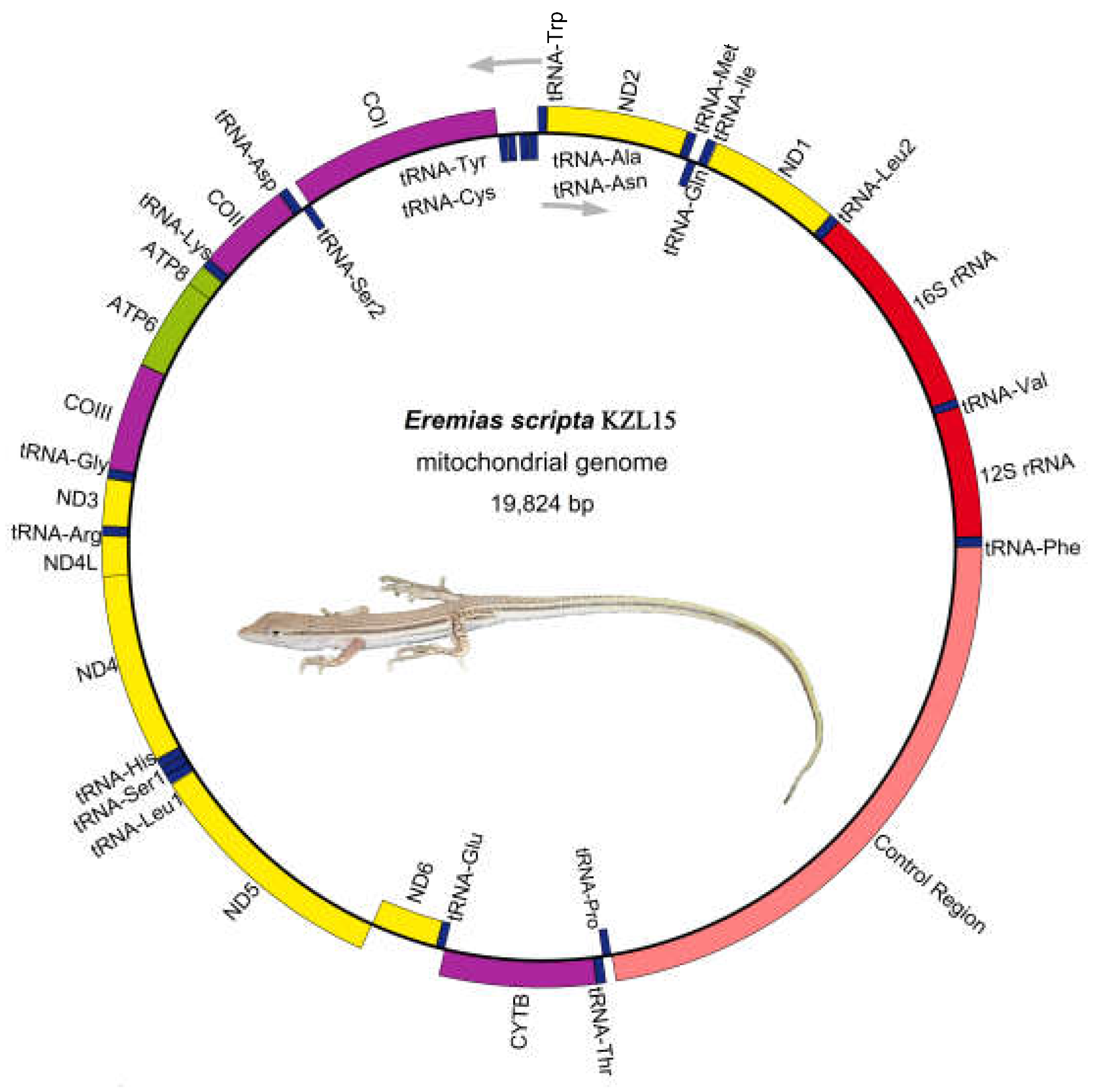
3.1. Genome Organization and Base Composition

The mitochondrial genome of E. scripta KZL15 (19,824 bp), E. scripta KZL44 (19,831 bp), E. nikolskii (20,840 bp), E. szczerbaki (19,650 bp), and E. yarkandensis (18,743 bp) were sequenced, annotated, and compared with the other 53 taxa of Lacertidae in several aspects. The composition and the arrangement of mitochondrial genes in these species were the same as those in most other typical vertebrates (Figure 1, Table S2). They all consisted of 13 PCGs, 22 tRNA genes, 2 rRNA genes (12S rRNA and 16S rRNA), and 2 non-coding regions (the CR and origin of replication on the light-strand (OL)). The length of OL ranged from 27 bp to 30 bp of the five racerunners. We found that the OL motifs of E. scripta KZL15, E. scripta KZL44, and E. nikolskii were relatively similar; they all contained the same composition, 5′-”TTCCCCCGTTANNNNNAAAACGGGGG”-3′. Additionally, E. szczerbaki and E. yarkandensis both contained the OL motif 5′-”TTCCCCCGTTANNNNAAAACGGGGG”-3′, and there was just one base difference between the two OL motifs. Most genes (12 PCGs, 2 rRNAs, and 14 tRNAs) were distributed on the H-strand, while 9 genes (ND6 and 8 tRNAs) were encoded on the L-strand.
The nucleotide composition, AT skew, and GC skew of total mitogenomes, PCGs, rRNAs, tRNAs, and CR of 58 taxa in the Lacertidae were calculated. The mean AT nucleotide content of the five complete mitogenomes was nearly similar: 61.33% in E. scripta KZL15, 61.16% in E. scripta KZL44, 58.54% in E. nikolskii, 59.47% in E. szczerbaki, and 59.46% in E. yarkandensis. Additionally, the nucleotide and composition skew values were conserved in the family Lacertidae (Table S3); they all showed a positive AT-skew (0.043 to 0.053) and a negative GC-skew (−0.351 to −0.324), suggesting a strong AT bias, and the AT content was higher in the CR (from 57.6% to 76.7%).

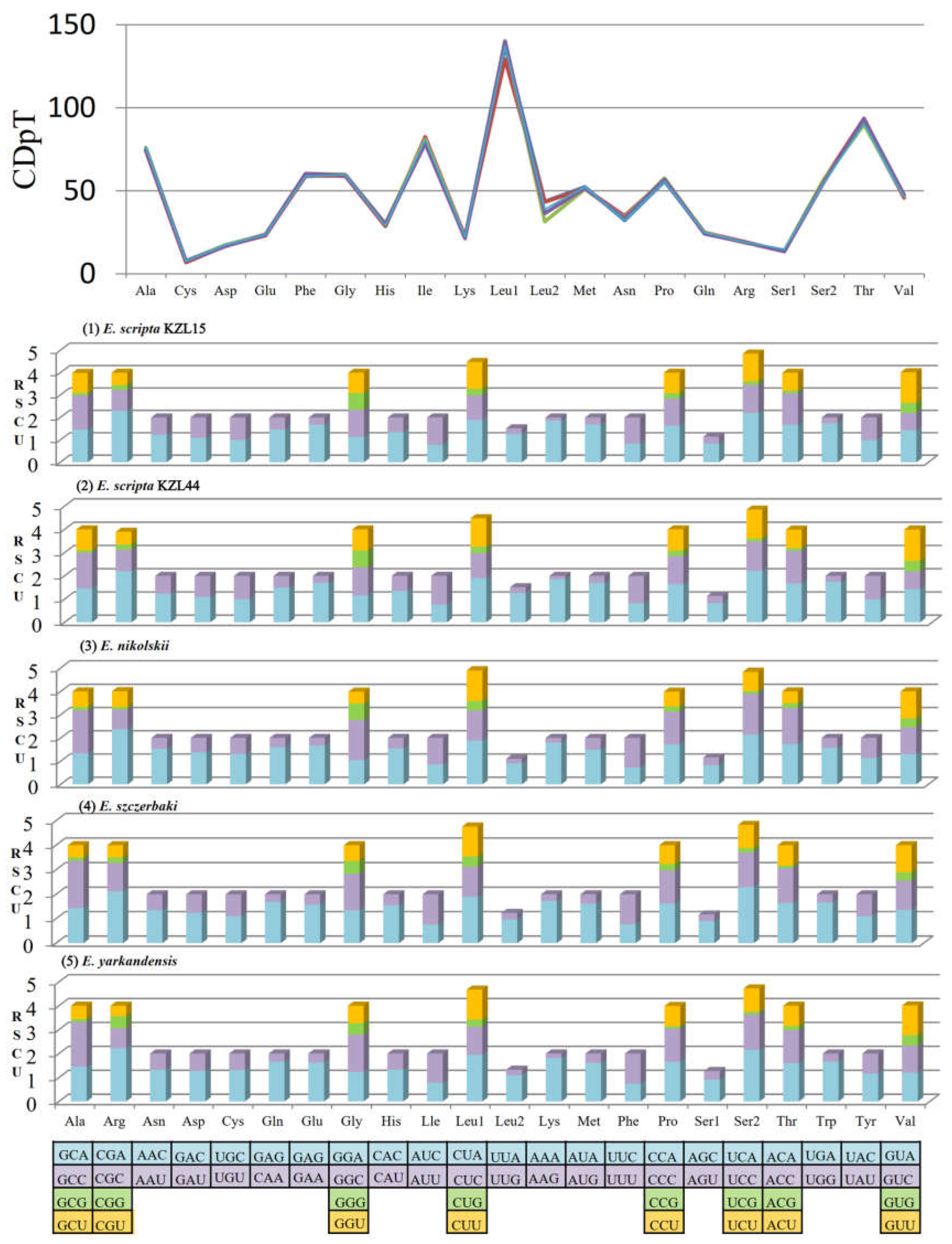
3.2. PCGs and Codon Usage

All newly sequenced Eremias mitogenomes contained 13 PCGs (ND1-ND6, ND4L, ATP8, ATP6, CYTB, and COI−COIII) ranging from 62 bp (ATP8) to 1824 bp (ND5). The total length of PCGs of the five racerunners ranged from 11,373 bp (E. yarkandensis) to 11,375 bp (E. nikolskii and E. szczerbaki). The start codons of 12 PCGs of E. scripta KZL15, E. scripta KZL44, E. nikolskii, and E. szczerbaki were ATG, whereas COI in the five racerunners and ND1 in E. yarkandensis showed the start codon GTG. In addition, there were five typical types of stop codons containing three canonical (TAA, TAG, and AGG) and two truncated stop codons (TA– – and T– –). By comparing the PCGs of 58 taxa in Lacertidae, we found two conserved motifs of the overlap, i.e., “ATGGNNNTAA” and “ATGANTAA” between ATP8 and ATP6. The overlap between ATP8 and ATP6 kept 10 bp in Lacertidae. More interestingly, the completely identical overlap motif “ATGGNNNTAA” was only presented across all species of Eremias. In addition, we compared the same structure of several other families of different orders in vertebrate, such as fishes in the subfamily Cobitinae [89], and found that the overlap composition of the majority of vertebrates contained the motif “ATGGNNNTAA”; the base of “NNN” may be their genus-specific feature and even family-specific feature. We also found the length of overlaps between ND4 and ND4L (7 bp), ND5 and ND6 (5 bp), ATP6 and COIII (1 bp) were consistent in the five racerunners. The relative synonymous codon usage (RSCU) and codon distribution of the five Eremias mitogenomes were analyzed (Figure 2). The total number of codons of the five racerunners was similar: 3790 in E. scripta KZL15, E. nikolskii, and E. szczerbaki; 3789 in E. scripta KZL44 and E. yarkandensis. The codon distribution among the five racerunners was coincident; the codons encoding Leucine (CUN), Threonine, and Isolecucine were the three most frequently present, while Cysteine was the rarest of them. In addition, the patterns of five racerunners were also consistent with one another. The codons were biased to utilize more A/U than G/C at the end, which resulted in the content of AT being higher than GC in the third position of Eremias PCGs.

3.3. Transfer RNAs and Ribosomal RNAs

The tRNA secondary structure and strand bias were coincident among the five racerunners and even in other lizards [31]. Among the 22 tRNA genes, only tRNAser(AGN) (Ser1) and tRNAcys could not be folded into a typical cloverleaf secondary structure and had no recognizable DHU arm (Figure S1). The length of single tRNA gene varied from 62 bp to 73 bp, and the total length of 22 tRNA genes ranged from 1513 bp to 1517 bp in the five Eremias mitogenomes.
The 12S rRNA and 16S rRNA were located between tRNAPhe and tRNALeu2 genes and interposed by tRNAVal. The length of 12S rRNA of the five specimens was 951 bp; however, the length of their 16S rRNA was variable, 1542 bp in E. scripta KZL15 and E. scripta KZL44, 1545 bp in E. nikolskii, 1537 bp in E. szczerbaki, and 1543 bp in E. yarkandensis. With reference to previous studies on secondary structures of 12S rRNA [31,45], we defined four domains of 12S rRNA of E. scripta KZL15 (as an example of Lacertidae) and defined 28 helices which contained 18 GU pairs. The pairing of guanine and uracil was permitted, in consideration of the structurally stable structure in RNAs [90]. As illustrated in Figure 3, Domain I contained helices 1–5; Domain II contained helices 11–19; Domain III contained helices 6−10 and helix 20; Domain IV contained helices 21–28. Of these 28 helices, helix 6 was the most stable one with fewer bulges and internal loops, while helix 4 was the most variable one with more unpaired bases. Meanwhile, according to the 16S rRNA of Darevskia genus within the Lacertidae from Brown in 2005 [20], we defined six domains of 16S rRNA of E. scripta KZL15, which contained 50 helices (Figure 4). Domain I consisted of helices 1−6 and helix 27; Domain II consisted of helices 8−16 and helices 19−24; Domain III contained helices 17–18 and helices 26–32; Domain IV included helices 33−44; Domain V contained helices 41–45, and Domain VI consisted of helices 46−50. Thirty GU pairs were found among the six domains. Meanwhile, we detected the most stable stem (helix 26) and the most variable structure (helix 48) for 16S rRNA.

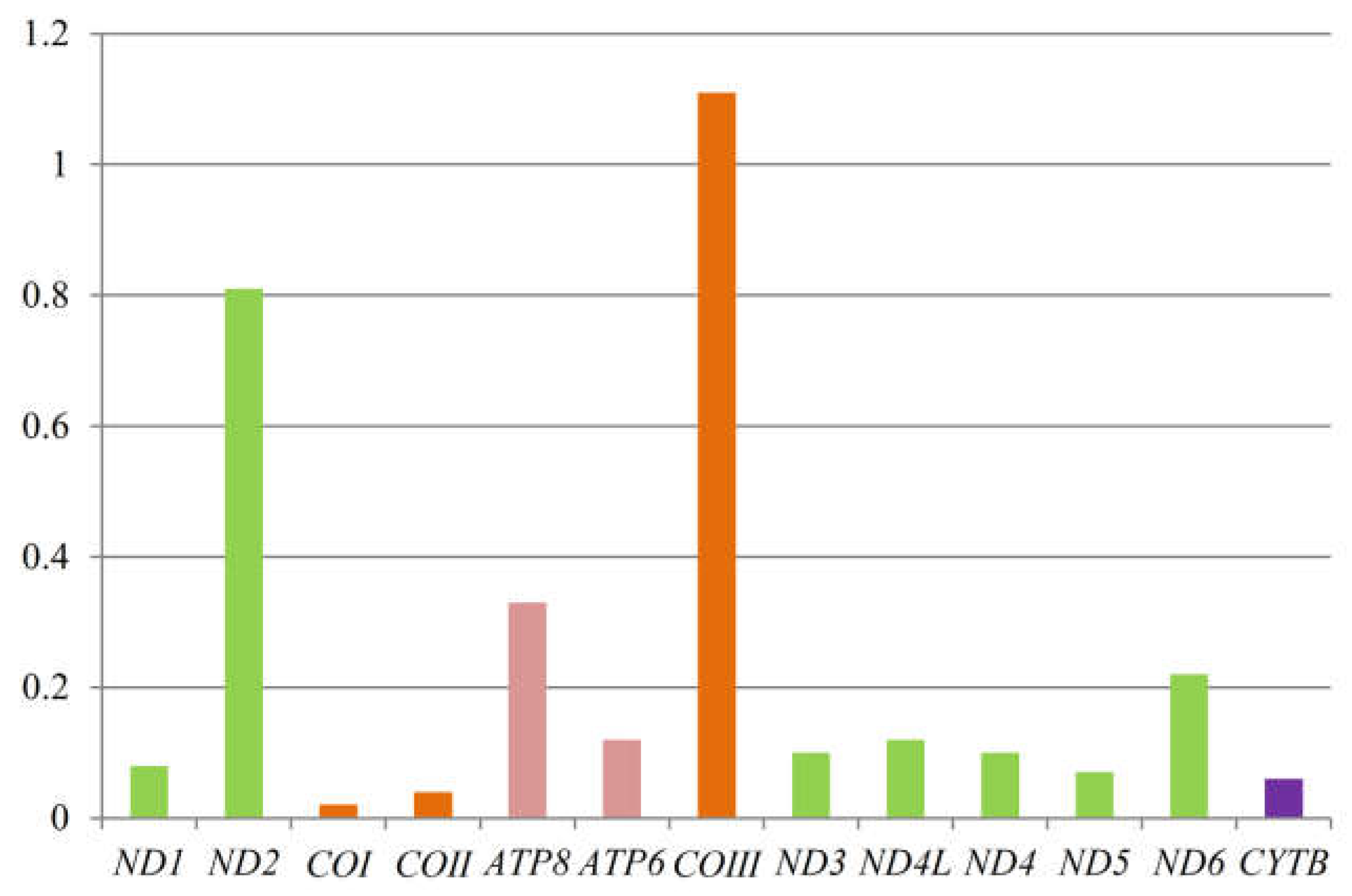
3.4. Non-Synonymous and Synonymous Substitutions

To further understand the role of selective pressure and the evolution of Lacertidae, we computed the average dN/dS value of each PCG of 58 taxa. We found that the Ka/Ks values for all PCGs except COIII were lower than one (between 0.02 and 0.81) (Figure 5), indicating that they are evolving under purifying selection. Among the 13 protein-coding genes, the average dN/dS of COIII was the highest (1.11), and ND2 (0.81) also had very high average dN/dS values.

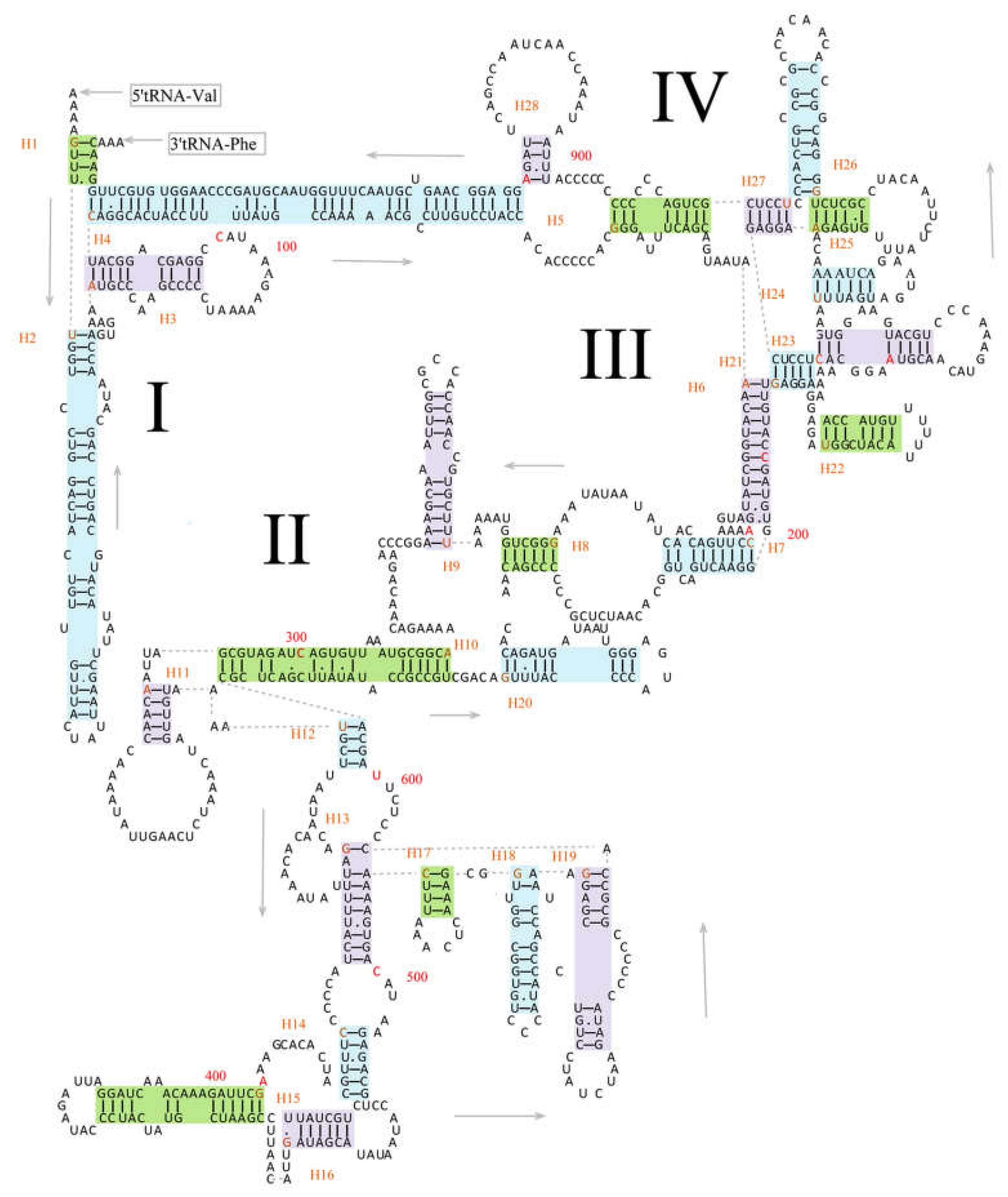
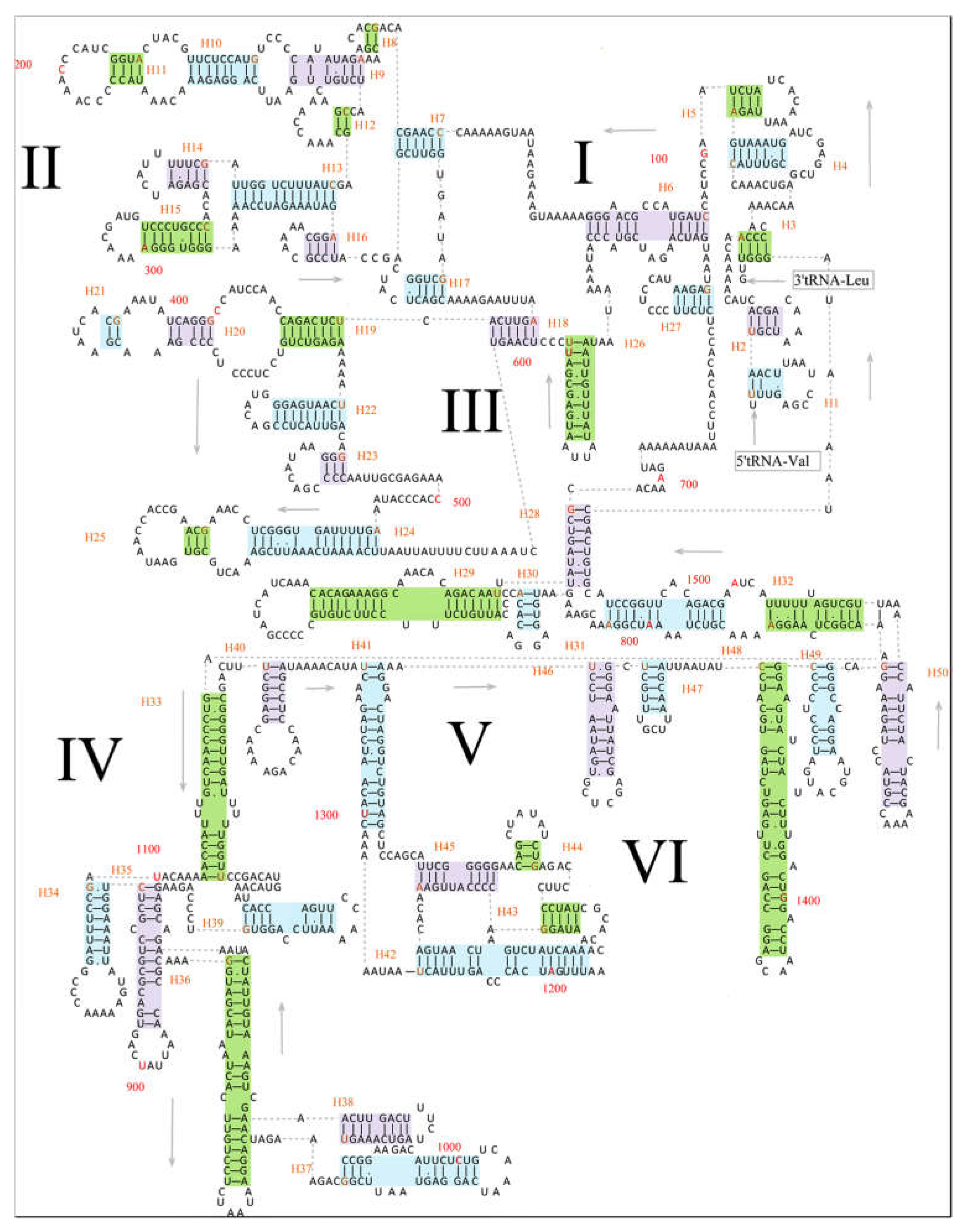
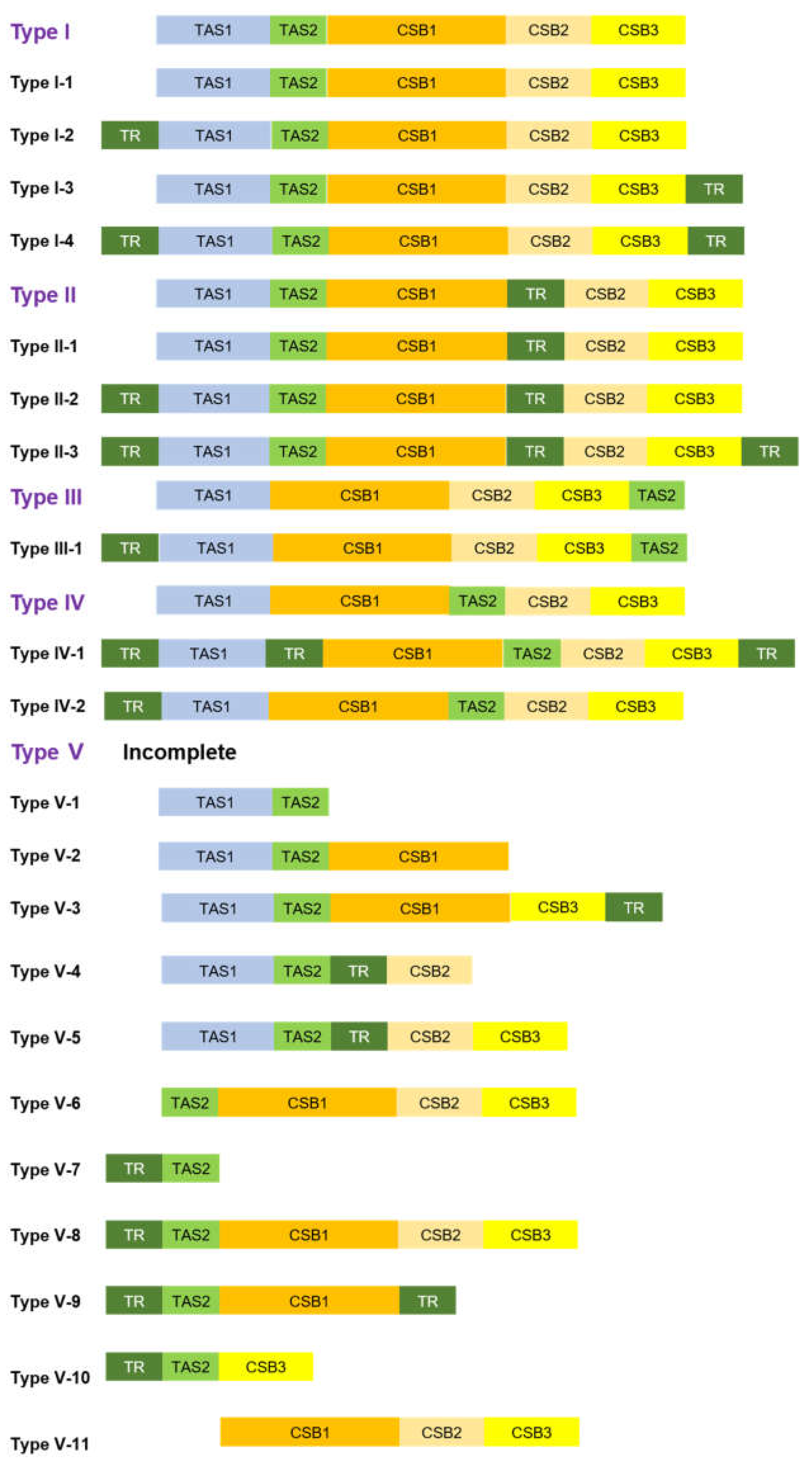
3.5. Structure of Control Region

Many studies have demonstrated that the CR in vertebrates shows a similar structure and conserved sequences [7,27,28,91,92], indicating evolutionary constraints and conservatism at various levels. We presented the composition of conserved structure elements of the CR in 58 taxa representing 13 genera in Lacertidae. Overall, there were more mitogenomes from four genera, Acanthodactylus, Eremias, Takydromus, and Darevskia, than those from other genera available in GenBank. We also analyzed the TR of CR in Lacertidae and found several motifs of Eremias and Takydromus. The CR was longest in E. nikolskii (5436 bp) and shortest in Al. nigropunctatus (146 bp).
Conserved structural elements of 58 lacertid taxa were analyzed and compared with the reference species of L. dugesii, which contained one or two termination-associated sequences (TASs) and three conserved sequence blocks (CSBs). The position of conserved structural elements is listed in Table S4. Overall, the compositions of TAS1, CSB1, and CSB2 were consistent with those of the reference sequence (Table 3, Table 4 and Table 5). Compared to the TAS1 (ACTATTATGTATATAGTGCATTAA) of L. dugesii, TAS1 of six species of Eremias and five species of Acanthodactylus were similar to one another, showing “·················A······” (see Type 2 in Table 3) (dots indicate the same base as the reference motif). The CSB1 of 12 species exhibited the standard motif “CTATATGGTATTATTGTCTTAATGCTTGGTAGACATAT” (see Type 1 in Table 4), which was used to compare and determine the presence of CSB1 of CR in the family Lacertidae and even reptiles, whereas this structure in the other 13 species was “···············C·T····················” (see Type 2 in Table 4). As such, the composition of CSB1 in Lacertidae warrants further study. As shown in Table 6, the TAS2 of only one species (L. viridis viridis) was similar to that (CATACATTAA) of L. dugesii, and the structures of 13 species were “T··G······“ (see Type 2 in Table 6). Additionally, the first base of 29 species was T; thus, the TAS2 of the standard reference sequence, which was traditionally used to compare and determine the presence of TAS2 of the CR in Lacertidae and even reptiles, should be updated to “TATGCATTAA”.
On the basis of the comparisons, we defined the standard compositions of TAS1, TAS2, and CSB1 in Eremias. In addition, there were also several genus-specific features of these three conserved structures in Takydromus and Darevskia. The TAS1 in Darevskia and CSB1 in Takydromus were exactly similar to that in the reference species L. dugesii. However, the TAS2 “T··G······” and CSB1 ”···············C·T····················” in Darevskia were specific at the genus level. Generally, CSB2 and CSB3 are conserved in the family Lacertidae [4,21,93], and they are very conserved in the four compared genera (Table 5, Table 6 and Table 7). For Eremias and Darevskia, the length between CSB2 and CSB3 kept 35 bp. The compositions of conserved structures of the CR in Lacertidae are listed in Table S5.
Tandem repeats are one of the factors accountable for extensive size variations in mitogenomes [7,92,93]. Tandem repeats were reported in the CR of several lacertid lizards [4,21,25]. In this study, we also compared the TR of 46 lacertids (see Table S6 for details), which contained the copy number, length, and motif information of TR. There are also several genus-specific features. The motif segment “TAGCGGCTTTTTTG” was present in the 11 examined species of Eremias. The motif segment “GCGGCTT” was present in the seven examined species of Takydromus excluding T. amurensis, while six species of Takydromus excluding T. wolteri showed the motif segment “TTTTCC”. Compared to these genera, the feature of TR of Darevskia was relatively weaker, and the motif segment “CAAAACTTTTAA” was present in just 9 of the 17 examined species. We found the position between TR and TAS1 of most species of Eremias, Takydromus, and Lacerta were conserved; there was one TR before TAS1 of these genera. The position between the TR and CSB1 was conserved in Darevskia; there was a TR located between CSB1 and CSB2. We also found the position between the TR and CSB3 were conserved in Eremias; there was one TR after CSB3. Overall, the composition and position of TR/TAS/CSB in Eremias was markedly conserved.
According to the criteria of the presence and position of TAS1, TAS2, CBS1, CSB2, and CSB3, as well as the anormal TR which is located between the conserved structure elements, we found five general types of the CR (Figure 6). Meanwhile, based on the criteria of general types, we further considered the presence and position of a normal TR, which is located outside the conserved structure elements, as the additional condition, and found a total of 21 subtypes of CR from Lacertidae (Figure 6). Notably, a close link between the types/subtypes of CR from some species of Lacertidae and their phylogeny can be observed (see Figure 7 for details).

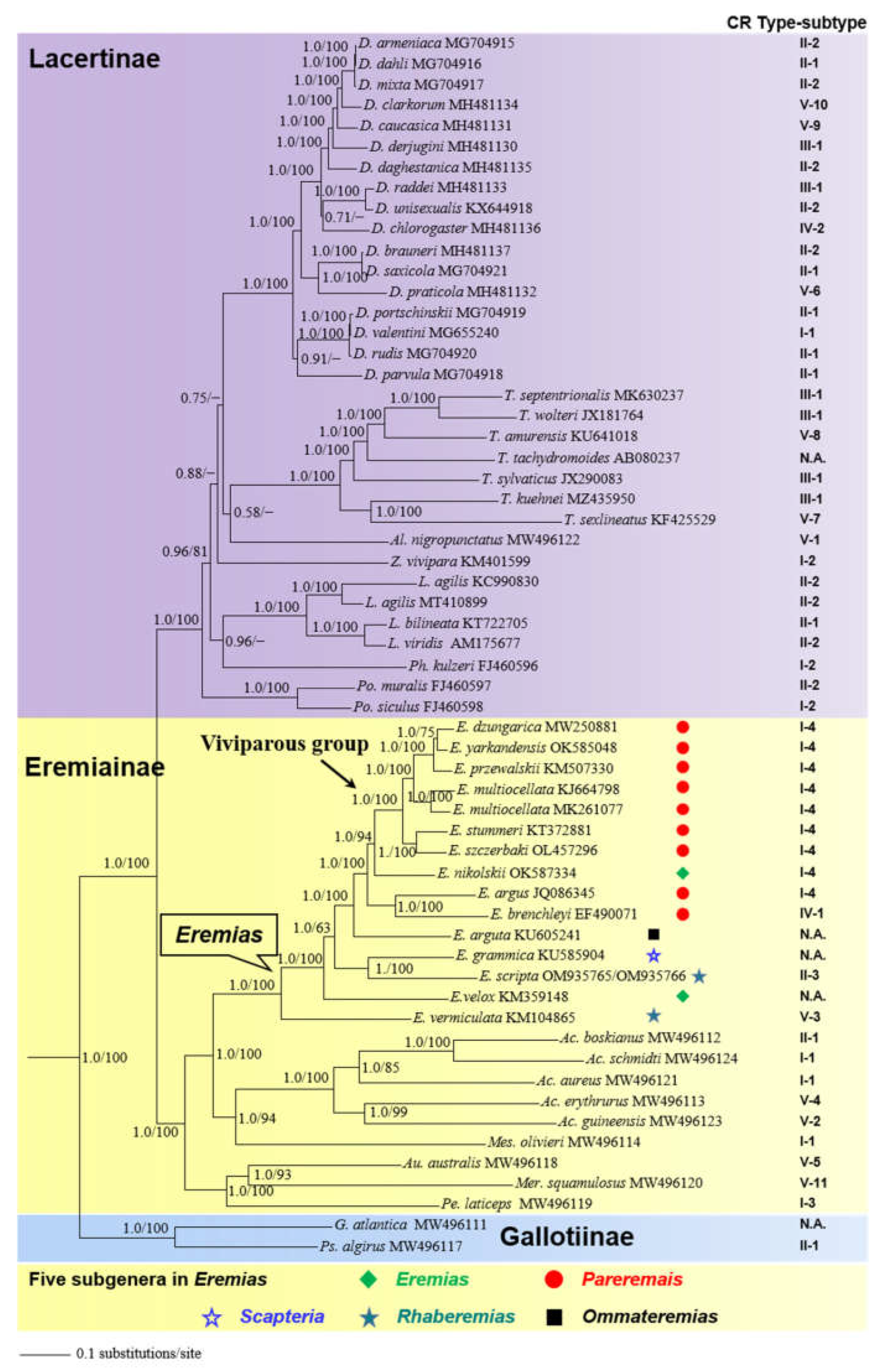
3.6. Phylogenetic Analysis

Bayesian inference and ML analyses produced highly congruent topology, with only minor differences on some nodes in the subfamily Lacertinae. Thus, only the BI tree with both PP and UFBoot from ML is presented (Figure 7), and see Figure S2 for ML tree. With limited taxon sampling, the monophyly of Lacertidae was recovered with strong support (PP = 1.0; UFBoot = 100), in accord with previous studies [9,10,76]. Moreover, the resulting trees confirmed a sister relationship between Eremiainae and Lacertinae with strong support (PP =1.0; UFBoot =100) [8,12]. In the subfamily Lacertinae, the phylogenetic position of most genera was unresolved due to lower support (PP < 0.95; UFBoot < 50). In the subfamily Eremiainae, the phylogenetic position of six sampling genera was resolved, with Eremias being more closely related to Acanthodactylus + Mesalina (PP =1.0; UFBoot =100). Within Eremias, the monophyly of the viviparous group was recovered with strong support (PP = 1.0; UFBoot = 100), which is consistent with previous studies [16,18,94]. However, E. nikolskii was inferred as the sister taxon to the viviparous group with strong support (PP = 1.0; UFBoot = 94). In addition, as demonstrated by p-distances (Table S7), the genetic divergence between E. nikolskii and the viviparous group (0.105–0.109) was smaller than that of E. arugs-E. brenchleyi versus the viviparous group (0.122–0.124).

4. Discussion

4.1. Secondary Structures of rRNA Are Useful for Phylogenetic Inference

In the present study, the secondary structures of 12S rRNA and 16S rRNA in E. scripta KZL15, as a representative lacertid, were presented in detail for the first time (see Figure 3 and Figure 4). Considering these secondary structural features, rRNA can be divided into paired (stem) and unpaired (loop) regions. Compensatory substitutions occur frequently in the paired regions; the property contradicts the assumption of independent mutations [95,96]. The analysis of RNA secondary structures is helpful to aid the alignment of rRNA sequences [97] and contributes to the increasingly sophisticated models of sequence evolution being applied in maximum likelihood and Bayesian approaches [20,98]. For instance, the doublet model of MrBayes [81,82] is intended for stem regions of ribosomal sequences, where nucleotides pair with each other to form doublets. There are various ways to model the evolution of nucleotide doublets. One method is to focus on the common doublets, A-T and C-G in particular. MrBayes uses a more complex model, originally formulated by Schöniger and von Haeseler [99], where all doublets are taken into account. Accordingly, the phylogenetic performance of the rRNA can be improved by incorporating information regarding its secondary structure in analyses for more accurate phylogenetic inference [84,100,101,102]. We believe that the secondary structure information of rRNAs described herein is useful for phylogenetic inference among species in Lacertidae.

4.2. Selection Pressure on PCGs

The phenomenon of purifying selection of PCGs is usually detected in most Metazoa [91]. As shown in Figure 5, the values of all PCGs except COIII were smaller than 1, which can be interpreted as meaning that the proteins evolve slowly under purifying selection, i.e., are more conserved [103,104]. This may be explained by the rationale that most of the nonsynonymous substitutions are detrimental to fitness and consequently have low fixation probabilities. A possible reason for the less extent of purifying selection of ND2 is that relaxed purifying selection drives the evolution of ND2 by mostly affecting regions that have lower functional relevance [105]. The COI gene showed the lowest value (0.02), and COIII showed the highest value (1.11). With the ratio dN/dS > 1, COIII may be considered under positive selection [103,104]. Further research is necessary to detect the variation of selective pressures among different lacertid lineages and to quantify the probability of positive selection on each site in each gene across all lacertids.

4.3. Phylogenetic Implications

Phylogenetic analyses based on 13 concatenated PCGs statistically recovered the higher-level relationships among the three subfamilies in Lacertidae (Figure 7). As for the subgenus assignment in Eremias, we acknowledge the plenary powers of the International Code on Zoological Nomenclature (ICZN) to designate Lacerta velox Pallas, 1771, as the type species of Eremias [106]. As such, the subgenera Eremias and Rhaberemias are not monophyletic, which is also congruent with previous results [16,17]. The phylogenetic position of E. nikolskii and its genetic affinity demonstrated by p-distances together challenged the monophyly of the subgenus Pareremias [16,18]. Nevertheless, the phylogenetic position of most genera in the subfamily Lacertinae was unresolved (PP < 0.95; UFBoot < 50), corroborating the hypothesis of a hard polytomy in the Lacertinae phylogeny due to fast radiation [10,14].

4.4. Structure and Evolution of Control Region

On the one hand, due to a lack of typical coding constraints, the CR is usually thought to be the fastest evolving region of the mitogenome [27], so it is broadly utilized to infer intraspecific and interspecific phylogenetic relationships. On the other hand, the CR constrains sequences related to the termination of H-strand replication, the origin of H-strand, and promoters of transcription to both L- and H-strand [7,28,107,108,109]. This indicates that the CR has evolutionary constraints. Indeed, many studies have demonstrated that the CR in vertebrates shows a similar structure and conserved sequences [7,27,28,91,92], indicating evolutionary constraints and conservatism at various levels. In addition, many CSBs identified suggest that many unknown functions exist. It is these known and unknown functions that put the CR under high evolutionary pressure and may lead to this conservation.
The mitochondrial control region had not been considered as a transcriptional region until 2018, when Gao et al. [110] documented that this region encodes two long non-coding RNAs (lncRNAs). However, current methods of the annotation of animal mitogenomes are still limited to blastx or structure-based covariance models [39]. Thus, it is necessary to further use a small RNA sequencing (sRNA-seq)-based method [111,112] to obtain improved annotations of the lacertids mitogenome at 1 bp resolution and to decipher TR in the CR.
By mapping the trait on the tree, we found that the CR from Mes. olivieri and the majority of species in Acanthodactylus and Eremias belonged to Type I, showing a close link with their phylogeny. Additionally, the CR from most species in Darevskia and species in Lacerta belonged to Type II; these species also presented closer affinities on the tree. The CR from minority species in Darevskia and four species in Takydromus belonged to Type III, demonstrating, to some extent, a close link with their phylogeny. Several similar, almost completely clade-specific insert and tandem repeat signatures were detected in the Lacerta viridis complex [23]. On the other hand, convergent evolution may be attributed to the mismatch for there not to be subfamily- or genus-specific types of the CR. In a framework of subtypes, we found several mismatches with phylogeny; this may imply the complexity of evolution in the CR. For example, in Type I-1, two species of Acanthodactylus and Mes. olivieri presented a closer relationship; however, D. valentini was somewhat distantly related to them. This pattern was also mirrored in Type I-2 and Type II-1. In addition, with regard to types of CR, several species also presented some independent evolutionary scenarios, such as E. argus, D. valentini, and D. chlorogaster, where no close link was observed between the types of CR and their phylogenetic positions. In other words, their CR types were different from those in most congeners; this phenomenon also reflected the complicated evolution of the CR. To decipher the evolutionary processes that drive the diversification of the CR in Lacertidae, further study is necessary to investigate the dynamics of the CR based on phylogenetic comparative methods in an explicit phylogenetic framework.

5. Conclusions

We comprehensively compared the complete mitochondrial genomes of five racerunners of Eremias (E. scripta KZL15, E. scripta KZL44, E. nikolskii, E. szczerbaki, and E. yarkandensis) for the first time. Additionally, the nucleotide composition and skew values as well as other characterizations of the mitogenomes in Lacertidae available in GenBank were comparatively analyzed. In addition, the secondary structures of 12S rRNA and 16S rRNA in E. scripta KZL15, as a representative lacertid, were presented in detail for the first time. Specifically, the tandem repeats, structure, and evolution of the control region from 58 taxa of Lacertidae were systematically analyzed for the first time. We found the reliable composition of TAS2 and the controversial composition of CSB1 in Lacertidae. Five general types and twenty-one subtypes of the CR in Lacertidae were unraveled. Meanwhile, we refined the composition of conserved structural elements at the genus level and found the motifs of tandem repeats in four genera, Acanthodactylus, Eremias, Takydromus, and Darevskia. Phylogenetic analyses recovered the higher-level relationships among the three subfamilies in Lacertidae and corroborated the hypothesis of a hard polytomy in the Lacertinae phylogeny due to fast radiation. E. nikolskii was inferred as the sister taxon to the viviparous group within Eremias; this challenged the monophyly of the subgenus Pareremias. Specifically, we found some close links of types of the CR and phylogeny, as well as some mismatches between them, which further verified the complexity of the evolutionary pattern of CR. In addition, our refinement of the secondary structures of rRNAs could promote the application of increasingly complex sequence evolution models in maximum likelihood and Bayesian approaches.

Supplementary Materials

The following supporting information can be downloaded at: https://www.mdpi.com/article/10.3390/genes13050726/s1. Figure S1: Putative secondary structures of the tRNAs identified in the mitogenomes of (a), E. scripta KZL15; (b), E. scripta KZL44; (c), E. nikolskii; (d), E. szczerbaki; (e), E. yarkandensis. Figure S2: Maximum likelihood tree inferred from 13 concatenated PCGs. Node numbers represent ultrafast bootstrap support. GenBank accession numbers are given with species names, and subfamily assignments are listed, along with viviparous group/subgenera in Eremias. Table S1: Best-fit models and partitioning schemes selected using PartitionFinder 2. Table S2: Organization of five mitochondrial genomes of Eremias determined in this study. (a), E. scripta KZL15; (b), E. scripta KZL44; (c), E. nikolskii; (d), E. szczerbaki; (e), E. yarkandensis. Table S3: The A+T composition of the complete mitogenomes, PCGs, rRNAs, tRNAs and CR, and composition skew values in Lacertidae. Table S4: Position of conserved structural elements of the CR in Lacertidae. Table S5: Compositions of conserved structures of the CR in Lacertidae. Table S6: Tandem repeats of the CR in Lacertidae. Table S7: Uncorrected genetic distances (p-distances) within and between the subgenus Pareremias and E. nikolskii based on 13 concatenated PCGs.

Author Contributions

Conceptualization, X.G.; methodology, L.T.; software, L.T.; validation, X.G. and L.T.; formal analysis, L.T.; investigation, X.G. and L.T. resources, X.G.; data curation, X.G.; writing—original draft preparation, L.T.; writing—review and editing, L.T. and X.G.; visualization, L.T. and X.G.; supervision, X.G.; project administration, X.G.; funding acquisition, X.G. All authors have read and agreed to the published version of the manuscript.

Funding

This work was supported by the Strategic Priority Research Program of the Chinese Academy of Sciences (Grant No. XDA20050201), the Third Xinjiang Scientific Expedition Program (Grant No. 2021xjkk0600), and the National Natural Science Foundation of China (Grant Nos. 32070433, 32000288).

Institutional Review Board Statement

Not applicable.

Informed Consent Statement

Not applicable.

Data Availability Statement

Illumina raw reads are deposited at NCBI Sequence Read Archive (SRA) under accession nos. SRR16509497 for E. nikolskii in the BioProject PRJNA773219, SRR16508134 for E. yarkandensis in the BioProject PRJNA773190, PRJNA779878 for E. szczerbaki in the BioProject PRJNA779878, SRR18381820 for E. scripta KZL15 in the BioProject PRJNA817793, and SRR18441150 for E. scripta KZL44 in the BioProject PRJNA817796. The mitochondrial genomes are deposited at GenBank with accession nos. OK587334, OK585048, OL457296, OM935765, and OM935766, respectively.

Acknowledgments

We thank Anar Alymkulova (the Institute of Biology and Soil, National Academy of Science of the Kyrgyz Republic) and Marina Chirikova (Institute of Zoology of Republic of Kazakhstan) for assistance in collecting the samples during the fieldwork in Kyrgyzstan. We are grateful to Tatjana N. Dusjebayeva for her logistic help and great assistance in the fieldwork in Kazakhstan, as well as to Jinlong Liu for collecting the samples in Kazakhstan. Thanks are extended to Na Wu, Song Wang, and Rui Xu for their discussion with regard to data analysis. This article benefited much from insightful comments by two anonymous reviewers.

Conflicts of Interest

The authors declare no conflict of interest. The funder had no role in the design of the study; in the collection, analyses, or interpretation of data; in the writing of the manuscript, or in the decision to publish the results.

References

  1. Anderson, S.; Bankier, A.T.; Barrell, B.G.; de Bruijn, M.H.L.; Coulson, A.R.; Drouin, J.I.; Eperon, C.; Nierlich, D.P.; Roe, B.A.; Sanger, F.; et al. Sequence and organization of the human mitochondrial genome. Nature 1981, 290, 457–465. [Google Scholar] [CrossRef]
  2. Boore, J.L. Animal mitochondrial genomes. Nucleic Acids Res. 1999, 27, 1767–1780. [Google Scholar] [CrossRef] [PubMed] [Green Version]
  3. Wolstenholme, D.R. Animal mitochondrial DNA: Structure and evolution. Int. Rev. Cytol. 1992, 141, 173–216. [Google Scholar] [PubMed]
  4. Brehm, A.; Harris, D.J.; Alves, C.D.; Jesus, J.D.; Thomarat, F.D.; Vicente, L.D. Structure and evolution of the mitochondrial DNA complete control region in the lizard Lacerta dugesii (Lacertidae, Sauria). J. Mol. Evol. 2003, 56, 46–53. [Google Scholar] [CrossRef]
  5. Hoelzel, A.R.; Lopez, J.V.; Dover, G.A.; O’Brien, S.J. Rapid evolution of a heteroplasmic repetitive sequence in the mitochondrial DNA control region of carnivores. J. Mol. Evol. 1994, 39, 191–199. [Google Scholar]
  6. Wu, N.; Liu, J.; Wang, S.; Guo, X. Comparative analysis of mitochondrial genomes in two subspecies of the sunwatcher toad-headed agama (Phrynocephalus helioscopus): Prevalent intraspecific gene rearrangements in Phrynocephalus. Genes 2022, 13, 203. [Google Scholar] [CrossRef] [PubMed]
  7. Sbisà, E.; Tanzariello, F.; Reyes, A.; Pesole, G.; Saccone, C. Mammalian mitochondrial D-loop region structural analysis: Identification of new conserved sequences and their functional and evolutionary implications. Gene 1997, 205, 125–140. [Google Scholar] [CrossRef]
  8. Harris, D.J.; Arnold, E.N.; Thomas, R.H. Relationships of lacertid lizards (Reptilia: Lacertidae) estimated from mitochondrial DNA sequences and morphology. Proc. Biol. Sci. B 1998, 265, 1939–1948. [Google Scholar] [CrossRef] [PubMed] [Green Version]
  9. Mayer, W.; Pavlicev, M. The phylogeny of the family Lacertidae (Reptilia) based on nuclear DNA sequences: Convergent adaptations to arid habitats within the subfamily Eremiainae. Mol. Biol. Evol. 2007, 44, 1155–1163. [Google Scholar] [CrossRef] [PubMed]
  10. Pavlicev, M.; Mayer, W. Fast radiation of the subfamily Lacertinae (Reptilia: Lacertidae): History or methodical artefact? Mol. Phylogenet. Evol. 2009, 52, 727–734. [Google Scholar] [CrossRef]
  11. Uetz, P.; Freed, P.; Aguilar, R.; Hošek, J. The Reptile Database. Available online: http://www.reptile-database.org (accessed on 22 March 2022).
  12. Arnold, E.N.; Arribas, O.; Carranza, S. Systematics of the Palaearctic and Oriental lizard tribe Lacertini (Squamata: Lacertidae: Lacertinae), with descriptions of eight new genera. Zootaxa 2007, 1430, 1–86. [Google Scholar] [CrossRef]
  13. Hipsley, C.A.; Himmelmann, L.; Metzler, D.; Müller, J. Integration of Bayesian molecular clock methods and fossil-based soft bounds reveals early Cenozoic origin of African lacertid lizards. BMC Evol. Biol. 2009, 9, 151. [Google Scholar] [CrossRef] [PubMed] [Green Version]
  14. Mendes, J.; Harris, D.J.; Carranza, S.; Salvi, D. Evaluating the phylogenetic signal limit from mitogenomes, slow evolving nuclear genes, and the concatenation approach. New insights into the Lacertini radiation using fast evolving nuclear genes and species trees. Mol. Phylogenet. Evol. 2016, 100, 254–267. [Google Scholar] [CrossRef] [PubMed]
  15. Kirchhof, S.; Lyra, M.L.; Rodríguez, A.; Ineich, I.; Müller, J.; Rödel, M.O.; Trape, J.F.; Vences, M.; Boissinot, M. Mitogenome analyses elucidate the evolutionary relationships of a probable Eocene wet tropics relic in the xerophilic lizard genus Acanthodactylus. Sci. Rep. 2021, 11, 4858. [Google Scholar] [CrossRef] [PubMed]
  16. Guo, X.; Dai, X.; Dali, C.; Papenfuss, T.J.; Ananjeva, N.B.; Melnikov, D.A.; Wang, Y. Phylogeny and divergence times of some racerunner lizards (Lacertidae: Eremias) inferred from mitochondrial 16S rRNA gene segments. Mol. Phylogenet. Evol. 2011, 61, 400–412. [Google Scholar] [CrossRef]
  17. Khan, M.A.; Jablonski, D.; Nadeem, M.S.; Masoor, R.; Kehlmaier, C.; Spitzweg, C.; Fritz, U. Molecular phylogeny of Eremias spp. from Pakistan contributes to a better understanding of the diversity of racerunners. J. Zool. Syst. Evol. Res. 2021, 59, 466–483. [Google Scholar] [CrossRef]
  18. Liu, J.L.; Dujsebayeva, T.N.; Chirikova, M.A.; Gong, X.; Li, D.J.; Guo, X.G. Does the Dzungarian racerunner (Eremias dzungarica Orlova, Poyarkov, Chirikova, Nazarov, Munkhbaatar, Munkhbayar &Terbish, 2017) occur in China? Species delimitation and identification with DNA barcoding and morphometric analyses. Zool. Res. 2021, 42, 287–293. [Google Scholar]
  19. Wang, S.; Liu, J.L.; Zhang, B.; Guo, X.G. The complete mitochondrial genome of Eremias dzungarica (Reptilia, Squamata, Lacertidae) from the Junggar Basin in Northwest China. Mitochondrial DNA B 2021, 6, 2012–2014. [Google Scholar] [CrossRef]
  20. Brown, R.P. Large subunit mitochondrial rRNA secondary structures and site-specific rate variation in two lizard lineages. J. Mol. Evol. 2005, 60, 45–56. [Google Scholar] [CrossRef]
  21. Böhme, M.U.; Fritzsch, G.; Tippmann, A.; Schlegel, M.; Berendonk, T.U. The complete mitochondrial genome of the green lizard Lacerta viridis viridis (Reptilia: Lacertidae) and its phylogenetic position within squamate reptiles. Gene 2007, 394, 69–77. [Google Scholar] [CrossRef]
  22. Podnar, M.; Pinsker, W.; Mayer, W. Complete mitochondrial genomes of three lizard species and the systematic position of the Lacertidae (Squamata). J. Zool. Syst. Evol. Res. 2009, 47, 35–41. [Google Scholar] [CrossRef]
  23. Jauss, R.-T.; Solf, N.; Kolora, S.R.R.; Schaffer, S.; Wolf, R.; Henle, K.; Fritz, U.; Schlegel, M. Mitogenome evolution in the Lacerta viridis complex (Lacertidae, Squamata) reveals phylogeny of diverging clades. Syst. Biodivers. 2021, 19, 682–692. [Google Scholar] [CrossRef]
  24. Stoneking, M.; Hedgecock, D.; Higuchi, R.G.; Vigilant, L.; Erlich, H.A. Population variation of human mtDNA control region sequences detected by enzymatic amplification and sequence-specific oligonucleotide probes. Am. J. Hum. Genet. 1991, 48, 370–382. [Google Scholar] [PubMed]
  25. Aquadro, C.F.; Greenberg, B.D. Human mitochondrial DNA variation and evolution: Analysis of nucleotide sequences from seven individuals. Genetics 1983, 103, 287–312. [Google Scholar] [CrossRef]
  26. Kasamatsu, H.; Robberson, D.L.; Vinograd, J. A novel closed-circular mitochondrial DNA with properties of a replicating intermediate. Proc. Natl. Acad. Sci. USA 1971, 68, 2252–2257. [Google Scholar] [CrossRef] [Green Version]
  27. Lee, W.J.; Conroy, J.; Howell, W.H.; Kocher, T.D. Structure and evolution of teleost mitochondrial control regions. J. Mol. Evol. 1995, 41, 54–66. [Google Scholar] [CrossRef]
  28. Randi, E.; Lucchini, V. Organization and evolution of the mitochondrial DNA control region in the avian genus Alectoris. Mol. Evol. 1998, 47, 449–462. [Google Scholar] [CrossRef]
  29. Liu, H.; Yang, G.; Wei, F.W.; Li, M.; Hu, J.C. Sequence variability of the mitochondrial DNA control region and population genetic structure of sika deers (Cervus nippon) in China. Acta Zool. Sinica 2003, 49, 53–60, (In Chinese with English abstract). [Google Scholar]
  30. Matson, C.W.; Baker, R.J. DNA sequence variation in the mitochondrial control region of red-backed voles (Clethrionomys). Mol. Biol. Evol. 2001, 18, 1494–1501. [Google Scholar] [CrossRef] [Green Version]
  31. Chen, M.; Liu, J.; Chen, D.; Guo, X. Complete mitochondrial genome of a blue-tailed skink Plestiodon capito (Reptilia, Squamata, Scincidae) and comparison with other Scincidae lizards. Genetica 2020, 148, 229–241. [Google Scholar] [CrossRef]
  32. Aljanabi, S.M.; Martinez, I. Universal and rapid salt-extraction of high quality genomic DNA for PCR-based techniques. Nucleic Acids Res. 1997, 25, 4692–4693. [Google Scholar] [CrossRef]
  33. Chen, S.; Zhou, Y.; Chen, Y.; Gu, J. fastp: An ultra-fast all-in-one fastq preprocessor. Bioinformatics 2018, 34, i884–i890. [Google Scholar] [CrossRef] [PubMed]
  34. Fastp. Available online: https://github.com/OpenGene/fastp (accessed on 22 March 2022).
  35. Bankevich, A.; Nurk, S.; Antipov, D.; Gurevich, A.A.; Dvorkin, M.; Kulikov, A.S.; Lesin, V.M.; Nikolenko, S.I.; Pham, S.; Prjibelski, A.D.; et al. SPAdes: A new genome assembly algorithm and its applications to single-cell sequencing. J. Comput. Biol. 2012, 19, 455–477. [Google Scholar] [CrossRef] [PubMed] [Green Version]
  36. SPAdes-v3.10.1. Available online: http://cab.spbu.ru/software/spades/ (accessed on 22 March 2022).
  37. Web BankIt. Available online: http://www.ncbi.nlm.nih.go-v/BankIt/index.html (accessed on 22 March 2022).
  38. Web BLAST. Available online: https://blast.ncbi.nlm.nih.gov/Blast.cgi (accessed on 22 March 2022).
  39. Bernt, M.; Donath, A.; Jühling, F.; Externbrink, F.; Florentz, C.; Fritzsch, G.; Pütz, J.; Middendorf, M.; Stadler, P.F. MITOS: Improved de novo metazoan mitochondrial genome annotation. Mol. Phylogenet. Evol. 2013, 69, 313–319. [Google Scholar] [CrossRef]
  40. MITOS Web Server. Available online: http://mitos.bioinf.uni-leipzig.de/index.py (accessed on 22 March 2022).
  41. tRNAscan-SE Web Server. Available online: http://lowelab.ucsc.edu/tRNAscan-SE/ (accessed on 22 March 2022).
  42. Lowe, T.M.; Chan, P.P. tRNAscan-SE On-line integrating search and context for analysis of transfer RNA genes. Nucleic Acids Res. 2016, 44, W54–W57. [Google Scholar] [CrossRef] [PubMed]
  43. Web RNAfold. Available online: http://rna.tbi.univie.ac.at//cgi-bin/RNAWebSuite/RNAfold.cgi (accessed on 22 March 2022).
  44. Comparative RNA Web (CRW) Site. Available online: http://www.rna.ccbb.utexas.edu/ (accessed on 22 March 2022).
  45. Hickson, R.E.; Simon, C.; Cooper, A.; Spicer, G.S.; Sullivan, J.; Penny, D. Conserved sequence motifs, alignment, and secondary structure for the third domain of animal 12S rRNA. Mol. Biol. Evol. 1996, 17, 530–539. [Google Scholar] [CrossRef] [PubMed] [Green Version]
  46. Cannone, J.J.; Subramanian, S.; Schnare, M.N.; Collett, J.R.; D’Souza, L.M.; Du, Y.; Feng, B.; Lin, N.; Madabusi, L.V.; Müller, K.M. The Comparative RNA Web (CRW) Site: An online database of comparative sequence and structure information for ribosomal, intron, and other RNAs. BMC Bioinform. 2002, 3, 2. [Google Scholar]
  47. Greiner, S.; Lehwark, P.; Bock, R. OrganellarGenomeDRAW (OGDRAW) version 1.3.1: Expanded toolkit for the graphical visualization of organellar genomes. Nucleic Acids Res. 2019, 47, W59–W64. [Google Scholar] [CrossRef] [Green Version]
  48. OGDRAW. Available online: https://chlorobox.mpimp-golm.mpg.de/OGDraw.html (accessed on 10 April 2022).
  49. Kumar, S.; Stecher, G.; Tamura, K. MEGA7: Molecular evolutionary genetics analysis version 7.0 for bigger datasets. Mol. Biol. Evol. 2016, 33, 1870–1874. [Google Scholar] [CrossRef] [Green Version]
  50. Benson, G. Tandem repeats finder: A program to analyze DNA sequences. Nucleic Acids Res. 1999, 27, 573–580. [Google Scholar] [CrossRef] [Green Version]
  51. Tandem Repeats Finder Web Server. Available online: http://tandem.bu.edu/trf/trf.html (accessed on 22 March 2022).
  52. Du, Y.; Qiu, Q.B.; Tong, Q.L.; Lin, L.H. The complete mitochondrial genome of Eremias przewalskii (Squamata: Lacertidae). Mitochondrial DNA A 2016, 27, 1918–1919. [Google Scholar]
  53. Zhou, T.; Li, D.; Dujsebayeva, T.N.; Liu, J.; Guo, X. Complete mitochondrial genome of Stummer’s Racerunner (Eremias stummeri) from Kazakhstan. Mitochondrial DNA A 2016, 27, 4340–4341. [Google Scholar] [CrossRef] [PubMed]
  54. Tong, Q.L.; Yao, Y.T.; Lin, L.H.; Ji, X. The complete mitochondrial genome of Eremias vermiculata (Squamata: Lacertidae). Mitochondrial DNA 2016, 27, 1447–1448. [Google Scholar] [CrossRef] [PubMed]
  55. Rui, J.L.; Wang, Y.T.; Nie, L.W. The complete mitochondrial DNA genome of Eremias brenchleyi (Reptilia: Lacertidae) and its phylogeny position within Squamata reptiles. Amphib-Reptil. 2009, 30, 25–35. [Google Scholar] [CrossRef]
  56. Li, D.; Song, S. Complete mitochondrial genome of Eremias multiocellata. 2015; unpublished work. [Google Scholar]
  57. Su, X.; Liu, J.; Chen, D.; Guo, X. Next-generation sequencing yields a nearly complete mitochondrial genome of the Multiocellated Racerunner (Eremias multiocellata) in Northwest China. Mitochondrial DNA B 2019, 4, 1430–1431. [Google Scholar] [CrossRef] [Green Version]
  58. Kim, S.K.; Yoo, D.U.; Hwang, U.W. Complete mitochondrial genome of Mongolia racerunner (Eremias argus). 2016; unpublished work. [Google Scholar]
  59. Yu, X.; Lin, L. The partial mitochondrial genome of Eremias arguta. 2021; unpublished work. [Google Scholar]
  60. Zhou, Z.S.; Li, H.; Tong, Q.L.; Lin, L.H.; Ji, X. The nearly complete mitochondrial genome of the rapid racerunner Eremias velox (Squamata: Lacertidae). Mitochondrial DNA A 2016, 27, 1781–1782. [Google Scholar]
  61. Murtskhvaladze, M.; Tarkhnishvili, D.; Anderson, C.L.; Kotorashvili, A. Phylogeny of caucasian rock lizards (Darevskia) and other true lizards based on mitogenome analysis: Optimisation of the algorithms and gene selection. PLoS ONE 2020, 15, e0233680. [Google Scholar] [CrossRef]
  62. Tao, C.R. The Complete Mitogenome of Lacerta agilis and the Phylogenetic Analysis of Squamata. Master’s Dissertation, Guangxi Normal University, Guilin, China, 2014. (In Chinese with English abstract). [Google Scholar]
  63. Margaryan, A. The partial mitochondrial genome of Lacerta agilis. 2020; unpublished work. [Google Scholar]
  64. Kolora, S.R.; Faria, R.; Weigert, A.; Schaffer, S.; Grimm, A.; Henle, K.; Sahyoun, A.H.; Stadler, P.F.; Nowick, K.; Bleidorn, C. The complete mitochondrial genome of Lacerta bilineata and comparison with its closely related congener L. viridis. Mitochondrial DNA A 2017, 28, 116–118. [Google Scholar] [CrossRef]
  65. Ma, W.W.; Liu, H.; Zhao, W.G.; Liu, P. The complete mitochondrial genome of Takydromus amurensis (Squamata: Lacertidae). Mitochondrial DNA B 2016, 1, 214–215. [Google Scholar] [CrossRef]
  66. Wu, L.X.; Luo, K.N.; Ding, G.H. Complete mitochondrial genome of Takydromus kuehnei. 2021; unpublished work. [Google Scholar]
  67. Hu, J.G.; Peng, L.F.; Tang, X.S.; Huang, S. The complete mitochondrial genome of Takydromus septentrionalis (Reptilia: Lacertidae). Mitochondrial DNA B 2019, 4, 2193–2194. [Google Scholar] [CrossRef] [Green Version]
  68. Qin, P.S.; Zeng, D.L.; Hou, L.X.; Yang, X.W.; Qin, X.M. Complete mitochondrial genome of Takydromus sexlineatus (Squamata, Lacertidae). Mitochondrial DNA 2015, 26, 465–466. [Google Scholar] [CrossRef] [PubMed]
  69. Tang, X.S.; Chen, J.M.; Huang, S. Mitochondrial genome of the Chung-an ground lizard Takydromus sylvaticus (Reptilia: Lacertidae). Mitochondrial DNA 2014, 25, 319–320. [Google Scholar] [CrossRef]
  70. Kumazawa, Y. Mitochondrial genomes from major lizard families suggest their phylogenetic relationships and ancient radiations. Gene 2007, 388, 19–26. [Google Scholar] [CrossRef] [PubMed]
  71. Yu, D.N.; Ji, X. The complete mitochondrial genome of Takydromus wolteri (Squamata: Lacertidae). Mitochondrial DNA 2013, 24, 3–5. [Google Scholar] [CrossRef] [PubMed]
  72. Liu, P.; Zhu, D.; Zhao, W.G.; Ji, X. The complete mitochondrial genome of the common lizard Zootoca vivipara (Squamata: Lacertidae). Mitochondrial DNA 2016, 27, 1944–1945. [Google Scholar]
  73. Macey, J.R.; Papenfuss, T.J.; Kuehl, J.V.; Fourcade, H.M.; Boore, J.L. Phylogenetic relationships among amphisbaenian reptiles based on complete mitochondrial genome sequences. Mol. Phylogenet. Evol. 2004, 33, 22–31. [Google Scholar] [CrossRef] [PubMed] [Green Version]
  74. Brunes, T.O.; Lyra, M.L.; Maldonado, J.A.; Pellegrino, K.C.M.; Rodrigues, M.T.; Fujita, M.K. The first mitochondrial genome of a South America parthenogenetic lizard (Squamata: Gymnophthalmidae). Mitochondrial DNA B 2021, 6, 2393–2395. [Google Scholar] [CrossRef]
  75. GenBank. Available online: https://www.ncbi.nlm.nih.gov/genbank/ (accessed on 22 March 2022).
  76. Wiens, J.J.; Hutter, C.R.; Mulcahy, D.G.; Noonan, B.P.; Townsend, T.M.; Sites, J.W., Jr.; Reeder, T.W. Resolving the phylogeny of lizards and snakes (Squamata) with extensive sampling of genes and species. Biol. Lett. 2012, 8, 1043–1046. [Google Scholar] [CrossRef] [Green Version]
  77. Pyron, R.A.; Burbrink, F.T.; Wiens, J.J. A phylogeny and revised classification of Squamata, including 4161 species of lizards and snakes. BMC Evol. Biol. 2013, 13, 93. [Google Scholar] [CrossRef] [Green Version]
  78. Zheng, Y.; Wiens, J.J. Combining phylogenomic and supermatrix approaches, and a time-calibrated phylogeny for squamate reptiles (lizards and snakes) based on 52 genes and 4162 species. Mol. Phylogenet. Evol. 2016, 94, 537–547. [Google Scholar] [CrossRef]
  79. Zhang, D.; Gao, F.; Li, W.X.; Jakovlić, I.; Zou, H.; Zhang, J.; Wang, G.T. PhyloSuite: An integrated and scalable desktop platform for streamlined molecular sequence data management and evolutionary phylogenetics studies. Mol. Ecol. Resour. 2020, 20, 348–355. [Google Scholar] [CrossRef] [PubMed]
  80. Lanfear, R.; Frandsen, P.B.; Wright, A.M.; Senfeld, T.; Calcott, B. PartitionFinder 2: New methods for selecting partitioned models of evolution for molecular and morphological phylogenetic analyses. Mol. Biol. Evol. 2017, 34, 772–773. [Google Scholar] [CrossRef] [PubMed] [Green Version]
  81. Ronquist, F.; Huelsenbeck, J.P. Mrbayes 3: Bayesian phylogenetic inference under mixed models. Bioinformatics 2003, 19, 1572–1574. [Google Scholar] [CrossRef] [PubMed] [Green Version]
  82. Ronquist, F.; Teslenko, M.; van der Mark, P.; Ayres, D.L.; Darling, A.; Höhna, S.; Larget, B.; Liu, L.; Suchard, M.A.; Huelsenbeck, J.P. MrBayes 3.2: Efficient Bayesian phylogenetic inference and model choice across a large model space. Syst. Biol. 2012, 61, 539–542. [Google Scholar] [CrossRef] [Green Version]
  83. Rambaut, A.; Drummond, A.J.; Xie, D.; Baele, G.; Suchard, M.A. Posterior summarization in Bayesian phylogenetics using Tracer 1.7. Syst. Biol. 2018, 67, 901–904. [Google Scholar] [CrossRef] [PubMed] [Green Version]
  84. Erixon, P.; Svennblad, B.; Britton, T.; Oxelman, B. Reliability of Bayesian posterior probabilities and bootstrap frequencies in phylogenetics. Syst. Biol. 2003, 52, 665–673. [Google Scholar] [CrossRef] [PubMed]
  85. Huelsenbeck, J.P.; Rannala, B. Frequentist properties of Bayesian posterior probabilities of phylogenetic trees under simple and complex substitution models. Syst. Biol. 2004, 53, 904–913. [Google Scholar] [CrossRef] [Green Version]
  86. Nguyen, L.T.; Schmidt, H.A.; von Haeseler, A.; Minh, B.Q. IQ-TREE: A fast and effective stochastic algorithm for estimating maximum-likelihood phylogenies. Mol. Biol. Evol. 2015, 32, 268–274. [Google Scholar] [CrossRef]
  87. Minh, B.Q.; Nguyen, M.A.T.; Haeseler, A.V. Ultrafast approximation for phylogenetic bootstrap. Mol. Biol. Evol. 2013, 30, 1188–1195. [Google Scholar] [CrossRef]
  88. FigTree. Available online: http://tree.bio.ed.ac.uk/software/figtree/ (accessed on 22 March 2022).
  89. Yu, P.; Zhou, L.; Yang, W.T.; Miao, L.J.; Li, Z.; Zhang, X.J.; Wang, Y.; Gui, J.F. Comparative mitogenome analyses uncover mitogenome features and phylogenetic implications of the subfamily Cobitinae. BMC Genomics 2021, 22, 50. [Google Scholar] [CrossRef]
  90. Topal, M.D.; Fresco, J.R. Complementary base pairing and the origin of substitution mutations. Nature 1976, 263, 285–289. [Google Scholar] [CrossRef] [PubMed]
  91. Castellana, S.; Vicario, S.; Saccone, C. Evolutionary patterns of the mitochondrial genome in Metazoa: Exploring the role of mutation and selection in mitochondrial protein coding genes. Genome Biol. Evol. 2011, 3, 1067–1079. [Google Scholar] [CrossRef] [PubMed] [Green Version]
  92. Southern, S.O.; Southern, P.J.; Dizon, A.E. Molecular characterization of a cloned dolphin mitochondrial genome. J. Mol. Evol. 1988, 28, 32–42. [Google Scholar] [CrossRef] [PubMed]
  93. Brown, G.G.; Gadaleta, G.; Pepe, G.; Saccone, C.; Sbisà, E. Structural conservation and variation in the D-loop-containing region of vertebrate mitochondrial DNA. J. Mol. Biol. 1986, 192, 503–511. [Google Scholar] [CrossRef]
  94. Orlova, V.F.; Poyarkov, N.A., Jr.; Chirikova, M.A.; Nazarov, R.A.; Munkhbaatar, M.; Munkhbayar, K.; Terbish, M. MtDNA differentiation and taxonomy of Central Asian racerunners of Eremias multiocellata-E. przewalskii species complex (Squamata, Lacertidae). Zootaxa 2017, 4282, 1–42. [Google Scholar] [CrossRef] [Green Version]
  95. Dixon, M.T.; Hillis, D.M. Ribosomal RNA secondary structure: Compensatory mutations and implications for phylogenetic analysis. Mol. Biol. Evol. 1993, 10, 256–267. [Google Scholar]
  96. Springer, M.S.; Hollar, L.J.; Burk, A. Compensatory substitutions and the evolution of the mitochondrial 12S rRNA gene in mammals. Mol. Biol. Evol. 1995, 12, 1138–1150. [Google Scholar]
  97. Kjer, K.M. Use of rRNA secondary structure in phylogenetic studies to identify homologous positions: An example of alignment and data presentation from the frogs. Mol. Phylogenet. Evol. 1995, 4, 314–330. [Google Scholar] [CrossRef]
  98. Hudelot, C.; Gowri-Shankar, V.; Jow, H.; Rattray, M.; Higgs, P.G. RNA-based phylogenetic methods: Application to mammalian mitochondrial RNA sequences. Mol. Phylogenet. Evol. 2003, 28, 241–252. [Google Scholar] [CrossRef] [Green Version]
  99. Schöniger, M.; von Haeseler, A. A stochastic model for the evolution of autocorrelated DNA sequences. Mol. Phylogenet. Evol. 1994, 3, 240–247. [Google Scholar] [CrossRef]
  100. Telford, M.J.; Wise, M.J.; Gowri-Shankar, V. Consideration of RNA secondary structure significantly improves likelihood-based estimates of phylogeny: Examples from the Bilateria. Mol. Biol. Evol. 2005, 22, 1129–1136. [Google Scholar] [CrossRef] [PubMed] [Green Version]
  101. Jow, H.; Hudelot, C.; Rattray, M.; Higgs, P.G. Bayesian phylogenetics using an RNA substitution model applied to early mammalian evolution. Mol. Biol. Evol. 2002, 19, 1591–1601. [Google Scholar] [CrossRef] [PubMed]
  102. Li, J.; Wang, X.; Kong, X.; Zhao, K.; He, S.; Mayden, R.L. Variation patterns of the mitochondrial 16S rRNA gene with secondary structure constraints and their application to phylogeny of cyprinine fishes (Teleostei: Cypriniformes). Mol. Phylogenet. Evol. 2008, 47, 472–487. [Google Scholar] [CrossRef] [PubMed]
  103. Kimura, M. Preponderance of synonymous changes as evidence for the neutral theory of molecular evolution. Nature 1977, 267, 275–276. [Google Scholar] [CrossRef]
  104. Yang, Z.; Bielawski, J.P. Statistical methods for detecting molecular adaptation. Trends Ecol. Evol. 2000, 15, 496–503. [Google Scholar] [CrossRef]
  105. Magnus, W.J.; Rute, R.D.F.; Louis, B.; Michael, M.H. Comparative analysis of complete mitochondrial genomes suggests that relaxed purifying selection is driving high nonsynonymous evolutionary rate of the NADH2 gene in whitefish (Coregonus ssp.). Mol. Phylogenet. Evol. 2016, 95, 161–170. [Google Scholar]
  106. Melville, R.V. Opinion 1318 (Opinion correcting the ruling given in Opinion 92) Lacerta velox Pallas, 1771 is the type species of Eremias Wiegmann, 1834. Bull. Zooll. Nomencl. 1985, 42, 171–172. [Google Scholar]
  107. Brown, W.M.; George, M., Jr.; Wilson, A.C. Rapid evolution of animal mitochondrial DNA. Proc. Natl. Acad. Sci. USA 1979, 76, 1967–1971. [Google Scholar] [CrossRef] [Green Version]
  108. Doda, J.N.; Wright, C.T.; Clayton, D.A. Elongation of displacement loop strands in human and mouse mitochondrial DNA is arrested near specific template sequences. Proc. Natl. Acad. Sci. USA 1981, 78, 6116–6120. [Google Scholar] [CrossRef] [Green Version]
  109. Saccone, C.; Pesole, G.; Sbisà, E. The main regulatory region of mammalian mitochondrial DNA: Structure-function model and evolutionary pattern. J. Mol. Evol. 1991, 33, 83–91. [Google Scholar] [CrossRef]
  110. Gao, S.; Tian, X.; Chang, H.; Sun, Y.; Wu, Z.; Cheng, Z.; Dong, P.; Zhao, Q.; Ruan, J.; Bu, W. Two novel lncRNAs discovered in human mitochondrial DNA using PacBio full-length transcriptome data. Mitochondrion 2018, 38, 41–47. [Google Scholar] [CrossRef] [PubMed]
  111. Ji, H.; Xu, X.; Jin, X.; Yin, H.; Luo, J.; Liu, G.; Zhao, Q.; Chen, Z.; Bu, W.; Gao, S. Using high-resolution annotation of insect mitochondrial DNA to decipher tandem repeats in the control region. RNA Biol. 2019, 16, 830–837. [Google Scholar] [CrossRef] [PubMed]
  112. Xu, X.; Ji, H.; Jin, X.; Cheng, Z.; Yao, X.; Liu, Y.; Zhao, Q.; Zhang, T.; Ruan, J.; Bu, W.; et al. Using pan RNA-seq analysis to reveal the ubiquitous existence of 5′ and 3′ end small RNAs. Front. Genet. 2019, 10, 105. [Google Scholar] [CrossRef] [PubMed]
Figure 1. Mitochondrial genome map of E. scripta KZL15. Genes encoded by the light or heavy strand are indicated inside or outside, respectively, showing the direction of transcription. The tRNAs are denoted by color blue and labeled according to the three-letter amino acid codes. The mitogenome maps of E. scripta KZL44, E. nikolskii, E. szczerbaki, and E. yarkandensis are similar to that of E. scripta KZL15. Lizard photo by Xianguang Guo.
Genes 13 00726 g001
Figure 2. The base composition and the relative synonymous codon usage (RSCU) values of E. scripta KZL15, E. scripta KZL44, E. nikolskii, E. szczerbaki, and E. yarkandensis, respectively. CDpT stands for codons per thousand codons. The color of blue, purple, green and yellow means the first, second, third and fourth type of each amino acid.
Genes 13 00726 g002
Figure 3. Predicted secondary structure of the 12S rRNA in E. scripta KZL15. Watson-Crick base pairings are indicated by the dashes (–); G–U base pairings are indicated by dots (•). Roman numerals indicate the conserved domain structures; I–IV indicate four domains in the secondary structure of 12S rRNA. Every 100th base is marked in red. Helices were marked with green, purple, and blue. The number of helices and the first base of helix were marked in orange; H means helix. Generated using RNAfold [43] and edited using Microsoft PowerPoint (14.0.4760.1000).
Genes 13 00726 g003
Figure 4. Predicted secondary structure of the 16S rRNA in E. scripta KZL15. Watson-Crick base pairings are indicated by the dashes (–); G–U bases pairings are indicated by dots (•). The numbering of helix follows Hickson et al. [45]. Roman numerals indicate the conserved domain structures; I–VI indicate six domains in the secondary structure of 16S rRNA. Every 100th base is marked in red. Helices were marked with green, purple, and blue. The number of helices and the first base of helix were marked in orange; H means helix. Generated using RNAfold [43] and edited using Microsoft PowerPoint (14.0.4760.1000).
Genes 13 00726 g004
Figure 5. The nonsynonymous/synonymous ratios (dN/dS) in 13 mitochondrial PCGs of 58 taxa in Lacertidae. The histogram represents the average dN/dS for each PCG.
Genes 13 00726 g005
Figure 6. Types of the control region in Lacertidae, with consideration of positions of TAS1, TAS2, CSB1, CSB2, CSB3 and TR. Type I-1: Ac. aureus, Ac. schmidti, D. valentini, Mes. olivieri; Type I-2: Ph. kulzeri, Po. siculus, Z. vivipara, D. chlorogaster; Type I-3: Pe. laticeps; Type I-4: E. multiocellata, E. stummeri, E. przewalskii, E. yarkandensis, E. szczerbaki, E. dzungarica, E. nikolskii, E. argus; Type II-1: D. dahli, D. parvula, D. portschinskii, D. rudis, D. saxicola, L. bilineata, Ac. boskianus, Ps. algirus; Type II-2: D. armeniaca, D. brauneri, D. daghestanica, D. mixta, D. unisexualis, L. agilis, L. viridis, Po. muralis; Type II-3: E. scripta; Type III-1: T. septentrionalis, T. wolteri, D. derjugini, T. kuehnei, T. sylvaticus, D. raddei; Type IV-1: E. brenchleyi; Type IV-2: D. chlorogaster; Type V-1: Al. nigropunctatus; Type V-2: Ac. guineensis; Type V-3: E. vermiculata; Type V-4: Ac. erythrurus; Type V-5: Au. australis; Type V-6: D. praticola; Type V-7: T. sexlineatus; Type V-8: T. amurensis; Type V-9: D. caucasica; Type V-10: D. clarkorum; Type V-11: Mer. squamulosus.
Genes 13 00726 g006
Figure 7. A 50% majority-rule consensus tree of the family Lacertidae inferred from partitioned Bayesian analyses based on the concatenated PCGs of 58 lacertids and 3 outgroup taxa (not shown for clarity). Node numbers indicate Bayesian posterior probabilities (PP) and ML ultrafast bootstrap values (UFBoot), respectively. Dashes represent nodes with bootstrap support lower than 50% or represent nodes that do not exist in ML tree. Branch lengths represent means of the posterior distribution. GenBank accession numbers are given with species names, and subfamily assignments are listed, along with the viviparous group/subgenera in Eremias. Types/subtypes of the CR are mapped on the tree and correspond to those depicted in Figure 6. N.A., not applicable, for species with incomplete CR available in GenBank.
Genes 13 00726 g007
Table 1. List of collection information for five racerunners in Eremias.
SpeciesVoucher NumberCollection DateCollection Site
E. scriptaKZL15July 2014Kazakhstan; 45.91764° N, 79.96855° E
E. scriptaKZL44July 2014Kazakhstan; 45.91764° N, 79.96855° E
E. nikolskiiGuo4717August 2014Kyrgyzstan; 41.38750° N, 73.93999° E
E. szczerbakiGuo4719August 2014Kyrgyzstan; 41.48393° N, 75.97943° E
E. yarkandensisGuo4722August 2014Kyrgyzstan; 39.64809° N, 73.86512° E
Table 2. Taxon information of Lacertidae and three outgroup species analyzed in this paper with GenBank accession numbers.
TaxonFamilySubfamilyAccession NumberLength (bp)Reference
Eremias
E. scripta KZL15LacertidaeEremiainaeOM93576519,824This study
E. scripta KZL44LacertidaeEremiainaeOM93576619,831This study
E. nikolskiiLacertidaeEremiainaeOK58733420,840This study
E. szczerbakiLacertidaeEremiainaeOL45729619,650This study
E. yarkandensisLacertidaeEremiainaeOK58504818,743This study
E. dzungaricaLacertidaeEremiainaeMW25088119,899[19]
E. przewalskiiLacertidaeEremiainaeKM50733018,225[52]
E. stummeriLacertidaeEremiainaeKT37288119,602[53]
E. vermiculataLacertidaeEremiainaeKM10486519,914[54]
E. brenchleyiLacertidaeEremiainaeEF49007119,542[55]
E. multiocellataLacertidaeEremiainaeKJ66479818,996[56]
E. multiocellataLacertidaeEremiainaeMK26107717,333[57]
E. argusLacertidaeEremiainaeJQ08634518,521[58]
E. argutaLacertidaeEremiainaeKU60524115,192[59]
E. grammicaLacertidaeEremiainaeKU58590415,338[59]
E. veloxLacertidaeEremiainaeKM35914818,033[60]
Acanthodactylus
Ac. aureusLacertidaeEremiainaeMW49612115,756[15]
Ac. boskianusLacertidaeEremiainaeMW49611217,143[15]
Ac. erythrurusLacertidaeEremiainaeMW49611316,827[15]
Ac. guineensisLacertidaeEremiainaeMW49612316,963[15]
Ac. schmidtiLacertidaeEremiainaeMW49612416,943[15]
Australolacerta
Au. australisLacertidaeEremiainaeMW49611817,019[15]
Meroles
Mer. squamulosusLacertidaeEremiainaeMW49612016,860[15]
Mesalina
Mes. olivieriLacertidaeEremiainaeMW49611416,899[15]
Pedioplanis
Pe. laticepsLacertidaeEremiainaeMW49611917,046[15]
Algyroides
Al. nigropunctatusLacertidaeLacertinaeMW49612215,844[15]
Darevskia
D. armeniacaLacertidaeLacertinaeMG70491517,521[61]
D. brauneriLacertidaeLacertinaeMH48113716,976[61]
D. caucasicaLacertidaeLacertinaeMH48113116,343[61]
D. chlorogasterLacertidaeLacertinaeMH48113617,479[61]
D. clarkorumLacertidaeLacertinaeMH48113416,301[61]
D. dahliLacertidaeLacertinaeMG70491617,528[61]
D. daghestanicaLacertidaeLacertinaeMH48113517,189[61]
D. derjuginiLacertidaeLacertinaeMH48113016,960[61]
D. mixtaLacertidaeLacertinaeMG70491717,532[61]
D. parvulaLacertidaeLacertinaeMG70491817,510[61]
D. portschinskiiLacertidaeLacertinaeMG70491917,529[61]
D. praticolaLacertidaeLacertinaeMH48113216,418[61]
D. raddeiLacertidaeLacertinaeMH48113320,478[61]
D. rudisLacertidaeLacertinaeMG70492017,534[61]
D. saxicolaLacertidaeLacertinaeMG70492117,524[61]
D. unisexualisLacertidaeLacertinaeKX64491821,433[61]
D. valentiniLacertidaeLacertinaeMG65524017,393[61]
Lacerta
L. agilisLacertidaeLacertinaeKC99083017,090[62]
L. agilisLacertidaeLacertinaeMT41089917,069[63]
L. bilineataLacertidaeLacertinaeKT72270517,154[64]
L. viridis viridisLacertidaeLacertinaeAM17657717,156[21]
Phoenicolacerta
Ph. kulzeriLacertidaeLacertinaeFJ46059617,199[22]
Podarcis
Po. muralisLacertidaeLacertinaeFJ46059717,311[22]
Po. siculusLacertidaeLacertinaeFJ46059817,297[22]
Takydromus
T. amurensisLacertidaeLacertinaeKU64101817,333[65]
T. kuehneiLacertidaeLacertinaeMZ43595017,224[66]
T. septentrionalisLacertidaeLacertinaeMK63023718,304[67]
T. sexlineatusLacertidaeLacertinaeKF42552918,943[68]
T. sylvaticusLacertidaeLacertinaeJX29008317,838[69]
T. tachydromoidesLacertidaeLacertinaeAB08023718,245[70]
T. wolteriLacertidaeLacertinaeJX18176418,236[71]
Zootoca
Z. viviparaLacertidaeLacertinaeKM40159917,046[72]
Psammodromus
Ps. algirusLacertidaeGallotiinaeMW49611717,118[15]
Outgroup
Amphisbaena
Am. schmidtiAmphisbaenidaeAY60547517,423[73]
Bipes
B. biporusBipedidaeAY60548116,430[73]
Loxopholis
Lo. percainatumGymnophthalmidaeMW86432915,875[74]
Table 3. Type of conserved structure element of TAS1 in Lacertidae.
TypeNumber of SpeciesTAS1
L. dugesiiACTATTATGTATATAGTGCATTAA
127························
211·················A······
31·······C·G······C······C
41··············T··A······
51··············C··A······
61··AT·········AT··A······
71··A············T······CA
81··AT···················G
91····C···················
Types of TAS1 of the control region in Lacertidae. Type 1: Al. nigropunctatus, D. armeniaca; D. brauneri, D. chlorogaster, D. daghestanica, D. dahli, D. derjugini, D. mixta, D. parvula, D. portschinskii, D. raddei, D. rudis, D. saxicola, D. unisexualis, D. valentini, E. nikolskii, E. przewalskii, L. agilis, L. bilineata, L. viridis viridis, Ph. kulzeri, Po. muralis, Po. siculus, T. kuehnei, T. septentrionalis, T. sylvaticus, T. wolteri; Type 2: Ac. aureus, Ac. boskianus, Ac. erythrurus, Ac. guineensis, Ac. schmidti, Au. australis, E. scripta, E. multiocellata, E. stummeri, E. vermiculata, E. yarkandensis; Type 3: E. argus; Type 4: E. brenchleyi; Type 5: E. szczerbaki; Type 6: Mes. olivieri; Type 7: Pe. laticeps; Type 8: Ps. algirus; Type 9: Z. vivipara.
Table 4. Type of conserved structure element of CSB1 in Lacertidae.
TypeNumber of SpeciesCSB1
L. dugesiiCTATATGGTATTATTGTCTTAATGCTTGGTAGACATAT
112······································
213···············C·T····················
32···············TC··G··················
41···············TC·····················
51···············TCT·G········C·········
61····T········C·TCA····················
71············G··C·T····················
81·········G······C··G··················
92················GA····················
102············G·························
112···················G··················
123····T··············G··················
131·········G······A··G··················
141········GGC·······G···················
151····T····G·········G··················
161····T··········TA··G··················
171·····A·······························A
181·················A··········A·········
Types of CSB1 of the control region in Lacertidae. Type 1: L. agilis, L. bilineata, L. viridis viridis, Ph. kulzeri, Po. muralis, Ps. algirus, T. kuehnei, T. amurensis, T. septentrionalis, T. sylvaticus, T. wolteri, Z. vivipara; Type 2: D. armeniaca, D. caucasica, D. chlorogaster, D. daghestanica, D. dahli, D. derjugini, D. mixta, D. parvula, D. portschinskii, D. raddei, D. rudis, D. unisexualis, D. valentini; Type 3: Mer. squamulosus, Pe. laticeps; Type 4: Po. siculus; Type 5: Mes. olivieri; Type 6: E. stummeri; Type 7: D. praticola; Type 8: E. vermiculata; Type 9: Ac. aureus, Ac. boskianus; Type 10: D. brauneri, D. saxicola; Type 11: E. scripta, E. nikolskii; Type 12: E. dzungarica, E. przewalskii, E. yarkandensis; Type 13: E. argus; Type 14: E. brenchleyi; Type 15: E. multiocellata; Type 16: E. szczerbaki; Type 17: Ac. guineensis; Type 18: Ac. schmidti.
Table 5. Type of conserved structure element of CSB2 in Lacertidae.
TypeNumber of SpeciesCSB2
L. dugesiiCAAACCCCCCTACCCCCC
141··················
21····T·············
31··············T···
41T····T··T········T
51····T·T·T·····T···
61·····T·····G·····T
Types of CSB2 of the control region in Lacertidae. Type 1: Ac. aureus, Ac. boskianus, Ac. schmidti, Au. australis, D. armeniaca, D. brauneri, D. chlorogaster, D. daghestanica, D. dahli, D. derjugini, D. mixta, D. parvula, D. portschinskii, D. praticola, D. raddei, D. saxicola, D. unisexualis, D. valentini, E. dzungarica, E. scripta, E. multiocellata, E. przewalskii, E. stummeri, E. szczerbaki, E. yarkandensis, L. agilis, L. bilineata, L. viridis viridis, Mer. squamulosus, Mes. olivieri, Pe. laticeps, Ph. kulzeri, Po. muralis, Po. siculus, Ps. algirus, T. kuehnei, T. amurensis, T. septentrionalis, T. sylvaticus, T. wolteri, Z. vivipara; Type 2: E. nikolskii; Type 3: D. rudis; Type 4: Ac. erythrurus; Type 5: E. argus; Type 6: E. brenchleyi.
Table 6. Type of conserved structure element of TAS2 in Lacertidae.
TypeNumber of SpeciesTAS2
L. dugesiiCATACATTAA
11··········
213T··G······
35T·········
45T········T
52T···T·····
61T···A·····
71T········C
81T······A·T
91T··C······
103A········T
111AG········
121A·········
131·········T
142···G······
151···GT·····
161·G········
171····T···T·
182····T·····
191·····T····
201····T··T··
211····AC··G·
221···C·GA···
231···A··T···
241·······A·T
251········C·
261·····A····
Types of TAS2 of the control region in Lacertidae. Type 1: L. viridis viridis; Type 2: D. armeniaca, D. brauneri, D. daghestanica, D. dahli, D. mixta, D. parvula, D. portschinskii, D. rudis, D. saxicola, D. valentini, E. przewalskii, L. agilis, Po. siculus; Type 3: D. derjugini, D. raddei, E. yarkandensis, Ph. kulzeri, L. bilineata; Type 4: Ac. schmidti, E. dzungarica, E. nikolskii, E. szczerbaki, Po. muralis; Type 5: D. chlorogaster, D. clarkorum; Type 6: T. amurensis; Type 7: D. unisexualis; Type 8: Ac. erythrurus; Type 9: D. caucasica; Type 10: Ac. boskianus, E. multiocellata, E. stummeri; Type 11: Ac. guineensis; Type 12: E. vermiculata; Type 13: Ac. aureus; Type 14: Au. australis, Pe. laticeps; Type 15: Mes. olivieri; Type 16: Z. vivipara; Type 17: Al. nigropunctatus; Type 18: T. septentrionalis, T. wolteri; Type 19: T. sexlineatus; Type 20: T. sylvaticus; Type 21: D. praticola; Type 22: E. argus; Type 23: E. brenchleyi; Type 24: E. scripta; Type 25: Ps. algirus; Type 26: T. kuehnei.
Table 7. Type of conserved structure element of CSB3 in Lacertidae.
TypeNumber of SpeciesCSB3
L. dugesiiTCGCCAAACCCCTAAAACGA
136····················
25············A·······
31···T······A·········
41········T···········
51C·······TT··CG····C·
61··········T·G·····A·
71········T·····G···AG
81AGG···ATT······T·T··
Types of CSB3 of the control region in Lacertidae. Type 1: Au. australis, D. brauneri, D. chlorogaster, D. clarkorum, D. daghestanica, D. dahli, D. derjugini, D. mixta, D. parvula, D. portschinskii, D. praticola, D. raddei, D. rudis, D. saxicola, D. unisexualis, D. valentini, E. dzungarica, E. scripta KZL15, E. scripta KZL44, E. multiocellata, E. przewalskii, E. stummeri, E. szczerbaki, E. yarkandensis, L. agilis, L. bilineata, L. viridis viridis, Ph. kulzeri, Po. muralis, Po. siculus, T. amurensis, T. kuehnei, T. septentrionalis, T. sylvaticus, T. wolteri, Z. vivipara; Type 2: Ac. aureus, Ac. boskianus, Ac. schmidti, Mer. squamulosus, Pe. laticeps; Type 3: D. armeniaca; Type 4: E. nikolskii; Type 5: E. argus; Type 6: E. brenchleyi; Type 7: E. vermiculata; Type 8: Ps. algirus.
Publisher’s Note: MDPI stays neutral with regard to jurisdictional claims in published maps and institutional affiliations.

