Article

The Di-Symbiotic Systems in the Aphids Sipha maydis and Periphyllus lyropictus Provide a Contrasting Picture of Recent Co-Obligate Nutritional Endosymbiosis in Aphids

1
Biodiversity Research Centre, Earth and Life Institute, Université Catholique de Louvain (UCLouvain), 1348 Louvain-la-Neuve, Belgium
2
Univ Lyon, INSA Lyon, INRAE, BF2I, UMR203, F-69621 Villeurbanne, France
3
Center for Applied Molecular Technologies, Institute of Experimental and Clinical Research, Université Catholique de Louvain (UCLouvain), 1200 Woluwe-Saint-Lambert, Belgium
4
Department of Plant Protection, National Institute of Agricultural Research, Avenue Ennasr, BP 415 Rabat Principale, Rabat 10090, Morocco
*
Author to whom correspondence should be addressed.
Microorganisms 2022, 10(7), 1360; https://doi.org/10.3390/microorganisms10071360
Submission received: 13 June 2022 / Revised: 1 July 2022 / Accepted: 4 July 2022 / Published: 6 July 2022
(This article belongs to the Section Environmental Microbiology)

Abstract

:
Dependence on multiple nutritional bacterial symbionts forming a metabolic unit has repeatedly evolved in many insect species that feed on nutritionally unbalanced diets such as plant sap. This is the case for aphids of the subfamilies Lachninae and Chaitophorinae, which have evolved di-symbiotic systems in which the ancient obligate nutritional symbiont Buchnera aphidicola is metabolically complemented by an additional nutritional symbiont acquired more recently. Deciphering how different symbionts integrate both metabolically and anatomically in such systems is crucial to understanding how complex nutritional symbiotic systems function and evolve. In this study, we sequenced and analyzed the genomes of the symbionts B. aphidicola and Serratia symbiotica associated with the Chaitophorinae aphids Sipha maydis and Periphyllus lyropictus. Our results show that, in these two species, B. aphidicola and S. symbiotica complement each other metabolically (and their hosts) for the biosynthesis of essential amino acids and vitamins, but with distinct metabolic reactions supported by each symbiont depending on the host species. Furthermore, the S. symbiotica symbiont associated with S. maydis appears to be strictly compartmentalized into the specialized host cells housing symbionts in aphids, the bacteriocytes, whereas the S. symbiotica symbiont associated with P. lyropictus exhibits a highly invasive phenotype, presumably because it is capable of expressing a larger set of virulence factors, including a complete flagellum for bacterial motility. Such contrasting levels of metabolic and anatomical integration for two S. symbiotica symbionts that were recently acquired as nutritional co-obligate partners reflect distinct coevolutionary processes specific to each association.

1. Introduction

Many insect species depend on inherited endosymbiotic bacteria to access certain nutrients and have evolved specialized cells, the bacteriocytes, to host these nutritional partners [1]. Bacteriocytes are the interface for metabolic exchanges between the host and the symbionts. They allow the host to control symbiont populations according to its nutritional needs and ensure stable vertical transmission of symbionts from mother to offspring [2,3]. This compartmentalization into bacteriocytes is considered as the outcome of a long coevolutionary history between the insect hosts and their obligate bacterial partners [4]. However, a consequence of this intracellular lifestyle is the isolation of symbionts from any environmental source of prokaryotic DNA, leading to Muller’s ratchet, i.e., the accumulation of irreversible deleterious mutations in the symbiont genome [5]. Combined with severe population bottlenecks during vertical transmission and the relaxation of purifying selection on genes that have become unnecessary in a context of interdependent association, this leads to the evolutionary degeneration of the symbiont genome [6,7]. Thus, extreme genomic reduction is a typical feature of obligate insect nutritional symbionts, often acquired tens of millions years ago [8,9]. This degeneration, once far advanced, can jeopardize the symbiotic functions of the nutritional symbiont and ultimately represents an evolutionary dead end for host lineages [10,11,12].
A compensatory mechanism to cope with the loss of these symbiotic functions is the acquisition by the host species of additional symbionts that evolve as co-obligate nutritional partners by forming a metabolic unit with the ancestral obligate symbiont that has become unable to perform its nutritional function alone [13]. The existence of such multi-symbiotic nutritional systems has been reported in various sap-feeding insect species [13,14,15,16,17,18,19,20,21,22] with evidence showing that the acquisition of additional obligate symbionts is a very dynamic process involving recurrent recruitments of new bacterial symbionts and repeated replacements of pre-existing intracellular ones [17,19,23,24]. While the nutritional basis of co-obligate symbioses has been examined in different sap-feeding insects through genomic analyses [13], many aspects of the biology of these associations remain unexplored, including the infection mechanisms that the co-obligate symbionts may use to colonize host tissues and the localization of these bacteria within the host. Examining these aspects in light of the diversity of co-obligate nutritional endosymbiosis is critical for understanding the evolution, development, and functioning of multi-symbiotic nutritional systems in insects.
Aphids (Hemiptera: Aphididae) of the subfamilies Lachninae and Chaitophorinae are valuable models for addressing the biology of co-obligate symbiosis in insects. In these aphids, the eroded metabolic abilities of the ancient obligate symbiont Buchnera aphidicola are complemented by those of a more recently acquired co-obligate symbiont, often belonging to the Serratia symbiotica species, one of the most common secondary symbionts found in aphids. Specifically, B. aphidicola and S. symbiotica metabolically complement each other for the production of certain B vitamins (in particular biotin/B7 and riboflavin/B2) and essential amino acids [18,19,20,21,22,25,26,27]. These systems, involving distinct aphid species and strains of S. symbiotica acquired independently through evolution, are suitable to study the diversity of multi-symbiotic nutritional symbiosis and to lay the groundwork for studying the development and functioning of these associations.
In the present study, we sequenced and annotated the genomes of the symbionts B. aphidicola and S. symbiotica composing the di-symbiotic systems associated with two Chaitophorinae aphids: the cereal aphid Sipha maydis and the Norway maple aphid Periphyllus lyropictus. The prevalence of S. symbiotica reaches 100% in these two aphid species and our genomic analyses suggest that the symbiont is a recently acquired co-obligate partner. We found that the nutritional symbiont exhibits very contrasting infection patterns in the two aphid species: while S. symbiotica exhibits an invasive phenotype in P. lyropictus, the co-obligate symbiont is strictly compartmentalized into bacteriocytes in S. maydis. We suggest that these differences in the anatomical integration of the co-obligate symbiont can be explained by the way in which S. symbiotica and B. aphidicola complement each other metabolically in their system, but also by the set of virulence factors that each co-obligate symbiont has retained over its evolution with its respective host. This work provides insight into the metabolic and anatomical integration of co-obligate symbionts in Chaitophorinae aphids in the early stages of co-obligate symbiosis.

2. Materials and Methods

2.1. Prevalence of S. symbiotica in S. maydis Populations

The prevalence of a symbiont in insect populations is an indication of its symbiotic status, with a prevalence of 100% indicating that the symbiont is fixed in the host species concerned and is an obligate partner. To examine the prevalence of S. symbiotica in S. maydis populations, specimens were collected on three common cereals grown in Morocco (Triticum turgidum, Triticum aestivum, and Hordeum vulgare) from the main cereal-growing areas of this country in two sampling campaigns: one in April 2014 covering six regions and locations with 21 colonies sampled, and one in April–May 2016 covering two regions with 76 colonies sampled (Figure S1; Table S1). The aphids collected consisted only of wingless parthenogenetic adult females that were stored in 95% ethanol at 4 °C until use.
Prior to DNA extraction, insect samples were surface sterilized with 99% ethanol and 10% bleach and rinsed with sterile water. Genomic DNA was extracted using the DNeasy Blood and Tissue kit (Qiagen, Hilden, Germany) following the manufacturer’s instructions. Each DNA extraction was performed on one individual per colony. DNA extractions were evaluated qualitatively and quantitatively using a Nanodrop spectrophotometer (Thermo Scientific, Waltham, MA, USA) and stored at −20 °C prior to PCR testing. Aphid samples were tested for the presence of S. symbiotica by amplifying a partial region of the 16S rRNA gene (0.4 kb region) using the specific primers 16SA1 and PASScmp [28]. PCR assays were performed as described previously [29]. The PCR products were stained with ethidium bromide and visualized on a 1% agarose gel. The amplicons were purified, sequenced, and then their identity was confirmed by BLAST search.

2.2. Establishment of Aphid Clonal Lines and Rearing Conditions

To perform the genomic approaches and other analyses, we established clonal lines for S. maydis and for P. lyropictus. For S. maydis, a colony sampled on H. vulgare in Midelt, (Morocco) in April 2016 was used to generate a clonal lineage from a single individual (clone Midelt). Aphids were reared on Triticum aestivum (bread wheat) under long day conditions (16 h light, 8 h dark) in a room maintained at a constant temperature of 20 °C to ensure parthenogenic reproduction.
The clonal line of P. lyropictus was established from one individual collected from Acer platanoides (Norway maple) in Louvain-la-Neuve in spring 2020 (clone LLN) [30]. Aphids were reared on young A. platanoides trees under the same conditions as the S. maydis clone Midelt.

2.3. Genome Sequencing and Assemblies

For whole-genome sequencing, DNA samples enriched with bacteria from S. maydis and P. lyropictus clone lines maintained in the laboratory were prepared following a previously described protocol [20,31]. Two sequencing approaches were performed on the DNA extracts: MinION long-read sequencing (Oxford Nanopore approach) and Illumina short-read sequencing. Both approaches were used to generate a hybrid assembly and thus obtain the most complete and accurate genomes possible.
Prior to the MinION nanopore sequencing, the integrity of genomic DNA was assessed using the 5200 Fragment Analyzer System (Agilent, Santa Clara, CA, USA) with the Genomic DNA 50 kb kit (Agilent). Libraries were then prepared from 400 ng of genomic DNA following the Rapid Barcoding Sequencing kit (SQK-RBK004) protocol (Oxford Nanopore Technologies, Oxford, UK). Briefly, the DNA molecules were cleaved by a transposase, and barcoded tags were attached to the cleaved ends. The barcoded samples were then pooled, and Rapid Sequencing Adapters were added to the tagged ends. The pooled libraries were sequenced into a FLO-MIN106 (R9.4.1) flow cell for a 48 h run according to the manufacturer’s instruction. After the run, fast5 files were basecalled on the MinIT using default settings in MinKNOWv21.02.5 and Guppy v4.3.4 and a Fast Basecalling. For the Illumina sequencing, libraries were prepared starting from 30 ng of genomic DNA using the Illumina DNA prep kit (Illumina, San Diego, CA, USA) following the manufacturer’s instruction. Briefly, genomic DNA was tagmented using Bead-Linked Transposomes, which fragments and tags the DNA with adapter sequences. After a post tagmentation clean-up, tagmented DNA were amplified using a 12-cycle PCR program and IDT® for Illumina® DNA/RNA UD Indexes Set A to add the index and adapter sequences. Libraries were equimolarly pooled and paired-end (2 × 300 bp) sequenced on an Illumina MiSeq platform (Illumina).
Raw sequences generated by MinION nanopore sequencing were assembled using meta-flye v.2.9, an assembler supporting metagenomic data that was specifically developed for the assembly of third-generation long-read sequences [32]. The resulting draft assemblies were then polished with Illumina short reads by using multiple rounds of Pilon v1.24 [33]. Four genomes were thus assembled: those of the B. aphidicola and S. symbiotica symbionts composing the di-symbiotic system of S. maydis (hereafter BaSm and SsSm), and those of the B. aphidicola and S. symbiotica symbionts of P. lyropictus (hereafter BaPl and SsPl).

2.4. Genome Annotation and Comparative Analyses

The four assembled genomes were annotated with the Prokaryotic Genome Annotation Pipeline (PGAP) [34]. The genome sequences were deposited on the MicroScope platform [35,36] and their metabolic capabilities were compared using MicroCyc, a collection of microbial Pathway/Genome Databases (PGDBs) based on the MetaCyc database and specifically dedicated to the analysis of microbial pathways [37]. Metabolic reconstructions generated by MicroCyc were manually verified and CDSs were considered as pseudogenes when their length was less than 80% of the one of their orthologs in reference bacterial genomes [38,39].
Virulomes of both S. symbiotica symbionts were analyzed with the virulence factor database (VFDB, http://www.mgc.ac.cn/VFs/, accessed on 30 April 2022) [40] integrated on the MicroScope platform using BLASTp (with a threshold of minimum 50% aa identity, 80% align. Coverage). MacSyFinder was used to identify secretion systems [41]. PHASTER was used to predict the intact prophage regions [42]. Assemblies and annotations are available on NCBI (BioProject accession numbers: PRJNA835797 for BaSm, PRJNA837269 for SsSm, PRJNA837360 for BaPl and PRJNA837363 for SsPl).

2.5. Molecular Phylogenetic Analyses

To examine the evolutionary relationships of SsSm and SsPl with other S. symbiotica symbionts and other bacteria in the genus Serratia, a phylogenetic tree was built using a set of single-copy core concatenated protein sequences shared across all S. symbiotica strains for which complete genome sequences are publicly available. S. symbiotica and outgroup genomes used for phylogenetic analysis are listed in Table S2 in the Supplementary Materials. Genomes were downloaded from the NCBI Assembly Database on 2 June 2022. All protein sequences of the different species and strains were analyzed with OrthoFinder (version 2.0) [43,44] to identify groups of orthologs. A set of 235 protein sequences encoded by single-copy genes present in each bacterial strain was then used for phylogeny estimation. The alignments of this set of protein sequences were concatenated using Phyutility [45]. By initially defining each protein as a separate partition, a ModelFinder analysis was performed [46] on the concatenated alignment with IQ-TREE (version 1.6.3) [47]; (options -m TESTMERGEONLY and -rcluster 10; set of models evaluated: mrbayes) to select the best partitioning of the alignment and the best model for each partition (-spp option, allowing each partition to have its own evolution rate). A maximal likelihood analysis was then performed on the best partitioning and modelling scheme (-spp option) with IQ-TREE (options -bb 1000 and -alrt 1000 for 1000 replicated using ultrafast bootstrapping (UFBoot; [48,49]) and approximate likelihood ratio test (SH-aLRT; [50]), respectively.

2.6. Examination of the Tissue Tropism of S. symbiotica

Tissue tropism of SsSm in S. maydis and of SsPl in P. lyropictus was examined using whole-mount fluorescence in situ hybridization (FISH). Young adult aphids were collected and preserved in acetone until fixation. The fixation, bleaching, and slide mounting steps were performed as previously described [51]. For S. maydis, the following oligonucleotide probes were used for in situ hybridization: Cy5-ApisP2a (5′-Cy5-CCTCTTTTGGGTAGATCC-3′) targeting 16 S rRNA of B. aphidicola and Cy3-PASSisR (5′-Cy3-CCCGACTTTATCGCTGGC-3′) targeting 16S rRNA of S. symbiotica [52]. For P. lyropictus, the probe Cy5-PeriBuch (5′-Cy5-CCTTTTTTGGGCAGATTC-3′) was specifically used to target 16S rRNA of B. aphidicola [22]. The samples were observed under a Zeiss LSM 710 confocal microscope equipped with Airyscan detector.

3. Results and Discussion

3.1. S. symbiotica Is a Fixed Symbiont in S. maydis

Examination of the prevalence of S. symbiotica in S. maydis populations shows that all 97 colonies surveyed were positive for the symbiont (Table S1), indicating that, in this aphid species, S. symbiotica is a fixed symbiont involved in a stable relationship with its host. Indeed, unlike facultative symbionts that show irregular prevalence in host insect populations because they are not essential to host survival, are often associated with fitness costs, and tend to exhibit imperfect maternal vertical transmission [53,54,55], obligate symbionts are universally present in the host species because both partners have evolved an interdependent relationship [12]. Their elimination from the host by antibiotic treatment typically results in developmental disorders and a significant reduction in fertility [56,57]. The universal presence of S. symbiotica in S. maydis suggests that it is an obligate symbiont, as previously reported in other aphid species of the subfamilies Lachninae and Chaitophorinae [19,20,21,22,27]. A 100% prevalence of S. symbiotica in P. lyropictus populations has been reported previously [22,30], indicating that the symbiont is also fixed in this Chaitophorinae aphid.

3.2. Genome Sequencing of the B. aphidicola/S. symbiotica Consortium

Sequencing of the B. aphidicola/S. symbiotica consortium genomes associated with each aphid species was performed using MinION sequencing (Oxford Nanopore approach) and Illumina MiSeq paired-end sequencing. For each technology, the total number of reads yielded, the average read length, and average genome coverage of each symbiont are summarized in Table S3. Using this hybrid assembly approach, highly contiguous de novo assemblies were obtained for each symbiont genome. The chromosomes of the S. maydis-associated B. aphidicola strain (BaSm) and the P. lyropictus-associated B. aphidicola strain (BaPl) could be circularized. The assembly approach also provided a circularized chromosome for the S. symbiotica co-obligate symbiont of P. lyropictus (SsPl) and identified an associated plasmid, namely pSsPl-LLN. The genome of the S. symbiotica strain associated with S. maydis (SsSm) consists of three contigs.

3.3. General Genomic Features of the Different Symbionts

The general genomic features of the different symbionts targeted in this study are summarized in Figure 1. Both B. aphidicola genomes are approximately 0.46 Mb in size, showing that the genomes of strains associated with S. maydis and P. lyropictus are much more eroded than Buchnera strains involved in mono-symbiotic nutritional systems (only Buchnera as nutritional symbiont), which are typically featured by a genome size of about 0.64 Mb [58]. These Buchnera symbionts with larger genomes are typically associated with aphids of the subfamily Aphidinae. In contrast, B. aphidicola symbionts with “small genomes” (i.e., around 0.45 Mb) are systematically found in aphids where this primary symbiont forms a metabolic consortium with a more recently acquired nutritional symbiont, typically in the Lachninae and Chaitophorinae aphids [20,21,22,25,26]. The small genome size of BaSm is thus further evidence suggesting that the cereal aphid S. maydis, similar to the Norway maple aphid P. lyropictus, hosts a di-symbiotic system where b. aphidicola is metabolically complemented by a S. symbiotica co-obligate symbiont.
The genomes of the two S. symbiotica symbionts differ in size: whereas the SsSm genome consists of a 2.48 Mb genome carrying 1360 presumably intact CDSs, the SsPl genome consists of a 3.05 Mb chromosome complemented by a large plasmid of 97.8 kb, carrying 2114 presumably intact CDSs. The genome size we report here for the S. symbiotica symbiont associated with P. lyropictus is larger than that reported by other authors in a previous study (2.58 Mb) that used another sequencing approach not compatible with chromosome circularization and plasmid identification [22]. This suggests that the version we report here provides a more accurate representation of the genome of the S. symbiotica co-obligate symbiont associated with P. lyropictus. Our data suggest that SsSm and SsPl were recently acquired by their host as they have genomes larger than 2.48 Mb, which is quite large for S. symbiotica co-obligate symbionts. For example, the S. symbiotica co-obligate symbiont associated with the conifer aphid Cinara cedri (Lachninae subfamily) has a genome size of 1.76 Mb, and the genome of the strain associated with the giant willow aphid Tuberolachnus salignus (Lachninae subfamily) is 0.65 Mb in size [59]. The genomes of SsSm and SsPl are actually similar in size to those of the facultative S. symbiotica strains reported in A. pisum (SsApIS and SsApTucson) and the S. symbiotica co-obligate symbionts associated with Cinara tujafilina (SsCt) and Cinara strobi (SsCs) reported as recently acquired co-obligate symbionts [20,26,59]. The still large genome size of SsSm and SsPl thus indicates that they are still in the early stages of genomic reduction, especially SsPl, which retains a larger genome. The hypothesis of recently integrated S. symbiotica symbionts as obligate nutritional partners is also supported by the presence in both genomes of a high proportion of pseudogenes: 1258 pseudogenes/2617 total CDSs (48%) in the SsSm genome and 1172 pseudogenes/3286 total CDSs (36%) in the SsPl genome. The presence of a high proportion of pseudogenes is an indicator of massive loss of functional genes and is another genomic feature typical of the early stages of genome reduction and evolutionary transition towards a host-dependent lifestyle [60]. The proportion of pseudogenes is much higher in the SsSm genome than in the SsPl genome, indicating that SsSm is probably more advanced in the evolutionary process of genomic degeneration than SsPl. This hypothesis is also supported by the smaller genome size of SsSm as well as the lower proportion of intact phage regions compared to SsPl (Figure 1 and Table S4).
Finally, it is interesting to note that, at 3.15 Mb (including chromosome and plasmid), the SsPl genome is the largest S. symbiotica genomes described so far for an aphid co-obligate symbiont. It is also larger than any genome of facultative S. symbiotica symbiont reported to date and approaches the genome size of cultivable pathogenic strains that have been isolated from aphids of the genus Aphis [61,62,63] and associated with a propensity to rapidly invade the aphid digestive tract [29,59,63,64,65]. This support the hypothesis that, in the evolution of bacterial mutualism, SsPl is at a very early stage of its establishment as a vertically transmitted obligate symbiont [30].

3.4. Phylogenetic Positioning of SsSm and SsPl

We examined the evolutionary relationships of SsSm and SsPl with all S. symbiotica strains for which complete genomic sequences are publicly available. Our phylogenetic analysis is based on 235 shared orthologous genes and is rooted with outgroups that included other members of the genus Serratia and Yersinia pestis as a more distant Enterobacterales species. Our phylogenomic tree shows that the S. symbiotica strains fall into two clades (Figure 2), as previously established [63,66]. Both SsSm (Midelt) and SsPl (LLN) are part of clade A, which is composed of strains that act as pathogens or mutualists in aphids of the subfamily Aphidinae, but also includes co-obligate strains associated with Chaitophorinae aphids and the co-obligate strain associated with Cinara tujafilina (subfamily Lachninae). Clade B includes only co-obligate strains associated with aphid species of the subfamily Lachninae.

3.5. Tissue Tropism of SsSm and SsPl

Regarding the tissue tropism of the S. symbiotica co-obligate symbionts in the two aphid species, it is marked by very contrasting localization patterns. SsPl exhibits a dual mode of life: intracellular by being housed in large syncytial secondary bacteriocytes embedded between the primary bacteriocytes containing B. aphidicola in a well-organized compartmentalization pattern; and extracellular by invading a wide variety of host tissues, including the digestive tract, hemolymph, and oviduct (Figure 3). The tissue tropism and infection dynamics of SsPl during P. lyropictus development have been studied in detail previously [30]. In contrast, SsSm appears to be only compartmentalized within the large secondary bacteriocytes embedded between the primary bacteriocytes hosting B. aphidicola, and SsSm is not found in hemolymph or other tissues (Figure 3). Compartmentalization within bacteriocytes is a typical feature of obligate nutritional symbionts [67,68] and a key adaptation that underpins the partnership between insects and these bacteria. Indeed, these specialized host cells mediate metabolic exchanges between the host and symbiotic bacteria and allow the host to control populations of its symbionts according to its nutritional needs [2,4,69]. The S. symbiotica co-obligate symbiont associated with P. lyropictus exhibits invasive traits and escape strict compartmentalization into bacteriocytes despite its integration into a cooperative lifestyle with its host and the ancestral symbiont B. aphidicola [30]. It has been suggested that the invasive phenotype of SsPl could be explained by the very recent acquisition of the symbiont by its host, which is not yet integrated into a fully stabilized relationship [30]. The evolutionary history of SsSm with its host is likely more ancient, resulting in further anatomical integration, i.e., strict compartmentalization of the co-obligate symbiont into S. maydis bacteriocytes. SsSm likely lost the ability to adopt an extracellular lifestyle, probably as a result of the loss of the genes required for it on the road to reductive evolution and which constrains it to an intracellular lifestyle. Such an anatomical configuration is expected to support the metabolic exchanges between B. aphidicola and S. symbiotica. Given the contrasting tissue tropism of S. symbiotica between P. lyropictus and S. maydis (invasive versus strictly compartmentalized), it is likely that host-specific mechanisms are used to control populations of the nutritional symbiont. In particular, host tolerance to the highly invasive nature of SsPL requires further study.

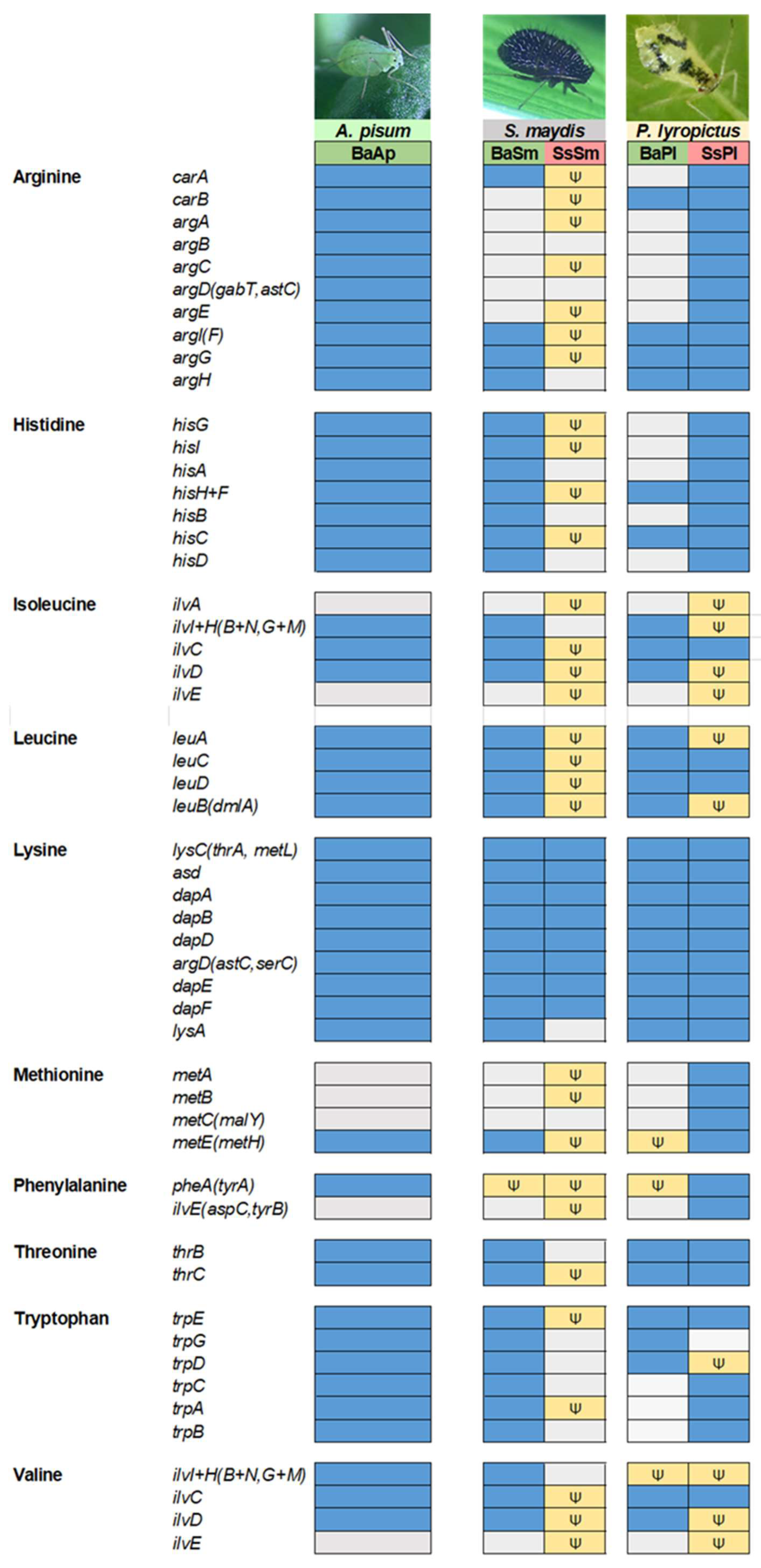
3.6. In the Two Chaitophorinae Aphids, S. symbiotica and B. aphidicola Complement Each Other Metabolically but in Distinct Layouts

In many Lachninae and Chaitophorinae aphids, the ancestral primary symbiont B. aphidicola has become unable to synthesize some essential nutrients on its own due to drastic genome degradation, and the synthesis of these nutrients is supported by the completion of Buchnera’s missing pathways with those of a more recently acquired co-obligate symbiont, with the whole forming a metabolic unit [18,22,26,27]. How the co-obligate symbiont metabolically complements B. aphidicola is usually inferred by genomic analyses. In Lachninae and Chaitophorinae aphids, the co-obligate symbiont is typically required for the biosynthesis of the vitamins riboflavin (B2) and biotin (B7) [20,21,22,25,26]. Analysis of the di-symbiotic systems of S. maydis and P. lyropictus shows that, in both cases, S. symbiotica likely takes charge of riboflavin biosynthesis (Figure 4). Indeed, unlike what has been observed in mono-symbiotic aphids [21], B. aphidicola in the studied Chaitophorinae aphids did not conserve any of the genes for synthesis of this vitamin. In contrast, all these genes are present in the genome of co-obligate symbionts, except for the gene encoding the 5-amino-6-(5-phospho-d-ribitylamino)uracil phosphatase enzyme in SsSm. yigB and the alternative gene ybjI are absent from the genome of SsSm and SsPl. yigL, reported as another alternative for this phosphatase activity [70] is present and intact in the SsPl genome, but pseudogenized in the SsSm genome (split into four pieces). The genes known to encode this enzymatic activity can be missing from the genome of Buchnera strains involved in mono-symbiotic systems (e.g., the APS strain) [71]. However, it has been postulated that this phosphatase activity may be performed by alternative enzymes whose identity is still unclear [70,72]. Regarding biotin biosynthesis, our results show contrasting biosynthetic capacities between the two co-obligate symbionts SsSm and SsPl. SsSm appears to complement B. aphidicola for the biosynthesis of this vitamin from 7-keto-8-aminopelargonate. Indeed, the genes bioA, bioB, and bioD are intact in the genome of the co-obligate symbiont associated with S. maydis, as is the case in the co-obligate symbionts associated with Lachninae aphids [21]. In contrast, bioA and bioD are pseudogenized in SsPl. The case of the di-symbiotic system associated with P. lyropictus for biotin biosynthesis thus differs from what has been previously reported for other Lachninae and Chaitophorinae aphids in which the genes bioA, bioB, and bioD of the co-obligate symbiont are intact [18,21,22]. One hypothesis is that the phloem sap of the Norway maple tree on which P. lyropictus specifically feeds contains this vitamin in sufficient quantities for the aphid to dispense with the biosynthetic capabilities of its bacterial partners.
Regarding essential amino acid (EAA) biosynthesis (Figure 5), the two di-symbiotic systems also show contrasting patterns. A notable finding is that in S. maydis, the co-obligate symbiont SsSm did not retain any intact genes for the synthesis of essential amino acids, except for lysine biosynthesis. In contrast, the B. aphidicola strain associated with this aphid, despite its highly eroded genome, fully supports the biosynthesis of several EAAs, including histidine and tryptophan. In P. lyropictus, histidine biosynthesis is performed by SsPl, and the tryptophan pathway is split between B. aphidicola and the co-obligate symbiont in a similar fashion to what has been reported in the Lachninae aphid Cinara cedri [73]. Overall, our results indicate that SsSm has no complementary role to B. aphidicola in EAAs synthesis and has evolved a dependence on the primary symbiont for the acquisition of most EAAs. The opposite situation occurs in P. lyropictus. Indeed, fewer genes involved in EAA biosynthesis are missing or pseudogenized in the SsPl genome compared to that of SsSm. The S. symbiotica co-obligate symbiont of P. lyropictus has, for example, all the genes required for arginine, histidine, lysine, methionine, and threonine biosynthesis and retains many redundant genes with BaPl. This broader capacity of SsPl for EAAs biosynthesis supports the hypothesis that this S. symbiotica symbiont was more recently acquired by its host compared to SsSm in S. maydis. In this scenario, relaxed selection on these genes may not yet have led to their pseudogenization or elimination in the context of reductive evolution.
In light of all these results, both SsSm and SsPl undertake riboflavin biosynthesis, which B. aphidicola is no longer able to perform. However, the way in which B. aphidicola and S. symbiotica complement each other metabolically for EAA biosynthesis differs greatly between the two di-symbiotic systems. One reason for this is that the EAA biosynthetic capabilities of the ancestral obligate symbiont B. aphidicola differ greatly between the two strains, in particular for histidine and tryptophan biosynthesis, whose pathways are complete in BaSm but incomplete in BaPl. This implies that, during the transition from facultative to co-obligate symbiont, the way in which relaxed selection on metabolism-related genes of the co-obligate symbiont is exerted depends on the genomic background of the primary symbiont and, in some cases, also that of the co-obligate symbiont it replaces. Added to this are the metabolic capabilities of the aphid host, which may differ between species, but which must play a role in the way di-symbiotic nutritional systems are shaped during evolution. This aspect has received little attention in studies of metabolic complementarity between symbionts, in part because it requires laborious sequencing and annotation of the host insect genome [74], a step that is, however, essential to establish the most accurate picture of the role of each partner in the functioning of a three-partner symbiotic system. Finally, another factor that future studies should consider is the nutrient content of the host plant phloem sap, which is expected to shape the evolution of specific biosynthetic pathways in nascent nutritional symbionts. Indeed, the nutrient content of phloem sap can differ greatly between plant species [75,76]. S. maydis and P. lyropictus are Chaitophorinae aphids that feed on contrasted host plants: while S. maydis feeds on the phloem sap of Poaceae (herbaceous plants), P. lyropictus feeds specifically on the phloem sap of the Norway maple A. platanoides (a woody plant). It is likely that the evolution of the di-symbiotic system associated with each of these aphid species is fashioned by the presumably very different nutritional content of the phloem sap of their respective host plant.

3.7. The Secretion Systems and the Array of Virulence Factors Encoded by S. symbiotica Genomes

The tissue tropisms of SsSm and SsPl suggest that the two co-obligate symbionts, although both performing a nutritional function, exhibit contrasting lifestyles within the host. Whereas SsSm is solely compartmentalized into secondary bacteriocytes, SsPl is capable of infecting a wide diversity of host tissues (Figure 3; [30]). These differences in invasion patterns should reflect the infection mechanisms that the two symbionts are able to express despite the ongoing reduction of their genome. The ability of bacteria to invade host tissues and cells depends in particular on the secretion systems they can express [77]. These protein complexes on bacterial cell membranes allow for the translocation of effector proteins that can modulate the host environment and facilitate its invasion, for example by enhancing attachment to eukaryotic cells, plundering resources in an environmental niche, or intoxicating target cells and disrupting their functions. Our genome analyses reveal that SsSm and SsPl are associated with a low diversity of secretory systems (Table S5). The two S. symbiotica co-obligate symbionts retained only the ability to synthesize type V secretion systems (T5SS). These results are in agreement with those of a previous study showing that both facultative and co-obligate mutualistic strains of S. symbiotica do not retain complete secretion systems other than T5SS, in contrast to pathogenic strains that tend to retain key elements of T3SSs [59]. All S. symbiotica strains whose genomes have been sequenced so far, including those with extremely small genomes, are virtually capable of expressing T5SSs [59], raising the question of their possible role in the symbiosis with the insect host. T5SSs include diverse forms of virulence factor-secreting autotransporters involved in cell-to-cell adhesion and biofilm formation [78,79]. They may be involved in various functions associated with the specific lifestyle of the bacteria, including protease activity, intracellular mobility, and nutrient acquisition in limited environments. SsSm and SsPl appear to have retained the ability to synthesize a FdeC-like autotransporter adhesin (Figure 6; Table S6). This T5SS has been shown to contribute to virulence of uropathogenic Escherichia coli (UPEC) strains by mediating their adhesion to mammalian cells [80], but the exact role of this autotransporter remains unknown outside this group of bacteria. Unlike SsPl, SsSm appears to have retained the genes encoding the autotransporters InvA (involved in the virulence of Yersinia bacteria [81]) and SinH (involved in the virulence of UPEC strains [82]). Currently, multiple functions of type V autotransporters have been identified recently in the context of eukaryotic host–pathogen interactions [78,83]. However, their potential roles in the context of bacterial mutualism in insects remain unknown.
The success of host colonization by invasive bacteria, whether in a host–pathogen relationship or mutualistic symbiosis, is largely determined by the ability of microorganisms to interact with hosts through the expression of virulence factors [84,85]. The content of virulence factors has been little studied in co-obligate symbionts. However, these nutritional symbionts, acquired more recently and exhibiting more moderate genome reduction compared to the ancient primary symbionts, are expected to have retained a greater diversity of these factors, which may allow them to colonize a greater diversity of host tissues. Examining virulence genes in the genome of co-obligate symbionts is therefore crucial to gain insight into the mechanisms underlying the early stages of insect endosymbiogenesis. In this context, our analyses show that SsSm and SsPl genomes differ greatly in their virulence factor content (Figure 6). Both genomes contain an equivalent number of virulence factor-encoding genes, but more genes are pseudogenized in SsSm than in SsPl (Figure 6; Table S6). This again suggests that SsSm is more advanced in reductive genome evolution compared to SsPl. The most striking example concerns the ability of both S. symbiotica strains to biosynthesize a complete flagellum, a macromolecular system that enables invasive bacteria to move through body fluids, attach the organ surfaces, and then promotes efficient colonization of host tissues [86]. The SsSm genome contains many genes encoding a flagellum, but those involved in hook and filament biosynthesis are either missing (including fliC and flgL) or pseudogenized (including flgK, flgE, flgC, flgF and flgG) (Table S6), suggesting that this structure has not retained any motility properties. Moreover, the remaining intact genes (including flgB, flgH, fliE, fliI, fliM, fliN) that correspond to the basal body of the flagellar structure are also not sufficient for its function as an injectisome, a macromolecular protein complex whose structure is an evolutionary homologue of the bacterial flagellum and which is used by many bacteria, including bacterial endosymbionts, to deliver secreted effector proteins to a eukaryotic host [87,88]. The remnants of the flagellar apparatus associated with SsSm hardly allow for the biosynthesis of a reduced flagellum as found in some strains of B. aphidicola [87]. Motility pathways are among the first to be altered upon transition from a free-living to a host-dependent lifestyle [89]. Given that the SsSm genome is already highly reduced and pseudogenized, it is not surprising that this co-obligate symbiont has lost the capacity for motility.
In contrast, the SsPl genome contains all the genes theoretically required for biosynthesis of a complete flagellum (Figure 6; Table S6), suggesting that this S. symbiotica co-obligate symbiont is endowed with motility and chemotaxis potential. This could explain the ability of this S. symbiotica co-obligate symbiont to exhibit a highly invasive phenotype [30]. The case of SsPl, capable to adopt both an intracellular and an extracellular lifestyle, is singular for a nutritional symbiont, but not unique. Indeed, there are a few cases of bacteriocyte-associated nutritional symbionts that, despite the significant reduction of their genome and their specific nutritional function, retain a complete flagellar system [90,91]. Wigglesworthia, the obligate symbiont of tsetse flies, can adopt both an intracellular lifestyle in bacteriocytes and an extracellular phase in milk gland secretions during which it expresses a functional flagellum despite a severely reduced genome [90,92]. The case of SsPl is not as extreme as that of Wigglesworthia, but it illustrates the existence of co-obligate strains of S. symbiotica that, despite their integration in a cooperative system with the host and the primary symbiont B. aphidicola, resemble the previously described pathogenic strains in that they retain a complete flagellar system and the ability to colonize the host’s digestive tract [59]. One hypothesis is that SsPl express a functional flagellum depending on the tissue it colonizes and its lifestyle (i.e., intracellular or extracellular) as is the case for the symbiont Wigglesworthia in the tsetse fly.
Iron uptake by siderophores are well known to promote host colonization not only by pathogenic bacteria, but also by insect symbionts such as Sodalis glossinidius, which uses this system to invade the tsetse fly [93]. Our analyses show that both S. symbiotica genomes contain many genes involved in siderophore biosynthesis. They both contain genes involved in the biosynthesis of enterobactin, a catechol-containing siderophore released by bacteria to bind ferric ions required for metabolic pathways [94]. However, in both SsSm and SsPl, most of the genes related to enterobactin biosynthesis (entA, entB, entC, entE and entF) are missing or pseudogenized, suggesting a relaxation of selection on these genes and that the production of enterobactin-like siderophores became dispensable during the evolution of both symbionts or was a limiting factor in the establishment of a persistent relationship between S. symbiotica and the host (Figure 6). Our analyses show that both symbionts retain intact genes for yersiniabactin (Ybt) biosynthesis, for which many identified genes are intact (e.g., irp1, irp2, fyuA, and ybtA), especially in SsPl genome. However, despite many intact genes in the SsPl genome for the ybt locus, close examination shows that the locus is incomplete with some genes missing, including ybtS, ybtu, ybtT, ybtD, and psn. Thus, it appears that SsPl has become unable to synthesize yersiniabactin.
Finally, our analyses detected few toxin-related genes in the genomes of both co-obligate symbionts. SsSm and SsPl appear, however, to be able to synthesize effectors of the YopJ family, which are well known to promote invasion of pathogenic bacteria, but whose potential roles in the establishment of the mutualistic symbiosis remain unknown [95].
Based on the analysis of the virulence factors of these two co-obligate symbionts, it can be concluded that SsSm and SsPl exhibit different sets of virulence factors that reflect their contrasting lifestyles. The most notable finding is that SsPl is potentially capable of synthesizing a complete flagellum that would endow it with motility, an ability that could explain the remarkable invasive phenotype of this co-obligate symbiont. However, both co-obligate symbionts tend to exhibit low virulence factor diversity compared to cultivable S. symbiotica and especially compared to entomopathogenic bacteria of the genus Serratia such as Serratia marcescens [59]. Our results suggest that SsSm and SsPl are derived from originally pathogenic bacteria capable of synthesizing enterobactin and yersiniabactin-like siderophores, an ability they seem to have lost. Loss of virulence factors is conducive to the establishment of a long-lasting, mutualistic lifestyle with the host, which underlies strong selection for attenuated virulence [7].

4. Conclusions and Perspectives

In conclusion, the S. symbiotica co-obligate symbionts associated with two Chaitophorinae species, S. maydis and P. lyropictus, provide a contrasting picture of recent co-obligate nutritional endosymbiosis in aphids. The di-symbiotic systems associated with these aphids show contrasting facets regarding genome evolution and the level of anatomical integration of the co-obligate symbionts into their respective hosts. The S. symbiotica co-obligate symbiont associated with P. lyropictus still has a large genome, comparable in size to that reported for pathogenic cultivable strains. Its dual intracellular and extracellular lifestyle indicate that host dependence on nutritional symbionts may evolve prior to compartmentalization in the host cells specialized for metabolic exchange and symbiont control (i.e., bacteriocytes) [30]. Its ability to infect a wide variety of host tissues is probably due to its ability to express a complete flagellum and thus display an ability to move in body fluids and on organ surface, a well-known invasive potential typically expressed by pathogenic bacteria. How such a nutritional symbiont, both compartmentalized into bacteriocytes and exhibiting such an invasive phenotype, is controlled by its host is a puzzle to be solved in future studies. The S. symbiotica co-obligate symbiont associated with S. maydis exhibits another side of co-obligate nutritional endosymbiosis, that of further anatomical integration into the host (i.e., strict compartmentalization into bacteriocytes), indicative of a presumably older association with its host compared to SsPl, which is now stabilized. In recent years, a growing number of studies have shown that many insect species harbor nutritional multi-symbiotic systems. However, how they develop and function remains largely unknown. Studying the diversity of these associations is crucial to tackle these aspects and address key questions in the evolutionary developmental biology of endosymbiosis including: How do metabolically complementary symbionts, forming a metabolic unit, coordinate with each other and with the host? How does the host control these nutritional symbionts, sometimes associated with an invasive phenotype? How do the different types of bacteriocytes (primary versus secondary) develop and interact with each other? The diversity of the di-symbiotic systems associated with Chaitophorinae aphids provides an ideal playing field to address these questions and better appreciate the sophistication of bacterial mutualism in insects.

Supplementary Materials

The following supporting information can be downloaded at: https://www.mdpi.com/article/10.3390/microorganisms10071360/s1, Figure S1: Geographic location of the collection sites of S. maydis colonies assayed in this study (for details, see Table S1). Green spots reference the 2014 sampling and red spots reference the 2016 sampling; Table S1: Collection details for S. maydis samples (collected in 2014 and 2016 in Morocco) and detection of S. symbiotica in the different sampled colonies (a Tt: Triticum turgidum; Ta: Triticum aestivum; Hv: Hordeum vulgare; b A: arid; pA: pre-arid; sA: semi-arid; sH: subhumid); Table S2: Accession numbers and details of the bacterial strains used for the multigene phylogenetic reconstruction; Table S3: Information on genome sequencing of symbiotic bacteria in this study; Table S4: Intact phage regions detected with PHASTER; Table S5: Macromolecular systems predicted by MacSyFinder in the SsSm-Midelt and SsPl-LLN strains; Table S6: Presence/absence map of S. symbiotica virulence genes. Presence of a virulence gene is labeled in gray (+) and absence in white (-). The yellow color (Ψ) means that the gene is pseudogenized. Note that the absence of a gene may be threshold dependent (minimum 50% aa identity, 80% align. Coverage).

Author Contributions

Conceptualization: F.R., Methodology: J.A., B.B., J.-L.G., N.P. and F.R. Formal analysis: J.A., N.P., S.F. and F.R. Writing—original draft preparation: F.R. Writing—review and editing: J.A., B.B., S.F., N.P., M.R.L., F.C. and T.H. Supervision: F.C. and T.H. Project administration: F.C. and T.H. All authors have read and agreed to the published version of the manuscript.

Funding

This study was funded by FNRS, grant no. 1B374.21. BF2i laboratory was specifically supported by INSA Lyon and INRAE.

Data Availability Statement

Publicly available datasets were analysed in this study. This data can be found on NCBI (BioProject accession numbers: PRJNA835797, PRJNA837269, PRJNA837360 and PRJNA837363).

Acknowledgments

The authors thank Karen Gaget for her help with the aphid pictures and the whole BF2I laboratory for the stimulating discussions about the topic. This paper is publication BRC 393 of the Biodiversity Research Centre (Université catholique de Louvain).

Conflicts of Interest

The authors declare no conflict of interest. The funders had no role in study design, data collection and analysis, decision to publish, or preparation of the manuscript.

References

  1. Baumann, P.; Moran, N.A.; Baumann, L.C. Bacteriocyte-Associated Endosymbionts of Insects. In The Prokaryotes: Prokaryotic Biology and Symbiotic Associations; Rosenberg, E., DeLong, E.F., Lory, S., Stackebrandt, E., Thompson, F., Eds.; Springer: Berlin/Heidelberg, Germany, 2013; pp. 465–496. ISBN 978-3-642-30194-0. [Google Scholar]
  2. Whittle, M.; Barreaux, A.M.G.; Bonsall, M.B.; Ponton, F.; English, S. Insect-Host Control of Obligate, Intracellular Symbiont Density. Proc. R. Soc. B Biol. Sci. 2021, 288, 20211993. [Google Scholar] [CrossRef] [PubMed]
  3. Koga, R.; Meng, X.-Y.; Tsuchida, T.; Fukatsu, T. Cellular Mechanism for Selective Vertical Transmission of an Obligate Insect Symbiont at the Bacteriocyte–Embryo Interface. Proc. Natl. Acad. Sci. USA 2012, 109, E1230–E1237. [Google Scholar] [CrossRef] [PubMed] [Green Version]
  4. Chomicki, G.; Werner, G.D.A.; West, S.A.; Kiers, E.T. Compartmentalization Drives the Evolution of Symbiotic Cooperation. Philos. Trans. R. Soc. B Biol. Sci. 2020, 375, 20190602. [Google Scholar] [CrossRef] [PubMed]
  5. Moran, N.A. Accelerated Evolution and Muller’s Rachet in Endosymbiotic Bacteria. Proc. Natl. Acad. Sci. USA 1996, 93, 2873–2878. [Google Scholar] [CrossRef] [Green Version]
  6. Wernegreen, J.J. Endosymbiont Evolution: Predictions from Theory and Surprises from Genomes. Ann. N. Y. Acad. Sci. 2015, 1360, 16–35. [Google Scholar] [CrossRef] [Green Version]
  7. McCutcheon, J.P.; Boyd, B.M.; Dale, C. The Life of an Insect Endosymbiont from the Cradle to the Grave. Curr. Biol. 2019, 29, R485–R495. [Google Scholar] [CrossRef]
  8. Tamas, I.; Klasson, L.; Canbäck, B.; Näslund, A.K.; Eriksson, A.-S.; Wernegreen, J.J.; Sandström, J.P.; Moran, N.A.; Andersson, S.G.E. 50 Million Years of Genomic Stasis in Endosymbiotic Bacteria. Science 2002, 296, 2376–2379. [Google Scholar] [CrossRef] [Green Version]
  9. Moran, N.A.; Bennett, G.M. The Tiniest Tiny Genomes. Annu. Rev. Microbiol. 2014, 68, 195–215. [Google Scholar] [CrossRef]
  10. Bennett, G.M.; Moran, N.A. Heritable Symbiosis: The Advantages and Perils of an Evolutionary Rabbit Hole. Proc. Natl. Acad. Sci. USA 2015, 112, 10169–10176. [Google Scholar] [CrossRef] [Green Version]
  11. Renoz, F.; Pons, I.; Hance, T. Evolutionary Responses of Mutualistic Insect–Bacterial Symbioses in a World of Fluctuating Temperatures. Curr. Opin. Insect Sci. 2019, 35, 20–26. [Google Scholar] [CrossRef]
  12. Moran, N.A.; McCutcheon, J.P.; Nakabachi, A. Genomics and Evolution of Heritable Bacterial Symbionts. Annu. Rev. Genet. 2008, 42, 165–190. [Google Scholar] [CrossRef] [PubMed] [Green Version]
  13. Douglas, A.E. How Multi-Partner Endosymbioses Function. Nat. Rev. Microbiol. 2016, 14, 731–743. [Google Scholar] [CrossRef] [PubMed]
  14. Wu, D.; Daugherty, S.C.; Aken, S.E.V.; Pai, G.H.; Watkins, K.L.; Khouri, H.; Tallon, L.J.; Zaborsky, J.M.; Dunbar, H.E.; Tran, P.L.; et al. Metabolic Complementarity and Genomics of the Dual Bacterial Symbiosis of Sharpshooters. PLoS Biol. 2006, 4, e188. [Google Scholar] [CrossRef] [PubMed]
  15. McCutcheon, J.P.; von Dohlen, C.D. An Interdependent Metabolic Patchwork in the Nested Symbiosis of Mealybugs. Curr. Biol. CB 2011, 21, 1366–1372. [Google Scholar] [CrossRef] [Green Version]
  16. Van Leuven, J.T.; Meister, R.C.; Simon, C.; McCutcheon, J.P. Sympatric Speciation in a Bacterial Endosymbiont Results in Two Genomes with the Functionality of One. Cell 2014, 158, 1270–1280. [Google Scholar] [CrossRef] [Green Version]
  17. Husnik, F.; McCutcheon, J.P. Repeated Replacement of an Intrabacterial Symbiont in the Tripartite Nested Mealybug Symbiosis. Proc. Natl. Acad. Sci. USA 2016, 113, E5416–E5424. [Google Scholar] [CrossRef] [Green Version]
  18. Manzano-Marín, A.; Latorre, A. Snapshots of a Shrinking Partner: Genome Reduction in Serratia Symbiotica. Sci. Rep. 2016, 6, 32590. [Google Scholar] [CrossRef]
  19. Manzano-Marín, A.; Szabó, G.; Simon, J.-C.; Horn, M.; Latorre, A. Happens in the Best of Subfamilies: Establishment and Repeated Replacements of Co-Obligate Secondary Endosymbionts within Lachninae Aphids. Environ. Microbiol. 2017, 19, 393–408. [Google Scholar] [CrossRef]
  20. Manzano-Marín, A.; Coeur D’Acier, A.; Clamens, A.-L.; Orvain, C.; Cruaud, C.; Barbe, V.; Jousselin, E. A Freeloader? The Highly Eroded Yet Large Genome of the Serratia Symbiotica Symbiont of Cinara Strobi. Genome Biol. Evol. 2018, 10, 2178–2189. [Google Scholar] [CrossRef] [Green Version]
  21. Manzano-Marín, A.; D’acier, A.C.; Clamens, A.-L.; Orvain, C.; Cruaud, C.; Barbe, V.; Jousselin, E. Serial Horizontal Transfer of Vitamin-Biosynthetic Genes Enables the Establishment of New Nutritional Symbionts in Aphids’ Di-Symbiotic Systems. ISME J. 2020, 14, 259–273. [Google Scholar] [CrossRef] [Green Version]
  22. Monnin, D.; Jackson, R.; Kiers, E.T.; Bunker, M.; Ellers, J.; Henry, L.M. Parallel Evolution in the Integration of a Co-Obligate Aphid Symbiosis. Curr. Biol. 2020, 30, 1949–1957.e6. [Google Scholar] [CrossRef] [PubMed]
  23. Koga, R.; Moran, N.A. Swapping Symbionts in Spittlebugs: Evolutionary Replacement of a Reduced Genome Symbiont. ISME J. 2014, 8, 1237–1246. [Google Scholar] [CrossRef] [PubMed] [Green Version]
  24. Matsuura, Y.; Moriyama, M.; Łukasik, P.; Vanderpool, D.; Tanahashi, M.; Meng, X.-Y.; McCutcheon, J.P.; Fukatsu, T. Recurrent Symbiont Recruitment from Fungal Parasites in Cicadas. Proc. Natl. Acad. Sci. USA 2018, 115, E5970–E5979. [Google Scholar] [CrossRef] [PubMed] [Green Version]
  25. Manzano-Marín, A.; Latorre, A. Settling Down: The Genome of Serratia Symbiotica from the Aphid Cinara Tujafilina Zooms in on the Process of Accommodation to a Cooperative Intracellular Life. Genome Biol. Evol. 2014, 6, 1683–1698. [Google Scholar] [CrossRef] [PubMed]
  26. Manzano-Marín, A.; Simon, J.-C.; Latorre, A. Reinventing the Wheel and Making It Round Again: Evolutionary Convergence in BuchneraSerratia Symbiotic Consortia between the Distantly Related Lachninae Aphids Tuberolachnus Salignus and Cinara Cedri. Genome Biol. Evol. 2016, 8, 1440–1458. [Google Scholar] [CrossRef] [Green Version]
  27. Meseguer, A.S.; Manzano-Marín, A.; d’Acier, A.C.; Clamens, A.-L.; Godefroid, M.; Jousselin, E. Buchnera Has Changed Flatmate but the Repeated Replacement of Co-Obligate Symbionts Is Not Associated with the Ecological Expansions of Their Aphid Hosts. Mol. Ecol. 2017, 26, 2363–2378. [Google Scholar] [CrossRef]
  28. Fukatsu, T.; Nikoh, N.; Kawai, R.; Koga, R. The Secondary Endosymbiotic Bacterium of the Pea Aphid Acyrthosiphon Pisum (Insecta: Homoptera). Appl. Environ. Microbiol. 2000, 66, 2748–2758. [Google Scholar] [CrossRef] [Green Version]
  29. Renoz, F.; Noël, C.; Errachid, A.; Foray, V.; Hance, T. Infection Dynamic of Symbiotic Bacteria in the Pea Aphid Acyrthosiphon Pisum Gut and Host Immune Response at the Early Steps in the Infection Process. PLoS ONE 2015, 10, e0122099. [Google Scholar] [CrossRef]
  30. Renoz, F.; Lopes, M.R.; Gaget, K.; Duport, G.; Eloy, M.-C.; Geelhand de Merxem, B.; Hance, T.; Calevro, F. Compartmentalized into Bacteriocytes but Highly Invasive: The Puzzling Case of the Co-Obligate Symbiont Serratia Symbiotica in the Aphid Periphyllus Lyropictus. Microbiol. Spectr. 2022, 10, e0045722. [Google Scholar] [CrossRef]
  31. Jousselin, E.; Clamens, A.-L.; Galan, M.; Bernard, M.; Maman, S.; Gschloessl, B.; Duport, G.; Meseguer, A.S.; Calevro, F.; D’acier, A.C. Assessment of a 16S RRNA Amplicon Illumina Sequencing Procedure for Studying the Microbiome of a Symbiont-Rich Aphid Genus. Mol. Ecol. Resour. 2016, 16, 628–640. [Google Scholar] [CrossRef]
  32. Kolmogorov, M.; Yuan, J.; Lin, Y.; Pevzner, P.A. Assembly of Long, Error-Prone Reads Using Repeat Graphs. Nat. Biotechnol. 2019, 37, 540–546. [Google Scholar] [CrossRef] [PubMed]
  33. Walker, B.J.; Abeel, T.; Shea, T.; Priest, M.; Abouelliel, A.; Sakthikumar, S.; Cuomo, C.A.; Zeng, Q.; Wortman, J.; Young, S.K.; et al. Pilon: An Integrated Tool for Comprehensive Microbial Variant Detection and Genome Assembly Improvement. PLoS ONE 2014, 9, e112963. [Google Scholar] [CrossRef] [PubMed]
  34. Tatusova, T.; DiCuccio, M.; Badretdin, A.; Chetvernin, V.; Nawrocki, E.P.; Zaslavsky, L.; Lomsadze, A.; Pruitt, K.D.; Borodovsky, M.; Ostell, J. NCBI Prokaryotic Genome Annotation Pipeline. Nucleic Acids Res. 2016, 44, 6614–6624. [Google Scholar] [CrossRef] [PubMed]
  35. Médigue, C.; Calteau, A.; Cruveiller, S.; Gachet, M.; Gautreau, G.; Josso, A.; Lajus, A.; Langlois, J.; Pereira, H.; Planel, R.; et al. MicroScope—an Integrated Resource for Community Expertise of Gene Functions and Comparative Analysis of Microbial Genomic and Metabolic Data. Brief. Bioinform. 2019, 20, 1071–1084. [Google Scholar] [CrossRef]
  36. Vallenet, D.; Calteau, A.; Dubois, M.; Amours, P.; Bazin, A.; Beuvin, M.; Burlot, L.; Bussell, X.; Fouteau, S.; Gautreau, G.; et al. MicroScope: An Integrated Platform for the Annotation and Exploration of Microbial Gene Functions through Genomic, Pangenomic and Metabolic Comparative Analysis. Nucleic Acids Res. 2020, 48, D579–D589. [Google Scholar] [CrossRef] [Green Version]
  37. Caspi, R.; Billington, R.; Keseler, I.M.; Kothari, A.; Krummenacker, M.; Midford, P.E.; Ong, W.K.; Paley, S.; Subhraveti, P.; Karp, P.D. The MetaCyc Database of Metabolic Pathways and Enzymes—A 2019 Update. Nucleic Acids Res. 2020, 48, D445–D453. [Google Scholar] [CrossRef] [Green Version]
  38. Lerat, E.; Ochman, H. Recognizing the Pseudogenes in Bacterial Genomes. Nucleic Acids Res. 2005, 33, 3125–3132. [Google Scholar] [CrossRef]
  39. Burke, G.R.; Moran, N.A. Massive Genomic Decay in Serratia Symbiotica, a Recently Evolved Symbiont of Aphids. Genome Biol. Evol. 2011, 3, 195–208. [Google Scholar] [CrossRef]
  40. Liu, B.; Zheng, D.; Jin, Q.; Chen, L.; Yang, J. VFDB 2019: A Comparative Pathogenomic Platform with an Interactive Web Interface. Nucleic Acids Res. 2019, 47, D687–D692. [Google Scholar] [CrossRef]
  41. Abby, S.S.; Néron, B.; Ménager, H.; Touchon, M.; Rocha, E.P.C. MacSyFinder: A Program to Mine Genomes for Molecular Systems with an Application to CRISPR-Cas Systems. PLoS ONE 2014, 9, e110726. [Google Scholar] [CrossRef]
  42. Arndt, D.; Grant, J.R.; Marcu, A.; Sajed, T.; Pon, A.; Liang, Y.; Wishart, D.S. PHASTER: A Better, Faster Version of the PHAST Phage Search Tool. Nucleic Acids Res. 2016, 44, W16–W21. [Google Scholar] [CrossRef] [PubMed] [Green Version]
  43. Emms, D.M.; Kelly, S. OrthoFinder: Solving Fundamental Biases in Whole Genome Comparisons Dramatically Improves Orthogroup Inference Accuracy. Genome Biol. 2015, 16, 157. [Google Scholar] [CrossRef] [PubMed] [Green Version]
  44. Emms, D.M.; Kelly, S. OrthoFinder: Phylogenetic Orthology Inference for Comparative Genomics. Genome Biol. 2019, 20, 238. [Google Scholar] [CrossRef] [PubMed] [Green Version]
  45. Smith, S.A.; Dunn, C.W. Phyutility: A Phyloinformatics Tool for Trees, Alignments and Molecular Data. Bioinformatics 2008, 24, 715–716. [Google Scholar] [CrossRef] [PubMed] [Green Version]
  46. Kalyaanamoorthy, S.; Minh, B.Q.; Wong, T.K.F.; von Haeseler, A.; Jermiin, L.S. ModelFinder: Fast Model Selection for Accurate Phylogenetic Estimates. Nat. Methods 2017, 14, 587–589. [Google Scholar] [CrossRef] [Green Version]
  47. Nguyen, L.-T.; Schmidt, H.A.; von Haeseler, A.; Minh, B.Q. IQ-TREE: A Fast and Effective Stochastic Algorithm for Estimating Maximum-Likelihood Phylogenies. Mol. Biol. Evol. 2015, 32, 268–274. [Google Scholar] [CrossRef]
  48. Minh, B.Q.; Nguyen, M.A.T.; von Haeseler, A. Ultrafast Approximation for Phylogenetic Bootstrap. Mol. Biol. Evol. 2013, 30, 1188–1195. [Google Scholar] [CrossRef]
  49. Hoang, D.T.; Chernomor, O.; von Haeseler, A.; Minh, B.Q.; Vinh, L.S. UFBoot2: Improving the Ultrafast Bootstrap Approximation. Mol. Biol. Evol. 2018, 35, 518–522. [Google Scholar] [CrossRef]
  50. Anisimova, M.; Gil, M.; Dufayard, J.-F.; Dessimoz, C.; Gascuel, O. Survey of Branch Support Methods Demonstrates Accuracy, Power, and Robustness of Fast Likelihood-Based Approximation Schemes. Syst. Biol. 2011, 60, 685–699. [Google Scholar] [CrossRef]
  51. Koga, R.; Tsuchida, T.; Fukatsu, T. Quenching Autofluorescence of Insect Tissues for in Situ Detection of Endosymbionts. Appl. Entomol. Zool. 2009, 44, 281–291. [Google Scholar] [CrossRef] [Green Version]
  52. Koga, R.; Tsuchida, T.; Fukatsu, T. Changing Partners in an Obligate Symbiosis: A Facultative Endosymbiont Can Compensate for Loss of the Essential Endosymbiont Buchnera in an Aphid. Proc. R. Soc. Lond. B Biol. Sci. 2003, 270, 2543–2550. [Google Scholar] [CrossRef] [PubMed] [Green Version]
  53. Oliver, K.M.; Degnan, P.H.; Burke, G.R.; Moran, N.A. Facultative Symbionts in Aphids and the Horizontal Transfer of Ecologically Important Traits. Annu. Rev. Entomol. 2010, 55, 247–266. [Google Scholar] [CrossRef] [PubMed] [Green Version]
  54. Rock, D.I.; Smith, A.H.; Joffe, J.; Albertus, A.; Wong, N.; O’Connor, M.; Oliver, K.M.; Russell, J.A. Context-Dependent Vertical Transmission Shapes Strong Endosymbiont Community Structure in the Pea Aphid, Acyrthosiphon Pisum. Mol. Ecol. 2018, 27, 2039–2056. [Google Scholar] [CrossRef]
  55. Zytynska, S.E.; Tighiouart, K.; Frago, E. Benefits and Costs of Hosting Facultative Symbionts in Plant-Sucking Insects: A Meta-Analysis. Mol. Ecol. 2021, 30, 2483–2494. [Google Scholar] [CrossRef]
  56. Wilkinson, T.L.; Ishikawa, H. Injection of Essential Amino Acids Substitutes for Bacterial Supply in Aposymbiotic Pea Aphids (Acyrthosiphon Pisum). Entomol. Exp. Appl. 2000, 94, 85–91. [Google Scholar] [CrossRef]
  57. Koga, R.; Tsuchida, T.; Sakurai, M.; Fukatsu, T. Selective Elimination of Aphid Endosymbionts: Effects of Antibiotic Dose and Host Genotype, and Fitness Consequences. FEMS Microbiol. Ecol. 2007, 60, 229–239. [Google Scholar] [CrossRef] [Green Version]
  58. Chong, R.A.; Park, H.; Moran, N.A. Genome Evolution of the Obligate Endosymbiont Buchnera Aphidicola. Mol. Biol. Evol. 2019, 36, 1481–1489. [Google Scholar] [CrossRef]
  59. Renoz, F.; Foray, V.; Ambroise, J.; Baa-Puyoulet, P.; Bearzatto, B.; Mendez, G.L.; Grigorescu, A.S.; Mahillon, J.; Mardulyn, P.; Gala, J.-L.; et al. At the Gate of Mutualism: Identification of Genomic Traits Predisposing to Insect-Bacterial Symbiosis in Pathogenic Strains of the Aphid Symbiont Serratia Symbiotica. Front. Cell. Infect. Microbiol. 2021, 11, 660007. [Google Scholar] [CrossRef] [PubMed]
  60. Latorre, A.; Manzano-Marín, A. Dissecting Genome Reduction and Trait Loss in Insect Endosymbionts. Ann. N. Y. Acad. Sci. 2017, 1389, 52–75. [Google Scholar] [CrossRef]
  61. Sabri, A.; Leroy, P.; Haubruge, E.; Hance, T.; Frere, I.; Destain, J.; Thonart, P. Isolation, Pure Culture and Characterization of Serratia Symbiotica Sp. Nov., the R-Type of Secondary Endosymbiont of the Black Bean Aphid Aphis Fabae. Int. J. Syst. Evol. Microbiol. 2011, 61, 2081–2088. [Google Scholar] [CrossRef] [Green Version]
  62. Grigorescu, A.S.; Renoz, F.; Sabri, A.; Foray, V.; Hance, T.; Thonart, P. Accessing the Hidden Microbial Diversity of Aphids: An Illustration of How Culture-Dependent Methods Can Be Used to Decipher the Insect Microbiota. Microb. Ecol. 2018, 75, 1035–1048. [Google Scholar] [CrossRef] [PubMed]
  63. Perreau, J.; Patel, D.J.; Anderson, H.; Maeda, G.P.; Elston, K.M.; Barrick, J.E.; Moran, N.A. Vertical Transmission at the Pathogen-Symbiont Interface: Serratia Symbiotica and Aphids. mBio 2021, 12, e00359-21. [Google Scholar] [CrossRef] [PubMed]
  64. Pons, I.; Renoz, F.; Noël, C.; Hance, T. New Insights into the Nature of Symbiotic Associations in Aphids: Infection Process, Biological Effects and Transmission Mode of Cultivable Serratia Symbiotica Bacteria. Appl. Environ. Microbiol. 2019, 85, e02445-18. [Google Scholar] [CrossRef] [PubMed] [Green Version]
  65. Elston, K.M.; Perreau, J.; Maeda, G.P.; Moran, N.A.; Barrick, J.E. Engineering a Culturable Serratia Symbiotica Strain for Aphid Paratransgenesis. Appl. Environ. Microbiol. 2020, 87, e02245-20. [Google Scholar] [CrossRef]
  66. Lamelas, A.; Pérez-Brocal, V.; Gómez-Valero, L.; Gosalbes, M.J.; Moya, A.; Latorre, A. Evolution of the Secondary Symbiont “Candidatus Serratia Symbiotica” in Aphid Species of the Subfamily Lachninae. Appl. Environ. Microbiol. 2008, 74, 4236–4240. [Google Scholar] [CrossRef] [PubMed] [Green Version]
  67. Braendle, C.; Miura, T.; Bickel, R.; Shingleton, A.W.; Kambhampati, S.; Stern, D.L. Developmental Origin and Evolution of Bacteriocytes in the Aphid–Buchnera Symbiosis. PLoS Biol. 2003, 1, e21. [Google Scholar] [CrossRef]
  68. Baumann, P. Biology of Bacteriocyte-Associated Endosymbionts of Plant Sap-Sucking Insects. Annu. Rev. Microbiol. 2005, 59, 155–189. [Google Scholar] [CrossRef]
  69. Simonet, P.; Duport, G.; Gaget, K.; Weiss-Gayet, M.; Colella, S.; Febvay, G.; Charles, H.; Viñuelas, J.; Heddi, A.; Calevro, F. Direct Flow Cytometry Measurements Reveal a Fine-Tuning of Symbiotic Cell Dynamics According to the Host Developmental Needs in Aphid Symbiosis. Sci. Rep. 2016, 6, 19967. [Google Scholar] [CrossRef]
  70. Haase, I.; Sarge, S.; Illarionov, B.; Laudert, D.; Hohmann, H.-P.; Bacher, A.; Fischer, M. Enzymes from the Haloacid Dehalogenase (HAD) Superfamily Catalyse the Elusive Dephosphorylation Step of Riboflavin Biosynthesis. ChemBioChem 2013, 14, 2272–2275. [Google Scholar] [CrossRef]
  71. Blow, F.; Bueno, E.; Clark, N.; Zhu, D.T.; Chung, S.H.; Güllert, S.; Schmitz, R.A.; Douglas, A.E. B-Vitamin Nutrition in the Pea Aphid-Buchnera Symbiosis. J. Insect Physiol. 2020, 126, 104092. [Google Scholar] [CrossRef]
  72. Szabó, G.; Schulz, F.; Manzano-Marín, A.; Toenshoff, E.R.; Horn, M. Evolutionarily Recent Dual Obligatory Symbiosis among Adelgids Indicates a Transition between Fungus- and Insect-Associated Lifestyles. ISME J. 2022, 16, 247–256. [Google Scholar] [CrossRef] [PubMed]
  73. Lamelas, A.; Gosalbes, M.J.; Manzano-Marín, A.; Peretó, J.; Moya, A.; Latorre, A. Serratia Symbiotica from the Aphid Cinara Cedri: A Missing Link from Facultative to Obligate Insect Endosymbiont. PLoS Genet. 2011, 7, e1002357. [Google Scholar] [CrossRef] [PubMed] [Green Version]
  74. Blaxter, M.; Archibald, J.M.; Childers, A.K.; Coddington, J.A.; Crandall, K.A.; Di Palma, F.; Durbin, R.; Edwards, S.V.; Graves, J.A.M.; Hackett, K.J.; et al. Why Sequence All Eukaryotes? Proc. Natl. Acad. Sci. USA 2022, 119, e2115636118. [Google Scholar] [CrossRef] [PubMed]
  75. Dinant, S.; Bonnemain, J.-L.; Girousse, C.; Kehr, J. Phloem Sap Intricacy and Interplay with Aphid Feeding. C. R. Biol. 2010, 333, 504–515. [Google Scholar] [CrossRef] [Green Version]
  76. Gallinger, J.; Gross, J. Unraveling the Host Plant Alternation of Cacopsylla Pruni—Adults but Not Nymphs Can Survive on Conifers Due to Phloem/Xylem Composition. Front. Plant Sci. 2018, 9, 484. [Google Scholar] [CrossRef]
  77. Green, E.R.; Mecsas, J. Bacterial Secretion Systems: An Overview. Microbiol. Spectr. 2016, 4. [Google Scholar] [CrossRef] [Green Version]
  78. Leo, J.C.; Grin, I.; Linke, D. Type V Secretion: Mechanism(s) of Autotransport through the Bacterial Outer Membrane. Philos. Trans. R. Soc. B 2012, 367, 1088–1101. [Google Scholar] [CrossRef] [Green Version]
  79. Meuskens, I.; Saragliadis, A.; Leo, J.C.; Linke, D. Type V Secretion Systems: An Overview of Passenger Domain Functions. Front. Microbiol. 2019, 10, 1163. [Google Scholar] [CrossRef] [Green Version]
  80. Nesta, B.; Spraggon, G.; Alteri, C.; Gomes Moriel, D.; Rosini, R.; Veggi, D.; Smith, S.; Bertoldi, I.; Pastorello, I.; Ferlenghi, I.; et al. FdeC, a Novel Broadly Conserved Escherichia Coli Adhesin Eliciting Protection against Urinary Tract Infections. mBio 2012, 3, e00010-12. [Google Scholar] [CrossRef] [Green Version]
  81. Leo, J.C.; Skurnik, M. Adhesins of Human Pathogens from the Genus Yersinia. In Bacterial Adhesion: Chemistry, Biology and Physics; Linke, D., Goldman, A., Eds.; Advances in Experimental Medicine and Biology; Springer: Dordrecht, The Netherlands, 2011; pp. 1–15. ISBN 978-94-007-0940-9. [Google Scholar]
  82. Shea, A.E.; Stocki, J.A.; Himpsl, S.D.; Smith, S.N.; Mobley, H.L.T. Loss of an Intimin-Like Protein Encoded on a Uropathogenic E. Coli Pathogenicity Island Reduces Inflammation and Affects Interactions with the Urothelium. Infect. Immun. 2022, 90, e00275-21. [Google Scholar] [CrossRef]
  83. Whelan, R.; McVicker, G.; Leo, J.C. Staying out or Going in? The Interplay between Type 3 and Type 5 Secretion Systems in Adhesion and Invasion of Enterobacterial Pathogens. Int. J. Mol. Sci. 2020, 21, 4102. [Google Scholar] [CrossRef] [PubMed]
  84. Hentschel, U.; Steinert, M.; Hacker, J. Common Molecular Mechanisms of Symbiosis and Pathogenesis. Trends Microbiol. 2000, 8, 226–231. [Google Scholar] [CrossRef]
  85. Bright, M.; Bulgheresi, S. A Complex Journey: Transmission of Microbial Symbionts. Nat. Rev. Microbiol. 2010, 8, 218–230. [Google Scholar] [CrossRef] [PubMed] [Green Version]
  86. Raina, J.-B.; Fernandez, V.; Lambert, B.; Stocker, R.; Seymour, J.R. The Role of Microbial Motility and Chemotaxis in Symbiosis. Nat. Rev. Microbiol. 2019, 17, 284–294. [Google Scholar] [CrossRef]
  87. Schepers, M.J.; Yelland, J.N.; Moran, N.A.; Taylor, D.W. Isolation of the Buchnera Aphidicola Flagellum Basal Body Complexes from the Buchnera Membrane. PLoS ONE 2021, 16, e0245710. [Google Scholar] [CrossRef]
  88. Diepold, A.; Armitage, J.P. Type III Secretion Systems: The Bacterial Flagellum and the Injectisome. Philos. Trans. R. Soc. B Biol. Sci. 2015, 370, 20150020. [Google Scholar] [CrossRef] [Green Version]
  89. Hendry, T.A.; de Wet, J.R.; Dunlap, P.V. Genomic Signatures of Obligate Host Dependence in the Luminous Bacterial Symbiont of a Vertebrate. Environ. Microbiol. 2014, 16, 2611–2622. [Google Scholar] [CrossRef] [Green Version]
  90. Akman, L.; Yamashita, A.; Watanabe, H.; Oshima, K.; Shiba, T.; Hattori, M.; Aksoy, S. Genome Sequence of the Endocellular Obligate Symbiont of Tsetse Flies, Wigglesworthia Glossinidia. Nat. Genet. 2002, 32, 402–407. [Google Scholar] [CrossRef]
  91. Maire, J.; Parisot, N.; Galvao Ferrarini, M.; Vallier, A.; Gillet, B.; Hughes, S.; Balmand, S.; Vincent-Monégat, C.; Zaidman-Rémy, A.; Heddi, A. Spatial and Morphological Reorganization of Endosymbiosis during Metamorphosis Accommodates Adult Metabolic Requirements in a Weevil. Proc. Natl. Acad. Sci. USA 2020, 117, 19347–19358. [Google Scholar] [CrossRef]
  92. Rio, R.V.M.; Symula, R.E.; Wang, J.; Lohs, C.; Wu, Y.; Snyder, A.K.; Bjornson, R.D.; Oshima, K.; Biehl, B.S.; Perna, N.T.; et al. Insight into the Transmission Biology and Species-Specific Functional Capabilities of Tsetse (Diptera: Glossinidae) Obligate Symbiont Wigglesworthia. mBio 2012, 3, e00240-11. [Google Scholar] [CrossRef] [Green Version]
  93. Smith, C.L.; Weiss, B.L.; Aksoy, S.; Runyen-Janecky, L.J. Characterization of the Achromobactin Iron Acquisition Operon in Sodalis Glossinidius. Appl. Environ. Microbiol. 2013, 79, 2872–2881. [Google Scholar] [CrossRef] [PubMed] [Green Version]
  94. Raymond, K.N.; Dertz, E.A.; Kim, S.S. Enterobactin: An Archetype for Microbial Iron Transport. Proc. Natl. Acad. Sci. USA 2003, 100, 3584–3588. [Google Scholar] [CrossRef] [PubMed] [Green Version]
  95. Ma, K.-W.; Ma, W. YopJ Family Effectors Promote Bacterial Infection through a Unique Acetyltransferase Activity. Microbiol. Mol. Biol. Rev. 2016, 80, 1011–1027. [Google Scholar] [CrossRef] [PubMed] [Green Version]
Figure 1. General genomic features of B. aphidicola and S. symbiotica symbionts associated with S. maydis and P. lyropictus, respectively.
Microorganisms 10 01360 g001
Figure 2. Maximum likelihood phylogeny of S. symbiotica based on concatenation of 235 single-copy orthologs shared across S. symbiotica, other Serratia species, and Yersinia pestis. Branch support (SH-aLRT and ultrafast bootstrap values) was >90% for all but one node *: S. symbiotica Af-2.3, S. symbiotica Af-24.1, S. symbiotica Apa-8A1, and S. symbiotica Ct. The S. symbiotica strains composing clade A are highlighted in blue while the strains composing clade B are highlighted in green. The outgroup is highlighted in gray. Strains SsSm-Midelt and SsPl-LLN are denoted in bold red. Genomes used for the phylogeny and relevant references for S. symbiotica genome features are listed in Table S2 in the Supplementary Materials.
Microorganisms 10 01360 g002
Figure 3. Tissue tropism of the S. symbiotica co-obligate symbiont associated with P. lyropictus and with S. maydis. Green, red, and blue signals indicate Buchnera cells, Serratia cells, and host insect nuclei, respectively. (A) In P. lyropictus, the S. symbiotica co-obligate symbiont resides extracellularly in the gut and intracellularly in secondary bacteriocytes (Sba) embedded between primary bacteriocytes (Pba) harboring the primary symbiont Buchnera. These clusters are distributed along the aphid’s abdomen. S. symbiotica can also be found in the gut of embryos (see embryo outlined in yellow frame). (B) S. symbiotica massively infecting the gut (here a portion of the midgut). (C) One of the bacteriocyte clusters forming the bacteriome composed of syncytial secondary bacteriocytes hosting S. symbiotica, embedded between the uninucleated primary bacteriocytes hosting Buchnera. Sheath cells that also house the co-obligate symbiont sparsely cover the periphery of the bacteriocytes (arrowheads). (D) S. symbiotica massively infect the periphery of the embryos in the ovarioles. The tissue tropism and infection dynamics of S. symbiotica in P. lyropictus has been thoroughly mapped previously [30]. (E) In S. maydis, the bacteriome has a horseshoe shape and consists of secondary bacteriocytes hosting S. symbiotica embedded between the primary bacteriocytes hosting Buchnera. (F) In S. maydis, the S. symbiotica co-obligate symbiont is strictly compartmentalized into secondary bacteriocytes. Unlike SsPl, SsSm does not evolve in the digestive tract of its host nor in the hemolymph, and it does not appear to reside in the sheath cells. (G) Close-up view of one of the lobes of the bacteriome showing S. symbiotica only present in the secondary syncytial bacteriocytes.
Microorganisms 10 01360 g003
Figure 4. B2 and B7 vitamins biosynthetic metabolic capabilities of the di-symbiotic system B. aphidicola (Ba)-S. symbiotica (Ss) in S. maydis (Sm) and P. lyropictus (Pl). The Buchnera APS strain forming the mono-symbiotic system (Buchnera-only) associated with the pea aphid A. pisum was used as a comparison. On the left are the names of the genes encoding the enzymes involved in the biosynthetic pathway. Each box is associated with the gene encoded by the genome of a symbiont. The following code is used to characterize the state of each gene: the blue color means that the gene (or an alternative gene that can perform the same enzymatic function) is present and not pseudogenized, the gray color indicates that the gene is missing, and the yellow color with the psi (Ψ) symbol means that the gene is pseudogenized (including its alternative(s)).
Microorganisms 10 01360 g004
Figure 5. Essential amino acid biosynthetic metabolic capabilities of the di-symbiotic system B. aphidicola (Ba)-S. symbiotica (Ss) in S. maydis (Sm) and P. lyropictus (Pl). The Buchnera APS strain forming the mono-symbiotic system (Buchnera-only) associated with the pea aphid A. pisum was used as a comparison. On the left are the names of the genes encoding the enzymes involved in the biosynthetic pathway. Each box is associated with the gene encoded by the genome of a symbiont. The following code is used to characterize the state of each gene: the blue color means that the gene (or an alternative gene that can perform the same enzymatic function) is present and not pseudogenized, the gray color indicates that the gene is missing, and the yellow color with the psi (Ψ) symbol means that the gene is pseudogenized (including its alternative(s)).
Microorganisms 10 01360 g005
Figure 6. Presence/absence map of S. symbiotica virulence genes. Presence of a virulence gene is labeled in gray (+) and absence in white (-). The yellow color (Ψ) means that the gene is pseudogenized. Note that the absence of a gene may be threshold dependent (minimum 50% aa identity, 80% align. Coverage).
Microorganisms 10 01360 g006
Publisher’s Note: MDPI stays neutral with regard to jurisdictional claims in published maps and institutional affiliations.

