
Antiarthritic Activities of Herbal Isolates: A Comprehensive Review
by
 , and
, and
Shweta Jain
1, 
Ankur Vaidya
2,*,
Pawan Kumar Gupta
3,
Jessica M. Rosenholm
4 and
and
Kuldeep K. Bansal
4,* 1
Sir Madan Lal Institute of Pharmacy, Etawah 206310, India
2
Pharmacy College Saifai, Uttar Pradesh University of Medical Sciences, Saifai 206130, India
3
Amity Institute of Pharmacy, AUMP, Gwalior 474009, India
4
Pharmaceutical Sciences Laboratory, Faculty of Science and Engineering, Åbo Akademi University, 20520 Turku, Finland
*
Authors to whom correspondence should be addressed.
Coatings 2021, 11(11), 1329; https://doi.org/10.3390/coatings11111329
Submission received: 29 September 2021
/
Revised: 22 October 2021
/
Accepted: 28 October 2021
/
Published: 29 October 2021
(This article belongs to the Special Issue Advanced Antimicrobial and Bioactive Materials and Coatings)
Abstract
:Numerous plant isolates with therapeutic properties, such as antimicrobial, antiinflammatory, antiviral, antimalarial, antiarthritic (AA), hepatoprotective, cardiotonic, and so forth, are reported in the literature. Usually, medicinal plants are widely used, and assumed to be safe and cheaper alternatives to chemically synthesized drugs. However, they are not regulated for potency and purity, and thus care must be taken for their safe use. In this review, we aimed to compile all of the herbal isolates possessing AA properties, including alkaloids (montanine, 3-acetylaconitine, sanguinarine, jatrorrhizine hydrochloride, and piperine), terpenoids (eugenol, nimbolide, bartogenic acid, cannabidiol, and curcumin), and flavonoids (quercetin, resveratrol, kaempferol, chebulanin, ellagic acid, rosmarinic acid, gallic acid, chlorogenic acid, ferulic acid, and brazilin). These isolates act through numerous pharmacological mechanisms such as inhibiting cytokines, chemokines, or matrix metalloproteinase, etc., to demonstrate AA activity. Animal models utilized for assessing the AA properties of these isolates, including adjuvant-induced arthritis mouse models, are also discussed. Furthermore, nanotechnology-based approaches to deliver these isolates are also reviewed, which have shown improved therapeutic efficacy of isolated compounds.
Keywords:
arthritis; herbal isolates; arthritic models; alkaloids; terpenoids; flavonoids; nanoparticles1. Introduction
Herbal products are of significant importance in traditional medicine. Numbers of plants and plant-based products have been utilized since ancient times. Ayurveda, Traditional Chinese Medicine (TCM), Traditional Korean Medicine (TKM), Kampo, and Unani employ herbal products which have been practiced all over the world for hundreds or even thousands of years. Herbal products have their incomparable advantages, such as abundant clinical experiences, and their unique diversity of chemical structures and biological activities. Herbal products have become some of the most important resources for developing new lead compounds and scaffolds, will undergo continual use toward meeting the urgent need to develop effective drugs, and will play a leading role in the discovery of drugs for treating human diseases, especially critical diseases [1].
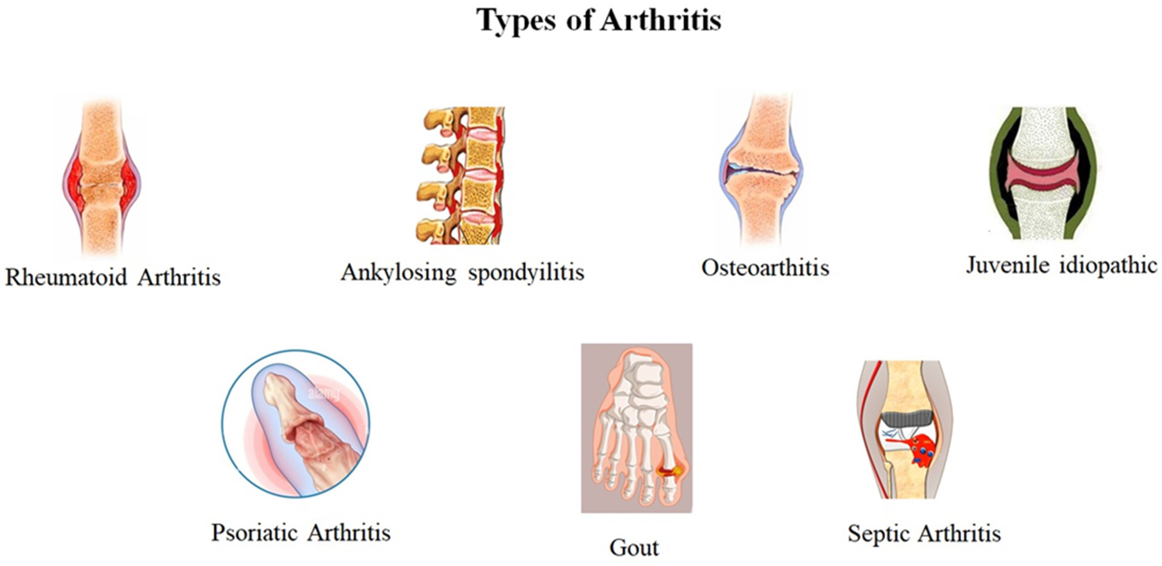
Arthritis is one of the most deceptive diseases globally, with 350 million individuals are currently affected. As per a recent report, one in four adults in the USA suffer from arthritis with severe joint pain [2]. Arthritis leads to the breakdown of cartilage which normally protects joints. Arthritis produces an inflammatory riposte as well as hyperplasia of synovial cells. Consequently, extra deposition of synovial fluid in the joints develops the sheets in the synovial cells that cause inflammation at joint sites. The pathology of the disease process often indicates that it also damages the articular cartilage and alkalosis of the joints [3]. Ankylosing spondylitis, juvenile idiopathic arthritis, reactive arthritis, psoriatic arthritis, rheumatoid arthritis, septic arthritis, osteoarthritis, and gout are the commonly reported types of arthritis (Figure 1).
Osteoarthritis (OA) is the commonest form of arthritis, affecting approximately 302 million individuals globally. The most affected areas by OA are the appendicular joints of the knees, hips, and hands [4]. Rheumatoid arthritis (RA) is another type of arthritis affecting the synovial joints and normally produces symmetrical arthritis, leads to considerable socioeconomic impact. RA is one of the most prevalent diseases, affecting approximately 0.5–1% of the world’s population. The cause of RA is not certain, but researchers believe that autoimmunity is the major cause The early detection of RA with timely treatment relieves symptoms arising from the RA condition [5]. Non-steroidal anti-inflammatory drugs (NSAIDs), including naproxen and aspirin, with rapid onset of action, corticosteroids (e.g., cortisone, dexamethasone etc.), biological agents (e.g., etanercept and infliximab), and disease modifying anti-rheumatic drugs (DMARDs) (e.g., methotrexate, sulfasalazine, leflunomide), either alone or in combination, are the most commonly used treatment strategies for arthritis [6]. DMARDs target the immune system, and thus they can also weaken the immune system’s ability to fight infections. Furthermore, higher cost and negative impacts on health have limited the use of synthetic drugs in arthritic treatment. Of these synthetic medicines, herbal medicines are also gaining popularity for arthritis treatment, due to fewer side effects.
2. Herbal Antiarthritic Drugs
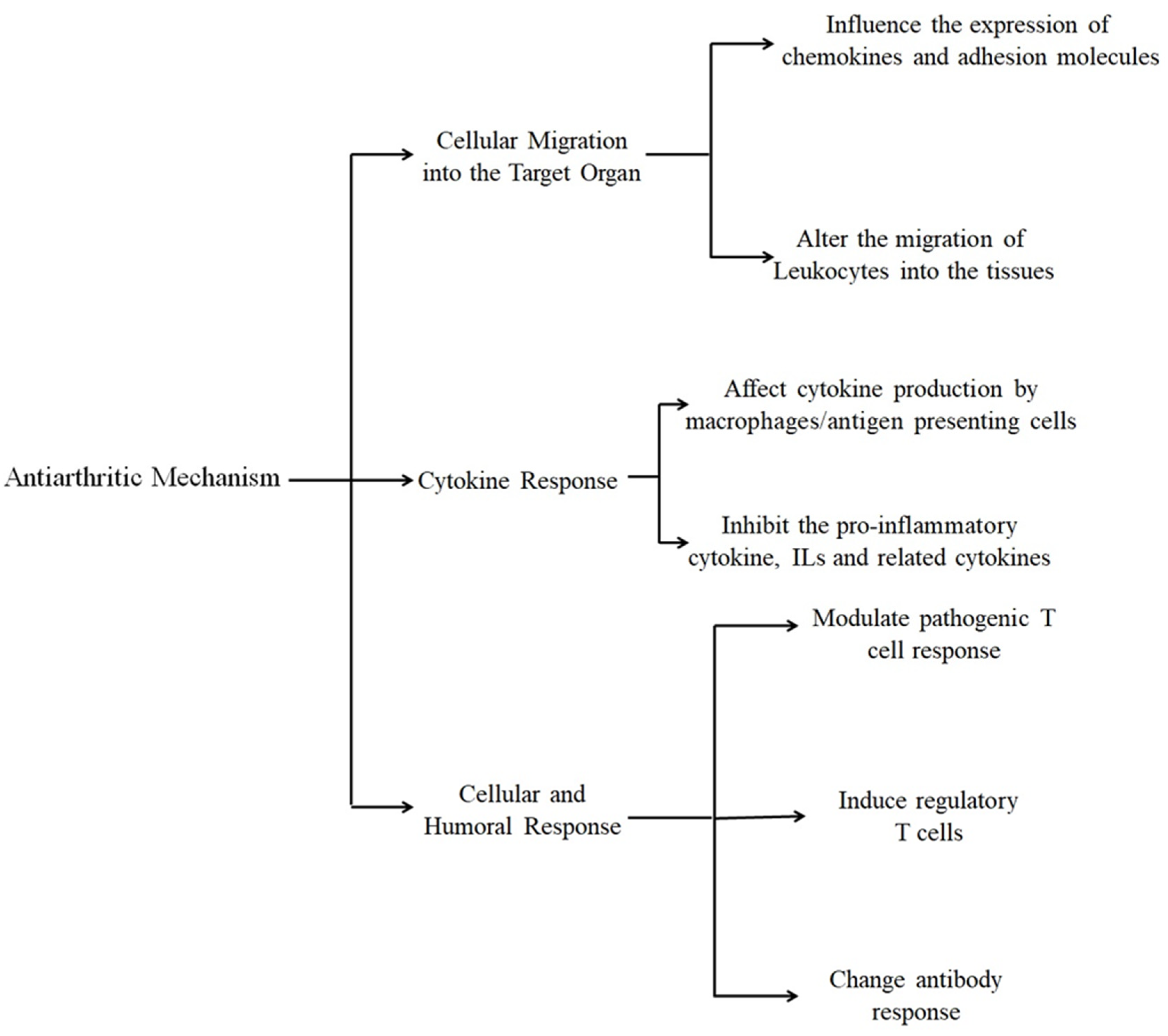
Herbal products have been widely used as medicine since ancient eras. These natural products have broad chemical diversity, pharmacological specificity, and molecular properties that make them potential candidates for lead structure identification [7,8]. Thousands of plant isolates possessing antiarthritic (AA) properties have been investigated and reported [9]. These plant isolates have been categorized into alkaloids, glycosides, terpenoids, flavonoids, etc. [10] In recent years, herbal products showing anti-inflammatory-mediated AA properties have been isolated [11,12]. These plants have been used either solely, or their extracts or isolates have been used for the treatment of RA or OA. Plant isolate is a pure compound obtained from a plant extract, having a defined structure which is responsible for particular biological activity, and helps to develop new potent compounds. Table 1 represents numerous plant isolates with their structure and IUPAC names. These plant isolates act through different mechanisms, which are summarized in Figure 2.
2.1. Alkaloids
2.1.1. Montanine
Plants belonging to the Amaryllidaceae family have a long history of usage globally, and are found to be a promising therapeutic tool for several human diseases. The plants belonging to this family have long been used as an alternative medicine in developing countries. The Amaryllidaceae alkaloids are secondary metabolites (alkaloids) of the Amaryllidaceae family, native to Argentina, Brazil and Uruguay [13]. Montanine has structural similarities to Amaryllidaceae alkaloids, and its pleiotropic pharmacologic activity raises the possibility of montanine possessing anti-arthritic properties [14].
Recently, montanine has received the considerable attention due to its strong anti-inflammatory action, which was isolated from the bulb of the plant Rhodophiala bifida (Herb.) through maceration in sulfuric acid 2% (v/v) [15]. The authors reported its significant AA activity by using in vitro effects on lymphocyte proliferation and on invasiveness of fibroblast-like synoviocytes (FLS). Later, the activity of isolate was evaluated on antigen-induced arthritis (AIA) Balb/c mice and collagen-induced arthritis (CIA) DBA/1J mice models. Study results revealed that montanine administration decreased nociception and leukocyte articular migration in the AIA model, and reduced the severity of arthritis and joint damage in CIA model. Histological results revealed considerable improvements in arthritis. The authors proposed that the inhibition of lymphocyte proliferation and decreased FLS invasion was responsible for AA activity. A median lethal dose (LD50) of montanine was reported to be 64.7 mg/kg for male mice, and the occurrence of side effects as altered motor activity, decreased respiratory rate, violent body tremors, and clonic convulsions.
2.1.2. 3-Acetylaconitine
3-Acetylaconitine (AAc) is a nitrogen-containing alkaloid, obtained from Aconitum flavum and Aconitum pendulum (Ranunculaceae). Tang et al. isolated AAc from the root of Aconitum flavum, and reported its AA activity in mouse and rat models [16]. An oral dose of 0.3–0.5 mg/kg of AAc impeded swelling of the hind paw in the formaldehyde-induced rat model, and inhibited the carrageenan-induced edema in the adrenalectomized rat model. Although AAc inhibited acetic acid and histamine-induced vascular permeability, it did not reduce the ascorbic acid content of the adrenal in rats, indicating that AAc did not act through stimulation of the pituitary adrenal axis.
2.1.3. Sanguinarine
Sanguinarine (SA) is a natural plant benzylisoquinoline alkaloid isolated from Argemone mexicana, Bocconia frutescens, Bocconia frutescens, Chelidonium majus, Macleaya cordata, and Sanguinaria Canadensis. SA is U.S.A Food and Drug Administration (FDA) approved; it inhibits osteoclast formation, and is recommended for inflammation [17]. Ma et al. isolated SA from the roots of Sanguinaria Canadensis, and investigated the therapeutic effect of SA against OA [18]. Results revealed that SA suppressed catabolic proteases expression in in vitro, in vivo, and ex vivo models. SA suppressed NF-κB and JNK activation, which presented a high level of specificity in repressing the production of catabolic factors. Additionally, SA also inhibited IL-1β-induced expression of matrix metalloproteinase (MMPs) 1, 3, and 13. It also suppressed a metalloproteinase and disintegrin with thrombospondin motifs-5 in chondrocytes. These results supported the potential application of SA in OA treatment.
2.1.4. Jatrorrhizine
Jatrorrhizine hydrochloride (JH) is a protoberberine alkaloid reported in many medicinal plants, including Berberis aristata and Coptis chinensis [19]. Qiu and colleagues recently investigated and reported the AA potential of commercially available isolate JH in a CIA rat model [20]. The results revealed the suppression of RA in the CIA rat model via an anti-inflammation action, and suppression of bone destruction. Furthermore, the in vitro assay showed inhibition of production of inflammatory mediators, and inhibition of proliferation and migration in MH7A cells. JH was found to suppress tumor necrosis factor-α (TNF-α)-stimulated activation of nuclear factor kappa B (NF-κB) and mitogen-activated protein kinases (MAPKs), leading to suppression of proinflammatory mediators. These results suggested JH as potential compound for AA treatment.
2.1.5. Piperine
Piperine is another alkaloid obtained from black pepper (Piper nigrum L.), responsible for the pungent taste, and found in the members of the Piperaceae family. Piper nigrum L. contains the highest amount of piperine, i.e., from 2% to 9%. Piper nigrum L. has been well reported in Ayurvedic and Chinese medicine [21]. Bang and colleagues in 2009 reported anti-inflammatory, nociceptive, and AA activities of piperine [22]. The AA activity was measured in a CI arthritis model in vivo by measuring the paw volume and weight distribution ratio. The results showed significant reduction of paw volume and weight distribution ratio. The authors also evaluated the levels of IL6, MMPs, COX-2, and PGE2 by ELISA and RT-PCR. An oral dose of piperine between 20 and 100 mg/kg/day for 8 days, inhibited the IL6 and MMP13 expression and PGE2 production. Surprisingly, piperine did not inhibit the expression of NFκB, but did suppress the migration of activator protein 1 (AP-1). Ultimately, on the fourth day, piperine reduced arthritic symptoms. These results suggested the potential of piperine in arthritis treatment.
2.1.6. Capsaicin
Capsaicin is an active component of chili peppers (genus Capsicum), and is produced as a secondary metabolite. It is a chemical irritant for mammals, including humans [23]. Ahmed and colleagues investigated capsaicin effects on substrate P (SP) and calcitonin gene-related peptide (CGRP) in the ankle joints and dorsal root ganglia (L2–L6) of adult female Lewis rats [24]. Subcutaneous injection of capsaicin in a dose of 200 mg/kg significantly reduced the level of substrate P (19%) and CGRP (42 %) in dorsal root ganglia of adjuvant-induced arthritic rats. In the ankle joint, capsaicin reduced the SP level by 40%, accompanied by a 40% reduction in inflammatory response. Furthermore, the capsaicin administration reduced the up-regulated levels of sensory neuropeptides in dorsal root ganglia and ankle joints in adjuvant-induced arthritis rats. These findings suggested that capsaicin is useful in arthritis treatment.
2.1.7. Tubastrine
The alkaloid tubastrine, obtained from the marine organism Aplidium orthium (Ascidiacea), possesses anti-inflammatory properties [25]. Tubastrine isolated from the frozen specimen of Aplidium orthium with methanolic acid, followed by chloroform, reduced superoxide synthesis in phorbol-12-myristate 13-acetate (PMA)-stimulated neutrophils in vitro and, in an in vivo study, reduced superoxide levels in a gouty arthritis model [26]. Additionally, tubastrine further showed an inhibitory effect on neutrophil infiltration in an in vivo model.
2.1.8. Orthidines
The orthidines (A–F) are a group of marine alkaloids isolated from the same ascidian Aplidium orthium. Orthidines (A–D) are benzodioxane, orthidine E (a cyclobutane dimer of tubastrine), and orthidine F (a biosynthetically unrelated dihomovanillamide derivative of spermine). Pearce and colleagues isolated orthidines (A–F) from the frozen specimen of New Zealand ascidian Aplidium orthium with methanolic acid, followed by chloroform, and evaluated anti-inflammatory and anti-arthritic activity in a gouty arthritis model [26]. Isolated orthidines (A–F) showed the in vitro production of superoxide by PMA-stimulated human neutrophils in a dose-dependent manner with IC50s of 10–36 µM, and this was associated within the inhibition of superoxide production by neutrophils in vivo in a murine model of gouty inflammation.
2.2. Terpenoids
Terpenoids are plant secondary metabolites, extracted from various parts of the plant, such as stalks, fruits, flowers, leaves, and roots. They are colorless liquids with a pleasant smell, and have a high refractive index [27,28]. The pharmaceutical importance of terpenoids has been proved and well documented in its anti-inflammatory, antibacterial, antiviral, antioxidant, and anti-carcinogenic properties [29]. Recently, Carvalho et al., identified and reported 24 terpenoids which were effective in the treatment of inflammation and arthritis [30].
2.2.1. Eugenol
Eugenol is a major phenolic component obtained from the clove bud (Eugenia caryophyllata), and constitutes 80–90% of clove bud oil. Sharma et al. first reported the suppressive effects of eugenol on arthritic symptoms [31]. A study was further carried out by Grespan et al. to estimate the AA activity of eugenol in a CIA mouse model [32]. The arthritic symptoms were induced with 100 µg of bovine collagen type II (CII) in male DBA1/J mice, and treated with orally administered eugenol (100 µg/mouse) from day 25 to day 40. Eugenol administration significantly decreased the levels of cytokines (i.e., TNF-α, tumor growth factor (TGF)-β, and interferon (IFN)-γ) within the ankle joints. Furthermore, the results indicated that eugenol also inhibited mononuclear cell infiltration into the knee joints of arthritic mice.
2.2.2. Nimbolide
Nimbolide is a triterpene, which is isolated from the leaves and flowers of the neem plant (Azadirachta indica), and has been widely used in treating numerous human ailments. Several bioactive compounds have been isolated from this plant species which exhibit multiple pharmacological effects. Cui et al. performed the AA activity of nimbolide on male albino rats against Freund’s adjuvant-induced arthritis [33]. A study was carried out to access the AA activities of nimbolide using different in vitro and in vivo analytical methods. AA activity of nimbolide (at a dose of 20 mg/kg per day, which was given orally) exhibited a noticeable reduction in edema formation, paw volume, organ indices, and arthritic score, along with considerable improvement in body weight. Histopathological studies revealed the protecting effects of nimbolide towards joints and inflammation. The outcomes of the study showed that nimbolide treatment inhibited inflammation by decreasing the proinflammatory cytokines (i.e., TNF-a, IL-6, IL-1b, and IL-10) manifestation in arthritic rats. Furthermore, nimbolide normalized the increased levels of iNOS, P-IkBa, Nf-kb, cox-2, and IKKa in treated rats.
2.2.3. Bartogenic Acid
Bartogenic acid (BA) is isolated from the fruits of the Barringtonia racemosa Roxb. (Lecythidaceae) plant [34]. A study was performed by Patil et al. in order to evaluate the AA activity of BA [35]. BA was isolated from the methanolic extract of fruits of Barringtonia racemosa. The in vivo results revealed noteworthy AA activity of BA against CFA-induced arthritis in rats by reducing serum markers, such as rheumatoid factor and C-reactive protein. BA protected against primary and secondary arthritis lesions with a dose of 2, 5, and 10 mg kg−1 day−1. It also normalized the raised WBC counts and increased hemoglobin counts, and reduced erythrocyte sedimentation rate in arthritic conditions. The possible mechanism to improve Hb count by BA was due to increased response of the bone marrow erythropoietin. BA also protected the rats from CFA-induced radiographic changes.
2.2.4. Cannabidiol
Cannabidiol (CBD) are meroterpenoids, terpenophenolic compounds which are isolated from the plant Cannabis sativa L., belonging to the Cannabaceae family, and cultivated worldwide. This plant contains a number of phytoconstituents including amides, amines, phytosterols, phenolic compounds, carbohydrates, terpenes, and fatty acids and their esters, along with CBD as main active constitute [36].
Several reports have clarified that CBD showed anti-inflammatory activity by inhibiting proliferative responses of T lymphocytes, nitric oxide (NO) production by macrophages, and suppression of macrophage function and antigen presentation. Malfait et al. demonstrated the AA therapeutic potential of cannabidiol in murine collagen-induced arthritis [37]. Arthritis was developed by bovine type II collagen (CII), and immunized completely against Freund’s adjuvant (CFA) in DBA/1 mice at the dose of 100 µg. Cannabidiol was found to be equipotent in both models (chronic relapsing CIA and in acute CIA), and the optimal dose of cannabidiol was found to be 5 mg/kg i.p. per day, or 25 mg/kg orally per day. Joints were protected against severe damage, and showed significant IFN-γ production and diminished CII-specific proliferation. Ex vivo results showed a decrease in TNF-α release and diminished CII-specific proliferation and IFN-g production by knee synovial cells in CBD-treated mice. A dose-dependent suppression of lymphocyte proliferation was also observed by CBD in in vitro studies. Additionally, cannabidiol suppressed the lipopolysaccharide-increased serum TNF level in C57/BL mice. These combined data showed immunosuppressive and anti-inflammatory actions which mediated the AA effect of CBD on CIA. A patent has also been filed describing the identification and use of CBD to treat inflammatory diseases [38].
2.2.5. Curcumin
Curcumin is the most active phytocomponent of Curcuma longa Linn, which belongs to the Zingiberaceae family, and is commonly cultivated in the region of south Asia. Curcuma longa is widely used in traditional Indian Ayurveda medicine as a popular home remedy, and its paste is applied with slaked lime for the treatment of inflammation and wounds [39]. Curcumin has received much interest in the scientific world due to its excellent pharmacological activities, which have been shown to target multiple signaling molecules at the cellular level. Curcumin has been known to possess AA effects in humans with OA and RA [40].
Huang et al. evaluated the anti-RA effect of curcumin in a CIA-induced DBA/1J mice model. A 50 mg/kg of curcumin was injected i.p. in the mice model, and the B cell-activating factor belonging to the TNF family (BAFF), IL6 and IFNγ production in serum were measured. Results revealed a decreased production of BAFF, IFNγ and IL-6 in serum. Furthermore, western blot analysis was also performed to measure IFNγ-related signal transducers and activators of transcription 1 (STAT1) signaling in B lymphocytes, which showed suppressed IFNγ-induced BAFF expression, STAT1 phosphorylation, and nuclear translocation after curcumin treatment [41]. Kuncha and colleagues reported the potentiate effect of curcumin with low dose of prednisolone against CFA-induced arthritis in a rat model [42].
An interesting study was performed by Yu et al., demonstrating the anti-neuroinflammatory response of curcumin in lipoteichoic acid (LTA)-stimulated BV-2 microglial cells [43]. Results revealed that curcumin inhibited the secretion of inflammatory cytokine NO and TNF-α, prostaglandin E2 (PGE2), and also inhibited COX-2 and iNOS expression. Additionally, curcumin also suppressed LTA-induced phosphorylation of MAPK expression. In LTA-stimulated microglial cells, curcumin inhibited hemeoxygenase-1(HO-1), which reversed the inflammatory mediator release, and produced their effect against neurodegenerative disorder neuroinflammation.
2.3. Flavonoids
Flavonoids are polyphenolic compounds isolated from plants and found in grains, fruits, flowers, vegetables, bark, stems, and roots. Flavonoids have been shown to possess anti-inflammatory properties, and these plant products have been widely used traditionally in the treatment of arthritis [44].
2.3.1. Quercetin or 3,5,7,3′,4′-Pentahydroxy Flavone
Quercetin (QTN) is a flavonoid obtained from apples, buckwheat, onions, and citrus fruits. Recently, Yuan et al. investigated and reported the mechanism of AA activity of QTN [45]. QTN significantly reduced ankle diameter and arthritic scores in adjuvant-induced arthritis (A42A) in a mouse model. The study revealed that QTN endorsed apoptosis of activated neutrophils, and inhibited neutrophil infiltration. Additionally, QTN inhibited ROS-mediated neutrophil extracellular traps (NETs) formation and autophagy. These findings suggested that QTN may be a potential agent for RA treatment by inhibiting neutrophil activities. QTN (30 mg/kg) oral administration showed a decrease in clinical sign of arthritis in a chronic rat (AA) model [46]. Gardi et al. showed a decrease in IL-1b level, monocyte chemotactic protein-1 (MCP-1) level, and also restored plasma antioxidant capacity in rat adjuvant arthritis after oral administration of QTN (150 mg/kg) [47].
Gaikwad et al. reported anti-inflammatory activity of ethanol extract of flowers of madhuca indica against formaldehyde-induced inflammation, carrageenan-induced inflammation, and cotton pellet granuloma in models of rats. Results revealed a superior dose-dependent anti-inflammatory action of ethanolic extract of m. indica, as compared to the reference drug diclofenac sodium in a formaldehyde-induced inflammation model [48]. The study was further extended by Tang and colleagues in 2021, who isolated QTN from methanolic leaves extract of madhuca indica, and evaluated for AA activity against FCA-induced arthritis in female rats (strain: Wistar). The in vivo results demonstrated a significant decrease in paw volume, joint diameter, and paw withdrawal threshold after QTN (10 and 20 mg/kg) treatment. The AA activity of QTN revealed reduced elevated inflammatory release (Ikβα, P2X&, COX-2 and NF-κβ), oxido-nitrosative stress, and pro-inflammatory cytokines (TNF-α and ILs) in experimental rats [49].
QTN was more effective alone than methotrexate, or in combination with methotrexate, to reduce joint inflammation in mice, and provided the highest protection against arthritis. The mechanisms included reduction of TNF-α, IL-1β, IL-17, and monocyte chemoattractant protein-1 (MCP-1) levels [50].
2.3.2. Resveratrol
Resveratrol (Res), a natural flavone, is widely present in medicinal plants including grape, cranberry, mulberry, pistachio, and peanut. The AA activity of Res was evaluated against a CFA-induced rat model by Chen and colleagues in 2013. Res was showed to inhibit the mRNA expression of IL-1β and TNF-α and, ultimately, IL-1β and TNF-α level after given through intragastric gavage (i.g. 10 mL/kg/day). Res stimulated synoviocytes, and the protein expression levels of p-ERK1/2 via protein kinase C (PKC) [51].
It has been reported that resveratrol inhibits the enzymatic activity of COX-1 and COX-2. Chen et al. investigated the AA effect of resveratrol on CFA-induced arthritis in a rat model [52]. The results revealed a significant paw swelling reduction with decreased arthritis scores (10 or 50 mg/kg, i.g.). Additionally, resveratrol suppressed the production of COX-2 and PGE2 inflammatory mediators. The study further revealed the histopathology improvement by resveratrol in AA rats.
Co-administration of Res and piperine significantly decreased the paw swelling and ameliorated the histopathological changes. The combined treatment highly reduced the serum TNF-a, IL-1b, thiobarbituric acid reactive substances (TBARS), and nitrate/nitrite (NOx). Moreover, a nearly negative expression of NF-κB p65 in the synovial tissue was observed by co-administration of piperine with Res. Results of the combination treatment were comparable to that of diclofenac treatment [53].
2.3.3. Kaempferol
Kaempferol (KAE), a natural flavanol, chemically known as 3,4′,5,7-tetrahydroxyflavone, is found in numbers of edible plants such as beans, tea, kale, broccoli, and spinach. KAE is used as a traditional medicine for numerous inflammatory disorders. Studies revealed that KAE reduces COX-2 levels in RAW 264.7 cells, and inhibits ROS production via inhibition of iNOS and TNF-α protein expression. KAE also inhibits IL-4, C-reactive protein (CRP) expression and NF-κB in liver cells [54,55]. Yoon et al. reported that KAE produces AA activity by inhibiting the proliferation of both unstimulated and IL-1β-stimulated RASFs, in addition to the mRNA and protein expression of MMP-1, MMP-3, PGE2, and COX-2 induced by IL-1β [56].
2.3.4. Chebulanin
Chebulanin is a natural polyphenolic compound isolated from the fruits of Terminalia chebula retzius (TC). Terminalia chebula retzius (TC) is widely used in medicine in Asian countries for its anti-microbial, anti-inflammatory, antioxidant, and AA properties. Zhao and colleagues investigated the chebulanin function as an AA agent in a CIA-animal model using DBA/1 mice [57]. Chebulanin was isolated from dry fruits of Terminalia chebula retzius with a 70% acetone solution (1:10, w/v) at room temperature (23 ± 2 °C). The authors measured the expression of inflammatory cytokines by immunohistochemical staining, and also performed a histopathological evaluation of the joints. Micro-CT was also performed to detect bone destruction and erosion. The results of above studies revealed the improved dose-dependent (oral dose of 40 mg/kg, 80 mg/kg or 160 mg/kg daily for 28 days) expression of IL-6, TNF-α, MMP-3, and COX-2 in joints and, ultimately, severity of arthritis. Additionally, histopathological studies revealed tissue improvement. Micro-CT results showed the dose-dependent reduction in cartilage destruction and bone erosion. These results confirm the potential role of chebulanin as a strong therapeutic agent for the treatment of RA. Recently, Liu et al. also confirmed the AA activity of chebulanin via inhibiting NF-κB and MAPK activation in a collagen-induced arthritis (CIA)mouse model [58]. Chebulanin significantly decreased the arthritic scores, paw swelling and IL-6 and TNF-α level in mice after being orally gavaged (80 mg/kg) daily for a total of 21 days. Moreover, chebulanin reduced the levels of excised phosphorylated (p)-p38, c-JUN, p-p65, N-terminal kinase (p-JNK), and phosphorylated NF-κB inhibitor alpha (p-IκBα), but did not alter extracellular-signal regulated kinase, which is implicated in many pathological conditions, including arthritis.
2.3.5. Ellagic Acid
Ellagic acid (EA) is a polyphenol bioactive compound richly existing in berries (strew berry, raspberry, and cloudberry), almonds, grapes, walnuts, and pomegranates [59,60]. Shruthi et al. isolated the ellagic acid from the methanol leaf extract of the plant Kirganelia reticulate, and tested its AA activity via in vitro, in vivo, and in silico assays [61]. The in vitro assay of EA showed maximum percentage inhibition of protein denaturation, membrane stabilization, and proteinase inhibitory action, which were observed at 250 µg/mL. The in vivo studies of EA against the formaldehyde-induced paw edema showed inhibition of cytokines and leukotriene infiltration, reduced paw edema volume, protected synovial membranes, and cartilage damage at both 100 µg/mL and 250 µg/mL concentration. The possible proposed mechanism was inhibition of hypoxia-inducible factor (HIF-2α). Other parameters including body weight, paw edema volume, and the movements of rats, were also studied, which showed a protective effect of EA similar to standard aspirin. The in silico study of EA revealed that it forms four hydrogen bonds with amino acid residues in the active pocket of Hypoxia-inducible factor (HIF-2α). The EA completely enfolded in the entire active pocket of HIF-2α, as compared to aspirin, and inhibited the activity of HIF-2α protein, thereby reducing remarkable anti-arthritic activity. The acute oral toxicity study of EA was also performed in albino rats via the OECD Organization of Economic Co-operation and Development guidelines (OECD No. 423). The results of toxicity studies revealed absence of any toxic effect up to 2500 mg/kg body weight.
Umar et al. investigated the combinatory effect of methotrexate and EA on the CIA-Wistar rat method. CIA rats were treated with solely methotrexate (1 mg/kg/week) and EA (60 mg/kg) daily, and the combination of methotrexate and EA for a period of 28 days [62]. Results revealed that the combination of methotrexate and EA potentiated the antiarthritic (decrease of hind paw volume and scoring) and the antioxidant effect (GSH and catalase), as well as suppression of lipid peroxidation. Combination therapy of methotrexate and EA significantly inhibited the development phase of arthritis, which is supported by histopathological and attenuation of pro inflammatory cytokines.
2.3.6. Rosmarinic Acid
Rosmarinic acid is a polyphenol, present in a number of herbs, including rosemary (Rosmarinus officinalis L.), mint (Mentha arvense L.), sage (Salvia officinalis L.), and basil (Ocimum basilicum L.). In 1958, it was first isolated and characterized by the Italian chemist Scarpatti from rosemary (Rosmarinus officinalis), and hence given its name rosmarinic acid [63]. Recently, Wei et al. isolated rosmarinic acid from Rosmarinus officinalis methanolic leaf extract, and reported its AA activity against CFA-induced arthritis in rats [64]. Oral administration of rosmarinic acid at a dose of 30 and 60 mg/kg exerts significant reduction in hind paw volume and various other arthritic symptoms, i.e., inflammation and joint stiffness. Rats treated with rosmarinic acid in a dose of 60 mg/kg, i.p improved locomotor movement from days 21 to 35. Additionally, rosmarinic acid also decreased TNF-α levels in serum in a dose-dependent manner in treated rats. Rosmarinic acid decreased the levels of inflammatory mediators TNF-α in treated animals. On the basis of these outcomes, the authors suggested rosmarinic acid as an encouraging candidate for the treatment of arthritis. Acute toxicity study of rosmarinic acid (according to OECD No. 423 guidelines) showed no sign of toxicity or onset of toxicity up to the dose of 2000 mg/kg body weight of albino rats.
2.3.7. Gallic Acid
Gallic acid (GA) is a natural polyphenol, present in gall nuts, oak bark, apple peels, sumac, grapes, tea leaves, and green tea. Anti-inflammatory, anti-microbial, anti-tumor, and pro-apoptotic activities of GA have been well documented. Shi et al. reported that tumor-like FLSs cells could migrate to cartilage and bone, producing pannus and accelerate the secretion of pro-inflammatory cytokines, chemokines, and MMPs [65]. GA has been found to inhibit cytokines, chemokines, and MMPs. Yoon et al. reported that GA triggers the apoptosis of FLSs cells in ≥10 µM concentration, and regulates the production of Bcl-2, Bax, p53, and pAkt in western blot analyses, and in the mRNA expressions analysis. Furthermore, GA also showed dose-dependent suppression of cytokines (IL-1, IL-6), chemokines (CCL-2/MCP-1, CCL-7/MCP3), COX-2, and MMPs-9 from RA FLS [66]. GA regulated apoptosis related protein expressions (i.e., fibroblast-like synoviocytes from patients with rheumatoid arthritis; RA FLS), and reduced the expression of pro-inflammatory genes in RA FLS. On the basis of these outcomes, the authors suggested that GA utilizes pro-apoptotic and anti-inflammatory therapeutic options for RA treatment. The acute toxicity of GA showed LD50 values greater than 2000 mg/kg in albino mice. However, at a higher dose (900 mg/kg) of GA up to 28 days did not alter the behavior, morphology, or histopathological parameters in mice.
2.3.8. Chlorogenic Acid
Chlorogenic Acid (CGA) is the most abundant phenolic acid, naturally occurring in tea and green coffee extracts. CGA. Chauhan and his team investigated the AA activities of CGA in an adjuvant induced-arthritis model using male Wistar rats [67]. At a dose of 40mg/kg, CGA controlled both total (CD3) and differentiated (CD4 and CD8) T-cell count, and effectively suppressed CD80/86, compared to ibuprofen. Flow cytometry analysis results demonstrate suppression of Th1 cytokines and elevation of Th2 cytokines by CGA. Fu and colleagues reported that CGA obstructed arthritis progression, and inhibited BAFF and TNF-α production in serum in a CIA-mice model [68]. Mechanistically, CGA showed constraints of TNF-α-induced BAFF expression, and reduced the DNA-binding activity of NF-κB to the BAFF promoter region in MH7A cells. These results suggested the potential of CGA as AA agent. Group of animals treated with CGA showed no abnormal behavior or mortality up to the dose of 2000 mg/kg, which showed the safety of CGA, even at high doses.
2.3.9. Ferulic Acid
Ferulic acid (FA) is the most common compound, present in numerous plants, particularly in grains, including rice and corn. FA exhibits free radical scavenger activity, and increases the expression of antioxidant proteins via the activation of NF-kB and COX-2, as well as inhibiting iNOS [69]. These properties of FA encouraged Zhu and colleagues to investigate FA effects in the treatment of CFA-induced arthritis in rats [70]. The authors also investigated whether the effect, if present, is due to the inhibition of the JAK/ STAT pathway or not. Results revealed that at an oral dose of 25 and 50 mg/kg of FA administered to CFA-induced arthritic rats showed significant reduction in arthritic index, ESR levels, and the percentage of lymphocytes. Both rheumatoid factor (RF) and C-reactive protein (CRP) are also reversed after FA treatment, and normalized the physiological condition. FA treatment also reduces the level of TNF-α, JAK2 levels, TGF-β, STAT-3, and STAT-4 levels. These findings suggested the potential use of FA in arthritic treatment by inhibition of the JAK pathway. Oral administration of FA showed low toxicity with an acute LD50 value of 3200 mg/kg in mice, while LD50 values were 2445 mg/kg and 2113 mg/kg for male and female rats, respectively.
2.3.10. Brazilin
Brazilin is a naturally occurring active compound (red dye) obtained from the wood of various plants such as Caesalpinia violacea, Haematoxylum brasiletto, Brazilin Paubrasilia echinata and heartwood of Caesalpinia sappan [71]. Jung et al. isolated brazilin from ethyl acetate extract of the heartwood plant Caesalpinia sappan L., and investigated its AA activity. HPLC was utilized to purify isolated brazilin, and its confirmation was performed using mass spectrometry and 1H/13C NMR analysis [72]. A type-II CIA mouse model was utilized for the determination of brazilin anti-rheumatoid activity. A 10 mg/kg body weight of purified brazilin and a 3 mg/kg body weight of methotrexate were administered intraperitoneally, and pro-inflammatory cytokines and stress enzyme markers were monitored. Results revealed a significant reduction in inflammatory cytokines, acute inflammatory paw edema, and arthritis index score in CIA-mice model. Additionally, the bone mineral density was improved adequately with both brazilin and methotrexate administration. The microstructural examinations showed joint prevention, bone formation, and prevention of surface erosion after brazilin administration. These results showed protective efficacy of brazilin in a CIA mouse model, and suggested that it would be useful to treat rheumatoid arthritis.
2.4. Plant Sterols
Beta-Sitosterol
Beta-sitosterol is a “plant sterol ester” found in fruits, vegetables, nuts, and seeds. It is commonly used for lowering cholesterol levels and improving symptoms of an enlarged prostate [73]. Liu et al. evaluated the effects of β-sitosterol of immune-regulation on macrophages and its potential role in rheumatoid arthritis (RA) [74]. β-sitosterol in a dose of 20 or 50 mg/kg i.p. boosts immunization of CIA in mice models. β-sitosterol-treated M1-polarized bone marrow-derived macrophages (BMDMs) reduced expression of CD86, IL-1b, iNOS, and MHCII by 87.1, 47.1, 50.2, and 31.3%, respectively. In CIA mice, β-sitosterol inhibited the production of proinflammatory cytokines, and reduced the levels of collagen-specific antibodies (IgG and IgG1, but not IgG2c). These results suggested the potential application of β-sitosterol in RA therapy.
3. Nano-Formulation of Isolated Compounds
Nanotechnology has been widely used for the treatment of severe diseases, with the objective of increasing efficacy and safety of active ingredients [75,76]. Nanocarriers have numerous advantages, including controlled and site-specific drug delivery, which minimizes unwanted side effects [77,78]. Nanocarriers have also gained much attention for the delivery of plant extracts and isolates. Several reports demonstrate the increment in AA activity of herbal extracts through utilizing nanotechnology by increasing their bioavailability. The details of plant isolates with their nanoformulation, method of preparation, and average particle size is given in Table 2.
Piperine-loaded solid lipid nanoparticle (SLN) were prepared through the melt emulsification method, and evaluated for particle size (128.80 nm), entrapment efficiency (78.71%), and zeta potential (−23.34 Mv). The prepared SLN was administered orally and topically to CFA-induced arthritic rats. An ex vivo study using Franz diffusion cells indicates that piperine from the SLN gel formulation accumulates in the skin. TNF α was significantly decreased by piperine-SLN, as compared to the arthritic control group, which may be attributed to the selective accumulation of piperine-SLN in inflamed sites thus reducing the secretion to TNF α from the activated macrophages. Histopathology studies revealed that piperine-SLN showed minimal infiltration of inflammatory cells and connective tissue proliferation, as compared to a control group, which showed moderate infiltration of inflammatory cells and connective tissue proliferation [79].
Sarwa and colleagues, in 2013, prepared and reported capsaicin-loaded transfersomal vesicular system for topical application in experimental arthritic rats [80]. Capsaicin-loaded transferosomes were prepared through the conventional thin film hydration method, and evaluated for numerous physical properties including morphology, size and size distribution, zeta potential, flexibility, and viscosity. The prepared transferosomes were nano sized (94 nm), with sufficient structural flexibility and negative surface charge (−14.5 mV). Furthermore, capsaicin-loaded transferosomes were compared to marketed Thermagel (standard) gel for antiarthritic activity. The results revealed enhanced permeability of transferosome formulations as compared to the marketed Thermagel formulation, and thus led to improved therapeutic concentration of the capsaicin at the desired site, ultimately improving the antiarthritic and anti-inflammatory activity of transferosome formulations over Thermagel formulation.
Jabbari and colleagues investigated the effects of encapsulated eugenol with chitosan nanoparticles on a neonatal CIA-rat model [81]. Eugenol-entrapped chitosan nanoparticles (Eug-CNPs) were prepared through the solvent dispersion method, and evaluated for anti-oxidative stress (malondialdehyde for assessment of lipid peroxidation) by an assay kit, FOXO3 protein as an antioxidant up-regulating by western blotting, and expression of the TGF-β and CCL2/MCP-1 genes by real-time PCR evaluation, supported by a cartilage histopathology analysis. Results revealed that Eug-CNPs significantly decreased the serum level of malondialdehyde and FOXO3 protein expression, in comparison to the control group. Additionally, Eug-CNPs decreased the expression of TGF-β and MCP-1 genes, and a significant positive correlation was observed between MCP-1 and TGF-β. Eug-CNPs also alleviated the symptoms of joint inflammation, synovial hyperplasia, and cartilage damage caused by the RA.
QTN was entrapped in cadmium telluride quantum dots (TGA-CdTe QDs) for enhancing its AA activity against adjuvant-induced arthritic in Wistar rats. Treatment with QTN-entrapped TGA-CdTe QDs (QDs-QE) reduced the expressions of lipid peroxidation, and improved antioxidant enzymes activity superoxide dismutase (SOD). Prepared QDs reduced the level of catalase (CAT), glutathione (GSH), and glutathione peroxidase (GPx) in paw tissue. Histopathology studies revealed the cartilage regeneration in arthritis-induced rats after QDs-QE treatment. These outcomes revealed QDs-QE have the potential ability to treat arthritis in rheumatic complications [82]. The QDs-QE complex showed improved AA activity at low concentrations via free radical quenching of QDs-QE by antioxidant enzymes, while QE showed AA potential only at the higher concentration. Furthermore, QDs, as nanocarrier of QE, exhibited enhanced AA effect even at a lower concentration of the drug. These findings suggested the utilization of QDs of QE as a nanocarrier to enhance the potential of QE for AA activity. Furthermore, Gokhale et al., developed QCT loaded nano-emulsion (NE) gel using spontaneous emulsification techniques for effective RA management [83]. The effect of QCT-NE on the production of inflammatory cytokines TNF-α was investigated on RAW264.7 cells. Results showed the significant inhibition of TNF-mediated inflammation, cartilage destruction, and lastly delay in arthritis progression. The inhibition of paw volume by QCT-NE gel was up to 51.13 ± 1.35 mm, as compared to the control group of 71.21 ± 0.33 mm. These results confirmed that QCT-NE gel will be a promising alternative in rheumatic complications via topical application.
Recently, Zhang and his team members (2020) evaluated β-sitosterol-loaded solid lipid nanoparticles (SLN) against CFA-induced arthritis in rats. β-sitosterol-loaded SLNs (β -sitosterol-SLNs) were prepared through double emulsion solvent displacement and cytokine levels were measured, which showed reduced expression levels of TNF-α, IL-2, IL-6, IL-16, IL-17, but increased IL-10 and transforming growth factor beta (TGF-β) levels [84]. A significant reduction of paw edema and arthritic index were reported by prepared β-sitosterol-SLNs. A significantly reduced level of COX-2, PGE2, VEGF, and NF-κB were reported with β-sitosterol-loaded SLNs. These results concluded that β-sitosterol-SLNs showed a potent antiarthritic effect via suppression of NF-κB and activation of the HO-1/Nrf-2 pathway.
A mixed micellar nano-system of Res was developed using different ratios of poloxamer 188 (Pluronic® F-68) and poloxamer 407 (Pluronic® F-127) through simple thin film hydration for the localized treatment of arthritis [85]. The optimized formulation (MM3) composed of P188: P407 in a ratio of 2:1 attained the most compromised properties (Particle size = 52.97 ± 4.52, encapsulation efficiency = 76.20 ± 4.51 and release efficiency = 76.26 ± 6.25), and then coated with poly-lactic acid (PLA). PLA-coated MM3 showed utmost anti-arthritic activity over MM3, followed by drug suspension in CFA-induced arthritis in rats after intra-articular (i.a.)-injection. This study demonstrated that intra-articular administration of the designed Res-loaded mixed micellar nano-systems reduced the severity of cartilage lesions and synovial inflammation in the experimental arthritis model, and proved the success of this site-specific restorative/anti-inflammatory treatment for combating the progress of rheumatoid arthritis.
Kaempferol (KAE), a strong antioxidant flavonoid compound, showed limited clinical application, due to its poor dissolution property. Tzeng et al. prepared kaempferol-entrapped Eudragit E100 nanoparticle (KAEN) through a nanoprecipitation technique to resolve the dissolution problem [86]. Prepared KAEN effectively increased the dissolution percentage by particle size reduction, high encapsulation efficiency, amorphous transformation, and hydrogen-bond formation with excipients. The antioxidant activity assays showed that the KAEN retained potent antioxidant activity after the nanoparticle engineering process, and showed better antioxidant activity than KAE dissolved in water (p < 0.05). These findings suggested that KAEN could be a low-dose alternative to KAE in the treatment of arthritis.
Yücel et al. prepared and reported rosmarinic acid-loaded ethosomes (ETHs) and liposomes (LPs), and evaluated their potential by determining rosmarinic acid penetration from ETHs and LPs in comparison with rosmarinic acid solution through the abdominal skin of mice [87]. ETHs were prepared through mechanical dispersion, while LPs were prepared through dry film hydration. Both formulations were optimized and characterized, and ex vivo permeation studies were performed mouse abdominal mouse skin, which showed enhanced permeation of ETHs over rosmarinic acid solution and LP formulations. Antioxidant activities such as lipid peroxidation and 2-deoxyribose degradation directed by specific and non-specific hydroxyl radicals and the inhibitory effects of formulations on collagenase and elastase enzymes were also measured and reported, showing improved activity of rosmarinic acid loaded ETHs and LPs.
Deligiannakis et al. introduced “nano-antioxidant” with antioxidant-functionalized silica nanoparticles (SiO2 NPs) and radical scavenging capacity (RSC) of GA [88]. GA was covalently grafted over well-characterized SiO2 NPs of various sizes (8−30 nm), and verified by FTIR spectroscopy and thermogravimetric analysis. Prepared hybrid SiO2-GA NPs claimed a first proof-of concept of engineered reused, low-cost nano-antioxidant materials capable of scavenging 2,2-diphenyl-1-picrylhydrazyl (DPPH●) radicals via fast H-atom transfer reactions are presented. The improved free radical scavenging capacity of SiO2-GA NPs over plain GA could be utilized in antioxidant-mediated arthritic treatment.
CGA-entrapped gold nanoparticles (CGA-AuNPs) were prepared through novel green synthesis without the use of other chemicals, and their anti-inflammatory efficacy was validated both in vitro and in vivo [89]. The prepared CGA-AuNPs were spherical in shape, with an average diameter of 22.25 ± 4.78 nm. CGA-AuNPs inhibited pro-inflammatory cytokines and other inflammation-related genes (including MMPs and Ninj1), and exhibited enhanced anti-inflammatory effects on NF-κB-mediated inflammatory over plain CGA, indicating that functionalization or the combination of AuNPs with green reductants, which are known to have therapeutic or preventive properties, can provide a new strategy for the development of novel anti-inflammatory-mediated anti-arthritic agents. These results recommended further clinical studies to ascertain the in vivo efficacy of CGA-AuNPs for anti-inflammatory or anti-arthritic activities.
A ferulic acid loaded nano-emulsion (FA-NE) based gel was prepared by Harwansh et al., using a spontaneous nano-emulsification method to enhance permeability and maximum antioxidant activity of FA against UVA-induced oxidative stress in rats [90]. The FA-NE was prepared by oil (isostearyl isostearate), aqueous system and Smix [surfactant (labrasol), and co-surfactant (plurol isostearique), respectively. Prepared FA-NE was characterized and evaluated for ex vivo skin permeation and in vivo UVA protection activity. The results revealed a sustained-release profile, better permeability and UVA protection activity, as compared to conventional dosage form. These results may be attributed towards increased solubility of the drug and enhanced permeability from nano-emulsion. The prepared nano-emulsion was suggested as a promising nanocarrier for topical delivery of FA, in response to better antioxidant activity in a sustained manner.
Curcumin’s solubility and stability at physiological condition hampered its potential in clinical conditions. Renewable poly(decalactone)-based micelles (~34 nm) and nano-emulsion (~268 nm) was employed for increasing its solubility and stability [91,92]. Zheng and colleagues prepared curcumin-loaded nano-emulsions (CM-Ns) using a high-pressure homogenizing method for the treatment of RA. The prepared CM-Ns were evaluated for particle size and morphology, and also studied for in vitro drug release. The diameter of the prepared CM-Ns was found to be 150 nm, with well-encapsulated curcumin in the Ns, without degradation in simulated GI conditions. Prepared CM-Ns showed an augmented area under the curve (AUC) and Cmax, and decreased the level of TNF-α and interleukin-1β more firmly in both synovial fluid and blood serum, as compared to the plain drug [93]. The prepared CM-Ns appeared to be a promising system that allowed RA therapy with curcumin to be converted from IV to oral administration.
4. Conclusions
Herbal isolates, referred to as “secondary metabolites”, have shown tremendous pharmacological activities, and thus are in use worldwide as medicines or supplements. Through knowing the molecular structures of isolate compounds, attempts could be made to synthesize the desired or more potent compounds. Herbal compound isolates, including alkaloid, terpenoids, flavonoids, and polyphenols, have been reported to possess AA activities. These plant isolates are easy to obtain, and produce remarkable AA activity. However, isolation from plants is still a challenging task, and several researchers are purchasing isolated compounds for their studies. Recently, the number of herbal-based products available in the market for the treatment of arthritis have increased; however, none of them contain pure isolates, but instead contain either mixtures of crude drugs or plant extracts. The AA activity of isolated compounds may be further aggravated by utilizing nano-formulations, possibly due to consequential enhancement in aqueous solubility/ bioavailability and the possibility for site-specific drug release.
Author Contributions
Conceptualization, A.V.; formal analysis, P.K.G.; resources, J.M.R.; writing—original draft preparation, A.V. and S.J.; writing—review and editing, K.K.B. and J.M.R.; supervision, K.K.B.; funding acquisition, K.K.B. and J.M.R. All authors have read and agreed to the published version of the manuscript.
Funding
This work was funded by Business Finland, grant number 1609/31/2021—JASMINE PRO to K.K.B and J.M.R. The financial support from Tor, Joe and Pentti Borg’s Memorial Fund (2021) to K.K.B is duly acknowledged.
Institutional Review Board Statement
Not applicable.
Informed Consent Statement
Not applicable.
Data Availability Statement
Not applicable.
Acknowledgments
This work is part of the activities within the strategic research profiling area Solutions for Health at Åbo Akademi University (Academy of Finland, # 336355).
Conflicts of Interest
The authors declare no conflict of interest.
References
- Karimi, A.; Majlesi, M.; Rafieian-Kopaei, M. Herbal versus synthetic drugs; beliefs and facts. J. Nephropharmacol. 2015, 4, 27–30. [Google Scholar] [PubMed]
- Laev, S.S.; Salakhutdinov, N.F. Anti-arthritic agents: Progress and potential. Bioorg. Med. Chem. 2015, 23, 3059–3080. [Google Scholar] [CrossRef]
- Katz, J.N.; Arant, K.R.; Loeser, R.F. Diagnosis and treatment of hip and knee osteoarthritis: A review. JAMA 2021, 325, 568–578. [Google Scholar] [CrossRef] [PubMed]
- Kolasinski, S.L.; Neogi, T.; Hochberg, M.C.; Oatis, C.; Guyatt, G.; Block, J.; Callahan, L.; Copenhaver, C.; Dodge, C.; Felson, D.; et al. 2019-American college of rheumatology/arthritis foundation guideline for the management of osteoarthritis of the hand, hip, and knee. Arthritis Care Res. 2020, 72, 149–162. [Google Scholar] [CrossRef]
- Bansod, M.S.; Kagathara, V.G.; Pujari, R.R.; Patel, V.B.; Ardeshna, H.H. Therapeutic effect of a polyherbal preparation on adjuvant induced arthritis in wistar rats. Int. J. Pharm. Pharm. Sci. 2011, 3, 186–192. [Google Scholar]
- Souliotis, K.; Golna, C.; Kani, C.; Nikolaidi, S.; Boumpas, D. Real world, big data cost of pharmaceutical treatment for rheumatoid arthritis in Greece. PLoS ONE 2019, 14, e0226287. [Google Scholar] [CrossRef]
- Siddiqui, A.A.; Iram, F.; Siddiqui, S.; Sahu, K. Role of natural products in drug discovery process. Int. J. Drug Dev. Res. 2014, 6, 172–204. [Google Scholar]
- Butt, M.S.; Sultan, M.T.; Butt, M.S.; Garlic, J.I. Nature’s protection against physiological threats. Crit. Rev. Food Sci. Nutr. 2009, 49, 538–551. [Google Scholar] [CrossRef] [PubMed]
- Schwager, J.; Mohajeri, M.H.; Fowler, A.; Weber, P. Challenges in discovering bioactives for the food industry. Curr. Opin. Biotechnol. 2008, 19, 66–72. [Google Scholar] [CrossRef]
- Mohiuddin, A.K. Secondary metabolism and therapeutic efficacy of medicinal plants. J. Pharm. Biol. Sci. 2020, 6, 104–108. [Google Scholar] [CrossRef]
- Raman, P.; DeWitt, D.L.; Nair, M.G. Lipid peroxidation and cyclooxygenase enzyme inhibitory activities of acidic aqueous extracts of some dietary supplements. Phytother. Res. 2008, 22, 204–212. [Google Scholar] [CrossRef]
- Roller, M.; Clune, Y.; Collins, K.; Rechkemmer, G.; Watzl, B. Consumption of prebiotic inulin enriched with oligofructose in combination with the probiotics Lactobacillus rhamnosus and Bifidobacterium lactis has minor effects on selected immune parameters in polypectomised and colon cancer patients. Br. J. Nutr. 2007, 97, 676–684. [Google Scholar] [CrossRef] [PubMed] [Green Version]
- Van Otterlo, W.A.L.; Green, I.R. A Review on Recent Syntheses of Amaryllidaceae Alkaloids and Isocarbostyrils (Time period mid-2016 to 2017). Nat. Prod. Commun. 2018, 13, 255–277. [Google Scholar] [CrossRef] [Green Version]
- Koutová, D.; Maafi, N.; Havelek, R.; Opletal, L.; Blunden, G.; Řezáčová, M.; Cahlíková, L. Chemical and biological aspects of montanine-type alkaloids isolated from plants of the amaryllidaceae family. Molecules 2020, 25, 2337. [Google Scholar] [CrossRef]
- Farinon, M.; Clarimundoa, S.V.; Pedrazzac, G.P.R.; Gulkod, P.S.; Zuanazzic, J.A.S.; Xaviera, R.M.; de Oliveira, P.G. Disease modifying anti-rheumatic activity of the alkaloid montanine on experimental arthritis and fibroblast-like synoviocytes. Eur. J. Pharmacol. 2017, 799, 180–187. [Google Scholar] [CrossRef] [PubMed]
- Tang, X.C.; Lin, Z.G.; Cai, W.; Chen, N.; Shen, L. Anti-inflammatory effect of 3-acetylaconitine. Acta Pharmacol. Sin. 1984, 5, 85–89. [Google Scholar]
- Li, H.; Zhai, Z.; Liu, G.; Tang, T.; Lin, Z.; Zheng, M.; Qin, A.; Dai, K. Sanguinarine inhibits osteoclast formation and bone resorption via suppressing RANKL-induced activation of NF-kappaB and ERK signaling pathways. Biochem. Biophys. Res. Commun. 2013, 430, 951–956. [Google Scholar] [CrossRef] [PubMed]
- Ma, Y.; Sun, X.; Huang, K.; Shen, S.; Lin, X.; Xie, Z.; Wang, J.; Fan, S.; Ma, J.; Zhao, X. Sanguinarine protects against osteoarthritis by suppressing the expression of catabolic proteases. Oncotarget 2017, 8, 62900–62913. [Google Scholar] [CrossRef] [Green Version]
- Slobodníková, L.; Kost’Álová, D.; Labudová, D.; Kotulová, D.; Kettmann, V. Antimicrobial activity of Mahonia aquifolium crude extract and its major isolated alkaloids. Phytother. Res. 2004, 18, 674–676. [Google Scholar] [CrossRef]
- Qiu, H.; Sun, S.; Ma, X.; Cui, C.; Chen, G.; Liu, Z.; Li, H.; Liu, M. Jatrorrhizine hydrochloride suppresses proliferation, migration, and secretion of synoviocytes in vitro and ameliorates rat models of rheumatoid arthritis in vivo. Int. J. Mol. Sci. 2018, 19, 1514. [Google Scholar] [CrossRef] [Green Version]
- Stojanović-Radić, Z.; Pejčić, M.; Dimitrijević, M.; Aleksić, A.; V Anil Kumar, N.; Salehi, B.; Cho, C.W.; Sharifi-Rad, J. Piperine-A major principle of black pepper: A review of its bioactivity and studies. Appl. Sci. 2019, 9, 4270. [Google Scholar] [CrossRef] [Green Version]
- Bang, J.S.; Oh, D.H.; Choi, H.M.; Sur, B.J.; Lim, S.J.; Kim, J.Y.; Yang, H.I.; Yoo, M.C.; Hahm, D.H.; Kim, K.S. Anti-inflammatory and antiarthritic effects of piperine in human interleukin 1beta-stimulated fibroblast-like synoviocytes and in rat arthritis models. Arthritis Res. Ther. 2009, 11, R49. [Google Scholar] [CrossRef] [Green Version]
- Gamse, R.; Leeman, S.E.; Holzer, P.; Lembeck, F. Differential effects ofeapsaicin on the content of somatostatin, substance P and neurotensin in the nervous system of the rat. Naunyn Schmiedebergs Arch. Pharmacol. 1981, 7, 140–148. [Google Scholar] [CrossRef] [PubMed]
- Ahmed, M.; Bjurholm, A.; Srinivasan, G.R.; Lundeberg, T.; Theodorsson, E.; Schultzberg, M.; Kreicbergs, A. Capsaicin effects on substance P and CGRP in rat adjuvant arthritis. Regul. Pept. 1995, 55, 85–102. [Google Scholar] [CrossRef]
- Ryuichi, S.; Tubastrine, H.T. A new guanidinostyrene from the coral tubastrea aurea. Chem. Lett. 1987, 16, 127–128. [Google Scholar]
- Pearce, N.; Chia, E.W.; Berridge, M.; Maas, E.W.; Page, M.J.; Harper, J.L.; Webb, V.L.; Copp, B. Orthidines A–E, tubastrine, 3,4-dimethoxyphenethyl-β-guanidine, and 1,14-sperminedihomovanillamide: Potential anti-inflammatory alkaloids isolated from the New Zealand ascidian Aplidium orthium that act as inhibitors of neutrophil respiratory burst. Tetrahedron 2008, 64, 5748–5755. [Google Scholar] [CrossRef]
- Ali, B.; Al-Wabel, N.A.; Shams, S.; Ahamad, A.; Khan, S.A.; Anwar, F. Essential oils used in aromatherapy: A systemic review. Asian Pac. J. Trop. Biomed. 2015, 5, 601–611. [Google Scholar] [CrossRef] [Green Version]
- Sharifi-Rad, J.; Sureda, A.; Tenore, G.C.; Daglia, M.; Sharifi-Rad, M.; Valussi, M.; Tundis, R.; Sharifi-Rad, M.; Loizzo, M.R.; Ademiluyi, A.O.; et al. Biological activities of essential oils: From plant chemoecology to traditional healing systems. Molecules 2017, 22, 70. [Google Scholar] [CrossRef]
- Osuntokun, O.T.; Ogunleye, A.J. Prospects of essential oils in drug discovery. ACP 2017, 2, 17–19. [Google Scholar] [CrossRef] [Green Version]
- Carvalho, A.M.; Heimfarth, L.; Santos, K.A.; Guimarães, A.G.; Picot, L.; Almeida, J.R.; Quintans, J.S.; Quintans-Júnior, L.J. Terpenes as possible drugs for the mitigation of arthritic symptoms—A systematic review. Phytomedicine 2019, 57, 137–147. [Google Scholar] [CrossRef]
- Sharma, J.N.; Srivastava, K.C.; Gan, E.K. Suppressive effects of eugenol and ginger oil on arthritic rats. Pharmacology 1994, 49, 314–318. [Google Scholar] [CrossRef]
- Grespan, R.; Paludo, M.; Lemos, H.D.P.; Barbosa, C.P.; Bersani-Amado, C.A.; Dalalio, M.M.D.O.; Cuman, R. Anti-arthritic effect of eugenol on collagen-induced arthritis experimental model. Biol. Pharm. Bull. 2012, 35, 1818–1820. [Google Scholar] [CrossRef] [Green Version]
- Cui, X.; Wang, R.; Bian, P.; Wu, Q.; Seshadri, V.D.D.; Liu, L. Evaluation of antiarthritic activity of nimbolide against Freund’s adjuvant induced arthritis in rats. Artif. Cells Nanomed. Biotechnol. 2019, 47, 3391–3398. [Google Scholar] [CrossRef] [Green Version]
- Dubey, V.K.; Budhauliya, A.; Jaggi, M.; Singh, A.T.; Rajput, S.K. Tumor-suppressing effect of bartogenic acid in ovarian (SKOV-3) xenograft mouse model. Naunyn Schmiedebergs Arch. Pharmacol. 2021, 394, 1815–1826. [Google Scholar] [CrossRef]
- Patil, K.R.; Patil, C.R.; Jadhav, R.B.; Mahajan, V.K.; Patil, P.R.; Gaikwad, P.S. Anti-Arthritic Activity of Bartogenic Acid Isolated from Fruits of barringtonia racemose roxb. (Lecythidaceae). Evid.-Based Complement. Altern. Med. 2011, 2011, 785245. [Google Scholar] [CrossRef] [Green Version]
- Pellati, F.; Borgonetti, V.; Brighenti, V.; Biagi, M.; Benvenuti, S.; Corsi, L. Cannabis sativa L. and nonpsychoactive cannabinoids: Their chemistry and role against oxidative stress, inflammation, and cancer. BioMed Res. Int. 2018, 2018, 1691428. [Google Scholar] [CrossRef] [PubMed] [Green Version]
- Malfait, A.-M.; Gallily, R.; Sumariwalla, P.F.; Malik, A.S.; Andreakos, E.; Mechoulam, R.; Feldmann, M. The nonpsychoactive cannabis constituent cannabidiol is an oral anti-arthritic therapeutic in murine collagen-induced arthritis. Proc. Natl. Acad. Sci. USA 2000, 97, 9561–9566. [Google Scholar] [CrossRef] [PubMed] [Green Version]
- Feldmann, M.; Malfait, A.M.; Gallily, R.; Mechoulam, R. Use of cannabinoids as anti-inflammatory agents. U.S. Patent 6,410,588 B1, 25 June 2002. [Google Scholar]
- Jacob, A.; Wu, R.; Zhou, M.; Wang, P. Mechanism of the anti-inflammatory effect of curcumin: PPAR-γ activation. PPAR Res. 2007, 2007, 89369. [Google Scholar] [CrossRef] [PubMed] [Green Version]
- Hewlings, S.J.; Kalman, D.S. Curcumin: A review of its’ effects on human health. Foods 2017, 6, 92. [Google Scholar] [CrossRef] [PubMed]
- Huang, G.; Xu, Z.; Huang, Y.; Duan, X.; Gong, W.; Zhang, Y.; Fan, J.; He, F. Curcumin protects against collagen-induced arthritis via suppression of BAFF production. J. Clin. Immunol. 2012, 33, 550–557. [Google Scholar] [CrossRef]
- Kuncha, M.; Naidu, V.G.; Sahu, B.D.; Gadepalli, S.G.; Sistla, R. Curcumin potentiates the anti-arthritic effect of prednisolone in Freund’s complete adjuvant-induced arthritic rats. J. Pharm. Pharmacol. 2014, 66, 133–144. [Google Scholar] [CrossRef]
- Yu, Y.; Shen, Q.; Lai, Y.; Park, S.Y.; Ou, X.; Lin, D.; Jin, M.; Zhang, W. Anti-inflammatory effects of curcumin in microglial cells. Front. Pharmacol. 2018, 9, 386. [Google Scholar] [CrossRef] [PubMed] [Green Version]
- Elisha, I.L.; Dzoyem, J.-P.; McGaw, L.J.; Botha, F.S.; Eloff, J.N. The anti-arthritic, anti-inflammatory, antioxidant activity and relationships with total phenolics and total flavonoids of nine South African plants used traditionally to treat arthritis. BMC Complement. Altern. Med. 2016, 16, 307. [Google Scholar] [CrossRef] [Green Version]
- Yuan, K.; Zhu, Q.; Lu, Q.; Jiang, H.; Zhu, M.; Li, X.; Huang, G.; Xu, A. Quercetin alleviates rheumatoid arthritis by inhibiting neutrophil inflammatory activities. J. Nutr. Biochem. 2020, 84, 108454. [Google Scholar] [CrossRef] [PubMed]
- Mamani-Matsuda, M.; Kauss, T.; Al-Kharrat, A.; Rambert, J.; Fawaz, F.; Thiolat, D.; Moynet, D.; Coves, S.; Malvy, D.; Mossalayi, M.D. Therapeutic and preventive 340 properties of quercetin in experimental arthritis correlate with decreased 341 macrophage inflammatory mediators. Ochem. Pharmacol. 2006, 72, 1304–1310. [Google Scholar]
- Gardi, K.; Bauerova, B.; Stringa, V.; Kuncirova, L.; Slovak, S.; Ponist, F.; Dra, L.; Bezakova, I.; Tedesco, A.; Acquaviva, S.; et al. Quercetin reduced inflammation and increased antioxidant defense in rat adjuvant arthritis. Biochem. Biophys. 2015, 583, 150–157. [Google Scholar] [CrossRef]
- Gaikwad, R.D.; Ahmed, M.L.; Khalid, M.S.; Swamy, P. Anti-infammatory activity of madhuca longifolia seed saponin mixture. Pharm. Biol. 2009, 47, 592–597. [Google Scholar] [CrossRef]
- Tang, Y.; Xie, D.; Gong, W.; Wu, H.; Qiang, Y. Pentahydroxy flavonoid isolated from Madhuca indica ameliorated adjuvant-induced arthritis via modulation of inflammatory pathways. Sci. Rep. 2021, 11, 17971. [Google Scholar] [CrossRef] [PubMed]
- Haleagrahara, N.; Miranda-Hernandez, S.; Alim, A.; Hayes, L.; Bird, G.; Ketheesan, N. Therapeutic effect of quercetin in collagen-induced arthritis. Biomed. Pharmacother. 2017, 90, 38–46. [Google Scholar] [CrossRef] [PubMed]
- Chen, X.-Y.; Wang, Z.-C.; Li, J.; Liu, X.-L.; Sun, Y.-H. Regulation of synoviocyte activity by resveratrol in rats with adjuvant arthritis. Exp. Ther. Med. 2013, 6, 172–176. [Google Scholar] [CrossRef] [Green Version]
- Chen, X.; Lu, J.; An, M.; Ma, Z.; Zong, H.; Yang, J. Anti-inflammatory effect of resveratrol on adjuvant arthritis rats with abnormal immunological function via the reduction of cyclooxygenase-2 and prostaglandin E2. Mol. Med. Rep. 2014, 9, 2592–2598. [Google Scholar] [CrossRef] [PubMed] [Green Version]
- El-Ghazaly, M.A.; Fadel, N.A.; Abdel-Naby, D.H.; El-Rehim, H.A.A.; Zaki, H.F.; Kenawy, S.A. Potential anti-inflammatory action of resveratrol and piperine in adjuvant-induced arthritis: Effect on pro-inflammatory cytokines and oxidative stress biomarkers. Egypt. Rheumatol. 2020, 42, 71–77. [Google Scholar] [CrossRef]
- Cortes, J.R.; Perez-G, M.; Rivas, M.D.; Zamorano, J. Kaempferol inhibits IL 4 induced STAT6 activation by specifically targeting JAK3. J. Immunol. 2007, 179, 3881–3887. [Google Scholar] [CrossRef] [Green Version]
- García-Mediavilla, M.V.; Crespo, I.; Collado, P.S.; Esteller, A.; Sánchez-Campos, S.; Tuñón, M.J.; González-Gallego, J. The anti-inflammatory flavones quercetin and kaempferol cause inhibition of inducible nitric oxide synthase, cyclooxygenase-2 and reactive C-protein, and down-regulation of the nuclear factor kappaB pathway in chang liver cells. Eur. J. Pharmacol. 2007, 557, 221–229. [Google Scholar] [CrossRef]
- Yoon, C.-H.; Chung, S.-J.; Lee, S.-W.; Park, Y.-B.; Lee, S.-K.; Park, M.-C. Gallic acid, a natural polyphenolic acid, induces apoptosis and inhibits proinflammatory gene expressions in rheumatoid arthritis fibroblast-like synoviocytes. Jt. Bone Spine 2013, 80, 274–279. [Google Scholar] [CrossRef] [PubMed]
- Zhao, Y.; Liu, F.; Liu, Y.; Zhou, D.; Dai, Q.; Liu, S. Anti-arthritic effect of chebulanin on collagen-induced arthritis in mice. PLoS ONE 2015, 10, e0139052. [Google Scholar] [CrossRef]
- Liu, F.; Liu, Y.; Zhan, S.; Lv, J.; Sun, F.; Weng, B.; Liu, S.; Xia, P. Chebulanin exerts its anti-inflammatory and anti-arthritic effects via inhibiting NF-κB and MAPK activation in collagen-induced arthritis mice. Int. Immunopharmacol. 2020, 88, 106823. [Google Scholar] [CrossRef]
- Bansal, N.; Kumar, M. Evaluation of neuroprotective properties of ellagic acid and caffeic acid phenethylester. Ann. Short Rep. 2018, 1, 1029. [Google Scholar]
- Sepúlveda, L.; Ascacio, A.; Rodriguez-Herrera, R.; Aguilera-Carbo, A.; Aguilar, C.N. ChemInform abstract: Ellagic acid: Biological properties and biotechnological development for production processes. Afr. J. Biotechnol. 2012, 43, 4518–4523. [Google Scholar] [CrossRef]
- Shruthi, S.D.; Ganapathy, S.P.S.; Kumar, R.; Kumara, S.; Dharshan, J.C.; Ramachandra, Y.L. In vivo, in vitro anti-arthritic studies of ellagic acid from kirganelia reticulata baill and its molecular docking. J. App. Pharm. Sci. 2014, 4, 024–031. [Google Scholar]
- Umar, S.; Ahmad, S.; Katiyar, C.K.; Khan, H.A. Combination therapy of methotrexate with ellagic acid attenuates the over expression of pro-inflammatory cytokines and modulates antioxidant status in collagen induced arthritis. Front. Immunol. 2013, 15, 22–27. [Google Scholar]
- Scarpati, M.; Oriente, G. Chicoric acid (dicaffeyltartic acid): Its isolation from chicory (Chicorium intybus) and synthesis. Tetrahedron 1958, 4, 43–48. [Google Scholar] [CrossRef]
- Wei, T.; Liu, Y.; Li, M. Anti-inflammatory and anti-arthritic activity of rosmarinic acid isolated from rosmarinus officinalis in an experimental model of arthritis. Indian J. Pharm. Educ. Res. 2021, 55, 507–516. [Google Scholar] [CrossRef]
- Shi, M.; Wang, J.; Xiao, Y.; Wang, C.; Qiu, Q.; Lao, M.; Yu, Y.; Li, Z.; Zhang, H.; Ye, Y.; et al. glycogen metabolism and rheumatoid arthritis: The role of glycogen synthase 1 in regulation of synovial inflammation via blocking AMP-activated protein kinase activation. Front. Immunol. 2018, 9, 1714. [Google Scholar] [CrossRef]
- Yoon, H.-Y.; Lee, E.-G.; Lee, H.; Cho, I.J.; Choi, Y.J.; Sung, M.-S.; Yoo, H.-G.; Yoo, W.-H. Kaempferol inhibits IL-1β-induced proliferation of rheumatoid arthritis synovial fibroblasts and the production of COX-2, PGE2 and MMPs. Int. J. Mol. Med. 2013, 32, 971–977. [Google Scholar] [CrossRef] [PubMed] [Green Version]
- Chauhan, P.S.; Satti, N.K.; Sharma, P.; Sharma, V.K.; Suri, K.A.; Bani, S. Differential effects of chlorogenic acid on various immunological parameters relevant to rheumatoid arthritis. Phytother. Res. 2012, 26, 1156–1165. [Google Scholar] [CrossRef]
- Fu, X.; Lyu, X.; Liu, H.; Zhong, D.; Xu, Z.; He, F.; Huang, G. Chlorogenic acid inhibits baff expression in collagen-induced arthritis and human synoviocyte mh7a cells by modulating the activation of the nf-κb signaling pathway. J. Immunol. Res. 2019, 22, 8042097. [Google Scholar] [CrossRef] [PubMed] [Green Version]
- Zhang, L.; Al-Suwayeh, S.A.; Hsiehc, P.; Fang, J. A comparison of skin delivery of ferulic acid and its derivatives: Evaluation of their efcacy and safety. Int. J. Pharm. 2010, 399, 44–51. [Google Scholar] [CrossRef]
- Zhu, L.; Zhang, Z.; Xia, N.; Zhang, W.; Wei, Y.; Huang, J.; Ren, Z.; Meng, F.; Yang, L. Anti-arthritic activity of ferulic acid in complete Freund’s adjuvant (CFA)-induced arthritis in rats: JAK2 inhibition. Inflammopharmacology 2019, 28, 463–473. [Google Scholar] [CrossRef]
- Nirmal, N.P.; Rajput, M.S.; Prasad, R.G.S.V.; Ahmad, M. Brazilin from caesalpinia sappan heartwood and its pharmacological activities: A review. Asian Pac. J. Trop. Med. 2015, 8, 421–430. [Google Scholar] [CrossRef] [Green Version]
- Jung, E.-G.; Han, K.-I.; Hwang, S.G.; Kwon, H.-J.; Patnaik, B.B.; Kim, Y.H.; Han, M.-D. Brazilin isolated from caesalpinia sappan L. inhibits rheumatoid arthritis activity in a type-II collagen induced arthritis mouse model. BMC Complement. Altern. Med. 2015, 15, 124. [Google Scholar] [CrossRef] [Green Version]
- Wang, K.; Senthil-Kumar, M.; Ryu, C.-M.; Kang, L.; Mysore, K.S. Phytosterols play a key role in plant innate immunity against bacterial pathogens by regulating nutrient efflux into the apoplast. Plant Physiol. 2012, 158, 1789–1802. [Google Scholar] [CrossRef] [PubMed] [Green Version]
- Liu, R.; Hao, D.; Xu, W.; Li, J.; Li, X.; Shen, D.; Sheng, K.; Zhao, L.; Xu, W.; Gao, Z.; et al. β-Sitosterol modulates macrophage polarization and attenuates rheumatoid inflammation in mice. Pharm. Biol. 2019, 57, 161–168. [Google Scholar] [CrossRef] [Green Version]
- Khan, T.; Vaidya, A.; Jain, R. Meropenem loaded pectin microspheres for colon delivery. Asian J. Biomater. Res. 2018, 4, 8–20. [Google Scholar]
- Vaidya, A.; Jain, S.; Pathak, K.; Pathak, D. Dendrimers: Nanosized multifunctional platform for drug delivery. Drug Deliv. Lett. 2018, 8, 3–19. [Google Scholar] [CrossRef]
- Jain, P.; Vaidya, A.; Jain, R.; Shrivastava, S.; Khan, T.; Jain, A. Ethyl cellulose coated chitosan microspheres of metronidazole as potential anti-amoebic agent. J. Bionanosci. 2017, 11, 599–607. [Google Scholar] [CrossRef]
- Vaidya, A.; Jain, S.; Jain, A.; Jain, A. Simvastatin-loaded PEGylated solid lipid nanoparticles: Lipid functionalization to improve blood circulation. Bionanoscience 2020, 10, 773–782. [Google Scholar] [CrossRef]
- Bhalekar, M.R.; Madgulkar, A.R.; Desale, P.S.; Marium, G. Formulation of piperine solid lipid nanoparticles (SLN) for treatment of rheumatoid arthritis. Drug Dev. Ind. Pharm. 2017, 43, 1003–1010. [Google Scholar] [CrossRef] [PubMed]
- Sarwa, K.K.; Mazumder, B.; Rudrapal, M.; Verma, V.K. Potential of capsaicin-loaded transfersomes in arthritic rats. Drug Deliv. 2015, 22, 638–646. [Google Scholar] [CrossRef] [PubMed]
- Jabbari, N.; Eftekhari, Z.; Roodbari, N.H.; Parivar, K. Evaluation of encapsulated eugenol by chitosan nanoparticles on the aggressive model of rheumatoid arthritis. Int. Immunopharmacol. 2020, 85, 106554. [Google Scholar] [CrossRef] [PubMed]
- Jeyadevi, R.; Sivasudha, T.; Rameshkumar, A.; Ananth, D.A.; Aseervatham, G.S.B.; Kumaresan, K.; Kumar, L.D.; Jagadeeswari, S.; Renganathan, R. Enhancement of antiarthritic effect of quercetin using thioglycolic acid-capped cadmium telluride quantum dots as nanocarrier in adjuvant induced arthritic Wistar rats. Colloids Surf. B Biointerfaces 2013, 112, 255–263. [Google Scholar] [CrossRef]
- Gokhale, J.P.; Mahajan, H.S.; Surana, S.J. Quercetin loaded nanoemulsion-based gel for rheumatoid arthritis: In vivo and in vitro studies. Biomed. Pharmacother. 2019, 112, 108622. [Google Scholar] [CrossRef]
- Zhang, F.; Liu, Z.; He, X.; Li, Z.; Shi, B.; Cai, F. β-Sitosterol-loaded solid lipid nanoparticles ameliorate complete Freund’s adjuvant-induced arthritis in rats: Involvement of NF-кB and HO-1/Nrf-2 pathway. Drug Deliv. 2020, 27, 1329–1341. [Google Scholar] [CrossRef]
- Kamel, R.; Abbas, H.; Shaffie, N.M. Development and evaluation of PLA-coated co-micellar nanosystem of resveratrol for the intra-articular treatment of arthritis. Int. J. Pharm. 2019, 569, 118560. [Google Scholar] [CrossRef]
- Tzeng, C.-W.; Yen, F.-L.; Wu, T.-H.; Ko, H.-H.; Lee, C.-W.; Tzeng, W.-S.; Lin, C.-C. Enhancement of dissolution and antioxidant activity of kaempferol using a nanoparticle engineering process. J. Agric. Food Chem. 2011, 59, 5073–5080. [Google Scholar] [CrossRef]
- Yücel, Ç.; Şeker, K.G.; Değim, İ.T. Anti-aging formulation of rosmarinic acid-loaded ethosomes and liposomes. J. Microencapsul. 2019, 36, 180–191. [Google Scholar] [CrossRef]
- Deligiannakis, Y.; Sotiriou, G.A.; Pratsinis, S.E. Antioxidant and antiradical SiO2 nanoparticles covalently functionalized with gallic acid. ACS Appl. Mater. Interfaces 2012, 4, 6609–6617. [Google Scholar] [CrossRef] [PubMed]
- Hwang, S.J.; Jun, S.H.; Park, Y.; Cha, S.-H.; Yoon, M.; Cho, S.; Lee, H.-J.; Park, Y. Green synthesis of gold nanoparticles using chlorogenic acid and their enhanced performance for inflammation. Nanomed. Nanotechnol. Biol. Med. 2015, 11, 1677–1688. [Google Scholar] [CrossRef]
- Harwansh, R.K.; Mukherjee, P.K.; Bahadur, S.; Biswas, R. Enhanced permeability of ferulic acid loaded nanoemulsion based gel through skin against UVA mediated oxidative stress. Life Sci. 2015, 141, 202–211. [Google Scholar] [CrossRef] [PubMed]
- Bansal, K.K.; Gupta, J.; Rosling, A.; Rosenholm, J.M. Renewable poly(δ-decalactone) based block copolymer micelles as drug delivery vehicle: In vitro and in vivo evaluation. Saudi Pharm. J. 2018, 26, 358–368. [Google Scholar] [CrossRef] [PubMed]
- Wik, J.; Bansal, K.K.; Assmuth, T.; Rosling, A.; Rosenholm, J.M. Facile methodology of nanoemulsion preparation using oily polymer for the delivery of poorly soluble drugs. Drug Deliv. Transl. Res. 2020, 10, 1228–1240. [Google Scholar] [CrossRef] [PubMed] [Green Version]
- Cai, H.; Zheng, Z.; Sun, Y.; Liu, Z.; Zhang, M.; Li, C. The effect of curcumin and its nanoformulation on adjuvant-induced arthritis in rats. Drug Des. Dev. Ther. 2015, 9, 4931–4942. [Google Scholar] [CrossRef] [PubMed] [Green Version]
Figure 1.
Common types of arthritis reported in the literatures.

Figure 2.
Mechanisms involved in the treatment of arthritis.


Table 1.
List of herbal isolates with their molecular formula, structures, and IUPAC names.
| Plant Isolate | Molecular Formula | Structure | IUPAC Name | Dose |
|---|---|---|---|---|
| Montanine | C32H48O8 |  | (1R,2R,6S,7S,8R,10S,11S,12R,16R,18R)-6,7-dihydroxy-8-(hydroxymethyl)-4,18-dimethyl-16-prop-1-en-2-yl-14-undecyl-9,13,15,19-tetraoxahexacyclo[12.4.1.0.0.0.0]nonadec-3-en-5-one | 0.5 and 1.5 mg/mL (i.p.) |
| 3-Acetylaconitine (AAc) | C36H49NO12 |  | [(2R,3R,5R,6S,8R,10R,17S)-8,14-diacetyloxy-11-ethyl-5,7-dihydroxy-6,16,18-trimethoxy-13-(methoxymethyl)-11-azahexacyclo[7.7.2.1.0.0.0]nonadecan-4-yl] benzoate | 0.3–0.5 mg/kg (oral) |
| Sanguinarine | C20H14NO4 |  | [(2R,3R,5R,6S,8R,10R,17S)-8,14-diacetyloxy-11-ethyl-5,7-dihydroxy-6,16,18-trimethoxy-13-(methoxymethyl)-11-azahexacyclo[7.7.2.1.0.0.0]nonadecan-4-yl] benzoate | 0.625 and 1.25 μM (i.v.) |
| Jatrorrhizine | C20H20NO4+1 |  | 2,9,10-trimethoxy-5,6-dihydroisoquinolino[2,1-b]isoquinolin-7-ium-3-ol | 20 and 50 mg/kg (oral) |
| Piperine | C17H19NO3 |  | (2E,4E)-5-(1,3-benzodioxol-5-yl)-1-piperidin-1-ylpenta-2,4-dien-1-one | 20 and 100 mg/kg (oral) |
| Capsaicin | C18H27NO3 |  | (E)-N-[(4-hydroxy-3-methoxyphenyl)methyl]-8-methylnon-6-enamide | 200 mg/kg (s.c.) |
| Tubastrine | C9H11N3O2 |  | 2-[(E)-2-(3,4-dihydroxyphenyl)ethenyl]guanidine | - |
| Orthidine F | C28H42N4O6 |  | N,N’-[1,4-Butandiylbis(imino-3,1-propandiyl)]bis[2-(4-hydroxy-3-methoxyphenyl)acetamid] | 25 µmol/kg (oral) |
| Eugenol | C10H12O2 |  | 2-methoxy-4-prop-2-enylphenol | 100 µg (oral) |
| Nimbolide | C27H30O7 |  | methyl 2-[6-(furan-3-yl)-7,9,11,15-tetramethyl-12,16-dioxo-3,17-dioxapentacyclo[9.6.1.0.0.0]octadeca-7,13-dien-10-yl]acetate | 20 mg/kg (oral) |
| Bartogenic acid | C30H46O7 |  | (2R,3R,4S,4aR,6aR,6bS,8aR,12S,12aS,14aR,14bR)-2,3,12-trihydroxy-4,6a,6b,11,11,14b-hexamethyl-1,2,3,4a,5,6,7,8,9,10,12,12a,14,14a-tetradecahydropicene-4,8a-dicarboxylic acid | 2, 5 and 10 mg/kg (oral) |
| Cannabidiol | C21H30O2 |  | 2-[(1R,6R)-3-methyl-6-prop-1-en-2-ylcyclohex-2-en-1-yl]-5-pentylbenzene-1,3-diol | 5 mg/kg (i.p.) or 25 mg/kg (oral) |
| Curcumin | C21H20O6 |  | (1E,6E)-1,7-bis(4-hydroxy-3-methoxyphenyl)hepta-1,6-diene-3,5-dione | 50 mg/kg (i.p.) |
| Quercetin | C15H10O7 |  | 2-(3,4-dihydroxyphenyl)-3,5,7-trihydroxychromen-4-one | 30 and 150 mg/kg (oral) |
| Resveratrol | C14H12O3 |  | 5-[(E)-2-(4-hydroxyphenyl)ethenyl]benzene-1,3-diol | 10 and 50 mg/kg (oral) |
| Kaempferol | C15H10O6 |  | 3,5,7-trihydroxy-2-(4-hydroxyphenyl)chromen-4-one | 25 mg/kg (oral) |
| Chebulanin | C27H24O19 |  | 2-[13,17,18,21-tetrahydroxy-7-(hydroxymethyl)-2,10,14-trioxo-5-(3,4,5-trihydroxybenzoyl)oxy-3,6,9,15-tetraoxatetracyclo[10.7.1.1.0]henicosa-1(19),16(20),17-trien-11-yl]acetic acid | 40, 80 and 160 mg/kg (oral) |
| Ellagic acid | C14H6O8 |  | 6,7,13,14-tetrahydroxy-2,9-dioxatetracyclo[6.6.2.0.0]hexadeca-1(15),4,6,8(16),11,13-hexaene-3,10-dione | 250 mg/kg (oral) |
| Rosmarinic acid | C18H16O8 |  | (2R)-3-(3,4-dihydroxyphenyl)-2-[(E)-3-(3,4-dihydroxyphenyl)prop-2-enoyl]oxypropanoic acid | 30 and 60 mg/kg (oral) Or 60 mg/kg (i.p) |
| Gallic acid | C7H6O5 |  | 3,4,5-trihydroxybenzoic acid | 100 mg/kg (i.a.) |
| Chlorogenic Acid | C16H18O9 |  | (1S,3R,4R,5R)-3-[(E)-3-(3,4-dihydroxyphenyl)prop-2-enoyl]oxy-1,4,5-trihydroxycyclohexane-1-carboxylic acid | 40 mg/kg (oral) |
| Ferulic acid | C10H10O4 |  | (E)-3-(4-hydroxy-3-methoxyphenyl)prop-2-enoic acid | 25 and 50 mg/kg (oral) |
| Brazilin | C16H14O5 |  | (6aS,11bR)-7,11b-dihydro-6H-indeno[2,1-c]chromene-3,6a,9,10-tetrol | 10 mg/kg (i.p.) |
| Beta-sitosterol | C29H50O |  | (3S,8S,9S,10R,13R,14S,17R)-17-[(2R,5R)-5-ethyl-6-methylheptan-2-yl]-10,13-dimethyl-2,3,4,7,8,9,11,12,14,15,16,17-dodecahydro-1H-cyclopenta[a]phenanthren-3-ol | 20 and 50 mg/kg (i.p.) |
Table 2.
Plant isolates with their nano-formulation for antiarthritic or antioxidant activity.
| Nanoformulation | Isolate Compound | Method of Preparation | Average Particle Size (nm) |
|---|---|---|---|
| Solid lipid nanoparticle | Piperine | Melt emulsification | 128.80 |
| Transferosome | Capsaicin | Conventional thin film hydration | 94 |
| Chitosan nanoparticles | Eugenol | Solvent dispersion method | 30.8–37.95 |
| Sodium alginate microcapsules | Cannabidiol | Ionic gelation | 0.400 ± 0.050 |
| Nanoemulsions | Curcumin | High-pressure homogenizing | 150 |
| Cadmium telluride quantum dots | Quercetin | N-acetyl-l-cysteine (nac) as stabilizer in aqueous solution | 185 |
| Nano-emulsion | Quercetin | Spontaneous emulsification techniques | 136.8 ± 1.2 |
| Mixed micellar nanosystem | Resveratrol | Thin film hydration method | 52.97 ± 4.52 |
| Eudragit nanoparticle | Kaempferol | Nanoprecipitation | 87.8 ± 1.67 |
| Ethosomes | Rosmarinic acid | Mechanical dispersion | 138 ± 1.11 |
| Liposomes | Rosmarinic acid | Dry film hydration | 202 ± 1.12 |
| Silica nanoparticles | Gallic acid | Covalent immobilization | 8−30 |
| Gold nanoparticles | Chlorogenic acid | Green synthesis | 22.25 ± 4.78 |
| Nanoemulsions | Ferulic acid | Spontaneous nano-emulsification | 100–200 |
| Solid lipid nanoparticles | β-sitosterol | Double emulsion solvent displacement | 146.7 |
Publisher’s Note: MDPI stays neutral with regard to jurisdictional claims in published maps and institutional affiliations. |
© 2021 by the authors. Licensee MDPI, Basel, Switzerland. This article is an open access article distributed under the terms and conditions of the Creative Commons Attribution (CC BY) license (https://creativecommons.org/licenses/by/4.0/).
Share and Cite
MDPI and ACS Style
Jain, S.; Vaidya, A.; Gupta, P.K.; Rosenholm, J.M.; Bansal, K.K. Antiarthritic Activities of Herbal Isolates: A Comprehensive Review. Coatings 2021, 11, 1329. https://doi.org/10.3390/coatings11111329
AMA Style
Jain S, Vaidya A, Gupta PK, Rosenholm JM, Bansal KK. Antiarthritic Activities of Herbal Isolates: A Comprehensive Review. Coatings. 2021; 11(11):1329. https://doi.org/10.3390/coatings11111329
Chicago/Turabian StyleJain, Shweta, Ankur Vaidya, Pawan Kumar Gupta, Jessica M. Rosenholm, and Kuldeep K. Bansal. 2021. "Antiarthritic Activities of Herbal Isolates: A Comprehensive Review" Coatings 11, no. 11: 1329. https://doi.org/10.3390/coatings11111329
Note that from the first issue of 2016, this journal uses article numbers instead of page numbers. See further details here.