Article

Bioactive Metabolites of the Stem Bark of Strychnos aff. darienensis and Evaluation of Their Antioxidant and UV Protection Activity in Human Skin Cell Cultures

by
Aikaterini Travasarou
1,
Maria T. Angelopoulou
2,
Konstantina Vougogiannopoulou
1,
Adamantia Papadopoulou
2,
Nektarios Aligiannis
1,
Charles L. Cantrell
3,
Dimitris Kletsas
2,
Nikolas Fokialakis
1,* and
Harris Pratsinis
2,*
1
Department of Pharmacognosy and Natural Products Chemistry, Faculty of Pharmacy, National and Kapodistrian University of Athens, Panepistimioupolis, 15771 Athens, Greece
2
Laboratory of Cell Proliferation and Ageing, Institute of Biosciences and Applications, National Center for Scientific Research “Demokritos”, 15310 Athens, Greece
3
Natural Products Utilization Research Unit, USDA/ARS, National Center for Natural Products Research, University, Mississippi, MS 38677, USA
*
Authors to whom correspondence should be addressed.
Cosmetics 2019, 6(1), 7; https://doi.org/10.3390/cosmetics6010007
Submission received: 4 December 2018 / Revised: 17 January 2019 / Accepted: 22 January 2019 / Published: 26 January 2019
(This article belongs to the Special Issue Anti-aging Properties of Natural Compounds)

Abstract

:
The genus Strychnos (Loganiaceae) is well-known as a rich source of various bioactive metabolites. In continuation of our phytochemical studies on plants from Amazonia, we examined Strychnos aff. darienensis, collected in Peru. This species has been traditionally used in South America and is still presently used as a drug by the Yanesha tribe in Peru. Phytochemical investigation of this plant led to the isolation and structure elucidation by ΝuclearΜagnetic Resonance and High Resolution Mass Spectroscopy of 14 compounds that belong to the categories of phenolic acids [p-hydroxybenzoic acid (1) and vanillic acid (2)], flavonoids [luteolin, (3),3-O-methyl quercetin (4), strychnobiflavone (5), minaxin (6) and 3’,4’,7-trihydroxy-flavone (7)], lignans [syringaresinol-β-D-glucoside (8), balanophonin (9) and ficusal (10)] and alkaloids [venoterpine (11), 11-methoxyhenningsamine (12), diaboline (13) and 11-methoxy diaboline (14)]. The isolated flavonoids—a class known for its anti-aging activities—were further evaluated for their biological activities on normal human skin fibroblasts. Among them, only (6), and to a lesser extent (7), exhibited cytotoxicity at 100 µg/ml. All five flavonoids suppressed intracellularreactive oxygen species (ROS) levels, either basal or following stimulation with hydrogen peroxide or both. Moreover, luteolin and strychnobiflavone protected skin fibroblasts against ultraviolet (UV)-irradiation-induced cell death. The isolated flavonoids could prove useful bioactive ingredients in the cosmetic industry.

Graphical Abstract

1. Introduction

The flora of the Amazonian forests has proven to be an important source of bioactive ingredients, with a high potential as pharmaceutical or protective agents. The rich ethnobotany of South and Central America is still under thorough investigation for the discovery of skin care products, based on the traditional uses of raw plants or preparations by the indigenous residents [1]. The genus Strychnos, although known as the source of highly toxic compounds, still remains a promising source of bioactive ingredients.
Species Strychnos darienensis, closely affiliated to the species studied herein, was initially identified by the botanist Seeman Berthold during an expedition in the Isthmus of Panama (or previously known as Isthmus of Darien), in 1845. Genus Strychnos is a rich source of secondary metabolites, although it is characterized by the presence of terpene indole alkaloids [2]. The highly toxic strychnine is without a doubt the most well-known Strychnos alkaloid, although the genus has provided more than 400 different indole alkaloids since its discovery. The presence of those compounds, as well as the traditional use of Strychnos in the preparation of the deadly curare, has led the researchers to focus mainly on the toxicity and less on the medicinal properties of the plant, although the Yanesha use is quite original and definitely medicinal.
The stem bark of Strychnos aff. darienensis has been used for centuries for the preparation of curare from the South American Indian hunters [3] and from the tribe Yanesha as medicine for respiratory and intestinal problems. The medicinal preparation to treat possible infection from worms or parasites involves the consumption of the decoction for three days, with extreme caution [4].
It is important to notice that not all compounds isolated from the genus Strychnos correspond to alkaloids. Among them, phenolic acids, lignans, terpenes, iridoids, and flavonoids have been isolated [5] with various medicinal properties.
Interestingly, other plants of the genus Strychnos have been used for the treatment of various skin disorders [6]; hence, in continuation of our interest inthe plants of Amazonia [7,8], we examine here for the first time the phytochemical composition of Strychnos aff. darienensis under the scope of the antioxidant and photoprotective activities of its components.

2. Materials and Methods

2.1. General

Fast Centrifugal Partition Chromatography (FCPC) was carried out on a Kromaton FCPC instrument equipped with a column of 1000 mL, adjustable rotation of 200–2000 rpm and a preparative Laboratory Alliance pump with a pressure safety limit of 50 bar. A manual sample injection valve was used to introduce the samples into the column. NMR spectra were recorded at 400 and 600 MHz (Bruker Advance III 600 MHz and Bruker DRX 400, Bruker, Karlsruhe, Germany) in MeOD. 2D-NMR experiments, including COSY, HSQC, and HMBC were performed using standard Bruckermicroprograms. The ESI-MS experiments were performed on an LTQ-Orbitrap XL (Thermo-Scientific, Brehmen, Germany). Analytical Thin Layer Chromatography (TLC) was performed on Merck Kieselgel 60 F254 or RP-8 F254 plates (Merck, Darmstadt, Germany). Spots were visualized by UV light (254 and 365 nm, Philips, Eindhoven, Netherlands) or by spraying with sulfuric vanillin. The plates were then heated for 5 min at 110 °C. Size exclusion chromatography was performed using Sephadex LH-20 (GE Healthcare, Uppsala, Sweden).

2.2. Plant Material

The stem bark of Strychnos aff. darienensis was collected from Peru in November 1997 and identified by Dr. Sydney McDaniel. The collected plant material was dried by freeze-drying, pulverized, and stored in dark glass bottles where oxygen was replaced by nitrogen for atmospheric stability. The pulverized plant material was kept in a cool, dry, and dark room until used. A voucher specimen (IBE12195-B) was deposited in the National Center for Natural Products Research in the University of Mississippi, USA.
The stem bark of Strychnos aff. darienensis (600 g) was initially treated with 400 mL EtOAc/EtOH/NH3 (96:3:1) and subsequently, extraction by maceration followed, using EtOAc three times (3 × 3.5 L) and then with MeOH three times (3 × 3.5 L) for 24 h each. Solvents were removed under vacuum and the corresponding dry extracts of EtOAc (3.18 g) and MeOH (27.01 g) were stored at −20 °C until used.

2.3. Isolation of Metabolites

Part of the EtOAc extract (490.1 mg) was subjected to size exclusion chromatography using Sephadex LH-20 as the stationary phase and MeOH as the mobile phase (1L). The flow rate was 0.4 mL/min and after combining the collected fractions, 17 final fractions (E1-E17) were generated. Fraction E9 was subjected to preparative TLC in CH2Cl2:MeOH (ratio 90:10) to afford compound 3 (0.9 mg). Fraction E12, also after preparative TLC in CH2Cl2:MeOH (ratio 86:14), gave compound 4 (7.0 mg). Fraction E10 was submitted to HPLC fractionation using an isocratic H2O:MeOH, (ratio 63:37) for 80 min and a flow rate 3 mL/min to yield compound 5 (3.7 mg).
The MeOH extract (27 g) was subjected to absorbent chromatography using the XAD4 resin (650 g) and was eluted with EtOH to afford 13.63 g of extract (yield 51.4%). This extract was diluted in 750 mL EtOAc and was subjected to liquid–liquid extraction with 750 mL (×3 times) of 4% HOAc to give fraction M1 (4.19 g). The remaining acidic (pH 3) solution was extracted by CH2Cl2 (1.2 L × 3 times) to give fraction M2 (241.0 mg), then basified to pH 8 with Na2CO3 and repeatedly extracted with CH2CL2 (1.2 L × 3 times) to give fraction M3 (240.5 mg). The same extraction was made at pH 10 (alkalinization with NH3) and pH 12 to afford fractions M4 (92.5 mg) and M5 (84.9 mg), respectively. The water after evaporation gave a residue of 8.5 g, see Supplementary Material, Figure S1.
Part of the fraction M1 (3.73 g) was submitted to fast centrifugal partition chromatography (FCPC) using a gradient system of c-Hex/EtOAc/EtOH/H2O, see Supplementary Material, Table S1. This fractionation gave 18 fractions (M1A1-M1A18). Fraction M1A6 afforded compound 6 (28 mg), fraction M1A7 afforded compound 4 (127.0 mg), fraction M1A12 compound 9 (16 mg) while further fractionation of M1A5 (113.4 mg) with size exclusion chromatography using Sephadex LH-20 and MeOHled to 18fractions (M1B1-M1B18). Fraction M1B-3 afforded compound 2 (4.6 mg), M1B7 compound 1 (2.1 mg), and M1B13 compound 8(2.0 mg)
Fraction M3 was subjected to size exclusion chromatography using Sephadex LH-20 and MeOH and gave 22 fractions (M3A1-M3A22). Fraction M3A3 yielded compound 11(5.7 mg). Fraction M3A11 wasfurther fractionated using a preparative HPLC gradient system starting from 99:1 H2O:MeOH to 100% MeOH for 45 min with a flow rate of 3 mL/min and afforded compounds 12 (2.9 mg), compound 7 (3.4 mg), and compound 10 (3.1 mg), while fraction M3A12 using the same HPLC system afforded compounds 13 (1.2 mg) and 14 (2.7 mg).

2.4. Cells and Cell Culture Conditions

A commercially available normal human neonatal foreskin fibroblast strain was used (AG01523c, Coriell Institute for Medical Research, Camden, NJ, USA). Cells were routinely cultured in Dulbecco’s Modified Eagle’s medium (DMEM) supplemented with 15% fetal bovine serum (FBS), as described previously [9], and subcultured twice a week at a 1:2 split ratio, using a trypsin-citrate solution (0.25–0.3%, respectively). Cell counting after trypsinization was performed using a Z1 Coulter counter (Beckman Coulter International SA, Nyon, Switzerland). Cells were tested periodically and found to be mycoplasma free. The cells were used within 10 passages from their purchase.

2.5. Assessment of Cytotoxicity

The cells were plated in 96-well flat-bottomed microplates at a density of 7000 cells/well in DMEM 15% Fetal Bovine Serum (FBS) and were left to adhere for 18 h. Then, the test compounds were added, appropriately diluted with Dimethyl Sulfoxide (DMSO) in serum-free DMEM. After a 72-h incubation, the medium was replaced with methylthiazolyldiphenyl-tetrazolium bromide (MTT; Sigma-Aldrich Chemie GmbH, Taufkirchen, Germany) dissolved at a final concentration of 1 mg/mL in serum-free, phenol-red-free DMEM (Biochrom) for a further 4 h of incubation. Then, the MTT formazan was solubilized in 2-propanol and the optical density was measured using an Infinite® M200 microplate reader (Tecan, Männedorf, Switzerland) at a wavelength of 550 nm (reference wavelength 690 nm).

2.6. IntracellularReactive Oxygen Species (ROS) Assay

The cells were plated in 96-well clear-bottomed black microplates at a density of 7000 cells/well in DMEM 15% FBS and were left to adhere for 18 h. Then, the test compounds were added and appropriately diluted with DMSO in serum-free, phenol-red-free DMEM. After a 24 h incubation, 2′,7′-Dichlorodihydrofluorescein diacetate (DCFH-DA) (Sigma-Aldrich) was added at a 10-μM final concentration for a further 45 min. Then, the medium was replaced by phosphate buffered saline (PBS), and, after a further 15 min, fluorescence emission was determined at 520 nm following excitation at 485 nm in an Infinite® M200 microplate reader (Tecan). Alternatively, 30 min after DCFH-DA addition, the cell cultures received H2O2 at a final concentration of 500 μM for 15 min, and then fluorescence determination, as described above, followed.

2.7. UV-Protection Assay

Photo-protective activity was assessed by a modification of the method used previously [10]. Briefly, cells were plated in 96-well flat-bottomed microplates at a density of 7000 cells/well in DMEM 15% FBS and were left to adhere for 18 h. Then, the test compounds were added and appropriately diluted with DMSO in serum-free, phenol-red-free DMEM. After pre-incubation for 24 h, cells were irradiated under aseptic conditions using four Sankyo Denki (Hiratsuka, Kanagawa, Japan) UV-B lamps (energy spectrum 280–360 nm peaking at 306 nm) for 10 min (726 mJ/cm2). Following further incubation for 72 h, viability was assessed by using the MTT assay, as described above.

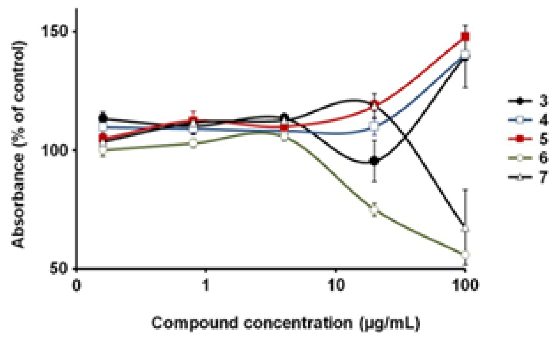
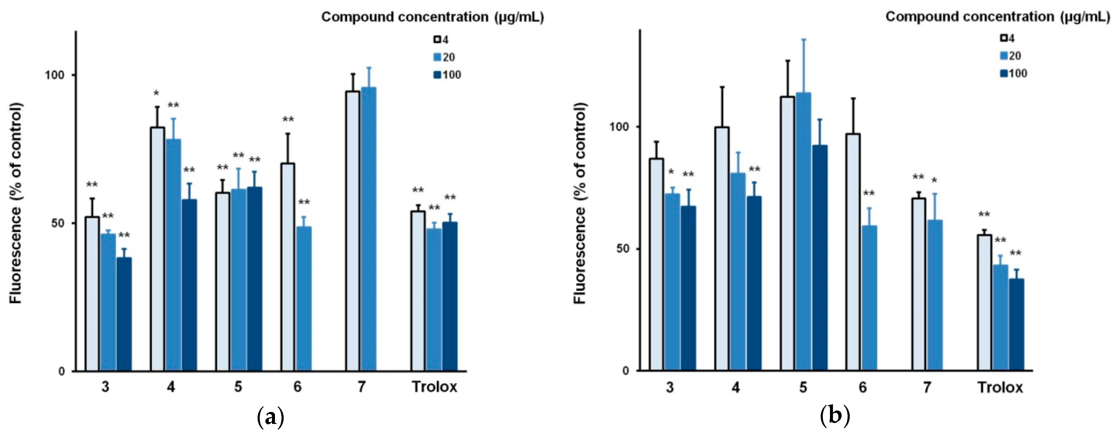
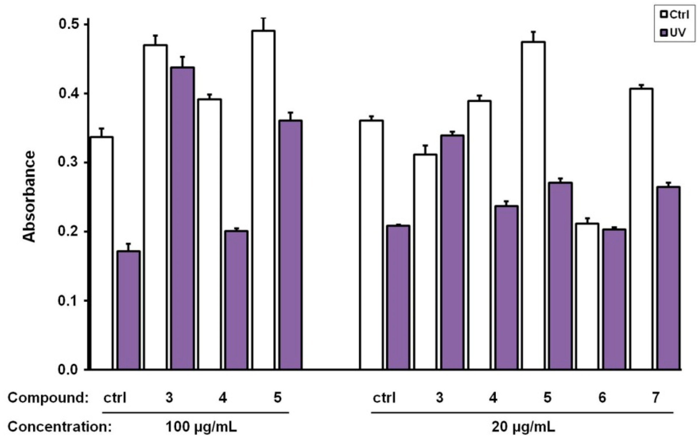
3. Results and Discussion

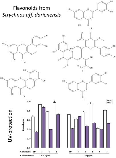
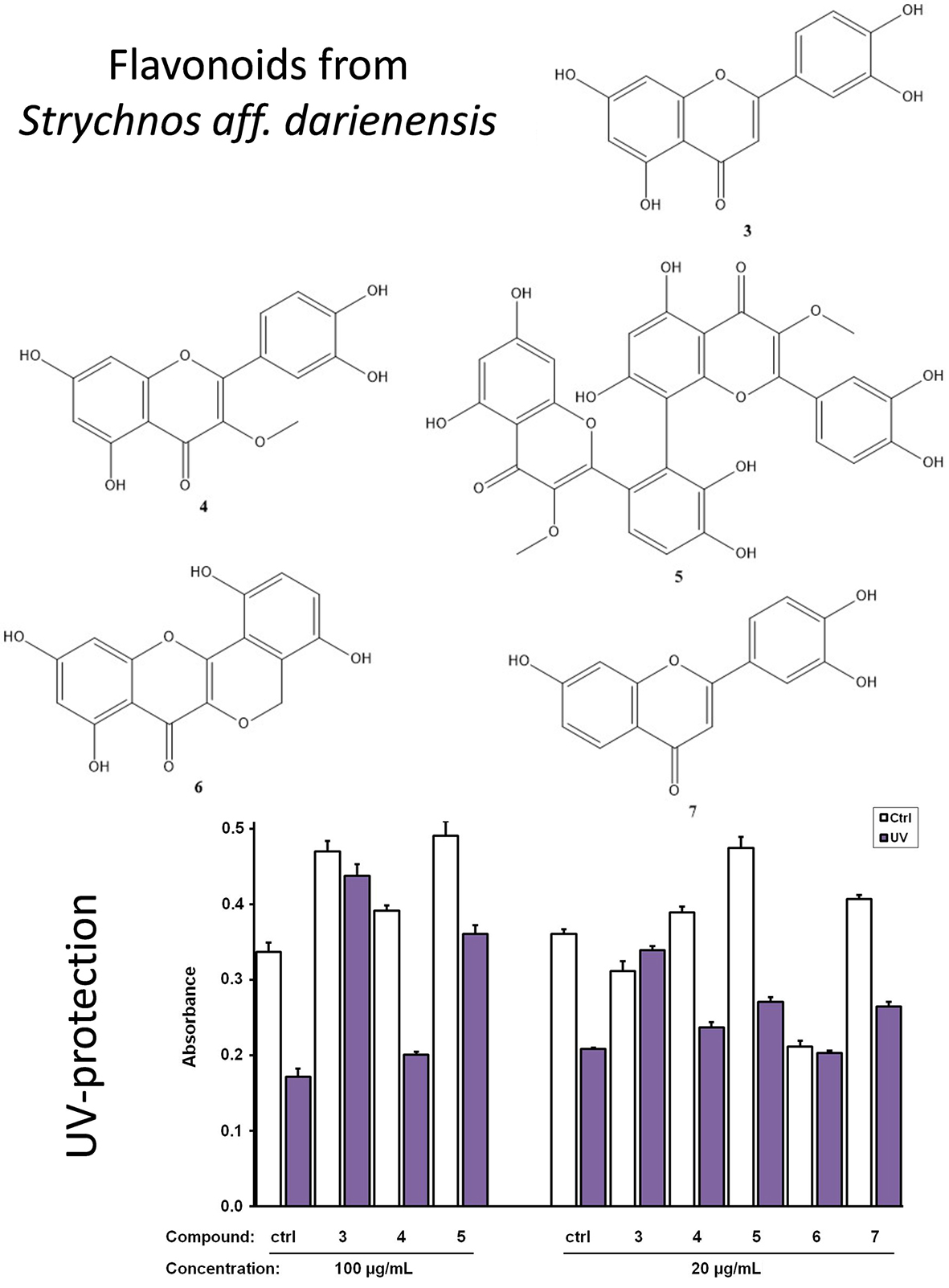
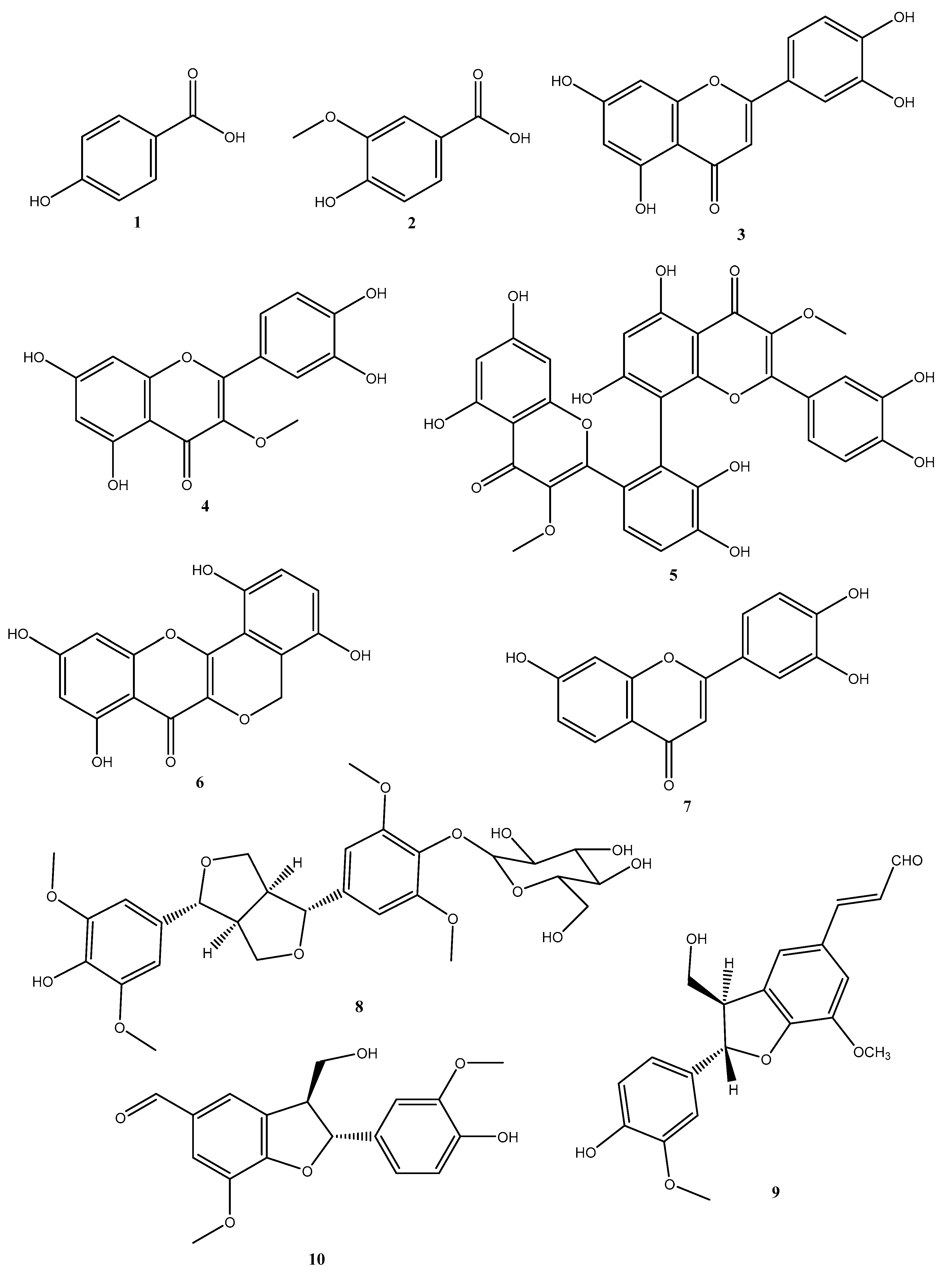
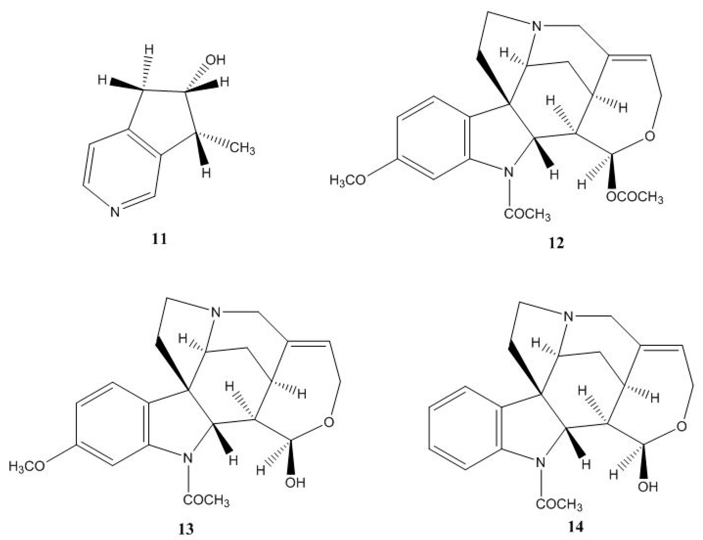
3.1. Isolation and Identification of Secondary Metabolites

The genus Strychnos is rich in alkaloids, whereas the content of other secondary metabolites is in many cases neglected. In continuation of our investigation of neotropical plants [7,8], we investigated phytochemically the stem bark of Strychnos aff. darienensis. Plant material collected in 1997 has been used for this research, as this material was stored under specific conditions and was considered appropriate for phytochemical investigation [11,12,13]. The evaluation of the best extraction procedure in order to recover a vast range of metabolites was of great importance. Profiling of the extracts with thin layer chromatography TLC revealed that the best approach was the maceration of the stem bark with EtOAc-EtOH-NH3 (96:3:1) and extraction with EtOAc and then with MeOH. The EtOAc extract was subjected to a size exclusion chromatography to afford luteolin (3) [14], and after additional purification, 3-O-methyl quercetin (4) [15] and strychnobiflavone (5) [16]. The MeOH extract was subjected to absorbent chromatography XAD4 and then treated with different pH, see Supplementary Material, Figure S1, giving five different extracts (M1-M5). Fraction M1 with further purification using fast centrifugal partition chromatography afforded minaxin (6) [17] and balanophonin (9) [18], and fraction M1B-3 treated with size exclusion chromatography afforded vanillic acid (2) [14], p-hydroxybenzoic acid (1) [19] and syringaresinol-β-D-glucoside (8) [20], see Figure 1. Fraction M3, using size exclusion chromatography and HPLC, afforded venoterpine (11) [21,22], 11-methoxyhenningsamine (12) [22], 3’,4’,7-trihydroxy-flavone (7) [23], ficusal (10) [24], diaboline (13) [25], and 11-methoxy diaboline (14) [17], see Figure 2.
In total, fourteen (114) compounds have been isolated and identified using spectroscopic methods. 1D and 2D NMR experiments, see Supplementary Material, Tables S2–S15, as well as, high-resolution MS led to unambiguous structure elucidation of the isolated compounds. All isolated compounds had identical spectroscopic data tothose previously described.

3.2. Biological Evaluation of The Isolated Flavonoids

For the biological evaluation, the well-studied class of flavonoids—known to possess limited cytotoxicity and significant anti-aging activity [26,27]—was selected to be evaluated for their antioxidant and UV-protection activity. Alkaloids were excluded for any further evaluation due to their high toxicity [3,28], while phenolic acids and lignans were less promising as antioxidants than flavonoids since they had fewerfree hydroxyl groups [29,30]. The first step was the evaluation of the cytotoxicity of the isolated compounds that was performed on human skin fibroblasts using the MTT-method. The compounds tested were luteolin (3), 3-O-methyl-quercetin (4), strychnobiflavone (5), minaxin (6), and 3’,4’,7-trihydroxyflavone (7). As shown in Figure 3, minaxin (6) was the most toxic compound for human fibroblasts, suppressing their viability to 56% (±1) of the control at the highest concentration tested (100 μg/ml), while at 20 μg/mL, viability was 75% (±3) of the control. 3’,4’,7-trihydroxyflavone (7) was less cytotoxic since it suppressed viability to 68% (±16) of the control only at the highest concentration (100 μg/mL). Hence, this concentration (i.e., 100 μg/mL) was excluded during the assessment of the antioxidant and UV-protection capacities of 6 and 7. The remaining three compounds werecompletely non-cytotoxic and actually enhanced cell viability to 140–148% of the control at the highest concentration of 100 μg/ml, see Figure 3.
Compounds 3, 4, 5, and 6 were found to inhibit the basal levels of intracellular reactive oxygen species (ROS) dose-dependently, see Figure 4A, with the most efficient being luteolin (3). In fact, 3 at 100 μg/mL was more efficient than Trolox, which was used as a positive control (38% vs. 50%, 0.0002). On the other hand, 3’,4’,7-trihydroxyflavone (7) was not attenuating the basal levels of intracellular ROS, as shown in Figure 4A. All compounds, with the exception of strychnobiflavone (5), were also observed to inhibit the hydrogen peroxide-induced ROS stimulation, see Figure 4B; although, none of them was as potent as the positive control (Trolox). Our results agree with literature reports from epithelial cells regarding the antioxidant activity of 3 [31] and 4 [32], while this is the first study to show the capacity of 5, 6, and 7 to suppress ROS in human skin fibroblasts.
Concerning the capacity of the compounds to protect from UV-B irradiation, the most active was luteolin (3), which at both concentrations of 20 and 100 μg/mL fully protected human skin fibroblasts from UV-B, as shown in Figure 5, in agreement with previous observations [33]. Strychnobiflavone (5) was also capable to reverse UV-B-induced lethality in a dose-dependent manner, while compounds 7 and 4 exhibited a minor protective effect at the concentration of 20 μg/mL, see Figure 5. Interestingly, strychnobiflavone has been identified as one of the metabolites from the Brazilian plant Strychnos pseudoquina possibly related with its anti-inflammatory and wound healing properties [34,35,36].
In conclusion, the flavonoids isolated from Strychnos aff. darienensis in the present study are capable of suppressing intracellular ROS levels, while two of them, luteolin (3) and strychnobiflavone (5), act as protective agents against UV-irradiation, properties that imply that they may be used as bioactive ingredients in the food and cosmetic industries.

Supplementary Materials

The following are available online at https://www.mdpi.com/2079-9284/6/1/7/s1, Figure S1: The extraction procedure using different pH treatments. Table S1: FCPC gradient system used for separation of fraction M1. Tables S2–S15: NMR Spectroscopic data of compounds 114.

Author Contributions

Conceptualization, N.F. and H.P.; Formal analysis, A.P.; Investigation, A.T., M.T.A. and K.V.; Methodology, A.T., M.T.A., K.V. and A.P.; Supervision, N.F. and H.P.; Validation, A.P., N.A. and C.L.C.; Visualization, H.P.; Writing—original draft, K.V., N.F. and H.P.; Writing—review & editing, N.A., C.L.C., D.K. and H.P.

Funding

This research received no external funding

Acknowledgments

The biological activity of the compounds was assessed within the framework of the project “An Open-Access Research Infrastructure of Chemical Biology and Target-Based Screening Technologies for Human and Animal Health, Agriculture and the Environment (OPENSCREEN-GR)” (MIS 5002691), which is implemented under the Action “Reinforcement of the Research and Innovation Infrastructure”, funded by the Operational Program “Competitiveness, Entrepreneurship and Innovation” (NSRF 2014-2020) and co-financed by Greece and the European Union (European Regional Development Fund).

Conflicts of Interest

The authors declare no conflict of interest.

References

  1. Burlando, B.; Cornara, L. Revisiting Amazonian Plants for Skin Care and Disease. Cosmetics 2017, 4, 25. [Google Scholar] [CrossRef]
  2. Massiot, G.; Delaude, C. Chapter 5 African Strychnos Alkaloids. In The Alkaloids: Chemistry and Pharmacology; Brossi, A., Ed.; Academic Press: New York, NY, USA, 1989; Volume 34, pp. 211–329. [Google Scholar]
  3. Philippe, G.; Angenot, L.; Tits, M.; Frederich, M. About the toxicity of some Strychnos species and their alkaloids. Toxicon 2004, 44, 405–416. [Google Scholar] [CrossRef] [PubMed]
  4. Valadeau, C.; Castillo, J.A.; Sauvain, M.; Lores, A.F.; Bourdy, G. The rainbow hurts my skin: Medicinal concepts and plants uses among the Yanesha (Amuesha), an Amazonian Peruvian ethnic group. J. Ethnopharmacol. 2010, 127, 175–192. [Google Scholar] [CrossRef] [PubMed]
  5. Guo, R.; Wang, T.; Zhou, G.; Xu, M.; Yu, X.; Zhang, X.; Sui, F.; Li, C.; Tang, L.; Wang, Z. Botany, Phytochemistry, Pharmacology and Toxicity of Strychnos nux-vomica L.: A Review. Am. J. Chin. Med. 2018, 46, 1–23. [Google Scholar] [CrossRef] [PubMed]
  6. De Wet, H.; Nciki, S.; van Vuuren, S.F. Medicinal plants used for the treatment of various skin disorders by a rural community in northern Maputaland, South Africa. J. Ethnobiol. Ethnomed. 2013, 9, 51. [Google Scholar] [CrossRef] [Green Version]
  7. Vougogiannopoulou, K.; Fokialakis, N.; Aligiannis, N.; Cantrell, C.; Skaltsounis, A.L. The raputindoles: Novel cyclopentylbisindole alkaloids from Raputiasimulans. Organ. Lett. 2010, 12, 1908–1911. [Google Scholar] [CrossRef] [PubMed]
  8. Vougogiannopoulou, K.; Fokialakis, N.; Aligiannis, N.; Cantrell, C.; Skaltsounis, A.L. Simple indole alkaloids from the neotropical rutaceous tree Raputiasimulans. Planta Med. 2011, 77, 1559–1561. [Google Scholar] [CrossRef]
  9. Vougogiannopoulou, K.; Angelopoulou, M.T.; Pratsinis, H.; Grougnet, R.; Halabalaki, M.; Kletsas, D.; Deguin, B.; Skaltsounis, L.A. Chemical and Biological Investigation of Olive Mill Waste Water—OMWWSecoiridoid Lactones. Planta Med. 2015, 81, 1205–1212. [Google Scholar] [CrossRef]
  10. Guldbrandsen, N.; De Mieri, M.; Gupta, M.; Liakou, E.; Pratsinis, H.; Kletsas, D.; Chaita, E.; Aligiannis, N.; Skaltsounis, A.L.; Hamburger, M. Screening of Panamanian Plants for Cosmetic Properties, and HPLC-Based Identification of Constituents with Antioxidant and UV-B Protecting Activities. Sci. Pharm. 2015, 83, 177–190. [Google Scholar] [CrossRef] [Green Version]
  11. Collins, T.L.; Andrew, R.L.; Greatrex, B.W.; Bruhl, J.J. Reliable analysis of volatile compounds from small samples of Eucalyptus magnificata (Myrtaceae). Aust. Syst. Bot. 2018, 31, 232–240. [Google Scholar] [CrossRef]
  12. Kao, D.; Henkin, J.M.; Soejarto, D.D.; Kinghorn, A.D.; Oberlies, N.H. Non-destructive chemical analysis of a Garcinia mangostana L. (Mangosteen) herbarium voucher specimen. Phytochem. Lett. 2018, 28, 124–129. [Google Scholar] [CrossRef] [PubMed]
  13. Phillipson, J.D. Chemical investigations of herbarium material for alkaloids. Phytochemistry 1982, 21, 2441–2456. [Google Scholar] [CrossRef]
  14. Lee, K.H.; Whang, W.K. Inhibitory Effects of Bioassay-Guided Isolation of Anti-Glycation Components from Taraxacumcoreanum and Simultaneous Quantification. Molecules 2018, 23, 2148. [Google Scholar] [CrossRef] [PubMed]
  15. Tian, X.; Guo, S.; He, K.; Roller, M.; Yang, M.; Liu, Q.; Zhang, L.; Ho, C.T.; Bai, N. Qualitative and quantitative analysis of chemical constituents of Ptychopetalumolacoides Benth. Nat. Prod. Res. 2018, 32, 354–357. [Google Scholar] [CrossRef] [PubMed]
  16. Nicoletti, M.; Goulart, M.O.; de Lima, R.A.; Goulart, A.E.; DelleMonache, F.; Marini Bettolo, G.B. Flavonoids and alkaloids from Strychnos pseudoquina. J. Nat. Prod. 1984, 47, 953–957. [Google Scholar] [CrossRef]
  17. Xu, N.; Xu, X.D.; Ma, L.Y.; Yuan, J.Q.; Miao, J.H.; Cheng, Y.; Yang, J.S. A new homoflavonoid from the seed of Caesalpinia minax Hance. Chin. Chem. Lett. 2010, 21, 696–698. [Google Scholar] [CrossRef]
  18. Haruna, M.; Koube, T.; Ito, K.; Murata, H. Balanophonin, A New Neo-Lignan from Balanophora japonica Makino. Chem. Pharm. Bull. 1982, 30, 1525–1527. [Google Scholar] [CrossRef]
  19. Chen, G.-Y.; Dai, C.-Y.; Wang, T.-S.; Jiang, C.-W.; Han, C.-R.; Song, X.-P. A new flavonol from the stem-bark of Premnafulva. Arkivoc 2010, 2010, 179–185. [Google Scholar]
  20. Lami, N.; Kadota, S.; Kikuchi, T.; Momose, Y. Constituents of the roots of Boerhaavia diffusa, L. III. Identification of Ca2+ channel antagonistic compound from the methanol extract. Chem. Pharm. Bull. 1991, 39, 1551–1555. [Google Scholar] [CrossRef]
  21. Ray, A.B.; Chatterjee, A. Venoterpine—A new monoterpenoid alkaloid from the fruits of Alstonia venenata R. Br. Tetrahedron Lett. 1968, 9, 2763–2766. [Google Scholar] [CrossRef]
  22. Thepenier, P.; Jacquier, M.-J.; Massiot, G.; Le Men-olivier, L.; Delaude, C. Alkaloids from Strychnos staudtii. Phytochemistry 1988, 27, 657–659. [Google Scholar] [CrossRef]
  23. Wu, J.H.; Tung, Y.T.; Chien, S.C.; Wang, S.Y.; Kuo, Y.H.; Shyur, L.F.; Chang, S.T. Effect of phytocompounds from the heartwood of Acacia confusa on inflammatory mediator production. J. Agric. Food Chem. 2008, 56, 1567–1573. [Google Scholar] [CrossRef] [PubMed]
  24. Li, Y.C.; Kuo, Y.H. Four new compounds, ficusal, ficusesquilignan A, B, and ficusolidediacetate from the heartwood of Ficus microcarpa. Chem. Pharm. Bull. 2000, 48, 1862–1865. [Google Scholar] [CrossRef] [PubMed]
  25. Ohshima, T.; Xu, Y.; Takita, R.; Shibasaki, M. Enantioselective total synthesis of (−)-strychnine: Development of a highly practical catalytic asymmetric carbon–carbon bond formation and domino cyclization. Tetrahedron 2004, 60, 9569–9588. [Google Scholar] [CrossRef]
  26. Martinez, S.E.; Davies, N.M.; Reynolds, J.K. Toxicology and Safety of Flavonoids. In Flavonoid Pharmacokinetics: Methods of Analysis, Preclinical and Clinical Pharmacokinetics, Safety, and Toxicology; Davies, N.M., Yáñez, J.A., Eds.; John Wiley & Sons: Hoboken, NJ, USA, 2013; Chapter 6; pp. 249–280. [Google Scholar] [CrossRef]
  27. Panche, A.N.; Diwan, A.D.; Chandra, S.R. Flavonoids: An overview. J. Nutr. Sci. 2016, 5, e47. [Google Scholar] [CrossRef] [PubMed]
  28. Levin, D.A.; York, B.M. The toxicity of plant alkaloids: An Ecogeographic perspective. Biochem. Syst. Ecol. 1978, 6, 61–76. [Google Scholar] [CrossRef]
  29. Lien, E.J.; Ren, S.; Bui, H.-H.; Wang, R. Quantitative structure-activity relationship analysis of phenolic antioxidants. Free Radic. Biol. Med. 1999, 26, 285–294. [Google Scholar] [CrossRef]
  30. Rice-Evans, C.A.; Miller, N.J.; Paganga, J. Structure-antioxidant activity relationships of flavonoids and phenolic acids. Free Radic. Biol. Med. 1996, 20, 933–956. [Google Scholar] [CrossRef]
  31. Wolfle, U.; Esser, P.R.; Simon-Haarhaus, B.; Martin, S.F.; Lademann, J.; Schempp, C.M. UVB-induced DNA damage, generation of reactive oxygen species, and inflammation are effectively attenuated by the flavonoid luteolin in vitro and in vivo. Free Radic. Biol. Med. 2011, 50, 1081–1093. [Google Scholar] [CrossRef]
  32. Kumar, A.D.; Bevara, G.B.; Kaja, L.K.; Badana, A.K.; Malla, R.R. Protective effect of 3-O-methyl quercetin and kaempferol from Semecarpus anacardium against H2O2 induced cytotoxicity in lung and liver cells. BMC Complement. Altern. Med. 2016, 16, 376. [Google Scholar] [CrossRef]
  33. Wolfle, U.; Haarhaus, B.; Schempp, C.M. The photoprotective and antioxidative properties of luteolin are synergistically augmented by tocopherol and ubiquinone. Planta Med. 2013, 79, 963–965. [Google Scholar] [CrossRef] [PubMed]
  34. Bieski, I.G.C.; Rios Santos, F.; de Oliveira, R.M.; Espinosa, M.M.; Macedo, M.; Albuquerque, U.P.; de Oliveira Martins, D.T. Ethnopharmacology of Medicinal Plants of the Pantanal Region (Mato Grosso, Brazil). Evid. Based Complement. Altern. Med. 2012, 2012, 36. [Google Scholar] [CrossRef] [PubMed]
  35. Bonamin, F.; Moraes, T.M.; Kushima, H.; Silva, M.A.; Rozza, A.L.; Pellizzon, C.H.; Bauab, T.M.; Rocha, L.R.; Vilegas, W.; Hiruma-Lima, C.A. Can a Strychnos species be used as antiulcer agent? Ulcer healing action from alkaloid fraction of Strychnos pseudoquina St. Hil. (Loganiaceae). J. Ethnopharmacol. 2011, 138, 47–52. [Google Scholar] [CrossRef] [PubMed]
  36. Santos, F.V.; Colus, I.M.; Silva, M.A.; Vilegas, W.; Varanda, E.A. Assessment of DNA damage by extracts and fractions of Strychnos pseudoquina, a Brazilian medicinal plant with antiulcerogenic activity. Food Chem. Toxicol. 2006, 44, 1585–1589. [Google Scholar] [CrossRef] [PubMed]
Figure 1. The isolated phenolic acids (1,2), flavonoids (37) and lignans (810) from the stem bark of Strychnos aff. darienensis.
Cosmetics 06 00007 g001
Figure 2. The isolated alkaloids (1114) from the stem bark of Strychnos aff. darienensis.
Cosmetics 06 00007 g002
Figure 3. Cytotoxicity of the isolated compounds. The cytotoxicity of the isolated compounds was evaluated in human skin fibroblasts using the MTT-method, as described in the Materials and Methods. Results are expressed as a percentage of the control and represent the mean of three independent experiments performed in quadruplicate.
Cosmetics 06 00007 g003
Figure 4. Intracellular antioxidant activity of the isolated compounds. The ability of the isolated compounds to attenuate basal (a) or hydrogen-peroxide-stimulated (b) (ROS) in human skin fibroblasts was evaluated using DCFH-DA, as described in the Materials and Methods. Vehicle (DMSO) was used as negative control and Trolox as positive one. Results are expressed as percentages of the value of the respective control concentration, and they represent the mean of three independent experiments performed in quadruplicate (* 0.01 < p ≤ 0.05; ** p ≤ 0.01).
Cosmetics 06 00007 g004
Figure 5. UV-protective capacity of the isolated compounds. The capacity of the isolated compounds to protect human skin fibroblasts from the lethal effects of UV-B-irradiation was assessed as described in the Materials and Methods. Results are expressed as absorbance values at 550 nm (reference wavelength 690 nm) and represent the mean of three independent experiments performed in quadruplicate.
Cosmetics 06 00007 g005

