Article

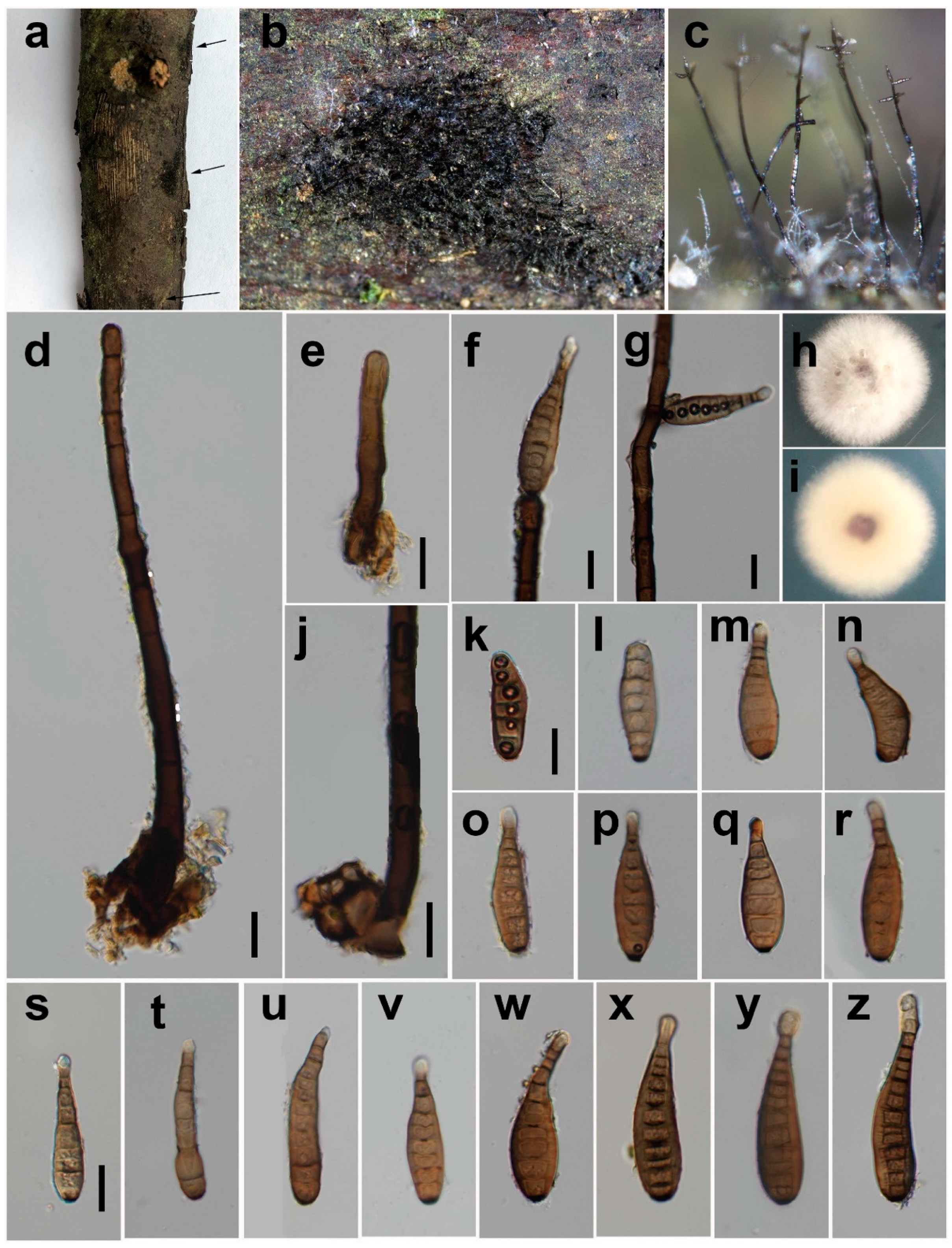
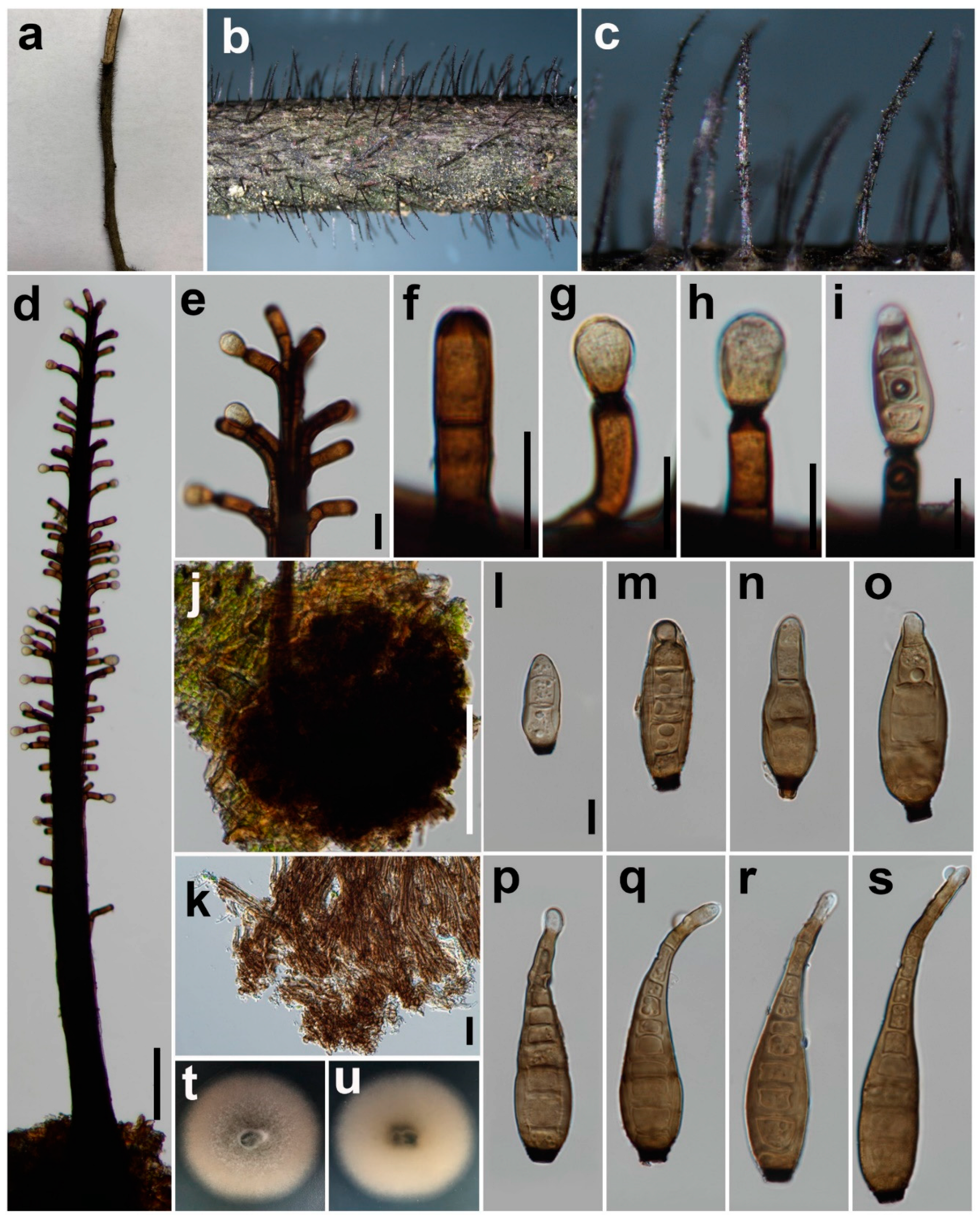
Synhelminthosporium gen. et sp. nov. and Two New Species of Helminthosporium (Massarinaceae, Pleosporales) from Sichuan Province, China

1
School of Life Science and Technology, Center for Informational Biology, University of Electronic Science and Technology of China, Chengdu 611731, China
2
Departamento de Tecnología Médica, Facultad de Ciencias de la Salud, Universidad de Tarapacá, Sede Iquique, Av. Luis Emilio Recabarren 2477, Iquique 1100000, Chile
*
Author to whom correspondence should be addressed.
J. Fungi 2022, 8(7), 712; https://doi.org/10.3390/jof8070712
Submission received: 10 June 2022 / Revised: 2 July 2022 / Accepted: 4 July 2022 / Published: 5 July 2022
(This article belongs to the Special Issue Polyphasic Identification of Fungi)

Abstract

:
Helminthosporium is a polyphyletic genus in Massarinaceae (Pleosporales). Species of Helminthosporium are characterized by having septate and erect conidiophores, acro-pleurogenous and distoseptate conidia with a ring-shaped scar at the base. During a survey of fungal diversity in Sichuan Province, China, six Helminthosporium-like isolates were collected from dead branches of unknown trees. Five barcodes, including ITS (ITS1-5.8S-ITS2), SSU, LSU, TEF1, and RPB2 were amplified and sequenced. Morphological examination and multi-locus phylogenetic analyses revealed two new Helminthosporium species (H. chengduense sp. nov., and H. chinense sp. nov.), a new genus (Synhelminthosporium gen. nov.) with a type species Synhelminthosporium synnematoferum sp. nov., and two known species (Helminthosporium submersum and H. velutinum) within Massarinaceae. The new genus Synhelminthosporium differs from the phylogenetically closest genus Helminthosporium by producing synnematous conidiophores. This work expands our understanding of the diversity of Helminthosporium-like taxa in Sichuan Province, China.

1. Introduction

Fungi consist of a highly diverse lineage of eukaryotes with a huge estimated number of between 2.2 and 3.8 million species [1]. Investigating fungal diversity is vital in Assembling the Fungal Tree Of Life (AFToL) [2], which significantly enhances our understanding of the history of life and also strengthens our ability to explore and use fungal resources [3].
Helminthosporium is an old, species-rich genus erected by Link in 1809 [4]. In addition to Helminthosporium, ten other genera are accepted in the family Massarinaceae (Pleosporales, Dothideomycetes), i.e., Byssothecium, Haplohelminthosporium, Helminthosporiella, Massarina, Mirohelminthosporium, Pseudodidymosphaeria, Pseudosplanchnonema, Semifissispora, Stagonospora, and Suttonomyces [5,6]. Based on multi-locus phylogenetic analysis, Konta et al. [7] confirmed that Helminthosporium is polyphyletic, where members were mixed with other taxa of Byssothecium, Helminthosporiella, and Pseudosplanchnonema. Most Helminthosporium species are saprobes feeding on dead or decaying woods [7,8]. However, one species, H. solani is an economically important pathogen causing silver scurf disease in potatoes worldwide [9,10]. Helminthosporium species are commonly collected from leaves and decaying wood in terrestrial habitats [7,8,11] and rarely reported in freshwater habitats [12].
The genus Helminthosporium, typified by H. velutinum, is characterized by producing macronematous, cylindrical, rather straight, septate, erect conidiophores with tretic conidiogenous cells and clavate or obclavate, distoseptate conidia with a flat, ringed pore at the base [8,13]. Conidia are produced mainly laterally from tretic conidiogenous cells and the production of a terminal conidium usually determines the end of conidiophore growth. Most Helminthosporium species were introduced based on their asexual morph and only six species, viz., H. tiliae, H. microsorum, H. oligosporum, H. massarinum, H. quercicola, and H. quercinum, were characterized based on both morphs [8,11]. Splanchnonema kalakadense was described as the sexual morph of H. velutinum, but this was only based on pure culture without sequence data [14]. Tanaka et al. [11] first connected the Massarina-like sexual morph and asexual morph of H. massarinum, which was confirmed based on pure culture and sequence data. Voglmayr and Jaklitsch [8] experimentally confirmed three Splanchnonema-like sexual morphs of Helminthosporium species based on pure culture, sequence data, and herbarium studies, which extends the knowledge of sexual morphs of Helminthosporium.
Sichuan Province, located in southwestern China, along the Yangtze River, has enormous fungal diversity [15,16,17,18]. We regularly conduct fungal diversity surveys in Sichuan Province. During the study of preliminary morphological examination and BLASTn analysis of ITS sequences (the ribosomal internal transcribed spacer), a total of six Helminthosporium-like isolates were obtained from July to September 2021. Based on the multi-locus phylogenetic analysis and morphological examination, two known Helminthosporium species including a new habitat record, two new Helminthosporium species, and a new genus Synhelminthosporium with the type species, S. synnematoferum sp. nov. are introduced. This study broadens our understanding of the diversity of Helminthosporium-like taxa.

2. Materials and Methods

2.1. Sample Collection, Isolation, and Morphological Examination

A survey of the diversity of ascomycetous fungi in Sichuan Province, China, was conducted between July and September 2021. Dead branches were collected from three locations in Sichuan Province (Yunqiao Wetland, Chengdu City; Baiyungou, Chongzhou City; Huilonggou, Pengzhou City). The specimens were taken to the laboratory in paper envelopes for examination. The morphological observation was consistently carried out from material on natural substrates. Tiny pieces of mycelium were mounted in a drop of sterilized water using syringe needles. Microscopic characters were observed and recorded using a Nikon SMZ800N stereo microscope equipped with a Nikon DS-Fi3 microscope camera and a Nikon ECLIPSE Ni-U microscope fitted with a Nikon DS-Ri2 microscope camera, respectively. Measurements were conducted using the Nikon NIS-Elements Documentation Imaging Software Version 5.21.00. All photos were processed using Adobe Photoshop software version 22.0. Isolates were obtained by picking up pieces of mycelium into sterilized water, spreading the suspension onto the surface of potato dextrose agar (PDA) plates, and incubated for 24 h at 25 °C. Germinated conidia were individually transferred to PDA plates and incubated under the dark at 25 °C. Culture characteristics were examined and recorded after one week and later at regular intervals.
The specimens were deposited in the Herbarium of Cryptogams Kunming Institute of Botany Academia Sinica (HKAS), Kunming, China, or the Herbarium of University of Electronic Science and Technology (HUEST), Chengdu, China. The living cultures were deposited in the China General Microbiological Culture Collection Center (CGMCC) in Beijing, China, and the University of Electronic Science and Technology Culture Collection (UESTCC) in Chengdu, China.

2.2. DNA Extraction, PCR Amplification, and Sequencing

Fungal genomic DNA was extracted from mycelia using the TreliefTM Plant Genomic DNA extraction Kit (TSINGKE Biotech, Shanghai, China) according to the manufacturer protocol. The DNA was stored at −20 °C for long-term storage. Five barcodes including the nuclear ribosomal internal transcribed spacer region (ITS: ITS1-5.8S-ITS2), the partial nuclear ribosomal small subunit rRNA gene (SSU), the partial nuclear ribosomal large subunit rRNA gene (LSU), the partial translation elongation factor 1-alpha gene (TEF1) and the partial second-largest subunit of RNA polymerase II gene (RPB2) were amplified by polymerase chain reaction (PCR). The corresponding primer pairs and PCR processes are listed in Table 1. The final PCR reaction volume was 25 µL containing 2 µL of DNA template, 1 µL each of the forward and reverse primer (10 µM), 8.5 µL of double-distilled water (ddH2O), and 12.5 µL of 2 × Flash PCR MasterMix (mixture of DNA Polymerase, dNTPs, Mg2+ and optimized buffer; CoWin Biosciences, Taizhou, China). The PCR products were visualized in 1% agarose gel electrophoresis. Sanger sequencing was conducted by Tsingke Biological Technology (Beijing, China).

2.3. Phylogenetic Analyses

According to the corresponding Sanger sequencing chromatograms, misleading data from the ends of raw sequencing fragments were manually trimmed and assembled into consensus sequences using SeqMan Pro version 7.1.0 (DNASTAR, Inc. Madison, WI, USA). Barcode sequences of all Helminthosporium species currently available in GenBank, representative strains from other genera in Massarinaceae, and the outgroup taxon Periconia pseudodigitata (CBS 139699) were downloaded from the NCBI nucleotide database using the function read.GenBank integrated within the R package Analysis of Phylogenetics and Evolution (APE) [29].
The multiple sequence alignment was conducted using MAFFT version 7.310 [30] with options “--adjustdirectionaccurately--auto”, and the alignment results were further trimmed using trimAl version 1.4 [31] with the option “-gapthreshold 0.5”, which only allows 50% of taxa with a gap in each site. The best-fit nucleotide substitution models for each alignment dataset were selected using PartitionFinder version 2.1.1 [32] under the Corrected Akaike Information Criterion (AICC).
Maximum Likelihood (ML) and Bayesian analysis were conducted based on individual and combined datasets. Five alignment datasets of SSU, ITS, LSU, TEF1, and RPB2 were concatenated using an in-house python script for multi-locus phylogenetic analysis. ML phylogenetic trees were obtained using the IQ-TREE version 2.0.3 [33], and the topology was evaluated using 1000 ultrafast bootstrap replicates. The Bayesian analysis was conducted using parallel MrBayes version 3.2.7a [34]. Two different runs with 20 million generations and four chains were executed, and the initial 25% of sample trees were treated as burn-in. Tracer version 1.7.1 [35] was used to confirm that the MCMC runs reached convergence with all ESS values above 200. Then, the ML tree was annotated by TreeAnnotator version 2.6.6 implemented in BEAST version 2.6.6 [36] based on MrBayes MCMC trees with no discard of burn-in, and no posterior probability limit. The ML trees were visualized using ggtree [37] and further edited in Adobe Illustrator version 16.0.0.

3. Results

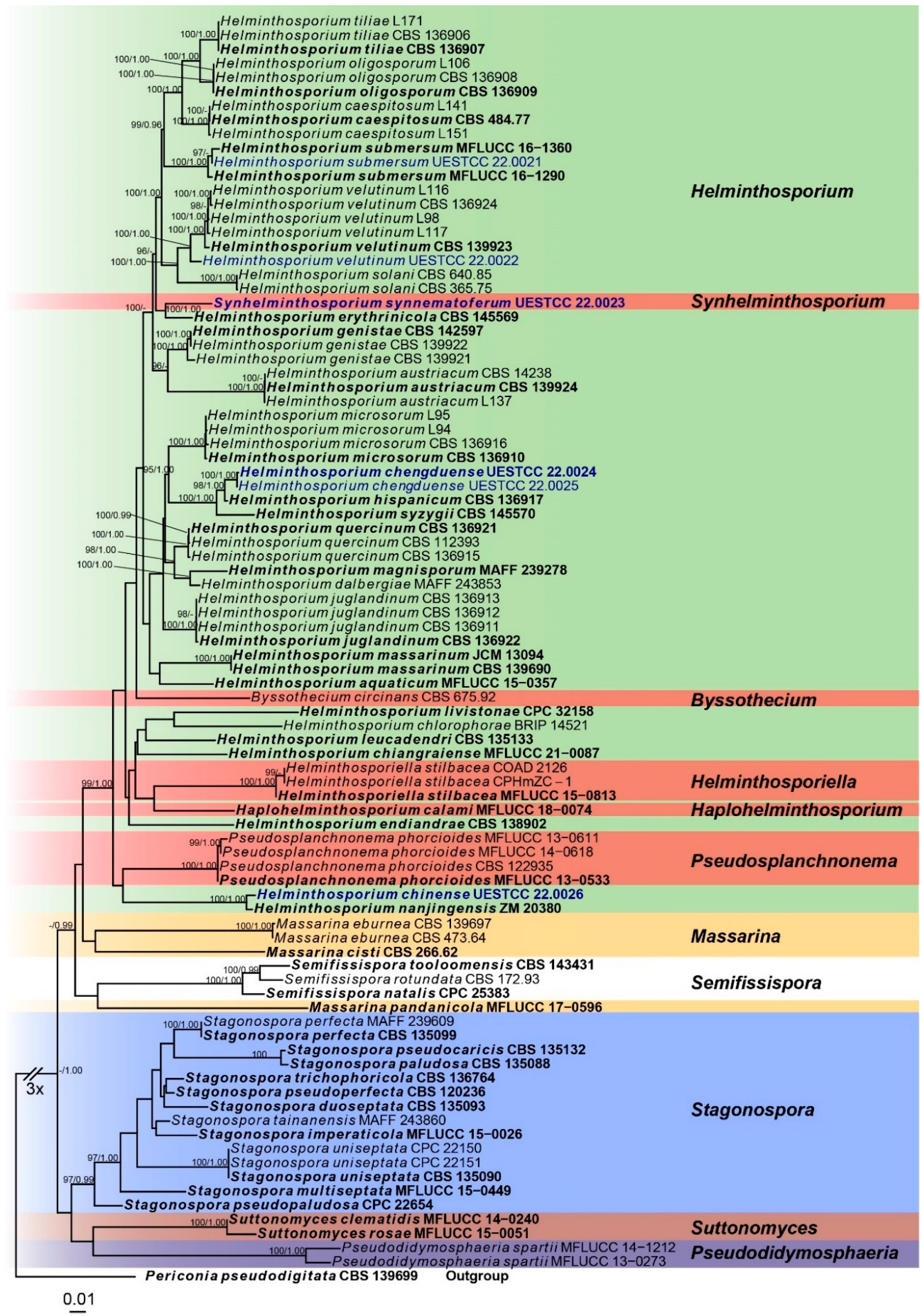
3.1. Molecular Phylogeny
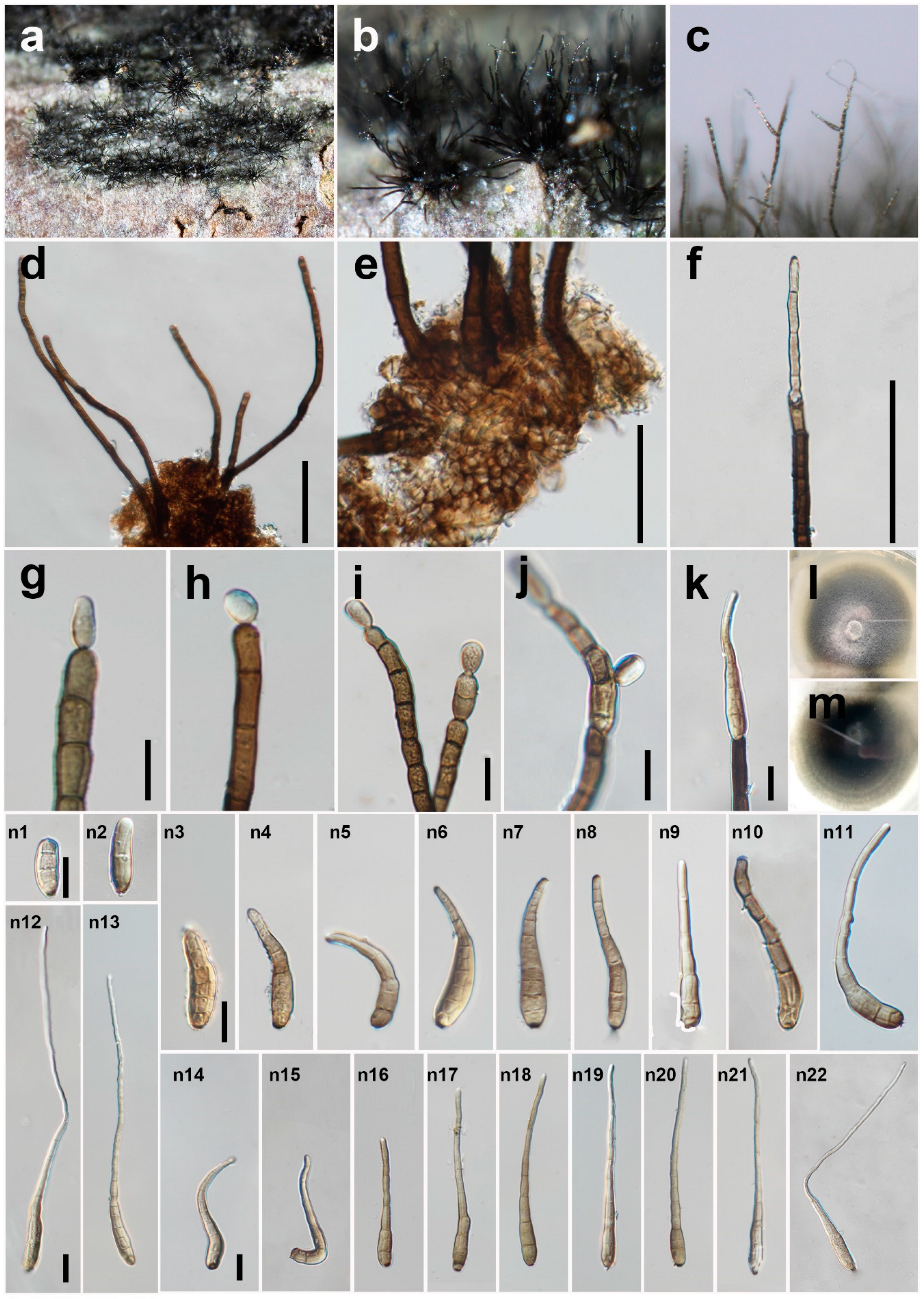
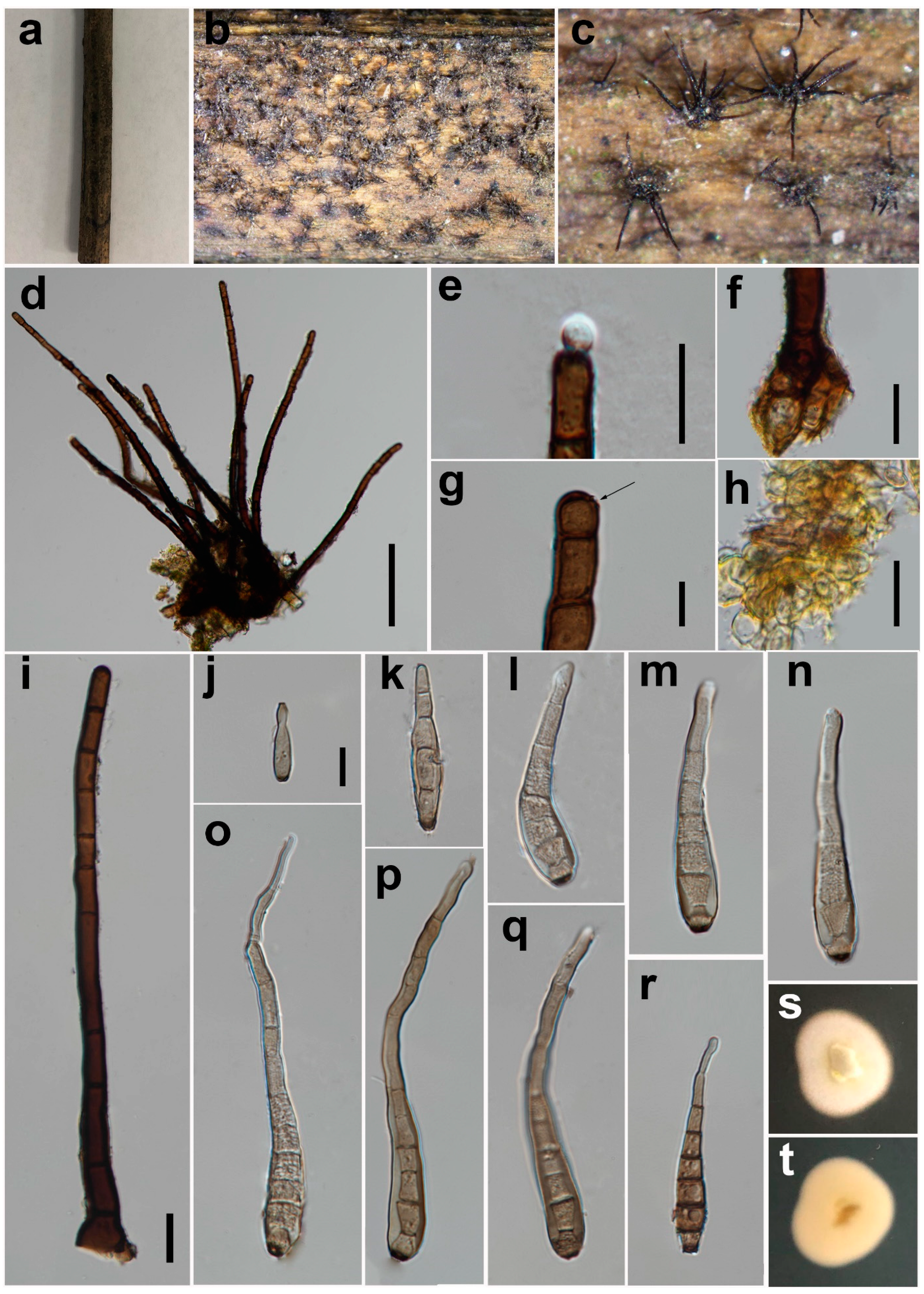
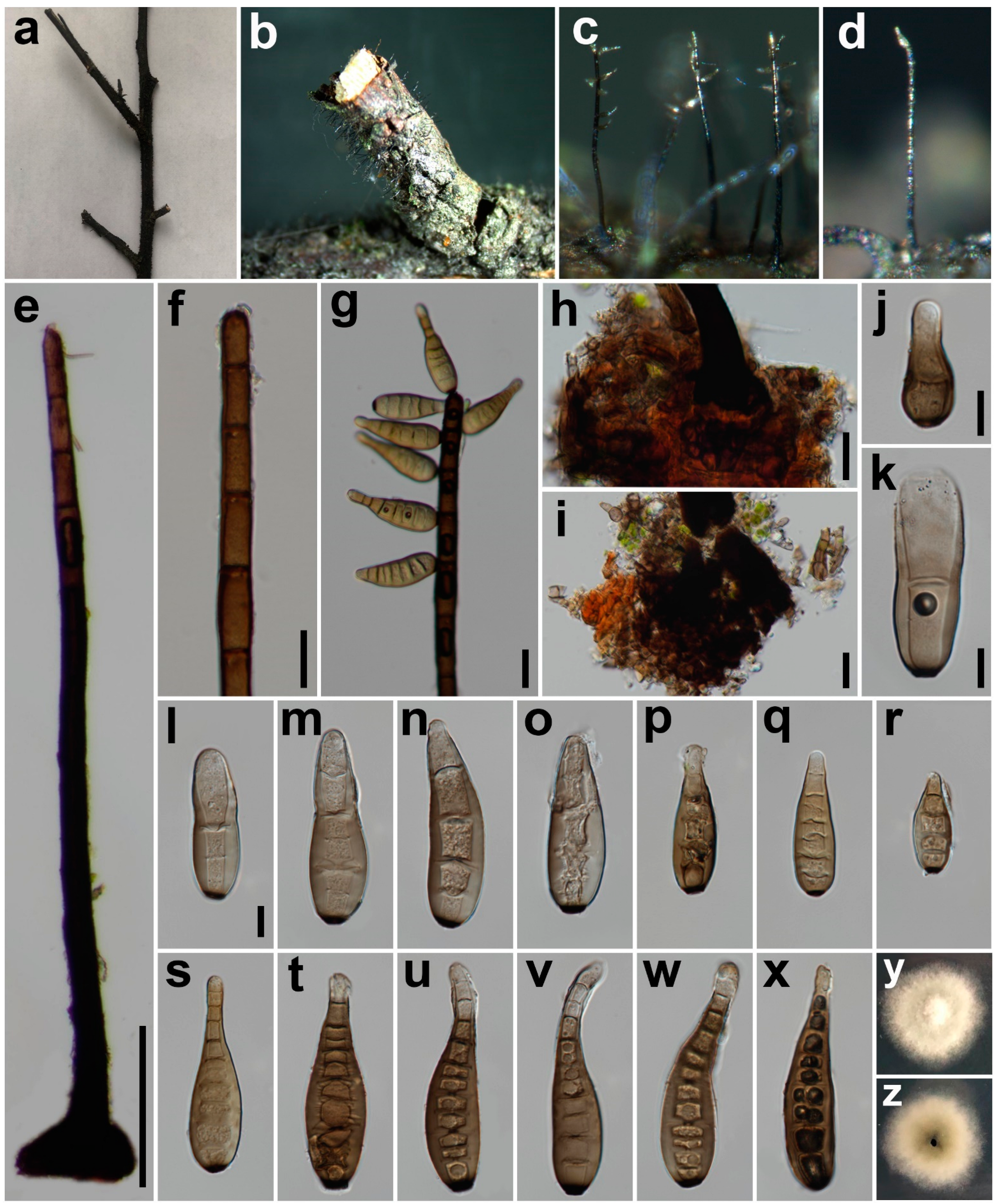
Five barcode sequences were obtained successfully except for RPB2 of H. velutinum (UESTCC 22.0022) and H. chengduense sp. nov. Newly generated sequences were deposited in GenBank and the accession numbers are listed in Table 2. The combined dataset (ITS:1-557, SSU:558-1594, LSU:1595-2485, TEF1:2459-3719, RPB2: 3720-4836) was composed of 1744 distinct patterns, 1138 parsimony-informative sites, 325 singleton sites and 3373 constant sites. Five single-locus datasets ITS, SSU, LSU, RPB2, and TEF1 contained 258, 69, 106, 353, and 357 parsimony informative sites, respectively. The best-fit evolution models were GTR+I+G for the ITS, LSU, TEF1 and RPB2 partitions and HKY+G for the SSU partition.
The best-scoring ML consensus tree (lnL = −21,360.862) with ultrafast bootstrap values from ML analyses and posterior probabilities from MrBayes analysis at the node is shown in Figure 1. Helminthosporium species are mixed with species of other genera, viz. Byssothecium, Pseudosplanchnonema, and Haplohelminthosporium, suggesting the genus is polyphyletic. Six newly obtained Helminthosporium isolates represent five different clades. Helminthosporium submersum (UESTCC 22.0021) clustered with two H. submersum isolates (MFLUCC 16-1360 and MFLUCC 16-1290). Helminthosporium velutinum (UESTCC 22.0022) and the five other H. velutinum strains including the epi-type (CBS 139923) are grouped into a statistically well-supported clade (100/1.00). Synhelminthosporium synnematoferum (UESTCC 22.0023) is separate from H. erythrinicola (CBS 145569) with strong statistical support (100/1.00). Two isolates (UESTCC 22.0025 and UESTCC 22.0024) of H. chengduense form a distinct clade sister to H. hispanicum with high support values (98/1.00). Helminthosporium chinense (UESTCC 22.0026) is a sister to H. nanjingensis (ZM 20380) with strong statistical support (100/1.00).

3.2. Taxonomy

Helminthosporium chengduense Y.P. Chen & Maharachch., sp. nov. (Figure 2).
MycoBank: MB 844416
Etymology: The name refers to Chengdu, the city where the fungus was collected.
Saprobic on decaying wood in a damp environment. Sexual morph: Unknown. Asexual morph: Colony on natural substrate punctiform, black, hairy. Mycelium mostly immersed, towards the surface forming stroma-like aggregations of light to dark brown pseudoparenchymatous cells. Conidiophores 133–391 μm long (x = 252, n = 40), 8–15 μm wide (x = 12, n = 40) at the base, tapering to 7–11 μm (x = 9, n = 40) at the apex, arising solitarily or in small groups from the stroma cells, erect, simple, straight or flexuous, thick-walled, subcylindrical, smooth, pale to dark brown, paler near the apex, with well-defined small pores at the apex and rarely laterally beneath the upper 1–2 septa. Conidiogenous cells mono- to poly-tretic, cylindrical, integrated, terminal and intercalary, pale brown to brown, secession schizo-lytic. Conidia 41–251 × 8–13 μm (x = 120 × 10, n = 60), tapering to 2–6 μm (x = 4, n = 55) at the distal end, with a blackish-brown 2–5 μm wide (x = 3, n = 25) scar at the base, obclavate, straight, flexuous, sigmoid, lunate or uncinate, thin-walled, smooth, grey-white to pale brown, 3–16-distoseptate (n = 52), with angular lumina; wall up to 2–4 μm thick (x = 3, n = 68).
Material examined: China, Sichuan Province, Chengdu City, Yunqiao Wetland, on decaying branch of unidentified host, N 30°52′32, E 103°53′23, elevation 570 m, 10 July 2021, Y.P. Chen, YQ 071048H (HKAS 124016, holotype), culture ex-type UESTCC 22.0024 = YQ 071,048 = CGMCC 3.23575; ibid., YQ 071047H (HUEST 22.0025, isotype), culture ex-isotype UESTCC 22.0025 = YQ 071047.
Culture characteristics: Colony on PDA 53 mm diam after 2 weeks in an incubator under dark conditions at 20 °C, pale green, irregular circular, surface velvety, with white and denser mycelium at the center, with creamy white, entire margin; reverse dark green at the center, pale green at the periphery, with growth rings.
Notes: The phylogenetic tree shows that the isolate UESTCC 22.0024 clusters with the ex-type strain of H. hispanicum (CBS 136917) [8]. However, the isolate UESTCC 22.0024 significantly differs from the holotype in the length of conidiophores (133–391 μm vs. 130–540 μm), the size of conidia (41–251 × 8–13 μm vs. 69–130 × 17–24 μm), the wall thickness of angular lumina (2–4 μm vs. 7 μm). In addition, H. hispanicum is fungicolous and grows on old conidiomata of Juglanconis juglandina [8], whereas H. chengduense (UESTCC 22.0024) is saprobic on decaying wood in damp environments. Konta et al. [7] summarized the morphological characteristics of 216 Helminthosporium species. Among them, only H. asterinum, H. longisinuatum, and H. makilingense produce larger conidia than H. chengduense UESTCC 22.0024. The conidia of H. chengduense (UESTCC 22.0024) are much shorter and narrower than H. asterinum (500–600 × 80 μm) [38]; shorter than H. longisinuatum (65–1000 μm) [39], but with longer conidiophores (133–391 μm vs. 20–75 μm) and a smaller number of distosepta (3–16 vs. 9–22); shorter than H. makilingense (100–300 μm) [40]. Considering the significant differences in morphology and molecular data, we introduce the isolate UESTCC 22.0024 as a new species H. chengduense.
Figure 2. Helminthosporium chengduense (HKAS 124016, holotype). (a,b) Colonies on the natural substrate; (c) Conidiophores with apical and lateral conidia; (d) Conidiophores and stroma cells; (e) Conidiophore bases and stroma cells; (f) Conidiophore; (gi) Conidiophore with young apical conidia; (j) Conidiophore with a young lateral conidium; (k) Conidiophore with an apical conidium; (l,m) Culture on PDA after 2 weeks (back and forth); (n1n22) Conidia. Scale bars: (d,f) = 100 μm; (e,g,ik,n1,n3,n12,n14) = 20 μm. Scale bar (g) applies to (h); (n1) applies to (n2); (n3) applies to (n4n11); (n12) applies to (n13); (n14) applies to (n15n22).
Jof 08 00712 g002
Helminthosporium chinense Y.P. Chen & Maharachch., sp. nov. (Figure 3).
MycoBank: MB 844417
Etymology: The name refers to China, the country where the fungus was collected.
Saprobic on decaying wood in damp environment. Sexual morph: Unknown. Asexual morph: Colony on natural substrate effuse, black, hairy. Mycelium mostly immersed, towards the surface forming stroma-like aggregations of light to brown pseudoparenchymatous cells. Conidiophores 214–461 μm long (x = 326, n = 40), 8–16 μm wide (x = 11, n = 38) at the base, tapering to 6–10 μm (x = 8, n = 38) at the apex, arising solitarily or in fascicles from the stroma cells, erect, simple, straight or flexuous, thick-walled, subcylindrical, smooth, pale to dark brown, with well-defined small pores at the apex and rarely laterally beneath the upper 1–5 septa. Conidiogenous cells mono- to poly-tretic, cylindrical, integrated, terminal and intercalary, brown, secession schizo-lytic. Conidia 42–109 × 5–11 μm (x = 61 × 8, n = 35), tapering to 2–6 μm (x = 4, n = 35) at the distal end, with a blackish-brown 3–5 μm wide (x = 4, n = 27) scar at the base, obclavate, straight or flexuous, thin-walled, smooth, pale gray to brown, 4–10-distoseptate, with angular lumina; wall up to 1–3 μm thick (x = 2, n = 36).
Material examined: China, Sichuan Province, Chengdu City, Yunqiao Wetland, on decaying branch of palm trees, N 30°52′32, E 103°53′23, elevation 570 m, 10 July 2021, Y.P. Chen, YQ 071005H (HKAS 124017, holotype), culture ex-type UESTCC 22.0026 = YQ 071,005 = CGMCC 3.23570.
Culture characteristics: Colony on PDA 31 mm diam after 2 weeks in an incubator under dark condition at 20 °C, white, irregular circular, surface velvety, with a clear margin; reverse white, with clear margin.
Notes: The phylogenetic tree shows that the isolate UESTCC 22.0026 clusters with the ex-type strain (ZM 20380) of H. nanjingensis, which was introduced by Wang et al. [41] from dead branches of an unidentified tree in Nanjing City, Jiangsu Province, China. Our collection (HKAS 124017) shares similar morphological characteristics in the shape and color of conidiophores and conidia with the holotype (HSAUP020198) [41] of H. nanjingensis on natural substrate. However, it differs from H. nanjingensis by having significantly shorter conidia (42–109 μm vs. 64.5–170.5 μm) and smaller number of disto-septa (4–10 vs. 6–17) [41]. The BLASTn analysis of ITS of our isolate UESTCC 22.0026 showed 98% identity (446/453 bp, no gap) with ex-type strain (ZM 20380) of H. nanjingensis. Helminthosporium nanjingensis produced yellow-green pigment on PDA media [41], but the isolate UESTCC 22.0026 does not produce pigment on PDA. Only the ITS sequence is available for H. nanjingensis. Therefore, we cannot compare the sequence difference of other barcodes. Thus, considering the difference in morphology and the ability to produce pigments, we describe the isolate UESTCC 22.0026 as a new species H. chinense.
Figure 3. Helminthosporium chinense (HKAS 124017, holotype). (a) Natural substrate; (b,c) Colonies on the substrate; (d) Colony and conidiophores; (e) Conidiophore with an apical conidium; (f) Conidiophore base and stroma cells; (g) Conidiophore with an apical pore; (h) Stroma cells; (i) Conidiophore; (j) Young conidium; (kq) Conidia; (r) Old conidium. (s,t) Front view and back view of culture on PDA after 2 weeks. Scale bars: (d) = 100 μm, (e,gi) = 20 μm, (f,j) = 10 μm. Scale bar j applies to (kr).
Jof 08 00712 g003
Helminthosporium submersum Z.L. Luo & H.Y. Su, Phytotaxa 348(4): 274 (2018) (Figure 4).
Saprobic on decaying wood in damp environment. Sexual morph: unknown. Asexual morph: Colony on natural substrate superficial, effuse, hairy, dark brown to black, glistening. Mycelium mostly immersed, composed of septate, unbranched, smooth, thick-walled hyphae, on the bark stroma-like aggregations of light to black dark brown pseudoparenchymatous cells at the hyphae base. Conidiophores mono-nematous, erect, simple, straight or flexuous, unbranched, smooth, thick-walled, subcylindrical, pale to dark brown, paler near the apex, 130–570 μm long, 15–31 μm wide (x = 21, n = 20) at the base, tapering to 9–12 μm (x = 10, n = 20) at the apex, with well-defined small pores at the apex and laterally beneath the upper 1–5 septa. Conidiogenous cells mono- to poly-tretic, integrated, terminal and intercalary, cylindrical, pale brown, secession schizo-lytic. Conidia acropleurogenous, simple, obclavate, straight or flexuous, thin-walled, smooth, grey-brown to brown, 49–86 × 14–25 μm (x = 70 × 18, n = 35), tapering to 6–12 μm (x = 8, n = 35) at the distal end, with a blackish-brown 3–6 μm wide (x = 5, n = 40) scar at the base, 1–12-distoseptate (n = 30), with angular lumina; wall up to 2–4 μm thick (x = 3, n = 35).
Material examined: China, Sichuan Province, Dujiangyan City, Longchi National Forest Park, on decaying branch of an unidentified host, N 31°6′37, E 103°33′55, elevation 1168 m, 19 September 2021, W. Tian, Sarah 08_3H (HUEST 22.0021), living culture UESTCC 22.0021 = Sarah 08_3 = CGMCC 3.23571.
Culture characteristics: Colony on PDA 19 mm diam after 2 weeks in an incubator under dark conditions at 20 °C, white, irregular circular, surface velvety, with denser mycelium at the center and becoming sparser towards the edge, with unclear margin; reverse pale green at the center, with unclear white margin.
Notes: The phylogenetic tree showed that our isolate (UESTCC 22.0021) from decaying wood in a damp environment clustered with the ex-type strain (MFLUCC 16–1360) of H. submersum, which was introduced by Zhao et al. [42] from submerged wood in freshwater. Morphologically, our collection fits well with H. submersum by having effuse, velvety, dark brown or black colonies, mono-nematous, straight or flexuous, unbranched, pale to dark brown conidiophores, and similar size (49–86 × 14–25 μm vs. 41–55 × 14.5–18.5 μm) conidia [42]. Based on the overlapping morphological characteristics and the multi-locus phylogenetic tree, we identify our isolate as H. submersum. This is the first report of H. submersum isolated from decaying wood in terrestrial habitats.
Figure 4. Helminthosporium submersum (HUEST 22.0021). (a) Natural substrate; (bd) Colonies on natural substrates; (e) Conidiophore; (f) Conidiophore apex; (g) Conidiophore, conidiophore apex, apical conidia and lateral conidia; (h,i) Conidiophore base and stroma cells; (jx) Conidia; (y,z) Front view and back view of a colony on PDA after 2 weeks. Scale bars: (eg) = 100 μm, (h,i) = 20 μm, (jl) = 10 μm. Scale bar (l) applies to (mx).
Jof 08 00712 g004
Helminthosporium velutinum Link [as ‘Helmisporium’], Mag. Gesell. naturf. Freunde, Berlin 3(1–2): 10, Table 1:9 (1809) (Figure 5).
Saprobic on decaying wood in damp environment. Sexual morph: Unknown. Asexual morph: Colony on natural substrate effuse, black, hairy, glistening. Mycelium mostly immersed, on the surface forming stroma-like aggregations of light to dark brown pseudoparenchymatous cells. Conidiophores 343–941 μm long (x = 715, n = 20), 12–30 μm wide (x = 20, n = 20) at the base, tapering to 9–16 μm (x = 107, n = 20) at the apex, arising solitarily or in fascicles from the stroma cells, erect, simple, straight or flexuous, cylindrical, thick-walled, smooth, dark to blackish brown, paler near the apex, with well-defined small pores at the apex and rarely laterally beneath the upper 1–11 septa. Conidiogenous cells mono- to poly-tretic, cylindrical, integrated, terminal and intercalary, brown, secession schizo-lytic. Conidia 53–99 × 13–22 μm (x = 76 × 17, n = 36), tapering to 5–8 μm (x = 7, n = 36) at the distal end, with a blackish-brown 3–5 μm wide (x = 4, n = 33) scar at the base, obclavate, straight or flexuous, thin-walled, smooth, pale brown, 6–13-distoseptate, with angular lumina; wall up to 2–5 μm thick (x = 4, n = 43).
Material examined: China, Sichuan Province, Chongzhou City, Baiyungou, on decaying branch of an unidentified host, N 30°47′56, E 103°24′15, elevation 990 m, 27 September 2021, Y.P. Chen, BY 14_2H (HUEST 22.0022), living culture UESTCC 22.0022 = BY 14_2 = CGMCC 3.23572.
Culture characteristics: Colony on PDA 19 mm after 2 weeks in an incubator under dark conditions at 20 °C, creamy white, irregular circular, surface velvety, with denser mycelium at the center and becoming sparser towards the edge, with clear margin; reverse white, pale green in the center, with clear margin.
Notes: The phylogenetic tree showed that the isolate HUEST 22.0022 clustered with the isolates of H. velutinum. Helminthosporium velutinum, the type of the genus, is a well-known and most commonly recorded species [8]. It has been recorded mainly from woody substrates, and it is known for more than 100 host records [43]. Zhu et al. [12] first reported H. velutinum from a freshwater habitat in China, which is a less common habitat for this species. Our collection (HUEST 22.0022) displays similar morphological characteristics with the type of H. velutinum in the shape and color of colonies, conidiophores, conidiogenous cells, and conidia on the natural substrate [8]. We identified the isolate UESTCC 22.0022 as H. velutinum, a new record from terrestrial habitats in china considering similar morphological characteristics.
Figure 5. Helminthosporium velutinum (HUEST 22.0022). (a,b) Colonies on the natural substrate; (c) Conidiophores with apical and lateral conidia; (d,e,j) Conidiophores with stroma cells; (f) Conidiophore apex with an apical conidium; (g) Conidiophore with a lateral conidium; (h,i) Front view and back view of culture on PDA after 1 week; (kz) Conidia. Scale bars: (dg,j,k,s) = 20 μm. Scale bar k applies to (lr); (s) applies to (tz).
Jof 08 00712 g005
Synhelminthosporium Y.P. Chen & Maharachch., gen. nov.
MycoBank: MB 844418
Etymology: Syn = together with its close phylogenetic relationship with Helminthosporium.
Saprobic on decaying wood in damp environment. Sexual morph: Unknown. Asexual morph: Colony on natural substrate evenly distributed, hairy, glistening. Mycelium mostly immersed. Conidiophores synnematous, compact, erect, straight or flexuous, unbranched, septate, smooth, subcylindrical, brown to dark brown, separated from the main body near the apex. Conidiogenous cells mono-tretic, cylindrical, integrated, terminal, brown, secession schizo-lytic. Conidia obclavate, straight or flexuous, smooth, pale brown, distoseptate, with angular lumina.
Type species: Synhelminthosporium synnematoferum Y.P. Chen & Maharachch. sp. nov.
Notes: The genus Synhelminthosporium is introduced based on the new species S. synnematoferum. Both BLASTn analysis results of five barcode sequences (SSU, ITS, LSU, TEF1, and RPB2) and multi-locus phylogenetic analyses showed that Synhelminthosporium is distinct and phylogenetically close to Helminthosporium. Synhelminthosporium differs from Helminthosporium by having synnematous conidiophores, which is a character only presented in Helminthosporiella within Massarinaceae [7,44]. Phylogenetic analysis shows that Synhelminthosporium is different from Helminthosporiella. In addition, Helminthosporiella shows catenate conidia, but this character is absent in Synhelminthosporium. Based on distinguishing morphological characteristics and multi-locus phylogenetic analyses, we introduce a new genus Synhelminthosporium to accommodate the new species S. synnematoferum in Massarinaceae.
Synhelminthosporium synnematoferum Y.P. Chen & Maharachch., sp. nov. (Figure 6).
MycoBank: MB 844419
Etymology: The name refers to the synnematous conidiophores.
Saprobic on decaying wood in damp environment. Sexual morph: Unknown. Asexual morph: Colony on natural substrate evenly distributed, hairy, glistening. Conidiophores 700–1500 μm long, synnematous, compact, erect, straight or flexuous, unbranched, septate, smooth, subcylindrical, brown to dark brown, separated from the main body from the middle part up to near the apex. Conidiogenous cells mono-tretic, cylindrical, integrated, terminal, brown, secession schizo-lytic. Conidia 52–116 × 14–20 μm (x = 80 × 16, n = 36), tapering to 5–10 μm (x = 7, n = 36) at the distal end, with a blackish-brown 3–7 μm wide (x = 5, n = 36) scar at the base, obclavate, straight or flexuous, smooth, pale brown, 4–11-distoseptate (n = 35), with angular lumina; wall up to 2–4 μm thick (x = 3, n = 36).
Material examined: China, Sichuan Province, Pengzhou City, Huilonggou, on decaying branch of an unidentified host, N 31°11′6, E 103°54′56, elevation 1400 m, 28 July 2021, Y.P. Chen, HLG 072894H (HKAS 124015, holotype), culture ex-type UESTCC 22.0023 = HLG 072,894 = CGMCC 3.23574.
Culture characteristics: Colony 52 mm diam after 1 week in an incubator under dark condition at 20 °C, creamy white, irregular circular, surface velvety, with denser mycelium at the center and becoming sparser at the edge, with a clear margin; reverse creamy white, pale green at the center, with clear margin.
Notes: The best BLASTn matches of five barcode sequences (SSU, ITS, LSU, TEF1, and RPB2) of the isolate UESTCC 22.0023 belong to the genus Helminthosporium. The phylogenetic tree shows that our isolate (UESTCC 22.0023) clusters with the ex-type strain H. erythrinicola (CBS 145569). The BLASTn analysis of H. erythrinicola (CBS 145569) and S. synnematoferum (UESTCC 22.0023) shows 94% identity (538/570, 5 gaps) using ITS, 99% identity (820/831, 3 gaps) using LSU and 93% identity (822/884, no gap) using RPB2. Helminthosporium erythrinicola was introduced by Crous et al. [45], it is a typical Helminthosporium species with simple, multiseptated, unbranched, brown conidiophores, tretic conidiogenous cells, and acro-pleurogenous, clavate or obclavate, distoseptate conidia. Our collection (HKAS 124015) differs from the H. erythrinicola and other Helminthosporium species in having synnematous conidiophores. Synnematous conidiophores are only present in Helminthosporiella in Massarinaceae [7,44]. The isolate UESTCC 22.0023 is different from Helminthosporiella phylogenetically and morphologically. The absence of catenate conidia is the most prominent characteristic distinguishing Synhelminthosporium synnematoferum from Helminthosporiella species. The multi-locus phylogenetic tree shows the placement of S. synnematoferum (UESTCC 22.0023) within Massarinaceae, which is distinct from other known species.
Figure 6. Synhelminthosporium synnematoferum (HKAS 124015, holotype). (ac) Colonies on natural substrates; (d) Synnematous conidiophores; (e) Conidiophores with apical conidia; (f) Conidiogenous cell with an apical pore; (gi) Conidiogenous cells with apical conidia; (j,k) Conidiophore bases; (l) Young conidium; (ms) Conidia; (t,u) Front view and back view of culture on PDA after 1 week. Scale bars: (d,j) = 100 μm, (ei,k) = 20 μm, (l) = 10 μm. Scale bar (l) applies to (ms).
Jof 08 00712 g006

4. Discussion

To date, there are 775 epithets of Helminthosporium (http://www.indexfungorum.org; accessed on 10 April 2022), whereas many of them are not congeneric with the generic type, and were reclassified into other groups in subsequent studies [46,47,48]. For instance, H. cynodontis was reassigned to the genus Bipolaris (Pleosporaceae, Pleosporales) due to the production of sympodial conidiogenous cells and by having darkly pigmented conidiogenous loci [46]. Recently, Konta et al. [7] accepted 216 Helminthosporium species, however many species are identified only based on morphological studies, and only 25 species have sequence data. The lack of a large amount of molecular data is mainly because most species were introduced before the advent of Sanger sequencing. Considering that numerous Helminthosporium species were characterized only based on morphological studies, it is likely that some of them belong to the same species or even to different genera. During phylogenetic analysis, abnormal long branches were observed in four Helminthosporium strains with incorrect taxonomic positions, viz. H. asterinum (CBS 203.35), H. decacuminatum (CBS 185.47), H. anomalum (CBS 161.27), and H. gibberosporum (CBS 200.32). These species were introduced and characterized before the 1950s [49,50,51,52], whereas the sequence data related to them were submitted to GenBank by Vu et al. [53]. BLASTn analyses of these sequences showed that the top hits of ITS and LSU sequences for H. anomalum (CBS 161.27) belong to Bipolaris, ITS and LSU sequences for H. asterinum (CBS 203.35) belong to Kirschsteiniothelia, ITS and LSU sequences for H. decacuminatum (CBS 185.47) and H. gibberosporum (CBS 200.32) belong to Curvularia. The present study introduces two new Helminthosporium species and a new genus Synhelminthosporium based on multi-locus phylogenetic analysis and morphological studies. This phylogeny needs to be expanded by re-examining type materials of old described Helminthosporium-like species without molecular data, collecting new fresh specimens, sequencing, and using multi-locus analysis to establish epi-types or neotypes as necessary. Our new species can be distinguished from all other Helminthosporium species by morphological features and multi-locus phylogenetic analysis, and thus we are confident that the newly introduced species are distinct.
Recent studies have no universally accepted standard in selecting barcodes for phylogenetic analysis. Boonmee et al. [54] introduced H. chiangraiense using ITS and LSU. Crous et al. [45] introduced H. erythrinicola and H. syzygii using ITS, LSU, and RPB2. Zhu et al. [12] introduced H. aquaticum using SSU, ITS, and LSU barcodes. Voglmayr and Jaklitsch [8] pointed out that only ITS and/or LSU sequences can be problematic in resolving the phylogeny of Massarinaceae. Other barcodes RPB2 and TEF1 were proposed in multi-gene phylogenetic analyses of Massarineae, as these barcodes usually significantly increase the phylogenetic resolution [8,11]. However, the majority of Helminthosporium species do not have RPB2 and TEF1 barcodes (19 species have SSU sequence data; 25 species have SSU sequence data; 23 species have LSU sequence data; 17 species have RPB2 sequence data and 15 species have TEF1 sequence data). The present study conducted both individual and combined phylogenetic analyses. ITS, RPB2, and TEF1 barcodes offered more parsimony informative sites than SSU and LSU. In addition, more powerful resolution in delineating species and higher bootstrap support values for most clades were observed in single-gene ML trees (Figures S1 and S2), indicating that these barcodes are better in resolving genera in Massarineae than the other two barcodes.

Supplementary Materials

The following supporting information can be downloaded at: https://www.mdpi.com/article/10.3390/jof8070712/s1, Figure S1: Single-gene trees. Novel isolates are indicated in dark blue. Type isolates are in bold. The ML ultrafast bootstrap values/Bayesian PP greater than 95%/0.95 are indicated at the respective nodes. The tree is rooted with Periconia pseudodigitata (CBS 139699) (Periconiaceae, Pleosporales). Figure S2: Comparisons of the single-gene ML trees and the multi-locus ML tree in delineating species. (a) The proportion of highly supported internal nodes, bootstrap values ≥ 95%. Protein-coding barcodes TEF1 and RPB2 offer higher proportions of highly supported internal nodes than three nuclear ribosomal regions (b) Pairwise tree distances. The smaller the value, the higher the similarity in topology.

Author Contributions

Conceptualization, Y.C. and S.S.N.M.; methodology, Y.C.; software, Y.C.; writing—original draft preparation, Y.C.; writing—review and editing, S.S.N.M. and H.M.; visualization, Y.C., W.T. and Y.G.; supervision, S.S.N.M.; funding acquisition, S.S.N.M. All authors have read and agreed to the published version of the manuscript.

Funding

This research was funded by Talent Introduction and Cultivation Project, University of Electronic Science and Technology of China, grant number A1098531023601245.

Institutional Review Board Statement

Not applicable.

Informed Consent Statement

Not applicable.

Data Availability Statement

All sequence data are available in NCBI GenBank following the accession numbers in the manuscript.

Acknowledgments

We thank the computing resources provided by the high-performance computing platform at the information technology center, School of Life Science and Technology, Center for Informational Biology, University of Electronic Science and Technology of China. Y.C. Chen thanks Xiandong Yu for the assistance in the preparation of manuscript.

Conflicts of Interest

The authors declare no conflict of interest.

References

  1. Hawksworth, D.L.; Lücking, R.; Heitman, J.; James, T.Y. Fungal Diversity Revisited: 2.2 to 3.8 Million Species. Microbiol. Spectr. 2017, 5, 79–95. [Google Scholar] [CrossRef] [PubMed]
  2. Celio, G.J.; Padamsee, M.; Dentinger, B.T.M.; Bauer, R.; McLaughlin, D.J. Assembling the Fungal Tree of Life: Constructing the Structural and Biochemical Database. Mycologia 2006, 98, 850–859. [Google Scholar] [CrossRef] [PubMed]
  3. Ryan, M.J.; McCluskey, K.; Verkleij, G.; Robert, V.; Smith, D. Fungal biological resources to support international development: Challenges and opportunities. World J. Microbiol. Biotechnol. 2019, 35, 139. [Google Scholar] [CrossRef] [PubMed] [Green Version]
  4. Link, H.F. Observationes in Ordines plantarum naturales. Dissertatio prima complectens Anandrarum ordines Epiphytas, Mucedines, Gastromycos et Fungos. Ges. Natur 1809, 3, 3–42. [Google Scholar]
  5. Hongsanan, S.; Hyde, K.; Phookamsak, R.; Wanasinghe, D.N.; McKenzie, E.H.C.; Sarma, V.V.; Lücking, R.; Boonmee, S.; Bhat, J.D.; Liu, N.G.; et al. Refined families of Dothideomycetes: Orders and families incertae sedis in Dothideomycetes. Fungal Divers. 2020, 105, 17–318. [Google Scholar] [CrossRef]
  6. Wijayawardene, N.; Hyde, K.; Dai, D.; Sánchez-García, M.; Goto, B.; Saxena, R.; Erdogdu, M.; Selçuk, F.; Rajeshkumar, K.; Aptroot, A. Outline of Fungi and fungus-like taxa-2021. Mycosphere 2022, 13, 53–453. [Google Scholar] [CrossRef]
  7. Konta, S.; Hyde, K.; Karunarathna, S.C.; Mapook, A.; Senwanna, C.; Dauner, L.A.P.; Nanayakkara, C.M.; Xu, J.; Tibpromma, S.; Lumyong, S. Multi-gene phylogeny and morphology reveal Haplohelminthosporium gen. nov. and Helminthosporiella gen. nov. associated with palms in Thailand and a checklist for Helminthosporium reported worldwide. Life 2021, 11, 454. [Google Scholar] [CrossRef]
  8. Voglmayr, H.; Jaklitsch, W.M. Corynespora, Exosporium and Helminthosporium revisited—New species and generic reclassification. Stud. Mycol. 2017, 87, 43–76. [Google Scholar] [CrossRef]
  9. Errampalli, D.; Saunders, J.M.; Holley, J.D. Emergence of silver scurf (Helminthosporium solani) as an economically important disease of potato. Plant Pathol. 2001, 50, 141–153. [Google Scholar] [CrossRef]
  10. Tian, S.M.; Chen, Y.C.; Zou, M.Q.; Xue, Q. First report of Helminthosporium solani causing silver scurf of potato in Hebei Province, North China. Plant Dis. 2007, 91, 460. [Google Scholar] [CrossRef]
  11. Tanaka, K.; Hirayama, K.; Yonezawa, H.; Sato, G.; Toriyabe, A.; Kudo, H.; Hashimoto, A.; Matsumura, M.; Harada, Y.; Kurihara, Y.; et al. Revision of the Massarineae (Pleosporales, Dothideomycetes). Stud. Mycol. 2015, 82, 75–136. [Google Scholar] [CrossRef] [PubMed] [Green Version]
  12. Zhu, D.; Luo, Z.L.; Baht, D.J.; Mckenzie, E.H.; Bahkali, A.H.; Zhou, D.Q.; Su, H.Y.; Hyde, K. Helminthosporium velutinum and H. aquaticum sp. nov. from aquatic habitats in Yunnan Province, China. Phytotaxa 2016, 253, 179–190. [Google Scholar] [CrossRef] [Green Version]
  13. Luttrell, E.S. Systematics of Helminthosporium and related genera. Mycologia 1964, 56, 119–132. [Google Scholar] [CrossRef]
  14. Subramanian, C.V.; Sekar, G. Three bitunicate ascomycetes and their tretic anamorphs. Kavaka 1987, 15, 87–97. [Google Scholar]
  15. Liang, Z.Q.; Wang, B.; Kang, J.C. Several rare entopathogenic fungi from the Western Sichuan mountains. Fungal Divers. 2003, 12, 129–134. [Google Scholar]
  16. Zhou, Y.; He, S.; Gong, G.; Zhang, S.; Chang, X.; Liu, N.; Sun, X.; Qi, X.; Ye, K.; Wang, Y. Soil fungal diversity in three nature reserves of Jiuzhaigou County, Sichuan Province, China. Ann. Microbiol. 2014, 64, 1275–1290. [Google Scholar] [CrossRef]
  17. Zhou, W.; Wei, Q.; Feng, R.; Liu, Y.; Liang, H.; Li, J.; Yan, K. Diversity and spatial distribution of endophytic fungi in Cinnamomum longepaniculatum of Yibin, China. Arch. Microbiol. 2021, 203, 3361–3372. [Google Scholar] [CrossRef]
  18. Yan, K.; Abbas, M.; Meng, L.; Cai, H.; Peng, Z.; Li, Q.; El-Sappah, A.H.; Yan, L.; Zhao, X. Analysis of the fungal diversity and community structure in Sichuan dark tea during pile-fermentation. Front. Microbiol. 2021, 12, 706714. [Google Scholar] [CrossRef]
  19. Toju, H.; Tanabe, A.S.; Yamamoto, S.; Sato, H. High-coverage ITS primers for the DNA-based identification of Ascomycetes and Basidiomycetes in environmental samples. PLoS ONE 2012, 7, e40863. [Google Scholar] [CrossRef] [Green Version]
  20. Vilgalys, R.; Hester, M. Rapid genetic identification and mapping of enzymatically amplified ribosomal DNA from several Cryptococcus species. J. Bacteriol. 1990, 172, 4238–4246. [Google Scholar] [CrossRef] [Green Version]
  21. Cubeta, M.A.; Echandi, E.; Abernethy, T.; Vilgalys, R. Characterization of anastomosis groups of binucleate Rhizoctonia species using restriction analysis of an amplified ribosomal RNA gene. Phytopathology 1991, 81, 1395–1400. [Google Scholar] [CrossRef]
  22. Hibbett, D.S. Phylogenetic evidence for horizontal transmission of group I introns in the nuclear ribosomal DNA of mushroom-forming fungi. Mol. Biol. Evol. 1996, 13, 903–917. [Google Scholar] [CrossRef] [PubMed] [Green Version]
  23. Carbone, I.; Kohn, L.M. A method for designing primer sets for speciation studies in filamentous ascomycetes. Mycologia 1999, 91, 553–556. [Google Scholar] [CrossRef]
  24. Rehner, S.A.; Buckley, E. A Beauveria phylogeny inferred from nuclear ITS and EF1-alpha sequences: Evidence for cryptic diversification and links to Cordyceps teleomorphs. Mycologia 2005, 97, 84–98. [Google Scholar] [CrossRef] [PubMed]
  25. Jaklitsch, W.M.; Komon, M.; Kubicek, C.P.; Druzhinina, I.S. Hypocrea voglmayrii sp. nov. from the Austrian Alps represents a new phylogenetic clade in Hypocrea/Trichoderma. Mycologia 2005, 97, 1365–1378. [Google Scholar] [CrossRef]
  26. Voglmayr, H.; Akulov, O.Y.; Jaklitsch, W.M. Reassessment of Allantonectria, phylogenetic position of Thyronectroidea, and Thyronectria caraganae sp. nov. Mycol. Prog. 2016, 15, 921–937. [Google Scholar] [CrossRef] [Green Version]
  27. Sung, G.H.; Sung, J.M.; Hywel-Jones, N.L.; Spatafora, J.W. A multi-gene phylogeny of Clavicipitaceae (Ascomycota, Fungi): Identification of localized incongruence using a combinational bootstrap approach. Mol. Phylogenet. Evol. 2007, 44, 1204–1223. [Google Scholar] [CrossRef]
  28. Liu, Y.J.; Whelen, S.; Hall, B.D. Phylogenetic relationships among ascomycetes: Evidence from an RNA polymerse II subunit. Mol. Biol. Evol. 1999, 16, 1799–1808. [Google Scholar] [CrossRef]
  29. Paradis, E.; Claude, J.; Strimmer, K. APE: Analyses of Phylogenetics and Evolution in R language. Bioinformatics 2004, 20, 289–290. [Google Scholar] [CrossRef] [Green Version]
  30. Katoh, K.; Misawa, K.; Kuma, K.i.; Miyata, T. MAFFT: A novel method for rapid multiple sequence alignment based on fast Fourier transform. Nucleic Acids Res. 2002, 30, 3059–3066. [Google Scholar] [CrossRef] [Green Version]
  31. Capella-Gutiérrez, S.; Silla-Martínez, J.M.; Gabaldón, T. trimAl: A tool for automated alignment trimming in large-scale phylogenetic analyses. Bioinformatics 2009, 25, 1972–1973. [Google Scholar] [CrossRef] [PubMed]
  32. Lanfear, R.; Frandsen, P.B.; Wright, A.M.; Senfeld, T.; Calcott, B. PartitionFinder 2: New methods for selecting partitioned models of evolution for molecular and morphological phylogenetic analyses. Mol. Biol. Evol. 2016, 34, 772–773. [Google Scholar] [CrossRef] [PubMed] [Green Version]
  33. Nguyen, L.T.; Schmidt, H.A.; von Haeseler, A.; Minh, B.Q. IQ-TREE: A fast and effective stochastic algorithm for estimating maximum-likelihood phylogenies. Mol. Biol. Evol. 2014, 32, 268–274. [Google Scholar] [CrossRef] [PubMed]
  34. Huelsenbeck, J.P.; Ronquist, F. MRBAYES: Bayesian inference of phylogenetic trees. Bioinformatics 2001, 17, 754–755. [Google Scholar] [CrossRef] [Green Version]
  35. Rambaut, A.; Drummond, A.J.; Xie, D.; Baele, G.; Suchard, M.A. Posterior summarization in Bayesian phylogenetics using Tracer 1.7. Syst. Biol. 2018, 67, 901–904. [Google Scholar] [CrossRef] [Green Version]
  36. Bouckaert, R.; Vaughan, T.G.; Barido-Sottani, J.; Duchêne, S.; Fourment, M.; Gavryushkina, A.; Heled, J.; Jones, G.; Kühnert, D.; De Maio, N.; et al. BEAST 2.5: An advanced software platform for Bayesian evolutionary analysis. PLoS Comput. Biol. 2019, 15, e1006650. [Google Scholar] [CrossRef] [Green Version]
  37. Yu, G. Using ggtree to Visualize Data on Tree-Like Structures. Curr. Protoc. Bioinform. 2020, 69, e96. [Google Scholar] [CrossRef]
  38. Cooke, M.C. Ravenel’s American fungi. Grevillea 1878, 6, 129–146. [Google Scholar]
  39. Matsushima, T. Matsushima Mycological Memoirs No. 7; Matsushima Fungus: Kobe, Japan, 1993; Volume 7, 141p. [Google Scholar]
  40. Sydow, P.; Sydow, H. Weitere neue Micromyceten der Philippinen-Inseln. Ann. Mycol. 1920, 18, 98–104. [Google Scholar]
  41. Wang, X.J.; Wu, H.Y.; Zhang, M. A new species of Helminthosporium from Jiangsu, China. Mycotaxon 2014, 127, 1–4. [Google Scholar] [CrossRef]
  42. Zhao, N.; Luo, Z.L.; Hyde, K.D.; Su, H.Y.; Bhat, D.J.; Liu, J.K.; Bao, D.F.; Hao, Y.E. Helminthosporium submersum sp. nov. (Massarinaceae) from submerged wood in north-western Yunnan Province, China. Phytotaxa 2018, 348, 269–278. [Google Scholar] [CrossRef]
  43. Farr, D.F.; Rossman, A.Y. Fungal Databases; U.S. National Fungus Collections, USDA: Washingtin, DC, USA, 2022. [Google Scholar]
  44. Crous, P.W.; Wingfield, M.J.; Richardson, D.M.; Leroux, J.J.; Strasberg, D.; Edwards, J.; Roets, F.; Hubka, V.; Taylor, P.W.J.; Heykoop, M.; et al. Fungal Planet description sheets: 400–468. Persoonia 2016, 36, 316–458. [Google Scholar] [CrossRef] [PubMed] [Green Version]
  45. Crous, P.W.; Carnegie, A.J.; Wingfield, M.J.; Sharma, R.; Mughini, G.; Noordeloos, M.E.; Santini, A.; Shouche, Y.S.; Bezerra, J.D.P.; Dima, B.; et al. Fungal Planet description sheets: 868–950. Persoonia 2019, 42, 291–473. [Google Scholar] [CrossRef] [PubMed]
  46. Shoemaker, R.A. Nomenclature of Drechslera and Bipolaris, grass parasites segregated from ‘Helminthosporium’. Can. J. Plant Pathol. 2006, 28, S212–S220. [Google Scholar] [CrossRef]
  47. Manamgoda, D.S.; Cai, L.; McKenzie, E.H.C.; Crous, P.W.; Madrid, H.; Chukeatirote, E.; Shivas, R.G.; Tan, Y.P.; Hyde, K. A phylogenetic and taxonomic re-evaluation of the Bipolaris-Cochliobolus-Curvularia complex. Fungal Divers. 2012, 56, 131–144. [Google Scholar] [CrossRef]
  48. Castañeda-Ruiz, R.F.; Li, D.W.; Zhang, X.G.; Kendrick, B.; Ramos-García, B.; Pérez-Martínez, S.; Sosa, D. Ellismarsporium gen. nov. and Stanhughesiella gen. nov. to accommodate atypical Helminthosporium and Corynesporella species. Mycotaxon 2018, 132, 759–766. [Google Scholar] [CrossRef]
  49. Cooke, M.C. Some Extra—European Fungi. Grevillea 1878, 7, 13–15. [Google Scholar]
  50. Thümen, V. Die Pilze des Weinstockes; W. Braumüller: Vienna, Austria, 1878. [Google Scholar]
  51. Gilman, J.C.; Abbott, E.V. A Summary of the Soil Fungi. Iowa State Coll. J. Sci. 1927, 1, 225–343. [Google Scholar]
  52. Curzi, M. Helminthosporium gibberosporum. C. R. Accad. Lincei. 1931, 6, 146. [Google Scholar]
  53. Vu, D.; Groenewald, M.; de Vries, M.; Gehrmann, T.; Stielow, B.; Eberhardt, U.; Al-Hatmi, A.; Groenewald, J.Z.; Cardinali, G.; Houbraken, J.; et al. Large-scale generation and analysis of filamentous fungal DNA barcodes boosts coverage for kingdom fungi and reveals thresholds for fungal species and higher taxon delimitation. Stud. Mycol. 2019, 92, 135–154. [Google Scholar] [CrossRef]
  54. Boonmee, S.; Wanasinghe, D.N.; Calabon, M.S.; Huanraluek, N.; Chandrasiri, S.K.U.; Jones, G.E.B.; Rossi, W.; Leonardi, M.; Singh, S.K.; Rana, S.; et al. Fungal diversity notes 1387–1511: Taxonomic and phylogenetic contributions on genera and species of fungal taxa. Fungal Divers. 2021, 111, 1–335. [Google Scholar] [CrossRef] [PubMed]
Figure 1. Phylogram of the best ML tree based on a combined dataset (SSU, ITS, LSU, TEF1, and RPB2) of Massarinaceae. Novel isolates are indicated in dark blue. Isolates from type materials are in bold. The ML ultrafast bootstrap values/Bayesian PP greater than 95%/0.95 are indicated at the respective nodes. The tree is rooted with Periconia pseudodigitata (CBS 139699) (Periconiaceae, Pleosporales).
Jof 08 00712 g001
Table 1. Loci used in this study with the corresponding PCR primers and conditions.
LocusPCR PrimersPCR: Thermal CyclesReference
ITSITS9mun/ITS4_KYO1(94 °C: 30 s, 56 °C: 30 s, 72 °C: 30 s) × 35 cycles[19]
LSULR0R/LR5(94 °C: 30 s, 56 °C: 30 s, 72 °C: 1 min) × 35 cycles[20,21]
SSUPNS1/NS41(94 °C: 30 s, 56 °C: 30 s, 72 °C: 1 min) × 35 cycles[22]
TEF1EF1-728F or EF1-983/EF1-2218R or TEF1LLErev(94 °C: 30 s, 52 °C: 30 s, 72 °C: 1 min) × 35 cycles[23,24,25]
RPB2dRPB2-5f or RPB2-5F2/dRPB2-7r or fRPB2-7cR(94 °C: 30 s, 52 °C: 30 s, 72 °C: 1 min) × 35 cycles[26,27,28]
Table 2. Isolates and GenBank accessions used in the phylogenetic analyses.
OrganismCulture/Specimen No. 1SSU 2,3LSUITSRPB2TEF1
Byssothecium circinansCBS 675.92GU205235GU205217OM337536DQ767646GU349061
Haplohelminthosporium calamiMFLUCC 18-0074 HTMT928160MT928156MT9281582
Helminthosporiella stilbaceaCOAD 2126MG668862MG682500
H. stilbaceaMFLUCC 15-0813 HTMT928161MT928157MT928159MT928151
H. stilbaceaCPHmZC-01KX228355KX228298
Helminthosporium aquaticumMFLUCC 15-0357 = S-096 HTKU697310KU697306KU697302
H. austriacumCBS 139924 = L132 HTKY984420KY984301KY984301KY984365KY984437
H. austriacumCBS 14238 = L169KY984303KY984303KY984367KY984439
H. austriacumL137KY984302KY984302KY984366KY984438
H. caespitosumCBS 484.77 = L99 ETKY984421JQ044448JQ044429KY984370KY984440
H. caespitosumL141KY984305KY984305KY984368
H. caespitosumL151KY984306KY984306KY984369
H. chengduenseUESTC 22.0024 = YQ 071048 = CGMCC 3.23575 HTON557757ON557745ON557751ON563073ON600598
H. chengduenseUESTC 22.0025 = YQ 071047ON557756ON557744ON557750ON563072ON600597
H. chiangraienseMFLUCC 21-0087 HTMZ538538MZ538504
H. chlorophoraeBRIP 14521AF120259
H. dalbergiaeMAFF 243853 = H 4628 = TS 36AB797231AB807521LC014555AB808497
H. endiandraeCBS 138902 = CPC 22194 HTKP004478KP004450
H. erythrinicolaCPC 35291 = CBS 145569 HTMK876432NR_165563MK876486
H. genistaeCBS 142597 = L142 ETKY984310KY984310KY984374
H. genistaeCBS 139922 = L129KY984423KY984309KY984309KY984373
H. genistaeCBS 139921 = L128KY984422KY984308KY984308KY984372
H. hispanicumCBS 136917 = L109 HTKY984424KY984318KY984318KY984381KY984441
H. juglandinumCBS 136922 = L118 HTKY984321KY984321KY984384KY984444
H. juglandinumCBS 136911 = L97KY984425KY984322KY984322KY984385KY984445
H. juglandinumCBS 136912 = L101KY984319KY984319KY984382KY984442
H. juglandinumCBS 136913 = L102KY984320KY984320KY984383KY984443
H. leucadendriCBS 135133 = CPC 19345 HTKF251654KF251150KF252159KF253110
H. livistonaeCPC 32158 = CBS 144413 HTNG_064539NR_160348
H. magnisporumMAFF 239278 = H 4627 = TS 33 HTAB797232AB807522AB811452AB808498
H. massarinumCBS 139690 = JCM 13095 = MAFF 239605 = KT 1564 HTAB797234AB807524AB809629AB808500
H. massarinumJCM 13094 = MAFF 239604 = KT 838 EPAB797233AB807523AB809628AB808499
H. microsorumCBS 136910 = L96 ETKY984427KY984329KY984329KY984390KY984448
H. microsorumL94KY984426KY984327KY984327KY984388KY984446
H. microsorumCBS 136916 = L108KY984323KY984323KY984386
H. microsorumL95KY984328KY984328KY984389KY984447
H. nanjingensisHHAUF020380 = ZM020380KF192322
H. oligosporumCBS 136909 = L93 ETKY984333KY984333KY984394KY984451
H. oligosporumCBS 136908 = L92KY984428KY984332KY984332KY984393KY984450
H. oligosporumL106KY984330KY984330KY984391KY984449
H. quercinumCBS 136921 = L90 HTKY984429KY984339KY984339KY984400KY984453
H. quercinumCBS 112393KY984334KY984334KY984395KY984452
H. quercinumCBS 136915 = L107KY984336KY984336KY984397
H. solaniCBS 365.75KY984430KY984341KY984341KY984402KY984455
H. solaniCBS 640.85KY984342KY984342KY984403
H. submersumMFLUCC 16-1360 HTMG098796MG098787MG098586
H. submersumMFLUCC 16-1290 PTMG098797MG098788MG098780MG098592MG098587
H. submersumUESTCC 22.0021 = Sara 08_3 = CGMCC 3.23571ON557759ON557747ON557753ON563075ON600600
H. syzygiiCPC 35312 = CBS 145570 HTMK876433NR_165564MK876487
H. tiliaeCBS 136907 = L88 ETKY984431KY984345KY984345KY984406KY984457
H. tiliaeCBS 136906 = L87KY984344KY984344KY984405
H. tiliaeL171KY984343KY984343KY984404KY984456
H. velutinumCBS 139923 = L131 ETKY984432KY984352KY984352KY984413KY984463
H. velutinumL98KY984433KY984359KY984359KY984417KY984466
H. velutinumCBS 136924 = L115KY984347KY984347KY984408KY984458
H. velutinumL116KY984348KY984348KY984409KY984459
H. velutinumL117KY984349KY984349KY984410KY984460
H. velutinumUESTCC 22.0022 = BY 14_2 = CGMCC 3.23572ON557761ON557749ON557755ON600602
H. chinenseUESTCC 22.0026 = YQ 071,005 = CGMCC 3.23570 HTON557760ON557748ON557754ON600601
Massarina cistiCBS 266.62 = JCM 14140 HTAB797249AB807539LC014568FJ795464AB808514
M. eburneaCBS 473.64GU296170GU301840OM337528GU371732GU349040
M. eburneaCBS 139697 = JCM 14422 = H 3953AB521718AB521735LC014569AB808517
M. pandanicolaMFLUCC 17-0596 = KUMCC 17-0293 HTMG646979MG646947MG646958MG646986
Periconia pseudodigitataKT 1395 = HHUF 29370 = CBS 139699 = JCM 13166 = MAFF 239676 HTNG_064850NG_059396NR_153490AB808540
Pseudodidymosphaeria spartiiMFLUCC 13-0273KP325438KP325436KP325434
P. spartiiMFLUCC 14-1212KP325439KP325437KP325435
Pseudosplanchnonema phorcioidesL16 = CBS 122935KY984434KY984360KY984360KY984418KY984467
P. phorcioidesMFLUCC 13-0533 = CGMCC 3.17583KM875455KM875454
P. phorcioidesMFLUCC 13-0611KP683377KP683376KP683375
P. phorcioidesMFLUCC 14-0618KP683374KP683373KP683372
Semifissispora natalisCPC 25383 = CBS 140659 HTKT950858KT950846KT950878
S. rotundataCBS 172.93 = CPC 549KT950859KT950847
S. tooloomensisCBS 143431 = CPC 31680 HTNG_058526NR_156674
Stagonospora duoseptataCBS 135093 = S618 HTKF251758KF251255KF252260KF253205
S. imperaticolaMFLUCC 15-0026 = ICMP 21563 HTKY706138KY706133KY706143KY706149KY706146
S. multiseptataMFLUCC 15-0449 = ICMP 21562 HTNG_068239NR_165854
S. paludosaCBS 135088 = S601 NTKF251760KF251257KF252262KF253207
S. perfectaKT 1726A = JCM 13099 = MAFF 239609AB797289AB807579AB809642AB808555
S. perfectaCBS 135099 = S656 HTKF251761KF251258KF252263
S. pseudocaricisCBS 135132 = S610 HTKF251763KF251259KF252265KF253210
S. pseudopaludosaCPC 22654 = CBS 136424 HTNG_058052NR_137840
S. pseudoperfectaCBS 120236 = JCM 13097 = MAFF 239607 HTAB797287AB807577AB809641AB808553
S. tainanensisKT 1866 = MAFF 243860AB797290AB807580AB809643AB808556
S. trichophoricolaCBS 136764 = D652 HTNG_058081NR_156586KJ869232
S. uniseptataCBS 135090 = S611 HTKF251767KF251264KF252269
S. uniseptataS607 = CPC 22151KF251768KF251265KF252270KF253213
S. uniseptataS608 = CPC 22150KF251769KF251266KF252271KF253214
Suttonomyces clematidisMFLUCC 14-0240 = GUCC 18KP842920KP842917
S. rosaeMFLUCC 15-0051 HTMG829185MG829085MG828973
Synhelminthosporium synnematoferumUESTCC 22.0023 = HLG 072894 = CGMCC 3.23574 HTON557758ON557746ON557752ON563074ON600599
1 Isolates from type materials are marked with ET (epi-type), HT (holotype), NT (neotype), and PT (paratype). 2 Missing sequences are indicated by “–”. 3 Newly generated sequences are in bold.
Publisher’s Note: MDPI stays neutral with regard to jurisdictional claims in published maps and institutional affiliations.

