

Bladder & urothelial tract
Urothelial carcinoma - invasive
With glandular differentiation
Last author update: 1 January 2016
Last staff update: 22 November 2023
Copyright: 2003-2024, PathologyOutlines.com, Inc.
PubMed Search: Urothelial carcinoma glandular differentiation renal pelvis


Table of Contents
Definition / general | Essential features | Epidemiology | Sites | Clinical features | Prognostic factors | Case reports | Treatment | Microscopic (histologic) description | Microscopic (histologic) images | Positive stains | Negative stains | Differential diagnosis | Additional referencesCite this page: Andeen NK, Tretiakova M. With glandular differentiation. PathologyOutlines.com website. https://www.pathologyoutlines.com/topic/kidneytumormalignanturothelialcarcinomasubtypesglandulardiff.html. Accessed April 30th, 2024.
Definition / general
- Glandular differentiation is defined by the presence of true glandular spaces, usually tubular or gland-like lumina, or with morphology similar to enteric adenocarcinomas and variable mucin production
- Rarely may contain signet ring component (Mod Pathol 2009;22:S96, Arch Pathol Lab Med 2007;131:1244)
- Cytoplasmic mucin containing cells are seen in normal urothelium and are not considered to represent glandular differentiation (Arch Pathol Lab Med 2007;131:1244)
Essential features
- Presence of true glandular spaces
- Cytoplasmic mucin is seen in normal urothelium and not diagnostic of glandular differentiation
- No known prognostic significance
Epidemiology
- Foci of glandular differentiation are less common than squamous differentiation, and seen in up to 10% of urothelial carcinomas (Mod Pathol 2009;22:S96)
Sites
- Bladder, kidney
Clinical features
- Similar to other urothelial carcinomas
Prognostic factors
- Prognostic implications of glandular differentiation are unclear; some studies suggest adverse outcome (PLoS One 2014;9:e107027, Mod Pathol 2009;22:S96), others show similar outcomes compared to pure urothelial carcinoma (Urol Oncol 2014;32:117)
Case reports
- Urothelial carcinoma of renal pelvis with gland-like lumina (Biomedical Research 2013;24:175)
Treatment
- Too few cases to establish specific treatment recommendations
Microscopic (histologic) description
- True glandular structures within a conventional urothelial carcinoma
- Glands are of either tubular or enteric type, with a single layer of neoplastic columnar cells radially arranged around a lumen, with or without mucin production
- May resemble enteric adenocarcinomas
- May have signet ring morphology, with neoplastic signet ring cells floating in pools of mucin
Microscopic (histologic) images
Contributed by Nicole K. Andeen, M.D. and Maria Tretiakova, M.D., Ph.D.
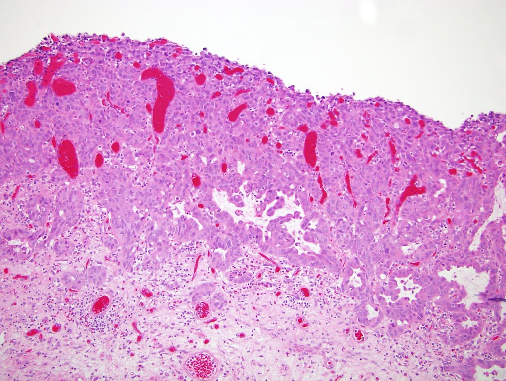
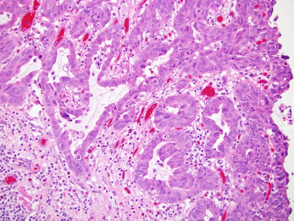
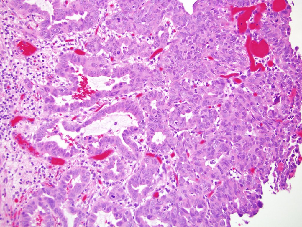
Various images
Positive stains
- MUC5AC (Virchows Arch 2001;439:609), CK7, CK20, S100P
- Variable with GATA3 and p63 (Hum Pathol 2014;45:1473)
Negative stains
- Villin (Arch Pathol Lab Med 2002;126:1057), CDX2 (Arch Pathol Lab Med 2007;131:1244)
- Uroplakin and thrombomodulin are often negative (Hum Pathol 2014;45:1473)
Differential diagnosis
- Collecting duct carcinoma of the kidney: does not contain conventional urothelial carcinoma, usually positive for PAX8, CD10 and Vimentin
- Cystitis cystica and glandularis: does not have malignant component
- Metastatic adenocarcinoma: clinical history, site specific antibodies
- Microcystic and nested variants of urothelial carcinoma: these may have lumina with central necrotic debris but not true glandular formation
- Urothelial carcinoma of renal pelvis with intratubular spread: smooth contours, limited to pyramids of medulla, no invasion or desmoplasia (Am J Clin Exp Urol 2014;2:102)
Additional references
