download - CINVESTAV Unidad Mérida
download - CINVESTAV Unidad Mérida
download - CINVESTAV Unidad Mérida

You also want an ePaper? Increase the reach of your titles
YUMPU automatically turns print PDFs into web optimized ePapers that Google loves.
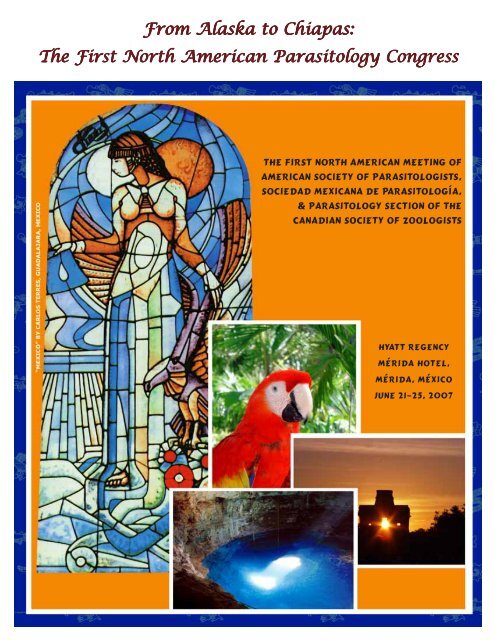
From From Alaska Alaska to to Chiapas:<br />
Chiapas:<br />
The The First First North North American American Parasitology Parasitology Congress<br />
Congress<br />
THE FIRST NORTH AMERICAN MEETING OF<br />
AMERICAN SOCIETY OF PARASITOLOGISTS,<br />
SOCIEDAD MEXICANA DE PARASITOLOGÍA,<br />
& PARASITOLOGY SECTION OF THE<br />
CANADIAN SOCIETY OF ZOOLOGISTS<br />
Hyatt Regency<br />
<strong>Mérida</strong> Hotel,<br />
<strong>Mérida</strong>, México<br />
June 21–25, 2007
The American Society of Parasitologists (ASP),<br />
the Sociedad Mexicana de Parasitología (SMP),<br />
and the Parasitology Section of the Canadian Society of Zoologists (PS-CSZ)<br />
gratefully acknowledge these companies and foundations<br />
for their support and sponsorship of<br />
the First North American Parasitology Congress.<br />
2<br />
THANK THANK Y YYOU!<br />
Y OU!<br />
CORPORA CORPORATE CORPORA TE SPONSORS<br />
SPONSORS<br />
AEROPUERTO INTERNACIONAL DE MÉRIDA, ASUR S.A. DE C.V.<br />
BIO-RAD, MÉXICO DF, MÉXICO<br />
CAMARA NACIONAL DE LA INDUSTRIA DE LA TRANSFORMACIÓN (CANACINTRA) DELEGACIÓN,<br />
YUCATÁN, MÉRIDA, YUCATÁN, MÉXICO<br />
CENTRO DE INVESTIGACIÓN Y DE ESTUDIOS AVANZADOS DEL IPN, UNIDAD MÉRIDA, MÉRIDA, YUCATÁN,<br />
MÉXICO<br />
CONSEJO ESTATAL DE CIENCIA Y TECNOLOGÍA DEL GOBIERNO DE YUCATÁN (CONCYTEY), MÉRIDA,<br />
YUCATÁN, MÉXICO<br />
CONSEJO NACIONAL DE CIENCIA Y TECNOLOGÍA (CONACYT), MÉXICO DF, MÉXICO<br />
CONTINENTAL AIRLINES, INC.<br />
EMBREX, DURHAM, NC, USA<br />
FACULTAD DE MEDICINA, UNIVERSIDAD NACIONAL AUTÓNOMA DE MÉXICO (UNAM), MÉXICO DF,<br />
MÉXICO<br />
GOBIERNO DE YUCATÁN, MÉRIDA, YUCATÁN, MÉXICO<br />
H. AYUNTAMIENTO DE MÉRIDA, YUCATÁN, MÉXICO<br />
INSTITUTO TECNOLÓGICO DE MÉRIDA, MÉRIDA, YUCATÁN, MÉXICO<br />
LAS CERVEZAS MODELO DEL SURESTE S. A. DE C. V., MÉRIDA, YUCATÁN, MÉXICO<br />
MÉXICANA DE AVIACIÓN S. A. DE C.V.<br />
NOVUS INTERNATIONAL, INC., ST. LOUIS MO, USA<br />
PRODUCTOS DE HARINA DONDE S.A. DE C.V., MÉRIDA, YUCATÁN, MÉXICO<br />
REFRESQUERA DE YUCATÁN S.A. DE C.V., MÉRIDA, YUCATÁN, MÉXICO<br />
REPOSTERÍA FINA TERE CAZOLA S.A. DE C.V., MÉRIDA, YUCATÁN, MÉXICO<br />
SCHERING–PLOUGH ANIMAL HEALTH, SUMMIT NJ, USA<br />
SECRETARÍA DE DESARROLLO RURAL Y PESCA DEL GOBIERNO DEL ESTADO DE YUCATÁN, MÉRIDA,<br />
YUCATÁN, MÉXICO<br />
SECRETARÍA DE EDUCACIÓN PÚBLICA DEL GOBIERNO DEL ESTADO DE YUCATÁN, MÉRIDA, YUCATÁN,<br />
MÉXICO<br />
UNIVERSIDAD AUTÓNOMA DE YUCATÁN (UADY), MÉRIDA, YUCATÁN, MÉXICO<br />
UNIVERSIDAD MARISTA, MÉRIDA, YUCATÁN, MÉXICO<br />
VETECH LABORATORIES, INC., GUELPH, ONTARIO, CANADA<br />
Scientific Program Committee<br />
Ana Flisser Steinbruch (SMP)<br />
Donald W. Duszynski (ASP)<br />
Guadalupe Ortega Pierres (SMP)<br />
Patricia Talamás Rohana (SMP)<br />
Local Organizing Committee<br />
Victor Manuel Vidal Martínez<br />
Ma. Leopoldina Aguirre Macedo<br />
Rossanna del Pilar Rodríguez Canul<br />
Reyna Rodríguez Olayo<br />
Felipe Torres Acosta<br />
Sergio Guillén Hernández<br />
Hugo Antonio Ruiz Piña<br />
Eduardo Alfonso Rebollar Téllez<br />
Roberto Cedillo Rivera
the First North American Meeting of<br />
American Society of Parasitologists,<br />
sociedad Mexicana de parasitología,<br />
& Parasitology section of the<br />
canadian Society of Zoologists<br />
From From Alaska Alaska to to Chiapas:<br />
Chiapas:<br />
The The First First North North American<br />
American<br />
Parasitology Parasitology Congress<br />
Congress<br />
Hyatt Regency<br />
<strong>Mérida</strong> Hotel,<br />
<strong>Mérida</strong>, México<br />
June 21–25, 2007<br />
3
JUNE JUNE 21 21 (Thursday)<br />
(Thursday)<br />
8:00 a.m. – Noon ASP Council Meeting Uxmal 1-2 8<br />
2:15 – 4:00 p.m. ASP & SMP Presidents’ Addresses Regency 2-3-4 8<br />
4:00 – 4:30 p.m. Break<br />
4:30 – 6:30 p.m. ASP President’s Symposium Regency 2-3-4 9<br />
7:30 – 10:30 p.m. Welcoming Reception Hotel Pool Area 9<br />
JUNE UNE 22 22 (F (Friday) (F riday)<br />
8:00 a.m – 12:30 p.m. Poster Session-1 Set Up Regency 1 9<br />
8:30 – 11:00 a.m. ASP Student Paper Competition-1 Uxmal 1-2 9<br />
9:00 – 11:00 a.m. Symposium 2: Parasite Genome Regency 2 10<br />
9:00 – 11:00 a.m. Symposium 3: Freshwater Fish Parasites<br />
in North America Regency 3 11<br />
9:00 – 11:00 a.m. Symposium 4: Drug Resistance &<br />
Chemotherapy Regency 4 11<br />
9:00 – 11:00 a.m. Symposium 5: Signaling Chichén Itzá 1-2 11<br />
11:00 – 11:30 a.m. Coffee Break Preconvene Area<br />
11:30 a.m. – 12:30 p.m. Plenary Session 1 Regency 2 12<br />
11:30 a.m. – 12:30 p.m. ASP Student Paper Competition-2 Regency 3 12<br />
12:30 – 2:30 p.m. Poster Session-1 & Lunch Regency 1 13<br />
2:30 – 3:00 p.m. Coffee Break Preconvene Area<br />
3:00 – 4:30 p.m. Removal of Posters (Session-1) Regency 1 19<br />
3:00 – 4:30 p.m. Ecology-1 Regency 2 19<br />
3:00 – 4:30 p.m. Biochemistry, Genetics, Molecular<br />
Biology-1 Regency 3 19<br />
3:00 – 4:30 p.m. Host–Parasite Interactions-1 Regency 4 20<br />
3:00 – 4:30 p.m. Taxonomy, Systematics, Phylogeny-1 Chichén Itzá 1-2 21<br />
3:00 – 4:30 p.m. ASP Student Paper Competition-3 Uxmal 1-2 21<br />
4:30 – 5:00 p.m. Break<br />
5:00 – 6:00 p.m. Eminent Parasitologist Lecture Regency 2-3 22<br />
5:00 – 7:30 p.m. Auction Set Up Regency 3-4 22<br />
(Regency 3 after 6:00 p.m)<br />
7:30 – 8:30 p.m. ASP–SMP Auction Preview Regency 3-4<br />
8:30 – 10:00 p.m. ASP–SMP Auction Regency 3-4 22<br />
JUNE JUNE 23 23 (Saturday)<br />
(Saturday)<br />
8:00 a.m – 12:30 p.m. Poster Session-2 Set Up Regency 1 22<br />
8:30 – 10:45 a.m. ASP Student Paper Competition-4 Uxmal 1-2 23<br />
9:00 – 11:00 a.m. Symposium 6: Proteomics Regency 2 23<br />
9:00 – 11:00 a.m. Symposium 7: Innate Immunity Regency 3 24<br />
9:00 – 11:00 a.m. Symposium 8: Malaria Vectors Regency 4 24<br />
9:00 – 11:00 a.m. Symposium 9: Water-borne Parasites Chichén Itzá 1-2 25<br />
11:00 – 11:30 a.m. Coffee Break Preconvene Area<br />
11:30 a.m. – 12:30 p.m. Plenary Session 2 Regency 2 25<br />
11:30 a.m. – 12:45 p.m. Ecology-2 Regency 3 25<br />
12:30 – 2:30 p.m. Poster Session-2 & Lunch Regency 1 26<br />
2:30 – 3:00 p.m. Coffee Break Preconvene Area<br />
3:00 – 4:30 p.m. Removal of Posters (Session-2) Regency 1 32<br />
3:00 – 6:00 p.m. Immunology-1 Regency 2 33<br />
3:00 – 6:00 p.m. Ecology-3 Regency 3 33<br />
3:00 – 5:45 p.m. ASP Student Paper Competition-5 Regency 4 35<br />
3:00 – 5:00 p.m. Genetics, Molecular Biology-2 Chichén Itzá 1-2 36<br />
3:00 – 5:15 p.m. Chemotherapy, Drug Resistance Uxmal 1-2 36<br />
4<br />
Time ime Activity/F Activity/Function<br />
Activity/F unction Meeting Meeting Room Room Page age age No.<br />
No.
Time ime Activity/F Activity/Function<br />
Activity/F unction Meeting Meeting Meeting Room Room Page age No.<br />
No.<br />
JUNE JUNE 23 23 (Saturday (Saturday, (Saturday (Saturday,<br />
, continued)<br />
continued)<br />
7:00 – 10:00 p.m. Folkloric Ballet of the Central Courtyard, 37<br />
University of Yucatán University of Yucatán<br />
JUNE JUNE JUNE 24 24 (Sunday)<br />
(Sunday)<br />
8:00 a.m – 12:30 p.m. Poster Session-3 Set Up Regency 1 38<br />
9:00 – 11:00 a.m. Symposium 10: Paleoparasitology Regency 2 38<br />
9:00 – 11:00 a.m. Symposium 11: Parasite Problems in<br />
Wild, Domestic or Cultured Hosts Regency 3 38<br />
9:00 – 11:00 a.m. Symposium 12: Coccidosis Conference Chickén Itzá 1-2 39<br />
9:00 – 11:00 a.m. Symposium 13: ASP–SMP Students Regency 4 39<br />
11:00 – 11:30 a.m. Coffee Break Preconvene Area<br />
11:30 a.m. – 12:30 p.m. Plenary Session 3 Regency 2 39<br />
11:30 a.m. – 12:30 p.m. Host–Parasite Interactions-2 Regency 3 40<br />
11:30 a.m. – 12:30 p.m. Life Cycles, Epidemiology-1 Regency 4 40<br />
11:30 a.m. – 12:30 p.m. Taxonomy, Systematics, Phylogeny-2 Chickén Itzá 1-2 41<br />
12:30 – 2:30 p.m. Poster Session-3 & Lunch Regency 1 41<br />
2:30 – 3:00 p.m. Coffee Break Preconvene Area<br />
3:00 – 4:30 p.m. Removal of Posters (Session-3) Regency 1 48<br />
3:00 – 5:00 p.m. Immunology-2 Regency 2 48<br />
3:00 – 6:00 p.m. Host–Parasite Interactions-3 Regency 3 49<br />
3:00 – 6:00 p.m. Life Cycles, Epidemiology-2 Regency 4 50<br />
3:00 – 5:30 p.m. Taxonomy, Systematics, Phylogeny-3 Chickén Itzá 1-2 51<br />
7:00 – 10:00 p.m. Banquet Quinta Montes Molina 52<br />
JUNE JUNE JUNE 25 25 25 (Monday)<br />
(Monday)<br />
9:00 – 10:00 a.m. Bueding/Von Brand Lecture Regency 3-4 52<br />
10:00 – Noon ASP Awards & Business Meeting Regency 3-4 52<br />
10:00 – Noon SMP Awards & Business Meeting Regency 2 53<br />
<br />
5
6<br />
HY HYATT HY TT REGENCY REGENCY MÉRID MÉRIDA MÉRID A HO HOTEL HO TEL<br />
Floor Floor Plans<br />
Plans
FOR FOR Y YYOUR<br />
Y OUR INFORMA INFORMATION<br />
INFORMA TION<br />
Registration<br />
Registration<br />
(Main Lobby, near the gazebo behind the Hyatt Registration desk)<br />
1:00–5:00 p.m., Thursday, June 21<br />
8:00 a.m.–5:00 p.m., Friday, June 22<br />
8:00 a.m.–5:00 p.m., Saturday, June 23<br />
Speak Speaker Speak er Ready Ready Room Room (Loltún)<br />
8:00 a.m.–5:00 p.m., Thursday, Friday, Saturday and Sunday<br />
Coffee Coffee Breaks Breaks (Preconvene Area)<br />
Friday, Saturday and Sunday<br />
11:00–11:30 a.m. and 2:30–3:00 p.m.<br />
Poster oster Sessions Sessions (Regency 1)<br />
Poster Sessions with Lunch:<br />
Friday, Saturday and Sunday, 12:30–2:30<br />
Set Up: 8:00–12:30 each day<br />
Removal: 3:00–4:30 each day<br />
Room Room Locations<br />
Locations<br />
Loltún First Floor<br />
Regency 1, 2, 3 & 4 Second Floor<br />
Chickén Itzá 1-2 Second Floor<br />
Uxmal 1-2 Second Floor<br />
Welcoming elcoming Reception Reception<br />
Reception (Hotel Pool Area)<br />
Thursday evening from 7:30–10:30 p.m.<br />
Other Other Meetings<br />
Meetings<br />
Friday, June 22 ASP–SMP Student Social, Mambo Café<br />
10:30 p.m.– (<br />
Saturday, June 23 ASP Journal of Parasitology Editorial Board Breakfast, Chichén Itzá 1-2<br />
7:00–8:30 a.m.<br />
Symbol Symbol Usage<br />
Usage<br />
† denotes a Student Competition Paper (ASP Oral and SMP Poster).<br />
Ground Ground T TTransportation<br />
T ransportation<br />
Transportation from the airport to the hotel: The airport is about 15 km from the Hyatt. The<br />
Local Organizing Committee (LOC) will provide ground transportation from the airport to the<br />
Hyatt from noon June 20 to midnight June 21. The bus will be available every hour outside the<br />
airport. There is also a 24-hour taxi service from the airport costing ~$13.50 USD one way.<br />
LOC telephone numbers are: ++52 9999 009646 (Victor Vidal), ++ 52 9999 009633 (Leo<br />
Aguirre), and ++52 9991 592598 (Reyna Rodríguez).<br />
7
THURSDAY MORNING, JUNE 21<br />
8:00–Noon ASP COUNCIL MEETING, Uxmal 1-2.<br />
Presiding: S.G. Kayes, University of South Alabama, Mobile AL, USA<br />
THURSDAY AFTERNOON, JUNE 21<br />
2:15–2:30 FORMAL WELCOME TO MÉXICO, TO MÉRIDA, AND TO ALL PARTICI-<br />
PANTS, Regency 2-3-4.<br />
Presiding: V.M. Vidal-Martínez and L. Aguirre-Macedo, <strong>CINVESTAV</strong>, <strong>Mérida</strong>, Yucatán, México<br />
Welcome:<br />
8<br />
DR. JOSÉ NARRO, Director, Faculty of Medicine, UNAM, México DF, México<br />
DR. JOSÉ ANTONIO DE LA PEÑA, Director Adjunto de Desarrollo Científico y<br />
Académico, CONACYT<br />
2:30–4:00 ASP & SMP PRESIDENTS’ ADDRESSES, Regency 2-3-4.<br />
2:30 WELCOME. V.M. Vidal-Martínez and M.L. Aguirre-Macedo.<br />
2:40 INTRODUCTION OF SMP PRESIDENT ANA FLISSER, UNAM, México DF,<br />
México.<br />
D. Correa, Instituto Nacional de Pediatria, SSA, México DF, México<br />
Paper<br />
Time No.<br />
DR. ANA FLISSER, SMP President,<br />
Microbiologia y Parasitologia,<br />
Facultad de Medicina, UNAM,<br />
México DF, México<br />
DR. STEPHEN KAYES, ASP President,<br />
University of South Alabama,<br />
Mobile AL, USA<br />
2:50 1 A NATIONAL MODEL FOR THE CONTROL OF A PARASITIC DISEASE:<br />
HUMAN CYSTICERCOSIS IN MEXICO. A. Flisser*, J. Narro, J. Calderón and G.<br />
Martínez.<br />
3:20 INTRODUCTION OF ASP PRESIDENT STEPHEN G. KAYES, University of<br />
South Alabama, Mobile AL.<br />
R.E. Kuhn, Wake Forest University, Winston-Salem NC, USA
3:30 2 CONVENTIONAL WISDOM AND A TALE OF TWO CYTOKINES. S.G. Kayes.<br />
4:00–4:30 BREAK.<br />
4:30–6:30 SYMPOSIUM 1: ASP PRESIDENT’S SYMPOSIUM, Regency 2-3-4.<br />
Presiding: S.G. Kayes, University of South Alabama, Mobile AL, USA<br />
Theme: Global Climate Change and Parasitic Infections.<br />
Paper<br />
Time No.<br />
4:30 3 GLOBAL CLIMATE CHANGE: CAUSES AND CONSEQUENCES. T.M. Hall.<br />
5:00 4 CLIMATE CHANGE AND PARASITISM IN ARCTIC AND SUBARCTIC ECO-<br />
SYSTEMS. S.J. Kutz*, R. Peacok and D. Bender.<br />
5:30 5 CLIMATE CHANGE, VECTOR-BORNE AVIAN DISEASES AND ENDEMIC<br />
HAWAIIAN FOREST BIRDS—WHAT WILL THE FUTURE BRING? C.T.<br />
Atkinson*, B.L. Woodworth, D.A. Lapointe and M.D. Samuel.<br />
6:00 6 CLIMATE CHANGE AS A DRIVER OF INFECTIOUS DISEASE CHANGE. K.D.<br />
Lafferty.<br />
THURSDAY EVENING, JUNE 21<br />
7:30–10:30 WELCOMING RECEPTION, Pool Area.<br />
FRIDAY MORNING, JUNE 22<br />
8:00–12:30 POSTER SESSION-1: SET UP, Regency 1.<br />
Authors of posters numbered 39–113 set up their posters.<br />
8:30–11:00 ASP STUDENT PAPER COMPETITION-1, Uxmal 1-2.<br />
Presiding: L. Couch, The University of New Mexico, Albuquerque NM, USA<br />
R. Rodríguez-Canul, <strong>CINVESTAV</strong>, <strong>Mérida</strong>, Yucatán, México<br />
Paper<br />
Time No.<br />
8:30 7† PROTEOMIC ANALYSIS OF SPOROZOITES REVEAL HIGHLY CONSERVED<br />
TRAP-LIKE MOLECULES IN TWO STRAINS OF EIMERIA MAXIMA M6 AND<br />
GS. S.A. El-Ashram* and J.R. Barta.<br />
8:45 8† PARASITE COMMUNITIES DISCERN DISTINCT PACIFIC SARDINE<br />
(SARDINOPS SAGAX) POPULATIONS IN THE CALIFORNIA CURRENT. R.E.<br />
Baldwin* and K.C. Jacobson.<br />
9
9:00 9† LIFE-HISTORY COST OF TREMATODE INFECTION IN HELISOMA ANCEPS<br />
USING MARK-RECAPTURE IN CHARLIE’S POND. N.J. Negovetich* and G.W.<br />
Esch.<br />
9:15 10† THE ENERGETIC COSTS OF PARASITISM IN AN INTERMEDIATE HOST. S.E.<br />
Lettini* and M.V. Sukhdeo.<br />
9:30 11† PATTERNS OF EUGREGARINE DIVERSITY IN DAMSELFLIES IN FOUR<br />
EAST TEXAS PONDS. J.C. Garcia*, T.J. Cook and R.E. Clopton.<br />
9:45 12† PREDICTING WEST NILE VIRUS (WNV) DYNAMICS USING LOCAL HABI-<br />
TAT CHARACTERISTICS. W.D. Rossiter*, M. Vitullo and M.V. Sukhdeo.<br />
10:00 13† POPULATION DYNAMICS OF DAUBAYLIA POTOMACA (NEMATODA:<br />
RHABDITIDA) IN HELISOMA ANCEPS. L.E. Camp*, N.J. Negovetich, G.W.<br />
Esch and H.E. Eure.<br />
10:15 14† HOW LARGE IS THE HAND INSIDE THE PUPPET? THE EVOLUTIONARY<br />
ECOLOGY OF INFECTION MASS OF 15 TREMATODE PARASITIC CASTRA-<br />
TORS OF THE ESTUARINE SNAIL, CERITHIDEA CALIFORNICA. R.F.<br />
Hechinger*, K.D. Lafferty, F.T. Mancini III and A.M. Kuris.<br />
10:30 15† COPROLOGICAL AND SEROLOGICAL DIAGNOSIS OF ANOPLOCEPHALA<br />
PERFOLIATA INFECTION IN SOUTHERN ALBERTA (CANADA) HORSES:<br />
PRELIMINARY DATA. S.L. Skotarek*, C.P. Goater and D.D. Colwell.<br />
10:45 16† LIFE CYCLE VARIATION IN THE GENUS RHABDIAS: A TALE OF SNAKES<br />
AND FROGS. G.J. Langford* and J. Janovy, Jr.<br />
9:00–11:00 SYMPOSIUM 2: PARASITE GENOME, Regency 2.<br />
Presiding: R. Hernández, UNAM, México DF, México<br />
K. Miska, ARS, USDA, Beltsville MD, USA<br />
Paper<br />
Time No.<br />
9:00 17 THE GENOME PROJECT OF TAENIA SOLIUM. A. Garcia-Rubio, R.J. Bobes,<br />
J.C. Carrero-Sánchez, M.A. Cevallos, K. Estrada, J.L. Fernández, G. Fragoso, P.<br />
Gaytán, V.M. González, L. Jiménez, V.M. José, M.S. Juárez, A. Landa, C.<br />
Larralde, L. Mendoza, J. Morales-Montor, E. Morett, E.L. Sciutto, X. Soberón, P.<br />
De La Torre, V. Valdés, J. Yánez and J.P. Laclette*.<br />
9:30 18 PARASITIC NEMATODES—FROM GENOMES TO CONTROL. M. Mitreva*, Y.<br />
Yin, J. Martin, S. Abubucker and R. Wilson.<br />
10:00 19 A MULTIDISCIPLINARY APPROACH TO UNDERSTANDING THE ROLE OF<br />
SAND FLY PROTEINS IN LEISHMANIA TRANSMISSION. J.G. Valenzuela*, F.<br />
Oliveira, J.M. Anderson, S. Kamhawi, R. Jochim, C. Teixeira, R. Gomes, D.<br />
Elnaiem and N. Collin.<br />
10:30 20 COMPARATIVE GENOMICS OF PARASITIC PROTISTS: BETTER THE BUG<br />
YOU KNOW. J.M. Carlton.<br />
10
9:00–11:00 SYMPOSIUM 3: FRESHWATER FISH PARASITES IN NORTH AMERICA,<br />
Regency 3.<br />
Presiding: G. Pérez-Ponce de León, UNAM, México DF, México<br />
A. Choudhury, St. Norbert College, DePere WI, USA<br />
Paper<br />
Time No.<br />
9:00 21 COLONIZATION OF FRESHWATER FISHES BY INTRODUCED PARASITES.<br />
W.F. Font.<br />
9:30 22 NORTH AMERICAN FRESHWATER FISHES AND THEIR PARASITES: PAT-<br />
TERNS AND PROCESSES IN BIOGEOGRAPHY. A. Choudhury.<br />
10:00 23 HELMINTH DISEASES IN AQUACULTURE, WITH AN EMPHASIS ON CAT-<br />
FISH, NEMATODES AND TREMATODES. R.M. Overstreet.<br />
10:30 24 PARASITES, FOOD WEBS AND ECOSYSTEM STRESS. D.J. Marcogliese.<br />
9:00–11:00 SYMPOSIUM 4: DRUG RESISTANCE AND CHEMOTHERAPY,<br />
Regency 4.<br />
Presiding: P. Mendoza de Gives, CENID-PAVET-INIFAP, Jiutepec, Morelos, México<br />
E.S. Loker, The University of New Mexico, Albuquerque NM, USA<br />
Paper<br />
Time No.<br />
9:00 25 PERTURBING THE DIMER INTERFACE OF TRIOSEPHOSPHATE ISOMER-<br />
ASE AND ITS EFFECT ON TRYPANOSOMA CRUZI. V. Olivares-Illana, A.<br />
Rodríguez-Romero, I.D. Becker, M. Berzunza-Cruz, J. García, N. Cabrera, F.<br />
López-Calahorra, M. Tuena De Gómez-Puyou, A. Gómez-Puyou and R. Pérez-<br />
Montfort*.<br />
9:30 26 ANTIPARASITIC DRUG DISCOVERY VIA MECHANISM-BASED SCREENING:<br />
WILL IT SUCCEED? T.G. Geary.<br />
10:00 27 UNDERSTANDING THE DIRECT AND INDIRECT EFFECTS OF SUPPLE-<br />
MENTARY FEEDING TO REDUCE THE NEED FOR ANTHELMINTIC TREAT-<br />
MENTS IN SMALL RUMINANTS. J.F. Torres-Acosta*, A.J. Aguilar-Caballero,<br />
C.A. Sandoval-Castro and H. Hoste.<br />
10:30 28 MECHANISMS AND MARKERS OF MACROCYCLIC LACTONE RESISTANCE<br />
IN NEMATODE PARASITES OF HUMANS AND ANIMALS. R. Prichard.<br />
9:00–11:00 SYMPOSIUM 5: SIGNALING, Chichén Itzá 1-2.<br />
Presiding: B. Espinoza, UNAM, México DF, México<br />
J. Hawdon, George Washington Medical Center, Washington DC, USA<br />
11
Paper<br />
Time No.<br />
9:00 29 TLR2 ACTIVATION IN NK CELLS OF PATIENTS INFECTED WITH LEISHMA-<br />
NIA MEXICANA. I.D. Becker*, I.C. Cañeda-Guzmán, E.A. Fernandez-Figueroa,<br />
N.L. Salaiza-Suazo and M.M. Aguirre-Garcia.<br />
9:30 30 PROINFLAMMATORY RESPONSES TO MALARIA INFECTION AND CELL<br />
SIGNALING MECHANISMS. C.D. Gowda.<br />
10:00 31 ACTIN STRUCTURAL ORGANIZATION IN ENTAMOEBA HISTOLYTICA<br />
TROPHOZOITES BY FIBRONECTIN SIGNALING. P. Talamás-Rohana*, A.<br />
Rios, V. Hernández-Ramírez and J.L. Rosales-Encina.<br />
10:30 32 MOLECULAR SIGNALING IN LARVAL SCHISTOSOMA MANSONI: GENE<br />
PROFILING THE MIRACIDIUM-TO-SPOROCYST TRANSITION. T.P. Yoshino.<br />
11:00–11:30 COFFEE BREAK, Preconvene Area.<br />
11:30–12:30 PLENARY SESSION 1, Regency 2.<br />
Presiding: J. Figueroa, CENID-PAVET-INIFAP, Jiutepec, Morelos, México<br />
A.M. Kuris, University of California, Santa Barbara CA, USA<br />
Paper<br />
Time No.<br />
11:30 33 CAVEOLIN-1 AND CHOLESTEROL PLAY A MAJOR ROLE IN THE DEVEL-<br />
OPMENT OF TRICHINELLA SPIRALIS OOCYTES. M.G. Ortega-Pierres.<br />
12:00 34 POCKET GOPHERS AND CHEWING LICE: NEW DISCOVERIES IN AN OLD<br />
SYSTEM. M.S. Hafner.<br />
11:30–12:30 ASP STUDENT PAPER COMPETITION-2, Regency 3.<br />
Presiding: R.S. Seville, University of Wyoming, Casper WY, USA<br />
V.M. Vidal-Martínez, <strong>CINVESTAV</strong>-IPN, <strong>Mérida</strong>, Yucatán, México<br />
Paper<br />
Time No.<br />
11:30 35† REEF FISHES HAVE HIGHER PARASITE DIVERSITY AT UNFISHED<br />
PALMYRA ATOLL COMPARED TO FISHED KIRITIMATI ISLAND. K.D.<br />
Lafferty, J.C. Shaw* and A.M. Kuris.<br />
11:45 36† FOOD WEB STABILITY DRIVES PARASITE SPECIES DIVERSITY. T.K. Anderson*<br />
and M.V. Sukhdeo.<br />
12:00 37† PARASITES OF FISHES FROM THE COLORADO RIVER AND SELECTED<br />
TRIBUTARIES IN GRAND CANYON, ARIZONA. C. Linder*, T. Hoffnagle, B.<br />
Persons, A. Choudhury and R. Cole.<br />
12
12:15 38† LEAVING THE NEST: THE GENETIC DISTRIBUTION OF PARASITES IN<br />
SNAILS THAT SERVE AS FIRST AND SECOND INTERMEDIATE HOSTS. J.T.<br />
Detwiler* and D.J. Minchella.<br />
FRIDAY AFTERNOON, JUNE 22<br />
12:30–2:30 POSTER SESSION-1 AND LUNCH, Regency 1.<br />
Authors stand by their posters.<br />
SMP SMP STUDENT STUDENT POSTER POSTER COMPETITION<br />
COMPETITION<br />
39† PATTERN OF PROTEIN CARBONYLATION FOLLOWING OXIDATIVE STRESS IN<br />
TRYPANOSOMA CRUZI. R. Martínez-Espinosa*, I. Martínez and B. Espinoza.<br />
40† CHARACTERIZATION AND EVALUATION OF AN ANTIGENIC EXTRACT FOR<br />
DIAGNOSIS OF TRYPANOSOMA CRUZI INFECTION IN SERA BY ELISA ASSAY.<br />
PRELIMINARY RESULTS. M.I. Bucio-Torres, E. Torres-Gutiérrez, E. Amador-Gaytán *,<br />
M. Cabrera-Bravo, A.L. Ruiz-Hernández, Y. Guevara-Gómez, L. Ruiz-González, J. Rojo-<br />
Medina, G.S. García-De La Torre, L. González-López, G.E. Rojas-Wastavino, M.O.<br />
Vences-Blanco, M. Gutiérrez-Quiroz and P.M.S. Salazar-Schettino.<br />
41† CLONING AND PURIFICATION OF A PROTEIN PHOSPHATASE 2C FROM LEISH-<br />
MANIA MEXICANA. A. Navarrete-Mena*, N. Cabrera-González, R. Pérez-Montfort,<br />
I.D. Becker and M.M. Aguirre-García.<br />
42† MECHANISMS OF ACTION OF DEHYDROEPIANDROSTERONE ON ENTAMOEBA<br />
HISTOLYTICA. C. Cervantes-Rebolledo*, E. Saavedra-Lira, M. Nequiz, J.P. Laclette and<br />
J.C. Carrero-Sánchez.<br />
43† IDENTIFICATION OF SURFACE PROTEINS OF TRYPOMASTIGOTES OF MEXICAN<br />
STRAINS OF TRYPANOSOMA CRUZI. M. Martínez-Velasco*, H. Lanz-Mendoza, G.<br />
Hurtado and B. Espinoza-Gutiérrez.<br />
44† DISTRIBUTION OF CYTOSKELETAL PROTEINS IN FLAME CELLS OF TAENIA<br />
SOLIUM CYSTICERCI. L.E. Valverde Islas*, O.A. Reynoso Ducoing, E. Vega Munguía<br />
and J.R. Ambrosio-Hernández.<br />
45† EHADH IS AN ENTAMOEBA HISTOLYTICA SURFACE BRO1 DOMAIN-CONTAIN-<br />
ING PROTEIN INVOLVED IN VESICLE BIOGENESIS. C. Bañuelos*, G. García-<br />
Rivera, I. López-Reyes, S. Castellanos-Castro, L. Mendoza, A. González-Robles and E.<br />
Orozco.<br />
46† TRICHINELLA SPIRALIS OOCYTE MATURATION IS INHIBITED BY CYCLOSPORIN<br />
A IN RATS INFECTED WITH THIS PARASITE. R. Hernández-Bello*, R.M. Bermúdez-<br />
Cruz, R. Fonseca-Liñán, P. García-Reyna, P. Boireau and M.G. Ortega-Pierres.<br />
47† LEISHMANIA MEXICANA-SPECIFIC ANTIGENS FOR SEROLOGIC DIAGNOSIS. N.<br />
Robles-Briones*, A. Ruiz-Remigio and I.D. Becker.<br />
48† IN VIVO EFFICACY OF NITAZOXANIDE AGAINST TOXOPLASMA GONDII. M.<br />
Galvan-Ramírez, G. Sánchez González *, L.R. Rodríguez Pérez, R.A. Franco Topete and<br />
M.A. Ramirez Herrera.<br />
13
49† PROTEOMICAL EVALUATION OF NEW POTENTIAL BENZIMIDAZOLE DERIVA-<br />
TIVES AGAINST GIARDIA INTESTINALIS. C.A. Méndez-Cuesta*, L. Velázquez-<br />
Márquez, M.A. Dea-Ayuela, R. Castillo-Bocanegra, F. Hernández-Luis, M.A. Hernández-<br />
Campos, L. Yépez-Mulia, F. Bolás-Fernández, O.A. Reynoso-Ducoing and J.R.<br />
Ambrosio-Hernández.<br />
50† IN VITRO ANTHELMINTIC ACTIVITY OF PLANT EXTRACTS FROM TROPICAL<br />
TANNINIFEROUS TREES AGAINST TRICHOSTRONGYLUS COLUBRIFORMIS. M.A.<br />
Alonso-Díaz*, J.F. Torres-Acosta, C.A. Sandoval-Castro, A.J. Aguilar-Caballero and H.<br />
Hoste.<br />
51† VARIATION IN THE P-GLYCOPROTEIN (PGP) GENE FROM ONCHOCERCA VOLVU-<br />
LUS MEXICAN ISOLATES. S. González-Guzmán*, G. Sánchez-Tejeda, J. Méndez-Galvan<br />
and A. Monroy-Ostria.<br />
52† COMMUNITIES OF HELMINTH PARASITES OF BUFO MARINUS (LINNAEUS, 1758)<br />
AND BUFO VALLICEPS (WIEGMANN, 1833) (ANURA: BUFONIDAE) IN THE LAGU-<br />
NAS DE YALAHAU, YUCATÁN. J.F. Espinola-Novelo* and S. Guillén-Hernández.<br />
53† BEHAVIORAL DISRUPTION ON FIDDLER CRAB, UCA SPECIOSA (IVES, 1891),<br />
CAUSED BY HEXAGLANDULA CORYNOSOMA (TRAVASSOS, 1915) IN THE<br />
CHUBURNÁ LAGOON, NORTHERN YUCATÁN PENINSULA: A PROGRESS RE-<br />
PORT. R.A. Pérez-Campos* and S. Guillén-Hernández.<br />
54† PRELIMINARY REPORT OF THE GENETIC TYPING OF ECHINOCOCCUS GRANU-<br />
LOSUS IN MÉXICO. D.E. Jiménez-González*, U. Rodríguez-Prado, A. López-Saavedra,<br />
L. Herrera, C. Mondragon, P. Mata, A. Flisser, J.J. Martínez-Maya and P.J. Maravilla-<br />
Campillo.<br />
55† DIAGNOSIS OF LYMNAEA SNAIL INFECTION BY FASCIOLA HEPATICA USING<br />
DUPLEX-PCR. C.P. Rico-Torres*, I. Cruz-Mendoza, H. Quiroz-Romero, L.B. Ortíz-<br />
Alegría and D. Correa.<br />
56† TRYPANOSOMA CRUZI SHSP16: THE FIRST MEMBER OF THE ALPHA-CRYSTAL-<br />
LIN-SMALL HEAT SHOCK PROTEIN FAMILY IN TRYPANOSOMATIDS. D. Pérez-<br />
Morales*, P. Ostoa-Saloma and B. Espinoza.<br />
57† IDENTIFICATION OF EHBLM, A PUTATIVE DNA HELICASE IN ENTAMOEBA<br />
HISTOLYTICA. M.S. Charcas-López*, C. López-Camarillo, M. López-Casamichana and<br />
L.A. Marchat.<br />
58† THE ESCRT MACHINERY OF ENTAMOEBA HISTOLYTICA. I. López-Reyes*, C.<br />
Bañuelos and E. Orozco.<br />
59† ENTAMOEBA HISTOLYTICA HAS THE CLEAVAGE FACTOR EHCF IM 25 THAT<br />
COULD BE INVOLVED IN PRE-MRNA 3' END POLYADENYLATION. J. Fernandez-<br />
Retana*, C. López-Camarillo and L.A. Marchat.<br />
60† PFSIR2 SILENCING COMPLEXES PURIFIED BY TANDEM AFFINITY PURIFICATION<br />
FROM PLASMODIUM FALCIPARUM. N.K. Mita-Mendoza*, S. Martínez-Calvillo and R.<br />
Hernández-Rivas.<br />
61† CHARACTERIZATION OF THE KAHRP GENE CORE PROMOTER REGION OF<br />
PLASMODIUM FALCIPARUM. M.E. Aranda-Barradas* and R. Hernández-Rivas.<br />
14
62† TLR2 AND TLR4 POLYMORPHISMS AND THEIR RELATIONSHIP TO CUTANEOUS<br />
LEISHMANIASIS. V. Becerril*, M. Berzunza-Cruz, G. Carrada-Figueroa, M. Maldonado<br />
and I.D. Becker.<br />
63† POSSIBLE ROLE OF A PUTATIVE GLUCOSAMINE-6-PHOSPHATE ISOMERASE OF<br />
ENTAMOEBA HISTOLYTICA IN THE FORMATION OF CYST-LIKE STRUCTURES. H.<br />
Aguilar-Diaz*, J.P. Laclette and J.C. Carrero-Sánchez.<br />
64† TAMOXIFEN TREATMENT INDUCES PROTECTION IN MURINE CYSTICERCOSIS.<br />
J.A. Vargas-Villavicencio*, C. Larralde, M.A. De León-Nava, G. Escobedo and J. Morales-Montor.<br />
65† FOLLOW-UP OF PHYSICAL DISCOMFORT AND HEALTH STATUS OF GOLDEN<br />
HAMSTERS USED AS EXPERIMENTAL DEFINITIVE HOSTS OF TAENIA SOLIUM,<br />
EMPLOYING TWO NON-STEROID DRUGS FOR IMMUNOSUPPRESSION. D.E.<br />
Jiménez-González*, R. Garcia-Cortes, G. Avila-Ramirez, A. Flisser and P.J. Maravilla-<br />
Campillo.<br />
66† PROCHRISTIANELLA SP. PARASITING THE OCTOPUS, OCTOPUS MAYA IN DZILAM<br />
DE BRAVO, YUCATÁN, NORTHERN YUCATÁN PENINSULA. A. López-Struck* and<br />
S. Guillén-Hernández.<br />
67† MIRACIDIA OF F. HEPATICA LYSES PROTEINS AND NUCLEIC ACIDS DURING<br />
INVASION OF THE INTERMEDIATE HOSTS. L.B. Ortíz-Alegría*, C.P. Rico-Torres, I.<br />
Cruz-Mendoza, H. Quiroz-Romero and D. Correa.<br />
68† EVALUATION OF TAENIA SOLIUM CALRETICULIN AS AN ORAL VACCINE IN<br />
EXPERIMENTAL TAPEWORM INFECTION. S. León-Cabrera*, M. Cruz-Rivera, G.<br />
Avila-Ramirez, F. Mendlovic and A. Flisser.<br />
69† CHARACTERIZATION OF THE ENDOCYTIC PATHWAY AND USE AS AN IRON<br />
SOURCE OF FERRITIN BY ENTAMOEBA HISTOLYTICA. F. López-Soto*, M. Reyes-<br />
López, N. León-Sicairos, A. González-Robles and M. de la Garza.<br />
70† LEISHMANIA MEXICANA INHIBITS THE APOPTOSIS IN MONOCYTE-DERIVED<br />
DENDRITIC CELLS. L. Valdes-Reyes*, M. Berzunza-Cruz, N.L. Salaiza-Suazo, M.M.<br />
Aguirre-Garcia, I.D. Becker, J. Moran and L. Gutiérrez-Kobeh.<br />
71† PARTICIPATION OF A PROTEIN TYROSINE PHOSPHATASE FROM LEISHMANIA<br />
MEXICANA IN THE INFECTION OF MACROPHAGES. J. Gómez-Sandoval, A.<br />
Escalona-Montaño, D. Pardavé-Alejandre, R. Cervantes, L. Gutiérrez-Kobeh, M.<br />
Gutiérrez-Quiróz, I.D. Becker and M.M. Aguirre-García*.<br />
72† LEISHMANIA MEXICANA LIPOPHOSPHOGLYCAN (LPG) STIMULATES THE EX-<br />
PRESSION AND FUNCTION OF NITRIC OXIDE SYNTHASE IN MURINE DEN-<br />
DRITIC CELLS. A. Wilkins-Rodríguez*, I.D. Becker and L. Gutiérrez-Kobeh.<br />
73† EFFECT OF SEXUAL AND ADRENAL HORMONES ON THE PROLIFERATION OF<br />
ENTAMOEBA HISTOLYTICA. C. Cervantes-Rebolledo*, J. Morales-Montor, N. Moreno,<br />
M. Nequiz, J.P. Laclette and J.C. Carrero-Sánchez.<br />
74† INDUCTION OF HIGH LEVELS OF TUMOR NECROSIS FACTOR α AND NITRIC<br />
OXIDE BY VIRULENT MEXICAN STRAIN OF TRYPANOSOMA CRUZI. A. Jiménez-<br />
Marin* and B. Espinoza.<br />
15
75† MACROPHAGE MIGRATION INHIBITORY FACTOR PLAYS A ROLE IN LEISHMA-<br />
NIA MEXICANA INFECTION. M. Romero-Grijalva*, I. Rivera-Montoya, L.I. Terrazas-<br />
Valdés and M. Rodríguez-Sosa.<br />
76† INCREASED SUSCEPTIBILITY TO TOXOPLASMA GONDII INFECTION IN AHR-<br />
NULL MICE. M. Rodríguez-Sosa*, L.I. Terrazas-Valdés, I. Rivera-Montoya, G. Elizondo<br />
and L. Vega.<br />
77† TAENIA CRASSICEPS: RELEVANCE OF PARASITE GENETIC CHANGES ON THE<br />
HOST SUSCEPTIBILITY AND IMMUNITY. G. Meneses, R.J. Bobes, E.L. Sciutto* and<br />
G. Fragoso.<br />
78† LOCALIZATION OF TSOL18 AND TSOL45 IN DIFFERENT STAGES OF TAENIA<br />
SOLIUM. J. Martínez-Ocana*, M. Garcia-De-León, C. Gauci, M. Lightowlers and A.<br />
Flisser.<br />
79† PROTEIN TYROSINE PHOSPHATASE FROM ENTAMOEBA HISTOLYTICA MODU-<br />
LATE THE ACTIVATION OF PHAGOCYTIC CELLS. J. Espejel-Zaragoza, A. Ruiz-<br />
Remigio, M. Nequiz, A. Escalona-Montaño, P. Talamás-Rohana and M.M. Aguirre-<br />
García*.<br />
80† CHLOROQUINE HAS AN IMMUNOMODULATORY ROLE IN BALB/C MICE IN-<br />
FECTED WITH PLASMODIUM YOELII 17XL. A. Ramos-Avila*, J.L. Ventura-Gallegos<br />
and M. Legorreta-Herrera.<br />
81† ANALYSIS OF TLR1, TLR2 AND TLR6 IN NK CELLS OF PATIENTS WITH CUTANE-<br />
OUS LEISHMANIASIS. I.C. Cañeda-Guzmán*, G. Carrada-Figueroa, N.L. Salaiza-<br />
Suazo and I.D. Becker.<br />
82† TOWARD TAENIA SOLIUM CONTROL: FIELD TRIAL EVALUATION OF THE<br />
S3PVAC ANTI-CYSTICERCOSIS VACCINE EXPRESSED IN FILAMENTOUS PHAGES.<br />
J. Morales*, J.J. Martínez, M. Hernández, K. Manoutcharian, G. Gevorkian, G. Acero, A.<br />
Blancas, A. Toledo, V.M. Maza, A. Aluja, A. Fleury, G. Fragoso, C. Larralde and E.L.<br />
Sciutto.<br />
83† DEVELOPMENT OF THE LIFE CYCLE OF TAENIA PISIFORMIS USING GOLDEN<br />
HAMSTER AS THE DEFINITIVE EXPERIMENTAL HOST. E. Toral-Bastida*, P.J.<br />
Maravilla-Campillo, A. Garza-Rodríguez, R. Garcia-Cortes, P. Palomares-Pérez, L.<br />
Fernandez-Maya, G. Avila-Ramirez and A. Flisser.<br />
84† VIABILITY OF TRICHINELLA SPIRALIS RECOVERED IN MEAT SUBMITTED TO<br />
DIFFERENT CONDITIONS OF HANDLING AND CONSERVATION. M. Medina-<br />
Lerena*, A. Ramírez-Alvarez, E. Pérez-Torres, C. Pacheco-Gallardo, S. Ruvalcaba-<br />
Barrera and J.L.A. de la Rosa.<br />
85† EFFICACY OF THREE DOSES OF A MEXICAN STRAIN OF DUDDINGTONIA<br />
FLAGRANS CHLAMYDOSPORES AGAINST HAEMONCHUS CONTORTUS LARVAE<br />
IN SHEEP FAECAL CULTURES. N.F. Ojeda-Robertos*, P. Mendoza-De- Gives, J.F.<br />
Torres-Acosta, A. Ayala-Burgos and A.J. Aguilar-Caballero.<br />
86† INFECTION DYNAMICS OF ECTOPARASITES DURING THE HATCHERY OF HY-<br />
BRID TILAPIA “PARGO-UNAM.” M.D. Pérez-Fosado*, M.I. Jiménez-García, M.<br />
Garduño-Lugo, G. Muñoz-Córdova and M.D. Castañeda-Chávez.<br />
16
87† TAENIA SOLIUM: ACTIVE TRANSMISSION OF PIG CYSTICERCOSIS IN SIERRA DE<br />
HUAUTLA, MORELOS. J. Morales*, J.J. Martínez, N. Peña, V.M. Maza, N. Villalobos,<br />
A. Aluja, A. Fleury, G. Fragoso, C. Larralde and E.L. Sciutto.<br />
88† PHYLOGENETIC AND BIOGEOGRAPHICAL RELATIONSHIPS OF SEVERAL POPU-<br />
LATIONS OF RHABDIAS SP. (NEMATODA), PARASITE OF LEPTODACTYLUS<br />
MELANONOTUS (ANURA) FROM MÉXICO. E.A. Martínez-Salazar* and V. León-<br />
Règagnon.<br />
89† GENETIC VARIATION AMONG POPULATIONS OF PHYLLODISTOMUM LACUSTRI<br />
(LOEWEN, 1929), PARASITE OF ICTALURIDS IN NORTH AMERICA, USING SE-<br />
QUENCES OF THE 28S rRNA GENE. R. Rosas-Valdez*, A. Choudhury and G. Pérez-<br />
Ponce De León.<br />
90† FIRST RECORD OF SALSUGINUS ANGULARIS (MUELLER, 1934) BEVERLY-BUR-<br />
TON, 1984 (MONOGENEA: ANCYROCEPHALINAE) PARASITIZING GOODEINAE<br />
FISHES (CYPRINODONTIFORMES: GOODEINAE) ENDEMIC TO MÉXICO. C.A.<br />
Mendoza Palmero* and G. Salgado-Maldonado.<br />
91† ENTOMOLOGICAL INDEXES OF TRIATOMA DIMIDIATA (HEMIPTERA: REDUVI-<br />
IDAE: TRIATOMINE) TO ASSESS RISK TRANSMISSION IN RURAL SAN PEDRO<br />
CHACABAL, MOTUL, YUCATÁN, MÉXICO. I.J. May-Concha*, S.J. Carballo-González,<br />
A. Polanco-Rodríguez, F.J. Escobedo-Ortegón, H.A. Ruiz-Piña, M.A. Barrera-Pérez and<br />
E.A. Rebollar-Téllez.<br />
92† PCR ANALYSIS OF INFECTION RATES OF SANDFLY SPECIES (DIPTERA: PSY-<br />
CHODIDAE: PHLEBOTOMINAE) FROM CALAKMUL, CAMPECHE, MÉXICO AND<br />
THEIR PUTATIVE ROLE AS VECTORS OF LEISHMANIA MEXICANA<br />
(KINETOPLASTIDA: TRYPANOSOMATIDAE). A. Pech-May*, F.J. Escobedo-Ortegón,<br />
M. Berzunza-Cruz and E.A. Rebollar-Téllez.<br />
93† DEXAMETHASONE AND PGE 2 MODULATION OF THE IMMUNE RESPONSE IN<br />
FAT BODY AND MIDGUT OF ANOPHELES ALBIMANUS. F.L. García-Gil De Muñoz*,<br />
J. Martínez Bartneche, H. Lanz Mendoza, M.H. Rodríguez López and F. Hernández-<br />
Hernández.<br />
94† THE EFFECT OF THE PROSTAGLANDINS ON THE PROTEOLITIC AND BACTERI-<br />
CIDE ACTIVITIES OF THE MIDGUT OF AEDES AEGYPTI MOSQUITO. M.G.<br />
Hernández-Estrada*, F. Hernández-Hernández, F.L. Garcia-Gil De Muñoz, H. Lanz-<br />
Mendoza and M.H. Rodríguez.<br />
BIOCHEMISTRY<br />
BIOCHEMISTRY, BIOCHEMISTRY , PHYSIOL PHYSIOLOGY<br />
PHYSIOL OGY<br />
95 GENERATION OF EHGEF1 PROTEIN MUTANTS FROM ENTAMOEBA<br />
HISTOLYTICA. N.A. Hernández-Cuevas*, A. Rojo-Domínguez, M.D. Almaraz-Barrera<br />
and M.Á. Vargas.<br />
96 PROTEIN CARBONYLATION IN TRYPANOSOMA CRUZI DURING DIFFERENTIA-<br />
TION IN VITRO. I. Martínez* and B. Espinoza.<br />
97 IDENTIFICATION OF A CYCLOOXYGENASE–LIKE ENZYME IN LEISHMANIA<br />
MEXICANA PROMASTIGOTES. J. Diaz-Gandarilla*, J.L. Rosales-Encina, A. Angel and<br />
P. Talamás-Rohana.<br />
17
98 CHARACTERIZATION OF TAENIA SOLIUM CYSTICERCI MICROSOMAL GLU-<br />
TATHIONE S-TRANSFERASE ACTIVITY. G. Nava* and A. Plancarte.<br />
99 IDENTIFICATION OF EXCRETION/SECRETION PRODUCTS DURING EVAGINA-<br />
TION AND IN VITRO DEGENERATION OF CYSTICERCI OF TAENIA SOLIUM. F.<br />
Mendlovic* and A. Flisser.<br />
100 KINETIC DETERMINATIONS TO RECOMBINANT GLUTATHIONE TRANSFERASE<br />
OF 26.5 KDA FROM TAENIA SOLIUM. A. Torres-Rivera* and A. Landa.<br />
101 IRON MODULATES THE DIFFERENTIAL EXPRESSION OF PROTEINASES IN<br />
TRICHOMONAS VAGINALIS. L.D. Ramón-Luing*, L. Ávila-González and R. Arroyo.<br />
102 ANALYSIS OF PKCβ-LIKE FROM GIARDIA DUODENALIS. M.L. Bazán-Tejeda*, R.<br />
Argüello-García, R.M. Bermúdez-Cruz, M. Robles-Flores and M.G. Ortega-Pierres.<br />
103 PURIFICATION OF α-MANNOSIDASES FROM ENTAMOEBA HISTOLYTICA. C.E.<br />
Santacruz-Tinoco*, E. López-Romero and J.C. Villagomez-Castro.<br />
18<br />
CELL CELL BIOL BIOLOGY BIOL OGY<br />
104 COMPARISON OF MEMBRANE LECTINS BETWEEN NAEGLERIA FOWLERI AND<br />
NAEGLERIA GRUBERI. A. Silva-Olivares*, I. Cervantes-Sandoval, J. Pacheco-Yepez, V.<br />
Tsutsumi and M. Shibayama.<br />
105 EFFECT OF CHOLESTEROL ON THE VIRULENCE OF ENTAMOEBA HISTOLYTICA.<br />
J.M. Gutiérrez-Meza*, R. Mejia-Zepeda, V. Tsutsumi, M. Shibayama and J.J. Serrano-<br />
Luna.<br />
106 VIRULENCE BEHAVIOR OF A BRAZILIAN ISOLATE OF ENTAMOEBA DISPAR. S.<br />
Santana-Dolabella*, J.J. Serrano-Luna, F. Navarro-Garcia, V. Tsutsumi and M.<br />
Shibayama.<br />
107 EHABP152, A NEW ACTIN BINDING PROTEIN FROM ENTAMOEBA HISTOLYTICA.<br />
A.D. Campos-Parra*, M.D. Almaraz-Barrera and M.A. Vargas-Mejia.<br />
108 SUBCELLULAR ORGANIZATION OF 11 ACTIN-LIKE AND ACTIN RELATED PRO-<br />
TEINS (ARPS) FROM ENTAMOEBA HISTOLYTICA. E. Guzmán-Huerta* and M.<br />
Vargas.<br />
109 ATLAS OF THE DEVELOPMENTAL STAGES OF TAENIA SOLIUM. F. Mendlovic*, J.<br />
Carillo-Farga, J. Torres and A. Flisser.<br />
110 ACTIN LOCALIZATION IN DIFFERENT STAGES OF TRYPANOSOMA CRUZI. Y.X.<br />
Segura-Kato*, H. Merchant-Larios, R.G. Manning-Cela, M.I. López-Villaseñor, R.<br />
Hernández and A.M. Cevallos.<br />
111 AP3 COMPLEX AND LEISHMANIA REMODELING. C. Rhodes, F. Shaw, Jr. and J.<br />
Porter-Kelley*.<br />
112 ACTIN CYTOSKELETON IN ACANTHAMOEBA CASTELLANII EVIDENCED BY<br />
MEANS OF RHODAMINE-PHALLOIDIN COMPLEX AND CRYO-ELECTRON MI-<br />
CROSCOPY TECHNIQUES. A. González-Robles*, G. Castañon-Gutiérrez and A.<br />
Martínez-Palomo.
113 PRELIMINARY FILAMENTOUS PROTEIN STUDIES IN DIFFERENT COMPART-<br />
MENTS FROM CYSTICERCI OF TAENIA SOLIUM. O.A. Reynoso-Ducoing*, X.M.<br />
González-Guerrero, Y. Romero-Aceff and J.R. Ambrosio-Hernández.<br />
2:30–3:00 COFFEE BREAK, Preconvene Area.<br />
3:00–4:30 REMOVAL OF POSTERS, Regency 1.<br />
Authors of Poster Session-1 remove their posters (39–113).<br />
3:00–4:30 ECOLOGY-1, Regency 2.<br />
Presiding: H. Quiroz-Romero, UNAM, México DF, México<br />
K. Jacobson, Hatfield Marine Science Center, Newport OR, USA<br />
Paper<br />
Time No.<br />
3:00 114 DIPLOSTOMIASIS IN FISH FROM TRES PALOS LAGOON, GUERRERO,<br />
MÉXICO. J. Violante-González* and A. Rojas-Herrera.<br />
3:15 115 FISH PREDATION ON TREMATODE CERCARIAE IN A CALIFORNIA ESTU-<br />
ARY. A.T. Kaplan*, S.E. Halling, K.D. Lafferty and A.M. Kuris.<br />
3:30 116 INFLUENCE OF FRESHWATER INFLOWS ON SHELLFISH RESPONSES IN<br />
SOUTHWEST FLORIDA ESTUARIES: UTILIZING SHELLFISH RESPONSES<br />
IN ECOSYSTEM MANAGEMENT AND RESTORATION. A.K. Volety*, G. Tolley,<br />
L. Haynes, A. Bridges, D.J. Crean and P.H. Doering.<br />
3:45 117 BIOINDICATORS OF CHEMICAL POLLUTION IN TROPICAL COASTAL<br />
LAGOONS: AN INTEGRATIVE APPROACH USING FISH BIOMARKERS AND<br />
HELMINTH PARASITES. D. Pech* and V.M. Vidal-Martínez.<br />
4:00 118 SPATIAL DISTRIBUTION AND COEXISTENCE OF DACTILOGYRIDAE<br />
(MONOGENEA) INHABITING WILD SPOTTED ROSE SNAPPER’S GILLS<br />
(LUTJANUS GUTTATUS) FROM THE MAZATLÁN BAY IN MÉXICO: PRELIMI-<br />
NARY STUDIES. L.C. Soler Jimenéz* and E.J. Fajer-Ávila.<br />
4:15 119 HELMINTH FAUNA OF THE GREY SNAPPER, LUTJANUS GRISEUS, ALONG<br />
THE SOUTHERN COAST OF QUINTANA ROO, MÉXICO. D. González-Solís.<br />
3:00–4:30 BIOCHEMISTRY, GENETICS, MOLECULAR BIOLOGY-1, Regency 3.<br />
Presiding: L. Roberts, Homestead FL, USA<br />
R. Manning, <strong>CINVESTAV</strong>, México DF, México<br />
Paper<br />
Time No.<br />
3:00 120 EXPRESSED SEQUENCE TAGS (ESTS) GENERATED FROM CYSTS AND<br />
TROPHOZOITES OF GIARDIA DUODENALIS BELONGING TO ASSEMBLAGE<br />
A, USING SUBTRACTIVE HYBRIDIZATION. K.B. Miska*, J.M. Trout and G.H.<br />
Rosenberg.<br />
19
3:15 121 THE UNIQUE FATTY ACID SYNTHETIC CAPABILITY OF THE OYSTER<br />
PARASITE, PERKINSUS MARINUS: IMPLICATION FOR CHEMOTHERAPEU-<br />
TIC TREATMENT. F.E. Chu*, E.D. Lund and J.A. Podbesek.<br />
3:30 122 A MONOADP-RIBOSYL TRANSFERASE ACTIVITY MODIFY A SECRETED<br />
GLYCERALDEHYDE-3-PHOSPHATE DEHYDROGENASE IN ENTAMOEBA<br />
HISTOLYTICA EXTRACELLULAR MEDIUM. A.H. Alvarez, G. Martínez-<br />
Cadena, M.E. Silva, E. Saavedra-Lira and E.E. Avila*.<br />
3:45 123 ANALYSIS OF CYTOADHERENCE AND CYSTEINE PROTEASE ACTIVITY IN<br />
FRESH AND LONG-TERM GROWN ISOLATES OF TRICHOMONAS<br />
VAGINALIS AND THEIR RELATIONSHIP WITH VIRULENCE. B.A. Alvarez-<br />
Sandoval*, A. Rangel-Serrano, L. Caracheo-Delgado, F. Anaya-Velázquez, F.<br />
Padilla-Vaca and J.F. Alderete.<br />
4:00 124 ENTAMOEBA INVADENS: INHIBITORS OF PHOSPHATASES AND N-GLYCAN<br />
PROCESSING α-GLYCOSIDASES BLOCK ENCYSTATION. L.M. Almanza-<br />
Villegas, C.E. Santacruz-Tinoco, E. López-Romero and J.C. Villagomez-Castro*.<br />
4:15 125 GENETIC POLYMORPHISM IN TAENIA SOLIUM CYSTICERCI RECOVERED<br />
FROM EXPERIMENTAL INFECTIONS IN PIGS. P.J. Maravilla-Campillo*, R.<br />
González-Guzmán, G. Zúñiga, A. Peniche, J.L. Dominguez-Alpizar, R. Reyes-<br />
Montes and A. Flisser.<br />
3:00–4:30 HOST–PARASITE INTERACTIONS-1, Regency 4.<br />
Presiding: J. Morales Montor, UNAM, México DF, México<br />
G. Mayer, Virginia Commonwealth University, Richmond VA, USA<br />
Paper<br />
Time No.<br />
3:00 126 WHAT IS DELUSIONAL PARASITOSIS? (I). O.M. Amin.<br />
3:15 127 WHAT IS DELUSIONAL PARASITOSIS? (II). O.M. Amin.<br />
3:30 128 HOST GENETIC BACKGROUND ALTERS SEX RATIOS AND GENOTYPE<br />
PATTERNS IN A COMPLEX-LIFE CYCLE PARASITE. M. Zavodna*, G.J. Sandland<br />
and D.J. Minchella.<br />
3:45 129 EXPRESSION PROFILING AND BINDING PROPERTIES OF FIBRINOGEN-<br />
RELATED PROTEINS (FREPS), PLASMA LECTINS FROM THE SNAIL BIOM-<br />
PHALARIA GLABRATA, THE INTERMEDIATE HOST OF HUMAN BLOOD<br />
FLUKE SCHISTOSOMA MANSONI. S. Zhang*, Y. Zeng and E.S. Loker.<br />
4:00 130 CO-INFECTION AND ITS CONSEQUENCES FOR HOST AND PARASITE LIFE<br />
HISTORIES. G.J. Sandland*, J.K. Rodgers and D.J. Minchella.<br />
4:15 131 TRANSCRIPTOMICS OF BIOMPHALARIA GLABRATA, SNAIL HOST OF<br />
SCHISTOSOMA MANSONI. B. Hanelt*, C. Lun and C.M. Adema.<br />
20
3:00–4:30 TAXONOMY, SYSTEMATICS, PHYLOGENY-1, Chichén Itzá 1-2.<br />
Presiding: G. Pérez-Ponce de León, UNAM, México DF, México<br />
F. Agustin-Jiménez, University of Nebraska, Lincoln NE, USA<br />
Paper<br />
Time No.<br />
3:00 132 MORPHOLOGY SHOWS PHYLOGENETIC CONCORDANCE BETWEEN THE<br />
GENERA OF FISH BLOOD FLUKES (DIGENEA: APOROCOTYLIDAE) AND<br />
THE PRIMARY LINEAGES OF NON-TETRAPOD GNATHOSTOMES. S.A.<br />
Bullard.<br />
3:15 133 A REVIEW OF THE GENUS PATAGIFER DIETZ, 1909 (DIGENEA: ECHINO-<br />
STOMATIDAE), PARASITES OF BIRDS. A. Faltynkova*, A. Kostadinova and T.<br />
Scholz.<br />
3:30 134 MONOGENEOUS PARASITES FROM CENTROPOMUS UNDECIMALIS AND C.<br />
PARALLELUS FROM TABASCO STATE. S. López-Jiménez* and L. Garcia-<br />
Magaña.<br />
3:45 135 PATHOLOGICAL AND HISTOLOGICAL CHANGES OCCURING IN THE<br />
LIVER OF VARIOUS BIRD SPECIES INFECTED WITH THE OPISTHORCHID<br />
TREMATODE AMPHIMERUS ELONGATUS. M.C. Sterner, III*, C. Meteyer, N.<br />
Thomas, V. Shearn-Bochsler and R. Cole.<br />
4:00 136 EVOLUTION OF THE FAMILY FASCIOLIDAE RAILLIET, 1895. W.M. Lofty,<br />
S.V. Brant*, T.H. Le, A. Demiaszkiewicz, J.M. Kinsella and E.S. Loker.<br />
4:15 137 GENETIC VARIABILITY IN MEMBERS OF DIDYMOZOIDAE FAMILY ISO-<br />
LATED FROM TWO BLUEFIN TUNA SPECIES. I. Mladineo* and B. Block.<br />
3:00–4:30 ASP STUDENT PAPER COMPETITION-3, Uxmal 1-2.<br />
Presiding: S. Sterner, National Wildlife Health Center, Madison WI, USA<br />
M.V.K. Sukhdeo, Rutgers University, New Brunswick NJ, USA<br />
Paper<br />
Time No.<br />
3:00 138† TRANSMISSION DYNAMICS OF CYATHOCOTYLE BUSHIENSIS (TREMA-<br />
TODA: CYATHOCOTYLIDAE) AND SPHAERIDIOTREMA GLOBULUS<br />
(TREMATODA: PSILOSTOMATIDAE) IN POOL 7 OF THE UPPER MISSIS-<br />
SIPPI RIVER NATIONAL WILDLIFE AND FISH REFUGE. K.K. Herrmann*<br />
and R.E. Sorensen.<br />
3:15 139† SPATIAL AND TEMPORAL ABUNDANCE PATTERNS OF THE COMMON<br />
GRASS SHRIMP, PALAEMONETES PUGIO, AND THE TREMATODE PARASITE,<br />
MICROPHALLUS TURGIDUS, IN THE NORTH CENTRAL GULF OF MÉXICO.<br />
K.L. Sheehan*, J. O’Brien and J. Cebrian.<br />
21
3:30 140† PATTERNS OF EUGREGARINE INFECTION IN DAMSELFLIES (ODONATA:<br />
COENAGRIONIDAE) OF THE TEXAS BIG THICKET. S. Dahlgren*, T.J. Cook<br />
and R.E. Clopton.<br />
3:45 141† PATTERNS OF GREGARINE INFECTIONS OF ARGIA SPP. (ODONATA:<br />
ZYGOPTERA) ACROSS BIOGEOGRAPHICAL PROVINCES IN TEXAS. J.J.<br />
Hays*, T.J. Cook and R.E. Clopton.<br />
4:00 142† GEOGRAPHICAL AND ECOLOGICAL DISTRIBUTION PATTERNS OF LEISH-<br />
MANIA VECTORS IN MÉXICO. C. González-Rosas*, I.D. Becker, E. Martínez-<br />
Meyer and V. Sánchez-Cordero.<br />
4:15 143† EFFECTS OF UVB ON LARVAE OF SCHISTOSOMA MANSONI AND THE<br />
SNAIL HOST, BIOMPHALARIA GLABRATA. D.S. Ruelas, D. Karentz and J.T.<br />
Sullivan.<br />
4:30–5:00 BREAK, no scheduled activities.<br />
FRIDAY EVENING, JUNE 22<br />
5:00–6:00 EMINENT PARASITOLOGIST LECTURE,<br />
Regency 2-3.<br />
Presiding: P.M. Schantz, NCZVED, CDC, Atlanta GA,USA<br />
Paper<br />
Time No.<br />
22<br />
144 THE INTERFACE BETWEEN PUBLIC HEALTH AND<br />
PARASITOLOGY: BRIDGING THE GAP. M. Eberhard.<br />
5:00–7:30 AUCTION SET UP, Regency 3-4.<br />
(Regency 3 after 6 p.m.)<br />
7:30–8:30 ASP–SMP AUCTION PREVIEW, Regency 3-4.<br />
8:30–10:00 ASP–SMP AUCTION, Regency 3-4.<br />
Presiding: L. Couch, The University of New Mexico, Albuquerque NM, USA<br />
K. Sapp, High Point University, High Point NC, USA<br />
10:30–( ASP–SMP Student Social, Mambo Café.<br />
SATURDAY MORNING, JUNE 23<br />
8:00–12:30 POSTER SESSION-2: SET UP, Regency 1.<br />
Authors of posters numbered 177–258 set up their posters.
8:30–10:45 ASP STUDENT PAPER COMPETITION-4, Uxmal 1-2.<br />
Presiding: L. Aguirre-Macedo, <strong>CINVESTAV</strong>, <strong>Mérida</strong>, Yucatán, México<br />
J.M. Porter-Kelley, Winston–Salem State University, Winston–Salem NC, USA<br />
Paper<br />
Time No.<br />
8:30 145† SPIRAL INTESTINE CESTODES IN THE PIKED DOGFISH (SQUALUS ACAN-<br />
THIAS) ACROSS SPACE AND TIME. M. Pickering* and J.N. Caira.<br />
8:45 146† PARASITE COMMUNITIES OF THE “CHECKERED PUFFER” SPHOEROIDES<br />
TESTUDINEUS FROM COASTAL LAGOONS OF YUCATÁN, MÉXICO. M.T.<br />
Sosa-Medina* and L. Aguirre-Macedo.<br />
9:00 147† SPATIAL STRUCTURE OF THE HELMINTHS OF TONGUEFISH SYMPHURUS<br />
PLAGIUSA ON THE CAMPECHE COAST, GULF OF MÉXICO. A. Rodríguez<br />
González* and V.M. Vidal Martínez.<br />
9:15 148† ASSESSING FACTORS EXERTING EVOLUTIONARY PRESSURES ON PARA-<br />
SITE SPECIES IN NATURE: THE CASE OF DACTYLOGYRUS. A.K. Knipes*<br />
and J. Janovy, Jr.<br />
9:30 149† MIGRATION AND SITE SELECTION OF ORNITHODIPLOSTOMUM PTYCHO-<br />
CHEILUS METACERCARIAE IN THE OPTIC LOBES OF FATHEAD MINNOWS.<br />
C.E. Matisz* and C.P. Goater.<br />
9:45 150† SYSTEMATICS AND BIOGEOGRAPHY OF INDOPACIFIC BLOODFEEDING<br />
TERRESTRIAL LEECHES (HIRUDINIDA: ARHYNCHOBDELLIDA: HIRU-<br />
DINIFORMES). E. Borda* and M. Siddall.<br />
10:00 151† A MULTI-GENE, MULTI-GENOME APPROACH TO INFERRING THE PHY-<br />
LOGENY OF MEMBERS OF THE APICOMPLEXA WITH PARTICULAR REF-<br />
ERENCE TO THE EIMERIID AND ADELEID COCCIDIA. J.D. Ogedengbe* and<br />
J.R. Barta.<br />
10:15 152† MOLECULAR ANALYSIS OF ACANTHOBOTHRIUM AND ITS IMPLICATIONS<br />
FOR GEOGRAPHIC VERSUS HOST ASSOCIATIONS AS DETERMINATES OF<br />
CESTODE PHYLOGENY. C.A. Fyler.<br />
10:30 153† HOMOPLASY IN BOTHRIDIAL POUCHES AND ITS IMPLICATIONS FOR<br />
THE IDENTITY OF THE TETRAPHYLLIDEAN GENUS CARPOBOTHRIUM.<br />
K. Christison-Lagay* and J.N. Caira.<br />
9:00–11:00 SYMPOSIUM 6: PROTEOMICS, Regency 2.<br />
Presiding: F. Mendlovic, UNAM, México DF, México<br />
V. Carruthers, Johns Hopkins University, Baltimore MD, USA<br />
Paper<br />
Time No.<br />
9:00 154 IDENTIFICATION OF PROTEINS, USING PROTEOMICS, DURING THE<br />
EVALUATION OF POTENTIAL ANTI-PARASITIC DRUGS. J.R. Ambrosio-<br />
23
24<br />
Hernández*, C.A. Méndez-Cuesta, O.A. Reynoso-Ducoing, L. Velazquez-<br />
Márquez, L. Ruiz-Martínez, R. Castillo, F. Hernández, A. Hernández, L. Yépez-<br />
Mulia, M.A. Dea-Ayuela, F. Bolas-Fernández, A. Pérez-Reyes and A. Ferrer.<br />
9:30 155 GENOME-WIDE ANALYSIS OF STAGE-SPECIFIC GENE EXPRESSION IN<br />
LEISHMANIA. B. Papadopoulou*, A. Rochette, M. Müller, F. McNicoll, F.<br />
Raymond, J. Corbeil and M. Ouellette.<br />
10:00 156 A PROTEOMIC APPROACH FOR THE ANALYSIS OF IMMUNE PROTEINS IN<br />
ANOPHELES ALBIMANUS INFECTED WITH PLASMODIUM. H. Lanz-<br />
Mendoza*, I. Castro-Romero, P. Mercado, S. Hernández-Martínez, V. Serrano-<br />
Pinto, M. Rodríguez, J. Martínez-Bartneche and M.H. Rodríguez.<br />
10:30 157 WIDE-SCALE ANALYSIS OF TOXOPLASMA INVASION PROTEIN PROCESS-<br />
ING. V. Lagal, E. Binder, R. Diaz, D. Chen, M. Gucek, R. Cole, K. Kim and V.<br />
Carruthers*.<br />
9:00–11:00 SYMPOSIUM 7: INNATE IMMUNITY, Regency 3.<br />
Presiding: B.E. Sánchez-Ramírez, Universidad Autónoma de Chihuahua, Chihuahua, México<br />
M. Belosevic, University of Alberta, Edmonton, Alberta, Canada<br />
Paper<br />
Time No.<br />
9:00 158 MAST CELLS PLAY AN IMPORTANT ROLE IN THE INNATE IMMUNE RE-<br />
SPONSE AGAINST TRICHINELLA SPIRALIS. N. Arizmendi-Puga, J. Enciso-<br />
Moreno, D. Befus, M.G. Ortega-Pierres and L. Yépez-Mulia*.<br />
9:30 159 INNATE IMMUNITY IN INVERTEBRATES: IS PATTERN RECOGNITION<br />
ENOUGH? E.S. Loker.<br />
10:00 160 IMPAIRED INNATE PRO-INFLAMMATORY RESPONSE AND RESISTANCE<br />
TO TOXOPLASMA GONDII INFECTION IN MICE LACKING MACROPHAGE<br />
MIGRATION INHIBITORY FACTOR. M. Rodríguez-Sosa*, M. Reyes, R.<br />
Saavedra, A.R. Satoskar and L.I. Terrazas-Valdéz.<br />
10:30 161 HELMINTHS INDUCE T REGULATORY CELL CIRCUITS AND MODULATE<br />
MUCOSAL INFLAMMATION. J.V. Weinstock.<br />
9:00–11:00 SYMPOSIUM 8: MALARIA VECTORS, Regency 4.<br />
Presiding: F. Hernández-Hernández, <strong>CINVESTAV</strong>, México DF, México<br />
G. Dimopoulos, Johns Hopkins University, Baltimore MD, USA<br />
Paper<br />
Time No.<br />
9:00 162 THE MOSQUITO’S ANTI-PLASMODIUM IMMUNE DEFENSE. G. Dimopoulos.<br />
9:30 163 VECTOR-PLASMODIUM INTERACTIONS: PLASMODIUM VIVAX DEVELOP-<br />
MENT IN THE MAIN MALARIA VECTORS IN MÉXICO. M.H. Rodríguez*, L.<br />
González-Cerón, M. Rodríguez, J.A. Nettel-Cruz and J.E. Hernández-Avila.
10:00 164 INNATE IMMUNITY IN MOSQUITO VECTORS: CELLS, MELANIN AND<br />
IMMUNE PEPTIDES. B.M. Christensen.<br />
10:30 165 SUITABILITY ENVIRONMENTAL MODEL FOR THE GEOGRAPHIC DISTRI-<br />
BUTION OF MALARIA VECTOR MOSQUITOES IN MÉXICO. J.E. Hernández-<br />
Avila*, M.H. Rodríguez and R. Santos.<br />
9:00–11:00 SYMPOSIUM 9: WATER-BORNE PARASITES, Chichén Itzá 1-2.<br />
Presiding: M. Shibayama, <strong>CINVESTAV</strong>, México DF, México<br />
M.L. Steinauer, The University of New Mexico, Albuquerque NM, USA<br />
Paper<br />
Time No.<br />
9:00 166 MECHANISM OF VIRULENCE OF ENTAMOEBA HISTOLYTICA. R. Pérez-<br />
Tamayo*, A. Olivos, E. Ramos, M. Nequiz, M. El Hafidi, A. Saralegui, E. Tello,<br />
R. López and I. Montfort.<br />
9:30 167 THE CRYPTOSPORIDIUM VOLUNTEER STUDY: BRIDGES OVER<br />
TROUBLED WATERS. C.L. Chappell*, P.C. Okhuysen and C. White.<br />
10:00 168 WATER-BORNE FREE-LIVING AMEBAE AS AGENTS OF CNS INFECTIONS.<br />
F.M. Marciano-Cabral* and G.A. Cabral.<br />
10:30 169 DIFFERENTIATION OF GIARDIA LAMBLIA: NEW STRUCTURAL INSIGHTS.<br />
A. Martínez-Palomo* and B. Chávez-Munguía.<br />
11:00–11:30 COFFEE BREAK, Preconvene Area.<br />
11:30–12:30 PLENARY SESSION 2, Regency 2.<br />
Presiding: S. Brant, The University of New Mexico, Albuquerque NM, USA<br />
P. Schantz, NCZVED, CDC, Atlanta GA, USA<br />
Paper<br />
Time No.<br />
11:30 170 UNIQUE PARASITIC SPECIALISATIONS ACROSS THE MÉXICO–U.S.A.<br />
BORDER. R.C. Tinsley.<br />
12:00 171 HELMINTH PARASITES OF FRESHWATER FISHES IN MÉXICO: UNCOVER-<br />
ING PATTERNS OF SPECIES RICHNESS. G. Pérez-Ponce De León.<br />
11:30–12:45 ECOLOGY-2, Regency 3.<br />
Presiding: H. Eure, Wake Forest University, Winston-Salem NC, USA<br />
D. González-Solís, Colegio de la Frontera Sur, Chetumal, Quintana Roo, México<br />
25
Paper<br />
Time No.<br />
11:30 172 SOCIAL RANK AND PARASITISM IN JAPANESE MACAQUES. A.D.<br />
Hernández* and M.A. Huffman.<br />
11:45 173 EPIDEMIOLOGY AND CHRONOBIOLOGY OF SCHISTOSOMES FROM LAKE<br />
VICTORIA, KENYA. M.L. Steinauer*, I.N. Mwangi, J.M. Kinuthia, G.M. Maina,<br />
G.M. Mkoji and E.S. Loker.<br />
12:00 174 FOOD WEBS AND SEASONAL DYNAMICS OF AN AMPHIBIAN PARASITE<br />
COMMUNITY: DO BOTTOM-UP EFFECTS DRIVE INFECTION? T.R. Raffel*,<br />
R.S. Huang, J.M. Kiesecker and P.J. Hudson.<br />
12:15 175 ESCAPING THE POOP COCOON: ARE THERE BEHAVIORAL ADAPTATIONS<br />
OF TOAD TAPEWORMS FOR TRANSMISSION? M.G. Bolek* and J. Janovy, Jr.<br />
12:30 176 PARASITES OF THE FIDDLER CRAB UCA THAYERI IN CELESTÚN, YUCA-<br />
TÁN, MÉXICO. N. Argáez-García*, L. Aguirre-Macedo and S. Guillén-<br />
Hernández.<br />
SATURDAY AFTERNOON, JUNE 23<br />
12:30–2:30 POSTER SESSION-2 AND LUNCH, Regency 1.<br />
26<br />
Authors stand by their posters.<br />
CHEMO CHEMOTHERAPY<br />
CHEMO THERAPY THERAPY, THERAPY,<br />
, DRUG DRUG RESIST RESIST RESISTANCE<br />
RESIST ANCE<br />
177 ANALYSIS OF AN ABCG GENE IN DRUG SENSITIVE AND RESISTANT LINES OF<br />
PLASMODIUM YOELII. I. Ferrer-Rodríguez*, B. González, J.A. García, G. González, J.<br />
Vega and A.E. Serrano.<br />
178 DETECTION OF NEOSPORA CANINUM ANTIBODIES IN MILK IN KOREA. S.<br />
Kang*, Y. Cho, E.H. Lee, S. Jung and Y. Jin.<br />
179 IN VITRO EFFICACY OF NITAZOXANIDE AGAINST TOXOPLASMA GONDII. M.<br />
Galvan-Ramírez*, J.A. Galvan Vega, L.R. Rodríguez Pérez, M.A. Ramirez Herrera and<br />
M.L. Mendoza-Magaña.<br />
180 EFFECT OF CURCUMIN ON G. LAMBLIA GROWTH, VIABILITY, ADHESION CA-<br />
PACITY, MORPHOLOGY, APOPTOTIC-LIKE CHANGES AND ION CURRENTS. M.A.<br />
Ramirez-Herrera*, M.L. Mendoza-Magaña, R.A. Navarro-Polanco, J.A. Sánchez-<br />
Chapula, R. Cortés-Zárate, J. Lara-Chávez and L. Pérez-Arriaga.<br />
181 DESIGN AND SYNTHESIS OF THE BENZYL ESTER OF N-PROPYL OXAMATE AS A<br />
POSSIBLE TRYPANOCIDAL PRO-DRUG. C. Aguirre-Alvarado *, L. Rodríguez-Páez,<br />
M.I. Baeza-Ramírez, B. Nogueda-Torres and C. Wong-Ramírez.<br />
182 DETERMINATION OF ANTHELMINTIC DRUG SUSCEPTIBILITIES OF TAENIA<br />
CRASSICEPS BY A FLUORESCENT LABEL ASSAY. D. Garcia-Vilchis*, J.R. Ambrosio-<br />
Hernández, O.A. Reynoso-Ducoing, R. Castillo, A. Hernández, F. Hernández and L.<br />
Yépez-Mulia.
183 APPARENT EMERGENCE OF PRAZIQUANTEL RESISTANCE IN A KENYAN ISO-<br />
LATE OF SCHISTOSOMA MANSONI. S.D. Melman*, M.L. Steinauer, E.S. Hatton,<br />
G.M. Mkoji, A. Aragon, C. Cunningham and E.S. Loker.<br />
184 GENE EXPRESSION ANALYSIS OF HEAT SHOCKED SCHISTOSOMA MANSONI<br />
AND ITS APPLICATION IN THE DEVELOPMENT OF ASSAYS TO MONITOR<br />
PRAZIQUANTEL RESISTANCE. R. Imani*, A. Aragon and C. Cunningham.<br />
185 STUDY OF THE REPRODUCTIVE CAPACITY OF TRICHINELLA SPIRALIS RECOV-<br />
ERED FROM EXPERIMENTALLY INFECTED MICE AFTER UNDER-DOSE TREAT-<br />
MENT WITH ALBENDAZOLE OR MEBENDAZOLE. J.L.A. de la Rosa, N. Álvarez and<br />
A. Gómez-Priego*.<br />
186 COMPARISON OF MOXIDECTIN + PRAZIQUANTEL, IVERMECTIN AND<br />
FEBANTEL + METRIPHONATE EFFICACY AGAINST HORSE PARASITES IN THREE<br />
MEXICAN REGIONS. M.C. Guerrero Molina*, E. Romero Callejas, P. Ochoa Galvan<br />
and Y. Alcala Canto.<br />
187 PREVALENCE OF SHEEP FARMS WITH ANTHELMINTIC RESISTANT NEMATODES<br />
IN TWO STATES OF TROPICAL MÉXICO. J.F. Torres-Acosta*, C. López-Cevantes,<br />
A.M. Ortíz-De-Montellano-Nolasco, R. Camara-Sarmiento, J. Canto-Dorantes, C.<br />
Martínez-Ortíz-de-Montellano, J. Rodríguez, H.L. Canul-Ku, F. Tirado-Munoz, A.J.<br />
Aguilar-Caballero and B. Roberts.<br />
188 USE OF REPETITIVE DOSES OF A COMBINATION OF AN ALBENDAZOLE–<br />
IVERMECTIN ASSOCIATION AGAINST TOXOCARA CANIS ENCYSTED LARVAE IN<br />
WHITE MICE. J.P. Martínez-Labat*, C. Fernández-González and C. Ortíz-Rivera.<br />
189 ANTHELMINTIC EFFECT OF ALBENDAZOLE IN DIFFERENT DOSES AGAINST<br />
TOXOCARA CANIS ENCYSTED LARVAE IN WHITE MICE. J.P. Martínez-Labat* and E.<br />
Jaramillo-Alcántara.<br />
190 ANTHELMINTICS AND INTESTINAL OBSTRUCTION DUE TO ASCARIS LUMBRI-<br />
COIDES. O. Vázquez-Tsuji*, M.A. Yamasaki-Nakashimada, T. Campos-Rivera, P.<br />
Gutiérrez-Castrellon and R.H. Medina-Campos.<br />
ECOL ECOLOGY ECOL OGY<br />
191 PRESENCE OF NAEGLERIA FOWLERI IN A THERMAL SPA WITH A GEYSER IN<br />
MÉXICO. E.M. Gallegos-Neyra*, A. Calderón-Vega, K. Rangel-Ruiz and P. Castillo-<br />
Nava.<br />
192 HETEROPHYID TREMATODES ARE CORRELATED WITH EMERGENT OCULAR<br />
PATHOLOGIES IN CICHLID FISHES FROM NICARAGUA. M.I. Jiménez-García*, J.K.<br />
McCrary and V.M. Vidal-Martínez.<br />
193 ENTOMOLOGICAL ASSESSEMENT OF CHAGAS DISEASE VECTORS IN SOUTH-<br />
ERN BELIZE. R. Polonio, M.J. Ramirez-Sierra and E. Dumonteil*.<br />
194 SURVIVORSHIP OF CYATHOCOTYLE BUSHIENSIS AND ITS SNAIL HOST FOLLOW-<br />
ING EXPERIMENTAL DESICCATION. E.M. Koppel* and R.E. Sorensen.<br />
27
195 PARASITE CHARACTERIZATION IN JUVENILE AND FRY TILAPIAS CULTURED IN<br />
VERACRUZ, MÉXICO. M.I. Jiménez-García*, M.D. Castañeda-Chávez, S.B. Cruz-<br />
Ordóñez and M.D. Pérez-Fosado.<br />
196 SELF-MEDICATION AS AN ANTI-PARASITIC ADAPTATION IN JAPANESE<br />
MACAQUES. C.J. Dagg.<br />
197 IN VITRO ACTIVITY OF CALCIUM HYDROXIDE AGAINST GIARDIA LAMBLIA<br />
CYSTS. B. Nogueda-Torres*, R.M. Sánchez-Manzano, B. Chávez-Munguía, A. Márquez-<br />
Navarro and A. Camacho-Vera.<br />
198 SEASONAL VARIATION OF THE METACERCARIA PARVATREMA POLYMESODA ON<br />
THE MARINE CLAM, POLYMESODA MARITIMA, IN A MANGROVE SYSTEM OF<br />
PROGRESO, NORTHERN YUCATÁN PENINSULA. K.P. Rodríguez Medina* and S.<br />
Guillén-Hernández.<br />
199 HELMINTH COMMUNITIES OF THE “LEOPARD FROG” RANA BROWNORUM<br />
SANDERS, 1973 (ANURA: RANIDAE) FROM YUCATÁN, MÉXICO. C.A. Yañez-<br />
Arenas* and S. Guillen-Hernández.<br />
200 ENDOHELMINTHS OF THE THREESPINE STICKLEBACK, GASTEROSTEUS<br />
ACULEATUS, FROM TWO LOCATIONS IN WESTERN NORTH AMERICA. A.<br />
Choudhury*, J. Cheng, J. Tracey, M. Kolipinski and S. Ghosh.<br />
201 SUSCEPTIBILITY OF FRY TILAPIAS (OREOCHROMIS NILOTICUS, STIRLING AND<br />
ROCKY MOUNTAIN, AND THE HYBRID PARGO CERESO) TO BE INFECTED BY<br />
ECTOPARASITES. S.B. Cruz-Ordóñez*, M.I. Jiménez-García, M.D. Castañeda-Chávez<br />
and C. Mato-López.<br />
202 CENTRAL AMERICA IS AN AREA OF ENDEMISM FOR HELMINTH PARASITES OF<br />
FRESHWATER FISH. G. Salgado-Maldonado.<br />
28<br />
GENETICS, GENETICS, MOLECULAR MOLECULAR BIOL BIOLOGY BIOL OGY OGY-1 OGY -1<br />
203 ISOLATION AND CHARACTERIZATION OF LOCUS EHCPADH IN A PHAGOCYTO-<br />
SIS-DEFICIENT MUTANT OF ENTAMOEBA HISTOLYTICA. R. Guzmán-Medrano*,<br />
B.A. Castillo-Juarez, A. Salas-Casas, E. Orozco and M.A. Rodríguez.<br />
204 ALTERATIONS IN RABB PROTEIN IN A PHAGOCYTOSIS-DEFICIENT MUTANT OF<br />
ENTAMOEBA HISTOLYTICA AND IN ENTAMOEBA DISPAR. R. Guzmán-Medrano*,<br />
B.A. Castillo-Juarez, R.M. Garcia-Pérez, A. Salas-Casas, E. Orozco and M.A. Rodríguez.<br />
205 MOLECULAR CHARACTERIZATION OF THE SUBUNITS C160, C128, C82 AND C37<br />
OF LEISHMANIA MAJOR RNA POLYMERASE III. L.E. Florencio-Martínez*, C.M.<br />
Gomez-Hurtado, I.I. Sánchez-Santamaria, N.E. Padilla-Mejia and S. Martínez-Calvillo.<br />
206 FUNCTIONAL ANALYSIS OF THE POLYADENYLATION SIGNALS IN TRICHOMO-<br />
NAS VAGINALIS. V. Fuentes-Morales*, M.G. Barrera-Andrade, L. López-Griego, R.<br />
Hernández-Fernández and M.I. López-Villaseñor.<br />
207 INSIGHTS IN DNA REPAIR AND HOMOLOGOUS RECOMBINATION IN ENTAM-<br />
OEBA HISTOLYTICA: CHARACTERIZATION OF THE EHRAD51 RECOMBINASE. M.<br />
López-Casamichana, C. López-Camarillo*, L.A. Marchat and E. Orozco.
208 ENTAMOEBA HISTOLYTICA DEAD-BOX RNA HELICASES FAMILY AND CHARAC-<br />
TERIZATION OF EHDEAD1, A CONSERVED RNA HELICASE WITH ATPASE AND<br />
ATP-DEPENDENT RNA UNWINDING ACTIVITIES. C. López-Camarillo*, M. García<br />
Hernández, L.A. Marchat, J.P. Luna-Arias and E. Orozco.<br />
209 IDENTIFICATION OF PEPTIDE SEQUENCES RELATED TO APICOMPLEXAN<br />
PROTEINS FROM SARCOCYSTIS NEURONA. J.W. Camp*, K. Kowalski and S.<br />
Dangoudoubiyam.<br />
210 DIFFERENTIAL GENE EXPRESSION PROFILES IN TAENIA SOLIUM CYSTICERCI<br />
OBTAINED FROM DIFFERENT ANATOMICAL REGIONS OF INFECTED PIGS. A.<br />
González, E.L. Sciutto*, R.J. Bobes, I. Estrada and G. Fragoso.<br />
211 CLONING, EXPRESSION AND PURIFICATION OF EHADH243, A POLYPEPTIDE<br />
INVOLVED IN ENTAMOEBA HISTOLYTICA VIRULENCE. S. Castellanos-Castro*, C.<br />
Bañuelos, M. Martínez, C. Martínez-López and E. Orozco.<br />
212 ASSOCIATION OF NRAMP1 GENE AND TNF- PROMOTER POLYMORPHISMS IN<br />
LEISHMANIASIS. A. Ortíz-Flores, G. de la Rosa, S. Chavez-López, J. Pastor-Santiago,<br />
C. Guzmán-Bracho, V. Martínez-Vilchis and A. Olivo-Diaz*.<br />
213 THE EXPRESSION OF THE TRICHOMONAS VAGINALIS TVCP12 CYSTEINE PRO-<br />
TEINASE IS REGULATED BY THE IRE/IRP SYSTEM. C.D. León-Sicairos*, A.L.<br />
Gutiérrez-Escolano and R. Arroyo.<br />
214 IDENTIFICATION OF THE CYSTEINE PROTEINASE TVCP4 OF TRICHOMONAS<br />
VAGINALIS. E. Solano-González*, L. Ávila-González, J. Ortega-López and R. Arroyo.<br />
215 IRP-LIKE PROTEINS IN TRICHOMONAS VAGINALIS. J.C. Torres-Romero* and R.<br />
Arroyo.<br />
216 THE PYRUVATE FERREDOXIN OXIDO-REDUCTASE A (PFOR A) IS LOCATED ON<br />
THE SURFACE OF T. VAGINALIS GROWN IN HIGH IRON CONDITIONS. P. Meza-<br />
Cervantez*, M.E. Alvarez-Sánchez and R. Arroyo.<br />
217 THE LEGUMAIN-LIKE TVLEGU-1 CYSTEINE PROTEINASE IS ANCHORED BY<br />
GLYCOSYLPHOSPHATIDYLINOSITOL ON THE SURFACE OF TRICHOMONAS<br />
VAGINALIS. F.J. Rendón-Gandarilla*, N.A. Rodríguez-Cabrera, J. Ortega-López and R.<br />
Arroyo.<br />
218 CIRCUMSPOROZOITE GENE POLYMORPHISM AMONG PLASMODIUM VIVAX<br />
VK210 AND VK247 PARASITE PHENOTYPES ISOLATED FROM SOUTHERN<br />
MÉXICO. L. González-Ceron, C. Montero-Solís* and F. Santilla-Valenzuela.<br />
219 STRUCTURAL AND FUNCTIONAL CHARACTERIZATION OF THE URE1-BINDING<br />
PROTEIN OF ENTAMOEBA HISTOLYTICA. M. Calixto-Gàlvez*, M. Romero-Díaz and<br />
M.A. Rodríguez-Rodríguez.<br />
220 IDENTIFICATION AND FUNCTIONAL CHARACTERIZATION OF THE ACTIVATOR<br />
REGION OF THE EHRABB GENE PROMOTER OF ENTAMOEBA HISTOLYTICA. M.<br />
Romero-Díaz*, C. Gómez, E. Orozco and M.A. Rodríguez.<br />
221 MOLECULAR CONFIRMATION OF TAENIA SOLIUM ISOLATES FROM SOUTHERN<br />
MÉXICO. G.R. Hernández-Cisneros*, R.D. Rodríguez-Canul, J.A. Pérez-Vega, H.<br />
Yamasaki, F. Cen-Aguilar, J.C. Allan and P.S. Craig.<br />
29
222 GENOTYPING GIARDIA INTESTINALIS IN MÉXICO: WHERE IS GENOTYPE B? F.<br />
Vargas-Puerto*, R. Moo-Puc, V. Suárez-Solís, R. Reyes and R. Cedillo-Rivera.<br />
223 INITIAL CHARACTERIZATION OF THE PUTATIVE POLYADENYLATION AND<br />
TRANSCRIPTIONAL FACTOR EHPC4 OF ENTAMOEBA HISTOLYTICA. O.N.<br />
Hernández De La Cruz*, L.A. Marchat, E. Orozco and C. López-Camarillo.<br />
224 IDENTIFICATION OF THE POLY (A) RIBONUCLEASES FAMILY IN ENTAMOEBA<br />
HISTOLYTICA AND INITIAL CHARACTERIZATION OF THE EHCAF1 DEADENYL-<br />
ASE. I. López-Rosas*, B. Gallo, C. López-Camarillo, E. Orozco and L.A. Marchat.<br />
225 POSITION EFFECTS AT TELOMERES THAT CONTROL VAR GENE REGULATION<br />
IN PLASMODIUM FALCIPARUM. R. Hernández-Rivas and A. Scherf*.<br />
226 IMMUNIZATION WITH TCSPA::TCHSP70ATP AND TCSPA::TCHSP70CHP RECOM-<br />
BINANTE PROTEINS PARTIALLY PROTECT AGAINST ACUTE PHASE OF CHAGAS’<br />
DISEASE IN THE MOUSE MODEL. B. Salgado-Jiménez*, L. Baylón Pacheco, P.<br />
Talamás-Rohana and J.L. Rosales-Encina.<br />
227 GENE TARGETING AND BIOCHEMICAL CHARACTERIZATION OF CLAN SB AND<br />
SC SERINE PROTEASES IN LEISHMANIA SPP. R.K. Swenerton*, B.L. Kelly, M. Sajid<br />
and J.H. McKerrow.<br />
228 VARIATION OF ONCHOCERCA VOLVULUS MEXICAN ISOLATES. A. Monroy-<br />
Ostria*, A. Ramirez-Ramirez and S. González-Guzmán.<br />
229 DETECTION OF TOXOPLASMA GONDII BY COPROPARASITOSCOPIC ELISA IN<br />
BLOOD SERUM PCR AND IN FECES OF ARTIFICIALLY INOCULATED CATS. N.<br />
Cárcamo-Aréchiga*, S.M. Gaxiola-Camacho and J.J. Portillo-Loera.<br />
230 CRYPTOSPORIDIUM IN OYSTERS SOLD IN MÉXICO CITY’S POPULAR MARKETS<br />
AND COMMERCIAL CENTRES. O. Vázquez-Tsuji*, T. Campos-Rivera, A. Rondán-<br />
Zárate, M. Ponce-Macotela, M. Martínez-Gordillo, M. Gutiérrez-Quiróz and D.<br />
Zaragoza-Alvarez.<br />
30<br />
HOST–P HOST–PARASITE HOST–P ARASITE INTERACTIONS<br />
INTERACTIONS<br />
231 TEMPORAL DYNAMICS OF PARASITIC MITE AGGREGATION (ACAROPHENAX<br />
TRIBOLII) ON RED FLOUR BEETLE POPULATIONS (TRIBOLIUM CASTANEUM).<br />
T.J. Dobrzeniecki* and J.E. López.<br />
232 LOCAL ADAPTATION FOR VIRULENCE OF THE ECTOPARASITIC MITE ACARO-<br />
PHENAX TRIBOLII ON RED FLOUR BEETLES (TRIBOLIUM CASTANEUM). K.L.<br />
Kolar*, G.K. Dunleavy and J.E. López.<br />
233 PREVALENCE AND TRANSOVARIAL TRANSMISSION OF TRYPANOSOMATID IN<br />
THE INSECT HOST CYRTOMENUS BERGI FROESCHNER (HEMIPTERA: CYDNI-<br />
DAE). A.M. Caicedo*, G. Gallego, G.A. Torres, J.E. Muñoz and J. Montoya-Lerma.<br />
234 AMOEBIC LIVER ABSCESS REGENERATION AFTER TREATMENT WITH MET-<br />
RONIDAZOLE. M. Sánchez-Palomero*, R. Gaxiola-Centeno, V. Tsutsumi and M.<br />
Shibayama.
235 PRESENCE OF GASTROINTESTINAL HELMINTHS IN THE ANTILLEAN MANATEE<br />
(TRICHECHUS MANATUS MANATUS) FROM THE STATE OF TABASCO, MÉXICO. A.<br />
Hernández-Olascoaga*, L.D. Olivera-Gomez and J.L. Dominguez-Alpizar.<br />
236 CHARACTERIZATION OF VESICLES WITH FIBRILAR CONTENT PRESENT IN<br />
ENTAMOEBA HISTOLYTICA TROPHOZOITES RECOVERED FROM EXPERIMEN-<br />
TAL LIVER ABSCESS. N. Segovia-Gamboa*, Y. Medina-Flores, L. Pérez-Castillo, A.<br />
Angel, V. Hernández-Ramírez, B. Cháve-Munguía, A. Martínez-Palomo and P. Talamás-<br />
Rohana.<br />
237 DETECTION OF SECRETORY AND CYTOSOLIC PHOSPHOLIPASE A 2 IN MAC-<br />
ROPHAGES STIMULATED WITH ENTAMOEBA HISTOLYTICA SOLUBLE PROTEINS.<br />
B.E. Sánchez-Ramirez*, H. Chaparro-Reyes, M.D. González-Horta and P. Talamás-<br />
Rohana.<br />
238 CHRONIC PSYCHOSOCIAL STRESS-INDUCED DOWN-REGULATION OF IMMU-<br />
NITY: THE EFFECT OF ISOLATION IN A MURINE MODEL OF EXPERIMENTAL<br />
CYSTICERCOSIS INFECTION. G. Fragoso, L. Mayagoitia, L. Pavon, E. Castillo, B.<br />
Hernández, M. Mañon, E.L. Sciutto* and G. Rosas.<br />
239 LIFE CYCLE OF TRIATOMA PALLIDIPENNIS (STALL,1872) AND OTHER ASPECTS<br />
ABOUT ITS BIOLOGY. J. Tay*, J.T. Sánchez-Vega, L. Calderón-Romero, R. Romero-<br />
Cabello, D. Ruiz-Sánchez and J.A. García-Tay.<br />
240 RELATIONSHIP OF FREPS TO ACQUIRED RESISTANCE IN THE SNAIL BIOM-<br />
PHALARIA GLABRATA. B.A. Stout*, S. Zhang, C.M. Adema and E.S. Loker.<br />
241 BOOPHILUS MICROPLUS TICK-TRANSMISSION OF TWO DIFFERENT MEXICAN<br />
ANAPLASMA MARGINALE STRAINS. R. Pérez-Munoz*, N.N. Mora-Contreras, E.E.<br />
Rojas-Ramirez, M.A. Garcia-Ortíz, J.F. Preciado-de-la-Torre, R. Hernández-Ortíz and<br />
S.D. Rodríguez.<br />
242 DIFFERENT ENDOCYTIC PATHWAYS FOR HUMAN HOLO-TRANSFERRIN AND<br />
HOLO-LACTOFERRIN PROTEINS IN ENTAMOEBA HISTOLYTICA. M. Reyes-López*,<br />
N. León-Sicairos, A. Canizalez-Roman and M. de la Garza.<br />
243 CO-EXPRESSION OF THE TVLEGU-1 OF TRICHOMONAS VAGINALIS WITH CHAP-<br />
ERONES FAVORS ITS EXPRESSION IN A SOLUBLE FRACTION. R. Arroyo*, N.A.<br />
Rodríguez-Cabrera, L. Brieba-De Castro and J. Ortega-López.<br />
244 ANTI-TREMATODE PARASITE RESPONSES OF THE SNAIL BIOMPHALARIA GLA-<br />
BRATA: ARCHITECTURE OF FREP LOCI. C. Lun*, T.M. Madrid, B. Hanelt and C.M.<br />
Adema.<br />
245 PARASITOLOGIC AND ULTRASONOGRAPHIC STUDY IN DOGS AND SHEEP<br />
FROM A COMMUNITY IN THE STATE OF MÉXICO. U.G. Rodríguez, P.J. Maravilla-<br />
Campillo*, A. Gutiérrez, P. Mata, J.J. Martínez and Ana Flisser.<br />
246 DERMATOPHAGOIDES SP. CLOSE TO D. FARINAE MITES IN COMMERCIAL HENS<br />
THAT CAUSE DERMATITIS AND LOSS OF FEATHERS. M.T. Quintero-Martínez*,<br />
I.G. Juárez-Vega, A. Eleno-Villa and E. Plascencia.<br />
247 ACTIN CYTOSKELETON OF MDCK CELLS WAS MODIFIED BY TOXOPLASMA<br />
GONDII. S. Muñiz-Hernández*, M. Mondragón, S. González and R. Mondragón.<br />
31
248 PARASITOSIS IN CHAETOGNATHS IN THE NORTH OF THE MEXICAN CARIB-<br />
BEAN SEA. H. Lozano-Cobo*, M. Gomez Del Prado-Rosas, J.N. Alvarez-Cadena and<br />
A.R. Almaral-Mendivil.<br />
249 INDUCTION AND CHARACTERIZATION OF THE CONOID EXTRUSION IN TOXO-<br />
PLASMA GONDII TACHYZOITES. M. González-Del Carmen*, M. Mondragón, S.<br />
González, I. Galván and R. Mondragón.<br />
250 SURVIVAL OF MYCOBACTERIUM TUBERCULOSIS H37RV INSIDE ENTAMOEBA<br />
HISTOLYTICA STRAIN HM1:IMSS. G.G. Sánchez-Cañas, F.J. Solís-Martínez, L.G.<br />
Cordoba-Fierro, J. Carrazco-Palafox, B.E. Sánchez-Ramirez*, V. Nevarez-Moorillon and<br />
B.E. Rivera-Chavira.<br />
251 COMMUNITY HELMINTH PARASITES OF FRESHWATER FISHES OF BAJA CALI-<br />
FORNIA SUR, MÉXICO. O. Méndez* and G. Salgado-Maldonado.<br />
252 COMPARING IN VITRO EFFECTS OF ANTIBIOTICS, ANTHELMINTICS AND ANTI-<br />
FUNGAL AGENTS ON THE REMOVAL OF MICROSPORIDIA, HETEROSPORIS<br />
ANGUILLARUM, AND SURVIVAL OF FISH CELLS. S.R. Monaghan*, C. Lo, N.C. Bols<br />
and L.E. Lee.<br />
253 CLINICAL STUDY OF DOGS NATURALLY INFECTED WITH TRYPANOSOMA CRUZI<br />
IN MERIDA, YUCATÁN, MÉXICO. J.V. Cruz-Chan*, M. Bolio-González, R. Colín-<br />
Flores, M.J. Ramirez-Sierra and E. Dumonteil.<br />
254 COMPARATIVE STUDY OF MUSCULAR HISTOTROPISM OF FIVE MEXICAN TRY-<br />
PANOSOMA CRUZI STRAINS. O.R. Dobrovinskaya*, V.G. Melnikov, F. Espinoza-<br />
Gómez, O. Newton-Sánchez, F. Guzmán-Rodríguez, F. Fierro-Velasco, B. Espinoza and I.<br />
Martínez.<br />
255 EFFECTS OF TAENIA CRASSICEPS INFECTION ON THE ESTRUS CYCLE AND<br />
SEXUAL BEHAVIOR PATTERN IN FEMALE MICE. M. Arteaga-Silva*, M. Rodríguez-<br />
Dorantes and J. Morales-Montor.<br />
256 SEXUAL DIMORPHISM OF CYTOKINES AND SEX STEROID RECEPTORS DURING<br />
MURINE CYSTICERCOSIS. M.A. De León-Nava*, J.A. Vargas-Villavicencio, C. Larralde<br />
and J. Morales-Montor.<br />
257 PROGESTERONE RECEPTOR EXPRESSION IN THE CENTRAL NERVOUS SYSTEM<br />
OF FEMINIZED INFECTED MALE MICE. M. Rodríguez-Dorantes*, M.A. Cerbón-<br />
Cervantes and J. Morales-Montor.<br />
258 PREVALENCE OF PERKINSUS MARINUS OF THE EASTERN OYSTER CRASSOSTREA<br />
VIRGINICA, SW GULF OF MÉXICO: ENVIRONMENTAL, PHYSIOLOGICAL AND<br />
IMMUNOLOGICAL FACTORS ASSOCIATED. M. Gullian-Klanian*, L. Aguirre-Macedo<br />
and R.D. Rodríguez-Canul.<br />
2:30–3:00 COFFEE BREAK, Preconvene Area.<br />
3:00–4:30 REMOVAL OF POSTERS, Regency 1.<br />
32<br />
Authors of Poster Session-2 remove their posters (177–258).
3:00–6:00 IMMUNOLOGY-1, Regency 2.<br />
Presiding: C.A. Hall, Berry College, Mount Berry GA, USA<br />
M. Bebsevic, University of Alberta, Edmonton, Alberta, Canada<br />
Paper<br />
Time No.<br />
3:00 259 PRELIMINARY EVIDENCE AND PATHOGENIC EFFECTS OF PANULIRUS<br />
ARGUS VIRUS 1 (PAV1) IN THE CARIBBEAN SPINY LOBSTER FROM THE<br />
REEF LAGOON, PUERTO MORELOS, MÉXICO. J.P. Huchin-Mian*, R.<br />
Rodríguez-Canul, E. Lozano-Álvarez, P. Briones-Fourzán, C. Pascual-Jiménez and<br />
E. Arias-Bañuelos.<br />
3:15 260 COCCIDIOSIS CONTROL IN POULTRY AS A MODEL FOR THE CONTROL<br />
OF MALARIA. E.H. Lee.<br />
3:30 261 IMMUNOMODULATORY ROLE OF PYRIMETHAMINE IN MALARIA-IN-<br />
FECTED MICE. M. Legorreta-Herrera* and A. Ramos-Avila.<br />
3:45 262 TOXOPLASMA GONDII-SPECIFIC CLASSES AND SUBCLASSES IN MOTHER/<br />
NEWBORN PAIRS. I. Cañedo-Solares*, M. Galván-Ramírez, H. Luna-Pastén, L.<br />
Rodríguez-Pérez, L.B. Ortíz-Alegría, C.P. Rico-Torres, M. Vela-Amieva, M. Pérez-<br />
Andrade and D. Correa.<br />
4:00 263 TOXOPLASMA GONDII INFECTION IN MOTHERS INDUCES CHANGES IN<br />
LYMPHOID ORGANS OF NEONATAL MICE. M.A. Cabañas-Cortes*, E.A.<br />
García-Latorre, E. Reyes-Maldonado and L.A. Jiménez-Zamudio.<br />
4:15 264 EVALUATION OF THE IMMUNOGENICITY OF LYT1 RECOMBINANT OF<br />
TRYPANOSOMA CRUZI. C.E. Angulo-Rojo*, L. Cedillo-Barron, J. Cabrera-<br />
Cordero and R.G. Manning-Cela.<br />
4:30 265 THE MULTIEPITOPE ANTICYSTICERCOSIS VACCINE FROM LABORATORY<br />
TO THE FIELD: NOVEL DELIVERY SYSTEMS AND ALTERNATIVE ROUTES<br />
FOR VACCINE ADMINISTRATION. E.L. Sciutto*, M. Hernández, J. Morales,<br />
G. Rosas, A. Toledo, M. Huerta, A. Diaz, J. Cervantes, J.J. Martínez, A. Aluja, G.<br />
Gevorkian, G. Acero, L. Herrera-Estrella, J.L. Cabrera-Ponce, F. López-Casillas,<br />
A. Blancas, K. Manoutcharian, G. Fragoso and C. Larralde.<br />
4:45 266 NEUROCYSTICERCOSIS: IMMUNOLOGICAL PREDICTIVE MARKERS FOR<br />
TREATMENT PROGNOSIS. D. San Juan, B.I. Saenz, A. Chavarria, C. Márquez,<br />
G. Fragoso, E.L. Sciutto and A. Fleury*.<br />
5:00 267 EXPLORING DIFFERENT SYSTEMIC AND ORAL ANTIGEN DELIVERY<br />
SYSTEMS TO IMPROVE THE ANTI-CYSTICERCOSIS VACCINE. J. Cervantes,<br />
M. Hernández, K. Manoutcharian, G. Gevorkian, G. Acero, J.L. Cabrera-Ponce,<br />
L. Herrera-Estrella, N. Ainciart, F.A. Goldbaum, G. Fragoso and E.L. Sciutto*.<br />
5:15 268 THE USE OF TAENIA SOLIUM SYNTHETIC PEPTIDES DERIVED FROM A 26<br />
KDA ANTIGENIC REGION TO ASSESS SERODIAGNOSIS OF PORCINE<br />
CYSTICERCOSIS. J.A. Pérez-Vega *, R.D. Rodríguez-Canul, F. Cen-Aguilar and<br />
P.S. Craig.<br />
33
5:30 269 EXPRESSION OF GROUP V SECRETORY PLA 2 IN MACROPHAGES DURING<br />
AMOEBIC LIVER ABSCESS FORMATION. B.E. Sánchez-Ramirez*, M. Moguel-<br />
Torres, E. Ramos-Martínez and P. Talamás-Rohana.<br />
5:45 270 INDUCTION OF AMOEBIC LIVER ABSCESS IN A IL-6 KO C57BL/6 MICE. M.<br />
Esquivel Velázquez*, E. Estrada-Villaseñor, J. Morales, E. Ramos-Martínez, M.<br />
Nequiz- Avendaño and P. Ostoa-Saloma.<br />
3:00–6:00 ECOLOGY-3, Regency 3.<br />
Presiding: K. Lafferty, University of California, Santa Barbara CA, USA<br />
C.P. Goater, University of Lethbridge, Lethbridge, Alberta, Canada<br />
Paper<br />
Time No.<br />
3:00 271 MALARIA PARASITES (PLASMODIUM) IN INVASIVE BROWN ANOLES<br />
(ANOLIS SAGREI) IN FLORIDA. S.L. Perkins*, A. Rothschild and E. Waltari.<br />
3:15 272 PHENOTYPIC TRADE-OFFS BETWEEN NUMBER AND SIZE OF EGGS: ARE<br />
PARASITES DIFFERENT FROM FREE-LIVING ORGANISMS? V. Herreras, F.E.<br />
Montero, D.J. Marcogliese, J.A. Raga and J.A. Balbuena*.<br />
3:30 273 DISENTANGLING HOST COLONIZATION AND HYBRIDIZATION PAT-<br />
TERNS IN HUMAN AND PIG ASCARIS: IS IT POSSIBLE? C.D. Criscione*, J.D.<br />
Anderson, D. Sudimack, W. Peng, M.E. Romero-Abal, J. Subedi, D.R. Rai, R.P.<br />
Upadhayay, B. Jha, S. Williams-Blangero and T.J. Anderson.<br />
3:45 274 GLOBAL WARMING AND DISEASE: EFFECTS ON TREMATODE CER-<br />
CARIAE. B.L. Fredensborg*, R. Sandoval, K.D. Lafferty and A.M. Kuris.<br />
4:00 275 DISINFECTANT ACTIVITY OF THREE COMMERCIAL FORMULATIONS<br />
AND MICRONIZED CALCIUM HYDROXIDE AGAINST BLASTOCYSTIS<br />
HOMINIS. G. Ibáñez-Cervantes*, R.M. Sánchez-Manzano, A. Márquez-Navarro<br />
and B. Nogueda-Torres.<br />
4:15 276 EXAMINATION OF PREZYGOTIC MATING BARRIERS BETWEEN SCHISTO-<br />
SOMA MANSONI AND SCHISTOSOMA RODHAINI. E.S. Hatton*, M.L.<br />
Steinauer and E.S. Loker.<br />
4:30 277 DISTRIBUTION AND ABUNDANCE OF PARASITES OF KELLETIA KELLETII,<br />
A MARINE WHELK WITH A RECENT RANGE EXPANSION. J. Lorda*, J.V.<br />
Hopper, C. White, S. Koch and A.M. Kuris.<br />
4:45 278 INTEGRATING PARASITE COMMUNITY ANALYSIS WITH FISHERIES<br />
OCEANOGRAPHY: A MORE COMPREHENSIVE LOOK AT THE OCEAN<br />
ECOLOGY OF JUVENILE PACIFIC SALMONIDS. K.C. Jacobson*, R.E.<br />
Baldwin, D.C. Reese and D.J. Teel.<br />
5:00 279 A SINGLE-SEXED GORDIID (NEMATOMORPHA: GORDIIDA) SPECIES<br />
FROM KENYA: ITS IMPLICATIONS FOR THE GENERAL BIOLOGY OF THE<br />
PHYLUM AND FOR THE NEED FOR A GLOBAL GORDIID SURVEY. B.<br />
Hanelt.<br />
34
5:15 280 REMOVAL OF THE INFECTIVE STAGES OF GIARDIA AND CRYPTOSPORIDI-<br />
UM SPECIES FROM ANIMAL WASTE STREAMS USING THERMOPHILIC<br />
ANAEROBIC DIGESTION. T.R. Ruhnke* and V. Carrasco.<br />
5:30 281 NEMATODE AND TREMATODE LIFE CYCLE STRATEGIES IN STRUCTUR-<br />
ING AMPHIBIAN HELMINTH COMMUNITIES. M.G. Bolek* and J. Janovy, Jr.<br />
5:45 282 LARVICIDAL POTENTIALS OF EUCALYPTUS CAMALDULENSIS (SCHLECT)<br />
AND EUCALYPTUS CITRIODORA (HOOK) ON CULEX QUINQUEFASCIATUS<br />
(SAY) LARVAE. H.S. Idris* and S.B. Lawal.<br />
3:00–5:45 ASP STUDENT PAPER COMPETITION-5, Regency 4.<br />
Presiding: V.A. Conners, University of South Carolina Upstate, Spartanburg SC, USA<br />
I. Rodríguez-Vivas, Universidad Autónoma de Yucatán, <strong>Mérida</strong>, Yucatán, México<br />
Paper<br />
Time No.<br />
3:00 283† SCHISTOSOME GENETIC DIVERSITY AND STRUCTURING IN A BRAZIL-<br />
IAN VILLAGE. E.A. Thiele*, R.E. Sorensen and D.J. Minchella.<br />
3:15 284† MOLECULAR CHARACTERIZATION AND EVIDENCE OF GENETIC EX-<br />
CHANGE IN U.S. ISOLATES OF TRYPANOSOMA CRUZI. D.M. Roellig*, A.W.<br />
Fujita, M.Y. Savage, E.L. Brown and M.J. Yabsley.<br />
3:30 285† CLONAL DIVERSITY OF THE MALARIA PARASITE, PLASMODIUM MEXI-<br />
CANUM: ALTERATIONS IN LIFE HISTORY TRAITS, VIRULENCE AND<br />
TRANSMISSION. A.M. Vardo* and J.J. Schall.<br />
3:45 286† EVALUATING WHOLE GENOME AMPLIFICATION FOR SMALL PARASITES:<br />
TYPING HUNDREDS OF MICROSATELLITE MARKERS FROM SINGLE<br />
MIRACIDIA OF SCHISTOSOMA MANSONI. C.L. Valentim*, P.T. Loverde, T.J.<br />
Anderson and C.D. Criscione.<br />
4:00 287† MODELING OF A POTENTIALLY UNIQUE SYLVATIC CYCLE FOR TRYPANO-<br />
SOMA CRUZI IN THE SOUTHEASTERN UNITED STATES. E.M. Pierce*, C.A.<br />
Hall, C. Kribs-Zaleta, A.N. Wimsatt, J.B. Meers and K. Newcomb.<br />
4:15 288† COMPONENTS OF HOST EPIDERMIS REDUCE INFECTIVITY OF TREMA-<br />
TODE CERCARIAE. C.T. James*, B.D. Wisenden and C.P. Goater.<br />
4:30 289† DETECTION OF NOVEL LINEAGES OF MALARIA PARASITES IN AFRICAN<br />
BATS. E. Stiner.<br />
4:45 290† IMMUNIZATION OF GOLDFISH WITH RECOMBINANT PARASITE β-TUBU-<br />
LIN CONFERS PROTECTION AGAINST TRYPANOSOMA DANILEWSKYI<br />
INFECTION. B.A. Katzenback*, D.A. Plouffe, G. Haddad and M. Belosevic.<br />
5:00 291† HUMORAL ANTIBODY RESPONSE OF THE TILAPIA OREOCHROMIS<br />
NILOTICUS AGAINST CICHLIDOGYRUS SPP. J.J. Sandoval-Gío*, R.D.<br />
Rodríguez-Canul and V.M. Vidal-Martínez.<br />
35
5:15 292† FIRST REPORT ON POPULATION STRUCTURE FOR THE LEISHMANIA<br />
MAJOR VECTOR, PHLEBOTOMUS PAPATASI SANDFLY, USING MICRO-<br />
SATELLITE LOCI. O. Hamarsheh*, W. Presber and G. Schönian.<br />
5:30 293† PROBIOTICS: IS THIS THE WAY OF THE FUTURE IN COCCIDIOSIS CON-<br />
TROL? J.L. McPherson-Komorowski* and J.R. Barta.<br />
3:00–5:00 GENETICS, MOLECULAR BIOLOGY-2 Chichén Itzá 1-2.<br />
Presiding: F.-L.E. Chu, Virginia Institute of Marine Science, Gloucester Point VA, USA<br />
P. Talamás, <strong>CINVESTAV</strong>, México DF, México<br />
Paper<br />
Time No.<br />
3:00 294 RNA POLYMERASE III TRANSCRIPTION COMPLEX IN LEISHMANIA MA-<br />
JOR. S. Martínez-Calvillo*, P.J. Myler, L.E. Florencio-Martínez, D.E. Velez-<br />
Ramirez, C. Flores-Pérez and E.E. Figueroa-Angulo.<br />
3:15 295 MONOXENIC GROWTH OF ENTAMOEBA HISTOLYTICA WITH ESCHERI-<br />
CHIA COLI 055 MODULATES AMOEBIC VIRULENCE AND GENE EXPRES-<br />
SION. C.L. Mendoza-Macías*, M.P. Barrios-Ceballos, L.P. Cárdenas-De La Peña,<br />
F. Anaya-Velázquez and F. Padilla-Vaca.<br />
3:30 296 BABESIA BOVIS MEROZOITES AND KINETES DIFFERENTIALLY EXPRESS<br />
MSA-2C AND HSP-20. J. Mosqueda*, Y. Rivera, A. Falcon, J.A. Ramos, J.V.<br />
Figueroa, J.A. Alvarez, J. Norimine and W.C. Brown.<br />
3:45 297 ANCYLOSTOMA CANINUM DAF-16 BINDS TO A CONSERVED DAF-16<br />
FAMILY MEMBER-BINDING ELEMENT. X. Gao and J.M. Hawdon*.<br />
4:00 298 CHANGES IN EXPRESSION OF HEAT SHOCK RESPONSE GENES DURING<br />
ANCYLOSTOMA CANINUM LARVAL ACTIVATION. A. Lorsong, X. Gao, L.<br />
Jennelle, A. Delaney and J.M. Hawdon*.<br />
4:15 299 BABESIA BIGEMINA GLYCOPROTEIN 45: IN SILICO COMPARATIVE ANALYSIS<br />
OF A VACCINAL STRAIN AND FIELD ISOLATES FROM MÉXICO AND<br />
ARGENTINA. J. Mosqueda*, L.A. Castro, A. Falcon, J.A. Ramos, D. Benitez, E.<br />
Alcaraz, J.V. Figueroa and J.A. Alvarez.<br />
4:30 300 SERIAL ANALYSIS OF GENE EXPRESSION (SAGE) IDENTIFIES STAGE-<br />
ASSOCIATED GENE TRANSCRIPTS DURING LARVAL SCHISTOSOMA MAN-<br />
SONI LARVAL DEVELOPMENT. A.S. Taft* and T.P. Yoshino.<br />
4:45 301 MOLECULAR EVIDENCE OF PERKINSUS MARINUS IN THE EASTERN<br />
OYSTER CRASSOSTREA VIRGINICA FROM THE GULF OF MÉXICO. J.P. Ek-<br />
Huchim*, R. Rodríguez-Canul, R. Varela-Valencia, V.M. Vidal-Martínez, R. Simá-<br />
Álvarez and L. Aguirre-Macedo.<br />
3:00–5:15 CHEMOTHERAPY, DRUG RESISTANCE, Uxmal 1-2.<br />
Presiding: A.J. Aguilar-Caballero, Universidad Autónoma de Yucatán, <strong>Mérida</strong>, Yucatán, México<br />
T.G. Geary, McGill University, Ste. Anne de Bellevue, Quebec, Canada<br />
36
Paper<br />
Time No.<br />
3:00 302 ANTHELMINTIC EFFECTS OF TANNIN-RICH PLANTS ON PARASITIC<br />
NEMATODES OF SMALL RUMINANTS: POSSIBLE MODES OF ACTION<br />
AGAINST THE INFECTIVE THIRD-STAGE LARVAE. H. Hoste*, S. Brunet, I.<br />
Fouraste, G. Vilarem, F.J. Jackson, M.A. Alonso-Díaz, and F.J. Torres-Acosta.<br />
3:15 303 AN OVERVIEW OF PARASITOLOGICAL RESEARCH ON THE SPOTTED<br />
ROSE SNAPPER, LUTJANUS GUTTATUS: IMPLICATIONS FOR AQUACUL-<br />
TURE IN MÉXICO. E.J. Fajer-Ávila*, F. García-Vargas, R.M. Medina-Guerrero<br />
and M. Betancourt-Lozano.<br />
3:30 304 NOVEL BENCIMIDAZOLE DERIVATES AGAINST TOXOCARA CANIS SEC-<br />
OND-STAGE LARVAE AND HYMENOLEPIS NANA. A. Márquez-Navarro*, J.P.<br />
Martínez-Labat, B. Nogueda-Torres, M.A. Hernández-Campos, L. Yépez-Mulia,<br />
R. Castillo-Bocanegra and F. Hernández-Luis.<br />
3:45 305 NEW NITRODERIVATES AGAINST TRYPANOSOMA CRUZI TRYPOMASTI-<br />
GOTES. J.C. Villalobos-Rocha*, B. Nogueda-Torres, A. Márquez-Navarro and E.<br />
Cortés-Cortés.<br />
4:00 306 ANTHELMINTIC ACTIVITY OF NINE NEW SYNTHETIC DERIVATIVES OF 4-<br />
HIDROXIPHENIL ETHYL CARBAMATE CHEMICALS AGAINST HAEMON-<br />
CHUS CONTORTUS: AN IN VITRO MODEL. J.P. Martínez-Labat*, E. Angeles-<br />
Anguiano and N. Pererira-Zuluaga.<br />
4:15 307 EFFECT OF DIFFERENT IVERMECTIN DOSAGES ON ENCYSTED LARVAE<br />
OF TOXOCARA CANIS IN WHITE MICE. J.P. Martínez-Labat* and N. Acosta-<br />
Sevilla.<br />
4:30 308 GIARDIAL CYSTEINE-CONTAINING PROTEINS AS POSSIBLE TARGETS OF<br />
THIOALLYL COMPOUNDS FROM GARLIC. R. Argüello-García*, M. De La<br />
Vega-Arnaud, I.J. Loredo-Rodríguez, A.M. Mejía-Corona, E. Melgarejo-Trejo,<br />
E.A. Espinoza-Contreras, A. González-Robles, N. Pérez-Hernández and M.G.<br />
Ortega-Pierres.<br />
4:45 309 DIFFERENTIAL DRUG METABOLIZATION AND EXPRESSION OF AN<br />
ANTIOXIDANT PEROXIREDOXIN BETWEEN ALBENDAZOLE-SENSITIVE<br />
AND RESISTANT CLONES OF GIARDIA DUODENALIS. R. Argüello-García*,<br />
M. Cruz-Soto, R. González-Trejo and M.G. Ortega-Pierres.<br />
5:00 310 ANALYSIS OF THE CYTOTOXIC ACTIVITY OF RECOMBINANT SCORPINE<br />
ON BACTERIA, MALARIA PARASITES AND DENGUE VIRUS. R. Carballar<br />
Lejarazu*, M.H. Rodríguez López, L.D. Possani Postay, F.D. Hernández-<br />
Hernández, R. Hernández and H. Lanz Mendoza.<br />
SATURDAY EVENING, JUNE 23<br />
7:00–10:00 FOLKLORIC BALLET OF THE UNIVERSITY OF YUCATÁN,<br />
Central Courtyard, Main Building of the University of Yucatán.<br />
37
SUNDAY MORNING, JUNE 24<br />
8:00–12:30 POSTER SESSION-3: SET UP, Regency 1.<br />
38<br />
Authors of posters numbered 341–419 set up their posters.<br />
9:00–11:00 SYMPOSIUM 10: PALEOPARASITOLOGY, Regency 2.<br />
Presiding: K. Reinhard, University of Nebraska, Lincoln NE, USA<br />
A. Araujo, Escola Nacional de Saude Publica, Fundação Oswaldo Cruz, Rio de Janeiro,<br />
Brasil.<br />
Paper<br />
Time No.<br />
9:00 311 ANCIENT MIGRATIONS BASED ON PALEOPARASITOLOGY. A. Araujo*, L.F.<br />
Ferreira, A. Iñiguez, D. Leles and K.J. Reinhard.<br />
9:30 312 CHAGAS DISEASE: FROM THE PAST TO THE PRESENT. K. Dittmar*, K.J.<br />
Reinhard, A. Fernandez, A. Adauto, M. Fink and A. Jansen.<br />
10:00 313 PREHISTORIC PATHOECOLOGY OF ANCESTRAL PUEBLO AND ARCHAIC<br />
PEOPLES. K.J. Reinhard.<br />
10:30 314 HELMINTH PARASITES IN PALEOFECES FROM CUEVA DE LOS MUERTOS<br />
CHIQUITOS, RIO ZAPE VALLEY, DURANGO, MÉXICO. F.A. Jiménez* and K.J.<br />
Reinhard.<br />
9:00–11:00 SYMPOSIUM 11: PARASITE PROBLEMS IN WILD, DOMESTIC OR<br />
CULTURED HOSTS, Regency 3.<br />
Presiding: V.M. Vidal-Martínez, <strong>CINVESTAV</strong>-IPN, <strong>Mérida</strong>, Yucatán, México<br />
V. León-Règagnon, UNAM, México DF, México<br />
Paper<br />
Time No.<br />
9:00 315 WHERE RODENT PESTS AND RESERVOIRS MEET: A GEOGRAPHICAL<br />
ANALYSIS OF AGRICULTURE RISK AREAS FOR TRANSMISSION OF<br />
CHAGAS DISEASE IN MÉXICO. V. Sánchez-Cordero*, J. Ramsey, C. Ibarra and<br />
T. Peterson.<br />
9:30 316 THE BIOMASS OF PARASITES AND THE ENERGETICS OF ECOSYSTEMS.<br />
A.M. Kuris.<br />
10:00 317 SPATIAL ANALYSIS OF BOOPHILUS MICROPLUS RESISTANCE TO ACARI-<br />
CIDES IN SOUTHEASTERN MÉXICO. A.L. Rivas* and R.I. Rodríguez-Vivas.<br />
10:30 318 THE ALIEN HELMINTH PARASITES OF MEXICAN FRESHWATER FISH. G.<br />
Salgado-Maldonado* and T. Scholz.
9:00–11:00 SYMPOSIUM 12: COCCIDIOSIS CONFERENCE, Chichén Itzá 1-2.<br />
Presiding: K. Miska and M. Jenkins, APDL, ARS, USDA, Beltsville MD, USA<br />
Theme: Eimeria Oocyst Vaccines: Different Approaches to Combating Avian Coccidiosis<br />
Paper<br />
Time No.<br />
9:00 319 COCCIDIOSIS VACCINATION IN COMBINATION WITH THE USE OF AN<br />
IONOPHORE IN THE GROWER FEED IMPROVED PERFORMANCE WHEN<br />
COMPARED TO A TRADITIONAL COCCIDIOSIS VACCINATION PROGRAM.<br />
M. Quiroz*, J. Dibner, C. Knight, B. Sánchez and T. Cherry.<br />
9:30 320 THE GEL SPRAY DELIVERY OF COCCIDIOSIS VACCINE. E.H. Lee*, A.<br />
Sunnucks, S. Andress and T. Cosstick.<br />
10:00 321 COCCIVAC–EIMERIA MAXIMA PROTECTED AGAINST FIELD ISOLATES.<br />
S.H. Fitz-Coy.<br />
10:30 322 BIOLOGIC AND MOLECULAR TOOLS IN THE USE OF LIVE OOCYST<br />
VACCINES. M.C. Jenkins* and K.B. Miska.<br />
9:00–11:00 SYMPOSIUM 13: ASP–SMP STUDENT SYMPOSIUM, Regency 4.<br />
Presiding: L.I. Terrazas, UNAM, México, México<br />
G. Sandland, Purdue University, West Lafayette IN, USA<br />
Theme: International Parasitology<br />
Paper<br />
Time No.<br />
9:00 323 IRON REGULATION IN TRICHOMONAS VAGINALIS. R. Arroyo*, C.D. León-<br />
Sicairos, E. Solano-González, J.C. Torres-Romero and J. Ortega-López.<br />
9:30 324 FIELD RESEARCH ON INTESTINAL PARASITES IN MALNOURISHED<br />
CHILDREN—IS THIS TYPE OF PROJECT FOR YOU? M.E. Scott.<br />
10:00 325 NEW TOOLS FOR THE CONTROL OF CHAGAS DISEASE AND LEISHMA-<br />
NIASIS. E. Dumonteil.<br />
10:30 326 EFFECTS OF SELECTIVE LOGGING AND FOREST FRAGMENTATION ON<br />
PRIMATE-PARASITE INTERACTIONS. T.R. Gillespie*, E.C. Greiner and C.A.<br />
Chapman.<br />
11:00–11:30 COFFEE BREAK, Preconvene Area.<br />
11:30–12:30 PLENARY SESSION 3, Regency 2.<br />
Presiding: B. Papadopoulou, Laval University, Québec, Canada<br />
D. Correa, Instituto Nacional de Pediatría, SSA, México DF, México<br />
39
Paper<br />
Time No.<br />
11:30 327 CAN THE COMMON BRAIN PARASITE, TOXOPLASMA GONDII, INFLUENCE<br />
HUMAN CULTURE? K.D. Lafferty.<br />
12:00 328 THE ROLE OF SEX STEROIDS IN THE HOST–PARASITE NEUROIMMUNO-<br />
ENDOCRINE NETWORK: CONSEQUENCES TO THE HOST AND THE<br />
PARASITE. J. Morales-Montor.<br />
11:30–12:30 HOST–PARASITE INTERACTIONS-2, Regency 3.<br />
Presiding: J. W. Camp, Purdue University, West Lafayette IN, USA<br />
R. Arroyo, <strong>CINVESTAV</strong>-IPN, México DF, México<br />
Paper<br />
Time No.<br />
11:30 329 A GALECTIN FROM HEMOCYTES OF THE OYSTER (CRASSOSTREA VIRGIN-<br />
ICA) IS A POTENTIAL RECEPTOR FOR THE PARASITE PERKINSUS MARIN-<br />
US. G.R. Vasta* and S. Tasumi.<br />
11:45 330 DEVELOPMENT OF CRYPTOSPORIDIUM PARVUM IN AVIAN EMBRYOS.<br />
K.M. Woods, C. Norris and S.J. Upton*.<br />
12:00 331 CHARACTERIZATION OF PLASMODIUM FALCIPARUM ERYTHROCYTE-<br />
BINDING LIGAND EBL-1. G.D. Mayer*, L. Mendoza and L.H. Miller.<br />
12:15 332 NEURON SPECIFIC ENOLASE (NSE) AND S-100B PROTEIN IN THE SERUM<br />
OF TOXOPLASMA GONDII CONGENITALLY INFECTED CHILDREN. J.<br />
Hernández-Islas*, M. Galván-Ramírez, D.N. Solís-Rios, I. Cañedo-Solares, H.<br />
Luna-Pastén, E. Calderón-Segura, M. Vela-Amieva, M. Pérez-Andrade, P.<br />
Gutiérrez-Calderón and D. Correa.<br />
11:30–12:30 LIFE CYCLES, EPIDEMIOLOGY-1, Regency 4.<br />
Presiding: W. Kozek, University of Puerto Rico, San Juan PR, USA<br />
P.M. Salazar, UNAM, México DF, México<br />
Paper<br />
Time No.<br />
11:30 333 TRYPANOSOMA CRUZI IN MESOMAMMALS FROM ARIZONA AND GEOR-<br />
GIA. M.J. Yabsley*, E.L. Brown, K.M. Wenning and D.M. Roellig.<br />
11:45 334 MODELING OF A POTENTIALLY UNIQUE SYLVATIC CYCLE FOR TRYPANO-<br />
SOMA CRUZI IN THE SOUTHEASTERN UNITED STATES. C.A. Hall*, C.<br />
Kribs-Zaleta, E.M. Pierce, A.N. Wimsatt, J.B. Meers and K. Newcomb.<br />
12:00 335 FIRST REPORT OF AUTOCHTHONOUS TRANSMISSION OF THE CHAGAS<br />
PARASITE, TRYPANOSOMA CRUZI, IN LOUISIANA AND SIXTH IN UNITED<br />
STATES. P.L. Dorn*, L. Perniciaro II, M.J. Yabsley, D.M. Roellig, G. Balsamo, J.<br />
Diaz and D. Wesson.<br />
40
12:15 336 SEROPREVALENCE OF ANTIBODIES AGAINST TRYPANOSOMA CRUZI IN<br />
PREGNANT WOMEN IN MÉXICO. R. Gamboa-León*, N. Padilla-Raygoza, O.<br />
Almendares, M. Cafferata, M. James, E. Dumonteil and P. Buekens.<br />
11:30–12:30 TAXONOMY, SYSTEMATICS, PHYLOGENY-2, Chichén Itzá 1-2.<br />
Presiding: T. Platt, St. Mary’s College, Notre Dame IN, USA<br />
J.A. Almeyda-Artigas, Universidad Autónoma Metropolitana, Xochimilco, México DF,<br />
México<br />
Paper<br />
Time No.<br />
11:30 337 INTERRELATIONSHIPS AND HOST ASSOCIATIONS OF THE ONCHOBOTH-<br />
RIID CESTODES OF CARCHARHINIFORM SHARKS. J.N. Caira*, K. Jensen,<br />
A. Waeschenbach and T.J. Littlewood.<br />
11:45 338 MORPHOLOGICAL AND MOLECULAR EVIDENCE FOR PATTERNS OF<br />
DIVERSITY AND HOST SPECIFICITY OF RHINEBOTHRIUM (CESTODA:<br />
TETRAPHYLLIDEA) FROM SOUTH AMERICAN FRESHWATER STINGRAYS.<br />
F.B. Reyda* and F.L. Marques.<br />
12:00 339 HOST PHYLOGENY AS AN EXPLANATION OF THE DIVERSIFICATION OF<br />
TETRAPHYLLIDEANS IN NEOTROPICAL FRESHWATER STINGRAYS: A<br />
CASE STUDY WITH RHINEBOTHROIDES. F.P. Marques*, N.M. Luchetti and<br />
V.M. Bueno.<br />
12:15 340 TAPEWORMS (CESTODA: PROTEOCEPHALIDEA) OF FIREWOOD CATFISH<br />
SORUBIMICHTHYS PLANICEPS (SILURIFORMES: PIMELODIDAE) FROM<br />
THE AMAZON RIVER: A SURVEY OF SPECIES AND KEY TO THEIR IDEN-<br />
TIFICATION. A. De Chambrier and T. Scholz*.<br />
SUNDAY AFTERNOON, JUNE 24<br />
12:30–2:30 POSTER SESSION-3 AND LUNCH, Regency 1.<br />
Authors stand by their posters.<br />
GENETICS, GENETICS, MOLECULAR MOLECULAR BIOL BIOLOGY BIOL OGY OGY-2 OGY-2<br />
-2<br />
341 EPIDEMIOLOGY TEST IN PREGNANT MOTHERS AND THEIR NEWBORNS WHO<br />
LIVE IN ENDEMIC AND NON-ENDEMIC ZONES OF CHAGAS DISEASE. J.C.<br />
Barrera-Ortíz*, L.V. Jiménez-Rojas, G. Campos-Valdez, R. Sánchez De La Luz, M.<br />
Caballero-Garcia and E. Jiménez-Cardoso, Sr.<br />
342 PRESENCE OF T. CRUZI IN PREGNANT WOMEN AND NEWBORNS IN ENDEMIC<br />
REGIONS OF MÉXICO. G. Campos-Valdez*, J.C. Barrera-Ortíz, L.V. Jiménez-Rojas, R.<br />
Sánchez de la Luz, M.L. Caballero-Garcia and E. Jiménez-Cardoso, Sr.<br />
343 SEROPREVALENCE OF GNATHOSTOMOSIS IN GUERRERO, MÉXICO. M.L. Caballero-Garcia*,<br />
S. López-Silva and E. Jiménez-Cardoso, Sr.<br />
41
344 GENETIC DIVERSITY OF TRYPANOSOMA CRUZI STRAINS ISOLATED FROM<br />
MÉXICO. L.V. Jiménez-Rojas*, J.C. Barrera-Ortíz, G. Campos-Valdez, R. Sánchez De La<br />
Luz and E. Jiménez-Cardoso, Sr.<br />
345 EXPRESSION OF GLUCOSAMINE 6-PHOSPHATE ISOMERASE, UBIQUITINE AND<br />
CYST WALL PROTEIN GENES DURING ENCYSTMENT OF GIARDIA INTESTINALIS<br />
BY REAL-TIME PCR ASSAY. E. Jiménez-Cardoso, Sr.*, L. Eligio-Garcia, Jr., M.<br />
Crisostomo-Vazquez, Jr. and A. Flores-Luna.<br />
346 GENOTYPING OF GIARDIA INTESTINALIS ISOLATED FROM DOGS BY RESTRIC-<br />
TION OF β-GIARDIN GENE. L. Eligio-Garcia, Jr.*, E. Jiménez-Cardoso, Sr., A. Cortes-<br />
Campos, S.D. Cota-Guajardo and N. Carcamo-Aréchiga.<br />
347 POLYMORPHISM OF THE β-GIARDIN GENE IN ALBENDAZOLE RESISTANT<br />
STRAINS OF GIARDIA INTESTINALIS. L. Eligio-Garcia, Jr.*, E. Jiménez-Cardoso, Sr.,<br />
A. Cortes-Campos and A. Flores-Luna.<br />
348 POLYMORPHISM RIBOSOMAL PROTEIN L30 GENE IN ENTAMOEBA HISTOLYTICA<br />
CDNA ISOLATED FROM LIVER ABSCESS IN HAMSTER AND MONOXENIC CUL-<br />
TURE. M. Crisostomo-Vazquez, Jr.*, L. Eligio-Garcia, Jr., V. Maravelez-Acosta III, J.<br />
Hernández-Garcia and E. Jiménez-Cardoso, Sr.<br />
349 GENETIC DIFFERENCE OF E. HISTOLYTICA BY SUBTRACTIVE HYBRIDIZATION.<br />
M. Crisostomo-Vazquez, Jr.*, L. Eligio-Garcia, Jr., V. Maravelez-Acosta III, J.<br />
Hernández-Garcia and E. Jiménez-Cardoso, Sr.<br />
42<br />
IMMUNOL IMMUNOLOGY<br />
IMMUNOL IMMUNOL OGY<br />
350 EIMERIA FALCIFORMIS EFFECT ON T HELPER 2-ASSOCIATED EOSINOPHILIC<br />
RESPONSES INDUCED BY NIPPOSTRONGYLUS BRASILIENSIS INFECTION. Z.A. Al-<br />
Dahwi* and L.F. Mayberry.<br />
351 THYMUS ATROPHY IN BALB/C MICE INFECTED WITH PLASMODIUM CHABAUDI<br />
CHABAUDI AS. G. Ortíz-Estrada, L.H. Fabila-Castillo and L.E. Sánchez-Torres*.<br />
352 DEVELOPMENT AND EVALUATION OF AN HDP2 SEMINESTED-PCR FOR DE-<br />
TECTING TAENIA SOLIUM DNA IN HUMAN CEREBROSPINAL FLUID: A NEW<br />
TOOL TO IDENTIFY SOLVED NEUROCYSTICERCOSIS? M. Hernández*, L.M.<br />
González, A. Fleury, B. Saenz, M. Avila, R.M. Parkhouse, L. Harrison, E.L. Sciutto and<br />
T. Garate.<br />
353 CYTOKINES AND NITRIC OXIDE DURING AMOEBIC LIVER ABSCESS DEVELOP-<br />
MENT IN IMMUNIZED HAMSTERS. J. Pacheco-Yepez, S. Galindo-Gomez*, V.<br />
Tsutsumi and M. Shibayama.<br />
354 IMPAIRED PRO-INFLAMMATORY CYTOKINE PRODUCTION AND TH1 BIASING<br />
ABILITY OF DENDRITIC CELLS EXPOSED TO TAENIA ANTIGENS. L.I. Terrazas-<br />
Valdéz*, C.A. Terrazas, I. Rivera-Montoya and M. Rodríguez-Sosa.<br />
355 REGULATORY T CELLS INDUCTION BY PARASITIC ANTIGENS. Y. Flores-García*,<br />
L. Pérez-Castillo, L. Baylón Pacheco, A. Angel, J.L. Rosales-Encina and P. Talamás-<br />
Rohana.
356 HISTOLOGICAL AND IMMUNOHISTOCHEMICAL DETECTION OF LEISHMANIA<br />
(L.) CHAGASI IN THE SKIN OF INFECTED DOGS. W.A. Starke-Buzetti*, N.G.<br />
Queiroz, R.S. Viveiros, A.F. Noronha, Jr., R.Z. Machado and T.F. Oliveira.<br />
357 EVALUATION OF THE EFFICACY OF A COMBINATION OF DNA VACCINES EN-<br />
CODING TSA-1 AND TC24 ANTIGENS IN MICE INFECTED WITH TRYPANOSOMA<br />
CRUZI. J.L. Tzec-Arjona*, P. López-López, W.G. Chale-Balboa, G. Sánchez-Burgos,<br />
M.J. Ramirez-Sierra and E. Dumonteil.<br />
358 EFFICACY OF A DNA VACCINE AGAINST LEISHMANIA MEXICANA IN GOLDEN<br />
HAMSTERS. W.G. Chale-Balboa*, J.L. Tzec-Arjona, M. Mut-Martin, M.J. Ramirez-<br />
Sierra, M.D. Garcia-Miss and E. Dumonteil.<br />
359 DIFFERENCES AND SIMILARITIES IN HUMAN AND PORCINE NEUROCYSTICER-<br />
COSIS: NECROPSIES STUDIES. B.I. Saenz*, A. S. De Aluja, A. Escobar, G. Fragoso, R.<br />
Pérez-Tamayo, F. Rosetti, E.L. Sciutto and A. Fleury.<br />
360 PREVALENCE OF ANTI-L-220 ANTIBODIES IN INDIVIDUALS PRESUMABLY<br />
INFECTED WITH ENTAMOEBA HISTOLYTICA IN CHILPANCINGO, GUERRERO,<br />
MÉXICO. R. Javier-Reyna*, D. Flores-Robles, J. Flores de la Cruz, V. Pérez-Castillo and<br />
P. Talamás-Rohana.<br />
361 ENTAMOEBA HISTOLYTICA AND ENTAMOEBA DISPAR INTERACTION WITH<br />
ENTEROPATHOGENIC BACTERIA SYNERGIZE DAMAGE TO EPITHELIAL CELLS,<br />
AMPLIFYING THE INFLAMMATORY RESPONSE. J.M. Galván Moroyoqui*, M.<br />
Domínguez Robles, E. Franco and I. Meza.<br />
362 TRYPANOSOMA CRUZI GENOTYPE I STRAINS INDUCE DIFFERENT INFLAMMA-<br />
TORY REACTIONS IN HEART, SKELETAL MUSCLE AND LARGE INTESTINE IN A<br />
MURINE MODEL. A. Vizcaino-Castillo*, R. Lira, I. Martínez and B. Espinoza.<br />
363 LEISHMANIA LPG ACTIVATES TLR2 SIGNALING PATHWAY IN NK CELLS. E.A.<br />
Fernandez-Figueroa*, I.C. Cañeda-Guzmán, N.L. Salaiza-Suazo, I.D. Becker and M.M.<br />
Aguirre-García.<br />
364 TOWARDS THE DEVELOPMENT OF AN ORAL VACCINE AGAINST CYSTICERCO-<br />
SIS AND TAENIASIS USING TRANSGENIC PLANTS. J. Cervantes, M. Hernández, L.<br />
Aguilar, N. Peña, J.L. Cabrera-Ponce, L. Herrera-Estrella, A. Guevara-Garcia, K. Willms,<br />
G. Fragoso and E.L. Sciutto*.<br />
365 NEUROCYSTICERCOSIS PATHOLOGY ASSOCIATED WITH THE LOCAL AND<br />
SYSTEMIC IMMUNE RESPONSE. B.I. Saenz*, A. Fleury, A. Chavarria, G. Fragoso, C.<br />
Márquez, J. Crispin, M. Vargas and E.L. Sciutto.<br />
366 ANALYSIS OF IGA, IGM AND IGG ANTIBODY RESPONSES TO TRICHINELLA<br />
SPIRALIS ADULT ANTIGENS DURING INFECTION IN THE RAT. D. Martínez-<br />
Alarcon*, E. Navarrete-Ramirez and M.R. Salinas-Tobon.<br />
367 SYSTEMIC AND INTESTINAL ANTIBODY RESPONSE AGAINST THE ADULT STAGE<br />
OF TRICHINELLA SPIRALIS. E. Navarrete-Ramirez*, B.M. Coaxiloa-Mantecon, T.<br />
Banda-Sánchez, F. Gomez-Martínez and M.R. Salinas-Tobon.<br />
368 INTESTINAL CYTOKINE PRODUCTION PROFILE IN HAMSTERS EXPERIMEN-<br />
TALLY INFECTED WITH TAENIA SOLIUM CYSTICERCI. M. Cruz-Rivera*, G.<br />
Vaughan, G. Avila and A. Flisser.<br />
43
44<br />
LIFE LIFE CYCLES, CYCLES, EPIDEMIOL<br />
EPIDEMIOLOGY<br />
EPIDEMIOL OGY<br />
369 DETECTION OF TOXOPLASMA IN PORK MEAT BY TISSUE CULTURE, BIOASSAY<br />
IN MICE, ELISA, POLYMERASE CHAIN REACTION AND HISTOPATHOLOGY. M.<br />
Galvan-Ramírez*, A.L. Madriz Elisondo, C.P. Rico Torres, H. Luna-Pastén, L.R.<br />
Rodríguez Pérez, A. Rincon Sánchez, R.A. Franco Topete and D. Correa.<br />
370 HUMAN CYSTIC ECHINOCOCCOSIS IN SLOVENIA. J. Logar*, B. Soba and T. Lejko-<br />
Zupanc.<br />
371 A HOST–PARASITE LIST OF THE ADVANCED THIRD-STAGE LARVA OF GNATHOS-<br />
TOMA BINUCLEATUM ALMEYDA-ARTIGAS, 1991 (NEMATODA: SPIRURIDA)<br />
FROM FRESHWATER AND ESTUARINE FISHES AND THEIR POTENTIAL ROLE IN<br />
LARVAL TRANSMISSION TO HUMANS IN MÉXICO. R.J. Almeyda-Artigas* and J.<br />
Gaspar-Navarro.<br />
372 EVALUATION OF A SALIVARY ANTIBODY IMMUNOASSAY FOR DETECTION OF<br />
CRYPTOSPORIDIUM INFECTIONS. S.M. Hunt*, G.S. Fout, M.M. Rothermich, T.<br />
Wade and A. Egorov.<br />
373 PRESENT AND POTENTIAL DISTRIBUTION OF GNATHOSTOMA SPP. (NEMA-<br />
TODA: GNATHOSTOMATIDAE) IN MÉXICO. Y. Pérez-Alvarez, L. García-Prieto, D.<br />
Osorio-Sarabia, V. León-Règagnon* and E. Martínez-Meyer.<br />
374 SNOOK PARASITES AND THEIR POTENTIAL EFFECT IN PUBLIC HEALTH. L.<br />
Garcia-Magaña*, S. López-Jiménez and R. Gamas-Triano.<br />
375 RATE OF CHAGAS DISEASE IN THE STATE OF QUERETARO FOR POPULATIONS<br />
UNDER 18 YEARS OF AGE. P.M.S. Salazar-Schettino*, G.S. García-De La Torre, M.<br />
Cabrera-Bravo, M.I. Bucio-Torres, G.E. Rojas-Wastavino, A.L. Ruiz-Hernández, Y.<br />
Guevara-Gómez, M.O. Vences-Blanco and E. Torres-Gutiérrez.<br />
376 IMPORTANCE OF CLINICAL SCREENING IN THE INFECTION FOR TRYPANO-<br />
SOMA CRUZI IN CANDIDATES TO BLOOD DONORS AND RISK ASSOCIATED<br />
BACKGROUND. A.L. Ruiz-Hernández*, J. Rojo-Medina, M.I. Bucio-Torres, M.<br />
Cabrera-Bravo, G. Estrada-García, P.M.S. Salazar-Schettino, G.E. Rojas-Wastavino, L.<br />
Ruíz-González, M. Gutiérrez-Quiróz and Y. Guevara-Gómez.<br />
377 CHARACTERIZATION OF INTERMEDIATE FORM DEVELOPMENTAL STAGES<br />
DURING AMASTIGOGENESIS OF TRYPANOSOMA CRUZI. L.A. Hernández-Osorio*<br />
and R.G. Manning-Cela.<br />
378 CAN THE ANALYSIS OF THE HELMINTH PREVALENCE IN DOGS BE AN INDICA-<br />
TOR OF THE EARTH WARMING? P.M. Acevedo-Ramírez *, A. Rodríguez-Caballero,<br />
R.I. Luna-Ochoa, G.E. Peralta-Abarca, M. Ponce-Macotela and M.N. Martínez-Gordillo.<br />
379 PREVALENCE OF SWINE CYSTICERCOSIS IN THREE RURAL COMMUNITIES<br />
FROM SOUTHERN MÉXICO. F. Cen-Aguilar*, R.D. Rodríguez Canul, J.A. Pérez Vega,<br />
J.L. Dominguez-Alpizar, J.C. Allan and P.S. Craig.<br />
380 A SEASONAL SURVEY OF HUMAN INTESTINAL PARASITES IN PATIENTS IN THE<br />
ROCKY MOUNTAIN REGION OF COLORADO, USA. A. Neill, A. Hodd and C.<br />
Church*.
381 MALACOLOGICAL FAUNA OF ACCOMPANIMENT OF FOSSARÍA HUMILIS AND F.<br />
BULIMOIDES IN TWO SITES OF MÉXICO. I. Cruz-Mendoza*, M.T. Quintero-Martínez<br />
and E. Naranjo-García.<br />
382 SEROEPIDEMIOLOGY OF GIARDIASIS IN MÉXICO. R. Cedillo-Rivera*, Y. Leal-<br />
Herrera, L. Yépez-Mulia, O. Muñoz and M.G. Ortega-Pierres.<br />
383 ECHINOCOCCUS SP. IN TRANSLOCATED ELK IN THE GREAT SMOKY MOUN-<br />
TAINS NATIONAL PARK (GSMNP): A RE-EMERGING DISEASE? C. Cross, E.C.<br />
Ramsay, S. Kania, A. Chapman and S. Patton*.<br />
384 SOCIOCULTURAL AND HEALTH PRACTICES ASSOCIATED WITH TAENIA SOLIUM<br />
TRANSMISSION. S. García-Cerecedo* and L. Vargas-Parada.<br />
385 CHARACTERIZATION OF INTERMEDIATE FORM DEVELOPMENT STAGES DUR-<br />
ING AMASTIGOGENESIS OF TRYPANOSOMA CRUZI. L.A. Hernández-Osorio*, S.<br />
Martínez-Calvillo and R.G. Manning-Cela.<br />
386 SEROLOGICAL EVIDENCE OF BORRELIA BURGDORFERI IN DOGS OF THE UR-<br />
BAN AREA OF MEXICALI, BAJA CALIFORNIA, MÉXICO. L. Tinoco-Gracia*, H.<br />
Quiroz-Romero, M.T. Quintero-Martínez, T.B. Rentería-Evangelista, A. Barreras-<br />
Serrano, G. López-Valencia, S. Hori-Oshima, A.R. Tamayo-Sosa, A.P. Haro-Alvarez, M.<br />
Moro and J. Vinasco.<br />
387 SEROPREVALENCE AND DETECTION OF EHRLICHIA IN DOGS FROM VETERI-<br />
NARY CLINICS IN MEXICALI (MÉXICO)—PRELIMINARY RESULTS. A.P. Haro-<br />
Alvarez, L. Tinoco-Gracia*, G. López-Valencia, T.R. Rentería-Evangelista, S. Hori-<br />
Oshima, G.E. Medina-Basulto and A. Barreras-Serrano.<br />
388 SEROLOGICAL REACTIVITY TO TRYPANOSOMA CRUZI IN THREE COMMUNI-<br />
TIES OF MICHOACÁN, MÉXICO, WITH SAMPLES OF BLOOD IN FILTER PAPER<br />
WITH HGI AND IFI. M. Gutiérrez-Quiroz*, S. Alvarado, L. Ruiz, A. Ruiz, G. Rojas and<br />
Y. García.<br />
389 POPULATION DYNAMICS OF PHILOMETRA CAROLINENSIS, AN OVARIAN PHILO-<br />
METRID IN THE SPOTTED SEATROUT (CYNOSCION NEBULOSUS) IN SOUTH<br />
CAROLINA, U.S.A. G.R. Pérez, W.A. Roumillat, E.J. Levesque and I. De Buron*.<br />
390 DEVELOPMENT OF PHILOMETRA OVERSTREETI AND PHILOMETROIDES PARA-<br />
LICHTHYDIS IN THE CYCLOPOID COPEPOD OITHONA COLCARVA. T. Bryan and I.<br />
De Buron*.<br />
391 BOOPHILUS MICROPLUS (ACARI: IXODIDAE) ON CATTLE DISTRIBUTION IN<br />
NAVOLATO Y MOCORITO, SINALOA, MÉXICO. S.M. Gaxiola-Camacho*, M.T.<br />
Quintero-Martínez, J.J. Portillo-Loera, N. Castro-Del Campo, J.E. Borbolla-Ibarra and J.<br />
Ordoñez-Manriquez.<br />
392 PRESENCE OF POPULATIONS OF HAEMATOBIA IRRITANS ON CATTLE OF<br />
DOUBLE PURPOSE (MEAT AND MILK) IN PASTURING IN THE SOUTH OF<br />
SINALOA STATE, MÉXICO. S.M. Gaxiola-Camacho*, M.T. Quintero-Martínez, J.E.<br />
Borbolla-Ibarra, J.J. Portillo-Loera and N. Castro- Del Campo.<br />
393 EVALUATION OF THE BIOLOGICAL REPRODUCTION OF TWO STRAINS OF<br />
BOOPHILUS MICROPLUS. S.M. Gaxiola-Camacho*, Z. García-Vázquez, C. Cruz-<br />
45
46<br />
Vázquez, J.J. Portillo-Loera, M.T. Quintero-Martínez, C. Vásquez-Peláez and R. Rosario-<br />
Cruz.<br />
394 POPULATION DYNAMICS OF THE PARASITIC STATE OF BOOPHILUS MICROPLUS<br />
IN BOVINES OF THE MUNICIPALITY OF CULIACÁN, SINALOA, MÉXICO. S.M.<br />
Gaxiola-Camacho, Z. García-Vázquez, C. Cruz-Vázquez, M.T. Quintero-Martínez*, J.J.<br />
Portillo-Loera, C. Vásquez-Peláez, R. Rosario-Cruz and J.E. Borbolla-Inarra.<br />
395 IDENTIFICATION OF ENDOPARASITES IN SNAKES BOIDAE, PITONIDAE AND<br />
VIPERIDAE FAMILIES, FROM THE ZOO PARK IN CULIACÁN, SINALOA. S.M.<br />
Gaxiola-Camacho, N. Castro-Del Campo, C. Barraza-Tizoc*, J.E. Borbolla-Ibarra, I.<br />
Quintero-Osuna, N. Cárcamo -Aréchiga, S.D. Cota-Guajardo, Y. Villalba-Robles, J.<br />
Gaxiola-Montoya, J.A. Pérez-Corrales, T. Martínez-Bastidas and M.A. Rodríguez-<br />
Gaxiola.<br />
396 NECROPSY RESULTS IN CAPUCHINO (ZEBUS APELLA) AND YELLOW HAND TITI<br />
(CALLICEBUS TORQUATOS) MONKEYS. S.M. Gaxiola-Camacho, N. Castro-Delcampo,<br />
C. Barraza-Tizoc*, J. Borbolla-Ibarra, N. Cárcamo -Aréchiga, Y. Villalba-Robles, S.D.<br />
Cota-Guajardo, J. Gaxiola-Montoya, J.A. Pérez-Corrales, I. Quintero-Osuna, T.<br />
Martínez-Bastidas, M.A. Rodríguez-Gaxiola, C. Sosa-Gutiérrez and E.G. Espínola-Ruiz.<br />
397 COMPARISON BETWEEN SNAP 3DX AND CYTOLOGICAL TEST IN DIAGNOSTIC<br />
FOR EHRLICHIA CANIS IN DOGS OF SINALOA, MÉXICO. C. Sosa-Gutiérrez*, S.M.<br />
Gaxiola-Camacho, S.D. Cota-Guajardo, M.T. Quintero-Martínez, N. Castro Del Campo,<br />
C. Barraza-Tizoc, N. Cárcamo-Aréchiga, J. Gaxiola-Montoya and J.A. Pérez-Corrales.<br />
398 ZOONOTIC DERMATOPATHIES IN COMPANION ANIMALS IN SINALOA, MÉXICO.<br />
S.M. Gaxiola-Camacho, S.D. Cota-Guajardo, N. Castro-Del Campo*, C. Sosa-Gutiérrez,<br />
N. Cárcamo-Aréchiga, C. Barraza-Tizoc, J. Gaxiola-Montoya, J.A. Pérez-Corrales, J.<br />
Borbolla-Ibarra, Y. Villalba-Robles, E.G. Espínola-Ruiz, K. Sicairos-Cañas, B. Romero-<br />
Méndez and M. Padilla.<br />
399 GASTROINTESTINAL PARASITES IN DOGS AND CATS: UNDERESTIMATED ZOO-<br />
NOSES. S.M. Gaxiola-Camacho, S.D. Cota-Guajardo*, N. Castro-Del Campo, C. Sosa-<br />
Gutiérrez, C. Barraza-Tizoc, N. Cárcamo-Aréchiga, J. Gaxiola-Montoya, J.A. Pérez-<br />
Corrales, J. Borbolla-Ibarra, Y. Villalba-Robles, M. Millán-Varela, C. Salgado-Vega and J.<br />
Daniel.<br />
400 TYPICAL AND ATYPICAL AMEBIC HEPATIC ABCESSES IN CHILDREN. EXPERI-<br />
ENCE OF 128 CASES TREATED AT THE INSTITUTO NACIONAL DE PEDIATRIA,<br />
MÉXICO. O. Vázquez-Tsuji*, M.P. Márquez-Aguirre, T. Campos-Rivera, Y. Garcia-Yanez<br />
and A. Rondán-Zárate.<br />
401 OCULAR LARVA MIGRANS IN PEDIATRIC PATIENTS: A REPORT OF 10 CASES. O.<br />
Vázquez-Tsuji*, T. Campos-Rivera and R.H. Medina-Campos.<br />
TAXONOMY<br />
AXONOMY<br />
AXONOMY AXONOMY, AXONOMY , SYSTEMA SYSTEMATICS, SYSTEMA TICS, PHYL PHYLOGENY<br />
PHYL OGENY<br />
402 DIGENEAN METACERCARIAE PARASITIZING THE HYDROMEDUSA CLYTIA<br />
FOLLEATA (McCRADY, 1859) FROM NORTHERN QUINTANA ROO, MÉXICO. M.Y.<br />
Morales-Hernández*, L. Aguirre-Macedo and M.L. Segura-Puertas.
403 INTESTINAL HELMINTHS IN CICONIIFORM BIRDS FROM THE CHUBURNA<br />
SALTMARSH IN THE YUCATÁN PENINSULA. A.O. Barrera-Guzmán* and S. Guillen-<br />
Hernández.<br />
404 RICHNESS AND ENDEMISM OF HELMINTH PARASITES OF FRESHWATER FISHES<br />
IN MÉXICO. R. Aguilar-Aguilar*, A. Martínez-Aquino and R. Contreras-Medina.<br />
405 COLECCION NACIONAL DE HELMINTOS (CNHE): 75 YEARS INVENTORYING<br />
MEXICAN PARASITE DIVERSITY. R.M. Lamothe-Argumedo*, G. Pérez-Ponce de<br />
León, L. García-Prieto and D. Osorio-Sarabia.<br />
406 DIVERSITY OF TICKS IN MÉXICO: AN ANALYSIS OF RECENT ADVANCES. C.<br />
Guzmán-Gornejo*, G. Montiel-Parra, R. Paredes-León and T.M. Pérez.<br />
407 PHYLOGENETIC ANALYSIS OF SOME SPECIES OF THE GENUS POLYMORPHUS<br />
LÜHE, 1911 FROM NORTH AMERICA (POLYMORPHIDAE: ACANTHOCEPHALA)<br />
BASED ON MITOCHONDRIAL GENE SEQUENCES. M. García-Varela.<br />
408 MORPHOLOGICAL AND MOLECULAR VARIATION IN OLIGOGONOTYLUS MAN-<br />
TERI WATSON, 1976 (DIGENEA: CRYPTOGONIMIDAE) IN MIDDLE-AMERICAN<br />
CICHLIDS (OSTEICHTHYES: CICHLIDAE). U. Razo-Mendivil*, R. Rosas-Valdez and<br />
G. Pérez-Ponce De León.<br />
409 SCANNING ELECTRON MICROSCOPY OF SEVEN SPECIES OF POLYMORPHID<br />
(ACANTHOCEPHALA) PARASITES OF BIRDS IN MÉXICO. B. Mendoza-Garfias*, M.<br />
García-Varela and G. Pérez-Ponce De León.<br />
410 KEY TO NEMATODES OF FRESHWATER FISHES IN MÉXICO. J.M. Caspeta<br />
Mandujano.<br />
411 CESTODES OF THE FAMILY DILEPIDIDAE FROM FISH-EATING BIRDS IN<br />
MÉXICO. M.P. Ortega-Olivares*, T. Scholz and G. Salgado-Maldonado.<br />
412 SPINILOCULUS (TETRAPHYLLIDEA) DIVERSITY IN BAMBOO SHARKS (ORECTI-<br />
LOBIFORMES: HEMISCYLLIIDAE) OF AUSTRALIA AND BORNEO. L. Desjardins*<br />
and J.N. Caira.<br />
413 GENETIC DIFFERENCES BETWEEN CYSTICERCI OF TAENIA SOLIUM ISOLATED<br />
FROM HUMAN BRAIN AND FROM PIGS. A.C. Hinojosa-Juarez, G. Zúñiga, M.<br />
Sandoval-Balanzario, S. González-Guzmán, D.P. McManus and A. Monroy-Ostria*.<br />
414 PHYLOGENETIC RELATIONSHIPS OF EIMERIA (APICOMPLEXA: EIMERIIDAE)<br />
PARASITES FROM SQUIRREL HOSTS BASED ON PLASTID ORF 470 DNA SEQUEN-<br />
CES. J. Casebolt*, D. Hofmann, C. Mandich, C. Oliver, D. Motriuk-Smith and R.S.<br />
Seville.<br />
415 PARASITE COLLECTIONS OF IMPORTANCE TO TROPICAL VETERINARY MEDI-<br />
CINE AT HARVARD UNIVERSITY’S MUSEUM OF COMPARATIVE ZOOLOGY. D.<br />
Conn.<br />
416 TAXONOMIC STUDY OF THE PHYLLOSOMA COMPLEX AND OTHER<br />
TRIATOMINE SPECIES (INSECTA: HEMIPTERA: REDUVIIDAE) OF EPIDEMIO-<br />
LOGICAL IMPORTANCE IN THE TRANSMISSION OF CHAGAS DISEASE: USING<br />
ITS-2 AND MTCYTB SEQUENCE. F. Martínez, G. Villalobos, A. Cevallos, P. De La<br />
Torre, J.P. Laclette, R. Alejandre-Aguilar and B. Espinoza*.<br />
47
48<br />
417 IDENTIFICATION OF A THIOESTER-CONTAINING PROTEIN (TEP) FROM THE<br />
MALARIA VECTOR ANOPHELES ALBIMANUS. M. Garrido-Armas*, M. González-<br />
Lázaro, L. Cortés-Martínez, J. Martínez-Bartneche and F.D. Hernández-Hernández.<br />
418 CHARACTERIZATION OF A SCAVENGER RECEPTOR IN THE MALARIA VECTOR<br />
MOSQUITO ANOPHELES GAMBIAE. M. González-Lázaro*, L. Flores-Romo, M.<br />
Rodríguez and F.D. Hernández-Hernández.<br />
419 TRIATOMINE’S INFESTATION ASSOCIATED TO INDIVIDUAL AND DWELLING<br />
FACTORS IN COMMUNITIES OF FOUR STATES OF MÉXICO (DGAPA IN205305). M.<br />
Cabrera-Bravo*, G.E. Rojas-Wastavino, M.O. Vences-Blanco, J.S. Rosales-Piña, A.L.<br />
Flores-Villegas, N.D. Luna-Chavira, G.S. García-De La Torre and P.M.S. Salazar-<br />
Schettino.<br />
2:30–3:00 COFFEE BREAK, Preconvene Area.<br />
3:00–4:30 REMOVAL OF POSTERS, Regency 1.<br />
Authors of Poster Session-3 remove their posters (341–419).<br />
3:00–5:00 IMMUNOLOGY-2, Regency 2.<br />
Presiding: C.D. Davis, Western Kentucky University, Bowling Green KY, USA<br />
G. Ávila, UNAM, México DF, México<br />
Paper<br />
Time No.<br />
3:00 420 EVALUATION OF CD4+, CD8+ AND GAMMA-DELTA (G-D) LYMPHOCYTES<br />
IN THE ABOMASAL MUCOSA IN SHEEP WITH HIGH AND LOW RESIS-<br />
TANCE TO HAEMONCHOSIS. M.A. Muñoz-Guzmán, R. Domínguez-Martínez,<br />
A. Buendía-Jiménez and F. Alba-Hurtado*.<br />
3:15 421 ABOMASUM PLASMATIC CELLS (IG+-CEL): IN SITU EVALUATION IN<br />
SHEEP WITH EXPERIMENTAL HAEMONCHOSIS. M.A. Muñoz-Guzmán*,<br />
S.A. Hernández-Rivera, A.A. Ayanegui, G. Valdivia-Anda and F. Alba-Hurtado.<br />
3:30 422 IDENTIFICATION OF NEW LEISHMANIA VACCINE CANDIDATES BY BIO-<br />
INFORMATIC ANALYSIS OF LEISHMANIA MAJOR GENOME AND IN VIVO<br />
VALIDATION. C. Najera-Herrera*, R. Piña-Aguilar, F. Xacur-Garcia, M.J.<br />
Ramirez-Sierra and E. Dumonteil.<br />
3:45 423 KINETICS OF THE IMMUNE RESPONSE IN THE INTESTINAL MUCOSA OF<br />
HAMSTERS INFECTED WITH TAENIA SOLIUM ADULTS. G. Avila-Ramirez*,<br />
L. Aguilar-Vega, S. Velasco-Velasco, F.J. Garcia-Vazquez, E. Farfan and A. Flisser.<br />
4:00 424 HSP60 E. COLI PARTICIPATE IN THE EXPRESSION OF HUMAN BETA<br />
DEFENSIN 2 IN PERIPHERAL BLOOD MONONUCLEAR CELLS (PBMC).<br />
M.D. Hernández-Córdova*, M.L. Domínguez-López and M.E. Cancino-Díaz.<br />
4:15 425 ALBENDAZOLE’S EFFECTS ON RECOGNITION OF A TRICHINELLA SPIRAL-<br />
IS NEWBORN LARVAE’S 49 KDA ANTIGEN. R.A. Avendaño-Rabiella*, M.R.<br />
Salinas-Tobon and J. Hernández.
4:30 426 PILOT CLINICAL TRIAL OF A THERAPEUTIC DNA VACCINE AGAINST<br />
TRYPANOSOMA CRUZI INFECTION IN DOGS. I. Quijano-Hernández*, M.<br />
Bolio-González, J. Rodríguez-Buenfil, M.J. Ramirez-Sierra and E. Dumonteil.<br />
4:45 427 EFFECT OF ALBENDAZOL ON ANTIBODY RESPONSE AND ESTABLISH-<br />
MENT OF TRICHINELLA SPIRALIS MUSCLE LARVAE IN INFECTED RATS. F.<br />
Velasco-Rivera, R.A. Avendaño-Rabiella, J. Hernández-Sánchez and M.R. Salinas-<br />
Tobon*.<br />
3:00–6:00 HOST–PARASITE INTERACTIONS-3, Regency 3.<br />
Presiding: L.F. Mayberry and J.R. Bristol, University of Texas, El Paso TX, USA<br />
Paper<br />
Time No.<br />
3:00 428 SUSCEPTIBILITY OF CRIOLLO AND SUFFOLK LAMBS TO HAEMONCHUS<br />
CONTORTUS AFTER AN EXPERIMENTAL INOCULATION. E. Romero-<br />
Escobedo*, G. Torres-Hernández, F. Alba-Hurtado, C.M. Becerril-Pérez, M.A.<br />
Muñoz-Guzmán and J. Solís-Ramírez.<br />
3:15 429 ANALYSIS OF VARIABILITY OF CLONES OF TRYPANOSOMA CRUZI DE-<br />
RIVED FROM MEXICAN STRAINS BY BEHAVIOR IN MICE AND CULTURE<br />
CELLS. M.A. Becerril-Flores* and P.M.S. Salazar-Schettino.<br />
3:30 430 SPLEEN CELL PROLIFERATION DURING AND AFTER SKIN MYIASIS BY<br />
HUMAN BOT FLY DERMATOBIA HOMINIS (DIPTERA: OESTRIDAE). A.C.<br />
Leite*, J.G. Gonçalves, N.M. Breynees, V.C. Fernandes and A.M. Goes.<br />
3:45 431 THE DIAGNOSIS OF PORCINE AND BOVINE CYSTICERCOSIS BY ULTRA-<br />
SONOGRAPHY. A. Schunemann-De Aluja*, S.C. Herrera-García, R.E. Méndez-<br />
Aguilar and E.L. Sciutto.<br />
4:00 432 PARASITIC LUNGWORMS ASSOCIATED WITH HISTOPATHOLOGICAL<br />
LESION IN NATURALLY INFECTED SHEEP. B. Coyote-Camacho*, M.U.<br />
Alonso-Fresan, M.E. López-Arellano, R. Montes-De-Oca-Jiménez and E. Liebano-<br />
Hernández.<br />
4:15 433 PARTICIPATION OF SEXUAL HORMONES DURING EXPERIMENTAL AME-<br />
BIC LIVER ABSCESS IN HAMSTER. C. Cervantes-Rebolledo*, C.A. Ortíz-<br />
Martínez, M. Nequiz, J.P. Laclette and J.C. Carrero-Sánchez.<br />
4:30 434 IN VITRO INTERACTIONS OF NEOPARAMOEBA PEMAQUIDENSIS WITH<br />
FISH AND SHELLFISH CELL CULTURES. L.E. Lee.<br />
4:45 435 LYMPHOMA DEVELOPED IN AN IMMUNE MOUSE WITH BURKITT HISTO-<br />
PATHOLOGY FEATURES AFTER REPEATED INFECTIONS BY PLASMODIUM<br />
YOELII YOELII. F. Malagón*, J. González, O. Castillo and E. Carrasco.<br />
5:00 436 DEHYDROEPIANDROSTERONE INHIBITS THE ESTABLISHMENT,<br />
GROWTH AND REPRODUCTION OF THE METACESTODE STAGE OF<br />
TAENIA CRASSICEPS. J.A. Vargas-Villavicencio* and J. Morales-Montor.<br />
49
5:15 437 SEX-STEROIDS ACCELERATE EVAGINATION OF THE SCOLEX IN THE<br />
HUMAN PARASITE TAENIA SOLIUM: IMPLICATIONS TO THE HOST-PARA-<br />
SITE RELATIONSHIP. G. Escobedo*, C. Larralde and J. Morales-Montor.<br />
5:30 438 IN VITRO DIRECT EFFECTS OF INSULIN ON TAENIA CRASSICEPS AND<br />
TAENIA SOLIUM: ANOTHER MOLECULAR MECHANISM MEDIATING THE<br />
HOST–PARASITE CROSSTALK. G. Escobedo*, M.C. Romano and J. Morales-<br />
Montor.<br />
5:45 439 DIFFERENCES IN GASTROINTESTINAL PARASITES BETWEEN TWO GOAT<br />
GENOTYPES IN THE DRY TROPIC OF MÉXICO. D. Camps Mota*, R.D.<br />
Martínez Rojero, G. Torres-Hernández, E. Romero Callejas, C.M. Becerril Pérez<br />
and J.C. García Carranza.<br />
3:00–6:00 LIFE CYCLES, EPIDEMIOLOGY-2, Regency 4.<br />
Presiding: K. Sapp, High Point University, High Point NC, USA<br />
Y. Garcia, UNAM, México DF, México<br />
Paper<br />
Time No.<br />
3:00 440 POPULATION AGE STRUCTURE OF DISCOCOTYLE SAGITTATA (MONO-<br />
GENEA) IN FARMED RAINBOW TROUT, ONCORHYNCHUS MYKISS. M.<br />
Rubio-Godoy* and R.C. Tinsley.<br />
3:15 441 GYRODACTYLUS SP. INFECTION IN FOUR GENETIC GROUPS OF TILAPIA<br />
FARMED IN VERACRUZ, MÉXICO. M. Rubio-Godoy*, G. Muñoz-Córdova, M.<br />
Garduño-Lugo, G. Mercado-Vidal and M. Salazar-Ulloa.<br />
3:30 442 IN VITRO REPRODUCTION AND SURVIVAL OF MICROPHALLUS<br />
TURGIDUS (TREMATODA: MICROPHALLIDAE). O.J. Pung*, M.H. Lancaster,<br />
C.E. Jarrous and E.D. Brown.<br />
3:45 443 ZOONOTIC PARASITES OF UNUSUAL OCCURRENCE IN CANADA. T.W.<br />
Gyorkos*, J. Dear, E. Kokoskin, A. Villeneuve, M. Ndao, B.J. Ward and J.D.<br />
MacLean.<br />
4:00 444 SEROPREVALENCE OF TOXOPLASMA GONDII ANTIBODIES IN SHEEP OF<br />
PUEBLA, MÉXICO. H. Caballero-Ortega, H. Quiroz-Romero, S. Olazarán-<br />
Jenkins, H. González Henkel and D. Correa*.<br />
4:15 445 BIOLOGY OF THE TROUT CECAL NEMATODE, TRUTTAEDACNITIS<br />
TRUTTAE IN THE COLORADO RIVER, GRAND CANYON, ARIZONA: AN<br />
UPDATE. R. Cole*, A. Choudhury and D. Reinitz.<br />
4:30 446 ANALYSIS OF HOST BEHAVIORS AND ENDOPARASITIC INFECTIONS IN<br />
MIGRATORY BIRDS FROM THE PURCHASE KNOB, GREAT SMOKY MOUN-<br />
TAINS NATIONAL PARK, LAKE JUNALUSKA, NORTH CAROLINA USA. V.R.<br />
Diderrich-Faulkner, C.T. Faulkner* and P.J. Super.<br />
4:45 447 VETERINARIAN’S ROLE IN PREVENTING ZOONOTIC TRANSMISSION OF<br />
INTESTINAL PARASITES FROM PET DOGS AND CATS TO PEOPLE. P.M.<br />
Schantz.<br />
50
5:00 448 TAENIA SOLIUM TAENIASIS AND CYSTICERCOSIS IN SOUTHERN MÉXICO,<br />
1996-2005. R. Rodríguez Canul*, J.A. Pérez Vega, J.L. Dominguez-Alpizar, F.<br />
Cen Aguilar, J.C. Allan and P.S. Craig.<br />
5:15 449 SCIENCE COMMUNICATION AS A MEANS OF PREVENTING INFECTIOUS<br />
DISEASES: DEVELOPMENT OF A HEALTH COMMUNICATION PROGRAM-<br />
ME FOR THE CRITICAL LEARNING OF HEALTH INFORMATION. L. Vargas-<br />
Parada*, S. García, U. Rodríguez and M. Lozano.<br />
5:30 450 MALARIA RELAPSE OR RE-INFECTION AND IMPACT IN THE TRANSMIS-<br />
SION AND PERSISTANCE OF THE DISEASE IN AFFECTED REGIONS OF<br />
MÉXICO: SOME ADVANCES TO MOLECULAR IDENTIFICATION. L.<br />
González-Ceron*, M.A. Sandoval, J.A. Nettel-Cruz, M.H. Rodríguez, V. Choy<br />
and R. Gómez.<br />
5:45 451 ULTRASTRUCTURE OF ORNITHODIPLOSTOMUM PTYCHOCHEILUS<br />
DIPLOSTOMULA DURING INVASION OF THE BRAIN OF THE FISH INTER-<br />
MEDIATE HOST. D. Conn*, C.P. Goater and D. Bray.<br />
3:00–5:30 TAXONOMY, SYSTEMATICS, PHYLOGENY-3, Chichén Itzá 1-2.<br />
Presiding: D. Minchella, Purdue University, West Lafayette, Indiana, USA<br />
F. Malagón, UNAM, México DF, México<br />
Paper<br />
Time No.<br />
3:00 452 THE IMPACT OF THE ARRIVAL OF HYSTRICOGNATH AND SIGMODON-<br />
TINAE RODENTS ON THE PHYLOGENY OF NEOTROPICAL NEMATODES.<br />
F.A. Jiménez* and S.L. Gardner.<br />
3:15 453 EVOLUTIONARY RELATIONSHIPS OF GNATHOSTOMA SPP. (NEMATODA:<br />
GNATHOSTOMATIDAE) INFERRED FROM MTDNA AND RDNA. V. León-<br />
Règagnon*, F. Bertoni-Ruiz, L. García-Prieto, D. Osorio-Sarabia, R.M. Lamothe-<br />
Argumedo, A. Zaldívar-Riverón, H. Akahane, K. Ando and V. Crichton.<br />
3:30 454 ANALYSIS OF DRYING METHODS FOR SCANNING ELECTRON MICROS-<br />
COPY OF PLATYHELMINTHES. J.G. Gates and S.S. Hendrix*.<br />
3:45 455 ITS1-ITS2 RDNA SEQUENCES USED TO RECOGNIZE CRYPTIC SPECIES OF<br />
EIMERIA (APICOMPLEXA: EIMERIIDAE) FROM SQUIRREL HOSTS. R.S.<br />
Seville*, C. Oliver, A. Smith, B. Hanelt, S.V. Brant, C.M. Adema and D. Motriuk-<br />
Smith.<br />
4:00 456 COMPARATIVE POPULATION GENETICS OF VETERINARY COCCIDIA IN<br />
THE AMERICAS. B.M. Rosenthal*, I.M. Asmundsson, D.B. Dunams, C.<br />
Madubata and J.P. Dubey.<br />
4:15 457 PHYLOGENETIC RELATIONSHIPS OF SOME SPIRURIDEAN NEMATODE<br />
GENERA PARASITIC IN NORTH AMERICAN FRESHWATER FISHES. A.<br />
Schemmel, R. Rosas-Valdez, G. Pérez-Ponce De León and A. Choudhury*.<br />
4:30 458 VARIATION IN THE COX1 MTDNA REGION FROM GEOGRAPHICALLY<br />
DIVERSE SAMPLES OF PARAORYGMATOBOTHRIUM COLLECTED FROM<br />
51
52<br />
THE BLACKTIP SHARK, CARCHARHINUS LIMBATUS. T.R. Ruhnke*, R.L.<br />
Turner and K. Jensen.<br />
4:45 459 A CAPSALID MONOGENEAN FROM THREE SPECIES OF PARALABRAX<br />
(PISCES: SERRANIDAE) FROM BAJA CALIFORNIA PENINSULA, MÉXICO.<br />
M. Gomez Del Prado-Rosas* and R.M. Lamothe-Argumedo.<br />
5:00 460 GLOBAL DEMOGRAPHIC AND PHYLOGENETIC CHARACTERIZATION OF<br />
THE RABIES VIRUS. E.J. Dunham*, E.C. Holmes and H. Bourhy.<br />
5:15 461 CHRONOLOGIC EVOLUTION OF THE ATTACHMENT OF NEOGRUBEA SPP.<br />
(MONOGENEA: MAZOCRAEIDAE) TO GILLS: ONTOGENETIC AND TAXO-<br />
NOMIC IMPLICATIONS. J.S. Hernández*, F.E. Montero, M. Del-Dedo, B.<br />
Beron-Vera, E.A. Crespo and J.A. Raga.<br />
SUNDAY EVENING, JUNE 24<br />
7:00–10:00 BANQUET, Quinta Montes Molina.<br />
MONDAY MORNING, JUNE 25<br />
9:00–10:00 BUEDING/VON BRAND LECTURE, Regency 3-4.<br />
Presiding: J. Janovy, Jr., University of Nebraska, Lincoln NE, USA<br />
Paper<br />
Time No.<br />
462 SOMETHING OLD, SOMETHING NEW, SOMETHING<br />
BORROWED, SOMETHING BLUE: MODES OF<br />
ACTION OF ANTHELMINTICS. R.J. Martin.<br />
10:00–Noon ASP AWARDS AND BUSINESS MEETING, Regency 3-4.<br />
Presiding: S. Kayes, University of South Alabama, Mobile AL, USA<br />
ASP ASP Awards<br />
Awards<br />
C.P. Read Mentor Award Lecture<br />
Introduction: E. Rowland, Ohio University, Athens OH,<br />
USA<br />
The recipient of the 2007 C.P. Read Mentor Award is<br />
RAYMOND E. KUHN, Department of Biology, Wake Forest<br />
University, Winston-Salem NC, USA<br />
463 WE ARE SCIENTISTS, BUT WE ARE IN THE PEOPLE<br />
BUSINESS. R.E. Kuhn
Ashton Cuckler New Investigator Award<br />
Introduction: V. Conners, University of South Carolina<br />
Upstate, Spartanburg SC, USA<br />
The recipient of the 2007 A. Cuckler New Investigator<br />
Award is GREGORY J. SANDLAND, Purdue University,<br />
West Lafayette IN, USA<br />
Willis A. Reid Jr. ASP Student Research Competition Awards<br />
Introduction: L. Couch, The University of New Mexico, Albuquerque NM, USA<br />
Undergraduate winner: Ken Kellner, Wheaton College, Norton MA, USA (Kristen<br />
Page, Advisor)<br />
Graduate winner: Jamie Kopper, Michigan State University, East Lansing MI (Linda<br />
Mansfield, Advisor)<br />
Best Student Paper and Travel Awards<br />
Introduction: T.J. Cook, Sam Houston State University, Huntsville TX, USA<br />
ASP ASP Business Business Meeting<br />
Meeting<br />
10:00–Noon SMP AWARDS AND BUSINESS MEETING, Regency 2.<br />
Presiding: A. Flisser, UNAM, México DF, México.<br />
<br />
53
ABSTRACTS<br />
The content of the submitted abstracts, authors’ names, and affiliations are those of the submitting authors. Discrepancies<br />
in hyphenated surnames and incomplete institutional affiliations are as they were entered into the abstract<br />
submission database. An attempt was made, however, to correct spelling and grammar, and to fill in missing institutional<br />
data, as time allowed before the publication deadline.<br />
54
ABSTRACTS<br />
1<br />
A National Model for the Control of a Parasitic Disease: Human Cysticercosis in México. A. FLISSER*,<br />
Microbiologia y Parasitologia, Facultad de Medicina, UNAM, J. NARRO, Dirección, Facultad de<br />
Medicina, UNAM, J. CALDERÓN, Cómputo, Facultad de Medicina, UNAM, and G. MARTÍNEZ,<br />
Facultad de Medicina, UNAM, México DF, México.<br />
Cysticercosis is acquired by pigs after ingesting Taenia solium eggs released by human tapeworm carriers,<br />
who acquire the infection after ingesting pork meat contaminated with cysticerci. Human neurocysticercosis<br />
(NCC) is due to the ingestion of T. solium eggs and has been considered a public health problem in<br />
México for more than 20 years. The analysis of the number of NCC cases and of human taeniosis<br />
reported in the National Epidemiologic Surveillance System between 1996 and 2005 shows a decreasing<br />
trend. Also, about 30% of pig production is in backyards, where pigs have access to human feces.<br />
Nowadays neurologists indicate commonly that fewer NCC patients request medical services and<br />
veterinarians note that it is more difficult to find infected pigs. This information indicates that T. solium<br />
has been controlled in México. This fact might be explained by the following actions: (1) General<br />
improvement of living conditions in the Mexican Republic—for example, since the last cholera epidemic<br />
in 1991, people wash their hands and fresh food; also, the streets in many cities and communities have<br />
been asphalted and sanitary infrastructure and animal husbandry have improved; (2) specific measures<br />
towards the control of T. solium, such as the publication of an Official Mexican Guideline for the control<br />
and prevention of taeniosis/cysticercosis in 1994; standardization of modern diagnosis (imaging and<br />
immunologic techniques); use of cestocidal drugs; and increase of expert neurologists and radiologists<br />
and, in general, of medical and veterinary human resources; (3) development of basic and applied<br />
research, since Mexican scientific publications have shown a trend from pathology to clinical aspects in<br />
the decade of 1980, to epidemiology and control a decade later, and to basic biology and prevention<br />
measures in this century. Also, about 20 Mexican research groups have been recognized nationally and<br />
internationally in specialized and general meetings, conferences, financial support and publication of<br />
books.<br />
2<br />
Conventional Wisdom and a Tale of Two Cytokines. S.G. KAYES, Department of Cell Biology and<br />
Neuroscience, University of South Alabama, Mobile AL, USA.<br />
Two cytokines active in inflammation have seemingly acquired functions that few would have predicted<br />
or imagined. A receptor molecule known as Duffy Antigen Receptor for Chemokines (DARC) is<br />
expressed on a subset of endothelial cells, which line the lumen of blood vessels. Following the interaction<br />
of IL-8 with DARC, neutrophils adhere to and cross the endothelium, thus establishing an acute<br />
inflammatory response in the extracellular connective tissues. DARC also is expressed on the surface of<br />
red blood cells where it participates in the adhesion of Plasmodium vivax (which expresses an IL-8-like<br />
molecule) to red blood cells. DARC’s absence in African Americans and West Africans has been postulated<br />
to protect these individuals from P. vivax infection. The conventional wisdom is that sickle cell<br />
anemia or not expressing Duffy antigen (i.e., DARC) protects these populations from malaria. The<br />
conventional wisdom may not be correct. The second cytokine that defies conventional wisdom is the T<br />
cell cytokine, macrophage migration inhibition factor (MIF). Macrophages take up and digest cellular<br />
debris and antigenic molecules and after breaking down the latter, can present them to the immune<br />
system to initiate an immune response. By inhibiting macrophage migration, their numbers build up at<br />
sites of MIF release and can lead to an inflammatory reaction known as granuloma formation which is<br />
seen in schistosomiasis, toxocariasis or tuberculosis. Downstream events of macrophage engagement in<br />
the face of helminthic infection frequently include eosinophilia and increased levels of IgE antibody.<br />
Defying the conventional wisdom, recent work has suggested that some helminths can release a MIF-like<br />
homolog that can activate macrophages and recruit eosinophils to sites of worm-derived MIF deposition.<br />
This would seem to raise the question of whether the inflammatory reaction observed following infection<br />
is beneficial rather than detrimental to the parasite. Had Charles Dickens been an evolutionary immuno-<br />
55
ABSTRACTS<br />
parasitologist, he may have begun The Tale of Two Cytokines with the words, “They were the best of<br />
cytokines, they were the worst of cytokines. . . . ”<br />
3<br />
Global Climate Change: Causes and Consequences. T.M. HALL, Goddard Institute for Space Studies,<br />
NASA, New York NY, USA.<br />
The global mean surface air temperature has increased by 0.8ºC since 1880, and the Earth is now<br />
warmer than it has been for thousands of years. The case is very strong that this warming is due to<br />
anthropogenic activity, and the greenhouse effect of industrial CO 2 is the single biggest contributor.<br />
Reponses to this warming are myriad and complex, and include both increases and decreases in regional<br />
precipitation, possible intensification of tropical cyclones, and stress on terrestrial and marine ecosystems.<br />
Sea level has risen by 2 mm/yr over the 20 th century, increasing to 3 mm/yr in the past decade, in response<br />
to thermal expansion of the warming ocean and melting of alpine glaciers. Recent satellite data<br />
shows slow, but accelerating reduction in the Greenland ice sheet volume, which may be a harbinger of<br />
much greater sea-level rise. I review the physical basis for the connection of global warming to anthropogenic<br />
activity, presenting the major radiative forcing agents and discussing the climate sensitivity to these<br />
forcings. Arguments based on ground-based and satellite measurements, paleo data, and climate model<br />
simulations are made. I focus on several consequences of the warming, particularly recent trends in sea<br />
level and the state of the Greenland and West Antarctic ice sheets. Finally, scenarios for future climate<br />
change are discussed, and issues related to carbon-cycle feedbacks raised, which depend in complex ways<br />
on ecosystem responses to warming.<br />
4<br />
Climate Change and Parasitism in Arctic and Subarctic Ecosystems. S.J. KUTZ*, Department of Ecosystem<br />
and Public Health, Faculty of Veterinary Medicine, University of Calgary, Calgary, Alberta, Canada,<br />
R. PEACOK and D. BENDER, Department of Geography, Faculty of Science, University of Calgary,<br />
Calgary, Alberta, Canada.<br />
Global climate change is altering the ecology of infectious agents and driving the emergence of disease in<br />
people, domestic animals, and wildlife. In the Arctic, where many biotic and abiotic responses to global<br />
climate change are already evident, dramatic alterations in biodiversity and epidemiology of infectious<br />
diseases are anticipated. Arctic terrestrial ecosystems are characterized by high seasonality with long cold<br />
winters and short, cool summers, and low parasite and host species diversity. Arctic species, including<br />
ungulates, their pathogens and the invertebrate vectors, have evolved a variety of strategies to persist<br />
under these constraints. As already demonstrated by shifted patterns of transmission for the muskox<br />
lungworm, climate warming will release some parasites from these constraints and is expected to change<br />
the dynamics and geographic distribution of other endemic parasites. Warming also will lead to the<br />
breakdown of ecological barriers and result in new host–parasite associations (e.g., northern range<br />
expansion of temperate host, parasite, and vector species). The response of various parasite taxa to<br />
climate changes will differ, constrained by life history traits and tempered by host diversity, density,<br />
immunocompetence, and behaviour. The seasonally defined, relatively simple arctic ecosystems provide<br />
an ideal opportunity to investigate in real time the responses of high latitude host–parasite systems to<br />
climate change and can provide insight into biotic implications of warming on a global scale. I will focus<br />
on parasitism in arctic ungulates to illustrate key principles and questions relevant to arctic disease<br />
ecology under a regime of climate change.<br />
5<br />
Climate Change, Vector-borne Avian Diseases and Endemic Hawaiian Forest Birds—What Will the<br />
Future Bring?. C.T. ATKINSON*, B.L. WOODWORTH, D.A. LAPOINTE, U.S. Geological Survey, Pacific<br />
Island Ecosystems Research Center, Hawaii Volcanoes National Park, Hawai’i HI, and M.D. SAMUEL,<br />
Wisconsin Cooperative Wildlife Research Unit, University of Wisconsin, Madison WI, USA.<br />
Hawaiian honeycreepers are spectacular examples of adaptive radiation, but face one of the highest rates<br />
of extinction in the world. of more than 50 species and subspecies documented since discovery of the<br />
islands by the Western world, only 13 are believed to be extant and more than half of these are critically<br />
endangered. Both population declines and dramatic changes in the altitudinal distribution of native birds<br />
56
ABSTRACTS<br />
have been tied closely to the introduction of mosquito vectors, avian malaria (Plasmodium relictum)and<br />
avian pox virus (Poxvirus avium). In this presentation, we will discuss how climate interacts with biotic<br />
components of this disease system, affecting transmission across steep altitudinal gradients of temperature<br />
and rainfall. Keys to sustaining the remaining diversity of this endemic avifauna likely lie at the<br />
extremes of these altitudinal gradients, both in the lowlands where natural selection is fostering evolution<br />
of disease resistance and in remaining high elevation refugia where restoration efforts are seeking to<br />
improve and expand habitat.<br />
6<br />
Climate Change as a Driver of Infectious Disease Change. K.D. LAFFERTY, Marine Science Institute,<br />
University of California, Santa Barbara CA, USA.<br />
Climate change scientists predict an increase in global average temperatures, increased sea level and<br />
altered precipitation and humidity patterns. Two crisis perspectives have developed surrounding climate<br />
change. These warn of a loss in biodiversity and an increase in infectious diseases. Both are important<br />
problems for society, but the present focus on the negative effects of climate change might obscure the<br />
scientific process by excluding from consideration changes that might increase biodiversity or decrease<br />
parasites. For instance, one can make a case that some aspects of climate change that decrease biodiversity<br />
will also decrease infectious disease (and vice versa). There may be value in stepping back from a<br />
crisis perspective of climate change so that we can address the full spectrum of potential changes in<br />
disease. Increased temperature is most likely to act on free-living parasite stages, or on parasites of<br />
ectotherms, or on the resistance of ectothermic hosts. The relationship between temperature and vital<br />
rates is hump-shaped (not linear). So, while host and parasite spatial and seasonal distributions will shift<br />
with increasing temperature, it is not necessarily the case that parasite distributions will expand with<br />
increasing temperature. The survivorship of free-living parasite stages and hosts with aquatic stages are<br />
likely to change with changes in humidity and precipitation, indicating that the direction of change in<br />
such parasites will vary from region to region. Overall, parasite biodiversity is tied directly to free-living<br />
biodiversity. Biodiversity loss due to climate change will lead to losses of parasite biodiversity because<br />
host extinction and decline will lead to parasite extinction and decline. For instance, sea-level rise will<br />
submerge low-lying islands and lead to loss of endemic hosts and their parasites. The same may happen<br />
for species with ranges limited to high altitudes or latitudes. Finally, while climate affects a species’<br />
potential range, control programs presently limit infectious diseases (such as malaria) to fragments of<br />
their former range, indicating that climatic factors are not the sole factors responsible for the distribution<br />
of diseases of modern humans. Future studies investigating the effect of climate on infectious disease<br />
should take into account the alternative hypotheses that parasites may decline as well as increase with<br />
climate change.<br />
7<br />
Proteomic Analysis of Sporozoites Reveal Highly Conserved Trap-like Molecules in Two Strains of<br />
Eimeria maxima M6 and GS. S.A. EL-ASHRAM* and J.R. BARTA, Department of Pathobiology, University<br />
of Guelph, Guelph, Ontario, Canada.<br />
Eimeria maxima is a widely distributed pathogen of chickens that causes both clinical and subclinical<br />
disease in commercial broiler and broiler-breeder operations. Two immunologically distinct strains of<br />
Eimeria maxima were examined in this study—the M6 strain (a single oocyst clonal line) and the Guelph<br />
strain (GS, a single oocyst-derived line). In an attempt to find differences between the strains that might<br />
explain observed strain-specific immune responses, the sporozoite proteome from each strain was<br />
examined using 1- and 2-dimensional SDS-PAGE analyses. Less than 10% of the detected proteins of the<br />
sporozoites of each strain differed from one another. One difference that was detectable using 1-D SDS-<br />
PAGE was a pair of high molecular weight proteins designated GS 267.1 and M6 272.5 to reflect their<br />
apparent molecular weights of 267 and 272kDa, respectively. Matrix-assisted laser desorption ionizationtime<br />
of flight (MALDI-TOF) analysis of the M6 272.5 band tentatively identified this protein as a<br />
thrombospondin-related adhesive protein (TRAP)-like molecule (EmTFP250) that had been characterized<br />
previously from a different strain of Eimeria maxima. Based on PCR with four pairs of specific<br />
primers using genomic template DNA from each strain, the TRAP-like molecule was shown to be<br />
present in both strains of E. maxima. Using a further pair of specific primers, we amplified the function-<br />
57
ABSTRACTS<br />
ally critical transmembrane region and determined that the TRAP-like molecules from both strains had<br />
identical cytoplasmic, transmembrane and adjoining extracellular sequence, and that this sequence also<br />
was identical to GenBank sequence data for this molecule from other E. maxima strains. We conclude<br />
that the differences in apparent molecular weight observed at the proteomic level between these strains<br />
may be differences in primary sequence of the molecules in the N-terminal regions of the extracellular<br />
portion of this molecule and/or post-translational modifications to one or either of these molecules.<br />
8<br />
Parasite Communities Discern Distinct Pacific Sardine (Sardinops sagax) Populations in the California<br />
Current. R.E. BALDWIN*, Oregon State University, CIMRS, Hatfield Marine Science Center, Newport<br />
OR, and K.C. JACOBSON, NMFS, NOAA Fisheries, Hatfield Marine Science Center, Newport OR,<br />
USA.<br />
The Pacific sardine (Sardinops sagax) fishery crashed in the 1950s off the coasts of Oregon and Washington<br />
states (USA); however, the fishery resumed in 1999. As a result, there is a renewed interest in<br />
reassessing their management in the California Current off western North America. Macroparasites have<br />
been used successfully as biological tags to differentiate between populations of various fish species in the<br />
past, but the potential to separate Pacific sardine populations using macroparasites is a new approach in<br />
an ongoing, multidisciplinary study between fisheries scientists from Canada, the United States and<br />
México. Sardines initially collected in 2005 were moderately infected with six parasite species, with an<br />
overall prevalence of less than 53%. Non-metric multidimensional scaling (MDS) ordination of parasite<br />
communities suggests that there are at least three sardine populations between Vancouver Island (British<br />
Columbia, Canada) and Point Arguello (California, USA). However, to assess the temporal stability of<br />
macroparasite communities, new sardine collections were made in the same regions in 2006. Three<br />
trematode species—Lecithaster gibbosus, Parahemiurus sp, and Myosaccium ecaude—have emerged as<br />
potential biological tags. These initial data support the idea that macroparasites can be good biological<br />
indicators important for the management of Pacific sardine populations in the California Current.<br />
9<br />
Life-history Cost of Trematode Infection in Helisoma anceps Using Mark–Recapture in Charlie’s Pond,<br />
N.J. NEGOVETICH* and G.W. ESCH, Department of Biology, Wake Forest University, Winston–Salem,<br />
NC, USA.<br />
Parasitism has the potential to affect key life-history traits of an infected host. Perhaps the most studied<br />
interactions are in snail–trematode systems, where infection can result in altered growth rates, survival,<br />
and/or fecundity of the individual. Positive correlations between host size and parasite prevalence are<br />
often attributed to changes in growth rates or mortality, which have been observed in the laboratory.<br />
Extending lab-based conclusions to the natural setting is problematic, especially when environmental<br />
conditions differ between the laboratory and the field. The present study utilizes reproduction experiments<br />
and mark–recapture methods to directly measure key life-history traits of the pulmonate snail<br />
Helisoma anceps in Charlie’s Pond. Based on previous laboratory and field experiments on H. anceps, we<br />
predict a significant reduction in fecundity, but not growth rate or survival, of infected snails. Individual<br />
capture histories were analyzed with multistate models to obtain estimates of survival and infection<br />
probabilities throughout the year. Recaptured individuals were used to calculate specific growth rates.<br />
Trematode infection resulted in complete castration of the host. However, neither survival nor growth<br />
rates were found to differ between infected and uninfected individuals. The probability of infection<br />
exhibited seasonal variation, but did not vary with size of the snail. These results suggest that the correlation<br />
between host size and trematode prevalence is not due to differential mortality or changes in growth<br />
rates. Instead, the infection accumulates in large snails via the growth of smaller, infected individuals.<br />
10 10<br />
10<br />
The Energetic Costs of Parasitism in an Intermediate Host. S.E. LETTINI* and M.V. SUKHDEO, Ecology,<br />
Evolution and Natural Resources, Rutgers University, New Brunswick NJ, USA.<br />
Acanthocephalan parasites often alter energy budgets in their arthropod intermediate hosts by directing<br />
energy away from reproduction towards growth. We investigated the ability of Acanthocephalus tehlequahensis<br />
to alter the energy budget in the isopod Caecidotea communis. Bomb calorimetry was used to<br />
58
ABSTRACTS<br />
quantify the allocation of ingested energy (kilojoules) to the host’s growth, reproduction and respiration,<br />
and to parasite growth in infected and uninfected isopods. Infected isopods ate significantly more leaf<br />
detritus and were significantly larger than uninfected isopods, but allocated significantly less energy to<br />
growth and reproduction and more energy to respiration than uninfected isopods. Additionally, the<br />
efficiency at which energy was converted to production (i.e., growth, reproduction, and the parasite) was<br />
significantly less in infected isopods. In infected isopods, 79% of the production energy was allocated to<br />
growth, 21% to the parasite, and 0% to reproduction, while in uninfected isopods, 61% of the production<br />
energy was directed to growth and 39% to reproduction. These data quantitatively support the idea<br />
that acanthocephalans have significant effects on the energy budgets of their intermediate isopod hosts.<br />
11<br />
11<br />
Patterns of Eugregarine Diversity in Damselflies in Four East Texas Ponds. J.C. GARCIA*, T.J. COOK,<br />
Department of Biological Sciences, Sam Houston State University, Huntsville TX, and R.E. CLOPTON,<br />
Department of Natural Sciences, Peru State College, Peru NE, USA.<br />
More than 25 species of eugregarines have been described from the insect order Odonata (dragonflies<br />
and damselflies). Intensive sampling over the past three years in the Primitive Big Thicket of East Texas<br />
has identified numerous odonate species (both larvae and adults) infected with gregarines. We examined<br />
the eugregarine fauna in damselfly larvae from four ponds in East Texas to investigate patterns of diversity.<br />
A total of 693 damselflies was collected, dissected and examined for gregarine infection from March–<br />
July 2006. Eleven species of damselflies were collected from Double Lake (9.3 hectares, surrounded by<br />
wooded camp sites); 10 species of damselflies were collected from Collins Pond (1.2 hectares in a<br />
wooded, secluded area of the Big Sandy Unit of the Big Thicket National Preserve); five species of<br />
damselflies were collected from Gibbs Pond 1 (0.4 hectares in open cattle pasture located at the Sam<br />
Houston State University, Gibbs Ranch) and four species of damselflies were collected from Gibbs Pond<br />
2 (8–10 m in diameter in open cattle pasture). Unfortunately, we did not recover gregarine oocysts from<br />
any damselfly and thus were not able to identify gregarines to the specific level. However, we were able<br />
to identify tentatively gregarines to the generic level based on epimerite structure. We recovered the<br />
genus Hoplorhynchus from Enallagma vesperum, Enallagma geminatum, Enallagma basidens and<br />
Nehalennia integricolis and the genus Steganorhynchus from the damselflies Ishnura posita and Ishnura<br />
hastata. We recovered gregarines from five damselfly species at Double Lake, three species at Collins<br />
Pond, one species at Gibbs Pond 1, and three species at Gibbs Pond 2. Assuming the paradigm of strict<br />
host specificity among gregarines, there was no correlation between host richness and gregarine richness.<br />
There also was no correlation between size of habitat or type of habitat (open vs. wooded) and number<br />
of damselfly species infected. (Supported by NSF grants DEB0340782, DEB0340774, and<br />
DBI0353538.)<br />
12<br />
12<br />
Predicting West Nile Virus (WNV) Dynamics Using Local Habitat Characteristics. W.D. ROSSITER*,<br />
Ecology and Evolution, Rutgers University, New Brunswick NJ, M. VITULLO, Center for Remote Sensing<br />
and Spatial Analysis, Rutgers University, New Brunswick NJ, and M.V. SUKHDEO, Ecology and Evolution,<br />
Rutgers University, New Brunswick NJ, USA.<br />
Spatial and temporal variation of environmental characteristics is thought to shape transmission dynamics<br />
in West Nile Virus (WNV). Habitat-based risk analyses can aid in understanding these dynamics and<br />
in forecasting potential outbreaks. We have created a predictive model for WNV presence in mosquito<br />
populations of New Jersey using a combination of mosquito trap collection data, remote sensing and<br />
geographic information system (GIS) technologies. A local reference point (1 km radius) was created<br />
around each collection location (N = 78), and each sample plot was characterized using a digital sampling<br />
technique for multiple habitat and community layers (land use, topography, hydrology, vector<br />
abundance, and mosquito community richness). Sample plots were grouped a priori as either WNVpositive<br />
or WNV-negative based on empirical data from the 2003 census for each county. Using a<br />
canonical analysis of discriminance (CAD), we determined that land use and topographic slope contributed<br />
most to the WNV presence in local mosquito populations. Using these predictive variables, we<br />
constructed a habitat characterization scheme to classify habitat types that could potentially harbor<br />
WNV-positive mosquito populations in the future.<br />
59
60<br />
ABSTRACTS<br />
13<br />
13<br />
Population Dynamics of Daubaylia potomaca (Nematoda: Rhabditida) in Helisoma anceps. L.E.<br />
CAMP*, N.J. NEGOVETICH, G.W. ESCH and H.E. EURE, Department of Biology, Wake Forest University,<br />
Winston–Salem NC, USA.<br />
Infection with the nematode parasite Daubaylia potomaca has been documented from Helisoma spp. in<br />
various locations throughout the United States. Most recently, infection with D. potomaca was found in<br />
H. anceps from Mallard Lake, a pond in the Piedmont region of North Carolina. Mallard Lake exhibits<br />
differences in the population dynamics of H. anceps compared to Charlie’s Pond, a pond in the same<br />
region where many years of work on H. anceps has been conducted. Another difference between the two<br />
bodies of water is the presence of D. potomaca–this parasite has not been detected in Charlie’s Pond. In<br />
Mallard Lake, the prevalence of D. potomaca is observed to be low in the fall (from 1–17%), while large<br />
increases in its prevalence (up to about 60%) are seen upon resumption of collections in the spring.<br />
Helisoma anceps are known to go into aestivation during the winter months, indicating that recruitment<br />
of parasites over this time period is due, in large part, to activity of D. potomaca at low temperatures.<br />
Infection intensity can be very high with this nematode, with some individual snails carrying D. potomaca<br />
adults in excess of 100 worms. The present study will detail characteristics of infection with D. potomaca<br />
in H. anceps, as well as review life cycle details of the nematode.<br />
14<br />
14<br />
How Large Is the Hand Inside the Puppet? The Evolutionary Ecology of Infection Mass of 15 Trematode<br />
Parasitic Castrators of the Estuarine Snail, Cerithidea californica. R.F. HECHINGER*, Department of<br />
Ecology, Evolution and Marine Biology, University of California, Santa Barbara CA, K.D. LAFFERTY and<br />
F.T. MANCINI, III, Marine Science Institute, University of California, Santa Barbara CA, and A.M.<br />
KURIS, Department of Ecology, Evolution and Marine Biology, University of California, Santa Barbara<br />
CA, USA.<br />
Parasitic castration is an adaptive strategy where the parasite usurps its host’s phenotype, including the<br />
host’s reproductive effort. Though castrators are generally known to be large relative to typical parasites,<br />
their mass has rarely been quantified and little is known about size variation, even if such variation exists.<br />
We hypothesize that castrator species vary in optimal allocation to survival versus production, and<br />
consequently differentially manipulate resource budgets of infected host phenotypes. Consequently, we<br />
predicted that castrator species using the same host would differ in body mass, which is used primarily<br />
for production. We quantified the parasite/host mass of 15 species of trematode that castrate the same<br />
snail host species. Trematode species took 14–39% (mean = 20.3%) of an infected snail’s mass. Body<br />
mass varied among trematode species according to family, host-tissues used, and position in their competitive<br />
dominance hierarchy. Intraspecific variation in castrator mass fluctuated with variables that<br />
covary with energy available for host reproduction. Specifically, trematode mass was 24% higher in<br />
summer than winter, 15% greater on flats than in channels, and increased with host mass to the 1.5<br />
power. Indicating absolute constraints of parasitic castration, mixed-species infections were not additive,<br />
increasing in mass only 38% more than single-species infections. Our findings suggest that parasitic<br />
castrator mass, although always large, does vary and is influenced by ecological constraints and life<br />
history trade-offs between reproduction and survival.<br />
15<br />
15<br />
Coprological and Serological Diagnosis of Anoplocephala perfoliata Infection in Southern Alberta<br />
(Canada) Horses: Preliminary Data. S.L. SKOTAREK*, C.P. GOATER, Department of Biological Sciences,<br />
University of Lethbridge, Lethbridge, Alberta, Canada, and D.D. COLWELL, Lethbridge Research<br />
Centre, Agriculture and Agri-Food Canada, Lethbridge, Alberta, Canada.<br />
The distribution of Anoplocephala perfoliata in western Canada is not well described. Given the potentially<br />
serious consequences of infection with this tapeworm, it has become important to determine the risk<br />
factors for horses in southern Alberta, Canada. A prior study of the spatial and temporal occurrence of A.<br />
perfoliata in faecal samples collected during the winter and summer of 2006 indicated prevalence of 18%<br />
and 8%, respectively. Significant differences in prevalence were noted between pastured and non-pastured<br />
horses during both sampling seasons. Regional differences also were noted, with drier pastures of the
ABSTRACTS<br />
eastern region having significantly lower prevalence than central and western regions, suggesting that<br />
pasture conditions are an important factor. Samples collected at a local abattoir served as the gold<br />
standard for evaluating diagnostic techniques. Feces and sera were collected and the caecum was inspected<br />
for presence of tapeworms. Examination of caecae indicated a preliminary prevalence of 7% for<br />
A. perfoliata. Western blots using 12/13kDa tapeworm excretory/secretory (E/S) antigen showed these<br />
horses also were serologically positive.<br />
16<br />
16<br />
Life Cycle Variation in the Genus Rhabdias: A Tale of Snakes and Frogs. G.J. LANGFORD* and J.<br />
JANOVY, JR., School of Biological Sciences, University of Nebraska, Lincoln NE, USA.<br />
Members of the nematode genus Rhabdias are among the most commonly encountered helminths of<br />
amphibians and reptiles around the world. In the United States and Canada, Rhabdias spp. infect the<br />
lungs of two host groups: amphibians and snakes. These host groups have drastically different life<br />
histories, yet they share morphologically similar parasites. This is a common theme in many parasite<br />
clades; however, few clades provide a host–parasite system that is amenable to both laboratory and field<br />
manipulations. The genus Rhabdias provides an ideal system to address these types of problems. To<br />
determine the life history strategies of lung nematodes, a series of experiments were designed to uncover<br />
the life cycles and host specificity of snake and anuran lungworms. These experiments demonstrated that<br />
anuran Rhabdias spp. display varying degrees of host specificity within anurans, infect host via skin<br />
penetration, have a heterogonic reproductive strategy, and have a relatively short lifespan. Snake lungworms<br />
exhibited almost no host specificity (even lizards were infected in the laboratory), infect host’s via<br />
oral ingestion, have both heterogonic and homogonic reproduction, and have a relatively long lifespan.<br />
Overall, these experiments showed a vast variation in life cycles between amphibian and snake Rhabdias<br />
spp. In an attempt to understand the subsequent variation in life history, since these two groups of<br />
lungworms shared a common ancestor, we explored several aspects of host ecology and abiotic factors<br />
that may affect transmission strategies in lungworms. Results suggest that host ecology plays a large role<br />
in transmission strategies used in different Rhabdias spp. by dictating the avenues of transmission<br />
available to each species.<br />
17<br />
17<br />
The Genome Project of Taenia solium. A. GARCIA-RUBIO, Instituto de Biotecnología, UNAM, México,<br />
R.J. BOBES, J.C. CARRERO-SÁNCHEZ, Instituto de Investigaciones Biomédicas, UNAM, México, M.A.<br />
CEVALLOS, Centro de Ciencias Genómicas, UNAM, México, K. ESTRADA, Instituto de Biotecnología,<br />
UNAM, México, J.L. FERNÁNDEZ, Centro de Ciencias Genómicas, UNAM, México, G. FRAGOSO,<br />
Instituto de Investigaciones Biomédicas, UNAM, México, P. GAYTÁN, Instituto de Biotecnología,<br />
UNAM, México, V.M. GONZÁLEZ, Centro de Ciencias Genómicas, UNAM, México, L. JIMÉNEZ,<br />
Facultad de Medicina, UNAM, México, V.M. JOSÉ, Instituto de Investigaciones Biomédicas, UNAM,<br />
México, M.S. JUÁREZ, Instituto de Biotecnología, UNAM, México, A. LANDA, C. LARRALDE, L.<br />
MENDOZA, Instituto de Investigaciones Biomédicas, UNAM, México, J. MORALES-MONTOR,<br />
Instituto de Investigaciones Biomédicas, UNAM, México, E. MORETT, Instituto de Biotecnología,<br />
UNAM, México, E.L. SCIUTTO, Instituto de Investigaciones Biomédicas, UNAM, México, X.<br />
SOBERÓN, Instituto de Biotecnología, UNAM, México, P. DE LA TORRE, Instituto de Investigaciones<br />
Biomédicas, UNAM, México, V. VALDÉS, Facultad de Ciencias, UNAM, México, J. YÁÑEZ, Instituto de<br />
Biotecnología, UNAM, México, and J.P. LACLETTE*, Instituto de Investigaciones Biomédicas, UNAM,<br />
México.<br />
We have constituted a consortium of key laboratories at UNAM to carry out a full genomic project for<br />
Taenia solium. Genome size estimated through cytofluorometry on isolated cyton nuclei is 270 Mb. In<br />
contrast, the rate of recovery of new sequences after sequencing 1.2 gigabases suggests a genome size of<br />
120–140 Mb. Final assembly will consider a coverage of 20X from the 454 sequencer and 3–5X from<br />
capillary sequencing. ESTs sequencing is completed; 14,113 adult and 9,157 larva quality checked ESTs<br />
have been deposited in GenBank. Out of the initial 23,290 ESTs, 19,067 were incorporated into 2,564<br />
genes (contigs). We have ~6,800 “genes,” including the contigs, plus 2,592 larva and 1,611 adult ESTs<br />
that remained as solitons. The 349 most highly expressed genes account for 50% of all transcripts. Many<br />
are differentially expressed between larva and adult. Approximately half of the 6,800 genes have a<br />
61
62<br />
ABSTRACTS<br />
significant match in SwissProt. Gene Ontology categories can be assigned to 36%: 2,448 genes. About<br />
27% of the genes have no match in SwissProt + TREMBL (expect > 1e-3), and could constitute new<br />
genes. Our results suggest that the genome of T. solium is not highly repetitive (~7%). One small (53<br />
bp) tandem-repeat represented 0.5% of the genome. Different tetranucleotide repeats of the form (Txxx)<br />
accounted for about 4.5% of the genome. The consortium makes a worldwide call for collaborative<br />
research. (An IMPULSA-UNAM Project.)<br />
18<br />
18<br />
Parasitic Nematodes—From Genomes to Control. M. MITREVA*, Y. YIN, J. MARTIN, S. ABUBUCKER<br />
and R. WILSON, Genome Sequencing Center, Washington University School of Medicine, St. Louis<br />
MO, USA.<br />
Parasitic nematodes of humans, animals and plants cause diseases of major socio-economic and agricultural<br />
importance globally. Molecular characterization of these parasites, as well as the development of<br />
new techniques for diagnostics and control, can benefit from genomic approaches. As an entrée to<br />
characterizing parasitic nematode genomes, we generated ~ 300,000 expressed sequence tags (ESTs)<br />
from 31 non-Caenorhabditis nematode species. These transcriptomic data, coupled with the existing<br />
whole genomic data, were used as a resource to identify phyla-specific proteins that nematodes have<br />
adapted after diverging evolutionarily from other animals. Proteins with specificity to nematodes may<br />
serve as excellent targets for drugs with low toxicity to humans and other vertebrates or for environmentally<br />
safe pesticides. We developed a computational based method that can detect highly conserved<br />
regions in a robust fashion. The identified putative coding sequences conserved across the phylum<br />
Nematoda or nematode subgroups were named ‘Nematode-specific Multi-species Conserved Sequences’<br />
(NMCSs). We identified 611 NMCSs, with, on average, 10 ± 8 species per group and an average<br />
alignment length of 176 ± 99 amino acids (aa). One hundred eighty of these (30%) had the<br />
Caenorhabditis elegans gene with known RNAi phenotype following transient gene knockout. In addition,<br />
509 groups included only parasitic nematode species, and therefore assigned as parasitism-specific,<br />
with an average of 6 ± 13 species per group and 119 ± 64 aa average alignment length. The strongest<br />
NMSC models are the ones spanning the phylum Nematoda. We will report on the progress of identification<br />
of NMCSs including extensive functional classification for those with homology. (Projects details<br />
available at www.nematode.net; project funded by NIH-NIAID-46593 grant.)<br />
19<br />
19<br />
A Multidisciplinary Approach to Understanding the Role of Sand Fly Proteins in Leishmania Transmission.<br />
J.G. VALENZUELA*, F. OLIVEIRA, J.M. ANDERSON, S. KAMHAWI, R. JOCHIM, C. TEIXEIRA, R.<br />
GOMES, D. ELNAIEM and N. COLLIN, Vector Molecular Biology Unit, Laboratory of Malaria and<br />
Vector Research, NIAID, NIH, Rockville MD, USA.<br />
Parasite–vector and vector–host interactions are fundamental for a successful pathogen transmission in<br />
vector-borne diseases. In order to understand these interactions at the molecular level, we followed a<br />
multidisciplinary approach based on cDNA sequencing, proteomics, bioinformatics, biochemical and<br />
immunological assays of sand fly salivary gland and midgut proteins. With this approach we have<br />
characterized the most abundant transcripts from the midgut of various sand flies including Phlebotomus<br />
papatasi and identified the receptor on this sand fly responsible for the parasite attachment and survival<br />
on this insect. Furthermore, we have characterized the transcripts that are modulated by the acquisition<br />
of a blood meal and more importantly the midgut transcripts that appear to be modulated by the parasite<br />
Leishmania major. Transcripts coding for the most abundant sand fly salivary gland secreted proteins also<br />
were identified. A functional genomic approach based on high-throughput DNA plasmid construction<br />
and immunization allowed us to identify the sand fly salivary proteins that produced a cellular immune<br />
response and protected animals against Leishmania infection. These data will contribute to the understanding<br />
of pathogen–vector, vector–host molecular interactions and in a more practical use these data<br />
suggest that vector midgut proteins are important targets for transmission blocking vaccines and that<br />
vector salivary proteins represent an important component for a vector-based anti-Leishmania vaccine.
ABSTRACTS<br />
20<br />
20<br />
Comparative Genomics of Parasitic Protists: Better the Bug You Know. J.M. CARLTON, Department of<br />
Medical Parasitology, New York University School of Medicine, New York NY, USA.<br />
In recent years, the study of eukaryotic parasites has undergone a revolution with the availability of vast<br />
amounts of genome sequence data from many of the species that make up this eclectic grouping. Two<br />
major groups of parasites, protists and helminths, account for most of the human suffering and agricultural<br />
loss caused by pathogenic eukaryotes, and in very many cases the available anti-parasitic therapeutics,<br />
i.e., drugs and vaccines, are woefully inadequate or becoming obsolete as parasite species develop<br />
resistance. Thus, genome sequence data, and in particular comparative genome sequence analysis,<br />
provides an alternative for development of novel therapeutics through the identification of parasitespecific<br />
genes, metabolic pathways and molecular mechanisms of parasite phenotypes. In this presentation,<br />
I will first describe the current status of parasitic protist genome sequencing and genomics. Then I<br />
will provide specific examples of how comparative genomics is being used to identify novel drugs,<br />
vaccines and genetic markers for the treatment, prophylaxis and surveillance of several important diseases,<br />
with particular emphasis on Plasmodium, the malaria parasite. Thus, the presentation will illustrate<br />
some of the first steps being taken to harness the power of comparative genomics in the discovery, design<br />
and application of therapies for diseases.<br />
21<br />
21<br />
Colonization of Freshwater Fishes by Introduced Parasites. W.F. FONT, Department of Biological<br />
Sciences, Southeastern Louisiana University, Hammond LA, USA.<br />
In México, the number of species of introduced freshwater fish helminths (n = 21) exceeds the number<br />
of endemic helminths (n = 15), a testament to the power of humans to alter natural fish parasite communities<br />
(Salgado-Maldonado, 2006). Anthropogenic introductions of parasites may bypass natural<br />
constraints to colonization such as geographic distance and geological barriers. Yet, even for human<br />
introductions, colonization may be difficult and taxa differ in their likelihood of becoming established.<br />
Biological characteristics of different taxa allow predictions regarding which parasites are more or less<br />
likely to colonize new geographic areas and fish hosts. In general, because they utilize more motile<br />
definitive hosts, allogenic parasites are more likely than autogenic species to colonize, and then subsequently<br />
to expand their geographic ranges once established in a new area. Monogenea have direct life<br />
cycles and are more likely to be introduced by transferring their fish hosts to new areas. Because of their<br />
narrow host specificity, however, monogenes are less likely to switch hosts and infect the native fishes<br />
found there. Because of their typically strict specificity to molluscan first intermediate hosts and their<br />
requirement for a second intermediate host, Trematoda are, in general, less likely to be successful colonists.<br />
However, Centrocestus formosanus is now widely distributed in North America because of the<br />
concomitant importation of its snail host, Melanoides tuberculata. Intermediate host requirements<br />
constrain colonization of Cestoda and many Nematoda as well, but for certain species like Bothriocephalus<br />
acheilognathi and Camallanus cotti, the occurrence of ubiquitous intermediate hosts like copepods may<br />
enhance opportunities for establishment. Insight into the probability of fish parasite colonizations in<br />
North America can be gained by comparing the occurrence of exotic parasites in Hawaiian stream fishes<br />
where constraints are even greater than on the continent.<br />
22<br />
22<br />
North American Freshwater Fishes and Their Parasites: Patterns and Processes in Biogeography. A.<br />
CHOUDHURY, Division of Natural Sciences, St. Norbert College, DePere WI, USA.<br />
The historical biogeography of enduring associations between North American freshwater fishes and<br />
their metazoan parasites is explored in the tradition of parascript studies. The North American freshwater<br />
fish fauna is a rich and diverse assemblage comprising clades of fishes showing various combinations<br />
of endemism, vicariant holarctic distribution, marine derivation, and continental colonization. Patterns of<br />
associations of the core helminth fauna (emphasis on trematodes and nematodes) in groups of fishes<br />
exhibiting such fundamental patterns of historical biogeography (e.g., sturgeons, catfishes, suckers and<br />
basses) are examined and reviewed. Results suggest that: (a) hypotheses of the historical biogeography of<br />
more basal holarctic families (acipenserids, salmonids, esocids) are largely consistent with the biogeogra-<br />
63
64<br />
ABSTRACTS<br />
phy of their parasite faunas; (b) more recent clades such as percids and cyprinids show considerable<br />
deviation from this pattern; (c) catostomids and ictalurids harbor a well-established core helminth fauna<br />
consistent with their biogeographical affinities; (d) there is increasing evidence of host-shifting in the<br />
southern transitional regions reflected in the parasite fauna of the nearctic fish fauna; and (e) the historical<br />
colonization of North American freshwater environments from marine environments by ancestors of<br />
hosts such as Centrarchidae allow us to develop testable hypotheses of parasite relationships.<br />
23<br />
23<br />
Helminth Diseases in Aquaculture, with an Emphasis on Catfish, Nematodes and Trematodes. R.M.<br />
OVERSTREET, Gulf Coast Research Laboratory, Department of Coastal Sciences, The University of<br />
Southern Mississippi, Ocean Springs MS, USA.<br />
Helminth parasites and resulting diseases can have a severe influence on the productivity and survival of<br />
freshwater fish aquaculture. These parasites consist of both adult and developmental stages and of many<br />
different taxonomic groups. Nematodes usually cause an esthetic problem because many are large enough<br />
to be seen by the seafood consumer. A few nematodes affect growth or reproduction of the fish host, and<br />
a few comprise a public health risk. Trematodes, in addition to those that affect the appearance of the<br />
product and pose public health risks, include several species that can kill or harm a large portion of the<br />
crops, resulting in greatly decreased production and even closing of the facilities. of special importance<br />
are diplostomoids, heterophyids, clinostomes and aporocotylids. Recently, a few diplostomoids have had<br />
a major influence on catfish culture in both the U.S. and Latin America, especially the metacercariae that<br />
have caused widescale mortalities of channel catfish at aquaculture farms in Louisiana, Mississippi and<br />
Arkansas, U.S.A. However, since harmful catfish parasites are transmitted by birds, management practices<br />
require considerable biological information on the fish, bird and snail hosts as well as the parasites.<br />
Snail control seems to provide the most feasible approach. (This material is based upon work supported<br />
by the National Science Foundation under Grant Nos. 0529684 and 0608603.)<br />
24<br />
24<br />
Parasites, Food Webs and Ecosystem Stress. D.J. MARCOGLIESE, Fluvial Ecosystem Research Section,<br />
Aquatic Ecosystem Protection Research Division, Environment Canada, Montreal, Quebec, Canada.<br />
Parasites are natural biological information units that can reveal profound insights about their hosts and<br />
their roles in local food web. Many parasites have complex life cycles that depend on predator–prey<br />
interactions for completion. Abundance and species composition of parasites, therefore, are influenced by<br />
the structure of the food web within ecosystems. Thus, any stressor that affects food web structure by<br />
altering any of its components also affects the flow of parasites through that web. Depending on the<br />
nature of the impact of a particular stressor in an ecosystem, certain predictions can be made concerning<br />
how parasite populations, guilds or communities may be affected, based on knowledge of their life<br />
cycles. For example, acidification causes a reduction in parasite diversity by eliminating digeneans<br />
because molluscs, their required intermediate hosts, are not tolerant of low pH. In the case of eutrophication,<br />
prevalence and richness of myxozoans may be enhanced due to proliferation of their oligochaete<br />
alternate hosts. However, predictions from other environmental perturbations such as contaminants may<br />
be less intuitively obvious, due to conflicting direct and indirect effects on host–parasite systems and<br />
unforeseen effects on the ecosystem itself. Direct effects include toxicity to free-living and exposed stages<br />
that, theoretically, should decrease parasite abundance, while indirect effects include host immunosuppression<br />
that, theoretically, should increase abundance. In addition, there may be a threshold contamination<br />
level that leads to clear effects on parasites below which effects are not easily discernible. To complicate<br />
matters further, confounding factors at a regional scale can affect patterns of parasite species<br />
abundance and composition at the local level. Such factors include distribution and abundance of avian<br />
hosts, climatic weather patterns and landscape fragmentation. Lastly, parasites themselves may affect food<br />
web structure by interacting with other stressors in polluted ecosystems.<br />
25<br />
25<br />
Perturbing the Dimer Interface of Triosephosphate isomerase and Its Effect on Trypanosoma cruzi. V.<br />
OLIVARES-ILLANA, Department of Biochemistry, Instituto de Fisiología Celular, UNAM, México DF,<br />
México, A. RODRÍGUEZ-ROMERO, Department of Biochemistry, Instituto de Química, UNAM,
ABSTRACTS<br />
México DF, México, I.D. BECKER, M. BERZUNZA-CRUZ, Department of Experimental Medicine,<br />
Facultad de Medicina, UNAM, México DF, México, J. GARCÍA, Facultad de Química, UNAM, México<br />
DF, México, N. CABRERA, Department of Biochemistry, Instituto de Fisiología Celular, UNAM, México<br />
DF, México, F. LÓPEZ-CALAHORRA, Department of Organic Chemistry, Universidad de Barcelona,<br />
Barcelona, Spain, M. TUENA DE GÓMEZ-PUYOU, Department of Molecular Genetics, Instituto de<br />
Fisiología Celular, UNAM, México DF, México, A. GÓMEZ-PUYOU and R. PÉREZ-MONTFORT*,<br />
Department of Biochemistry, Instituto de Fisiología Celular, UNAM, México DF, México.<br />
In the American Continent, approximately 18 million people are affected by Chagas disease, caused by<br />
Trypanosoma cruzi. There is no satisfactory cure for the disease because current drugs have serious side<br />
effects and are useless in the chronic stage of the disease. Thus, more effective new drugs are urgently<br />
needed. We have been searching for molecules that are detrimental to the life of the parasite by targeting<br />
a central enzyme of the glycolytic pathway: triosephosphate isomerase. This enzyme, which is a homodimer<br />
that is catalytically active only in its dimeric form, has been validated as a target in a closely related<br />
trypanosomatid: Trypanosoma brucei. Since there are important differences in the interface of the enzymes<br />
from the parasite and humans, we have searched for small molecular weight compounds that specifically<br />
disrupt the contacts between the two subunits of the enzyme from T. cruzi without affecting the enzyme<br />
of Homo sapiens, and examined if such a compound could kill the parasite. We found that dithiodianiline<br />
(DTDA) causes total inactivation of recombinant triosephoshate isomerase from T. cruzi (TcTIM) at<br />
nanomolar concentrations. In contrast, the concentration needed to inactivate the recombinant human<br />
enzyme was 400 times higher. Since DTDA is a disulfide, we tried its effect in four mutants of TcTIM in<br />
which each of its four cysteines was replaced with either valine or alanine. The sensitivity of the mutants<br />
to DTDA was very similar to that of wild type TcTIM. The crystal structure of TcTIM that had been<br />
soaked in DTDA, together with the data on the mutants, showed that inactivation resulted from alterations<br />
of the dimer interface. We also found that DTDA prevented the growth of Escherichia coli cells that<br />
had been transformed with TcTIM, but had no effect on normal E. coli. In addition, DTDA killed<br />
epimastigotes of T. cruzi in culture. We show that by targeting the dimer interface of oligomeric enzymes<br />
from parasites, it is possible to discover small molecules that kill the parasite with a high level of selectivity.<br />
26<br />
26<br />
Antiparasitic Drug Discovery Via Mechanism-based Screening: Will It Succeed? T.G. GEARY, Institute<br />
of Parasitology, McGill University, Montreal, Quebec, Canada.<br />
The current paradigm for drug discovery in the pharmaceutical industry, high-throughput mechanismbased<br />
screening, relies on the testing of millions of compounds in micro-assay formats for the ability to<br />
affect the function of a recombinant protein. It was introduced widely in the mid-1980s and into antiparasitic<br />
drug discovery programs, mostly in veterinary contexts, shortly thereafter. In the larger pharmaceutical<br />
arena, the rate of success of this technology in finding new kinds of drugs that affect novel<br />
targets has been much lower than hoped. In the parasite arena, we have yet to see a new class of compound<br />
introduced to the field after having been discovered in a biotechnology-based screen. As this<br />
strategy becomes entrenched in academic/NGO-driven drug discovery programs, it is important to<br />
consider the reasons underlying the lack of success to date and to suggest modifications that may increase<br />
productivity. of all therapeutic areas, chemotherapy of infectious disease, including parasitisms, should be<br />
the most rewarding for mechanism-based screening. Choice of a target for screening is not the successlimiting<br />
step. Is the quality of the library of chemicals available for screening the key variable? Most<br />
chemical collections have been assembled based on activity against non-parasite related diseases, and<br />
contain a low proportion of structures with high affinity for invertebrate or protozoal proteins. One key<br />
task for groups interested in target-based antiparasitic drug discovery is to generate, for wide use, a highquality<br />
chemical collection, including natural products, that maximizes structural diversity, so that this<br />
hypothesis can be tested.<br />
27<br />
27<br />
Understanding the Direct and Indirect Effects of Supplementary Feeding to Reduce the Need for<br />
Anthelmintic Treatments in Small Ruminants. J.F. TORRES-ACOSTA*, A.J. AGUILAR-CABALLERO and<br />
65
66<br />
ABSTRACTS<br />
C.A. SANDOVAL-CASTRO, FMVZ, Universidad Autónoma de Yucatán, <strong>Mérida</strong>, Yucatán, México, and<br />
H. HOSTE, UMR 1225 INRA/DGER, Ecole Nationale Veterinaire Toulouse, Toulouse, France.<br />
Sustainability of grazing/browsing small ruminant production systems depends on the successful control<br />
of infections with gastrointestinal nematodes (GIN). However, farmers need to reduce their reliance on<br />
the sole use of commercial anthelmintic (AH) drugs for the control of GIN. Several novel approaches<br />
that reduce the dependence on AH treatment for the control of GIN have been sought. Manipulation of<br />
the animal’s diet is a promising example. Evidence produced from recent trials in the tropics is used to<br />
confirm that sheep and goats may improve their resilience and resistance against GIN through supplementary<br />
feeding. If an adequate level of supplementation is achieved, improvement of resilience can<br />
reach the productivity of a worm-free animal. Also, supplemented animals may have a reduction in worm<br />
burdens in the region of 50 to 60% compared to non-supplemented animals browsing under the same<br />
conditions (P > 0.05). These trials help to illustrate that this novel approach can be economically viable<br />
with browsing sheep and goats in spite of previous expectations from controlled infection trials. However,<br />
the possible mechanisms in charge of improving resilience and/or resistance warrants further<br />
investigation. Some supplements (energy, protein, energy and protein) may increase the availability of<br />
nutrients in the animal, which in turn may fuel its growth and/or immune response (indirect mechanism).<br />
However, under some browsing conditions, the use of a supplement may induce a shift towards<br />
more consumption of browsing legumes and less grass and herbs. Two possible consequences may be: (a)<br />
an increased consumption of tannins with a direct anthelmintic effect on the worms, and (b) a reduction<br />
in the consumption of infective larvae from this high stratum of vegetation. Further studies are needed to<br />
explore these mechanisms in order to understand and manipulate them for the benefit of farmers.<br />
(Acknowledgement: ECOS-France/ANUIES-CONACYT-México [Project M03-A03].)<br />
28<br />
28<br />
Mechanisms and Markers of Macrocyclic Lactone Resistance in Nematode Parasites of Humans and<br />
Animals. R. PRICHARD, McGill University, Institute of Parasitology, Ste. Anne de Bellevue, Quebec,<br />
Canada.<br />
Macrocyclic lactone (ML) endectocides, such as ivermectin (IVM) and moxidectin, act by opening<br />
glutamate- or GABA-gated chloride channels (GluCl and GABA Cl, respectively). Resistance to MLs,<br />
and particularly to IVM, has become widespread in nematode parasites of livestock and IVM resistance<br />
has now developed in the human parasite, Onchocerca volvulus. Despite the published effects of gene<br />
deletion of GluCls on reducing the susceptibility of Caenorhabditis elegans to IVM, there is limited<br />
evidence for the involvement of GluCl or GABA Cl genes in ML resistance. Recent studies, however,<br />
indicate that changes in a number of ABC transporter genes, including the expression levels of some Pglycoproteins,<br />
are implicated in ML resistance in Haemonchus contortus and other nematodes, with<br />
different ABC transporters responding to different MLs. In addition, MLs appear to be selecting on the<br />
β-tubulin gene. This has implications not only for ML resistance, but also for benzimidazole (BZ)<br />
resistance selection and BZ-resistance SNP detection. Similar to H. contortus, IVM resistance in O.<br />
volvulus, which appears to be manifested by reduced suppression of reproduction, also involves selection<br />
on some ABC transporters and β-tubulin. ABC transporters may be involved in regulating the concentrations<br />
of ML anthelmintics in the parasite, while IVM appears to interact directly with β-tubulin to alter<br />
the stability of microtubules. Based on a developing knowledge of the mechanisms of IVM resistance, a<br />
number of markers have been identified which may be useful for monitoring for IVM resistance in H.<br />
contortus and O. volvulus.<br />
29<br />
29<br />
TLR2 Activation in NK Cells of Patients Infected with Leishmania mexicana. I.D. BECKER*, Departamento<br />
de Medicina Experimental, Facultad de Medicina, UNAM, I.C. CAÑEDA-GUZMÁN, E.A.<br />
FERNANDEZ-FIGUEROA, N.L. SALAIZA-SUAZO and M.M. AGUIRRE-GARCÍA, Departamento de<br />
Medicina Experimental, Facultad de Medicina, UNAM.<br />
Leishmania mexicana produces two forms of cutaneous leishmaniasis: localized cutaneous leishmaniasisis<br />
(LCL), where an ulcer forms at the site of the parasite inoculation, which is associated with a strong<br />
cellular immune response, and diffuse cutaneous leishmaniasis (DCL), characterized by nodules containing<br />
large numbers of parasites that spread throughout the skin devoid of hair. These patients have a poor
ABSTRACTS<br />
cellular response. The cause of the uncontrolled spread of the parasite remains unknown; currently, it is<br />
accepted that the decisive events of the immune response occur during the innate phase. Recently, our<br />
group described that Leishmania LPG activates NK cells through TLR2, leading to the production of<br />
IFN-γ, TNF-α and an increase in the TRL2 expression. In the current work, we analyzed the signaling<br />
pathway leading to the NK cell activation by LPG through TLR2. By means of immunoprecipitations,<br />
we found that TLR2 stimulation through LPG leads to the recruitment of MyD88, IRAK1, TRAF6,<br />
IKK-α, IKK-β and NEMO, IkB-α with nuclear translocation of NF-kB p65 and p50. We also analyzed<br />
the NK cell response of patients with LCL and DCL stimulated with Leishmania mexicana LPG. We<br />
found differences between the NK cell response of LCL and DCL patients. Whereas in LCL patients,<br />
LPG induces a significant increase in IFN-γ and TNF-α production as well as TLR2 upregulation, in<br />
DCL patients there was no response in cytokine production nor modification of the TLR2 expression. In<br />
LCL patients, there was an increase in NF-kB p65 and p50 translocation when cells were stimulated with<br />
LPG. On the other hand, in a DCL patient, there was an intense expression of NF-kB p65 in basal<br />
conditions, which was not altered with the LPG stimulus. These data indicate that in DCL patients there<br />
is a dysregulation in the stimulation of TLR2 with LPG. In DCL patients there is a possible exhaustion<br />
of NK cells due to the continued intense stimulus to which the cells are subjected in the heavily parasitized<br />
DCL patients. (Grants: CONACyT 47256 and DGAPA IN221806.)<br />
30<br />
30<br />
Proinflammatory Responses to Malaria Infection and Cell Signaling Mechanisms. C.D. GOWDA,<br />
Department of Biochemistry and Molecular Biology, Pennsylvania State University College of Medicine,<br />
Hershey PA, USA.<br />
Malaria, caused by various Plasmodium species of protozoan parasites, is a major public health crisis<br />
around the world, afflicting ~500 million people and causing ~two million deaths annually. Unregulated,<br />
excessive production of proinflammatory mediators during parasite infection are thought to play an<br />
important role in malaria pathogenesis. Several lines of evidence indicate that glycosylphosphatidylinositol<br />
(GPI) anchor glycolipids of Plasmodium falciparum are the major, if not exclusive, molecules responsible<br />
for inducing production of proinflammatory responses by the host innate immune system. Parasite<br />
GPIs consist of EtN-P-6(Manα1-2)Manα1-2Manα1-6Manα1-4GlcN, linked to insoitol-acylated phosphatidylinositol<br />
(PI) by an α(1-6)-glycosidic bond. The structure of P. falciparum GPIs differ considerably<br />
from those of animals or other protozoan parasites. Previously, it has been proposed that GPImediated<br />
cell activation and innate immune responses are mediated by the activation of a cell-surface<br />
protein tyrosine kinase and a cytoplasmic protein kinase C. These two kinases collaborate to initiate<br />
downstream activation of NF-kB and the production of proinflammatory mediators. Recent studies,<br />
however, have shown that the parasite GPIs function as specific pattern recognition molecules and are<br />
recognized mainly by TLR2 and to a lesser extent by TLR4. TLR1 is the major co-receptor for TLR2<br />
recognition of parasite GPIs. The signals initiated by TLR2/TLR1 complex are transduced to the cells<br />
through the MyD88 adaptor protein, leading to the activation of MAPK (ERK, p38 and JNK) and NFkB<br />
signaling pathways and cytokine gene transcription. The various MAPK pathways and the members<br />
of NF-kB cascade differentially contribute to GPI-mediated production of various proinflammatory<br />
mediators. In vivo studies using a mouse malaria model have shown that GPI-mediated signaling contribute<br />
significantly to malaria pathogenesis. (Supported by the Grant AI 41139 from the NIAID, NIH.)<br />
31<br />
31<br />
Actin Structural Organization in Entamoeba histolytica Trophozoites by Fibronectin Signaling. P.<br />
TALAMÁS-ROHANA*, Departamento de Patologia Experimental, <strong>CINVESTAV</strong>, A. RIOS, <strong>Unidad</strong><br />
Monterrey, <strong>CINVESTAV</strong>-IPN, Monterrey, N.L, V. HERNÁNDEZ-RAMÍREZ and J.L. ROSALES-ENCINA,<br />
Departamento de Patologia Experimental, <strong>CINVESTAV</strong>.<br />
It has been shown that actin cytoskeleton and the functions that depend on this organelle are important<br />
for E. histolytica pathogenesis. Using an in vitro model of trophozoites interaction with human fibronectin<br />
(FN), it has been possible to gain insight as to how the actin cytoskeleton functions. During<br />
invasion, trophozoites establish contact with host components through cell surface molecules that act as<br />
receptors. In our laboratory, we have worked for several years with a FN receptor molecule (EhFNR),<br />
which shows functional and antigenic characteristics similar to integrins and is able to trigger a signaling<br />
67
68<br />
ABSTRACTS<br />
process conducting to the formation of a signaling molecular complex. Rho A, a small GTPase, controls<br />
the generation of stress fibers and focal adhesions in response to mitogens, lisophosphatidic acid (LPA),<br />
bombesin, and fetal calf serum. Evidence has accumulated that Rho-like GTPases in amoeba participate<br />
as key regulators in the assembly of the actin cytoskeleton. We explored whether the multiplicity of actin<br />
structures in FN-stimulated trophozoites depended on RhoA activation. A comparative analysis was<br />
done of the effect of LPA, serum or absence of serum in RhoA activation in trophozoites incubated on<br />
FN or glass. RhoA was found in its active state in cells stimulated by FN with or without LPA or serum.<br />
Trophozoites incubated on glass showed RhoA activation only when LPA was added. A very low GAP<br />
activity was found with FN, serum or LPA, accordingly with the activated state of RhoA, able to bound<br />
to Rock-2, confirming the active state of the GTPase and the kinase. The effect of a Rock-2 inhibitor was<br />
evident for LPA-treated cells, but it was not as effective for cells incubated on FN. In addition, the<br />
activation of MAPK and LIMK-2 was analyzed by western blot, showing a differential activation pattern<br />
depending on the stimulation condition. Taken together, these results strongly suggest that in addition to<br />
G-protein coupled receptors, other signaling pathways are participating in the actin structuration when<br />
cells are triggered by FN through the amoebic EhFNR.<br />
32<br />
32<br />
Molecular Signaling in Larval Schistosoma mansoni: Gene Profiling the Miracidium-to-Sporocyst<br />
Transition. T.P. YOSHINO, Department of Pathobiological Sciences, University of Wisconsin, Madison<br />
WI, USA.<br />
Parasitic helminths, like other organisms, rely on chemical signals to communicate with their external<br />
environment, as well as for communication within and between cells comprising their internal environment.<br />
However, due to the sheer complexity of molecular events associated with host invasion, multistage<br />
development, growth and proliferation, little is known about the signaling networks involved in<br />
coordinating the differential gene expression required for carrying out the “programs” necessary for<br />
parasite survival within its host. Recently, we applied gene microarray (MA) and serial analysis of gene<br />
expression (SAGE) approaches to identify genes associated with common signaling pathways and their<br />
differential expression during the in vitro transition from the free-living miracidium to the mother<br />
sporocyst stage of Schistosoma mansoni. Combining gene profiling analyses with this simple culture<br />
system, we have started to identify and evaluate functionally the signaling pathways associated with larval<br />
stages undergoing fundamental changes from the free-living to parasitic states. Functional analyses<br />
include: (1) differential expression of specific signaling genes that provide hints of their direct involvement<br />
in larval development, (2) determining the expression of signaling genes in response to specific<br />
stimuli (e.g., oxidative stress) indicating linkages between signaling pathways and putative functions, and<br />
(3) targeting identified signaling genes for knockdown by RNAi to evaluate their potential role in larval<br />
development. Using SAGE and MA methods, numerous signal-related genes have been identified<br />
including G-protein coupled receptors, kinases, accessory proteins, etc. A substantial portion appears to<br />
be expressed differentially between miracidia and sporocysts.<br />
33<br />
33<br />
Caveolin-1 and Cholesterol Play a Major Role in the Development of Trichinella spiralis Oocytes. G.M.<br />
ORTEGA-PIERRES, Departamento de Genética y Biología Molecular, <strong>CINVESTAV</strong>, México DF, México.<br />
Caveolins are integral membrane proteins implicated in cholesterol homeostasis and transport, endocytosis<br />
mechanisms and regulation of signal transduction in differentiated cells. We have identified a caveolin-<br />
1 gene from the nematode parasite T. spiralis (CavTs ). For this, a stage-specific cDNA library of 3-day-old<br />
adult worms was screened using a stage-specific cDNA labeled-probe. A selected clone contained a<br />
cDNA insert of 1,427-bp and a full length ORF of 687-bp, which encodes for a 229 polypeptide with a<br />
MW of 26 kDa. Confocal laser microscopy analysis using antibodies against CavTs and cross-sections of T.<br />
spiralis adult parasites showed that CavTs is gradually accumulated in the ova and later on in the oocyte<br />
membrane, reaches a maximum expression at day 3 pi and decreases during newborn larva development.<br />
RT-PCR assays with specific CavTs primers using 1 to 4 days old parasites showed a similar gene expression<br />
profile as the one observed for CavTs suggesting a developmental regulation for CavTs gene. Free<br />
cholesterol in female worms as detected by filipin staining was mainly distributed in germ line and in the<br />
oocyte membranes as was observed for CavTs suggesting a temporal membrane association of cholesterol
ABSTRACTS<br />
with Cav Ts for proper functions. When cyclosporin A (CsA) was given to mice during infection with T.<br />
spiralis and worms were collected a different times post-infection it was observed that development of<br />
oocytes was practically abolished in female worms. Also, indirect immunofluorescence staining in<br />
sections of adult worms using anti-Cav Ts antibodies showed that Cav Ts had a lower expression and an<br />
irregular distribution in the oocyte membranes in female worms collected from CsA treated rats as<br />
compared with parasites obtained from untreated control rats. All together these results suggest that<br />
Cav Ts plays a role in oocyte maturation during development of ML to adult parasites demonstrating a<br />
specific gender expression of this gene.<br />
34<br />
34<br />
Pocket Gophers and Chewing Lice: New Discoveries in an Old System. M.S. HAFNER, Museum of<br />
Natural Science, Louisiana State University, Baton Rouge LA, USA.<br />
The high incidence of cospeciation between pocket gophers and their chewing lice was first reported<br />
almost two decades ago in the journal Nature. That study was the first to document cospeciation statistically<br />
in any host–parasite system, and it was the first to base such documentation on molecular evidence.<br />
Subsequent to that discovery, cospeciation has been documented for many additional species of pocket<br />
gophers and their lice, and the gopher–louse system has become literally a text-book example of cospeciation.<br />
Although interesting in their own right, continued reports of cospeciation in previously unstudied<br />
species of pocket gophers and their lice have contributed relatively little to our understanding of the<br />
behavioral, ecological, physiological, and other evolutionary mechanisms underlying cospeciation. This<br />
report focuses on the few studies that have attempted to address the “how” and “why” of cospeciation in<br />
the gopher–louse system. Some of these new discoveries lead to the intriguing possibility that chewing<br />
lice are not parasites, but rather commensals or mutalists of pocket gophers.<br />
35<br />
35<br />
Reef Fishes Have Higher Parasite Diversity at Unfished Palmyra Atoll Compared to Fished Kiritimati<br />
Island. K.D. LAFFERTY, USGS, Western Ecological Research Center, University of California, Santa<br />
Barbara CA, J.C. SHAW* and A.M. KURIS, Department of Ecology, Evolution and Marine Biology,<br />
University of California, Santa Barbara CA, USA.<br />
Parasites of fishes were lower at Kiritimati than at Palmyra, two coral atolls in the Central Pacific Line<br />
Islands. We sampled five fish species—Chromis margaritifer, Plectroglyphidodon dickii, Paracirrhites arcatus,<br />
Acanthurus nigricans and Lutjanus bohar—for helminth and arthropod endoparasites. Parasite diversity<br />
was higher at Palmyra compared to Kiritimati for all five fish species. Fishes from Palmyra also tended to<br />
have more parasites species per individual fish, higher parasite prevalence and higher parasite abundance<br />
than did fishes from Kiritimati. Lower parasitism at Kiritimati may result from a simplified food web<br />
due to overfishing. Low biodiversity could impair parasite transmission by reducing the availability of<br />
hosts required by parasites with complex life cycles. Most notably, abundances of larval shark tapeworms<br />
at Kiritimati were lower, presumably because fishing has depleted sharks in comparison to Palmyra.<br />
36<br />
36<br />
Food Web Stability Drives Parasite Species Diversity. T.K. ANDERSON* and M.V. SUKHDEO, Department<br />
of Ecology, Evolution and Natural Resources, Rutgers, The State University of New Jersey, New<br />
Brunswick NJ, USA.<br />
Parasite species richness, heterogeneity and abundance are functions of host community composition and<br />
dynamics. This study examined the metazoan parasite community in the mummichog, Fundulus heteroclitus,<br />
within the New York–New Jersey Harbor Estuary complex. Four distinct salt marsh areas were<br />
selected, reflecting a gradient in host species diversity (H = 0.29, 0.31, 0.33, 0.37) and time postrestoration<br />
(control, 0 year, 9 year, 19 year). A total of 480 fish were studied, 30 collected in each of the<br />
four seasons between 2006–07. Eleven taxa of metazoan parasites were identified: nematodes Dichelyne<br />
bullocki and Contracaecum sp.; the digenean Lasiotocus minutus and metacercaria of Ascocotyle diminuta,<br />
Mesostephanus appendiculatoides, Posthodiplostomum minimum; monogeneans Fundulotrema prolongis and<br />
Swingleus ancistrus; acanthocephalans Paratenuisentis ambiguous and Southwellina hispida (cystacanth); the<br />
copepod Ergasilus funduli. These taxa infected more than 70% of the mummichogs examined. Parasite<br />
intensity per host ranged from 1 to 127. Species richness and abundance of birds, fish and benthic<br />
69
70<br />
ABSTRACTS<br />
macroinvertebrates varied between sites; increasing diversity in host species was not positively or negatively<br />
correlated with diversity in the parasite community. A neutral relationship between host and<br />
parasite diversity was observed; parasite species richness and abundance did not vary between sites.<br />
Measures of food web stability (connectance) and structure (nestedness) were strong predictors of the<br />
parasite community.<br />
37<br />
37<br />
Parasites of Fishes from the Colorado River and Selected Tributaries in Grand Canyon, Arizona. C.<br />
LINDER*, Department of Biology, University of Wisconsin, La Crosse WI, T. HOFFNAGLE, Oregon<br />
Department of Fish and Wildlife, La Grande OR, B. PERSONS, Arizona Game and Fish, Phoenix AZ, A.<br />
CHOUDHURY, Department of Biology, St. Norbert College, De Pere WI, and R. COLE, National<br />
Wildlife Health Center, USGS, Madison WI.<br />
Since the closure of the Glen Canyon Dam (GCD) in 1963, the Colorado River has seen drastic changes<br />
and many exotic species introduced. These new fish species introduced non-native parasites that may<br />
cause increased stress on and mortality of native fish populations. Proposed thermo-controlling devices<br />
on GCD would return the main stem of the river closer to former natural conditions to favor growth and<br />
reproduction of native fishes. Those conditions, however, also favor growth and development of Asian<br />
tapeworm Bothriocephalus acheilognathi, which has been implicated in the decline of humpback chub<br />
(Gila cypha). More information was requested by the Grand Canyon Monitoring and Research Center<br />
before any restorative measures are implemented. During June and July 2006, we sampled eight tributaries<br />
and seven sections of the Colorado River from Lee’s Ferry to Diamond Creek using a variety of<br />
sampling techniques, including electroshocking, seining and angling. Our objectives were to document<br />
prevalence and distribution of fish parasites. A total of 717 fish were collected belonging to four native<br />
species and eight non-native species. To date, one species of crustacean (Lernaea cyprinacea), two species<br />
of trematodes (Ornithodiplostomum sp. and Posthodiplostomum sp.), one species of monogena (Octomacrum<br />
sp.), three species of cestodes (B. acheilognathi, Megathylacoides giganteum and Corallobothrium<br />
fimbriatum), and four species of nematodes (Rhabdochona sp, Truttadaecnitis truttae, Contracaecum sp.<br />
and Eustrongylides sp.) have been identified. Parasite–host associations were typical of those documented<br />
previously. Identifications continue on leeches and myxozoans, and examinations of blood smears and<br />
gills for protozoan and monogenean parasites, respectively, continue.<br />
38<br />
38<br />
Leaving the Nest: The Genetic Distribution of Parasites in Snails That Serve as First and Second Intermediate<br />
Hosts. J.T. DETWILER* and D.J. MINCHELLA, Biological Sciences, Purdue University, West<br />
Lafayette IN, USA.<br />
A fundamental question in evolutionary parasitology is: why do parasites utilize several host species at<br />
multiple points in their life cycle? In our echinostome–snail system, the snail species Lymnaea elodes<br />
serves as a first intermediate host, and several snail species, including L. elodes, can serve as second<br />
intermediate hosts. Given that a single host can serve simultaneously as both a first and second intermediate<br />
host, a new question arises: do parasites propagules re-colonize the same host from which they are<br />
produced? Selection would be expected to favor colonization of novel second intermediate hosts because<br />
of the highly virulent larval stages within the first intermediate host. For echinostomes, the redial stage<br />
produces many propagules, castrates the host, and leads to early mortality, whereas the subsequent<br />
metacercarial stage has significantly fewer fitness effects on the host. We assessed genotypes of both<br />
rediae and metacercariae in single snails to test the prediction that genotypes between the two larval<br />
stages would differ. Hosts were collected from spatially explicit points in a wetland where two snail<br />
species co-occur with L. elodes. Utilizing microsatellite markers, we found snails were (1) infected<br />
exclusively by metacercariae with the same genotypes as rediae, (2) infected with the same as well as<br />
different metacercarial genotypes from that of the rediae, and (3) individuals with no overlapping<br />
genotypes between the parasite stages. Our results suggest that in the environment, parasite genotypes<br />
tend to exhibit several transmission patterns. The role of biotic and abiotic factors in generating these<br />
patterns will be discussed.
ABSTRACTS<br />
39<br />
39<br />
Pattern of Protein Carbonylation Following Oxidative Stress in Trypanosoma cruzi. R. MARTÍNEZ-<br />
ESPINOSA*, I. MARTÍNEZ and B. ESPINOZA, Departamento de Inmunología, Instituto de Investigaciones<br />
Biomédicas, UNAM, México DF, México.<br />
Trypanosoma cruzi, the causative agent of Chagas disease in America, is constantly exposed to reactive<br />
oxygen species (ROS) generated either by endogenous processes or by external influences, such as<br />
immune responses and drug metabolism. These toxic species generate oxidative stress that can damage<br />
important proteins by processes like carbonylation, in which proteins function is inhibited and can form<br />
cytotoxic aggregates. Some proteins are more susceptibles than others. In the case of T. cruzi, there are<br />
no reports about the pattern of proteins that are mainly carbonylated under oxidative stress. Also, it has<br />
been proposed that this parasite is very sensitive to oxidative damage induced by ROS. To investigate the<br />
amount and pattern of proteins that were the target of oxidative damage, carbonyls were derivatized with<br />
2,4-dinitrophenylhydrazine, separated by SDS-PAGE and the product was detected by western blot<br />
analysis in both control and hydrogen peroxide exposed samples of three Mexican strains of T. cruzi<br />
epimastigotes. At the same time, we evaluated the expression of HSP70 whose relevance under oxidative<br />
stress is well-known, as well as its susceptibility to be carbonylated. The overall level of protein carbonylation<br />
increased in the samples exposed to hydrogen peroxide and showed differences between strains.<br />
The proteins affected correspond to molecular masses of approximate 115, 95, 75, 60, 53 and 45 kDa.<br />
On the other hand, densitometric analysis showed that the expression of HSP70 diminished when<br />
parasites were exposed to hydrogen peroxide. Differences between strains in mobility and growth<br />
capacity after hydrogen peroxide challenge were detected.<br />
40<br />
40<br />
Characterization and Evaluation of an Antigenic Extract for Diagnosis of Trypanosoma cruzi Infection in<br />
Sera by Elisa Assay. Preliminary Results. M.I. BUCIO-TORRES, Departamento de Microbiología y<br />
Parasitología, Facultad de Medicina, UNAM, México DF, E. TORRES-GUTIÉRREZ, E. AMADOR-<br />
GAYTÁN *, M. CABRERA-BRAVO, A.L. RUIZ-HERNÁNDEZ, Y. GUEVARA-GÓMEZ, L. RUIZ-<br />
GONZÁLEZ, J. ROJO-MEDINA, G.S. GARCÍA-DE LA TORRE, L. GONZÁLEZ-LÓPEZ, G.E. ROJAS-<br />
WASTAVINO, M.O. VENCES-BLANCO, M. GUTIÉRREZ-QUIRÓZ and P.M.S. SALAZAR-SCHETTINO,<br />
Medicina, UNAM, México DF, México.<br />
Different serological tests have been used to diagnose Chagas disease in indeterminate and chronic phase;<br />
OPS/OMS recommended the use of IFI, HAI and ELISA. In México, commercial tests are very expensive,<br />
and for this reason they are not used routinely for laboratory diagnosis. The aim of this research was<br />
to obtain a low-cost Mexican antigenic reactive to be used in the diagnostic centers in our country. The<br />
antigenic extract was obtained by sonication from three different strains of Trypanosoma cruzi that were<br />
characterizations by total protein quantification, electrophoretic pattern (SDS-PAGE) and reactivity in<br />
Western-blot. The evaluation was by ELISA test and sensitivity, specificity, positive and negative predictive<br />
values were determined by comparison between our antigenic extract and a commercial reagent<br />
(Chagatek ELISA). We analyzed 100 samples (50 reactives and 50 non-reactives). The protein concentration<br />
was 4.95 mg/ml, the electrophoretic pattern showed components in a range between 10 and 166<br />
kDa, and antigenic extract showed reactivity with a reference serum. The sensitivity was 100%, specificity<br />
98%, and positive and negative predictive values were 100% and 98%, respectively. These preliminary<br />
results give us the possibility of using this extract for serodiagnosis of Trypanosoma cruzi infection.<br />
41<br />
41<br />
Cloning and Purification of a Protein Phosphatase 2C From Leishmania mexicana. A. NAVARRETE-<br />
MENA*, Departamento de Medicina Experimental, Facultad de Medicina, UNAM, México DF, México,<br />
N. CABRERA-GONZÁLEZ, R. PÉREZ-MONTFORT, Departamento de Bioquimica, Instituto de Fisiologia<br />
Celular, UNAM, I.D. BECKER and M.M. AGUIRRE-GARCÍA, Departamento de Medicina Experimental,<br />
Facultad de Medicina, UNAM, México DF, México.<br />
Parasitic protozoa of the genus Leishmania cycle between two main developmental stages: the promastigotes<br />
in the digestive tract of the sandfly and the amastigotes in the mononuclear cells of the mammalian<br />
host. In higher eukaryotes, the reversible phosphorylation of proteins on serine, threonine, and<br />
71
72<br />
ABSTRACTS<br />
tyrosine plays a key role in the integration of the signals involved in cellular proliferation and differentiation.<br />
In Leishmania, several protein kinases have been cloned and characterized. However, information<br />
about protein phosphatases and their function is limited. Ser/Thr protein phosphatases are divided into<br />
two distinct families: the phosphoprotein phosphatases (PPPs) and the Mg +2 or Mn +2 dependent protein<br />
phosphatases (PPMs). The PPP family is divided into three distinct subfamilies (protein phosphatases 1,<br />
2A and 2B) and the PPM family consists only of protein phosphatases 2C (PP2Cs). In parasites such as<br />
Trypanosoma brucei and Toxoplasma gondii, two protein phosphatase have been cloned and the expression<br />
of the gene and protein product have been analyzed. In Plasmodium falciparum, the presence of a protein<br />
phosphatase (PP1) is relevant in the release of merozoites. In this work, we show that Leishmania<br />
mexicana possesses a protein phosphatase. A gene for PP2C of L. mexicana was amplified by PCR using<br />
the specific oligonucleotides of PP2C of L. major. The optimum conditions for protein expression were<br />
2 h at 37 o C in the bacterial strain Rosetta-gami. The recombinant protein was purified by niquelcolumn<br />
and eluted with imidazole gradient. This enzyme showed a molecular weight of 44.9 kDa and<br />
dephosphorylated the substrates: pNPP and serine/threonine peptides. The analysis the participation of<br />
L. mexicana PP2C in the regulation of cellular proliferation of this parasites and possibly mechanism to<br />
control the disease represents an interesting in the study of Leishmania evasion. (This work was supported<br />
by grants IN221606 from DGAPA and 45052-M from CONACyT México.)<br />
42<br />
42<br />
Mechanisms of Action of Dehydroepiandrosterone on Entamoeba histolytica. C. CERVANTES-<br />
REBOLLEDO*, Departmento de Immunología, Instituto de Investigaciones Biomédicas, UNAM,<br />
México DF, E. SAAVEDRA-LIRA, Departmento de Bioquímica, Instituto Nacional de Cardiologìa,<br />
México DF, M. NEQUIZ, Departmento de Medinica Experimental, Facultad de Medicina, UNAM,<br />
México DF, J.P. LACLETTE, Departmento de Immunología, Instituto de Investigaciones Biomédicas,<br />
UNAM, México DF, and J.C. CARRERO-SÁNCHEZ, Departmento de Immunología, Instituto de<br />
Investigaciones Biomédicas, UNAM, México DF.<br />
Dehydroepiandrosterone (DHEA) and its ester sulphate (DHEA-S) are the more abundant steroids<br />
hormones secreted by the adrenal gland. Our previous studies demonstrated that DHEA inhibits the in<br />
vitro proliferation of Entamoeba histolytica trophozoítes. The anti-amebic effect of DHEA is of great<br />
interest due to the serious problems of public health caused by this parasite and the lack of antecedents<br />
about the hormones effect on the amebic infection. It has been reported that the anti-proliferative effect<br />
of DHEA on human cancer cell lines is due to the inhibition of the enzyme 3-hydroxy-3-methylglutaryl-<br />
CoA reductase (HMGR). In view of this, we evaluated the effect of DHEA on a putative HMGR<br />
activity detected in clarified extracts from E. histolytica trophozoites. The addition of DHEA to the<br />
enzymatic reaction at different concentrations inhibited the activity of this enzyme in a dose-dependent<br />
manner, obtaining an IC50 of 0,089 mM. The possibility that DHEA might interact with an androgen<br />
receptor, and in this way affect the proliferation of the amoeba, also was evaluated through the use of a<br />
compound lacking steroid activity that inhibits the binding of the androgen to its receptor. Assays<br />
showed that the treatment of ameba cultures with 1 and 5 ug/ml of flutamide during 2 and 4 h reverted<br />
the anti-proliferative effect of DHEA (1 and 5 ug/ml). Moreover, binding of tritiated-DHEA to fixed<br />
and non-fixed trophozoites, as well as to some bands in different amebic fractions (Alley technique), also<br />
were observed. In conclusion, our results suggest that DHEA inhibits the in vitro growth of E. histolytica<br />
trophozoites by inhibiting its HMGR-like activity, rate-limiting enzyme in the biosynthesis of mevalonic<br />
acid, a precursor of the cholesterol, necessary for cellular duplication, or/and by binding to an androgenreceptor<br />
type molecule, which activates nuclear factors that affect the proliferation of E. histolytica.<br />
43 43<br />
43<br />
Identification of Surface Proteins of Trypomastigotes of Mexican Strains of Trypanosoma cruzi. M.<br />
MARTÍNEZ-VELASCO*, Departamento de Inmunología, Instituto de Investigaciones Biomédicas,<br />
UNAM, México DF, H. LANZ-MENDOZA, G. HURTADO, Centro de Investigación Sobre Enfermedades<br />
Infecciosas, Instituto Nacional de Salud Pública, Cuernavaca, Morelo, México, and B.<br />
ESPINOZA-GUTIÉRREZ, Departamento de Inmunología, Instituto de Investigaciones Biomédicas,<br />
UNAM, México DF.
ABSTRACTS<br />
Trypanosoma cruzi is the causative parasite of Chagas disease, which presents genetic heterogeneity that is<br />
reflected in variability in his biological properties. The Mexican strains of T. cruzi have been classified as<br />
genotype I. Previous results with two Mexican strains concluded that they presented different infectivity<br />
and virulence capacities. In this work the trypomastigotes surface proteins of two Mexican strains were<br />
studied and identified. The parasite surface proteins were labeled with biotin, purified by avidin affinity<br />
chromatography and analyzed by two-dimensional electrophoresis. Some purified surface proteins were<br />
identified by MALDI-TOF. The results showed abundant proteins with acid pI minor to six. An organization<br />
in protein groups with very similar molecular weight and pI suggested that they could be isoforms<br />
or families of proteins. Finally, purified surface protein identification was realized in both strains.<br />
MALDI-TOF analysis showed the presence of one Mucins Associated Surface Protein (MASP) and one<br />
gp63, proteins recently described in the genome of this parasite and proposed as possible virulence<br />
factors. Besides, other surface proteins such as three transialidases, one gp82 and three different HSP<br />
were identified. These results can contribute to the better understanding of the complexity of the surface<br />
coat of this parasite.<br />
44<br />
44<br />
Distribution of Cytoskeletal Proteins in Flame Cells of Taenia solium Cysticerci. L.E. VALVERDE-ISLAS*,<br />
O.A. REYNOSO-DUCOING, Departamento de Microbiología y Parasitología, Facultad de Medicina,<br />
UNAM, E. VEGA-MUNGUÍA, Departamento de Visualización, DGSCA, UNAM, México DF, and J.R.<br />
AMBROSIO HERNÁNDEZ, Departamento de Microbiología y Parasitología, Facultad de Medicina,<br />
UNAM, México DF, México.<br />
It is known that tapeworms contain an osmotic regulatory and excretory system composed of an extremely<br />
complex system of ducts with tubules that end in flame cells (FC) and these tubules are connected<br />
to collecting ducts. FC are characterized by a large body and a tuft of cilia, the “flame,” which can<br />
be observed beating rapidly under the light microscope. At ultrastructural level, each cilium consists of<br />
the typical 9+2 pattern of microtubules. Until now, there were no studies that demonstrated if the FC<br />
architecture is related to excretory functions. Using immunohistochemistry strategies for platyhelminthes<br />
S. mansoni and D. dendriticum, some information was obtained regarding the distribution of some<br />
cytoskeletal proteins as myosin II and actin. The aim of the present studies was to demonstrate how<br />
monoclonal and polyclonal antibodies, raised against cytoskeleton proteins, can be useful for establishing<br />
the distribution of cytoskeletal proteins in FC of T. solium cysticerci; muscular myosin II, F-Actin, α- and<br />
γ-tubulins were studied using confocal laser scanning microscopy (CLSM) and transmission electronic<br />
microscopy (TEM). Z Images recovered from CLSM were processed for 3D reconstructions with the<br />
computer-based software Amira 3.1 and the produced images were animated. The results demonstrate<br />
how FC are distributed in cysticerci, the cellular distribution of α- and γ-tubulins and F-Actin in these<br />
cells, and how these FC are supported by muscular contractile proteins in parasite tissues. Integrations of<br />
images from CLSM and TEM could indicate how FC are participating in excretory activities in T. solium<br />
cysticerci. (Work supported by CONACYT-México [43629] and DGAPA-UNAM [IN 201003 and IN<br />
216107].)<br />
45<br />
45<br />
EhADH Is an Entamoeba histolytica Surface Bro1 Domain-containing Protein Involved in Vesicle<br />
Biogenesis. C. BAÑUELOS*, G. GARCÍA-RIVERA, I. LÓPEZ-REYES, S. CASTELLANOS-CASTRO,<br />
Departamento de Patología Experimental, <strong>CINVESTAV</strong>-IPN, México DF, México, L. MENDOZA,<br />
Posgrado en Ciencias Genómicas, UACM, México DF, México, A. GONZÁLEZ-ROBLES, and E.<br />
OROZCO, Departamento de Patología Experimental, <strong>CINVESTAV</strong>-IPN, México DF, México.<br />
EhADH is a part of the Entamoeba histolytica EhCPADH complex, and is a key player in the parasite’s<br />
virulence. EhADH has a Bro1 domain at its N-terminus. Bro1 domain-containing proteins have been<br />
involved in a broad spectrum of cellular functions, including multivesicular bodies (MVB) formation.<br />
Here, we studied the EhADH protein and the EhADH-Bro1 domain functions, including their contribution<br />
to endocytosis and phagocytosis. ANeoBRO trophozoites, which overexpress the first 166<br />
EhADH amino acids (EhADH-Bro1 domain), were dominant negative for phagocytosis in comparison<br />
with ANeo wild-type trophozoites. Through confocal microscopy, EhADH was detected in the trophozoite<br />
plasma membrane and in cytoplasmic vacuoles, whereas the EhADH-Bro1 protein (BRO-FLAG)<br />
73
74<br />
ABSTRACTS<br />
was accumulated in vesicles. Ultrastructural studies showed EhADH in plasma membrane, endosomes,<br />
vacuoles, fusing vesicles, MVB and lysosomes. During erythrophagocytosis, EhADH appeared in<br />
phagosomes, RBC and MVB. In contrast, BRO-FLAG was located in huge vesicles, without reaching<br />
the plasma membrane. During erythrophagocytosis, we rarely found BRO-FLAG molecules within<br />
phagosomes. By subcellular fractioning, endogenous EhADH and BRO-FLAG were located in soluble<br />
and membrane fractions. Both proteins seem to associate to different molecules that form complexes<br />
probably involved in the MVB pathway. Our findings define EhADH as a member of a novel subfamily<br />
of Bro1 domain-containing proteins present at the cellular surface. Through its Bro1 domain, EhADH<br />
could be participating in endosomal trafficking and protein sorting.<br />
46<br />
46<br />
Trichinella spiralis Oocyte Maturation Is Inhibited by Cyclosporin A in Rats Infected with this Parasite.<br />
R. HERNÁNDEZ-BELLO*, Departamento de Genética y Biología Molecular, <strong>CINVESTAV</strong>, México DF,<br />
R.M. BERMÚDEZ-CRUZ and R. FONSECA-LIÑÁN, Departamento de Genética y Biología Molecular,<br />
<strong>CINVESTAV</strong>, México DF, México, P. GARCÍA-REYNA, Facultad de Veterinaria, UAEH, Pachuca, Hidalgo,<br />
P. BOIREAU, UMR, BIPAR, Maisons-Alfort, France, and G.M. ORTEGA-PIERRES, Departamento de<br />
Genética y Biología Molecular, <strong>CINVESTAV</strong>, México DF, México.<br />
Cyclosporin A (CsA), a potent immunosuppressive drug, has been used as anti-parasitic drug against a<br />
variety of helminths. In previous studies on the effect of CsA in mice treated with this drug during T.<br />
spiralis infection, a total absence of new-born larva (NBL) deposition by female worms obtained from<br />
these animals was observed. This correlated with the lack of larval development in uterus. Based on this<br />
and on recent observations by our group on the role of caveolin-1 of T. spiralis (CavTs ) in oocyte maturation<br />
in female worms, we studied the effect of CsA in the oogenesis of T. spiralis using CavTs protein as a<br />
marker of oocyte maturation. This was approached using light microscopy, immunohistochemical<br />
techniques with anti-CavTs antibodies together with confocal laser microscopy. Observation in light<br />
microscopy of parasites obtained at day 1 post-infection (pi) from untreated and CsA treated rats did not<br />
shown differences in oocytes maturation. However, parasites obtained at 2 and 3 days pi from untreated<br />
rats showed mature and fertilized oocytes, respectively. In contrast, parasites from CsA treated rats<br />
showed only immature oocytes. Female parasites obtained at 5 days pi from untreated rats contained<br />
fully developed NBL in uterus, while females from CsA treated rats contained only immature oocytes.<br />
Confocal analysis of cross-sections of female worms from untreated rats using anti-CavTs antibodies<br />
showed that CavTs is accumulated in the oocyte membrane until fertilization occurs. Similar analysis of<br />
female worms from CsA treated rats obtained at 1 to 5 days pi showed a lower CavTs expression in the<br />
immature oocytes with irregular membrane localization. These results suggest that CsA may prevent the<br />
correct membrane localization and function of CavTs in oocyte maturation, probably by depleting cholesterol<br />
from the oocyte membrane, as has been shown in other cell systems.<br />
47<br />
47<br />
Leishmania mexicana-specific Antigens for Serologic Diagnosis. N. ROBLES-BRIONES*, A. RUIZ-<br />
REMIGIO and I.D. BECKER, Departamento de Medicina Experimental, Facultad de Medicina, UNAM,<br />
México DF, México.<br />
In México, leishmaniasis is caused by Leishmania mexicana, which can cause localized (LCL) and diffuse<br />
(DCL) cutaneous leishmanaisis. The disease is present in the same ecological habitat as Trypanosoma cruzi<br />
and immunological cross-reactions occur between both parasites, making a specific serological diagnosis<br />
difficult. In the current project we developed a Leishmania-specific serological diagnosis with identification<br />
of antigens that do not cross-react with T. cruzi antigens. Material and Methods: A Western-blot<br />
analysis using protein extracts from both parasites was made with serum from patients with Chagas<br />
disease and patients with LCL and DCL. The proteins were visualized with alkaline phosphatase.<br />
Results: We found that the sera from the three groups of patients recognized many proteins present in<br />
both parasites, yet we also identified a 39 kDa L.mexicana specific protein that was recognized by sera<br />
from LCL and DCL patients. Additionally, we identified L.mexicana proteins that were only recognized<br />
by DCL patients (18.5, 17.5, 11 and 8 kDa). These proteins were considered Leishmania specific, since<br />
they were not recognized by sera of patients with Chagas disease. It is noteworthy that this study<br />
identified proteins that were only recognized by DCL patients, thus providing a specific diagnostic tool
ABSTRACTS<br />
that permits the discrimination between LCL and DCL patients. This finding will help optimize serological<br />
diagnosis of leishmaniasis and Chagas disease and thereby improve treatment designs for patients<br />
with LCL, DCL and Chagas disease. (Grants: CONACyT: 47256 and DGAPA: 221806-3.)<br />
48<br />
48<br />
In vivo Efficacy of Nitazoxanide Against Toxoplasma gondii. M. GALVÁN-RAMÍREZ, G. SÁNCHEZ<br />
GONZÁLEZ *, L.R. RODRÍGUEZ PÉREZ, Departamento de Fisiología, Centro Universitario de Ciencias<br />
de la Salud, Universidad de Guadalajara, Guadalajara, Jalisco, R.A. FRANCO TOPETE, Departamento<br />
de Patología del Nuevo Hospital Civil de Guadalajara, and M.A. RAMIREZ HERRERA, Departamento<br />
de Fisiología, Centro Universitario de Ciencias de la Salud, Universidad de Guadalajara, Guadalajara,<br />
Jalisco, México.<br />
Background: Nitazoxanide (NTZ 2-acetolyloxil-N-5-nitro-2-thiazoly benzamide) has shown effectiveness<br />
at about 85% to 97% in the treatment of intestinal parasites. Pyrimethamine (PYR) is the drug widely<br />
used in toxoplasmosis treatment; nevertheless, it presents collateral effects in patients by decreasing folic<br />
acid. Objective: The purpose of this study was to evaluate the in vivo efficacy of NTZ in Swiss mice<br />
infected with T. gondii Rh strain tachyzoites. Methods: Three groups of Swiss mice were studied, with<br />
each group of 12 mice infected with 2 x 10³ tachyzoites of T. gondii. Group 1 was treated with 10, 25,<br />
50, 75 or 100 mg/Kg of NTZ; Group 2 was treated with 12.5, 25, 50, 75 or 100 mg/Kg of PYR;<br />
Group 3 (control) was treated with 500 µl of sterile phosphate-buffered saline (0.01 M pH 7.4 and<br />
DMSO al 0.25% ). Each drug was dissolved in 500 µl solution group control. After 10 days of treatment<br />
or when the mice died, the tachyzoites were counted in a Neubauer chamber and the survival rate was<br />
observed. To study NTZ and PYR toxicity, two groups of 12 mice were treated with 100 mg/Kg of<br />
NTZ. The mice brains were stained with hematoxilin and eosin. Group 4 was treated with 100 mg/Kg of<br />
NTZ, and Group 5 was treated with 100 mg/Kg of PYR. This treatment was given for 10 days to assay<br />
toxicity, and mice were sacrified. Results: The efficacy of NTZ with the 75 and 100 mg/Kg dose was<br />
similar to that of PYR, and a higher survival rate was observed with the 25 to 100 mg/Kg NTZ dose<br />
than with PYR. The 100 mg/Kg dose of PYR was toxic as was shown by piloerection and death. Following<br />
PYR treatment, weight loss was considerable and moderate lethargy was observed; with NTZ, there<br />
was slight weight loss, which was gained immediately. Conclusions: NTZ effectively reduced the number<br />
of T. gondii Rh strain tachyzoites and showed increased survival rate in infected mice. NTZ shows less<br />
toxicity and fewer side effects. NTZ can be a useful drug for the treatment of toxoplasmosis without<br />
severe side effects.<br />
49<br />
49<br />
Proteomical Evaluation of New Potential Benzimidazole Derivatives Against Giardia intestinalis. C.A.<br />
MÉNDEZ-CUESTA*, L. VELÁZQUEZ-MÁRQUEZ, Departamento de Microbiología y Parasitología,<br />
Facultad de Medicina, UNAM, M.A. DEA-AYUELA, Departamento de Parasitología, Facultad de<br />
Farmacia, Universidad Complutense de Madrid, Madrid, España, R. CASTILLO-BOCANEGRA, F.<br />
HERNÁNDEZ-LUIS, M.A. HERNÁNDEZ-CAMPOS, Departamento de Farmacia, Facultad de Química,<br />
UNAM, L. YÉPEZ-MULIA, UIMEI, Centro Médico Nacional Siglo XXI, IMSS, México DF, F. BOLÁS-<br />
FERNÁNDEZ, Departamento de Parasitología, Facultad de Farmacia, Universidad Complutense de<br />
Madrid, Madrid, España, O.A. REYNOSO-DUCOING and J.R. AMBROSIO-HERNÁNDEZ, Departamento<br />
de Microbiología y Parasitología, Facultad de Medicina, UNAM, México.<br />
The drug of choice for the treatment of giardiasis has been metronidazole (MTZ) and nitazoxanide<br />
(NTZ). Recently, it has been shown that albendazole (ABZ) has giardicidal activity due to the inhibition<br />
of the polymerization of tubulin. In recent years, however, resistance to MTZ and ABZ has been developed<br />
by the parasites, in clinic trails and in vitro. As part of our efforts to find new giardicidal compounds,<br />
a series of 2-(methylthio)- N-(1,3-thiazol-2-yl)-1H-benzimidazole-5-carboxamide derivatives<br />
were synthesized and tested in vitro against G. intestinalis. One of them is compound CMC-20. Another<br />
compound, RSD-8, was designed and synthesized to learn the importance of the hydrogen at position 1<br />
on the antiprotozoal activity. In the present study, a proteomic assay was carried out on the in vitro effect<br />
of CMC-20 and RSD-8, as well as MTZ, NTZ and ABZ, on giardia. Following drug treatment, the<br />
trophozoites were submitted to thaw–freeze cycles, and the product was used for the analysis. 1D<br />
electrophoresis was carried out in 18 cm immobilized pH 4-7 gradient strips. For 2D electrophoresis,<br />
75
76<br />
ABSTRACTS<br />
samples were separated onto 12.5% SDS-PAGE gels and proteins were visualized by colloidal<br />
Coomassie. Approximately 200 spots were resolved in each treated and untreated samples, and some of<br />
them were excised for mass spectrometry analysis (MALDI-TOF). Analysis revealed an increase or<br />
decrease in the expression of some proteins that could be identified. SALP-1 and thioredoxin–peroxidase<br />
increased in MTZ, NTZ and ABZ treated samples compared to control samples. CMC-20, RSD-8 and<br />
NTZ reduced the expression of giardins, arginine deiminase and UPL-1. Giardins and the axonemeassociated<br />
protein are a group of cytoskeletal proteins that, together with tubulins, are the main components<br />
of ventral disk and flagella. These variations on protein expression correlated with observations<br />
performed by Scanning Electronic Microscopy (SEM). These approaches could be useful for evaluating<br />
the action of drugs and also they can be a good chance for learning what kind of proteins are involved.<br />
(This work was supported by Conacyt-México 43629, DGAPA-UNAM IN201003, IN216107.)<br />
50<br />
50<br />
In vitro Anthelmintic Activity of Plant Extracts from Tropical Tanniniferous Trees Against Trichostrongylus<br />
colubriformis. M.A. ALONSO-DÍAZ*, CEIEGT-FMVZ, UNAM, Martínez de la Torre, Veracruz, México,<br />
J.F. TORRES-ACOSTA, C.A. SANDOVAL-CASTRO, A.J. AGUILAR-CABALLERO, FMVZ, Universidad<br />
Autónoma de Yucatán, <strong>Mérida</strong>, Yucatán, México, and H. HOSTE, Ecole Nationale Vétérinaire Toulouse,<br />
Toulouse, France.<br />
As for temperate foragers, some tropical tannin-rich plants from browsing might be an alternative to<br />
chemical anthelmintics. Recent studies have shown that extracts from leaves of tropical legume trees have<br />
an in vitro anthelmintic (AH) effect against Haemonchus contortus. It is important to assess whether these<br />
effects depends on the nematode species. Extracts of four tanniniferous plants (Acacia pennatula, Lisyloma<br />
latisiliquum, Piscidia piscipula, Leucaena leucocephala) were tested against Trichostrongylus colubriformis<br />
using two in vitro assays. First, the effects of increasing concentrations of lyophilized extracts (150, 300,<br />
600, 1200 µg/ml PBS) were tested on T. colubriformis larvae using the Larval Migration Inhibition (LMI)<br />
test. Levamisole and PBS were used, respectively, as positive and negative controls. An inhibitor of<br />
tannin, PVPP, was used to verify whether tannins were responsible of the effects. Second, the effects of<br />
extracts on larval exsheathment were examined. After a three-hour contact with extracts (1200 µg/ml);<br />
the larvae were exposed to an artificial exsheathment procedure, with observation of the process at 10minute<br />
intervals. A GLM test was used to determine the dose effect in the LMI test. A Kruskal-Wallis<br />
test was used to determine the effect of PVPP on LMI results. No anthelmintic effect was found for P.<br />
piscipula, but the LMI test showed a dose-dependent effect for the three other plants (P < 0.001). PVPP<br />
confirmed the role of tannins in the observed effects. All four plant extracts interfered with the process of<br />
L3 exsheathment, which might be involved as a mechanism of action of CT on T. colubriformis larvae.<br />
(This trial was funded by CONACYT-SAGARPA-COFUPRO [project no. 12441] and the ECOS-<br />
France/ANUIES-CONACYT-México [project no. M03A03].)<br />
51<br />
51<br />
Variation in the P-glycoprotein (PgP) Gene from Onchocerca volvulus Mexican Isolates. S. GONZÁLEZ-<br />
GUZMÁN*, Departamento de Inmunología, Escuela Nacional de Ciencias Biológicas, IPN, México DF,<br />
G. SÁNCHEZ-TEJEDA, J. MÉNDEZ-GALVAN, Centro Nacional de Vigilancia Epidemiológica, Secretaria<br />
de Salud México, and A. MONROY-OSTRIA, Departamento de Inmunología, ENCB, IPN, México DF,<br />
México.<br />
Onchocerca volvulus is a filarial nematode transmitted by Simulium spp. black flies in endemic regions of<br />
Africa and South America. Clinical disease can result from an inflammatory response to the microfilariae<br />
(mf) in the skin and the eyes, progressing in some cases to blindness. Ivermectin is the selected drug for<br />
treatment of onchocerciasis and is administrated to the population at risk. Ivermectin reduces parasite mf<br />
counts in the skin and reduces the fecundity of the surviving female parasite. In other nematodes,<br />
parasites have demonstrated resistance to ivermectin mediated mainly by phosphoglycoproteins (PgP).<br />
In this study, the PgP gene of several Mexican samples (onchocercomata) from Chiapas and Oaxaca<br />
endemic states from México was amplified by the Polimerase Chain Reaction (PCR) with the specific<br />
primers OVPL1 and OVPL2 (generated from sequences from African O. volulus). The amplicons were<br />
cloned with TA Cloning (pCRR II Vector) (Invitrogen, USA) and transformed into E. coli INV αF´ and<br />
sequenced using the kit (ABI PRISM Dye Terminator Cyclers Secuencing Ready Reaction Kit, Perkin
ABSTRACTS<br />
Elmer) and an automated sequencer Abi Prism M 310 Genetic Analizer, Perkin Elmer. The sequences<br />
were edited with DNAMAN; Chromas Ver. 2.0 and Seaview. Multiple sequence alignment was carried<br />
out using Clustal-X Ver. 1.83. Phylogenetic trees were constructed by the neighbour-joining method.<br />
Evolutionary distances were calculated using a Kimura’s two-parameter method with MEGA Ver. 3.1.<br />
program. Sequences data were compared with previously published sequences of Pgp of O. volvulus. Only<br />
14 out of 64 samples were amplified with OVPL1 and OVPL2 primers; with the sequences of Mexican<br />
samples, a couple of primers were designed to amplify more samples. The phylogentic tree shows that<br />
the samples from Chiapas are placed in the same group, except one sample, which was placed alone in a<br />
group apart. Samples from Oaxaca formed another group. (A. Monroy-Ostria is supported by COFAA<br />
and EDI, IPN, México; S. González-Guzmán was sponsored by CONACyT, México.)<br />
52<br />
52<br />
Communities of Helminth Parasites of Bufo marinus (Linnaeus, 1758) and Bufo valliceps (Wiegmann,<br />
1833) (Anura: Bufonidae) in the Lagunas de Yalahau, Yucatán. J.F. ESPINOLA-NOVELO*, Campus de<br />
Ciencias Biologicas y Agropecuarias, Depto. Biología Marina, and S. GUILLÉN-HERNÁNDEZ, Campus<br />
de Ciencias Biologicas y Agropecuarias, Depto. Biología Marina, Universidad Autónoma de Yucatán.<br />
Most studies on helminth communities of amphibians have been done mainly in the Nearctic regions of<br />
America. There are few works related to the helminth fauna of this group of vertebrates in México. In the<br />
Yucatán Peninsula, reports of parasitic helminths of some amphibians have been done, but none has been<br />
done to the community level. In this work, the helminth communities of Bufo marinus and Bufo valliceps<br />
(Anura: Bufonidae) from Lagunas de Yalahau are described. The analysis was conducted at community<br />
and infracommnity levels. A total of 80 hosts were collected: B. marinus (N = 40) and B. valliceps (N =<br />
40). Seven different helminth species were found: one digenean, Langeronia macrocirra, one acantochephalan<br />
(Cystacant), and five nematodes, Rhabdias fulleborni, Aplectana sp, Cruzia morleyi, Ozwaldocruzia<br />
sp. and one in the larvae stage. Six species were present in B. marinus and four in B. valliceps. Two species<br />
are reported for the first time in B. valliceps and three are first records for the Yucatán State. Diversity and<br />
dominance values showed departure and dominated communities. The dominating species was Aplectan<br />
sp. in both communities. This pattern was observed in host species at component and infracommunity<br />
levels.<br />
53<br />
53<br />
Behavioral Disruption on Fiddler Crab, Uca speciosa (Ives, 1891), Caused by Hexaglandula corynosoma<br />
(Travassos, 1915) in the Chuburná Lagoon, Northern Yucatán Peninsula: A Progress Report. R.A.<br />
PÉREZ-CAMPOS* and S. GUILLÉN-HERNÁNDEZ, Laboratorio de Biología Marina, Campus de Ciencias<br />
Biológicas y Agropecuarias, Universidad Autónoma de Yucatán.<br />
Many studies have demonstrated that parasites can manipulate specific aspects of the behavior of their<br />
hosts to ensure their permanence in the environment. Hexaglandula corynosoma is an acanthocephalan<br />
parasite very common in crabs of the species Uca speciosa from northern Yucatán Peninsula. It is known<br />
that acanthocephalan parasites are able to disrupt the behavior of their host by increasing probabilities of<br />
predation on its definitive host by seabirds. Nevertheless, studies on this topic have not been reported for<br />
México. From September 2006 to February 2007, monthly collections were made in two localities of the<br />
lagoon system of Chuburná, Yucatán. In those collections, crabs were separated as (1) inside burrows<br />
and (2) outside. A total of 303 individuals was examined, 153 outside and 150 inside. The values of<br />
prevalence, mean intensity, and mean abundance of parasites in crabs outside burrows were higher<br />
compared with those inside. Parasites in these latter were in an earlier stage of development compared<br />
with those parasites in crabs outside. This suggests that parasite presence is potentially causing a disruption<br />
in the behavior of the crabs, making these latter to remain outside burrows, consequently making<br />
them more vulnerable to predation by seabirds.<br />
54<br />
54<br />
Preliminary Report of the Genetic Typing of Echinococcus granulosus in México. D.E. JIMÉNEZ-<br />
GONZÁLEZ*, Departamento de Ecologia de Agentes Patogenos, Hospital General “Dr. Manuel Gea<br />
González,” SSA, México DF, U. RODRÍGUEZ-PRADO, Facultad de Medicina Veterinaria y Zootecnia,<br />
UNAM, México DF, A. LÓPEZ-SAAVEDRA, L. HERRERA, Instituto de Investigaciones Biomedicas,<br />
77
78<br />
ABSTRACTS<br />
UNAM, México DF, C. MONDRAGON, Universidad de Zacatecas, Zacatecas, Zac, P. MATA, Departamento<br />
de Ecologia de Agentes Patogenos, Hospital General “Dr. Manuel Gea González,” SSA, México<br />
DF, A. FLISSER, Departamento de Microbiologia y Parasitologia, Facultad de Medicina, UNAM, México<br />
DF, J.J. MARTÍNEZ-MAYA, Facultad de Medicina Veterinaria y Zootecnia, UNAM, México DF, and P.J.<br />
MARAVILLA-CAMPILLO, Departamento de Ecologia de Agentes Patogenos, Hospital General “Dr.<br />
Manuel Gea González,” SSA, México, México DF.<br />
Little is the known about Echinococcus granulosus strains in México. There are a few reports suggesting<br />
the existence of the G7 swine strain; however, a recent publication described an autochthonous case of<br />
cystic echinococcosis in a woman who had the bovine G5 strain, demonstrating the existence of more<br />
than one strain in our country. The objective of our work was to perform genetic typing of E. granulosus<br />
strains according to the cytochrome c oxidase subunit 1(COI) sequences of swine and sheep from rural<br />
areas in Central México. The parasites were identified in vivo by ultrasonography of sheep abdomens or<br />
by visual inspection of the viscera during necropsy in rural slaughterhouses. No hydatid cysts were found<br />
in sheep. Cysts from 10 infected pigs were collected in the states of Aguascalientes (2), Morelos (1) and<br />
Zacatecas (7). For molecular analysis, cyst fluid was centrifuged and hydatid sand was incubated with<br />
proteinase K overnight at 53ºC. DNA was purified by the phenol-chloroform conventional method.<br />
PCR was performed with primers COI-F 5´TTTTTTGGGCATCCTGAGGTTTAAT and COI-R<br />
5´TAAAGAAAGAACATAATGAAAAT. Thermocycler conditions were as follows: initial denaturation<br />
of 94°C for 2 min, 54°C annealing for 1 min, 72°C extension for 1 min; 35 cycles of 94°C for 30 s, 54°C<br />
for 30 s, 72°C for 30 s; and a final extension at 72°C for 7 min and hold at 15°C. PCR products were<br />
visualized using ethidium bromide in 1% agarose gel after electrophoresis for 60 min at 70V. Amplicons<br />
of 444 pb were obtained from all hydatid cysts, which were purified and sequenced. Sequence results<br />
were analyzed using ChromasPro version1.33 software and aligned using Clustal W software. All the<br />
studied cysts had 95–97% identity with the G7 Echinococcus granulosus strain, confirming the higher<br />
frequency of the swine strain and host in México and supporting the low frequency of the human disease<br />
since the swine strain seemed to be poorly infective for humans, although it is apparent that infection<br />
with this strain does occur.<br />
55 55<br />
55<br />
Diagnosis of Lymnaea Snail Infection by Fasciola hepatica Using Duplex-PCR. C.P. RICO-TORRES*,<br />
Instituto Nacional de Pediatría, SSA, México DF, I. CRUZ-MENDOZA, H. QUIROZ-ROMERO, FMVZ,<br />
UNAM, L.B. ORTÍZ-ALEGRÍA and D. CORREA, Instituto Nacional de Pediatría, SSA, México DF,<br />
México.<br />
Fasciola hepatica, the common bile duct fluke, is widely distributed in temperate and subtropical areas<br />
around the world, infecting numerous species of mammals as definitive hosts and various species of snails<br />
of the genum Lymnaea, Pseudosuccinea and Stagnicola. Lymnaeid species that commonly harbor the<br />
parasite in México include L. humilis and L. bulimoides. Traditional methods for F. hepatica detection are<br />
based on snail exposure to light, in order to induce cercarial release or by dissection and search of<br />
sporocysts, rediae or cercariae. Such methods are of low sensitivity, compromising field trials. Molecular<br />
techniques such as multiplex PCR have been used extensively as diagnosis tools. In this work, the<br />
presence of F. hepatica in L. humilis and L. bulimoides was detected at different times of infection by<br />
Duplex-PCR. A PCR assay was standardized using a primer pair that amplifies the 28S rRNA. A 618 bp<br />
product was observed using genomic DNA of adults and eggs of the trematode, but two amplicons of<br />
618 and 500 bp were amplified from snail DNA. Therefore, this PCR reaction was used as an internal<br />
control, and another primer pair that amplifies a region of 405 bp of the F. hepatica cytochrome c oxidase<br />
subunit 1 gene was tested. L. humilis and L. bulimoides were infected with two recently hatched miracidia,<br />
and DNA was extracted at 0, 4, 24 and 72 hours to perform a duplex PCR. Parasite infection of snails<br />
was detected after four hours of infection, as shown by appearance of the three expected bands. The use<br />
of this PCR may help F. hepatica detection in snail populations, providing information on pasture<br />
contamination, an important issue concerning livestock fasciolosis control.
ABSTRACTS<br />
56<br />
56<br />
Trypanosoma cruzi sHSP16: The First Member of the Alpha-crystallin-small Heat Shock Protein Family<br />
in Trypanosomatids. D. PÉREZ-MORALES*, P. OSTOA-SALOMA and B. ESPINOZA, Departamento de<br />
Inmunología, Instituto de Investigaciones Biomédicas, UNAM, México DF, México.<br />
Trypanosoma cruzi is a parasite responsible for Chagas disease. As with many parasites, T. cruzi is exposed<br />
to dramatic temperature changes throughout its complex life cycle. Adaptation to these temperature<br />
variations is crucial for the parasite’s survival, reproduction and therefore transmission. These conditions<br />
may increase the synthesis of a set of proteins commonly known as heat shock proteins (HSPs). The<br />
small heat shock proteins (sHSPs) are one of the four most common groups of the HSPs and some of<br />
them are evolutionary related to the vertebrate lens protein α-crystallin. This work describes for the first<br />
time the identification and characterization of a sHSP from T. cruzi. The nucleotide sequence of this<br />
gene was determined by a heterologue search on the T. cruzi genome. The newly identified 16 kDa<br />
protein was shown to posses the characteristic α-crystallin domain, hence it is named SHSP16 and<br />
categorized as a new member of the α-crystallin-sHSP family. We identified putative heat-shock factor<br />
binding sites upstream of the SHSP16 translation start site. To investigate its gene expression, we<br />
analyzed the corresponding mRNA levels in epimastigote cells heat shocked and demonstrated by semiquantitative<br />
RT-PCR analysis that the expression of SHSP16 is heat-inducible, thus demonstrating that it<br />
is a HSP in vivo. Protein extracts from T. cruzi were probed with a anti-SHSP16 polyclonal antibody, and<br />
Western blot analysis showed some bands that migrate more slowly than its expected molecular size and<br />
specifically react with the antibody, probably representing oligomeric forms. We show, in vitro, chaperone-like<br />
activity of SHSP16 in preventing thermal aggregation of malate dehydrogenase. The predicted<br />
3D model of SHSP16 was compared with the wheat HSP16.9 monomer, showing that they were<br />
similar. Thus, SHSP16 appears to be a true member of the α-crystallin-sHSP family. These results open<br />
an interesting horizon in the understanding of heat shock response mechanisms on protozoan parasites.<br />
57<br />
57<br />
Identification of EhBLM, a Putative DNA Helicase in Entamoeba histolytica. M.S. CHARCAS-LÓPEZ*,<br />
Programa Institucional de Biomedicina Molecular, Escuela Nacional de Medicina y Homeopatía-IPN,<br />
México DF, C. LÓPEZ-CAMARILLO, M. LÓPEZ-CASAMICHANA, Posgrado en Ciencias Genómicas,<br />
Universidad Autónoma de la Ciudad de México, México DF, and L.A. MARCHAT, Programa Institucional<br />
de Biomedicina Molecular, Escuela Nacional de Medicina y Homeopatía-IPN, México DF,<br />
México.<br />
Double strand break (DSB) of DNA, the most lethal DNA damage, is caused by UV rays, ionizing<br />
radiations or chemical agents. It also occurs in collapsed forks restoration during replication, mitosis and<br />
meiosis. DSB generates mutations, affecting cell functions and survival. Homologous recombination<br />
(HR) is one of the mechanisms involved in DSB repair. Moreover, it allows genetic diversity in meiosis.<br />
HR process involves about 11 proteins, including the Bloom DNA helicase (BLM). BLM belongs to the<br />
RecQ family that catalyzes ATP-dependent DNA unwinding. Four main RecQ homologues have been<br />
described in humans. Mutations in three of them have been associated with cancer and premature death,<br />
indicating their relevance for genomic integrity and cellular viability maintenance. Entamoeba histolytica,<br />
the protozoan responsible for human amebiasis, presents different virulence grades that have been related<br />
with genetic variability. By computational screening of E. histolytica genome, we identified the machinery<br />
involved in HR in this parasite. Particularly, we detected a gene (3549 pb) that codes for a possible DNA<br />
helicase of the RecQ family (EhBLM). The predicted polypeptide (1183 aa) possesses the helicase<br />
domain with the seven motifs that are conserved evolutionary in the RecQ family. The EhBLM helicase<br />
domain was PCR-amplified and cloned into the pRSET-A vector to express the 59 kDa rEhBLM protein<br />
in bacteria, which was recognized by anti-histidine antibodies in Western blot assays. rEhBLM was<br />
submitted to affinity chromatography using Ni-agarose columns to perform functional assays related to<br />
DNA helicases catalytic activities.<br />
58<br />
58<br />
The ESCRT Machinery of Entamoeba histolytica. I. LÓPEZ-REYES*, C. BAÑUELOS and E. OROZCO,<br />
Departamento de Patología Experimental, <strong>CINVESTAV</strong>-IPN, México DF, México.<br />
79
80<br />
ABSTRACTS<br />
The EhCPADH complex is a key molecule in the Entamoeba histolytica virulence. EhCPADH is formed<br />
by a cysteine protease (EhCP112) and an adhesin (EhADH) and participates in trophozoite adherence to<br />
phagocytosis and cytolisis of target cells. EhADH is a surface protein also located in vacuoles that is<br />
translocated during erythrophagocytosis. EhADH is structurally similar to mammalian ALIX and yeast<br />
BRO1 proteins. These proteins have a Bro1 domain at their N-terminal, which is conserved along the<br />
evolutionary scale, and they participate in several processes that include multivesicular bodies (MVB)<br />
formation, apoptosis, virus budding and pH regulation, among others. Bro1 domain-containing proteins<br />
interact with proteins of the Endosomal Sorting Complex Required for Transport (ESCRT) machinery<br />
to perform the sorting and transport of transmembrane proteins through endosomes. In yeast, about 20<br />
proteins named Vacuolar Protein Sorting (Vps) have been involved in the endosomal pathway, being<br />
required for the assembly of ESCRT-0, - I, - II and - III during MVB formation. Nevertheless, in E.<br />
histolytica, we do not know if this biological process occurs through ESCRT proteins. Since E. histolytica<br />
is a highly endocytic organism, even considered a professional phagocyte, we initiated the study of the<br />
protein machinery that orchestrates MVB formation in this parasite. Here, by an in silico analysis, we<br />
showed the presence of yeast or human ESCRT homologues in E. histolytica. Our results showed the<br />
presence of 18 Vps-encoding sequences, possibly involved in MVB formation during the endocytic<br />
pathway of trophozoites. Further studies will determine more precisely the putative interaction of<br />
EhADH with some elements of the ESCRT machinery.<br />
59<br />
59<br />
Entamoeba histolytica Has the Cleavage Factor EhCF Im25 That Could Be Involved in Pre-mRNA 3' End<br />
Polyadenylation. J. FERNANDEZ-RETANA*, Programa Institucional de Biomedicina Molecular, Escuela<br />
Nacional de Medicina y Homeopatía, IPN, México DF, C. LÓPEZ-CAMARILLO, Posgrado en Ciencias<br />
Genómicas, Universidad de la Ciudad de México, México DF, and L.A. MARCHAT, Programa Institucional<br />
de Biomedicina Molecular, Escuela Nacional de Medicina y Homeopatía, IPN, México DF,<br />
México.<br />
In eukaryotes, genetic expression regulation takes place at multiple levels including transcription initiation,<br />
elongation and termination. Additional controls points involve transcript translation and degradation.<br />
DNA transcription produces immature mRNA (pre-mRNA) that undergoes post-transcriptional<br />
modifications, such as 5' end capping, splicing and 3' end polyadenylation. Pre-mRNA polyadenylation<br />
requires two coupled reactions, cleavage and polyadenylation, in a coordinated way through interaction<br />
with the carboxyl terminal domain of RNA polymerase II. Both processes depend on trans-acting factors<br />
that coordinately interact with specific cis-sequences in 3' UTR to synthesize the poly(A) tail that has<br />
been shown to be essential for mRNA nuclear export, stability and translation efficiency. Recently, we<br />
identified the cleavage and polyadenylation machinery of Entamoeba histolytica, the protozoan parasite<br />
responsible for human amoebiasis. Here we focused on the EhCF Im25 protein, the homologue of the<br />
human CFIm-25 factor that is thought to be involved in RNA 3' end cleavage. Interestingly, E. histolytica<br />
also possesses the EhCF Im-bis gene that seems to result from EhCF Im25 gene duplication associated<br />
with mutations events. In silico analysis showed that EhCF Im25 (31 kDa) shares 28–35% identity with<br />
homologous eukaryotic proteins. Moreover, it has the central NUDIX domain described in distinct<br />
enzymes and a nuclear localization signal. The EhCF Im25 gene was PCR-amplified and cloned into the<br />
pRSET-A vector. The recombinant rEhCF Im25 protein expressed in bacteria was used to generate<br />
specific rabbit antibodies, which recognized a 55 kDa band in nuclear and cytoplasmic extracts of<br />
trophozoites in Western Blot assays. Our results suggested that EhCF Im25 could be interacting with<br />
another protein through a biochemical link that could not be disrupted under reducing and denaturing<br />
electrophoresis conditions.<br />
60<br />
60<br />
PfSir2 Silencing Complexes Purified by Tandem Affinity Purification from Plasmodium falciparum. N.K.<br />
MITA-MENDOZA*, Departamento de Biomedicina Molecular, <strong>CINVESTAV</strong>-IPN, México DF, S.<br />
MARTÍNEZ-CALVILLO, Departamento de Biomedicina, Instituto de Investigaciones Biomédicas,<br />
Universidad Nacional Auntóma de México, México DF, and R. HERNÁNDEZ-RIVAS, Departamento de<br />
Biomedicina Molecular, <strong>CINVESTAV</strong>-IPN, México DF, México.
ABSTRACTS<br />
Silent Information Regulator 2 (Sir2) is a histone deacetylase, present in all organisms ranging from<br />
archaea to humans, that has been associated with process of gene regulation. Silencing complexes of Sir2<br />
exist as large assemblies of many different proteins that control gene expresion, rDNA replication and<br />
pairing in Saccharomyces cerevisiae. PfSir2 protein from Plasmodium falciparum was immunolocalized at<br />
telomeres and nucleolar regions, and recently associated with the silencing of var genes located near the<br />
telomeres. Although there are orthologous genes for some silencing-complexes proteins in P. falciparum,<br />
few of them are described. Here we have addressed proteins that are members of PfSir2 native complexes.<br />
We obtained transgenic parasites of FCR3 P. falciparum line, which epissomally express a PfSir2<br />
protein that harbor a C-terminal TAP, which consists of a domain of a calmodulin-binding protein, a<br />
TEV protease cleavage site, two protein A domains, and a HA flag. By immunolocalization using HA<br />
antibody, we found that PfSir2-TAPtagHA protein is located at telomeres, nucleolus and citoplasmatic<br />
regions of P. falciparum. Purification of PfSir2-TAPtagHA from crude extracts and SDS-PAGE followed<br />
by Sypro rubi stain show that there are four proteins that could be new members of PfSir2 silencing<br />
complexes.<br />
61<br />
61<br />
Characterization of the kahrp Gene Core Promoter Region of Plasmodium falciparum. M.E. ARANDA-<br />
BARRADAS* and R. HERNÁNDEZ-RIVAS, Departamento de Biomedicina Molecular, <strong>CINVESTAV</strong>-IPN,<br />
Ciudad de México DF, México.<br />
RNA pol II promoters in Plasmodium falciparum, as in most of eukaryotic organisms, consist of a core<br />
promoter and upstream regulatory sequences. Some of the cis elements that constitute the eukaryotic<br />
core promoter that have been described in P. falciparum are the initiator and the TATA box. The last one<br />
is located at 81 base pairs upstream of the transcription start site of the kahrp gene; a comparable<br />
location for this element has been described in yeast (yTATA), but not in eukaryotic cells (-30bp). In<br />
higher eukaryotic the general transcription factor TFIIB recognizes the BRE element (TFIIB Recognition<br />
Element), which is located adjacent to the TATA box. Recently, by in silico analysis, an orthologous<br />
of TFIIB was identified in the P. falciparum genome (PfTFIIB), and a putative BRE element (PfBRE)<br />
was identified in the promoter region of kahrp gene. Surprisingly, the composition of this sequence is<br />
more similar to that in archaea (aBRE) than the one in eukaryotes (eBRE). These findings suggest that<br />
cis elements that compose the kahrp promoter region of P. falciparum have characteristics of archaeal and<br />
yeast elements. In order to know if the BRE element of P. falciparum is a functional cis element, we<br />
obtained the recombinant protein Maltose-PfTFIIB, which was used to generate antibodies, and finally<br />
supershift assays were performed. This assay shows that PfTFIIB is present in the protein complex that<br />
binds to the promoter region of the kahrp gene. Moreover, by EMSA assays we demonstrated that<br />
Matose- PfTFIIB protein is able to bind specifically to the PfBRE element. Also, we found that this<br />
element can be replaced by eBRE; however, this interaction is less stable than PfBRE-PfTFIIB complex.<br />
62<br />
62<br />
TLR2 and TLR4 Polymorphisms and Their Relationship to Cutaneous Leishmaniasis. V. BECERRIL*,<br />
UNAM, Departamento de Medicina Experimental, Facultad de Medicina, M. BERZUNZA-CRUZ,<br />
UNAM, Facultad de Medicina, Departamento de Medicina Experimental, G. CARRADA-FIGUEROA,<br />
Universidad Autónoma de Tabasco, Secretaría de Salud Tabasco, M. MALDONADO, Centro Médico<br />
Nacional Siglo XXI, IMSS, and I.D. BECKER, Departamento de Medicina Experimental, Facultad de<br />
Medicina, UNAM.<br />
In leishmaniasis, the innate immune response has been considered decisive in determining the disease<br />
outcome in patients with localized (LCL) and diffuse (DCL) cutaneous leishmaniasis. Toll-like receptors<br />
(TLRs) have been considered to perform a central role in innate response activation in many diseases.<br />
Also in leishmaniasis, TLR2 and TLR4 are considered crucial in the disease outcome, and resistance or<br />
susceptibility has been related to molecular variations in these receptors. The purpose of this study was to<br />
analyze polymorphisms in TLR2 (Arg753Gln) and in TLR4 (Asp299Gly/Thr399lle) in patients with<br />
LCL and DCL, as well as in healthy individuals from the same geographical region, in order to determine<br />
if there is a correlation between the polymorphisms and the disease severity. One hundred thirtytwo<br />
blood samples were analyzed (62 LCL, 7 DCL, 63 healthy controls). Real-time PCR with allelic<br />
discrimination was done. The results show that 8.5% of the samples showed polymorphisms for TLR4,<br />
81
82<br />
ABSTRACTS<br />
and specifically that Asp299Gly was present in 4.85% of LCL patients and 8.9% of the healthy controls.<br />
The genotypic frequency of polymorphisms in Thr399lle for TLR4 was 5.17% for LCL patients and<br />
14.28% for DCL patients. No TLR2 polymorphism in Arg753 was found in the patients analyzed. No<br />
statistical correlation was found between the polymorphisms in TLR4 and the disease outcome, which<br />
could be due to the small sample size. The fact that only 9.23% of the patients with leishmaniasis had<br />
polymorphisms for TLR4 and none for TLR2 suggests that possibly additional genes are involved in<br />
determining the disease outcome. (Grants: DGAPA: 221806-3 and CONACyT: 47256.)<br />
63<br />
63<br />
Possible Role of a Putative Glucosamine-6-phosphate Isomerase of Entamoeba histolytica in the<br />
Formation of Cyst-like Structures. H. AGUILAR-DIAZ*, J.P. LACLETTE and J.C. CARRERO-SÁNCHEZ,<br />
Department of Immunology, Institute of Biomedical Research, UNAM.<br />
The cyst, the infective form of the Entamoeba histolytica, is a structure surrounded by a chitin wall highly<br />
resistant to adverse environment conditions. The encystment process represents a target to inhibit the<br />
amoeba life cycle and impede its spreading. Unfortunately, E. histolytica has not been encysted in vitro.<br />
Giardia intestinalis cyst wall consist of a N-acetylgalactosamine polymer, which is synthesized by a<br />
biosynthetic pathway where the rate limiting enzyme is the Glucosamine-6-phosphate isomerase<br />
(GlcN6pI). For that reason, it could be possible that E. histolytica encystment depends on the controlled<br />
expression of this enzyme. In order to induce the formation of cyst-like structures, 100 mM of H O in 2 2<br />
combination with chitin synthasa cofactors were added to trophozoites in TYI-S33 medium. The<br />
rounded structures obtained were stained with white calcofluor to evaluate the chitin presence. Nacetylglucosamine<br />
polymers were determined in a quantitative way by as ELISA test using wheat germ<br />
agglutinin. The expression of the E. histolytica GlcN6PI was evaluated by RT-PCR in treated trophozoites.<br />
Three fragments of the promotor (-1 to –280; -470; –680) were cloned into pBSCAT vector in<br />
order to carry out CAT assay studies. The results revealed positive fluorescence of the cyst-like structures<br />
stained with white calcofluor, suggesting the presence of chitin, which also was detected with the ELISA<br />
test. The treated trophozoites display enzyme mRNA over-expression with respect to the untreated ones,<br />
which diminished about a half in mature cysts. CAT activity was observed with fragments –680 and –<br />
470bp, but it was loosed with the fragment –280bp, suggesting that the elements needed for the transcription<br />
of this gene are present within the –470bp upstream. The obtaining of E. histolytica cyst-like<br />
structures in vitro and the understanding at molecular level of the encystment process will help to identify<br />
possible cyst targets that could be the base for the development of new anti-amoebic drugs and the<br />
eradication of this parasite.<br />
64<br />
64<br />
Tamoxifen Treatment Induces Protection in Murine Cysticercosis. J.A. VARGAS-VILLAVICENCIO*, C.<br />
LARRALDE, M.A. DE LEÓN-NAVA, G. ESCOBEDO and J. MORALES-MONTOR, Departamento de<br />
Inmunología, Instituto de Investigaciones Biomédicas, UNAM, México DF, México.<br />
Administration of Tamoxifen (an anti-estrogen) produced an 80% parasite-load reduction in female<br />
mice, and a weaker effect of 50% in male mice. This protective effect was associated in both sexes with<br />
an increase in the mRNA levels of IL-2 (a cytokine associated in protection against cysticerci) and IL-4<br />
(innocuos against infection). Tamoxifen treatment modified 17-β estradiol production in females, while<br />
serum testosterone was not affected. However, the expression of the two types of estrogen receptor, ERα<br />
and ER-β, in the spleen of infected mice of both sexes, was decreased by Tamoxifen treatment. In vitro<br />
treatment of T. crassiceps with Tamoxifen reduced reproduction and loss of motility. These results indicate<br />
that Tamoxifen treatment is a new therapeutic possibility to treat cysticercosis, since it can act at both<br />
ends of the host–parasite relationship: increasing the cellular immune response protective against the<br />
parasite and affecting directly the parasite´s reproduction and survival.<br />
65<br />
65<br />
Follow-up of Physical Discomfort and Health Status of Golden Hamsters Used as Experimental Definitive<br />
Hosts of Taenia solium, Employing Two Non-steroid Drugs for Immunosuppression. D.E. JIMÉNEZ-<br />
GONZÁLEZ*, R. GARCIA-CORTES, Departamento de Ecologia de Agentes Patogenos, Hospital<br />
General “Dr. Manuel Gea González,” SSA, México DF, G. AVILA-RAMIREZ, A. FLISSER, Departamento
ABSTRACTS<br />
de Microbiologia y Parasitologia, Facultad de Medicina, UNAM, México DF, and P.J. MARAVILLA-<br />
CAMPILLO, Departamento de Ecologia de Agentes Patogenos, Hospital General “Dr. Manuel Gea<br />
González,” SSA, México DF, México.<br />
The golden hamster as an experimental model of human taeniosis has been shown to be a good alternative<br />
for studying Taenia solium. However, elevated physical discomfort, wasting syndrome and infections<br />
are frequent in immunosuppressed rodents. The objective of this work was to evaluate the effect of<br />
methotrexate (MTX) and mycophenolate mophetyl (MPM) as immunosuppressive non-steroid drugs as<br />
compared to the drug usually used, methyl-prednisolone acetate (MPA), in the health of golden hamsters<br />
infected with T. solium; the effect of these drugs also was evaluated during evagination of cysticerci. After<br />
infection and immunosuppression, weekly physical examinations included behavioral and physical<br />
parameters. Each parameter was scored between 0 and 3, depending on the degree of discomfort.<br />
Animals were euthanized humanely when the addition of all parameters exceeded 12 points, which<br />
represented a critical condition in the state of the host. At necropsy, tapeworms were searched and<br />
recovered from the small intestine. Behavioral and physical parameters showed that MTX and MPM<br />
generated a low physical discomfort on rodents, the highest average score was 7.5 for MTX and 6 for<br />
MPM at six weeks post-infection (WPI), but at necropsy, no tapeworms were found. AMP immunosuppressed<br />
hamsters had an average score of 10 at six WPI, and at necropsy 100% of the hamsters in this<br />
group were infected, with eight tapeworms measuring between 28 and 39 cm obtained. All cysticerci<br />
cultured in 25% bile evaginated, while adding MTX or MPM, evagination was reduced in 50 and 60%,<br />
respectively. Our data suggest that other non-steroid drugs should be assessed in order to reduce the<br />
effects on the general state of health and to avoid unnecessary suffering of the experimental animals, but<br />
assuring that tapeworms will develop.<br />
66<br />
66<br />
Prochristianella Sp. Parasiting the Octopus, Octopus maya in Dzilam de Bravo, Yucatán, Northern<br />
Yucatán Peninsula. A. LÓPEZ-STRUCK* and S. GUILLÉN-HERNÁNDEZ, Campus de Ciencias Biológicas<br />
y Agropecuarias, Departamento de Biología Marina, Universidad Autónoma de Yucatán,<br />
<strong>Mérida</strong>, Yucatán, México.<br />
The octopus (O. maya) is one of the most important resources of the Yucatán Peninsula due to its great<br />
commercial value. Despite its great importance, however, there is a lack of studies on parasite ecology, a<br />
very important aspect for the development of the potential mariculture of this species. In this preliminary<br />
study, a cestode larvae (Prochristianella sp.) was found showing the highest value of prevalence, mean<br />
intensity and mean abundance in O. maya. Therefore, for the first time we describe a host–parasite<br />
relationship between O. maya and the larvae of Prochristianella sp. during a four-month period of sampling.<br />
From August to November 2006, a total of 43 individuals (O. Maya) taken from the commercial<br />
catch at Dzilam de Bravo, Yucatán were examined. Thirty-three individuals were infected by the cestode<br />
larvae (Prochristianella sp.), showing a prevalence of 76.74%, a mean intensity of 11.33 and a mean<br />
abundance of 16.28. Ninety-nine percent of the collected parasites were found in the buccal cavity.<br />
67<br />
67<br />
Miracidia of F. hepatica Lyses Proteins and Nucleic Acids During Invasion of the Intermediate Hosts.<br />
L.B. ORTÍZ-ALEGRÍA*, Instituto Nacional de Pediatría, SSA, Torre Investigación, México DF, C.P. RICO-<br />
TORRES, Instituto Nacional de Pediatría, SSA, Torre Investigación, México DF, I. CRUZ-MENDOZA, H.<br />
QUIROZ-ROMERO, FMVZ, UNAM, and D. CORREA, Instituto Nacional de Pediatría, SSA, Torre<br />
Investigación, México DF, México.<br />
Fasciolosis is a prevalent and economically important disease of livestock having a significant impact on<br />
growth, development and productivity in ruminants. Newly excysted juvenile forms and adult proteinases<br />
hydrolyze a variety of proteins and are involved in invasion. Infection of the intermediate hosts (Lymnaeid<br />
snails) by the miracidia of the trematode follows penetration of epithelial barriers. The miracidium<br />
of F. hepatica presents vesicles in the apical gland, which are emptied at the host–parasite interface during<br />
invasion. These vesicles could contain proteolytic enzymes. In fact, this aspect has been studied more<br />
extensively in the Schistosoma mansoni miracidia, which shows proteolytic activity (mainly by thiolproteases)<br />
in lateral glands, total extracts and excretory–secretory products. In this project, the role of<br />
proteinases in invasion is under study. For this purpose, the first gross experiment in order to determine<br />
83
84<br />
ABSTRACTS<br />
if there is partial degradation of the mollusk proteins, Lymnaea humilis snails are infected by incubation<br />
with one to twenty freshly hatched miracidia, and directly analyzed by SDS-PAGE under reducing<br />
conditions; in a first experiment, no degradation was observed, although it was only possible to use one<br />
miracidium per host. Nucleic acids were purified from these snails as well, and, unexpectedly, we found<br />
degradation, even with these low parasite burdens. Apparently, the DNA was preserved, and the bands<br />
for the ribosomal RNA were seen partially degraded. This degradation inhibited the PCR technique for<br />
rRNA and cytochrome oxidase c subunit 1. The addition of RNAse completed the RNA degradation<br />
and the PCR performance was restored. The role of these processes in snail invasion deserves further<br />
investigation.<br />
68<br />
68<br />
Evaluation of Taenia solium Calreticulin as an Oral Vaccine in Experimental Tapeworm Infection. S.<br />
LEÓN-CABRERA*, M. CRUZ-RIVERA, G. AVILA-RAMIREZ, F. MENDLOVIC and A. FLISSER, Departamento<br />
de Microbiología y Parasitología, Facultad de Medicina, UNAM.<br />
Human neurocysticercosis is caused by the larval stage of the cestode Taenia solium. It is a public health<br />
problem in many developing countries and an emerging disease in some developed ones. Many epidemiological<br />
studies have demonstrated that a human carrier of the intestinal T. solium is the main risk<br />
factor in the transmission of neurocysticercosis. There are few studies on taeniosis because humans are<br />
the only natural definitive hosts, and no effective vaccines have been produced. Recombinant T. solium<br />
calreticulin (rTsCRT) has been identified and characterized. It is a functional protein found in subtegumentary<br />
and muscle cells of cysticerci and tapeworms and it is expressed during embryogenesis. Therefore,<br />
we used rTsCRT to evaluate if it induces protection against experimental T. solium tapeworm<br />
infections in hamsters. Three oral immunizations with 100 µg of rTsCRT alone or with cholera toxin<br />
(CT) or crude extract of Macracanthorhynchus hirudinaceus (CE) as adjuvant were given to hamsters.<br />
Control groups were immunized with CT, CE or PBS. Three weeks after the last immunization, hamsters<br />
were challenged with four cysticerci that had 95% evagination, obtained from a naturally infected pig. In<br />
the necropsy, the groups of animals immunized with rTsCRT and TC or without TC did not develop<br />
tapeworms 21 days after challenge. Control groups immunized with CT, CE or PBS developed tapeworms<br />
measuring up to 6.5 cm. Our preliminary results suggest that the immune response induced by<br />
rTsCRT prevents the establishment of the tapeworm; therefore, it can be a good candidate for the<br />
development of a vaccine against human taeniosis, which would interrupt the life cycle of T. solium.<br />
69<br />
69<br />
Characterization of the Endocytic Pathway and Use as an Iron Source of Ferritin by Entamoeba histolytica.<br />
F. LÓPEZ-SOTO*, M. REYES-LÓPEZ, Depto. de Biologia Celular, <strong>CINVESTAV</strong>-IPN, México DF, N.<br />
LEÓN-SICAIROS, Depto. de Investigacion del Hospital Pediatrico de Sinaloa, Culiacan, Sinaloa, A.<br />
GONZÁLEZ-ROBLES, Depto. de Patologia Experimental, <strong>CINVESTAV</strong>-IPN, México DF, and M. DE LA<br />
GARZA, Depto. de Biologia Celular, <strong>CINVESTAV</strong>-IPN, México DF, México.<br />
Iron is an essential element for growth of nearly all organisms; it is not free in the cell due to its toxicity.<br />
Ferritin is an iron-storage protein containing up to 4,500 atoms of Fe (III) that is found throughout<br />
evolution from bacteria to humans with small differences in structure. It is mainly found in the liver,<br />
spleen and brain of mammals. In infectious processes, ferritin synthesis increases in order to sequester<br />
iron, avoiding its availability to invaders. Reports of pathogens using host ferritin as a source of iron are<br />
few. Fewer studies have been done regarding ferritin endocytosis; in oligodendrocytes, ferritin is internalized<br />
via clathrin-coated vesicles. Previous work by our group has shown that Entamoeba histolytica, the<br />
parasite causing amoebiasis, utilizes human hemoglobin, transferrin and lactoferrin as sources of iron for<br />
growth. In the liver, an amoeba could interact with ferritin, which probably provides enough iron for the<br />
parasite’s survival. We researched whether E. histolytica uses ferritin as an iron source for growth in axenic<br />
cultures; we also characterized the endocytic pathway by confocal and electronic microscopy, flow<br />
cytometry, and substrate gel electrophoresis. Trophozoites maintained their viability (80%) in 50 µg/ml<br />
ferritin (100 µM iron). Cysteine proteases from culture supernatants and crude extracts cleaved ferritin,<br />
producing mainly three fragments of 180, 120 and 23 kDa. Amoebas endocytosed FITC-ferritin and<br />
cationized-ferritin, a process initiated at 1–2 min of incubation; after this time, ferritin was located in<br />
cytosol vesicles. Ferritin endocytosis was through clathrin vesicles, since it was inhibited with chloro-
ABSTRACTS<br />
quine and other classical clathrin inhibitors. Also, it was dependent on a ferritin binding-protein, because<br />
this process was inhibited by trypsin treatment. Ferritin endocytosis behaved as a regulated process,<br />
leading to the saturation of the ferritin binding-protein. The capacity of amoebas to use ferritin for<br />
survival could help explain the parasite’s virulence in the liver and the cause of hepatic abscesses.<br />
70<br />
70<br />
Leishmania mexicana Inhibits the Apoptosis in Monocyte-derived Dendritic Cells. L. VALDES-REYES*,<br />
M. BERZUNZA-CRUZ, N.L. SALAIZA-SUAZO, M.M. AGUIRRE-GARCÍA, I.D. BECKER, Departamento<br />
de Medicina Experimental, Facultad de Medicina, UNAM, J. MORAN, Departamento de Neurociencias,<br />
Instituto de Fisiología Celular, UNAM, and L. GUTIÉRREZ-KOBEH, Departamento de Medicina<br />
Experimental, Facultad de Medicina, UNAM.<br />
Leishmania mexicana is a protozoan parasite that infects macrophages and dendritic cells (DC) and causes<br />
diverse clinical forms of leishmaniasis. The mechanism of apoptosis has been recognized as an important<br />
way of defense during the innate and adaptive immunity for its role in the elimination of infected cells<br />
and maintenance of homeostasis in the immune system. In this work, we analyzed the effect of Leishmania<br />
in the apoptosis of DC and showed that the parasite inhibited the translocation of phosphatidylserine<br />
and activation of caspase-3 in DC. Mononuclear cells were obtained from human peripheral blood using<br />
Histopaque. Monocytes were isolated with α-CD14 magnetic beads and differentiated to moDC with<br />
GM-CSF and IL-4 for seven days. One x 106 moDC were incubated with a different number of promastigotes<br />
of Leishmania mexicana for 12 hours, dyed with Annexin V-FITC and analyzed by flow cytometry.<br />
The effect on the activity of caspase-3 was determined by fluorometry using a fluorogenic peptide<br />
substrate. moDC had a basal apoptosis of 1%, which increased to 46% when treated with the inducer<br />
camptothecin. The incubation of moDC with the parasites caused an inhibition of the induced apoptosis<br />
in 98% in a dose-dependent manner. Also, Leishmania mexicana exerted a reduction in the activity of<br />
caspase-3 in moDC and inhibited the camptothecin-induced DNA fragmentation, as analyzed by gel<br />
electrophoresis. The inhibition of the apoptosis of moDC by Leishmania mexicana could provide a safe<br />
niche for the parasites to survive and can have significant consequences for disease development. (Supported<br />
by grants 34828-M from CONACyT, México and IN-220207 from DGAPA, UNAM.)<br />
71<br />
71<br />
Participation of a Protein Tyrosine Phosphatase from Leishmania mexicana in the Infection of Macrophages.<br />
J. GÓMEZ-SANDOVAL, A. ESCALONA-MONTAÑO, D. PARDAVÉ-ALEJANDRE, R. CERVANTES<br />
and L. GUTIÉRREZ-KOBEH, Departamento de Medicina Experimental, Facultad de Medicina, UNAM,<br />
México DF, M. GUTIÉRREZ-QUIRÓZ, Departamento de Microbiology, Facultad de Medicina, UNAM,<br />
México DF, and I.D. BECKER and M.M. AGUIRRE-GARCÍA*, Departamento de Medicina Experimental,<br />
Facultad de Medicina, UNAM, México DF, México.<br />
Leishmania mexicana is an intracellular protozoan parasite that causes chronic cutaneous disease. Although<br />
many enzymatic activities have been reported in this parasite, the presence of kinase and phosphatases<br />
has been poorly studied. The protein tyrosine kinase (PTK) phosphorylate tyrosine residues and<br />
protein tyrosine phosphatases (PTPases) dephosphorylate tyrosine residues. PTPase activities have been<br />
reported as pathogenic factors in various infectious microorganisms such as viruses, bacteria, and parasites.<br />
Also, it has been shown that the induction of one or more PTPase activites represents an important<br />
factor in the pathogenicity of Leishmania. Recently, we reported membrane-bound PTPase activity in<br />
promastigotes of Leishmania major. In the present work, we give evidence that promastigotes of Leishmania<br />
mexicana are able to secrete a PTPase in culture medium. The pattern of sensitivity and resistance to<br />
specific phosphatase inhibitors allows us to characterize it as a protein tyrosine phosphatase. The mAb<br />
anti-PTPase1B from human placenta recognized a molecule of 50-55 kDa in supernatant of cultured<br />
promastigotes. The incubation of promastigotes with 20 µM sodium vanadate and Peroxovanadium<br />
(PTPase inhibitors) inhibited the PTPase activity 50% and 75%, respectively. Also, the infection of<br />
murine macrophages with Leishmania promastigotes, treated with Peroxovanadium, showed a rate of<br />
infection of 80% as compared to untreated parasites. However, when both PTPase of the Leishmania and<br />
macrophages were treated with peroxovanadium, the percentage of the infection decreased to 60%.<br />
These results suggest the importance of PTPases in the infection by Leishmania promastigotes. (This<br />
work was supported by grants IN221606 from DGAPA and 45052-M from CONACyT México.)<br />
85
86<br />
ABSTRACTS<br />
72<br />
72<br />
Leishmania mexicana Lipophosphoglycan (LPG) Stimulates the Expression and Function of Nitric Oxide<br />
Synthase in Murine Dendritic Cells. A. WILKINS-RODRÍGUEZ*, I.D. BECKER and L. GUTIÉRREZ-<br />
KOBEH, Laboratorio de Inmunoparasitología, Departamento de Medicina Experimental, Facultad de<br />
Medicina, UNAM.<br />
Leishmania are dimorphic protozoan parasites, which are transmitted by the phlebotomine sandfly to<br />
mammalian hosts. Leishmania species cause a wide range of pathologies in humans and may be fatal if<br />
left untreated. In cutaneous leishmaniasis, skin macrophages (Mφ) and dendritic cells (DC) are sequentially<br />
parasitized, and DC, rather than Mφ, are responsible for Th priming and the initiation of Th<br />
education in this disease. Professional phagocytic cells express a number of antimicrobial compounds,<br />
aiding the control of microbial intracellular proliferation. Among these, nitric oxide (NO) has been<br />
described as a potent agent capable of limiting the growth of several intracellular parasites. NO appears<br />
to be the primary molecule associated with the killing of leishmanial amastigotes. Since DC internalizes<br />
amastigotes and somehow destroys them to prime T cells, we investigated if LPG from L. mexicana was<br />
able to induce the expression and function of iNOS. Bone marrow-derived dendritic cells were stimulated<br />
with LPG from L. mexicana. In some cases, cells were primed with IFN-γ. LPG and IFN-γ/LPG<br />
upregulated the expression of mRNA for iNOS, as determined by densitometric analysis of the PCR<br />
products resolved in 2% agarose gels. Also, the activity of the enzyme was upregulated, as shown by NO<br />
production, quantitated as nitrites by Griess reagent. DC in medium alone produced less than 2 µM<br />
NaNO , the stimulation with LPG and IFN-γ/LPG increased the NaNO production to 7 and 28 µM,<br />
2 2<br />
respectively. Our findings show that LPG is able to activate the NO machinery, which suggests that<br />
besides Mφ, DC could function in the innate response against Leishmania. (Work supported by grants<br />
IN220207 from DGAPA, UNAM and 34828-M from CONACyT, México.)<br />
73<br />
73<br />
Effect of Sexual and Adrenal Hormones on the Proliferation of Entamoeba histolytica. C. CERVANTES-<br />
REBOLLEDO*, Department of Immunology, Instituto de Investigaciones Biomédicas, UNAM, México<br />
DF, J. MORALES-MONTOR, Department of Immunology, Instituto de Investigaciones Biomédicas,<br />
UNAM, México DF, N. MORENO, Department of Cell Biology and Physiology, Instituto de Investigaciones<br />
Biomédicas, UNAM, México DF, M. NEQUIZ, Department of Experimental Medicine, Facultad<br />
de Medicina, UNAM, Mèxico DF, J.P. LACLETTE, Department of Immunology, Instituto de Investigaciones<br />
Biomédicas, UNAM, México DF, and J.C. CARRERO-SÁNCHEZ, Department of Immunology,<br />
Instituto de Investigaciones Biomédicas, UNAM, México DF, México.<br />
Recently, interactions between the nervous, endocrine and immune systems have acquired importance as<br />
a determinant in the control of parasitic infections. The Hypothalamic-Pituitary-Adrenocortical (HPA)<br />
and Hypothalamic-Pituitary-Gonadal (HPG) axes are involved in those regulations due to the direct<br />
influence of hormones on parasites growth and an indirect effect on the host immune response. Since the<br />
sex is a determinant factor in the outcome of many infections, and there are few related reports in the<br />
Entamoeba histolytica infection, in the present work we evaluated the direct in vitro influence of several<br />
hormones on the proliferation of E. histolytica trophozoites. The results showed that the treatment of<br />
cultures with variable concentrations of the HPA hormones, dehydroepiandrosterone (DHEA) and<br />
cortisol, affected the trophozoite growth in a dose-dependent manner: DHEA inhibiting it from very<br />
low concentrations (0.1 g/ml) and cortisol stimulating it. On the other hand, sexual hormones such as<br />
progesterone, estradiol and testosterone did not affect the parasite proliferation. The inhibitory effect of<br />
HPA hormones on ameba proliferation was confirmed by evaluating the level of DNA replication in<br />
tritiated-thymidine incorporation assays. In order to determine the mechanism by which DHEA killed<br />
the trophozoites, apoptosis and necrosis assays by TUNEL and propidium iodide staining were carried<br />
out, suggesting that trophozoites were dead by a necrotic process. In contrast to the in vitro anti-proliferative<br />
property of DHEA, the intraperitoneal treatment of hamsters with different doses of this hormone<br />
increased the size and lesions of amebic liver abscesses with respect to non-treated, but infected<br />
animals. This in vivo result suggests that DHEA or its metabolites interact with host factors that favor<br />
the development of the amebic liver abscess. A possibility is that DHEA upregulates the host´s cellular
ABSTRACTS<br />
immune system, which, in turn, contributes to the tissue damage during the liver infection by E. histolytica,<br />
as has been suggested elsewhere.<br />
74<br />
74<br />
Induction of High Levels of Tumor Necrosis Factor α and Nitric Oxide by Virulent Mexican Strain of<br />
Trypanosoma cruzi. A. JIMÉNEZ-MARIN* and B. ESPINOZA, Departamento de Inmunologia, Instituto<br />
de Investigaciones Biomedicas, UNAM, Cuidad Universitaria, México DF, México.<br />
Trypanosoma cruzi is an obligate intracellular protozoan parasite of mammals and the etiologic agent of<br />
Chagas disease. This parasite infects a variety of host cell types, including macrophages, which is the first<br />
cell that interacts with the parasite immediately after crossing the cutaneous barrier. In T. cruzi infections,<br />
gamma interferon (IFN-γ) produced by NK cells and T cells, activates macrophages to release nitric<br />
oxide (NO) and kill the obligate intracellular amastigote forms of the parasite. Tumor necrosis factor<br />
alpha (TNF-α), another cytokine associated with macrophage activation, provides a second signal to<br />
induce microbicidal activity in IFN-γ-activated macrophages by stimulating NO production. The aim of<br />
this work was to analyze the response of IFN-γ-activated J774 macrophages to the infection of two T.<br />
cruzi Mexican strains that display different degrees of virulence and pathogenicity in the murine model.<br />
The results revealed differences in the capacity of invasion of both strains, being the infection of the<br />
virulent strain two-fold higher than the non-virulent strain. With respect to the trypanocidal response of<br />
the macrophages, when NO production was evaluated by the Griess method, the results showed that the<br />
non-virulent strain induced NO 15 times more than the virulent strain. There were no differences in the<br />
production of cytokines IL-12, IL-10 in the expression of chemokines RANTES and MCP-1. TNF-α<br />
production, however, was higher in cells infected with the virulent strain, both with or without IFN-γ<br />
incubation. These results suggest that the analyzed strains of T. cruzi may establish different interactions<br />
with the surface of macrophages or inducing different signal pathways during the first stages of the<br />
infection.<br />
75<br />
75<br />
Macrophage Migration Inhibitory Factor Plays a Role in Leishmania mexicana Infection. M. ROMERO-<br />
GRIJALVA*, I. RIVERA-MONTOYA, L.I. TERRAZAS-VALDÉS and M. RODRÍGUEZ-SOSA, Laboratorio<br />
de Inmunoparasitología, UBIMED, FES, Iztacala, UNAM, México.<br />
MIF is a 12 KDa highly conserved trimeric protein that acts as an enzyme hormone and cytokine. MIF<br />
has been associated with the delayed type hypersensitivity response, regulation of innate immunity and<br />
promotion of protective immunity or as a factor involved in the development of adverse pathology.<br />
These roles of MIF in immunity have prompted investigators to study its role in parasitic infection.<br />
Using MIF+/+ and MIF-/- males and females BALB/c mice, we analyzed the role of endogenous MIF<br />
in regulating the innate and adaptive immune response after intra-dermal Leishmania mexicana infection.<br />
No differences in the lesion size were observed between the groups MIF+/+ vs MIF-/-, but, interestingly,<br />
MIF-/- mice displayed greater parasitic load than MIF+/+ mice at three, five and eight weeks<br />
post-infection. Moreover, MIF-/- females displayed greater parasitic loads at 3 and 5 weeks post-infection<br />
compared with MIF-/- males. After eight weeks post-infection, the differences in parasite load between<br />
the genders were lost, but not the differences between groups MIF+/+ vs MIF-/-. In the adaptive<br />
immunity, it was observed that MIF-/- mice males and females at 2, 4 and 6 weeks post-infection displayed<br />
low levels of IL-4, TNF-alfa compared with MIF+/+. Interestingly, MIF-/- mice expressed higher<br />
levels of IFN-gama than MIF+/+ mice. In the innate immunity, TNF-alfa, Arginase-1 and iNOS<br />
expression were downregulated in the MIF-/- mice. The expression of IL-12p35 was greater in MIF+/+<br />
mice than mice MIF-/-; in contrast, IL-12 p40 was smaller in the MIF+/+ females than the MIF-/females.<br />
Together these data indicate that MIF contributes in the protective immune response to Leishmania<br />
mexicana, modulating the expression of some cytokines from the innate immune response such as<br />
TNF-alfa, IFN-gama and the enzyme iNOS, all of them necessary to control the early replication of the<br />
parasite.<br />
87
88<br />
ABSTRACTS<br />
76<br />
76<br />
Increased Susceptibility to Toxoplasma gondii Infection in AhR-null Mice. M. RODRÍGUEZ-SOSA*, L.I.<br />
TERRAZAS-VALDÉS, I. RIVERA-MONTOYA, <strong>Unidad</strong> de Biomedicina, FES, Iztacala, UNAM, México, G.<br />
ELIZONDO and L. VEGA, Toxicología, <strong>CINVESTAV</strong>-IPN, México.<br />
Aryl hydrocarbon receptor (AhR) is a ligand-activated transcription factor for CYP1A1. Recently, it has<br />
been suggested that AhR plays a role in the immune system by down-regulating Th1-type responses.<br />
Here, we used AhR-/- (AhR-null) and AhR+/+ (WT) C57BL6 mice to determine the role of AhR in<br />
the regulation of host immune response against Toxoplasma gondii infection. Following i.p. infection with<br />
40 cysts of ME49 strain, AhR-null mice developed severe clinical signs of sickness, a higher reduction in<br />
body weight and succumbed to T. gondii infection faster than WT mice. Enhanced susceptibility of AhRnull<br />
mice was associated with higher levels of TNF-α in their sera. AhR-null and WT mice produced<br />
comparable levels of IFN-gamma, IL-10 and IL-4 along the infection. AhR-null mice showed significantly<br />
fewer eosinophils than WT mice. Brains from T. gondii-infected AhR-null mice accumulated less<br />
parasite cysts when compared to WT infected mice. These data show that AhR-null mice display increased<br />
susceptibility to T. gondii infection likely associated with a high pro-inflammatory response that<br />
may control parasite replication but causing damage to the host during the infection. (Work supported<br />
by grants 36272 CONACyT and IN208606 PAPIIT-UNAM.)<br />
77<br />
77<br />
Taenia crassiceps: Relevance of Parasite Genetic Changes on the Host Susceptibility and Immunity. G.<br />
MENESES, R.J. BOBES, E.L. SCIUTTO* and G. FRAGOSO, Instituto de Investigaciones Biomedicas,<br />
UNAM, Ciudad Universitaria, México DF, México.<br />
Taenia crassiceps cysticerci develop in the peritoneal cavity of BALB/cAnN mice (susceptible) and, to a<br />
lesser extent, in BALB/cJ (resistant) mice. Susceptibility differences are shown during the first days after<br />
infection, pointing to the relevance of differences in the early immunity between these strains. Studies of<br />
the peritoneal cell population and proinflammatory cytokines levels by flow conducted from 2001 to<br />
2004 revealed that resistant BALB/cJ mice exhibited a significant increase in macrophages, NK cells and<br />
IFN-γ; in respect to those susceptible mice. In additional studies performed after 2004, differences in the<br />
susceptibility were modified, with BALB/cAnN being more resistant than BALB/J. BALB/cAnN also<br />
exhibited an increase in macrophages, NKcells and IFN-γ, all features related to resistance. Differences<br />
between parasites from 2001 and 2004 were determined by RAPIDs, which could account for these host<br />
differences in the susceptibility. Nevertheless, results shown herein point to the relevance of the innate<br />
immunity in the early control of Taenia crassiceps cysticercosis.<br />
78<br />
78<br />
Localization of TSOL18 and TSOL45 in Different Stages of Taenia solium. J. MARTÍNEZ-OCANA*,<br />
Hospital General “Dr. Manuel Gea González,” México DF, M. GARCIA-DE-LEÓN, Departamento de<br />
Medicina Experimental, Facultad de Medicina, UNAM y Hospital General de México, México DF, C.<br />
GAUCI, M. LIGHTOWLERS, The University of Melbourne, Veterinary Clinical Centre, Victoria, Australia,<br />
and A. FLISSER, Departamento de Microbiología y Parasitología, Facultad de Medicina, UNAM,<br />
Mèxico DF, México.<br />
Tenia solium causes cysticercosis in swine and humans. An alternative measure to control this disease is by<br />
vaccination. In experimental trials with swine, vaccines prepared as the recombinant antigens TSOL18<br />
and TSOL45 prepared from Tenia solium oncosphere mRNA showed 99.5% protection against cysticercosis.<br />
The distribution and localization of these antigens in Tenia solium is unknown. The objective of the<br />
present study was to identify the expression of TSOL18 and TSOL45 antigens in the parasite. Immunoperoxidase<br />
labeling was carried out using pig hyperimmune serum collected during the vaccine trial.<br />
Cysticerci and adult tapeworms (scolex, immature, mature and gravid proglottids) were fixed in formalin,<br />
embedded in paraffin blocks, and continuous 4 um sections were prepared. All tissues were deparaffinized<br />
and hydrated using graded alcohols and incubated in hydrogen peroxide in PBS to quench<br />
endogenous peroxidase activity. As the negative control, the pre-immune pig serum was used. After<br />
incubation with specific or control sera, the tissues were submitted to biotin anti-pig IgG antibody,<br />
avidin biotin reagent and diaminobenzidine, in sequential incubations; finally, they were counterstained
ABSTRACTS<br />
in hematoxylin. The results showed that TSOL45 antigen was distributed mainly in the numerous testes<br />
and the ovaries of gravid proglottids. Nevertheless, TSOL18 and TSOL45 antigens did not react with<br />
oncopheres, probably because these antigens have an up-regulated expression when oncospheres are<br />
activated, and this is lacking in oncospheres located within proglottids.<br />
79<br />
79<br />
Protein Tyrosine Phosphatase from Entamoeba histolytica Modulate the Activation of Phagocytic Cells.<br />
J. ESPEJEL-ZARAGOZA, A. RUIZ-REMIGIO, M. NEQUIZ, A. ESCALONA-MONTAÑO, Departamento<br />
de Medicina Experimental, Facultad de Medicina, UNAM, México DF, P. TALAMÁS-ROHANA, Departamento<br />
de Patología Experimental, <strong>CINVESTAV</strong>-IPN, and M.M. AGUIRRE-GARCÍA*, Departamento de<br />
Medicina Experimental, Facultad de Medicina, UNAM, México DF, México.<br />
Entamoeba histolytica is an invasive organism capable of lysing a variety of host cells, including hepatocytes,<br />
intestinal cell lines, neutrophils, lymphocytes, and macrophages. Although many enzymatic<br />
activities have been reported in E. histolytica, the presence of phosphatases has been poorly studied.<br />
Protein tyrosine phosphatases (PTPases) dephosphorylate tyrosine residues. PTPase activities have been<br />
reported as pathogenic factors in various infectious microorganisms, such as the PTPase of Yersinia that<br />
inhibits phagocytosis and the oxidative burst in macrophages. Our group has reported the existence of a<br />
membrane-bound protein tyrosine phosphatase (mPTPase), as well as one secreted to the extracellular<br />
medium (sPTPase). The mPTPase has been purified and characterized. The interaction of this mPTPase<br />
with HeLa cells altered the cell actin cytoskeleton by disruption of the actin stress fibers. In the present<br />
study, we analyzed the effect of this mPTPase on the oxidative burst of neutrophils activated with fMLP<br />
and PMA and cytokines production by macrophages incubated with LPS. Results show that mPTPase<br />
inhibits the oxidative burst in neutrophils activated with fMLP in a dose-dependent manner. When<br />
neutrophils were activated with PMA, however, the effect of the amoebic enzyme on the oxidative burst<br />
was not observed. Pre-incubation of the cells with amoebic mPTPase increased the levels of TNFα, IL-10<br />
and decreased the level of IL-12. Our results suggest that the mPTPase of E. histolytica may modulate the<br />
activation of phagocytic cells by inhibiting the production of oxygen radicals and, at the same time,<br />
through pro-inflammatory mechanisms that favour trophozoites dissemination and migration into the<br />
liver, contributing to the amoebic abscess formation. (Work supported by grants IN221606 from<br />
DGAPA and 45052-M from CONACyT México.)<br />
80<br />
80<br />
Chloroquine Has an Immunomodulatory Role in BALB/c Mice Infected with Plasmodium yoelii 17xl. A.<br />
RAMOS-AVILA*, FES Zaragoza, Posgrado en Ciencias Biológicas, UNAM, México DF, J.L. VENTURA-<br />
GALLEGOS, Instituto de Investigaciones Biomédicas, UNAM, México DF, and M. LEGORRETA-<br />
HERRERA, FES Zaragoza, UNAM, Posgrado en Ciencias Biológicas, México DF, México.<br />
Treatment with the antimalarial drug chloroquine (CLQ) has been used successfully in autoimmune<br />
diseases as systemic lupus erythematosus and rheumatoid arthritis, even when the mechanism of action is<br />
not well understood. The improvement in patient’s clinical condition has been attributed to the induction<br />
of apoptosis in lymphocytes. Since these cells are central to control the malaria infection in this work<br />
we study how the treatment with chloroquine modifies the apoptosis of spleen cells in mice infected with<br />
Plasmodium yoelii 17XL (Py 17XL). Furthermore, we evaluated the mRNA expression level of IL-10, an<br />
anti-inflammatory cytokine and TNF-a a proinfammatory cytokine. Groups of BALB/c mice were<br />
infected with Py 17XL. On day 7 post-infection, the mice were treated with a therapeutic dose of<br />
chloroquine; 24 hrs later, the mice were sacrificed, and the spleen cells were used to evaluate apoptosis<br />
and the expression level of cytokines IL-10 and TNF-a by a semi quantitative RT-PCR. The results<br />
indicate that chloroquine treatment increases apoptosis of spleen cells in Py 17XL infected mice, decreases<br />
the level of IL-10 expression, and increases the level of TNF-a. These results suggest that chloroquine<br />
not only eliminates the parasite, but also induces apoptosis of spleen cells and modulates the<br />
expression of pro-inflammatory and anti-inflammatory cytokines during a malaria infection. (This work<br />
was supported by PAPIIT IN214007 and 5782-Q CONACyT Grants.)<br />
89
90<br />
ABSTRACTS<br />
81<br />
81<br />
Analysis of TLR1, TLR2and TLR6 In NK Cells of Patients with Cutaneous Leishmaniasis. I.C. CAÑEDA-<br />
GUZMÁN*, Departamento de Medicina Experimental, Facultad de Medicina, UNAM, G. CARRADA-<br />
FIGUEROA, Universidad Autónoma de Tabasco, N.L. SALAIZA-SUAZO and I.D. BECKER, Departamento<br />
de Medicina Experimental, Facultad de Medicina, UNAM.<br />
The family of Toll-like receptors (TLRs) recognize conserved patterns present in microorganisms and<br />
participate in the activation of innate immune cells. Each TLR recognizes distinct classes of molecules<br />
present in pathogens. TLR2 recognizes a variety of molecules present in pathogens due to its association<br />
with TLR1 or TLR6. LPG (lipophosphoglycan) from Leishmania major is a glycoconjugate abundantly<br />
expressed on the surface of different Leishmania species. Previously, we have shown that TLR2 recognizes<br />
LPG, leading to NK cell activation and cytokine production. Yet it has not been established whether<br />
TLR2 forms heterodimers with TLR1 or TLR6 and if this association correlates with the disease<br />
outcome in leishmaniasis. In the present study, we analyzed the expression of TLR2, TLR1 and TLR6<br />
by flow cytometry and the IFN-gamma and TNF-alpha production by ELISA in NK cells of three<br />
healthy controls and five patients with localized cutaneous leishmaniasis (LCL). The cells were stimulated<br />
with 10 ug/ml LPG during 18 hrs. Results: Our results show that when NK cells are stimulated<br />
with LPG, they upregulate their TLR1, TLR2 and TLR6 expression in healthy controls as well as<br />
patients with LCL, yet the NK cells of LCL patients secrete twice as much TNF-alpha and four times as<br />
much IFN-gamma as healthy controls. Apparently, there is no clear association between the TLR expression<br />
and the cytokine production in NK cells. We conclude that the upregulation of both TLR1 and<br />
TLR6 in association with TLR2 possibly indicates that the recognition of LPG requires the participation<br />
of heterodimes formed by TLR1 and TLR2 or of TLR6 and TLR2. It remains to be established if this<br />
combination of TLR1, TLR2 and TLR6 also occurs in patients with diffuse cutaneous leishmaniasis.<br />
(Grants: DGAPA 221806-3 and CONACyT 47256-M.)<br />
82<br />
82<br />
Toward Taenia solium Control: Field Trial Evaluation of the S3Pvac Anti-cysticercosis Vaccine Expressed<br />
in Filamentous Phages. J. MORALES*, J.J. MARTÍNEZ, Facultad de Medicina Veterinaria y Zootecnia,<br />
UNAM, Ciudad Universitaria, México DF, M. HERNÁNDEZ, Departamento de Inmunologìa, Instituto<br />
de Investigaciones Biomedicas, UNAM, Ciudad Universitaria, México DF, K. MANOUTCHARIAN,<br />
Departamento de Biotecnologìa, Instituto de Investigaciones Biomedicas, UNAM, Ciudad Universitaria,<br />
México DF, G. GEVORKIAN, G. ACERO, Departamento de Inmunologìa, Instituto de Investigaciones<br />
Biomedicas, UNAM, Ciudad Universitaria, México DF, A. BLANCAS, Instituto de Investigaciones<br />
Biomedicas, UNAM, Planta Piloto, Ciudad Universitaria, México DF, A. TOLEDO,<br />
Departamento de Inmunologìa, Instituto de Investigaciones Biomedicas, UNAM, Ciudad Universitaria,<br />
México DF, V.M. MAZA, Secretaria de Desarrollo Agropecuario, Programa de Salud Animal, Cuernavaca,<br />
Mor. México, A. ALUJA, Facultad de Medicina Veterinaria y Zootecnia, UNAM, Ciudad Universitaria,<br />
México DF, A. FLEURY, Instituto Nacional de Neurologia y Neurocirugia Manuel Velasco Suarez,<br />
Tlalpan, México DF, G. FRAGOSO, C. LARRALDE and E.L. SCIUTTO, Departamento de Inmunologìa,<br />
Instituto de Investigaciones Biomedicas, UNAM, Ciudad Universitaria, México DF, México.<br />
Taenia solium is a major parasitic disease that seriously and frequently affects human health and economy<br />
in developing countries. Since pigs are an indispensable intermediate host, transmission could be reduced<br />
by reducing pig cysticercosis through vaccination. The S3Pvac synthetic vaccine has proved to induce<br />
high level of protection against pig cysticercosis in the field and reduce the viability of those established.<br />
S3Pvac is composed by peptides of 8 (KETc12), 12 (KETc1) and 18 (GK1) amino acid peptides derived<br />
from the KETc7 protective peptide. To improve the developed vaccine reducing its cost and increasing its<br />
immunogenecity, the vaccine peptides were recombinantly expressed in M13 filamentous phage. This<br />
multiepitope recombinant bacteriophage anti-cysticercosis vaccine inactivated induces high levels of<br />
protection against pig cysticercosis under experimental conditions. Herein, the efficacy of this new<br />
vaccine against pig cysticercosis in 16 rural communities of “Sierra de Huautla” in the State of Morelos,<br />
México was determined. Before the vaccination program began, a prevalence of 14 % of pig cysticercosis<br />
was found by tongue inspection in a total of 632 of the 1,461 pigs estimated in these communities. A<br />
total of 1,047 pigs of three to four months of age were included in the vaccination schedule (626
ABSTRACTS<br />
vaccinated + 421 controls). Six to seven months later, the effect of vaccination was determined by<br />
tongue inspection in 532 of the 1,047 pigs. Vaccination significantly reduced pig cysticercosis from<br />
12.9% (29/225) to 3.9% (12/307). The effect of the vaccine also was determined in 331 pigs (197<br />
vaccinated + 134 controls) sacrificed by their owners for their consumption. The masseters, tongue,<br />
diaphragm and heart of each one were inspected and the number of cysticerci determined. Vaccination<br />
reduced in 54.2% the number of infected pigs and in 81.2% the number of parasites established with<br />
some differences depending on risk and sexual factors. These results support the usefulness of this new<br />
vaccine to be extensively applied in control campaigns to effectively interrupt the T. solium transmission.<br />
83<br />
83<br />
Development of the Life Cycle of Taenia pisiformis Using Golden Hamster as the Definitive Experimental<br />
Host. E. TORAL-BASTIDA*, P.J. MARAVILLA-CAMPILLO, A. GARZA-RODRÍGUEZ, R. GARCIA-<br />
CORTES, P. PALOMARES-PÉREZ and L. FERNANDEZ-MAYA, Hospital General “Dr. Manuel Gea<br />
González,” México DF, G. AVILA-RAMIREZ and A. FLISSER, Facultad de Medicina, UNAM, México<br />
DF, México.<br />
We reproduced the life cycle of Taenia pisiformis in the laboratory, using rabbits as natural intermediary<br />
hosts and golden hamsters as experimental definitive hosts. T. pisiformis eggs were obtained from naturally<br />
infected dogs, their viability was evaluated by six techniques, and 1,500 eggs were used to infect<br />
each rabbit. After seven to 10 weeks, cysticerci were recovered and used to infect immunosuppressed<br />
hamsters; T. pisiformis cysticerci from naturally infected rabbits also were used to infect hamsters. The<br />
average viability values obtained were: propidium yodine (PY) 79%, trypan blue (TB) 58%, neutral red<br />
(NR) 47%, oncosphere activation (OA) 8%, and 0% for MTT. At necropsy, 89 cysticerci (41 viable and<br />
48 calcified) were obtained from 25 of the 51 rabbits infected. To obtain adult parasites, female hamsters<br />
were infected with one to three morphologically intact cysticerci obtained from experimental infections<br />
and kept immunosuppressed by injection of methyl prednisolone acetate each 14 days; feces were sieved<br />
to follow up infections, no gravid proglottids were found, and, during necropsy performed at four to<br />
eight weeks post-infection, no tapeworms was recovered. Instead, when cysticerci from two naturally<br />
infected rabbits were used to infect six immunosuppressed female hamsters, proglottid release started<br />
after 21 days and 16 weeks post-infection, and one gravid T. pisiformis measuring 21 cm was recovered at<br />
necropsy; egg viability was 98% by PY, 70% by TB, 68% by NR and 0% by OA. Five rabbits were<br />
inoculated with 1,500 eggs each and at necropsy eight cysticerci (six alive and two calcified) were found.<br />
Our results indicate that gravid adult T. pisiformis tapeworms develop in immunosuppressed hamsters if<br />
cysticerci come from natural infections. Propidium yodide and trypan blue apparently are the best assays<br />
to evaluate viability of oncospheres since they are simple to perform, easy to interpret and gave good<br />
correlation with infection.<br />
84<br />
84<br />
Viability of Trichinella spiralis Recovered in Meat Submitted to Different Conditions of Handling and<br />
Conservation. M. MEDINA-LERENA*, A. RAMÍREZ-ALVAREZ, E. PÉREZ-TORRES, C. PACHECO-<br />
GALLARDO and S. RUVALCABA-BARRERA, Laboratorio de Medición Paraclínica, Centro Universitario<br />
de Ciencias Biológicas y Agropecuarias, Zapopan Jalisco, and J.L.A. DE LA ROSA, Laboratorio de<br />
Helmintos Tisulares, Instituto de Nacional de Diagnóstico y Referencia Epidemiológicos, México DF,<br />
México.<br />
Trichinellosis is a worldwide parasitic zoonoses caused by the muscle larvae (ML) of the nematode<br />
Trichinella spiralis. Transmission involves both domestic and wild animals. Human beings can be infected<br />
by the ingestion of raw or insufficiently cooked meat containing the ML. The procedures of cure, flavor,<br />
drying, refrigeration and freezing of meat to human consumption do not guarantee the complete destruction<br />
of T. spiralis. In México, the government Norm, NOM-194-SSA1-2004, defines that the pig<br />
meat should be free of Trichinella spiralis to be considered apt for human consumption. Thus, the aim of<br />
this work was to determine the viability of T. spiralis ML recovered from pork meat subjected to different<br />
handling conditions and conservation. Three male pigs three months old were infected experimentally by<br />
oral with 5,000 ML. At day 105 post-infection, when pigs weighted 90 kg and specific antibodies were<br />
detected by ELISA, the animals were sacrificed; the diaphragm, masseters and tongue were obtained and<br />
pooled, then separate portions of 50 g were used for treatments. Handling and conservation procedures<br />
91
92<br />
ABSTRACTS<br />
were (1) freezing by 15 or 30 days at -18°C or -20°C; (2) refrigeration for 105 days at 4°C or 0°C; (3)<br />
drying carried out in an oven for 24 hours at 60°C or 57°C, then submitted to 4°C by 15 or 30 days; (4)<br />
two commercial products, “Doña Chonita” and “Doña Paula,” were used to flavor the meat, which was<br />
then submitted to 4°C for 105 days; and (5) cure were made by 3% brine during 24 hours, then submitted<br />
to 4°C or 0°C for 105 days. After treatment, the meat was digested with 1% pepsin-HCl solution to<br />
recover the ML and naïve mice were infected with 50 ML. After 40 days post-infection, the diaphragms<br />
were obtained and ML were detected by trichinoscopy. No larvae were recovered of mice infected with<br />
larvae recovered from meat freezing or drying.<br />
85<br />
85<br />
Efficacy of Three Doses of a Mexican Strain of Duddingtonia flagrans Chlamydospores Against haemonchus<br />
Contortus Larvae in Sheep Faecal Cultures. N.F. OJEDA-ROBERTOS*, FMVZ, Universidad<br />
Autónoma de Yucatán, <strong>Mérida</strong>, Yucatán, P. MENDOZA-DE-GIVES, CENID-PAVET, Instituto Nacional de<br />
Investigación Agricola y Pecuaria, J.F. TORRES-ACOSTA, A. AYALA-BURGOS and A.J. AGUILAR-<br />
CABALLERO, FMVZ, Universidad Autónoma de Yucatán, <strong>Mérida</strong>, Yucatán, México.<br />
Anthelmintic-resistant Haemonchus contortus is becoming a common feature in tropical and sub-tropical<br />
areas of the world. Alternative methods for the control of these nematodes are sought. The objective was<br />
to evaluate the efficacy of three doses of Duddingtonia flagrans chlamydospores against H. contortus<br />
infective larvae from sheep faecal cultures. Twelve male hair sheep, infected with H. contortus, were<br />
divided into four groups of three animals each. Groups 1, 2 and 3 received doses of 1 x 10⁶ , 2.5 x 10⁶ and 5 x 10⁶ chlamydospores per kg body weight. Animals received their doses on seven consecutive days.<br />
The fungal material was dosed orally in a molasses–oat mix. Animals in group 4 were the control group.<br />
Faeces were collected from rectum of sheep on days 2, 4, 6, 8 and 10 post-inoculation of fungal chlamydospores.<br />
Four faecal cultures from every sheep were prepared per day in a Petri dish. Each faecal culture<br />
(experimental unit) was incubated for eight days. After the incubation period, infective larvae were<br />
harvested from the faecal cultures (Baermann technique). The estimation of the number of recovered H.<br />
contortus infective larvae was obtained by counting the number of larvae in 10 aliquots of five µl. Animals<br />
in group 1 had a maximum larvae reduction on day four post-administration (80.5%). Animals in group<br />
2 had a maximum larvae reduction of 91% on day two and 79.3% on day six. A variable response in the<br />
larvae reduction was observed in group 3 (maximum reduction of 72.7% at the fourth day). The three<br />
D. flagrans chlamydospore doses showed similar reduction percentages. Therefore, the lower dose (1 x<br />
106 chlamydospores per kg of body weight) was considered sufficient to obtain a satisfactory larvae<br />
reduction in faecal cultures. (Trial funded by SAGARPA-CONACYT-COFUPRO 12441.)<br />
86<br />
86<br />
Infection Dynamics of Ectoparasites During the Hatchery of Hybrid Tilapia “Pargo-UNAM.” M.D.<br />
PÉREZ-FOSADO*, M.I. JIMÉNEZ-GARCÍA, M. GARDUÑO-LUGO, G. MUÑOZ-CÓRDOVA and M.D.<br />
CASTAÑEDA-CHÁVEZ, División de Estudios de Posgrado e Investigación y Centro de Estudios, Investigación<br />
y Extensión, Instituto Tecnológico de Boca del Río, Veracruz, México.<br />
Although the economic and social potential of tilapia aquaculture in México, until now, it is scarce the<br />
knowledge about the infection behavior of parasites in such activity. According to tilapias examination in<br />
several farms in Veracruz State, Trichodina and the monogeneans, Gyrodactylus sp. and Cichlidogyrus spp.,<br />
are relatively frequent and abundant parasites; however, there is no information about possible effects of<br />
such parasites in the tilapia growth. Therefore, we studied experimentally the effect of ectoparasite<br />
infection dynamics in the growth of fry tilapias (0.3 ± 0.1 gr) during a period of 90 days. Tilapias were<br />
set in four tanks of 1,000 L: control (without parasites) and treatment (with parasites), with one replica<br />
each one (90 fishes/tank). Before the experiment, 20 fishes were examined and no parasites were found.<br />
Physicochemical water features were maintained in similar values. To ensure that every treatment contained<br />
infected fishes, fry Oreochromis niloticus parasitized with Trichodina, Gyrodactylus and Cichlidogyrus<br />
were introduced during two weeks in both tanks, in small cages, in order to expose to the tilapia Pargo-<br />
UNAM to the infective forms of the parasites. All the material was disinfected in control systems, even<br />
though control fishes were colonized by parasites, therefore, we decided to apply a treatment with<br />
formalin (1:4000) for 30 minutes every two weeks. The monitoring of the infection dynamics was made<br />
every two weeks, checking 12–15 fishes randomly chosen from each tank. Each examined host was
ABSTRACTS<br />
measured and weighed, parasite were censed to evaluate the infection intensities. At the end of the study,<br />
one way ANOVA analysis showed that there were no significant differences F 3,287 = 1.36, p = 0.25<br />
between the final weight between control (23.8 ± 10.9 gr.) and treatment tanks (22.7 ± 13.9 gr.), even<br />
though fishes from treatments had significantly more parasites than those of the controls: Gyrodactylus<br />
sp. F 3,287 = 7.67, p < 0.0001; Cichlidogyrus F 3,287 = 4.85, p < 0.003; and Trichodina sp. F 3,284 = 6.72, p<br />
< 0.0002.<br />
87<br />
87<br />
Taenia solium: Active Transmission of Pig Cysticercosis in Sierra de Huautla, Morelos. J. MORALES*, J.J.<br />
MARTÍNEZ, N. PEÑA, Facultad de Medicina Veterinaria y Zootecnia, UNAM, Ciudad Universitaria,<br />
México DF, V.M. MAZA, Secretaria de Desarrollo Agropecuario, Departamento del Programa de Salud<br />
Animal, N. VILLALOBOS, A. ALUJA, Facultad de Medicina Veterinaria y Zootecnia, UNAM, Ciudad<br />
Universitaria, México DF, A. FLEURY, Instituto Nacional de Neurologia y Neurocirugia Manuel Velasco<br />
Suarez, México DF, G. FRAGOSO, C. LARRALDE and E.L. SCIUTTO, Instituto de Investigaciones<br />
Biomedicas, UNAM, Ciudad Universitaria, México DF, México.<br />
Taenia solium is a parasitic disease that frequently affects human health and rustic porciculture in México.<br />
This study was designed to determine pig cysticercosis prevalence in 16 rural communities of “Sierra de<br />
Huautla” in the State of Morelos, México, to design a strategic control program in this area. In a first<br />
survey, a prevalence of 13% was determined in a total of 562 pigs by tongue inspection from the 1,500<br />
pigs estimated in the area. Absence of latrines and the degree of confination were found related to<br />
cysticercosis. Cyticercotic pigs exhibited a significant lower weight than those not infected. In a second<br />
survey, 1,081 piglets of three to four months of age were inspected and a frequency of 6.2% was determined.<br />
These data indicate that almost half of the pigs infected acquired cysticercosis early during their<br />
lives. The relevance of pregnancy, castration and localization of the pigs in the community were determined.<br />
Overall, this information will be of usefulness for the design of a control program with major<br />
impact to effectively interrupt the infection in these communities.<br />
88<br />
88<br />
Phylogenetic and Biogeographical Relationships of Several Populations of Rhabdias Sp. (Nematoda),<br />
Parasite of Leptodactylus melanonotus (Anura) from México. E.A. MARTÍNEZ-SALAZAR* and V. LEÓN-<br />
RÈGAGNON, Lab. Helmintología, Departamento de Zoología, Instituto de Biología, UNAM, México<br />
DF, México.<br />
Ten of about 54 species of the cosmopolitan genus Rhabdias Stiles and Hassall, 1905 are recorded in<br />
México (R. americanus, R. ranae, R. füelleborni; R. cf. elegans; R. cf. tobagoensis; R. savagei, R. lamothei,<br />
and R. leonae; and two species considered originally as Palaearctic: R. cf. sphaerocephala and R. cf.<br />
fuscovenosa) as parasitizing the lungs of several species of amphibians and reptiles. The identification of<br />
Rhabdias is complicated principally due to their morphological similitude and poor knowledge of its host<br />
specificity; however, the molecular sequences of mitochondrial DNA (e.g., cytochrome oxidase subunit 1<br />
(cox1)) or rDNA (ITS-1) provide a powerful tool to differentiate related species. The phylogenetic and<br />
biogeographical relationships of several populations in México using partial sequences of cytochrome<br />
oxidase I, and cytochrome b of mtDNA (964 pb), and partial sequences of 18S and 28S, complete<br />
sequences of ITS-1, 5.8S, and ITS-2 of rDNA (~2,000 pb) from several samples the Rhabdias of<br />
Leptodactylus were analyzed (including Rhabdias sp, R. leonae and Entomelas floresvillelai as the outgroup).<br />
Maximum parsimony analysis was performed using PAUP. We obtained eight equally parsimonious trees<br />
(CI = 0.86). Two clades of Rhabdias from Leptodactylus were revealed by the phylogenetic analyses.<br />
These included populations from Pacific and Gulf Slopes of México, respectively, showing high host<br />
preference. Richness of amphibians and reptiles in México suggests the existence of several species of<br />
Rhabdiasidae still waiting to be discovered. (Supported by NSF DEB-0102383; ASP Willis A. Reid, Jr.<br />
Student Research Grant 2006.)<br />
89<br />
89<br />
Genetic Variation Among Populations of Phyllodistomum lacustri (Loewen, 1929), Parasite of Ictalurids<br />
in North America, Using Sequences of the 28s rRNA Gene. R. ROSAS-VALDEZ*, Instituto de Biologia,<br />
93
94<br />
ABSTRACTS<br />
UNAM, A. CHOUDHURY, St. Norbert College, De Pere WI, USA, and G. PÉREZ-PONCE DE LEÓN,<br />
Instituto de Biologia, UNAM, Mèxico.<br />
Phyllodistomum lacustri is a digenean commonly found in ictalurids in North America. The distributional<br />
range of this species extends from Canada to Central México. This species is distinguished easily by its<br />
ridges (crenulations) in the hindbody margin. Specimens of P. lacustri were collected from five locations:<br />
Rio Bravo (Grande), Tamaulipas State, San Juanico Reservoir, Michoacán State, and Rio Nazas,<br />
Durango State (México), Nemaha River, Nebraska (U.S.A.), and Assiniboine River, Manitoba (Canada).<br />
The objective of this study is to analyze the genetic variation of P. lacustri along its distributional range<br />
by using partial sequences of the 28S ribosomal RNA gene. Sequences were obtained also for two other<br />
species of Phyllodistomum (P. staffordi from La Salle River, Manitoba and P. brevicaecum from Broken<br />
Head River, Manitoba), as well as one species of gorgoderid (Gorgoderina sp. from Lake Catemaco,<br />
Veracruz). The sequences of individual specimens of P. lacustri from the Assiniboine, Nemaha and Bravo<br />
Rivers were identical; only individuals corresponding with endemic hosts in Central México (Ictalurus<br />
dugesi and Ictalurus sp.) showed some level of genetic variation with respect to the other three populations,<br />
although this variation is very low. In our analysis, P. lacustri is the sister species of P. staffordi, with<br />
P. folium as the sister taxa of these two, and P. brevicaecum as the sister taxa to all of them. Both P. lacustri<br />
and P. staffordi are parasites of ictalurid catfishes, and they show some morphological differences, although<br />
the presence of crenulations (inconspicuous in P. staffordi) in the hindbody margin may represent<br />
a synapomorphy. Some authors have proposed that P. lacustri is a junior synonym of P. staffordi. A more<br />
detailed analysis in which more species of Phyllodistomum are included would corroborate or reject this<br />
proposal.<br />
90 90<br />
90<br />
First Record of Salsuginus angularis (Mueller, 1934) Beverly-Burton, 1984 (Monogenea: Ancyrocephalinae)<br />
Parasitizing Goodeinae Fishes (cyprinodontiformes: Goodeinae) Endemic to México. C.A.<br />
MENDOZA PALMERO* and G. SALGADO-MALDONADO, Laboratorio de Helmintología, Departamento<br />
de Zoología, Instituto de Biología, UNAM, México DF, México.<br />
The freshwater fish of the subfamily Goodeinae include 36 species endemics to Central México. Some of<br />
these species have been studied for helminth parasites. However, studies about the monogenean fauna,<br />
which parasitize these fish, are scare. In recent studies, we have identified Salsuginus angularis as parasitizing<br />
goodeinae fish endemic to México. This species had been registered previously as Fundulus diaphanus<br />
from North America, and we registered it for the first time to the family Goodeidae. The objective of<br />
this work was to carry out a taxonomical survey of dactylogyrids, such as collecting of goodeinae fish,<br />
comparing the morphology of its sclerotized structures with other species of Salsuginus, and providing<br />
host and locality records. We registered S. angularis as parasitizing 12 goodeinae species of 10 genera in<br />
Central México. The presence of this monogenean on endemic fish of México could suggest an evolutionary<br />
history in common between Fundulidae and Goodeidae, like recent phylogenetic studies of<br />
Cyprinodontiformes have suggested.<br />
91<br />
91<br />
Entomological Indexes of Triatoma dimidiata (Hemiptera: Reduviidae: Triatomine) to Assess Risk<br />
Transmission in Rural San Pedro Chacabal, Motul, Yucatán, México. I.J. MAY-CONCHA*, S.J.<br />
CARBALLO-GONZÁLEZ, Instituto Tecnológico de Conkal, Conkal, Yucatán, A. POLANCO-<br />
RODRÍGUEZ, Departamento de Medicina Social y Salud Pública, Centro de Investigaciones Regionales<br />
“Dr. Hideyo Noguchi,” Universidad Autónoma de Yucatán, <strong>Mérida</strong>, Yucatán, F.J. ESCOBEDO-<br />
ORTEGÓN, H.A. RUIZ-PIÑA, M.A. BARRERA-PÉREZ and E.A. REBOLLAR-TÉLLEZ, Departamento de<br />
Enfermedades Infecciosas y Transmitidas por Vector, Centro de Investigaciones Regionales “Dr. Hideyo<br />
Noguchi,” Universidad Autónoma de Yucatán, <strong>Mérida</strong>, Yucatán, México.<br />
Trypanosoma cruzi is the causative agent of Chagas Disease in Latin America. The disease is endemic in<br />
México, including the Yucatán Peninsula. In Yucatán, there have been several studies on clinical, reservoir<br />
hosts, vectors and parasite strains. However, the fully epidemiological extend to which Chagas disease<br />
occurs in Yucatán, especially in rural and poor communities, is unknown. With this in mind, we selected<br />
a community to assess the risk of T. cruzi transmission to inhabitants. To achieve an empowerment of<br />
people towards the disease, we imparted several workshops in the elementary school and afterwards we
ABSTRACTS<br />
began the collection of bugs in a group of selected households of the community. Sampling of triatomines<br />
was carried out weekly from May 2006 to March 2007. In addition, we also have placed resting/<br />
hiding traps made out of milk and soft drink containers, which were placed in the peridomestic and<br />
domestic environments. Furthermore, once per month we carried out night captures (1800 to 2100 h) of<br />
bugs in a selvatic ecotope 1.3 km away from the houses. Night catches were carried out using a blank<br />
cloth (1.5 x 1.5 m) suspended from branches of trees, illumination to the cloth was provided by one 6-v<br />
fluorescent lamp. Overall, we caught 191 triatomines as follows: 147 intra-domestic, 24 peri-domestic,<br />
and 20 sylvatic. The majority of bugs captured were adults, females (57.9 %, n = 106) being more<br />
abundant than males (42.2%, n = 77). We have found evidence of colonization by the finding of eight<br />
nymphs of T. dimidiata. Infection rate was found low as revealed by parasitological examination (4.76%,<br />
n = 42) of faeces and PCR analysis of mid and hindguts (0%, n = 36). The abundance of T. dimidiata<br />
was found highest during August and September, which taken together represented 64.4% of all collected<br />
bugs so far. The lowest abundance was during December, January and February, which represented<br />
together only the 4.2%. The analysis of spatial distribution of bugs within the community has been<br />
shown to be uneven among the selected premises.<br />
92<br />
92<br />
PCR Analysis of Infection Rates of Sandfly Species (Diptera: Psychodidae: Phlebotominae) from<br />
Calakmul, Campeche, México and their Putative Role as Vectors of Leishmania mexicana (Kinetoplastida:<br />
Trypanosomatidae). A. PECH-MAY*, F.J. ESCOBEDO-ORTEGÓN, Departamento de Enfermedades<br />
Infecciosas y Transmitidas por Vector, Centro de Investigaciones Regionales “Dr. Hideyo<br />
Noguchi,” Universidad Autónoma de Yucatán, <strong>Mérida</strong>, Yucatán, M. BERZUNZA-CRUZ, Departamento<br />
de Medicina Experimental, Facultad de Medicina, UNAM, México DF, and E.A. REBOLLAR-TÉLLEZ,<br />
Departamento de Enfermedades Infecciosas y Transmitidas por Vector, Centro de Investigaciones<br />
Regionales “Dr. Hideyo Noguchi,” Universidad Autónoma de Yucatán, <strong>Mérida</strong>, Yucatán, México.<br />
Leishmania mexicana is the main parasite causing cutaneous leishmaniasis (CL) in the Yucatán Peninsula.<br />
In Campeche, the disease is endemic and the sandfly Lutzomyia olmeca olmeca has hithertho been incriminated<br />
as the main vector, although recent studies suggest that other species may act as vectors as well.<br />
Based on recent data of human cases of CL, we selected two endemic villages to undertake sandfly<br />
catches. The villages were Dos Lagunas Sur and Ejido La Union 20 de Junio, Calakmul, Campeche.<br />
Catches were made from November 2005 to February 2006, a period that previously has been documented<br />
to include the transmission season of Le. mexicana. Sampling was carried out using a Shannon<br />
trap and rodent-baited Disney traps. Specimens were preserved in ethanol in Eppendorf vials. Later, in<br />
the laboratory in <strong>Mérida</strong>, the heads of female bodies were dissected and used for sandfly identification,<br />
while the remaining bodies were kept frozen until their analysis by PCR. In Dos Lagunas Sur, 892<br />
specimens of nine species were caught, whereas in Ejido la Union 20 de Junio, 1,354 specimens of nine<br />
species were collected. In Dos Lagunas Sur, the most abundant sandfly species were Lu. panamensis<br />
(39.57%), Lu. olmeca olmeca (29.93%) and Lu. shannoni (11.88%), whereas in Ejido la Unión 20 de<br />
Junio, the most abundant species were Lu. olmeca olmeca (27.4%), Lu. panamensis (24.29%), Lu. cruciata<br />
(20.53%) and Lu. shannoni (17.94%). The highest biting rate of female sandflies was recorded during<br />
December. of the 769 sandfly females analysed by PCR, we found 27 positive females (3.51%). The<br />
highest infection rate (5.30%) was found in Ejido La Unión 20 de Junio. Positive females were Lu.<br />
olmeca olmeca, Lu. shannoni, Lu. panamensis, Lu. cruciata, and Lu. ylephiletor. With this study, we confirm<br />
the role of Lu. olmeca olmeca as a primary vector of Le. mexicana as well as the suspected role of Lu.<br />
shannoni, Lu. panamensis, Lu. cruciata and Lu. ylephiletor. Further studies on vector competence are<br />
required to fully incriminate those species.<br />
93<br />
93<br />
Dexamethasone and PGE Modulation of the Immune Response in Fat Body and Midgut of Anopheles<br />
2<br />
albimanus. F.L. GARCÍA-GIL DE MUÑOZ*, Department of Experimental Pathology, <strong>CINVESTAV</strong>-IPN,<br />
México DF, J. MARTÍNEZ BARTNECHE, H. LANZ MENDOZA, M.H. RODRÍGUEZ LÓPEZ, CISEI-<br />
National Health Public Institute, Cuernavaca Mor, and F.D. HERNÁNDEZ-HERNÁNDEZ, Department<br />
of Experimental Pathology, <strong>CINVESTAV</strong>-IPN, México DF, México.<br />
95
96<br />
ABSTRACTS<br />
Studies in lepidopteran and hemipteran insects have involved prostaglandins (PGs) in the regulation of<br />
insect immune system. In mosquito vectors, an understanding of PGs participation in the modulation of<br />
immune system could be useful to design novel strategies for malaria control. In this work, PGE 2 was<br />
identified and measured in midgut and fat body cultured in vitro of Anopheles albimanus (a major vector<br />
of malaria in México), using an specific enzyme immunoassay (EIA) and the effects of follow PGs<br />
synthesis inhibitors described: Dexamethasone, a Phospholipase A 2 inhibitor; and Ibuprofen, an Cyclooxygenase<br />
I- II inhibitor. The results demonstrated that an eicosanoid biosynthesis system is present in<br />
An. albimanus and that the system responds, in cultured midguts, to the presence of Gram-positive<br />
bacteria (Micrococcus luteus) and Gram-negative bacteria (Klebsiella pneumoniae). These observations<br />
indicate that PGE 2 could be an important regulator of the immune response to bacterial invasion. RT-<br />
PCR experiments evaluating the production of mRNA for three antimicrobial peptides, which are<br />
described for forts time in this specie, attacin, cecropin and gambicin-homologue in cultured fat body<br />
and midgut, showed a decrease at 30′ of treatment with dexamethasone and production was restored by<br />
PGs precursor Arachidonic Acid (AA). In other experiments adding PGE 2 to cultured organs cecropin<br />
mRNA increased at 30′<br />
and, in contrast, attacin and gambicin-homologue mRNA production decreased. This is the first<br />
demonstration that mosquitoes immune responses are modulated by a physiological system, including<br />
PGs biosynthesis. Molecular mechanisms of regulation by PGs should involve different receptors,<br />
including coupled G-receptors or intracellular receptors, as those used by steroid hormones as PPAR γ<br />
molecules, which has been proposed in other models, and research will be oriented to answer these<br />
issues.<br />
94<br />
94<br />
The Effect of the Prostaglandins on the Proteolitic and Bactericide Activities of the Midgut of Aedes<br />
aegypti Mosquito. M.G. HERNÁNDEZ-ESTRADA*, F.D. HERNÁNDEZ-HERNÁNDEZ, F.L. GARCIA-GIL<br />
DE MUÑOZ, Universidad Simón Bolivar y <strong>CINVESTAV</strong>-IPN, Lab. Entomología Molecular, Dpto.<br />
Patología Experimental, Cd. México DF, H. LANZ-MENDOZA and M.H. RODRÍGUEZ, Centro de<br />
Investigación Sobre Enfermedades Infecciosas e Instituto Nacional de Salud Pública, Cuernavaca,<br />
Morelos, México.<br />
Aedes aegypti mosquitoes are efficient vectors of viral diseases including Dengue. Transmission occurs<br />
when female mosquitoes feed with blood, because during probing. mosquito injects saliva (transmitter<br />
vehicle). Mosquitoes present different defense mechanisms against infectious agents: the production of<br />
enzymes, serine proteases type (24–36 kDa) involved in digestion of food, and activation of defense<br />
mechanisms like melanization and encapsulation; and the synthesis of antimicrobial peptides (4–24<br />
kDa), small proteins that eliminate invader microorganisms modifying the permeability of their cellular<br />
membrane. In mammals, prostaglandins, lipidic molecules derived from arachidonic acid, coordinate the<br />
responses of the immune system, but in insects, their presence and activity is scarcely described. Objective:<br />
To study the immune response of Ae. aegypti, the proteases activity, and the bactericide effect on<br />
Serratia marscesens (gram-) and Micrococcus luteus (gram +) of midgut extracts of adult females, which<br />
was measured and compared with the response in presence of prostaglandine E2 (PGE ) or its synthesis<br />
2<br />
inhibitor Dexamethasone (Dex). Material and Methods: To observe proteases activity, zymographies<br />
were conducted in acrylamide gels copolimerized with gelatin. Bactericide activity was evaluated by a<br />
modification of the routine zone of inhibition method. Total midgut proteins were analyzed by PAGE-<br />
SDS and one differential protein was identified by MALDI-TOF. Results: The proteolytic activity of the<br />
midgut extracts decreased in presence of Dex. The antimicrobial activity, which was measured by the size<br />
of the inhibition haloes, was affected, too, by the treatment with Dex. On the other hand, the proteins<br />
pattern observed by PAGE-SDS changed with the treatment of PGE . Finally, one protein of 37 kDa<br />
2<br />
with homology to one Dihydrodiol dehydrogenasa, disappear of the tissue by Dex effect. Conclusions:<br />
The prostaglandins modulate several immunological activities in the midgut of Aedes mosquito. This<br />
information might be useful to design new strategies of Dengue control.<br />
95<br />
95<br />
Generation of EhGEF1 Protein Mutants From Entamoeba histolytica. N.A. HERNÁNDEZ-CUEVAS*,<br />
<strong>CINVESTAV</strong>-IPN, Dept. of Molecular Biomedicine, México DF, A. ROJO-DOMÍNGUEZ, UAM-Cuaji-
ABSTRACTS<br />
malpa, M.D. ALMARAZ-BARRERA and M.Á. VARGAS-MEJIA, <strong>CINVESTAV</strong>-IPN, Dept. of Molecular<br />
Biomedicine, México DF, México.<br />
The Rho family GTPases are molecular switches that are involved in the majority of actin-dependent<br />
processes such as those related to migration, actin cytoskeleton, morphogenesis, phagocytosis, cell<br />
polarity, cell cycle progression and gene expression. The Rho GTPases are low molecular weight proteins<br />
that have a cycle of two states: one inactive GDP-bound and the other active GTP-bound. The Rho<br />
family GTPases’ cycle of activation and inactivation is regulated by the GEF proteins or Dbl family<br />
guanine nucleotide exchange factors. These proteins have an important role as positive regulators of the<br />
Rho proteins. The GEF proteins are able to bind to their inactive target GTPase bound to GDP and to<br />
promote GDP disassociation so that the latter can be activated in GTP binding. Recently, our study<br />
group reported the first GEF in parasites, the EhGEF1 protein from Entamoeba histolytica: this protein is<br />
able to promote the guanine nucleotide exchange on the EhRacG and EhRho1 GTPases of amoeba. In<br />
this study, we produced models of the EhGEF1–EhRacG and EhGEF1–EhRho1 complexes. Through<br />
mutation in silico, we estimated the contribution of the residues of the interface region of both complexes.<br />
We identified two EhGEF1 residues not preserved among the members of the Dbl family, which<br />
are important for the selective activity on EhRacG, since by mutating them with alanine, the guanine<br />
nucleotide exchange activity in vitro on EhRacG, and not on EhRho1, was affected. Moreover, we were<br />
able to obtain three candidate residues that could dictate selectivity on the EhRho1 GTPase.<br />
96<br />
96<br />
Protein Carbonylation in Trypanosoma cruzi during Differentiation in vitro. I. MARTÍNEZ* and B.<br />
ESPINOZA, Departamento de Inmunología, Instituto de Investigaciones Biomédicas, UNAM, Ciudad<br />
Universitaria, México DF, México.<br />
Protein carbonylation is a natural process that occurs in all cells to label proteins that will be degraded in<br />
the proteasome. This process can be increased by oxidative stress, starving and aging. The phenomenon<br />
of protein carbonylation has not been studied in T. cruzi, the causal agent of Chaga’s Disease, and it is<br />
unknown if there is an increase in protein carbonylation during differentiation of the parasite in vitro.<br />
The aim of this work was to determine the carbonylated proteins in epimastigotes of T. cruzi during the<br />
differentiation process from epimastigotes to tripomastigotes in vitro. Epimastigotes of Silvio (T. cruzi<br />
major group I) and Cl Brener (T. cruzi major group II) strains were cultured in LIT medium starting<br />
with five million of parasites per milliliter. Parasites were counted and collected at 2, 4 and 10 days of<br />
culture. Cellular pellets were lysate in buffer (Hepes 10 mM, SDS 6%). Total protein was quantified by<br />
DC protein assay (Bio-RAD) and 10 micrograms of protein was separated by SDS-PAGE and transfered<br />
to the nitrocellulose membrane. Carbonylated proteins in the membrane were detected using Oxiblot Kit<br />
(Chemicon, USA). Seven proteins ranging 40–90 KDa were carbonylated at early log phase (2 days),<br />
middle log (4 days) and stationary (10 days) culture in both strains. No changes in number of carbonylated<br />
proteins during differentiation in vitro was observed. However, the levels of carbonylation in T.<br />
cruzi were higher than the observed in other microorganisms such as E. coli or Leishmania mexicana.<br />
High levels of carbonylated proteins observed in T. cruzi are maintained in the presence of 10% and 20%<br />
of fetal bovine serum, indicating that the protein carbonylation observed was not caused by nutritional<br />
stress in the culture. We concluded that T. cruzi have higher levels of carbonylated proteins than other<br />
microorganisms and that the number of carbonylated proteins does not change during the differentiation<br />
process.<br />
97<br />
97<br />
Identification of a Cyclooxygenase–like Enzyme in Leishmania mexicana Promastigotes. J. DIAZ-<br />
GANDARILLA*, J.L. ROSALES-ENCINA, A. ANGEL and P. TALAMÁS-ROHANA, Departamento de<br />
Patologia Experimental, <strong>CINVESTAV</strong>.<br />
Leishmania sp. are trypanosomatid parasites that infect humans and mammals. Infections with these<br />
parasites are associated with an overproduction of arachidonic acid (AA) metabolites. The cyclooxygenases<br />
1 and 2 (COX-1/2) reaction convert AA to PGG2 and PGH2 as intermediaries used for the generation<br />
of prostaglandins (PGs) such as PGE2, PGF2α; and PGD2. PGs influence the pathogenesis of the<br />
infection, favoring a Th2 type response and suppressing the Th1 type response in the infected host, as<br />
well as inhibiting macrophages response against the parasite. The aim of this study was to identify and<br />
97
98<br />
ABSTRACTS<br />
characterize a COX-like activity in L. mexicana promastigotes by means of a chemiluminescent assays to<br />
measure COX activity. Enzymatic activity is represented as Relative Light Units (RLU) at integration<br />
times of 300 ms. Enzymatic reactions on 96 well microtiter plates were recorded in a luminometer.<br />
Enzymatic activity in total extracts was 2200 RLU. This enzyme showed its optimal activity at a pH of<br />
7.5, and in order to see whether the enzyme was an inducible one, assays were performed with total<br />
extracts from parasites cultivated in the presence of 33 µM of AA. Under this condition, there was a 2.6fold<br />
increase in the enzymatic activity in comparison with total extracts from parasites cultivated without<br />
AA. COX-1 and 2 are membrane-bound enzymes; therefore, a fractionation procedure was followed in<br />
order to obtain soluble and membrane fractions (SF, MF). Results showed that 27.7% of activity was<br />
associated with SF and 72.3% with MF. These results suggest that L. mexicana displays a COX-like<br />
activity, which could play a major role in the pathogenesis and immune evasion mechanisms.<br />
98<br />
98<br />
Characterization of Taenia solium Cysticerci Microsomal Glutathione S-transferase Activity. G. NAVA*<br />
and A. PLANCARTE, Departamento de Microbiología y Parasitología, Facultad de Medicina, UNAM,<br />
México DF, México.<br />
Glutathione S-transferase activity has been shown to be associated firmly with the microsomal fraction of<br />
Taenia solium. Activity was still observed after washing of the microsomes, and after carrying out procedures<br />
that release loosely bound material from membranes. Electron microscopy and subcellular enzyme<br />
markers criteria indicate the high purity of the microsomal fraction that presents the glutathione Stransferase<br />
activity. T. solium microsomes were solubilized using Triton X-100 at a similar concentration<br />
as used for solubilize integral microsomal proteins. This procedure proved necessary to obtain enzymatic<br />
activity. Neither N-ethylmaleimide nor iodoacetamide activated the parasite microsomes, as also observed<br />
for Escherichia coli, Synechocystis sp., Arabidopsis thaliana and the frog Xenopus leavis. In order to characterize<br />
this parasite enzyme activity, several substrates and inhibitors were used. The optimum activity for<br />
microsomal glutathione S-transferase was found at pH 6.6, and had a specific enzyme activity of 0.9, 0.1,<br />
0.067, 0.03, 0.05 µmol min-1 mg-1 the substrates 1-chloro-2,4-dinitrobenzene (CDNB), 1,2-dichloro-4nitrobencene,<br />
4-hydroxynonenal, 2,4-hexadienal and trans-2-nonenal respectively. No activity was<br />
observed with ethacrynic acid, H O and cumene hydroperoxide. T. solium microsomes were inhibited by<br />
2 2<br />
Cibacron Blue (IC µM), sulfobromophthalein (IC 53.5 µM), triphenyltin choride (IC 8.0 µM)<br />
50 50 50<br />
and Rose Bengal (IC 14.3 µM) and it was possible to establish the simple inhibition system for each<br />
50<br />
inhibitor. These results indicate that the T. solium microsomal glutathione S-transferase is different from<br />
the parasite cytoplasmic enzymes catalyzing similar reactions. (Work supported by a grant from the<br />
UNAM [PAPIIT-IN201906-3]).<br />
99<br />
99<br />
Identification of Excretion/Secretion Products during Evagination and in vitro Degeneration of Cysticerci<br />
of Taenia solium. F. MENDLOVIC* and A. FLISSER, Departamento de Microbiología y Parasitología,<br />
Facultad de Medicina, UNAM, México DF, México.<br />
The ability of cysticerci to evaginate depends on the regulation of expression of different proteins. In<br />
addition, neurocysticercosis remains asymptomatic for long periods while the parasite is intact; however,<br />
clinical symptoms are thought to be a result of degenerating cysts, suggesting the presence of immunomodulatory<br />
products synthethized by the live parasite. Identification of molecules involved in both these<br />
phenomena will bring some insight into the mechanisms necessary for establishment and pathogenesis of<br />
T. solium. We characterized the parasite’s proteome, including excretion–secretion products of cysticerci<br />
incubated in the presence of trypsin and praziquantel, which constitute in vitro models of evagination and<br />
degeneration, respectively. Protein patterns of evaginated and degenerating cysticerci were compared to<br />
that of controls at different times before, during and after induction of evagiantion or degeneration.<br />
Preliminary results of one-dimensional electrophoresis show a ~43 kDa band in cysticerci induced to<br />
evaginate, which is absent in the controls. The band is present after 15 minutes, even though, macroscopically,<br />
the parasites have not evaginated; this occurs after 90 minutes. Two-dimensional gels of both<br />
the proteome and excretory–secretory products will be generated to identify differentially expressed<br />
proteins that will be submitted to mass spectrometry. The T. solium genome is being characterized by
ABSTRACTS<br />
Laclette et al. and, together with other available databases, will help us identify proteins involved in both<br />
evagination and degeneration of cysticerci, which are crucial in the pathology associated with T. solium.<br />
100<br />
100<br />
Kinetic Determinations to Recombinant Glutathione Transferase of 26.5 kDa From Taenia solium. A.<br />
TORRES-RIVERA* and A. LANDA, Departamento de Microbiología y Parasitología, Facultad de<br />
Medicina, UNAM, México DF, México.<br />
Taeniosis and cysticercosis caused by Taenia solium are not minor diseases and actually are widely distributed.<br />
In fact, WHO and FAO have developed a control plan for them. Cysticercosis represents the main<br />
cause of neurological illness with parasitic origin, and part of epileptic seizures is associated to it, generating<br />
a great morbility. Despite of diverse interventions using vaccines and chemotherapy with antihelminthics<br />
to erradicate those diseases, until now there have been no conclusive advances. Therefore, new<br />
ways are necessary to deal with the problem, such as developing inhibitors against essential parasitic<br />
enzymes. Our choice is focused toward detoxification metabolism. Cytosolic glutathione transferases<br />
(GST, E.C. 2.5.1.18) are a versatile family of enzymes that are part of the second phase of detoxification;<br />
they catalyze conjugation of glutathione (GSH) to a wide range of electrophilic compounds. In T. solium,<br />
two dimeric GST with a monomeric molecular mass of 25.5 and 26.5 kDa have been identified. The<br />
recombinant GST26.5 kDa (GST26.5r) was expressed, purified by affinity chromatography and its<br />
kinetic parameters determined according to Habig (1981, Methods in Enzymology). Enzyme activity was<br />
determined using 1-Chloro-2,4-nitrobenzene (CNDB) as a universal substrate and other compounds like<br />
ethacrynic acid, bromosulfophtalein, cumene hydroperoxide, and alkenals were used as class markers. The<br />
optimum activity for the GST26.5r was found at the pH range of 6.5–8.5 and between 4–50°C. Kinetic<br />
results showed that enzyme had a Vmax = 51.5 µM/minmg and Km = 1.1 mM for CDNB. Activity<br />
analysis with the class markers showed a hybrid behavior between mu and alpha classes Also, kinetic<br />
analysis suggested that catalytic mechanism is influenced by a cooperativity process.<br />
101<br />
101<br />
Iron Modulates the Differential Expression of Proteinases in Trichomonas vaginalis. L.D. RAMÓN-<br />
LUING*, L. ÁVILA-GONZÁLEZ and R. ARROYO, Depto. Patología Experimental, <strong>CINVESTAV</strong>-IPN,<br />
México DF, México.<br />
The expression, proteolytic activity, and surface location of many Trichomonas vaginalis CPs are regulated<br />
differentially by iron. Recently, in the T. vaginalis genome project, 48 genes encoding papain-like cysteine<br />
peptidase were reported. In two-dimensional (2-D) zymograms, however, 23 spots with proteolytic<br />
activity between 23 and 110 kDa and pI between 4.5 and 7.0 have been detected. The aim of this work<br />
was to study the differential expression of peptidases to identify the iron up- and down-regulated trichomonad<br />
CPs. Total protein and proteinase samples were separated by isoelectric focusing and used for<br />
the second dimension on SDS-PAGE acrylamide gels or gels co-polymerized with gelatin. Approximately<br />
51 and 149 spots were observed in total protein extracts from normal, grown parasites in the silver- and<br />
Coomassie blue brilliant-stained 2-D gels, respectively. In contrast, only 25 spots were observed in the<br />
total proteinase extracts in the 2-D silver-stained gels. Interestingly, a similar proteolytic pattern was<br />
observed in the 2-D zymogram. These data suggest that the silver-stained spots may correspond to<br />
proteinases. To confirm this, we analyzed the spot #16 by LC-MS/MS. The peptide sequence obtained<br />
from this spot identified it as a novel cathepsin L-like CP. Differential 2-D zymograms and silver-stained<br />
patterns of proteinase extracts from parasites grown in different iron concentrations showed 15 and 22<br />
spots at low- and high-iron concentrations, respectively. These results confirm that iron controls the<br />
differential expression of T. vaginalis proteinases. This strategy will be useful to separate and identify by<br />
mass spectrometry the distinct trichomonad peptidases that are expressed in the parasite.<br />
102 102<br />
102<br />
Analysis of PKC β-like from Giardia duodenalis. M.L. BAZÁN-TEJEDA*, R. ARGÜELLO-GARCÍA, R.M.<br />
BERMÚDEZ-CRUZ, Departamento de Genética y Biología Molecular, Centro de Investigación y de<br />
Estudios Avanzados-IPN, México DF, M. ROBLES-FLORES, Departamento de Bioquímica, Facultad de<br />
Medicina, UNAM, México DF, and G.M. ORTEGA-PIERRES, Departamento de Genética y Biología<br />
Molecular, Centro de Investigación y de Estudios Avanzados-IPN, México DF, México.<br />
99
100<br />
ABSTRACTS<br />
PKC isoforms are members of a serine/threonine kinases family that are involved in regulation of several<br />
cellular processes. Recently, we demonstrated that Giardia duodenalis expresses several PKC isoforms.<br />
However, only one conventional PKC β-like was detected. In this study, we characterized the gPKC β<br />
expression and subcellular localization during G. duodenalis encystment. We also determined the catalytic<br />
activity of gPKC β. This kinase has a MW of 150 kDa and showed an increase expression during the<br />
inductive phase of encystment. IIF assays showed that, in trophozoites, gPKC β was localized in the<br />
cytoplasm. By 10 minutes post-encystment induction, it was detected in the membrane. In vitro phosphorylation<br />
assays showed that gPKC β displays kinase catalytic activity that depends on classic conventional<br />
PKC cofactors. In the Giardia genome database, we identified an ORF with homology to catalytic<br />
domain of PKC2B from C. elegans and PKC1 from S. cerevisiae. This belongs to ORF 88644 that<br />
encodes for a 46.6 kDa protein. This ORF was cloned and the recombinant protein was recognized by<br />
polyclonal antibodies against the mammal PKC β II isoform. Upstream to ORF 86444, ORF 13775 is<br />
located. This ORF contains sequences homologus to the C1 and C2 domains of calcium-dependent<br />
PKCs. We hypothesized that this ORF encodes for the regulatory domain of gPKC β. The ORF 137754<br />
encodes for a 107.8 kDa protein, while both ORFs encode for a hypotethical protein of 154 kDa.<br />
However, these ORFs are separated by 90 bp with two translation end codons TAA. It has been reported<br />
the presence of alternative codon usage in Giardia that exhibit a slight bias to UGA, which would allow<br />
readthrough of TAA codons. We amplified by RT-PCR two sequences of 536 bp and 1994 bp upstream<br />
of the ORF 86444 using two primers: the gPKC3-F 8 (for the 536 bp) and the gPKC4-F (for the 1994<br />
bp), located upstream in the ORF 137754, and the primer gPKC3-R, located at the 5´ end of ORF<br />
86444. The results suggest that both ORFs encode for gPKC β mRNA.<br />
103<br />
103<br />
Purification of α-Mannosidases from Entamoeba histolytica. C.E. SANTACRUZ-TINOCO*, E. LÓPEZ-<br />
ROMERO and J.C. VILLAGOMEZ-CASTRO, Instituto de Investigacion en Biologia Experimental,<br />
Facultad de Quimica, Universidad de Guanajuato, Guanajuato, Guanajuato, México.<br />
The Entamoeba histolytica parasite represents a serious public health problem in the least developed<br />
countries, ranking third in mortality due to protozoans, and causing nearly 70,000 annual deaths worldwide.<br />
In the first events of the pathogenic process from this parasite, specific surface glycoproteins<br />
(adhesins) are very important; however, little is known on the mechanisms of biosynthesis, maturation<br />
and secretion of these macromolecules in E. histolytica. Previous results from this laboratory have demonstrated<br />
that about 45% of α-mannosidase activity in this organism is associated with internal membranes<br />
and that it may be involved in N-glycan processing. Here, we describe a purification protocol of a<br />
membranal and soluble α-mannosidases from E. histolytica. Trophozoites were incubated up to the<br />
exponential phase of growth in TYI-S-33 medium, harvested and washed twice with PBS. Later, they<br />
were ballistically disrupted, resuspended in 20 mM Hepes, buffer pH 7.2 containing 2 mM E and 64<br />
centrifuged al 100,000 x g for 1 h. The supernatant was subjected to anionic exchange chromatography<br />
in a HR 5/5 Mono Q column and eluted with a 0.–0.3 M lineal gradient of NaCl in the same buffer, 1<br />
ml fractions were colleted. Most active fractions were pooled and subjected to preparative native PAGE<br />
in 6% gels. The enzyme were visualize and bands were cut off and eluted overnight at 4°C with buffer.<br />
The membranal enzyme was first solubilized with Tx-114 and subjected to same protocol. Only 30 and 6<br />
folds were purified soluble and membranal enzymes, respectively. SDS-PAGE showed two bands of<br />
protein for soluble enzyme and six for membranal activity. Now we are recovering more pure protein to<br />
biochemical characterization of both enzymes. (Work supported by Grant C-02-39529-Q from SEP-<br />
CONACyt, México.)<br />
104<br />
104<br />
Comparison of Membrane Lectins Between Naegleria fowleri and Naegleria gruberi. A. SILVA-<br />
OLIVARES*, I. CERVANTES-SANDOVAL, Department of Experimental Pathology, <strong>CINVESTAV</strong>-IPN, J.<br />
PACHECO-YEPEZ, Electron Microscopy Laboratory, Mexican Faculty of Medicine, La Salle University,<br />
México DF, V. TSUTSUMI, Department of Experimental Pathology, <strong>CINVESTAV</strong>-IPN, and M.<br />
SHIBAYAMA, Department of Experimental Pathology, <strong>CINVESTAV</strong>-IPN, México.<br />
Naegleria fowleri is a free-living ameboflagellate and the etiologic agent of the primary amoebic meningoencephalitis<br />
(PAM), an acute and rapidly fatal central nervous system (CNS) disease. Adhesion of
ABSTRACTS<br />
amoebae to the host cells is an important step in the invasion process; therefore, surface molecules such<br />
as glycoproteins must have an important role during this event. The aim of this study was to determine<br />
the differences in the glycoprotein composition between the pathogenic N. fowleri and the non-pathogenic<br />
N.gruberi strains using lectins with different carbohydrates specificity. We analyzed by flow cytometry,<br />
the expression of surface glycoproteins in axenic trophozoites. Cells of both amoebic strains were<br />
fixed and then incubated with the following biotinilated lectins: Canavalia ensiformis (Con A) that<br />
recognize glucose, Tetragonolobus purpurea (lotus agglutinin) that has an affinity for fucose residues, Helix<br />
pomatia (HPA agglutinin) that identify specific galactose and Pisum sativum (PSA agglutinin) and<br />
Galanthus nivalis (GNL lectin) that both recognize manose residues. After washing, amoebae were<br />
incubated with streptavidin–fluorescein and analyzed by Fluorescent Activation Cell Sorting (FACS).<br />
Results showed that the pathogenic strain N. fowleri expressed with stronger intensity (2.7 fold higher)<br />
the glycoproteins recognized by both, Con A and lotus agglutinin, in comparison with N. gruberi. No<br />
differences were found with the other lectins. To identify glycoproteins recognized by these two lectins,<br />
extracts of both strains were electrophoresed, transferred to nitrocellulose membranes and then incubated<br />
with the biotinilated lectins. Con A recognized several glycoproteins from 15 to 150 kDa in both strains,<br />
but with higher intensity in N. fowleri as compared with N. gruberi. Lotus agglutinin recognized a single<br />
protein of 80 kDa in both strains, although the band intensity was stronger in N. fowleri. These results<br />
suggest that carbohydrates residues of glucose and fucose may be involved in cell adhesion, and consequently<br />
in the invasion process to the host cell.<br />
105<br />
105<br />
Effect of Cholesterol on the Virulence of Entamoeba histolytica. J.M. GUTIÉRREZ-MEZA*, Department<br />
of Experimental Pathology, <strong>CINVESTAV</strong>-IPN. México DF, R. MEJIA-ZEPEDA, UBIMED FES-Iztacala,<br />
UNAM, México DF, V. TSUTSUMI, M. SHIBAYAMA, Department of Experimental Pathology,<br />
<strong>CINVESTAV</strong>-IPN, México DF, and J.J. SERRANO-LUNA, Department of Cell Biology, <strong>CINVESTAV</strong>-IPN,<br />
México DF, México.<br />
Polyxenic and axenic strains of Entamoeba histolytica lose their virulence after prolonged in vitro culture. It<br />
is known, however, that trophozoites can maintain their virulence by adding cholesterol in the culture<br />
medium. To understand better this phenomenon, we analyzed the effect of cholesterol lyposomes on<br />
E.histolytica trophozoites. Amoebae of different virulence and culture conditions were tested: (a) Wild<br />
type HM1-IMSS strain (W) grown in a normal Diamond medium; (b) the same W strain, but grown in<br />
a cholesterol lyposomes-enriched medium (WC), (c) Virulent HM1-IMSS strain (V) (previously passed<br />
through hamster liver), cultured in a normal Diamond medium; and (d) the same V strain, but cultured<br />
in a cholesterol-enriched medium (VC). In vivo virulence of each strain of E. histolytica was evaluated in<br />
the hamster model by intrahepatic inoculation of 1 x 10⁶ trophozoites. Animals were sacrificed at the 7th day post-inoculation and the percentage of liver abscess was determined. For erytrophagocytic assay,<br />
trophozoites interacted with human erythrocytes at a 1:100 ratio (amoeba:erythrocytes) for 5 and 15<br />
min. Absorbance of each sample was measured in a spectrophotometer at 405 nm. Endocytic activity<br />
was determined by using inert particules. Amoebae interacted with fluorescent microspheres (1µm) at a<br />
1:100 ratio, for 5 and 15 minutes, and measured by flow cytometry. Strains V and VC were able to<br />
produce large amoebic liver abscess (43.5 and 43.7%, respectively). At 15 min, these strains also had a<br />
higher erytrophagocytic rate than in strain W. In comparison with W strain, strain VC showed more<br />
endocytic microspheres at 5 min. This endocytic activity diminished at 15 min of interaction. Contrarily,<br />
no difference in endocytic capacity was detected between V and VC strains. In conclusion, cholesterol<br />
lyposomes added to the culture medium appears to maintain the virulence in E. hitolytica trophozoites<br />
without requiring the previous passage through a hamster liver. Molecular and biochemical mechanisms<br />
involved in the virulence activation, however, remain to be determined.<br />
106 106<br />
106<br />
Virulence Behavior of a Brazilian Isolate of Entamoeba dispar. S. SANTANA-DOLABELLA*, Department<br />
of Parasitology, Institute of Biological Sciences, Federal University of Minas Gerais, Brasil, J.J.<br />
SERRANO-LUNA, F. NAVARRO-GARCIA, Department of Cell Biology, <strong>CINVESTAV</strong>-IPN, México DF, V.<br />
TSUTSUMI, Department of Experimental Pathology, <strong>CINVESTAV</strong>-IPN, México DF, and M. SHIBAYAMA,<br />
Department of Experimental Pathology, <strong>CINVESTAV</strong>-IPN, México DF, México.<br />
101
102<br />
ABSTRACTS<br />
Entamoeba dispar is a species responsible for most of the cases of non-invasive amoebiasis, considered<br />
non-pathogenic. Other studies have shown that the amoeba does not secrete significant quantities of<br />
proteases, thus it is incapable of producing damage in the host. There is some evidence, however, of<br />
temporal and small lesions in hamster liver and intestine produced by E. dispar. In the present work, we<br />
analyzed the profile of proteases of E. dispar (ADO) strain isolated in 1997 from an asymtomatic patient<br />
in Brazil. The ADO strain was maintained in a polyxenic culture with the host’s original flora, containing<br />
Escherichia coli and Shigella sp. This strain presents a non-pathogen isoenzimatic profile type I, according<br />
with Sargeaunt. The virulence was tested in the hamster liver and in MDCK cells. The ADO strain<br />
showed extensive damage in the hepatic parenchyma, as well as the destruction of MDCK monolayer<br />
cells. Proteolytic assays of total extracts (20 µg) and conditioned medium (20 µl) have been carried out<br />
in copolymerized porcine gelatin gels. Two main bands of 97KDa and 55KDa in the total extracts were<br />
detected. Meanwhile, a 97KDa was present in the conditioned medium. To quantify the proteolytic<br />
activity of the E. dispar strain, a colorimetric assay with azure hide powder and azocoll substrates was<br />
carried out with total extracts (100 µg) and conditioned medium (100 µl). A stronger proteolytic<br />
activity was present in both, total crude extracts and in conditioned medium of ADO strain in comparison<br />
with E. histolytica HM1:IMSS strain. On the other hand, the proteolytic activity was abolished in<br />
total extracts as well as in conditioned medium with cysteine protease inhibitor, para-hydroxymercuribenzoic<br />
acid (PHMB). These results showed the pathogenic behavior of this isolate, contrary to<br />
reports of the axenic culture of E. dispar (SAW-760). More studies are need to elucidate the pathogenic<br />
mechanisms involved in the virulence of this Brazilian isolate of E. dispar.<br />
107 107<br />
107<br />
EhABP152, a New Actin Binding Protein from Entamoeba histolytica. A.D. CAMPOS-PARRA*, M.D.<br />
ALMARAZ-BARRERA and M.Á. VARGAS-MEJIA, <strong>CINVESTAV</strong>-IPN, Depto. of Molecular Biomedicine,<br />
México DF, México.<br />
Microfilaments, microtubules and intermediate filaments comprise the cytoskeleton that maintains the<br />
intracellular architecture. In the parasite pathogen Entamoeba histolytica, the actin cytoskeleton has a great<br />
importance in different cellular processes related to the invasion of different cell targets. Nevertheless, the<br />
mechanisms and proteins related to the movement of E. histolytica are poorly defined. It is known,<br />
however, that the actin and the actin binding proteins (ABPs) in high eukaryotic cells can form highly<br />
ordered structures that provide shape and support to the cells. Due to the importance of ABPs during the<br />
reorganization of the cytoskeleton and in the mechanism of the pathogenicity from E. histolytica, in this<br />
project we focused on a new ABP protein from E. histolytica denominated EhABP152. The EhABP152<br />
has a MW of 152 kDa, and a pI 5.22. This protein has a 23% of identity and 50% of homology with the<br />
α-actinin from Aspergillus fumigatus. It has a conserved putative ABD domain (Gln20–Phe234), a<br />
putative binding to microtubules (MAP) region (Lys705–Lys1021), and at the C-terminal region a DUF<br />
domain of unknown function. By RT-PCR assays, it was determined that the gene EhABP152 is transcribed<br />
in the amoeba. By fluorescence microscopy of PExEhNeo/HSV-tagged-EhABP152 transfected<br />
cells, it was observed that the EhABP152 protein is located in the plasmatic membrane as well as in the<br />
nucleus of E. histolytica.<br />
108 108<br />
108<br />
Subcellular Organization of 11 Actin-like and Actin Related Proteins (ARPS) from Entamoeba histolytica.<br />
E. GUZMÁN-HUERTA* and M.Á. VARGAS-MEJIA, Departamento de Biomedicina Molecular,<br />
<strong>CINVESTAV</strong>, México DF, México.<br />
Conventional actin is one of the principal components of the eukaryotic cytoskeleton, and it has a<br />
principal role in cellular processes from the cell motility to cell organization. It is also well known that<br />
actin, together with actin related proteins (ARPs), participates in activities inside the nucleus such as<br />
regulation transcriptional, export nucleocytoplasmic of the mRNA and as a component of the nucleoskeleton.<br />
In the protozoan parasite Entamoeba histolytica, the actin cytoskeleton is very important for the<br />
mechanisms of pathogenicity of this parasite. It is unknown, however, if in E. histolytica’s cytoskeleton<br />
there are actin’s isoforms as in higher eukaryotes, if there are ARPs, and if these together with actin could<br />
be participating in the transcriptional regulation of this parasite. In this study, we identified 11 new<br />
genes of E. histolytica actin-like and ARP, which, through bioinfomatic analysis, we determined have an
ABSTRACTS<br />
actin domain. By fluorescence microscopy of pExEhNeo/HSV-tagged-Eh (actin, actin-like, ARP)<br />
transfected cells, it was observed that these proteins are located in the plasmatic membrane, cytoplasm<br />
and nucleus of E. histolytica. By RT-PCR assay, it was determined that the 11 genes of E. histolytica are<br />
transcribed in the parasite.<br />
109<br />
109<br />
Atlas of the Developmental Stages of Taenia solium. F. MENDLOVIC*, Departamento de Microbiología<br />
y Parasitología, Facultad de Medicina, UNAM, México DF, J. CARILLO-FARGA, J. TORRES, Instituto de<br />
Hematopatología, Cuajimalpa, and A. FLISSER, Departamento de Microbiología y Parasitología,<br />
Facultad de Medicina, UNAM, México DF, México.<br />
We have elaborated an atlas of the different life stages of Taenia solium. Three different stains were used:<br />
Giemsa, Masson’s trichromic, and periodic-acid Schiff (PAS). Two µm tissue sections were obtained from<br />
invaginated and evaginated cysticerci, as well as from proglottids at different stages of maturation,<br />
including gravid proglottids. In the evaginated scolex stained with Giemsa, evident differences in the<br />
tegument and number of tegumentary cytons are clearly apparent when compared with the apical region<br />
and neck, where stem cells have been reported. At the region where the neck and vesicular membrane<br />
join, a large number of calcareous corpuscles can be observed. The Masson stain revealed a different<br />
composition of the parasite’s tegument at this interface. A layer of collagen fibers between the tegument<br />
and tegumentary cytons is present at the neck portion that is lacking in the vesicular membrane. Interestingly,<br />
the rostellum, which is armed with a double crown of hooks, shows very similar cytology to that of<br />
the suckers, suggesting a common origin of these structures. In sections stained with PAS, which detects<br />
glycogen, a high abundance in the musculature of suckers and rostellum was evident, as well as in the<br />
tegumentary cytons and tegument. The tegument at the apical portion of the scolex is also highly<br />
positive to PAS stain. In developing proglottids of 36-day tapeworms, carbohydrates are not detected at<br />
the genital anlagen. A similar staining pattern was observed in mature proglottids, where the differentiated<br />
sexual organs are PAS negative. Finally, oncospheres show a surrounding PAS positive membrane<br />
closely associated with the oncospheral cells. This atlas is an important contribution, since little information<br />
on the histology of the life cycle of Taenia solium is available in the literature.<br />
110<br />
110<br />
Actin Localization in Different Stages of Trypanosoma cruzi. Y.X. SEGURA-KATO*, Departamento de<br />
Biología Molecular y Biotecnología, Instituto de Investigaciones Biomédicas, UNAM, México DF, H.<br />
MERCHANT-LARIOS, Departamento de Biología Celular y Fisiología, UNAM, México DF, R.G. MAN-<br />
NING-CELA, Departamento de Biomedicina Molecular, <strong>CINVESTAV</strong>, México DF, I. LÓPEZ-<br />
VILLASEÑOR, R. HERNÁNDEZ and A.M. CEVALLOS, Departamento de Biología Molecular y Biotecnología,<br />
Instituto de Investigaciones Biomédicas, UNAM, México DF, México.<br />
Trypanosoma cruzi (T. cruzi) is the aetiological agent of Chagas disease. T. cruzi cytoskeleton is peculiar<br />
because it is formed basically by tubulin. Although we know actin is expressed (43kDa) in T. cruzi, there<br />
is no clear evidence about actin or microfilaments localization and function. In this study, we determined<br />
actin localization in the different stages of T. cruzi (epimastigotes, trypomastigotes and amastigotes) by<br />
confocal microscopy immunolocalization using a polyclonal rabbit serum against a GST-T. cruzi actin<br />
recombinant protein. In the three stages, the protein is expressed and it was distributed in a diffuse way<br />
throughout the cell. We observed, however, concentrations in replicative stages. In epimastigotes, actin<br />
was concentrated mainly in specific sites: in the posterior region of kinetoplast (endocytosis site) and in<br />
several spots along the flagella. While in amastigotes population, actin concentrations with heterogeneous<br />
signal levels and localizations were observed. In conclusion, our results indicate a different actin<br />
localization throughout the T. cruzi life cycle. Also, they suggest an actin role for epimastigotes in<br />
endocytosis and possibly in adhesion through its flagella, an important process for the transformation to<br />
trypomastigotes. Finally, they suggest a high actin dynamic in amastigotes whose importance have to be<br />
determined.<br />
111<br />
111<br />
AP3 Complex and Leishmania Remodeling. C. RHODES, F. SHAW, JR. and J. PORTER-KELLEY*,<br />
Department of Life Sciences, Winston–Salem State University, Winston–Salem, NC, USA.<br />
103
104<br />
ABSTRACTS<br />
Leishmaniasis is a human and animal disease caused by the protozoan parasite Leishmania. It is a major<br />
affliction in the tropical and oubtropical parts of the world, including areas U.S. soldiers currently<br />
occupy. One longstanding question in Leishmania biology is how the parasite transitions from one<br />
developmental form to the other. Recent studies have shown that this developmental transition occurs by<br />
protein and lipid synthesis and degradation, also known as cellular remodeling. Protein and lipid synthesis<br />
has been well characterized in eukaryotes; however, there is not much known about protein and lipid<br />
degradation. Trafficking to the lysosomes appears to be involved in the degradation process whereby<br />
proteins and lipids are degraded by transport to lysosomes. We are interested in unraveling the pathway<br />
by which these proteins and lipids are transported to the lysosomes for degradation. Based on our<br />
literature searches, the AP3 complex may play an important role in the transport process. Here we show<br />
the initial molecular characterization studies of the four components that make up the AP3 complex in L.<br />
major promastigotes.<br />
112<br />
112<br />
Actin Cytoskeleton in Acanthamoeba castellanii Evidenced by Means of Rhodamine–Phalloidin Complex<br />
and Cryo-electronmicroscopy Techniques. A. GONZÁLEZ-ROBLES*, G. CASTAÑON-GUTIÉRREZ,<br />
Departamento de Patología Experimental, Centro de Investigación y de Estudios Avanzados del IPN,<br />
México DF, and A. MARTÍNEZ-PALOMO, Departamento de Patología Experimental, Centro de Investigación<br />
y de Estudios Avanzados del IPN, México DF, México.<br />
Acanthamoebae are ubiquitous free-living amoebae. Acanthamoeba castellanii is recognized as the etiological<br />
agent of amoebic keratitis, a progressively painful, sight-threatening infection of the eyes occurring<br />
in immunocompetent persons. The mechanisms involved during the invasion of the amoeba in the<br />
cornea are poorly understood. It has been recognized that the initial step of corneal infection involves the<br />
adhesion of trophozoites to the corneal epithelium. It is accepted that actin cytoskeleton plays a structural<br />
and dynamic role in diverse functions of the cell such as adhesion, motility, phagocytosis and other<br />
processes. During locomotion, coordinated actin assembly near the plasma membrane creates lamelipodia<br />
and filopodia. These structures, along with the movements of the amoebae, play an important<br />
function in the first steps of the cytophatic effect. By means of light and electron microscopy, we described<br />
different structures formed by actin in Acanthamoeba castellanii. Trophozoites were cultured at<br />
30ºC and harvested after three days during the logarithmic phase of growth by chilling the culture tubes<br />
in an ice-water bath for 15 min. After centrifugation at 2,000 rpm for 5 min, the pellets were resuspended<br />
in a culture medium. Polimerized actin was visualized by fluorescence microscopy and cryoelectron<br />
microscopy techniques. For fluorescence microscopy, trophozoites were fixed with 4% paraformaldehide,<br />
permeabilized with 0.2% Triton-X and treated with 1:25 rhodamine-tagged phalloidin<br />
complex. Cryo-fixation and cryo-substitution were used for visualization of actin filaments, along with<br />
cryo-sections where actin was detected by means of a monoclonal anti-actin antibody followed by a<br />
secondary antibody conjugated to gold particles. Actin cytoskeletons were clearly detected in the acanthopodia,<br />
adhesion plates and in endocytic structures.<br />
113<br />
113<br />
Preliminary Filamentous Protein Studies in Different Compartments from Cysticerci of Taenia solium.<br />
O.A. REYNOSO-DUCOING*, X.M. GONZÁLEZ-GUERRERO, Y. ROMERO-ACEFF and J.R.<br />
AMBROSIO-HERNÁNDEZ, Facultad de Medicina, UNAM.<br />
The cellular cytoskeleton is constituted by actin filaments, microtubules, intermediate filaments and their<br />
associated proteins assemble into network of proteins filaments that organized the contents of the<br />
cytoplasm. The cytoskeleton also plays the preeminent role in determinate the shape and polarity of the<br />
cell and the nature of its movements, like muscle contraction in Taenia solium is produced by the sliding<br />
of actin filament against myosin II and the transport role for microtubules is the traffic of vesicles inside<br />
the cell body. Our group has been studying the expression of cytoskeleton proteins in taeniids parasites<br />
and we had found the existence of several isoforms in these parasites, which are probably associated to<br />
physiological requirements of each developmental stage, thus they may have important roles in determining<br />
the direction and strength of movements required by the parasites in response to various physiological<br />
situations. In preliminary studies, we had obtained biochemical information, using SDS-PAGE and<br />
Western Blot strategies that indicates that contractile proteins like actin, myosin II and tubulin are in all
ABSTRACTS<br />
cysticercus compartments: tissues and vesicular fluid and the level of protein expression and quality is<br />
different in each section. Some interesting questions then emerge: Are these contractile proteins fully<br />
functional in all compartments? Have these proteins differents isoforms that express in each structure<br />
that integrate it? Why are these proteins in vesicular fluid? (Work supported by Conacyt-México 46629<br />
and DGAPA-UNAM IN201003 and IN216107.)<br />
114<br />
114<br />
Diplostomiasis in Fish from Tres Palos Lagoon, Guerrero, México. J. VIOLANTE-GONZÁLEZ* and A.<br />
ROJAS-HERRERA, <strong>Unidad</strong> Académica de Ecología Marina (UAG), Acapulco Guerrero, México.<br />
An analysis was done to determine the distribution of diplostomiasis (eye fluke) in fish in Tres Palos<br />
Lagoon, Guerrero, México. A total of 1,240 fish from 10 species were examined between April 2000 and<br />
February 2003. Diplostomiasis diagnosis was done by necropsy with microdissection of eyes. The<br />
helminth parasite causal agent was identified as Austrodiplostomum compactum metacercariae. Degree of<br />
infection varied by host species. The prevalence of infection was as high as 20% in Cichlasoma trimaculatum,<br />
Astianax fasciatus and Poecilia sphenops, and less than 1% in Gobiomorus maculatus and Eleotris<br />
pictus. The mean infection intensity was more than three helminths per infected host in Ariopsis guatemalensis<br />
and Centropomus robalito, while four host species had only a single parasite. The maximum number<br />
of A. compactum metacercariae taken from a single host was 28, although 50% of hosts had only one to<br />
two parasites. Mean host size did not correlate with prevalence or mean intensity parameters. Differences<br />
in infection levels were attributed to host habitat. Host species with higher infection percentages, such as<br />
C. trimaculatum, A. fasciatus or P. sphenops, tend to inhabit shallow areas near lagoon margins, placing<br />
them in contact with the mollusks (snails) that act as first intermediary hosts for A. compactum. Mature<br />
A. compactum have been identified in the duck Phalacrocorax olivaceus, a resident piscivorous bird.<br />
Diplostomiasis in Tres Palos Lagoon does not cause mortality in fish populations. Its presence in the<br />
lagoon is important to consider, however, if fish from Tres Palos Lagoon are be to used in aquaculture<br />
applications or to repopulate other bodies of water.<br />
115<br />
115<br />
Fish Predation on Trematode Cercariae in a California Estuary. A.T. KAPLAN*, S.E. HALLING, K.D.<br />
LAFFERTY and A.M. KURIS, Ecology, Evolution and Marine Biology Department, University of California,<br />
Santa Barbara CA, USA.<br />
In salt marsh ecosystems where the snail Cerithidea californica is infected with digenean trematodes,<br />
trillions of cercariae are shed into the estuary every day, but a large percentage of cercariae do not reach<br />
their second intermediate hosts. Cercarial mortality factors are little studied and the role of predators as a<br />
mortality source is unknown. Our laboratory studies indicate that several zooplanktivorous fishes are<br />
potential predators. Seven local estuarine fish species were brought into the lab and offered cercariae<br />
from 12 trematode species that reside with them in the marsh. Thirty minutes after the fish were presented<br />
with cercariae, they were examined for the presence of cercariae in the gut. Most of these fishes<br />
rapidly engorged on cercariae. We also examined the relationship between the size of these fishes and<br />
cercarial feeding behavior; juvenile fish of some species were much more likely to consume cercariae. The<br />
species of fishes that preyed on cercariae in the lab were then examined in the field. We collected juvenile<br />
fishes form very shallow water on the rising tide, as cercariae are released from snails under these conditions.<br />
These fishes were dissected immediately (in the field) and their gut contents were examined. of 75<br />
fish dissected, we found cercariae in the foregut of three of those fish. We believe these juvenile fishes are<br />
a significant source of mortality for trematode cercariae in the salt marshes along the coast of California.<br />
116<br />
116<br />
Influence of Freshwater Inflows on Shellfish Responses in Southwestern Florida Estuaries: Utilizing<br />
Shellfish Responses in Ecosystem Management and Restoration. A.K. VOLETY*, G. TOLLEY, L.<br />
HAYNES, A. BRIDGES, Florida Gulf Coast University, Fort Myers FL, D.J. CREAN and P.H. DOERING,<br />
South Florida Water Management District, West Palm Beach FL, USA.<br />
Ecosystem restoration and management seek to repair or improve a suite of desired environmental<br />
conditions for a specific ecosystem. Alterations in freshwater inflow, resulting from watershed development<br />
and water management practices, have impacted salinity and water quality within southwestern<br />
105
106<br />
ABSTRACTS<br />
Florida estuaries. For example, in the Caloosahatchee estuary, where oyster abundances have declined<br />
precipitously from historic values, altered hydrology including unnatural high and low water deliveries to<br />
the estuary have been identified as key stressors. To investigate the effects of seasonal changes and<br />
watershed management on oysters, relationship between freshwater inflows, salinities and oyster responses<br />
were investigated from three estuaries (Caloosahatchee River, Estero River and Henderson<br />
Creek) in southwestern Florida. A significant relationship exists between freshwater inflows and salinities<br />
in the estuaries (R2 65–84%). Prevalence and intensity of Perkinsus marinus infection varied over the<br />
sampling period and decreased with an increase in freshwater inflows. Depending on the estuary, inflows<br />
between 50 and 3,000 cubic feet per second would result in optimal salinities that would support and<br />
enhance oyster reefs in southwestern Florida estuaries. The condition index of oysters varied with the<br />
reproductive activity of oysters. Oysters in southwestern Florida estuaries continuously spawn between<br />
May–October, a period that coincides with rainfall and freshwater discharges through water management<br />
structures. Results further suggest that well-timed freshwater releases into the estuaries may lower P.<br />
marinus infections to non-lethal levels in oysters, thereby increasing survival. Freshwater inflows should<br />
be minimized during the spawning months to facilitate the spat recruitment of oysters onto oyster reefs.<br />
Limited freshwater releases during winter coupled with decreased releases in summer should result in<br />
suitable conditions for survival and enhancement of oyster reefs in the estuaries. These results suggest<br />
that the responses of oysters can be a useful tool for managing southwestern Florida estuaries.<br />
117<br />
117<br />
Bioindicators of Chemical Pollution in Tropical Coastal Lagoons: An Integrative Approach Using Fish<br />
Biomarkers and Helminth Parasites. D. PECH* and V.M. VIDAL-MARTÍNEZ, Departamento de<br />
Recursos del Mar, <strong>CINVESTAV</strong>, <strong>Mérida</strong>, Yucatán, México.<br />
The suitability of using fish biomarkers and helminth communities as bioindicators of environmental<br />
quality of the Yucatán coastal lagoons status was tested on the checkered puffer (Spheroides testudineus). A<br />
multidisciplinary approach was undertaken to measure the concentration of organic and inorganic<br />
sediment pollutants, water hydrology parameters and the relative abundance of helminth parasite infracommunities,<br />
as well as the fish physiological responses to pollution, using biomarkers. Sediment analysis<br />
revealed the presence of hydrocarbons, organic pesticides and polychlorinated biphenyls with different<br />
degrees of concentration in each of the lagoons. Results from fish biomarkers showed a high catalase<br />
activity and a low fish condition index related to the presence of chemical pollutants. Moreover, significant<br />
negative associations among organoclhorine pesticides (DDTs and TCBs), infracommunity characteristic<br />
and fish physiological response were observed. The general findings suggest that these chemical<br />
pollutants seem to be causing a negative effect on abundance, richness and diversity in helminth infracommunity<br />
patterns despite the degree of pollution of the lagoon. Overall, this finding suggests that<br />
helminth parasites seem to be promising bioindicators of the environmental status of coastal lagoons.<br />
Additionally, fish physiological response, such as catalase activity and condition index, appear to be useful<br />
tools for early detection of potential effects of chemical pollutants on the fish host.<br />
118<br />
118<br />
Spatial Distribution and Coexistence of Dactilogyridae (Monogenea) Inhabiting Wild Spotted Rose<br />
Snapper’s Gills (Lutjanus guttatus) from the Mazatlán Bay in México: Preliminary Studies. L.C. SOLER<br />
JIMENÉZ*, Centro de Investigación en Alimentación y Desarrol, and E.J. FAJER-ÁVILA, Centro de<br />
Investigación en Alimentación y Desarrollo A.C, <strong>Unidad</strong> Mazatlan en Acuicultura y Manejo Ambiental,<br />
Sinaloa, México.<br />
First attempts at culturing spotted rose snapper, Lutjanus guttatus, have shown recurrent dactylogyrids<br />
monogenean infestation, causing hyperplasia of gill lamellae and anorexia. Ecological studies have been<br />
started in order to develop effective management strategies. The spatial distribution and coexistence of<br />
different dactylogyrids species from a wild spotted rose snapper’s population in Mazatlán, México are<br />
being investigated. The aim of this study is to define the microhabitat of each parasite species, describing<br />
their location, prevalence and abundance on the gills. Gill arches are dissected, numbered and divided<br />
into three equal regions: front, middle and back. Parasites are localized on each region by a dissecting<br />
microscope and isolated alive from the gill filaments to be identified and counted under a compound<br />
microscope. Two Euryhaliotrema and four Haliotrema species have been found. Preliminary results
ABSTRACTS<br />
showed that Euryhaliotrema sp. 1 and Haliotrema sp. 2 had the highest prevalence (90 and 73%) and<br />
similar mean abundance (28.12 and 28.7 parasite per fish), respectively, while the lowest prevalence was<br />
found to Euryhaliotrema sp. 2 (22 %). Haliotrema sp. 2 was the dominant species with 1,177 individuals,<br />
followed by Euryhaliotrema sp. 1 with 1,153 individual from the total sampling time so far. The role of<br />
interspecific and intraspecific relationships in the spatial distribution of monogeneans using the “aggregation<br />
model of co-existence,” as well as niche breadth and overlap niche of the parasites species, will be<br />
done.<br />
119<br />
119<br />
Helminth Fauna of the Grey Snapper, Lutjanus griseus, along the Southern Coast of Quintana Roo,<br />
México. D. GONZÁLEZ-SOLÍS, Parasitología del Necton, El Colegio de la Frontera Sur, <strong>Unidad</strong><br />
Chetumal, Chetumal, Quintana Roo, México.<br />
Along the Caribbean coast of México, several commercially important fishes are very common. In spite<br />
of economic and social importance of these aquatic vertebrates in the region, surveys dealing with the<br />
parasites affecting wild fish populations and their community structure are scarce. Therefore, the main<br />
goal of this investigation was to determine and characterize the helminth community structure of one of<br />
the most valuable fish species, the grey snapper (Lutjanus griseus) along the southern coast of Quintana<br />
Roo. A total of 360 specimens were collected from June 2004 to May 2005 in 13 localities mainly<br />
located within the Chetumal Bay and southern coast of Quintana Roo. All fishes examined were infected<br />
by at least one helminth species. The results showed that the richness of the helminth community of the<br />
gray snapper was very high, with a total of 53 species found (six monogeneans, 20 trematodes, two<br />
cestodes, 18 nematodes and seven acanthocephalans). This is the richest helminth community so far<br />
described from marine fishes in México. At the infracommunity level, the highest number of species was<br />
14. Haliotrema gracilihamus (70.3%) and Metadena adglobosa (66.9%) were the most prevalent helminths,<br />
the latter being also the most abundant species (100.38). Trematodes were the parasitic group<br />
with the highest number of species recovered during this survey, followed by nematodes and acanthocephalans.<br />
Most helminths were adult forms (32 species), autogenic (38) and specialists (23), although<br />
larval stages (21), allogenic (12) and generalist (28) species, also were present.<br />
120<br />
120<br />
Expressed Sequence Tags (ESTS) Generated from Cysts and Trophozoites of Giardia duodenalis Belonging<br />
to Assemblage A, Using Subtractive Hybridization. K.B. MISKA*, J.M. TROUT, APDL, ARS, USDA,<br />
Beltsville MD, and G.H. ROSENBERG, Department of Biology, The University of New Mexico, Albuquerque<br />
NM, USA.<br />
To characterize genes expressed by assemblage A of Giarida duodenalis, sequences of 1,391 ESTs were<br />
obtained from cDNAs enriched for transcripts expressed by the trophozoite and cysts stages. To obtain<br />
these libraries, cDNA from G. duodenalis cysts was subtracted via hybridization with cDNA from trophozoites.<br />
A reverse subtraction also was carried out, in which cDNA from trophozoites was subtracted with<br />
cDNA from cysts. This procedure enriches the presence of transcripts specific to these two stages. When<br />
EST sequences were assembled, 924 unique contiguous (contigs) sequences were identified. Two hundred<br />
and twenty two contigs were composed of more than one overlapping sequence. When compared<br />
to the nr database using Blastx, it was found that all sequences encode proteins hypothetically encoded in<br />
the Giardia genome. Additionally, blastn comparison against the Giardia genome confirmed that all<br />
sequences obtained in this study are derived from Giardia.<br />
121<br />
121<br />
The Unique Fatty Acid Synthetic Capability of the Oyster Parasite, Perkinsus marinus: Implication for<br />
Chemotherapeutic Treatment. F.E. CHU*, E.D. LUND and J.A. PODBESEK, Department of Environmental<br />
and Aquatic Animal Health, Virginia Institute of Marine Science, College of William and Mary,<br />
Williamsburg VA, USA.<br />
The protozoan, Perkinsus marinus, is presently the most prevalent parasite of the eastern oyster Crassostrea<br />
virginica in mid-Atlantic water of the U.S. Similar to Plasmodium sp, this parasite is capable to synthesize<br />
phospholipids and, as most other parasitic protozoans, can acquire and metabolize exogenous lipids.<br />
However, unlike other parasitic protozoans, P. marinus is able to synthesize a range of saturated and<br />
107
108<br />
ABSTRACTS<br />
unsaturated fatty acids, including the essential fatty acid, arachidonic acid. To determine whether the<br />
antibiotic, triclosan, is a useful chemotherapeutic agent to target the parasite fatty acid synthesis, we<br />
tested its effects on the parasite and its host. While causing minimal effect on oyster hemocyte viability,<br />
we found that triclosan not only inhibits growth and greatly reduces the viability of P. marinus meronts,<br />
the primary disease transmission stage, but also inhibits the fatty acid synthetic ability of the parasite.<br />
Due to the importance of temperature on disease progression in the field, we also tested the effects of<br />
triclosan on the viability of P. marinus meronts and oyster hemocytes at various temperatures. Parasite or<br />
oyster hemocytes were exposed to triclosan (0, 2, 5, or 10 µM) for 24 hr at different temperatures and<br />
their viabilities were assessed by MTS/PMS assay. Exposure of P. marinus meronts to 2, 5, or 10 µM<br />
triclosan at 20, 26 and 28°C significantly reduced their viabilities (40–60%). Meronts had a better<br />
triclosan tolerance at 13°C. Oyster hemocytes treated with triclosan exhibited less than 10% mortality at<br />
≤ 10 µM triclosan at 13°C and viabilities reduced slightly (10–16%) at 5 and 10 µM triclosan exposure<br />
at 20°C. No significant reduction in viability occurred in hemocytes at 2 to 10 µM triclosan at 26°C.<br />
Triclosan slightly affected the reactive oxidative intermediate (ROI) production measured by chemiluminescence<br />
assay. A dose-dependent response of ROI production was noted when hemocytes were exposed<br />
to 0, 2, 5, or 10 µM triclosan at 20°C. (This study is supported by ODRP, NOAA [Grant V710720].)<br />
122<br />
122<br />
A Monoadp-ribosyl Transferase Activity Modify a Secreted Glyceraldehyde-3-phosphate Dehydrogenase<br />
in Entamoeba histolytica Extracellular Medium. A.H. ALVAREZ, CIATEJ, Guadalajara, Jalisco, G.<br />
MARTÍNEZ-CADENA, M.E. SILVA, Instituto de Investigacion en Biologia Experimental, Facultad de<br />
Quimica, Universidad de Guanajuato, Guanajuato, E. SAAVEDRA-LIRA, Departamento de Bioquimica,<br />
Instituto Nacional de Cardiologia, México DF, and E.E. AVILA*, Instituto de Investigacion en Biologia<br />
Experimental, Facultad de Quimica, Universidad de Guanajuato, Guanajuato, México.<br />
Due to the important role of monoADP-ribosyl transferases in physiological events and as the mechanism<br />
of action of several bacterial toxins, we investigated whether the protozoan parasite Entamoeba<br />
histolytica has monoADP-ribosyl transferase activity. Reactions were performed using ameba-free medium<br />
as the source of both enzyme and ADP-ribosylation substrate(s) and [ 32P]NAD + as source of ADPribose.<br />
Proteins were analyzed by electrophoresis, and [ 32P]-labeled proteins were detected by autoradiography.<br />
A major labeled product of 37,000 Da was observed in the extracellular ameba-free medium.<br />
The 37-kDa substrate was enzymatically ADP-ribosylated because the reaction depended on time,<br />
temperature, pH, and the appropriate controls excluded the unspecific binding of NAD or ADP-ribose.<br />
The 37,000 Da ADP-ribosylated protein was identified by MaldiTof as glyceraldehyde-3-phosphate<br />
dehydrogenase. We observed that glyceraldehyde-3-phosphate dehydrogenase is secreted from ameba in a<br />
time- and temperature-dependent manner and its enzymatic activity was not inhibited by ADP-ribosylation.<br />
On the basis of sensitivity to NH OH of ADP-ribose bound to the 37,00 Da protein, it was<br />
2<br />
determined that the extracellular glyceraldehyde-3-phosphate dehydrogenase was ADP-ribosylated in an<br />
arginine residue. These results demonstrate the existence in E. histolytica of a monoADP-ribosyl transferase<br />
that modify a glyceraldehyde-3-phosphate dehydrogenase, whose localization depends upon a<br />
secretion process. In other organisms, glyceraldehyde-3-phosphate dehydrogenase has been implicated in<br />
many activities such as membrane fusion, binding to host proteins and signal transduction, in addition to<br />
its classic glycolytic role. Extracellular glyceraldehyde-3-phosphate dehydrogenase from ameba may play<br />
an important role in the survival of this important human pathogen or in interaction with host molecules,<br />
as occurs in other organisms.<br />
123<br />
123<br />
Analysis of Cytoadherence and Cysteine Protease Activity in Fresh and Long-term Grown Isolates of<br />
Trichomonas vaginalis and their Relationship with Virulence. B.A. ALVAREZ-SANDOVAL*, A. RANGEL-<br />
SERRANO, L. CARACHEO-DELGADO, F. ANAYA-VELÁZQUEZ, F. PADILLA-VACA, Instituto de Investigación<br />
en Biología Experimental, and J.F. ALDERETE, Departament of Microbiology, University of Texas<br />
Health Science Center, San Antonio TX, USA.<br />
Trichomonas vaginalis is a protozoan parasite that causes the most common sexually transmitted disease of<br />
nonviral origin in humans, which infects the urogenital tracts of more than 200 million people worldwide.<br />
Trichomoniasis is associated clinically with preterm delivery, low-birth-weight infants, infertility,
ABSTRACTS<br />
pelvic inflammatory disease, chronic pelvic pain, and increases in human immunodeficiency virus transmission.<br />
The clinical spectrum varies from an asymptomatic to a severe symptomatic state. T. vaginalis<br />
infects the epithelium of the genital tract and several studies seem to agree that parasite adhesion to<br />
epithelium cells is the initial step leading to infection in women. Cytoadherence is crucial for T. vaginalis<br />
to establish an infection, and it has been shown to involve multiple surface adhesion proteins and<br />
lipophosphoglycans. The parasite’s cysteine proteinase activity is necessary for recognition and adhesion<br />
of the parasite to the superficial epithelial cells of the host. However, the mechanism by which it infects<br />
leading to the disease is not thoroughly understood. To address this issue, we determined the capability<br />
of fresh and long term grown isolates of T. vaginalis to adherence and destroy to monolayers of MDCK<br />
cells. The proteolytic activity also was evaluated in those isolates. Our data show that the long-term<br />
grown isolates (GT-21 and 9910E) have the highest cytopathic activity, cytoadherence and cysteine<br />
protease activity as high as the fresh isolate 9910F. Meanwhile, the low-virulent isolates, GT-7 and GT-<br />
15, present the lowest cell adhesion and proteolytic activity. This research will help to identify additional<br />
molecules involved in virulence and to decipher the pathogenic mechanism of T. vaginalis.<br />
124<br />
124<br />
Entamoeba invadens: Inhibitors of Phosphatases and N-glycan Processing α-glycosidases Block Encystation.<br />
L.M. ALMANZA-VILLEGAS, Instituto de Investigación en Biología Experimenta, C.E. SANTACRUZ-<br />
TINOCO, E. LÓPEZ-ROMERO and J.C. VILLAGOMEZ-CASTRO*, Instituto de Investigación en Biología<br />
Experimental, Facultad de Química, Universidad de Guanajuato, Guanajuato, Guanajuato, México.<br />
Knowledge of encystation of Entamoeba histolytica is limited because no axenic encystation conditions are<br />
presently available for this parasite. Studies have been carried out on in vitro axenic encystation of<br />
Entamoeba invadens, a related reptilian parasite because of its close similarity with E. histolytica both in<br />
morphology and development. During trophozoite differentiation into cysts, many events take place,<br />
among which the expression of specific surface glycoproteins are very important. Little is known,<br />
however, on the mechanisms of biosynthesis, maturation and secretion of these macromolecules during<br />
encystations. Here, we analyzed the effect of 1-deoxinojirimicine and swansonine, which specifically<br />
inhibit α-glucosidase and α-mannosidase, respectively, and ammonium vanadate and sodium pirophosphate,<br />
both in specific inhibitors of phosphatases, on differentiation of E. invadens in LG and LG47<br />
media. Trophozoites were incubated up to the exponential phase of growth in TYI-S-33 medium,<br />
washed with LG or LG47 media an inoculated in these media supplemented with 5% of serum in the<br />
presence or absence of inhibitors. Encystment, cell viability by trypan blue exclusion and enzyme specific<br />
activity were determined every 24 h in cyst whole homogenates. In both differentiation media, encystation<br />
of E. invadens was inhibited 80–90% by 42 µM 1-deoxinojirimicine and 6.7 mM sodium pirophosphate,<br />
55–67% by 133 µM swansonine, and 5–10% by ammonium vanadate. Cell viability was lower<br />
only in the LG47 medium with respect to the control without inhibitor. Specific activity of α-glucosidase<br />
showed a major peak after 48 h and 96 h in LG47 and LG media, respectively, whereas α-mannosidase<br />
activity was optimum after 96 h in both media. These results strongly suggest that some phosphatase and<br />
glycoprotein processing α-glycosidases are involved in differentiation of E. invadens. (Work supported by<br />
Grant C-02-39529-Q from SEP-CONACyT, México.)<br />
125 125<br />
125<br />
Genetic Polymorphism in Taenia solium Cysticerci Recovered from Experimental Infections in Pigs. P.J.<br />
MARAVILLA-CAMPILLO*, R. GONZÁLEZ-GUZMÁN, Departamento de Ecologia de Agentes Patogenos,<br />
Hospital General “Dr. Manuel Gea González,” México DF, G. ZÚÑIGA, Departamento de<br />
Zoologia, Escuela Nacional de Ciencias Biologicas, IPN, México DF, A. PENICHE, J.L. DOMINGUEZ-<br />
ALPIZAR, Departamento de Parasitologia, Facultad de Medicina Veterinaria y Zootecnia, UADY,<br />
<strong>Mérida</strong>, Yucatán, R. REYES-MONTES and A. FLISSER, Departamento de Microbiologia y Parasitologia,<br />
Facultad de Medicina, UNAM, México DF, México.<br />
The information about the genetic structure of Taenia solium can be applied to the epidemiology and<br />
transmission of cysticercosis, since genetic variants may differ in infectivity and pathogenicity. Until now,<br />
however, the knowledge about this has been modest; T. solium cysticerci recovered from naturally<br />
infected pigs from México, Honduras and Tanzania show a clonal structure and local lineages with<br />
probable events of genetic recombination without genetic flow within them, as revealed by RAPD. To<br />
109
ABSTRACTS<br />
evaluate the genetic polymorphism from cysticerci recovered from experimental infections, four piglets<br />
received T. solium eggs obtained from tapeworms released by three human carriers, a 10-year-old female,<br />
a 25-year-old female, and a 44-year-old male; the fourth pig was infected with a mixture of eggs from the<br />
three tapeworms. Each pig was orally inoculated with 50,000 eggs. After 16 weeks, the pigs were<br />
humanely euthanized and cysticerci were excised. Parasites recovered from each pig were analyzed by<br />
RAPD. The proportion of polymorphic loci, mean heterozygosity, dendogram, principal coordinate<br />
analysis and minimum spanning tree were obtained. All four pigs developed viable cysticerci; the percentages<br />
of infection, obtained by the ratio of eggs used and all cysticerci counted in each pig, varied<br />
from 0.2 to 4.2; in general, polymorphic loci proportion (0.14–0.55) and average expected heterozygosity<br />
(0.06–0.22) displayed higher values than those reported in the literature. The dendogram clustered<br />
cysticerci into two main groups, and minimum spanning tree allowed corroboration of the data obtained<br />
in the dendogram, and gave a better discrimination because all cysticerci from each tapeworm were<br />
clustered among themselves. Cysticerci recovered from experimental infections might have a higher<br />
polymorphic genetic pool than those coming from natural infections, because environmental factors and<br />
genetic selection agents present in nature, influence natural infections, but do not participate in experimental<br />
ones.<br />
126<br />
126<br />
What Is Delusional Parasitosis? (I). O.M. AMIN, Parasitology Center Inc., Tempe AZ, USA.<br />
We have been seeing an increasing number of patients with pathogenic bacterial and fungal infections<br />
associated with recurrent open skin sores/lesions and with crawling and tingling (pin prick) sensations,<br />
often interpreted as and confused with the presence and movements of parasites under the skin and in<br />
body cavities. The presence of parasites could not be substantiated upon thorough testing. Patients were<br />
classified by health care practitioners as delusional. They were found to represent, however, genuine<br />
clinical cases, but not of parasitic infections. Our studies of a few hundred patients over the last seven<br />
years have resulted in the description and incrimination of a new disease, Neuro-Cutaneous Syndrome<br />
(NCS) as the cause of “delusional parasitosis.” NCS is a dental toxicity disorder caused by the use of<br />
toxic liners (bases or sealants), adhesives, cements, and related dental materials during routine dental<br />
procedures, e.g., fillings, root canals, etc. NCS is an epidemic in disguise that affects many people,<br />
subject to their degree of reactivity.<br />
127<br />
127<br />
What Is Delusional Parasitosis? (II). O.M. AMIN, Parasitology Center Inc., Tempe AZ, USA.<br />
More than 20 cases of NCS are presented here. They show variable degrees of hyper-reactivity depending<br />
on their level of sensitivity to the toxicity of the dental materials used, how much of it was used, and how<br />
long they have been in place. Symptoms, dental materials used and mode of their action, and associated<br />
opportunistic infections are discussed. Relationships to dental sites, storage organs, and use of recreational<br />
drugs also are reported. Testing and treatment protocols are presented along with a number of<br />
case histories highlighted with photos of patients before and after rehabilitation. All patients who have<br />
followed and completed our protocol have invariably recovered.<br />
128<br />
128<br />
Host Genetic Background Alters Sex Ratios and Genotype Patterns in a Complex-life Cycle Parasite. M.<br />
ZAVODNA*, G.J. SANDLAND and D.J. MINCHELLA, Department of Biological Sciences, Purdue<br />
University, West Lafayette IN, USA.<br />
For parasites that require a number of hosts to complete their development, genetic interplay with one<br />
host may impact parasite establishment, transmission and population genetic structure in their subsequent<br />
hosts. In this study, we address whether the genetic background (assessed via microsatellite loci) of<br />
snail intermediate hosts influences life-history traits and genetic patterns of dioecious trematode parasites<br />
in their definitive hosts. We performed experimental Schistosoma mansoni infections utilizing two allopatric<br />
populations of Biomphalaria glabrata snails and assessed the intensities, sex ratios and genetic patterns<br />
of adult parasites in mouse definitive hosts. Our results provide the first evidence that the genetic<br />
background of hosts at one point in a parasite’s life cycle can significantly impact the intensities and<br />
genetic structure of parasites in subsequent hosts.<br />
110
ABSTRACTS<br />
129<br />
129<br />
Expression Profiling and Binding Properties of Fibrinogen-related Proteins (FREPs), Plasma Lectins From<br />
The Snail Biomphalaria glabrata, The Intermediate Host of Human Blood Fluke Schistosoma mansoni.<br />
S. ZHANG*, Y. ZENG and E.S. LOKER, Department of Biology, The University of New Mexico, Albuquerque<br />
NM, USA.<br />
To gain insight into the function of fibrinogen-related proteins (FREPs), plasma lectins present in the<br />
snail Biomphalaria glabrata, an intermediate host for the human blood fluke Schistosoma mansoni, recombinant<br />
FREP proteins were produced in vitro in bacteria. Antibodies were raised against the corresponding<br />
recombinant FREPs. Using the newly developed antibodies, we show that most, but not all FREP<br />
proteins form as multimeric proteins in snail hemolymph. Our Western blot analyses reveal the differential<br />
expression of multiple FREPs or individual FREP protein (e.g., FREP4) in the hemolymph of S.<br />
mansoni resistant and susceptible snail strains after exposure to the trematode Echinostoma paraensei or S.<br />
mansoni. Finally, binding assays demonstrate that snail plasma FREP proteins were able to bind to a<br />
variety of microorganisms, trematodes and their derived components. This suggests that FREPs may<br />
play a role in defense against a wide range of pathogens from prokaryotic to eukaryotic organisms. The<br />
observation that different types of microorganism were bound by specific FREP populations suggests<br />
that there is a recognition specificity of FREPs to invading microorganisms.<br />
130 130<br />
130<br />
Co-infection and Its Consequences For Host and Parasite Life Histories. G.J. SANDLAND*, J.K.<br />
RODGERS and D.J. MINCHELLA, Department of Biological Sciences, Purdue University, West<br />
Lafayette IN, USA.<br />
Co-infection of host organisms by multiple parasite species has evolutionary consequences for all participants<br />
in the symbiosis. In this study, we co-exposed aquatic snails (Biomphalaria glabrata) to two of their<br />
trematode parasites, Schistosoma mansoni and Echinostoma caproni. In co-exposed snails, E. caproni prevalence<br />
was 63% compared to only 23% for S. mansoni. Co-exposed E. caproni-infected snails exhibited<br />
reduced fecundity, higher mortality, and higher parasite reproduction (higher virulence) compared to<br />
hosts exposed to echinostomes alone. Conversely, co-exposed S. mansoni-infected snails released fewer<br />
parasites and produced greater numbers of eggs compared to hosts exposed to S. mansoni alone. These<br />
results suggest that co-exposure not only influences the establishment (presence or absence) of particular<br />
parasite species, but also impacts host life history, parasite reproduction, and the virulence of the interaction.<br />
131<br />
131<br />
Transcriptomics of Biomphalaria glabrata, Snail Host of Schistosoma mansoni. B. HANELT*, C. LUN and<br />
C.M. ADEMA, Department of Biology, The University of New Mexico, Albuquerque NM, USA.<br />
The prominent role of gastropod intermediate hosts in transmission and subsequent epidemiology of<br />
schistosomiasis merits detailed study of the biology of the snail Biomphalaria glabrata. Along with<br />
ongoing efforts that include molecular methods, current sequencing of the genome of B. glabrata is<br />
anticipated to yield extensive novel data. Interpretation of this sequence information, especially as it<br />
relates to the biology processes that function in the context of snail–schistosome immune interactions<br />
and compatibility, will depend considerably on the study of the transcriptome of B. glabrata. Cataloguing<br />
the transcripts that are expressed by the snail will inform on the genomic sequence by aiding gene<br />
finding and prediction. Additionally, analysis of the response to various pathogens and parasites will<br />
reveal aspects of the immune function (effectors and regulation) of B. glabrata. In this study, we analyzed<br />
10,000 ESTs collected from B. glabrata snails exposed to various pathogens (schistosomes and bacteria).<br />
In addition to providing examples of (groups) of immune-relevant genes, these data helped describing<br />
the molecular biology of this snail intermediate host. An approach at this scale may reveal relevant<br />
patterns of the transcriptome if parasite–host compatibility depends on multiple factors. Results will be<br />
presented in light of parasite–host interactions. (Supported by NIH RO1 AI052363.)<br />
111
112<br />
ABSTRACTS<br />
132<br />
132<br />
Morphology Shows Phylogenetic Concordance Between the Genera of Fish Blood Flukes (Digenea:<br />
Aporocotylidae) and the Primary Lineages of Non-tetrapod Gnathostomes. S.A. BULLARD, Gulf Coast<br />
Research Laboratory, The University of Southern Mississippi, Ocean Springs MS, USA.<br />
“Fish blood flukes” are an ancient and taxonomically diverse group of digeneans that infect an array of<br />
distantly related, non-tetrapod gnathostome lineages or “fishes.” Although aporocotylids have drawn the<br />
occasional, but fleeting interest of taxonomists for more than107 years, and although taxonomic interest<br />
in the group is presently high, aporocotylids have yet to be the focus of a phylogenetic analysis. Herein, I<br />
present the first phylogenetic hypothesis for the genera of Aporocotylidae and compare that phylogeny<br />
with the most widely accepted phylogeny for Gnathostomata. The foundation of this work comprises<br />
dissections of more than 2,500 individual fish of more than 200 species in 122 genera, 67 families, and<br />
20 orders from marine and estuarine systems in the northwestern and eastern Atlantic Ocean, eastern and<br />
western Pacific Ocean, Sea of Cortez, Gulf of México, Caribbean Sea, and Mediterranean Sea as well as<br />
from lakes and rivers in North America, South America, and Africa. The strict consensus tree of the 22<br />
most parsimonious trees generated from the analysis of 204 unordered, unweighted morphological<br />
characters and 36 taxa, including two outgroups, indicated that (1) phylogenetic host specificity among<br />
aporocotylids is structured at the level of higher order gnathostome subdivisions, (2) basal aporocotylids<br />
infect basal gnathostome lineages, (3) highly derived aporocotylids infect highly derived gnathostome<br />
lineages, (4) most of the basal aporocotylids infect freshwater fishes, and (5) probable host switching<br />
events may involve euryhaline fishes acquiring freshwater fish blood flukes. This pattern may have been<br />
obscured previously by the void of information on aporocotylids that infect primitive gnathostomes,<br />
collections that targeted euteleost aporocotylids, and incomplete taxonomic descriptions. (Work supported<br />
by the National Science Foundation under Grant Nos. 0508856 and 0608603.)<br />
133<br />
133<br />
A Review of the Genus Patagifer Dietz, 1909 (Digenea: Echinostomatidae), Parasites of Birds. A.<br />
FALTYNKOVA*, Biology Centre, Institute of Parasitology, Academy of Sciences of the Czech Republic,<br />
„eské Bud•jovice, Czech Republic, A. KOSTADINOVA, Central Laboratory of General Ecology,<br />
Bulgarian Academy of Sciences, Sofia, Bulgaria, and T. SCHOLZ, Biology Centre, Institute of Parasitology,<br />
Academy of Sciences of the Czech Republic, „eské Bud•jovice, Czech Republic.<br />
The genus Patagifer Dietz, 1909 is revised, its generic diagnosis is amended, and a key to the species is<br />
included. The type-species, Patagifer bilobus (Rudolphi, 1819), as well as P. parvispinosus Yamaguti, 1933,<br />
P. chandrapuri Srivastava, 1952 and P. vioscai Lumsden, 1962 are redescribed on the basis of museum and<br />
newly collected material. The variations in the number and size of the collar spines and other metrical<br />
characters in P. bilobus are studied in two host species from Europe, Plegadis falcinellus and Platalea<br />
leucorodia. Other valid species recognised are: P. consimilis Dietz, 1909, P. acuminatus Johnston, 1917, P.<br />
fraternus Johnston, 1917, P. wesleyi Verma, 1936, P. brygooi Richard, 1964, P. toki Onda, Imai & Ishii,<br />
1983, plus a new yet undescribed species. P. plegadisi Sakla, Monib & Mandour, 1988 and P. simarai<br />
Nigam, 1944 are considered synonyms of P. bilobus and P. sarai Saksena, 1957 is placed in synonymy<br />
with P. chandrapuri. Forms considered species inquirendae are: P. bilobus of Machida (1966), P. simerai [sic]<br />
of Mehra (1980), P. skrjabini Hilmy, 1948, P. srivastavai Peter, 1954, Patagifer sp. of Odening (1962) and<br />
Patagifer sp. of Gupta & Mehrotra (1971). The list of all records for the 11 valid species is compiled and<br />
presented with comments on the hosts and the geographical distribution. The geographical range of<br />
Patagifer spp. extends to the Americas, Europe, Africa, Asia and Australia with the bulk of the species<br />
originally described and recorded in Asia (five species). The life-cycle of P. bilobus is known only; cercariae<br />
in Planorbis planorbis and metacercariae in other pulmonate and prosobranch snails were recorded<br />
in Russia and the Ukraine (rivers Dnepr, Dniestr).<br />
134<br />
134<br />
Monogeneous Parasites from Centropomus undecimalis and C. parallelus from Tabasco State. S.<br />
LÓPEZ-JIMÉNEZ* and L. GARCIA-MAGAÑA, División Académica de Ciencias Biológicas, UJAT,<br />
Villahermosa, Tabasco, México.
ABSTRACTS<br />
White snook and the snook known as chucumite, Centropomus undecimalis and C. parallelus have great<br />
commercial importance in Tabasco State constituting fisheries in which they capture them so much in the<br />
coastal lagoons, tidelands, and in the numerous rivers that end to these lagoons in the Gulf of México.<br />
Nowadays in the State, as in other zones of the country, research projects are developing in order to start<br />
to cultivate as test the snook, especially the white snook. From other research done in other zones of the<br />
State, no species had been registered of monogeneo as parasite the gills of snooks, for which in this work<br />
our aim was studying that species of monogeneos that are as parasite the gills of these fish species. A<br />
review of the gills of 11 withe snooks was done of (C. undecimalis) and eight of the C. parallelus specie<br />
proceeding from the surrounding areas of the Station of the Reserve of the Biosphere “Centla’s marshes,”<br />
of which they found two species: Rhabdosynochus rhabdosynochus and a species of the Microcotyle genre,<br />
which is subject to review to determine to what species it belongs since this constitutes a new record of<br />
this genre of helminth in the snook it so much inside like out of the country. The prevalence found for<br />
Microcotyle sp. was 45% in C. undecimalis and 37% for C. parallelus, whereas the abundance belonged to<br />
11.7 and to 0.37, respectively. For Rhabdosynochus rhabdosynochus, the prevalence was 36% in C. undecimalis<br />
and 12% for C. parallelus, whereas the abundance belonged to 1.0 and to 0.12, respectively. It is<br />
considered necessary to continue realizing parasitologics studies of the snooks in other regions of the<br />
State to determine the parasite of these species of great economic importance.<br />
135 135<br />
135<br />
Pathological and Histological Changes Occuring in the Liver of Various Bird Species Infected with the<br />
Opisthorchid Trematode Amphimerus elongatus. M.C. STERNER, III*, C. METEYER, N. THOMAS, V.<br />
SHEARN-BOCHSLER and R. COLE, USGS National Wildlife Health Center, Madison WI, USA.<br />
Amphimerus elongatus, an Opisthorchid trematode, has been reported in three species of birds and various<br />
waterfowl in the literature. From 1993 to 2006, the National Wildlife Health Center (NWHC) diagnosed<br />
hepatic trematodiasis caused by A. elongatus in Phalacrocorax auritus (double-crested cormorant),<br />
Gavia immer (common loon), and Haliaeetus leucocephalus (bald eagle). The birds ranged from 17 days of<br />
age to adults and were collected in Minnesota, Wisconsin, Maine and New Hampshire. Pathological<br />
changes in the liver of a double-crested cormorant (P. auritus) have been documented in the literature;<br />
however, to date, no one has compared pathological changes across species. In the six birds examined at<br />
the NWHC, one of the eagles had little pathology, but the other three and the cormorant had enlarged<br />
livers with rounded margins. The capsular surfaces were nodular with areas of depression and numerous<br />
sinuous tracts that were either white or deep green to black. Pale foci with dark centers also were seen in<br />
the eagles. Microscopic changes consisted primarily of granulomas with centers of necrotic cellular<br />
debris, often containing trematode eggs, trematode pigment and, occasionally, colonies of bacteria. These<br />
necrotic centers were surrounded by multinucleated giant cells, which, in turn, were surrounded by<br />
fibrous connective tissue and inflammatory cells. In addition, the delicate structures of gravid trematodes<br />
were present within portal areas with peripheral compression of hepatocytes. This trematode uses fish as<br />
a second intermediate host and the metacercariae are very resistant. Thus, the piscivorous habits of birds<br />
are the common risk factor that all these birds share.<br />
136<br />
136<br />
Evolution of the Family Fasciolidae Railliet, 1895. W.M. LOFTY, Parasitology Department, Medical<br />
Research Institute, Alexandria, Egypt, S.V. BRANT*, Department of Biology, The University of New<br />
Mexico, Albuquerque NM, , USA, T.H. LE, Immunology Department, Institute of Biotechnology,<br />
Hanoi, Vietnam, A. DEMIASZKIEWICZ, Witold Stefanski Institute of Parasitology, Polish Academy of<br />
Sciences, Warszawa, Poland, J.M. KINSELLA, HelmWest Laboratory, Missoula MT, USA, and E.S.<br />
LOKER, Department of Biology, The University of New Mexico, Albuquerque NM, USA.<br />
Members of the family Fasciolidae are rather large, leaf-shaped trematodes that parasitize mammals (e.g.,<br />
cattle, pigs, deer, sheep, elephants) as definitive hosts. The adults are found mainly in the liver or bileducts;<br />
however, two species are known to occur in the small intestine. It is remarkable that, although<br />
species in this family are responsible also for disease in both human and domesticated animals, there is no<br />
modern systematic treatment. Using internal transcribed spacer 1 and 2 (ITS) rDNA and NADH<br />
dehydrogenase subunit 1 (ND1) mtDNA, the phylogenetic relationships among genera in the family<br />
Fasciolidae were reconstructed. Monophyly of the family and the three recognized subfamilies was<br />
113
114<br />
ABSTRACTS<br />
supported. Protofasciola, which occurs in the small intestine of elephants, was the basal genus. Although<br />
the snail intermediate host of Protofasciola is yet unknown, the other basal genera, Fasciolopsis and Parafascioloides,<br />
use planorbid snails. Fascioloides and Fasciola were derived and are found in the bile-duct of<br />
their mammalian host and use lymnaeid snails as intermediate hosts. In light of these results, host–<br />
parasite coevolution will be discussed.<br />
137<br />
137<br />
Genetic Variability in Members of Didymozoidae Family Isolated from Two Bluefin Tuna Species. I.<br />
MLADINEO* and B. BLOCK, Hopkins Marine Station, Stanford University, Pacific Grove CA, USA.<br />
Atlantic (Thunnus thynnus) and Pacific bluefin tuna (Thunnus orientalis) are migratory marine teleosts<br />
that are declining in abundance due to overexploitation in the Atlantic and Mediterranean Sea. Recently,<br />
molecular and electronic tag data have shown that at least two populations exist in the North Atlantic<br />
and mix on foraging grounds in the North Atlantic. One population spawns in the Gulf of México and<br />
the other spawns in the Mediterranean Sea. Tuna also comprises the most valuable finfish aquaculture<br />
product currently recognized, with the production accounting for more than a half of the world total,<br />
concentrated in the Mediterranean Sea. While the use of parasites as natural tags is not a novel practice,<br />
it is employed mainly to assess stocks of fish taxa with high commercial value (hake, Atlantic cod,<br />
rockfish). In past studies, bluefin tuna parasite communities were assessed both in wild and reared fish,<br />
showing remarkable prevalence/ abundance of Didymozoidae (Trematoda, Digenea). They parasitize a<br />
wide number of tissues (body surface, fins, mouth and gill cavity, visceral organs and kidney) and are able<br />
to elicit pathological effect in reared fish. Given the wide geographical distribution of bluefin tuna and<br />
substantial efforts to distinguish between the stocks, we analyzed molecular variation in 63 Didymozoidae<br />
members using mitochondrial (COI) and genomic markers (28S rDNA and ITS). Forty-four<br />
specimens were collected from Atlantic bluefin tuna reared in Adriatic Sea pans, and 19 were sampled<br />
from Pacific bluefin tuna reared in the Gulf of México. The aim of the study was to discern if the genetic<br />
diversity of Didymozoidae is correlated with the origin of the bluefin from the Mediterranean or Gulf of<br />
México stock, in order to assess the usefulness of didymozoans as biological tags. In contrast with<br />
previous results on genetic diversity of digenea Cradicola forsteri and monogenean Hexostoma thynni from<br />
four tuna species, our results showed evidence in favor of genetic divergence between didymozoids from<br />
Gulf of México and Adriatic Sea, respectively.<br />
138<br />
138<br />
Transmission Dynamics of Cyathocotyle bushiensis (Trematoda: Cyathocotylidae) and Sphaeridiotrema<br />
globulus (Trematoda: Psilostomatidae) in Pool 7 of the Upper Mississippi River National Wildlife and<br />
Fish Refuge. K.K. HERRMANN* and R.E. SORENSEN, Department of Biological Science, Minnesota<br />
State University, Mankato MN, USA.<br />
Recurrent mortality of migrating waterbirds, primarily lesser scaup (Aythya affinis) and American coot<br />
(Fulica americana), has been occurring in Pool 7 of the Upper Mississippi River National Wildlife and<br />
Fish Refuge since 2002. The mortality is associated with two trematodes, Cyathocotyle bushiensis and<br />
Sphaeridiotrema globulus. Birds become infected by ingesting metacercariae-infected Bithynia tentaculata<br />
snails, an invasive species that serves as first and second intermediate host for both trematode species.<br />
This study examined temporal and spatial patterns in the transmission and distribution of C. bushiensis<br />
and S. globulus around Arrowhead and Broken Gun Islands in Pool 7. A total of 2,970 snails were<br />
collected during six dates in 2005 and 2006. Bird hosts, 18 of each species, were collected in the fall of<br />
2005. Prevalence and abundance in the snail host population varied among dates and sites. During the<br />
fall of 2005, the abundance of mature metacercariae of C. bushiensis at 5.76 ± 0.621 metacercariae per<br />
snail was greater than the abundance of S. globulus at 3.51 ± 0.355 metacercariae per snail. All 18 coot<br />
examined were infected with C. bushiensis, but only one was infected with S. globulus. All 18 scaup<br />
examined were infected with S. globulus, and 16 were infected with C. bushiensis. The abundance of C.<br />
bushiensis was greater in coot at 107.9 ± 17.97 worms per bird than in scaup at 36.6 ± 8.79 worms per<br />
bird. Conversely, abundance of S. globulus was greater in scaup at 3,440.1 ± 582.93 worms per bird than<br />
in coot at 0.2 ± 0.17 worms per bird. The ratio of metacercarial abundance of C. bushiensis to S. globulus<br />
of 1.64:1 is not the same ratio found in the bird hosts. In contrast, the ratio of C. bushiensis to S. globulus<br />
is 108:1 in coot, but only 0.01:1 in scaup. This suggests that the ratio found in bird hosts is affected by
ABSTRACTS<br />
host susceptibility and that susceptibility to these two trematode species is very different between coot<br />
and scaup.<br />
139<br />
139<br />
Spatial and Temporal Abundance Patterns of the Common Grass Shrimp Palaemonetes pugio, and the<br />
Trematode Parasite, Microphallus turgidus in the North Central Gulf of México. K.L. SHEEHAN*,<br />
Dauphin Island Sea Lab, Dauphin Island AL, and Department of Marine Sciences, University of South<br />
Alabama, Mobile AL, J. O’BRIEN, Department of Biological Sciences, University of South Alabama,<br />
Mobile AL, J. CEBRIAN, Dauphin Island Sea Lab, Dauphin Island AL, and Department of Marine<br />
Sciences, University of South Alabama, Mobile AL, USA.<br />
This study was undertaken to understand how biological and environmental parameters may affect the<br />
prevalence and intensity of the trematode, Microphallus turgidus, in its second intermediate host, the<br />
common grass shrimp, Palaemonetes pugio, in the marshes of Mobile Bay, Alabama. Regional surveys of<br />
habitats throughout the Bay during winter and spring of 2007 documented that the distribution of P.<br />
pugio differed significantly among habitats, as did the prevalence of metacercarial larval stages of the<br />
trematode, M. turgidus. Parasite abundance and prevalence varied with salinity, season, and host size.<br />
Two shrimp populations within the Mobile Bay have been sampled periodically since March 2006: a<br />
dredge spoil island (Gaillard Island) that serves as a brown pelican rookery and a nearby marsh creek<br />
(Deer River) on the mainland. Trematode prevalence was always lower on the island [Gaillard Island,<br />
maximum 12%; Deer River, minimum 25%] and prevalence in both populations was higher during<br />
summer than winter. Water column, pore water, and sediment samples were collected from both sites.<br />
Nutrient concentrations in the water column and pore water were elevated on Gaillard Island during<br />
nesting season. One of many explanations under consideration is that although birds nesting on Gaillard<br />
Island may be a source of trematode eggs, the degree of guano produced by the rookery may raise<br />
nutrient concentrations to toxic levels that may somehow impede the ability of M. turgidus to infect<br />
successfully populations of either the first intermediate host (hydrobiid snails) or P. pugio. The higher<br />
prevalence of M. turgidus in P. pugio at Deer River supports this hypothesis. Birds visit, hunt, and<br />
defecate eggs at the mainland site, but nutrient concentrations do not become as elevated as on the island<br />
because the birds do not roost there. This investigation provides information regarding the magnitude of<br />
spatial and temporal variability of the prevalence of M. turgidus in P. pugio that may be related to the<br />
presence/absence of a shorebird rookery.<br />
140<br />
140<br />
Patterns of Eugregarine Infection in Damselflies (Odonata: Coenagrionidae) of the Texas Big Thicket. S.<br />
DAHLGREN*, T.J. COOK, Department of Biological Sciences, Sam Houston State University, Huntsville<br />
TX, and R.E. CLOPTON, Department of Natural Science, Peru State College, Peru NE, USA.<br />
Eugregarines are apicomplexan parasites of invertebrates. Most species have been described from insects<br />
and typically infect the midgut. As part of a larger inventory of eugregarine diversity within the Texas Big<br />
Thicket, we examined the infection patterns in 17 species of damselflies. We identified differences in<br />
eugregarine prevalence between the 14 species of damselflies collected in 2006 and compared these<br />
results with prevalence data from 10 species collected in 2005, and discuss possible explanations for<br />
variability. Within a host species, we compared prevalence between collection sites, and in instances<br />
where we collected both adults and naiads, we compared prevalence across stadia. In 2006, Argia sedula<br />
and A. translata had prevalence levels of 81.3% and 92.5%, respectively. Neither of these species was<br />
collected the previous year and A. moesta, which was collected the previous year with 100% prevalence,<br />
was not collected in 2006. Overall, the genus Argia demonstrates the highest overall prevalence per<br />
species. Enallagma basidens, E. civile, E. daeckii, E. divagans, E. traviatum and E. vesperum were all collected<br />
in 2006, with prevalence varying from 0% to 83.3%. Unlike 2005, E. dubium, E. exulsans and E.<br />
signatum were not collected in 2006, but E. civile and E. daeckii were. The great variation in prevalence<br />
may be accounted for by the greater diversity of species caught within the genus. Prevalence for Ishnura<br />
hastata and I. posita changed from 23.6 to 28.3% (2005 to 2006) and from 29 to 21.2%, respectively. L.<br />
kellicotti and L. ramburii were collected in 2006 with a 0% prevalence. This may be due, in part, to a<br />
small sample size for each species. A new species collected in 2006 was Nehalennia integricollis, with a<br />
53.8% prevalence.<br />
115
116<br />
ABSTRACTS<br />
141<br />
141<br />
Patterns of Gregarine Infections of Argia spp. (Odonata: Zygoptera) Across Biogeographical Provinces in<br />
Texas. J.J. HAYS*, T.J. COOK, Department of Biological Science, Sam Houston State Universtiy, Huntsville<br />
TX, and R.E. CLOPTON, Department of Natural Science, Peru State College, Peru NE, USA.<br />
Gregarines (Apicomplexa: Eugregarinida) are ubiquitous parasites of invertebrates, especially insects.<br />
Gregarines are typically thought to be highly host-specific, leading some authors to propose a “one host,<br />
one gregarine species” model. More than 20 gregarine species have been described from the insect order<br />
Odonata (dragonflies and damselflies) in the Old World, but only six species have been described from<br />
odonates in the Western Hemisphere. Preliminary sampling in Primitive Big Thicket of East Texas has<br />
identified numerous odonate species infected with gregarines. Our project focuses on gregarines in the<br />
damselfly genus Arigia, which is widely distributed in, but limited to the New World. In 2006, we<br />
recovered gregarines from four species of Arigia: Arigia translata, Argia sedula, Argia moesta and Argia<br />
tibialis. Based on morphological data, it appears that gregarines infecting A. translata and A. sedula are<br />
the same species, but that the gregarines infecting the other two species of Argia are different from each<br />
other and from the species infecting A. translata and A. sedula. Based on these preliminary results, we are<br />
investigating the nature of host specificity of gregarines parasitizing Argia spp. across biogeographic<br />
provinces in Texas and latitudinal gradients in North America. (Supported by NSF Grant<br />
DEB0340782.)<br />
142<br />
142<br />
Geographical and Ecological Distribution Patterns of Leishmania Vectors in México. C. GONZÁLEZ-<br />
ROSAS*, Instituto de Biología, UNAM, México DF, I.D. BECKER, <strong>Unidad</strong> de Medicina Experimental,<br />
Facultad de Medicina, UNAM, México DF, E. MARTÍNEZ-MEYER and V. SÁNCHEZ-CORDERO,<br />
Instituto de Biología, UNAM, México DF, México.<br />
The Leishmaniases consist of a group of diseases caused by at least 13 species of parasites of the genus<br />
Leishmania, which are transmitted to humans by phlebotominae blood-feeding female insects. The<br />
disease may take different forms depending on the parasite species involved: visceral, muco-cutaneous,<br />
and cutaneous (diffuse or located). The WHO reports at least two million cases each year: 1.5 million<br />
involving the cutaneous form and 500,000 the visceral form. Their distribution is highly related to<br />
anthropogenic environmental changes, favoring an increase in vector, parasite and reservoir abundances.<br />
Species’ ecological niche modeling projected as potential distributions (EN) offers new insight for<br />
identifying risk areas and, consequently, priorities for launching programs for disease prevention. Here,<br />
we modeled the EN projected as potential distributions of the main vectors of visceral leishmaniasis in<br />
México and Colombia, and those of cutaneous leishmaniasis in México, using point occurrence data,<br />
environmental digital coverages, and a GIS platform. We used the comuter algorithms MaxEnt (Maximum<br />
Entropy Modeling) and GARP (Genetic algorithm for rule-set prediction) software packages<br />
(http://homepages.<br />
inf.ed.ac.uk/s0450736/maxent.html; http://nhm.ku.edu/desktopgarp/). Species suspected of being<br />
involved in the transmission of cutaneous leishmaniasis in México showed a wide geographical distribution,<br />
with two prominent regions located in the northern and southern limit of the country. The visceral<br />
form of the disease showed different distributions of the two species involved, L. longipalpis and L. evansi,<br />
and they vary between geographical regions. GARP tended to predict larger areas of potential distribution<br />
than MaxEnt. We discussed the applicability of species’ ecological niche modeling in public health<br />
control and prevention.<br />
143<br />
143<br />
Effects of UVB on Larvae of Schistosoma mansoni and the Snail Host, Biomphalaria glabrata. D.S.<br />
RUELAS*, D. KARENTZ and J.T. SULLIVAN, Department of Biology, University of San Francisco, San<br />
Francisco CA, USA.<br />
Although Schistosoma mansoni occurs mainly in the tropics, where intense levels of solar radiation are<br />
present, the effects of ultraviolet (UV) light on schistosome transmission are not known. The purpose of<br />
this study was to investigate potential effects of UVB (290–320 nm) on aspects of the life cycle of S.<br />
mansoni. Larval schistosomes (miracidia) and the snail intermediate host (Biomphalaria glabrata) were
ABSTRACTS<br />
exposed to measured doses of UVB from UV-fluorescent lamps, and the following were measured: snail<br />
survival, parasite development, photoreactivation (light-mediated DNA repair), effects on snail innate or<br />
acquired resistance to infection, and effects on snail behavior. Exposure to UVB is lethal to B. glabrata in<br />
a dose-dependent manner. The shell offers some, but not complete protection. Wild-type (pigmented)<br />
snails are less susceptible to lethal effects of UVB than albino snails, and they may be more capable of<br />
photoreactivation. UVB, however, does not affect the innate resistance to infection with S. mansoni in<br />
genetically resistant snails, nor do UV-irradiated schistosome miracidia induce acquired resistance in<br />
susceptible snails; both negative results are similar to those reported using ionizing radiation. UVB<br />
affects the development of S. mansoni such that, at high doses, infection is retarded or does not occur,<br />
and most irradiated parasites degenerate and die within the snail. Sporocysts are able to photoreactivate,<br />
even at three days post-infection. Finally, UVB exposure inhibits snail feeding behavior, and causes<br />
abnormalities (branching) in snail tentacles. Thus, UVB may influence the life cycle of S. mansoni by<br />
affecting parasite development, snail survival, and snail behavior. The ability of both snails and parasites<br />
to photoreactivate, however, may mitigate these effects. (Supported by NIH grant AI 50546, and by the<br />
Fletcher Jones Foundation.)<br />
144<br />
144<br />
The Interface Between Public Health and Parasitology: Bridging the Gap. M. EBERHARD*, Division of<br />
Parasitic Diseases, CDC, Atlanta GA,USA.<br />
Prevention of disease, the hallmark of public health, is based on sound science and hard data, generated<br />
through research and epidemiologic study. At the core of public health, and effective prevention and<br />
control, is the need for basic understanding of parasite biology, life cycle, mode of transmission, pathology,<br />
host factors, and, in some cases, an understanding of intermediate host(s). In the United States,<br />
three infections, Babesia, Cryptosporidium and Cyclospora, serve to highlight how basic parasitology has<br />
contributed to public health efforts to control these infections. Yet these same infections also provide<br />
striking examples of where much basic biology is still needed and the absence of this information poses<br />
real challenges to public health. These three infections also highlight vulnerabilities related to blood,<br />
water and food safety, respectively. Water safety, further subcategorized into areas of drinking water and<br />
recreational water, along with food safety, also engage components of bioterrorism preparedness (intentional<br />
harm). By contrast, we have considerably more understanding of the basic biology of several<br />
parasitic infections, known since antiquity, such as malaria, Guinea worm, lymphatic filariasis, and<br />
onchocerciasis. For these infections, proven, sustainable and relatively inexpensive interventions exist,<br />
and massive country or region-wide control programs are under way, and for several, we are on the verge<br />
of global eradication or regional elimination. As efforts to reduce the burden of these diseases progresses,<br />
there are important, relevant research questions remaining for parasitologists to address. Nonetheless,<br />
these control programs provide excellent opportunities to reduce the burden these diseases cause. The<br />
level of renewed interest, both domestically and globally, in parasitic diseases provides an incredible<br />
opportunity for scientists engaged in public health and parasitology to contribute to these efforts. The<br />
interface between parasitology and public health continues to be dynamic and the need for parasitology<br />
research as great as ever, particularly in the areas of food, water, and blood safety. Parasitologists will<br />
continue to bridge this gap between public health and other allied disciplines to explore new areas,<br />
conduct research, and provide critical answers that will effectively move public health forward with the<br />
control and prevention of parasitic diseases.<br />
145<br />
145<br />
Spiral Intestine Cestodes in the Piked Dogfish (Squalus acanthias) Across Space and Time. M.<br />
PICKERING* and J.N. CAIRA, Ecology and Evolutionary Biology, University of Connecticut, Storrs CT,<br />
USA.<br />
The squaliform shark, Squalus acanthias, is one of the few elasmobranch species that appears to exhibit a<br />
truly cosmopolitan distribution and thus can serve as a source of information on the cosmopolitan nature<br />
of cestode species and seasonal variation in cestode faunas globally. The literature includes extensive data<br />
on the cestode fauna of S. acanthias from coastal Ireland, and minimal published data are available from<br />
other locations, including New Zealand, the North Sea, the Black Sea, and the Mediterranean Sea. By<br />
compiling these past records and augmenting them with data from new collections from Japan, Alaska,<br />
117
118<br />
ABSTRACTS<br />
New Zealand, New England, and Chile, we can begin to trace cestode infections through space and time.<br />
In total, three cestode species have been found to infect the spiral intestine. These are the two tetraphyllideans,<br />
Phyllobothrium squali and Trilocularia acanthiaevulgaris, and the trypanorhynch, Gilguina<br />
squali. At least the latter two species appear to be cosmopolitan. Cestode prevalence was found to vary<br />
among locations and between seasons. Published data from Ireland support the existence of a seasonal<br />
cycle in T. acanthiaevulgaris. Winter prevalence of T. acanthiaevulgaris was found to be greater in New<br />
England than reported from Ireland (46% vs. 11%). Trilocularia acanthiaevulgaris prevalence was found<br />
to range from zero (n = 5) in New England in spring to 57% (n = 7) in New Zealand in summer.<br />
Gilguina squali prevalence ranged from zero in New England in spring and Japan in fall (n = 5 and 7), to<br />
76.9% (n = 13) in New England in winter. Phyllobothrium squali was found to be the least ubiquitous,<br />
occurring only in Japan in fall (42.8%, n = 7), New England in spring (40%, n = 5), and Alaska in<br />
summer (7.7%, n = 13). It also exhibited the lowest mean abundance (0.18 vs. 0.9 for T. acanthiaevulgaris<br />
and 1.45 for G. squali). These results suggest that intraspecific comparisons of infection parameters<br />
across seasons and broad geographic ranges are productive lines of investigation.<br />
146<br />
146<br />
Parasite Communities of the “Checkered Puffer” Sphoeroides testudineus from Coastal Lagoons of<br />
Yucatán, México. M.T. SOSA-MEDINA* and L. AGUIRRE-MACEDO, <strong>CINVESTAV</strong>, Lab. Patología<br />
Acuatica, Depto. Recursos del Mar, <strong>Mérida</strong>, Yucatán, México.<br />
The parasites of the “Checkered puffer,” Sphoeroides testudineus, were studied at component community<br />
and infracommunity levels in four coastal lagoons from Yucatán, México during the dry season of 2005.<br />
Sampling was conducted using a 15-m-long beach seine with a 2.5-cm mesh. A total of 211 fish were<br />
collected. Parasite species composition, richness and diversity were used to compare parasite communities<br />
between and within lagoons. A total of 2,391 parasites belonging to 14 species were recovered: one<br />
monogenean, seven trematodes, one cestode, four nematodes, and one acanthocephala. From all species<br />
found, seven were larval stages and seven were adults; two species were allogenic and 12 autogenic. The<br />
majority of the species found were generalist. At the component level, species richness was between<br />
seven and eight species per sampled site, and the Simpson’s diversity index was between 0.441 and<br />
0.554. Values of Jaccard index qualitative similarity and quantitative percentage of similarity were above<br />
47.96%. At the infracommunity level, species richness was between 2.290 ± 1.30 and 3.034 ± 1.010<br />
and Brillouin’s diversity between 0.040 ± 0.081 and 0.149 ± 0.142. Similarity, at this community level,<br />
it ranged between 40.9% (± 43.4) and 64.2% (± 41.4). There were no significant differences in any of<br />
the community parameters between lagoons. Trematodes were the most dominant group in all communities;<br />
the metacercariae of Apharingostrigea sp. and Stephanostomum sp. were the only species present in<br />
the four coastal lagoons; nevertheless, a large number of species were present in at least three of four<br />
lagoons, suggesting a high parasite exchange between and within lagoons.<br />
147<br />
147<br />
Spatial Structure of the Helminths of Tonguefish Symphurus plagiusa on the Campeche Coast, Gulf of<br />
México. A. RODRÍGUEZ GONZÁLEZ* and V.M. VIDAL MARTÍNEZ, Departamento de Recursos del<br />
Mar, <strong>CINVESTAV</strong>-IPN, <strong>Unidad</strong> <strong>Mérida</strong>, Yucatán, México.<br />
The distribution of the organisms in a space can exhibit some type of spatial structure (random, uniform<br />
or aggregated) as a result of the influence of environmental variables in restricted areas. The information<br />
about the influence of spatial structure over helminths of marine fish is practically nil. Thus, our aims<br />
were to determine if there is a spatial structure in the abundance of two helminth species of S. plagiusa, as<br />
well as to determine possible associations between environmental and biotic variables in a spatially<br />
structured environment. The helminths considered were the most frequent and abundant species in S.<br />
plagiusa: the metacercariae Stephanostomum sp. and the larval cestode Trypanoryncha gen. sp. The data set<br />
was built by sampling monthly (May to September, 2003) 37 sites along the Campeche coast. The data<br />
were analyzed with SADIE, a program that generates indices of spatial aggregation. The main advantage<br />
of SADIE is that it allows determination of possible correlations between the abundance of the organisms<br />
(helminths) and environmental parameters, while controlling the spatial autocorrelation. Significant<br />
correlations between environmental variables and helminth abundance means that the former can be<br />
influencing the spatial distribution of the latter. This, in turn, means that these helminths would be living
ABSTRACTS<br />
in a spatially structured environment. Significant positive spatial associations were found between the<br />
abundance of Stephanostomum sp. and depth in May, June and September. There also were significant<br />
positive correlations between the abundance of Trypanoryncha gen. sp. and depth in May, June, July and<br />
September. There also were positive correlations between salinity and the abundance of Stephanostomum<br />
sp. and Trypanorhyncha gen. sp. from May to September. The results suggest that the spatial structure of<br />
these two helminth species is strongly influenced by seasonal water discharges of large rivers in the study<br />
area.<br />
148<br />
148<br />
Assessing Factors Exerting Evolutionary Pressures on Parasite Species in Nature: The Case of Dactylogyrus.<br />
A.K. KNIPES* and J. JANOVY, JR., School of Biological Sciences, University of Nebraska, Lincoln<br />
NE, USA.<br />
Factors exerting evolutionary pressures on parasite species in nature are difficult to identify, though they<br />
are presumably affecting host–parasite encounter dynamics and modes of parasite attachment. Dactylogyrus,<br />
a large genus of approximately 970 species, is distributed widely in the world, parasitizes fish<br />
belonging to three different families, and exhibits a high degree of host-specificity, making the genus a<br />
prime candidate for evolutionary studies likely to shed light on evolutionary factors generating diversity<br />
in parasites. A 2004–2006 study of the community and population structures of nine Dactylogyrus<br />
species, on three fish species, in three convergent streams in the Salt Valley Watershed of Lancaster<br />
County, Nebraska has revealed evidence that allows one to sort through the factors that are likely to<br />
influence the evolutionary trajectories of the parasites involved. Such factors, including morphological<br />
diversity, host-partitioning by season, and host specificity have enabled the formulation of hypotheses as<br />
to the selective forces at work in the system, including diversity in host gill structure, habitat variability,<br />
limitation of resources, and low diversity of available hosts. Combined with the facilitation of transmission<br />
through selection for lifecycles that coincide with certain host behaviors, such factors are likely to<br />
play a role in the evolution of parasite and host interactions, and ultimately the distribution of parasites<br />
in nature.<br />
149<br />
149<br />
Migration and Site Selection of Ornithodiplostomum ptychocheilus Metacercariae in the Optic Lobes<br />
of Fathead Minnows. C.E. MATISZ*, Department of Biological Sciences, University of Lethbridge,<br />
Lethbridge, Alberta, Canada, and C.P. GOATER, Biological Sciences, University of Lethbridge, Lethbridge,<br />
Alberta, Canada.<br />
Trematodes exhibit tremendous interspecific variation in the extent to which their metacercariae are site<br />
specific. Thus, while cercariae of Strigeoid trematodes migrate to, and encyst within, highly specific<br />
regions within host tissues, cercariae of Fasciolid trematodes encyst on almost any substrate. We do not<br />
understand the factors that lead to this variation. Here, we characterize extreme site specificity of the<br />
metacercariae of Ornithodiplostomum ptychocheilus (Strigeida: Diplostomidae) within the optic lobes of<br />
the brains of fathead minnows. Electron and confocal imagery analyses involving experimentally infected<br />
minnows indicate that diplostomules migrate along the central nervous system to the ventral regions of<br />
the brain. Two days after exposure to cercariae, diplostomules are restricted within only one–three of a<br />
total of 15 laminar layers of the minnow optic lobe. Specifically, diplostomules develop and ultimately<br />
encyst within the stratum opticum. This region contains few dendrites and few cell bodies, yet it is<br />
heavily vascularized and highly regenerative. These results indicate that metacercariae site specificity may<br />
result from nutrient requirements associated with metacercariae growth and development.<br />
150<br />
150<br />
Systematics and Biogeography of Indopacific Bloodfeeding Terrestrial Leeches (Hirudinida: Arhynchobdellida:<br />
Hirudiniformes). E. BORDA*, Department of Biology, City Unversity of New York, New York<br />
NY, and M. SIDDALL, Division of Invertebrate Zoology, American Museum of Natural History, New<br />
York NY, USA.<br />
IndoPacific bloodfeeding terrestrial leeches, or haemadipsid leeches, are adapted to damp tropical<br />
terrestrial environments and are ectoparasites that feed on vertebrate blood. This group of leeches is<br />
particularly interesting because they have a strict distribution that mirrors the breakup of Gondwana.<br />
119
120<br />
ABSTRACTS<br />
Haemadipsid leeches have been classified under a single family, Haemadipsidae (Sawyer, 1986) or have<br />
been divided into three families based on jaw morphology and geography (Richardson, 1975). There are<br />
two major groups of bloodfeeding terrestrial leeches, including the trignathous (three-jawed) leeches,<br />
which are distributed throughout the Indian subcontinent (below the Himalayan Mountains), East Asia<br />
and Southeast Asia, and the duognathous (two-jawed) leeches found in isolation on Australia, Papua<br />
New Guinea, Indonesia, Madagascar, Seychelle Islands, and the Pacific Islands, including the Juan<br />
Fernandez Archipelago. The phylogenetic and biogeographic relationships of Haemadipsidae are assessed<br />
based on a broad taxonomic sampling of leeches from across their range and from the combined data<br />
analyses of nuclear 18S rDNA and 28S rDNA and mitochondrial cytochrome c oxidase subunit 1<br />
sequence data. The combined data analyses suggest that Haemadipsidae is monophyletic and that the<br />
duognathous haemadipsids also are monophyletic, yet are nested within a trignathous haemadipsid clade.<br />
The monophyly of duognathous leeches may support that Haemadipsidae has origins that date back to<br />
more than 150 MYA, before the IndoMalagasy landmass separated from Gondwana. In contrast, some<br />
trignathous haemadipsid leeches that are distributed throughout India and Asia may have more recent<br />
origins due dispersal and diversification events that occurred as recently as ~35–25 MYA following<br />
India’s collision into Asia.<br />
151<br />
151<br />
A Multi-gene, Multi-genome Approach to Inferring the Phylogeny of Members of the Apicomplexa<br />
with Particular Reference to the Eimeriid and Adeleid Coccidia. J.D. OGEDENGBE* and J.R. BARTA,<br />
Department of Pathobiology, University of Guelph, Guelph, Ontario, Canada.<br />
The phylum Apicomplexa, members of which are mostly parasites, are important in understanding the<br />
evolution of parasitism and adaptation to their hosts from marine to terrestrial habitats. Two important<br />
members of the group include the eimeriids and the adeleids. The relationships among and within these<br />
recognizable groups of apicomplexan parasites have been poorly understood; the adeleids, in particular,<br />
have not benefited from molecular phylogenetic analysis, and phylogenetic hypotheses have been based<br />
primarily on morphological details. We used aligned sequences from the nuclear 18S rRNA, plastid rPo<br />
and mitochondrial COX I genes, individually and collectively, to infer the interrelationships among these<br />
various apicomplexan taxa. Maximum parsimony, maximum likelihood and Bayesian methods were used<br />
for the various analyses with appropriate taxonomic or functional outgroup taxa included. Taxa belonging<br />
to recognized groups within the Apicomplexa formed monophyletic clades. Analysis using the<br />
individual genes reasonably resolved the various apicomplexan groups with strong bootstrap support and<br />
high Bayesian posterior probabilities. The multi-gene, multi-genome (18S, rPo and COX I) analysis, to<br />
which was added relevant morphological details in a total evidence approach, appeared to indicate that<br />
some members of the Apicomplexa, especially the eimeriids, occupied similar niches in different hosts<br />
before their present adaptive hosts. New sequences generated in our lab from marine and terrestrial<br />
gregarines and adeleids were supplemented with additional sequences from GenBank representing a<br />
range of other apicomplexan taxa to provide a phylogenetic reconstruction that suggests that gregarines<br />
form a monophyletic clade with the haemosporinids and cryptosporidia, and the adeleids form a monophyletic<br />
clade with the piroplasms. These well-supported clades are at odds with classical taxonomic<br />
groupings based solely on morphological features and highlight the value of a multi-gene, and genome,<br />
molecular analysis for resolving evolutionary relationships among apicomplexan parasites.<br />
152<br />
152<br />
Molecular Analysis of Acanthobothrium and Its Implications for Geographic Versus Host Associations as<br />
Determinates of Cestode Phylogeny. C.A. FYLER*, Department of Ecology and Evolutionary Biology,<br />
University of Connecticut, Storrs CT, USA.<br />
The cestode genus Acanthobothrium is by far the most ubiquitous genus of elasmobranch cestodes,<br />
parasitizing 10 of the 14 elasmobranch orders. Most species in this genus are remarkably host specific,<br />
exhibiting essentially oioxenous specificity for their definitive hosts. A common assumption in the<br />
parasite literature is that high host specificity will be maintained through evolutionary time, resulting in<br />
parasite phylogenies that mirror the phylogenies of their hosts. The goal of this study was to determine if<br />
evolution of the highly specific Acanthobothrium species is mirroring evolution of their elasmobranch<br />
hosts. Broad geographic and host sampling of Acanthobothrium species was necessary to address this
ABSTRACTS<br />
question. Molecular sequence data from the nuclear ribosomal subunit 28S were generated for 44<br />
parasite species from eight elasmobranch orders collected off the coasts of Africa, North America,<br />
Australia and Borneo. Bayesian inference and maximum parsimony were used to analyze the molecular<br />
dataset. In all analyses, Acanthobothrium was found to form a single, well-supported clade, which consisted<br />
of three well-supported subclades. Based on these molecular results, host associations do not<br />
appear to be determinates of phylogeny. Acanthobothrium species parasitizing an elasmobranch order<br />
were never each other’s closest relatives. Instead, Acanthobothrium species of an order were distributed<br />
throughout the phylogeny. The molecular results supported geography as a good determinate of parasite<br />
phylogeny. Regardless of host associations, two of the three subclades showed strong geographic affinities,<br />
with one subclade of species being distributed throughout the Atlantic basin and Baja California,<br />
and the second distributed in localities throughout the Indo-Pacific. These results suggest that evolution<br />
of this diverse and host specific genus is not being driven by host ordinal relationships. Host switching<br />
must have played a major role early on in the evolutionary history of Acanthobothrium.<br />
153<br />
153<br />
Homoplasy in Bothridial Pouches and Its Implications for the Identity of the Tetraphyllidean Genus<br />
Carpobothrium. K. CHRISTISON-LAGAY* and J.N. CAIRA, Department of Ecology and Evolutionary<br />
Biology, University of Connecticut, Storrs CT, USA.<br />
The diversity of bothridial morphologies seen among tetraphylidean cestodes has challenged cestode<br />
systematists for decades. As in other systems, attempts to generate a natural classification for such taxa<br />
have emphasized conspicuous and relatively unique morphological features, often to the exclusion of<br />
other characters. Carpobothrium is an example of such a taxon. This genus has come to house species<br />
with bothridia that bear pouches, but these species exhibit variation in other bothridial features such as<br />
apical suckers and marginal loculi and parasitize a wide array of elsamobranchs. New collections from<br />
sharks and rays in Borneo and Australia allowed more detailed investigation of the bothridial pouches of<br />
taxa assigned to Carpobothrium. Pouch-bearing specimens from eight species of sharks and rays were<br />
prepared as whole mounts and sections for light microscopy, and also were examined with scanning<br />
electron microscopy. The results suggest that two distinct types of pouches exist among these species. In<br />
some species, the bothridia are pedicellate and the pouch takes up the majority of the bothridium. In<br />
such cases, the anterior and posterior regions of the bothridium can be withdrawn into the pouch. In<br />
contrast, in other species, the bothridia are essentially sessile and the pouch occupies only the central<br />
one-third of the bothridium; complete retraction of the anterior and posterior regions of the bothridium<br />
into the pouch is not possible in these taxa. Thus, the pouches in these two suites of cestodes are not<br />
homologous. Whereas species bearing the first pouch type are restricted to bamboo sharks (e.g., Chiloscyllium),<br />
species exhibiting the second pouch type parasitize batoids (e.g., Himantura and Rhina). The<br />
erection of a new genus for species exhibiting one of the two pouch types is supported by sequence data<br />
from 28S rDNA, which shows that species bearing the two different pouch types do not form a monophyletic<br />
group. Given that the type species of Carpobothrium exhibits the first bothridial pouch type and<br />
was collected from Chiloscyllium indicum, the new genus is proposed for species exhibiting the second<br />
pouch type.<br />
154 154<br />
154<br />
Identification of Proteins, Using Proteomics, During the Evaluation of Potential Anti-parasitic Drugs. J.R.<br />
AMBROSIO-HERÁNDEZ*, Dep. Microb. y Parasitol, Fac. Med. UNAM, C.A. MÉNDEZ-CUESTA, O.A.<br />
REYNOSO-DUCOING, L. VELAZQUEZ-MÁRQUEZ, L. RUIZ-MARTÍNEZ, Dep. Microb. y Parasitol,<br />
Fac. Med. UNAM, R. CASTILLO, F. HERNÁNDEZ, A. HERNÁNDEZ, Dep. Farmacia, Fac. Química,<br />
UNAM, L. YÉPEZ-MULIA, Hospital de Pediatria, IMSS Siglo XXI, México, M.A. DEA-AYUELA, F.<br />
BOLÁS-FERNÁNDEZ, Dept. Parasitología, Fac. Farmacia, UCM, España, A. PÉREZ-REYES and A.<br />
FERRER, Dep. Microb. y Parasitol, Fac. Med, UNAM, México.<br />
There is a lack in the development of new drugs for curing parasitic diseases, and in recent years, some<br />
drugs used are presenting clinical resistance and secondary effects on the patients. Developing a drug is a<br />
long and an expensive process, however, the evaluation of potential drugs could be an easier task using<br />
new strategies such as medicinal chemistry, chemoinformatics, bioinformatics, cellular biology and<br />
proteomics. With a multidisciplinary team, that involves chemists, parasitologists, biochemists, cellular<br />
121
122<br />
ABSTRACTS<br />
biologists, we are working with designed and synthesized benzymidazole derivatives (BZM) and hybrid<br />
molecules that combine features of nitazoxanide (NTZ) and some BZM known as carboxamides; these<br />
BZM were tested in vitro using several parasitic models in order to evaluate their potential anti-parasite<br />
activity and some of them were selected, because they were found in vitro to be more potent than<br />
albendazole, metronidazole and NTZ. Parasite extracts were assessed, using 1D and 2D electrophoresis,<br />
in order to establish changes in the expression of proteins. For G. intestinalis, approximately 500 spots<br />
were resolved in each treated and untreated sample and, because we know the genome of G. duodenalis,<br />
after using Mass Spectrometry (MALDI TOF-TOF), several proteins were identified. Later, using a PD-<br />
Quest analysis, we found that there was an increase or decrease in the expression of several proteins; for<br />
example, three BZM significantly reduced the expression of giardins, a group of cytoskeletal proteins,<br />
that together with tubulins are the main components of the ventral disk and an important structure on<br />
the flagella; these variations on protein expression correlated with observations performed by Scanning<br />
Electronic Microscopy (SEM) of parasites. Using this parasite model, we found that these approaches<br />
(proteomic and SEM analysis) could be useful for evaluating the action of drugs, or potential drugs, and,<br />
at first glance, there is a good chance for learning what type of proteins involved. (Supported by<br />
CONACYT-México [43629] and DGAPA-UNAM [IN201003 and IN216107].)<br />
155<br />
155<br />
Genome-wide Analysis of Stage-specific Gene Expression in Leishmania. B. PAPADOPOULOU*, A.<br />
ROCHETTE, M. MÜLLER, F. McNICOLL, F. RAYMOND, J. CORBEIL and M. OUELLETTE, Medical<br />
Biology, Laval University, Quebec, Quebec, Canada.<br />
Leishmania encounters drastic environmental changes during developmental switches from the free-living<br />
promastigotes in the alimentary tract of the sand fly vector to intracellular amastigotes in the mammalian<br />
macrophages. These developmental stages display distinct morphologic and metabolic characteristics<br />
consistent with a highly regulated level of differential gene/protein expression, which is central to the<br />
parasite’s intracellular survival. We have applied a genome-wide approach consisting of protein prefractionation,<br />
global proteomics and DNA oligonucleotide full-genome microarrays to analyze stagespecific<br />
gene expression both in L. infantum and L. major. Two-dimensional gel and LC-MS/MS analyses<br />
showed that 6.1% of the L. infantum proteins were overexpressed or unique in the promastigote stage,<br />
whereas 12.4% were increased in amastigotes. Many proteins were present in multiple spots, suggesting<br />
that post-translational modifications or processing is extensive in this parasite. Advanced statistical<br />
analyses identified a relatively low degree of differential mRNA expression. Comparison between L.<br />
infantum/L. major promastigotes and intracellular amastigotes revealed that 1.5–4% of the total genes are<br />
overexpressed in promastigotes and 2.3–2.7% are more expressed in amastigotes. Recently, we have<br />
identified two new families of widespread extinct retroposons (~2,000) that are located predominantly<br />
within 3′-untranslated regions of Leishmania mRNAs. We showed that members of these retroposons<br />
could either enhance mRNA translation or promote destabilization of retroposon-bearing mRNAs.<br />
Interestingly, a large number of developmentally regulated Leishmania transcripts carry these retroposons,<br />
which suggests a potential link between stage-regulated gene expression and these 3′UTR elements.<br />
Overall, these studies allowed the identification of novel genes or pathways that are developmentally<br />
regulated and depicted novel mechanisms of stage-regulated gene expression in Leishmania. The followup<br />
studies are expected to provide new insights about the intracellular development of this parasite and<br />
eventually lead to the identification of novel therapeutic targets.<br />
156<br />
156<br />
A Proteomic Approach for the Analysis of Immune Proteins in Anopheles albimanus Infected with<br />
Plasmodium. H. LANZ-MENDOZA*, I. CASTRO-ROMERO, P. MERCADO, S. HERNÁNDEZ-<br />
MARTÍNEZ, V. SERRANO-PINTO, M. RODRÍGUEZ, J. MARTÍNEZ-BARTNECHE and M.H.<br />
RODRÍGUEZ, Instituto Nacional de Salud Pública, México.<br />
Malaria remains the main parasitological disease in tropical areas around the world. Its control relies<br />
heavily on the abatement of free parasite transmission by controlling the anppheline vectors, but their<br />
increasing resistance to insecticides adds difficulties to stopping transmission. This disease is caused by a<br />
protozoan parasite of the genus Plasmodium and is transmitted by mosquitoes of the genus Anopheles. An<br />
essential step towards the development of new molecular approaches to fight malaria is a thorough
ABSTRACTS<br />
understanding of the parasite’s interaction with mosquito defense responses. Our main purpose is to<br />
understand the immune response in An. albimanus and its role in the transmission of Plasmodium. We<br />
used a high throughput approach to identify An. albimanus immune proteins and peptides against<br />
Plasmodium berghei in two main tissues that respond to parasitic invasion: the hemolymph and the<br />
carcass (containing mainly the fat body). Carcass proteins were separated using 2 D gels and identified<br />
by MALDI-TOF. The analysis of the carcass showed 10 differential proteins that appear only in mosquitoes<br />
fed with Plasmodium-infected blood. Interesting proteins, such as wingless, cytochrome P 450 and a<br />
protein that probably participates in the protein ubiquitination, were identified. The protein wingless is<br />
very important in morphogenesis and also in the immune response of Drosophila, but its function has not<br />
been reported in mosquitoes. The cytochrome P450 has diverse functions and participates mainly in<br />
xenobiotics elimination. Hemolymph peptides were analyzed by RP-HPLC and MALDI-TOF. In the<br />
hemolymph of mosquitoes fed infected blood a peptide of 861 I/m with homology to Manduca sexta<br />
protease H15 and an antibacterial peptide cecropin C3 were identified. Functional studies of the proteins<br />
and peptides already identified currently are in progress to determine their role in malaria parasite<br />
elimination<br />
157<br />
157<br />
Wide-scale Analysis of Toxoplasma Invasion Protein Processing. V. LAGAL, E. BINDER, Departments of<br />
Medicine and Microbiology and Immunology, Albert Einstein College of Medicine, Bronx NY, R. DIAZ,<br />
D. CHEN, M. GUCEK, R. COLE, Mass Spectrometry/Proteomics Facility, Johns Hopkins School of<br />
Medicine, Baltimore MD, K. KIM, Departments of Medicine and Microbiology and Immunology, Albert<br />
Einstein College of Medicine, Bronx NY and V. CARRUTHERS*, Department of Microbiology &<br />
Immunology, University of Michigan, Ann Arbor MI, USA.<br />
Like other apicomplexans, Toxoplasma gondii is an obligate intracellular parasite that relies critically on<br />
active cell invasion to access the protective and fertile interior of a target host cell. Although it has been<br />
known for several years that the activity of a parasite enzyme called microneme protein proteases 2<br />
(MPP2) is responsible for trimming proteins involved in cell and tissue invasion, the identity of this<br />
protease remained unknown. Trimming of invasion proteins occurs only upon secretion and is sensitive<br />
to the protease inhibitors ALLN and chymostatin, suggesting that MPP2 is a secreted cysteine or serine<br />
protease. Proteomic analysis by differential gel electrophoresis (DIGE) showed a limited number of<br />
ALLN-sensitive substrates among the secreted material, including the invasion protein complex MIC2–<br />
M2AP, MIC4, and the subtilisin-like serine protease, SUB1. Since SUB1 is the most abundant secreted<br />
protease, we created a knockout strain deficient in SUB1 to test whether it is involved in the processing<br />
of invasion proteins. Strikingly, DIGE analysis of secreted material from SUB1 knockout parasites almost<br />
perfectly mirrored that of ALLN-treated parasites. No MPP2 processing of MIC2–M2AP or MIC4 was<br />
observed in the knockout, and these defects were reversed upon re-expression of SUB1. SUB1 is normally<br />
GPI anchored. Interestingly, genetic complementation with a non-anchored mutant failed to<br />
restore MPP2 processing, suggesting that trimming occurs on the parasite surface and not after release<br />
into the culture supernatant. We conclude that SUB1 most likely is MPP2 and currently, we are assessing<br />
the effects of SUB1 loss of function on Toxoplasma infection.<br />
158 158<br />
158<br />
Mast Cells Play an Important Role in the Innate Immune Response Against Trichinella spiralis. N.<br />
ARIZMENDI-PUGA, UIMEIP-Pediatría, Centro Médico Nacional Siglo XXI, IMSS, México DF, México,<br />
J.A. ENCISO-MORENO, UIMS-Zacatecas, IMSS, México, D. BEFUS, Pulmonary Research Group,<br />
Department of Medicine, and Department of Physiology, University of Alberta, Edmonton, Alberta,<br />
Canada, G.M. ORTEGA-PIERRES, Departamento de Genética y Biología Molecular, <strong>CINVESTAV</strong>,<br />
México DF, México, and L. YÉPEZ-MULIA*, UIMEIP-Pediatría, Centro Médico Nacional Siglo XXI,<br />
IMSS, México DF, México.<br />
Mast Cells (MC) play an important role in the expulsion of T. spiralis adult worms from the intestine of<br />
infected animals. This process can be induced by T. spiralis muscle larvae, and IL-4 and TNFα, participate<br />
both in the parasite expulsion and in the enteropathy observed in the infected host. Our group has<br />
focused on the study of the participation of specific antigens from T. spiralis muscle larvae (TSL-1) in the<br />
induction of innate immune response in the host through the activation of MC by an independent IgE<br />
123
124<br />
ABSTRACTS<br />
pathway and the release of specific mediators. We demonstrated that direct activation of unsensitized<br />
normal MC by TSL-1 antigens induces histamine, IL-4 and TNF release. Interestingly, in these studies,<br />
no β-hexosaminidase release was detected from these cells when stimulated with TSL-1 antigens, suggesting<br />
that histamine and β-hexosaminidase are released in a highly selective and differential manner.<br />
Further studies showed that histamine secretion by MC activated with TSL-1 shares some features with<br />
substance P induced histamine secretion; however, it is Ca 2+ independent. These results suggest that MC<br />
stimulated by TSL-1 could be a source for IL-4 and TNFα, providing important evidence on the role of<br />
MC secreted regulatory molecules in the induction of a Th2 type immune response during intestinal<br />
infections by T. spiralis.These observations indicate that MC are important in the host innate immunity<br />
to T. spiralis.<br />
159<br />
159<br />
Innate Immunity in Invertebrates: Is Pattern Recognition Enough? E.S. LOKER, Department of Biology,<br />
The University of New Mexico, Albuquerque NM, USA.<br />
The completion of invertebrate genome sequences and the application of a broad array of molecular tools<br />
to the study of model organisms like Caenorhabditis elegans and Drosophila melanogaster have given us<br />
dramatic new means to view the internal defense capabilities of invertebrates. This is relevant to parasitologists<br />
both because invertebrates often serve as vectors and intermediate hosts of parasites and because<br />
many parasites themselves are invertebrates and have to contend with their own pathogens. The power<br />
of genetic tools for model invertebrates also has lead directly to fundamental new insights into the<br />
workings of innate immunity in vertebrates. Principally because of the parallel structure of fly and<br />
mammalian receptor and cell signaling pathways, there has been a tendency to homogenize our view of<br />
innate immune responses across phyla, and to assume innate immune recognition depends to a large<br />
extent on the presence of receptors that recognize invariant molecular patterns associated with major<br />
groups of pathogens. However, given the extraordinary diversity of invertebrate phyla, and that many<br />
invertebrates live for decades in environments laden with potentially fast-evolving pathogens, can reliance<br />
on relatively invariant pattern recognition really be enough to protect them? Emerging results from<br />
several invertebrate models have pointed to the discovery of means to diversify molecules believed to be<br />
of relevance to internal defense, suggesting that innate immunity may not be as limiting as it might<br />
seem. Some of these discoveries will be reviewed and their implications for future studies of innate<br />
immunity in invertebrates discussed. (Supported by NIH RO1 AI24340.)<br />
160<br />
160<br />
Impaired Innate Pro-inflammatory Response and Resistance to Toxoplasma gondii Infection in Mice<br />
Lacking Macrophage Migration Inhibitory Factor. M. RODRÍGUEZ-SOSA*, M. REYES, <strong>Unidad</strong> de<br />
Biomedicina, FES-Iztacala, UNAM, R. SAAVEDRA, Depto Inmunologia, IIB, UNAM, A.R. SATOSKAR,<br />
Department of Microbiology, The Ohio State University, Columbus OH, USA, and L.I. TERRAZAS-<br />
VALDÉS, <strong>Unidad</strong> de Biomedicina, FES-Iztacala, UNAM, México.<br />
Macrophage-migration inhibitory factor (MIF) is a pro-inflammatory cytokine that is involved in the<br />
host defense against several pathogens. Here, we used MIF-/- BALB/c mice to determine the role of<br />
endogenous MIF in the regulation of host immune response against Toxoplasma gondii infection. Following<br />
i.p. infection with tachyzoites of the virulent RH strain, MIF-/- mice developed severe clinical signs<br />
of sickness and succumbed to T. gondii infection faster than MIF+/+ mice. In contrast, following<br />
infection with the avirulent strain Me49 of T. gondii WT mice showed no mortality, whereas close to<br />
95% of MIF-/- mice succumbed by day 40 after infection. Enhanced susceptibility of MIF-/- mice to T.<br />
gondii was associated with reduced levels of pro-inflammatory cytokines such as TNF-α, IL-18, IFN-γ<br />
and IL-1β in their sera. Along the infection, MIF+/+ and MIF-/- mice produced comparable levels of<br />
IL-10, IL-4, antigen specific IgG1 and IgG2a. Assays using dendritic cells (DCs) ex-vivo as well as<br />
maturated in vitro, showed that MIF-/- mice have a similar percentage of DCs, but with significantly less<br />
ability to produce TNF-α and IL-12 in response to Toxoplasma soluble antigen stimulation. Lastly, brains<br />
from T. gondii-infected MIF-/- mice displayed increased number of parasite cysts, as early as 10 days after<br />
infection, when compared to MIF+/+ mice. Taken together, our findings show that MIF plays a critical<br />
role in the early pro-inflammatory response that is necessary to control acute Toxoplasmosis. (Work<br />
supported by DGAPA-UNAM Grant IN208606, CONACYT J-49911 and PAPCA FES-Iztacala.)
ABSTRACTS<br />
161<br />
161<br />
Helminths Induce T Regulatory Cell Circuits and Modulate Mucosal Inflammation. J.V. WEINSTOCK,<br />
Tufts University, Boston MA, USA.<br />
Inflammatory bowel disease (IBD) and other immune-mediated illnesses are common in industrialized<br />
countries, but are rare in tropical less-developed countries. Immune-mediated diseases emerge as countries<br />
develop. Among the many changes that come with socio-economic improvement is a loss of<br />
exposure to helminths. It is possible that exposure to helminths inhibits IBD and the development of<br />
other immune-related diseases. We find that mice harboring helminths are protected from developing<br />
colonic inflammation. Previously established colonic inflammation resolves in mice that are exposed to<br />
helminths. In clinical trials, patients with IBD improve when given viable embryonated Trichuris suis ova.<br />
Helminths alter host immune responses in several ways that can inhibit IBD. Lamina propria mononuclear<br />
cells (LPMC) isolated from mice with helminths make less IL12p40 and IFNg, but more IL10<br />
and TGFb than LPMC from worm-free mice. Colonization with Heligmosomoides polygyrus increases<br />
Foxp3 expression by mesenteric lymph node (MLN) and LP T cells. Transfer of MLN T cells from<br />
colonized mice to colitic mice ameliorates inflammation. Colonization induces a LP Tlr4+ T cell population<br />
that produces TGFb in response to LPS. Colonization also induce a LP CD8+ regulatory T cell that<br />
can inhibit antigen-induced T cell proliferation and prevent colitis in an IBD animal model. Thus,<br />
helminths increase T regulatory activity that may protect from immune-mediated diseases such as IBD.<br />
162<br />
162<br />
The Mosquito’s Anti-Plasmodium Immune Defense. G. DIMOPOULOS, Molecular Microbiology and<br />
Immunology, Johns Hopkins School of Public Health, Baltimore MD, USA.<br />
Transmission of malaria requires successful completion of complex interactions between the Anopheles<br />
vector and the Plasmodium parasite. These interactions involve mosquito immune and other physiological<br />
responses to the invading ookinetes and other components of infected blood, and accurate execution of<br />
Plasmodium’s gene expression program that directs its developmental transitions and interactions with<br />
the vector. Major obstacles are encountered in the midgut tissue, where most parasites are killed by the<br />
mosquito’s immune system. A comparative analysis of A. gambiae transcript responses to midgut invasion<br />
of P. berghei and P. falciparum ookinetes showed broad variations and have identified factors that can<br />
modulate infection levels of both or only one of the two parasite species. Invasion by P. berghei had a<br />
more profound impact on the mosquito transcriptome, including a variety of functional gene classes,<br />
while P. falciparum elicited a broader immune response at the gene transcript level. Ingestion of human<br />
malaria-infected blood lacking invasive ookinetes also induced a variety of immune genes, including<br />
several anti-Plasmodium factors. The mosquito is capable of sensing infected blood constituents in the<br />
absence of invading ookinetes, thereby inducing anti-Plasmodium immune responses. Seven of the 12<br />
functionally tested genes were found to influence mosquito resistance to both parasite species. While all<br />
genes that affected Plasmodium development also influenced mosquito resistance to bacterial infection,<br />
four of the antimicrobial genes had no effect on Plasmodium development. The defense against the two<br />
Plasmodium species is mediated by immune factors with both universal and Plasmodium-species specific<br />
activities. The bacterial exposure plays a major role in priming the mosquito’s anti-Plasmodium defense.<br />
163<br />
163<br />
Vector–Plasmodium Interactions: Plasmodium vivax Development in the Main Malaria Vectors in<br />
México. M.H. RODRÍGUEZ*, L. GONZÁLEZ-CERÓN, M. RODRÍGUEZ, J.A. NETTEL-CRUZ and J.E.<br />
HERNÁNDEZ-AVILA, Instituto Nacional de Salud Pública, México.<br />
An. albimanus and An. pseudopunctipennis are the principal malaria vectors in México; these mosquito<br />
species are widely distributed in the Mexican territory. P. vivax produces more than 99% of malaria cases<br />
in this country, and the two P. vivax phenotypes (VK210 and VK247) have been identified. A comparison<br />
of the mosquito susceptibilities using insectary reared An. albimanus and An. pseudopunctipennis, fed<br />
simultaneously with P. vivax infected blood obtained from patients, indicate that these two species have<br />
different susceptibilities to both phenotypes. In An. albimanus high VK210 oocyst densities developed<br />
while few or none VK247 oocyst did. The opposite was observed in An. pseudopunctipennis. The susceptibilities<br />
of An. pseudopunctipennis colonies from areas separated by 2,000 km, Tapachula in the south and<br />
125
126<br />
ABSTRACTS<br />
Abasolo in the north were similar suggesting that susceptibilities of An. pseudopunctipennis to the P.<br />
vivax phenotypes are similar in the whole country. VK210 y VK247 ookinete development in the blood<br />
meal bolus differs bin time and synchronization, and the possible participation of receptor molecules on<br />
the internal surface of the epithelial midgut determines the susceptibility-resistance in both mosquito<br />
species. VK210 parasite, of show development are destroyed by midgut digestive enzymes that are early<br />
produced in An. pseudopunctipennis, while VK247 parasites of rapid development invade the midgut<br />
epithelium of this mosquito before they could be damaged by the digestive enzymes. VK210 parasites<br />
can survive in An.albimanus, because their aminopeptidases are of show release. This parasites invade the<br />
epithelium of these mosquitoes by the recognition of calreticulin, while VK247 parasites are halted at<br />
this level and later destroyed by digestive enzymes.<br />
164<br />
164<br />
Innate Immunity in Mosquito Vectors: Cells, Melanin and Immune Peptides. B.M. CHRISTENSEN,<br />
Department of Pathobiological Sciences, University of Wisconsin, Madison WI, USA.<br />
The mosquito innate immune response is a major factor governing the interaction between vector and<br />
pathogen; consequently, it is a primary determinant of vector competence. Innate immunity in insects<br />
employs both cellular and humoral components in response to invading pathogens, but studies of the<br />
humoral aspects of innate immunity have dominated, with a primary emphasis on the identification of<br />
antimicrobial peptides (AMPs) and the signaling pathways involved in their production. Far less emphasis<br />
has been placed on hemocytes and cellular immune responses, but data clearly verify that cellular<br />
responses play a critical role in clearing pathogens by phagocytosis, encapsulation and/or melanization.<br />
There is a void in the data required to accurately define regulatory processes, hemocyte-produced effector<br />
molecules, and the role these cells and cell products play in innate immunity. In addition, it is not known<br />
how cellular and AMP-based humoral responses function in concert to clear infections. It now seems<br />
evident that a reductionist approach that studies one gene, or a small set of genes, in relation to a phenotype<br />
has limitation for determining the complex interrelationships that undoubtedly exist in mosquito<br />
innate immune responses. Our general hypothesis is that these responses are a highly complex network of<br />
processes that employ several distinct, but not mutually exclusive, humoral and cellular responses to clear<br />
pathogens from the hemolymph. We have been testing this hypothesis with two culicine mosquitoes and<br />
a number of infectious agents in high-throughput molecular approaches to profile gene transcription and<br />
to identify distinct patterns underlying innate immune responses. Results from microarray analyses were<br />
further analyzed using RNAi methodologies, synthetic peptides and phenotype evaluations to better<br />
define the interaction of various genes and gene products. The best summary I can provide of our<br />
findings to date is that we now realize we really do not know very much about innate immunity in these<br />
insects.<br />
165<br />
165<br />
Suitability Environmental Model for the Geographic Distribution of Malaria Vector Mosquitoes in<br />
México. J.E. HERNÁNDEZ-AVILA*, M.H. RODRÍGUEZ and R. SANTOS, Instituto Nacional de Salud<br />
Pública, México.<br />
Malaria is a permanent public health problem in México; the main vectors in the country are An. albimanus<br />
and An. Pseudopunctipennis and their geographic distribution is linked directly to the risk of<br />
malaria infection in the population. The size of the country makes it very hard to collect specimens so it<br />
is necessary to develop models to predict or at least identify the most suitable conditions for the vector<br />
mosquitoes to breed and survive. Objective: to develop a suitability model for the geographic distribution<br />
of malaria mosquito vectors in México. Material and Methods: environmental and climatic information<br />
of the entire country was incorporated into a geographic information systems (GIS), all historical<br />
data about mosquito collections was geo-referenced and also incorporated into the GIS. New layer were<br />
constructed in the GIS based frequency of collection by each level of the environmental/climatic variables<br />
to form a suitability index for each variable, these layers were spatially added up to create a composite<br />
suitability index. Results: maps of the distribution of suitable environmental and climatic conditions for<br />
the presence of An. albimanus and An. Pseudopunctipennis were developed, there is a strong spatial<br />
correlation with the distribution of historical malaria cases.
ABSTRACTS<br />
166<br />
166<br />
Mechanism of Virulence of Entamoeba histolytica. R. PÉREZ-TAMAYO*, A. OLIVOS, E. RAMOS, M.<br />
NEQUIZ, Departamento Medicina Experimental, Facultad de Medicina, UNAM, M. EL HAFIDI,<br />
Departamento Bioquímica, Instituto Nacional de Cardiología, A. SARALEGUI, <strong>Unidad</strong> de Microscopía<br />
Confocal IBT, UNAM, E. TELLO, R. LÓPEZ and I. MONTFORT, Departamento Medicina Experimental,<br />
Facultad de Medicina, UNAM, México.<br />
Entamoeba histolytica is the protozoan parasite responsible for human amebiasis and causes approximately<br />
100,000 deaths each year worldwide. Parasite virulence has been attributed to many molecules, such as<br />
adhesins, amebopores and cystein proteinases, but evidence supporting such views is either partial or<br />
indirect. In this work, we compared virulent E. histolytica (vEh) (achieved through continuous liver<br />
passages in hamsters) with non-virulent E. histolytica (nvEh) (achieved through continuous in vitro<br />
cultures) in some of their functions, such as: complement resistance, cysteine and aspartic proteinase<br />
activities, hemolytic, phagocytic and cytotoxic capacities, transepithelial electrical resistance and oxygen<br />
susceptibility. We found that the only strong difference was a marked susceptibility to oxygen in nvEh.<br />
Next we explored the molecular mechanism of this difference. We analyzed superoxide and hydrogen<br />
peroxide molecules in both groups of amebas after high oxygen exposure. We found more superoxide<br />
production in vEh, whereas the highest hydrogen peroxide concentration was observed in nvEh. On the<br />
other hand, dihidroethydium, a specific superoxide scavenger, was capable of increasing the nvEh oxygen<br />
resistance to levels comparable to vEh. Taken together, our results suggest for the first time that of all the<br />
amebic functions analyzed, only oxygen resistance is correlated with virulence. Finally, the molecular<br />
explanation of oxygen susceptibility in nvEh could be due to differences in: unknown superoxide producer,<br />
FeSOD, NADPH flavin-oxide-reductase and/or amebic peroxirredoxin activities, or in non<br />
enzymatic molecules. Currently, these possibilities are being pursued in our laboratory. (Supported by<br />
grant PAPIIT IN205407 from UNAM.)<br />
167<br />
167<br />
The Cryptosporidium Volunteer Study: Bridges over Troubled Waters. C.L. CHAPPELL*, Center for<br />
Infectious Diseases, The University of Texas School of Public Health, Houston TX, P.C. OKHUYSEN,<br />
Division of Infectious Diseases, The University of Texas School of Medicine, Houston TX, and C.<br />
WHITE, Division of Infectious Diseases, Baylor College of Medicine, Houston TX, USA.<br />
Cryptosporidium causes outbreaks of waterborne diarrheal illness in communities. To better understand its<br />
infectivity and disease outcomes, healthy adult volunteers were studied. After informed consent, healthy<br />
volunteers were fed a known number of Cryptosporidium oocysts and monitored for six weeks. The data<br />
presented represent the collective experience from 186 volunteers. Infectivity was established for C.<br />
parvum and C. hominis isolates by generating dose–response curves in antibody-negative individuals. ID50 of isolates varied from 10–1000 oocysts. Parallel studies were done with two C. parvum isolates in<br />
persons with pre-existing anti-Cryptosporidium IgG. ID ’s increased 14–20 fold, and protection from<br />
50<br />
infection and illness was seen at low dosages. In addition, infectivity of C. muris and C. meleagridis in<br />
healthy subjects was established using a single, large oocyst dose. All volunteers shed oocysts, and some<br />
developed diarrhea characteristic of C. parvum and C. hominis infection. Unlike the other Cryptosporidium<br />
species, C. muris persisted for months in some persons as an asymptomatic infection. Studies in antibody-negative<br />
individuals indicate that: asymptomatic infections occur; symptoms range from mild GI<br />
complaints to profuse diarrhea; illness outcomes vary with isolate, but not species; and the illness attack<br />
rate is independent of ID . Illness in volunteers with pre-existing serum IgG occurred only at high<br />
50<br />
oocyst dosages and was more severe than in the antibody-negative persons. In all volunteers, enteroscopic<br />
biopsies revealed little mucosal damage, but significant changes in mucosal lymphocyte populations<br />
were seen in infected versus uninfected subjects. Certain cytokines, such as interferon g, was<br />
associated with control of the infection. Volunteer studies have added significant knowledge regarding<br />
Cryptosporidium infectivity and illness and contributed key data for risk assessment modeling of waterborne<br />
transmission.<br />
127
128<br />
ABSTRACTS<br />
168<br />
168<br />
Water-borne, Free-living Amebae as Agents of CNS Infections. F.M. MARCIANO-CABRAL* and G.A.<br />
CABRAL, Department of Microbiology and Immunology, Virginia Commonwealth University School of<br />
Medicine, Richmond VA, USA.<br />
Naegleria fowleri and several species of Acanthamoeba are free-living amebae that are ubiquitous and<br />
found in a variety of fresh-water habitats. In nature, the amebae feed on bacteria and yeast. Under<br />
conditions yet to be defined, however, they can cause infections in humans and animals. Several species<br />
of Naegleria have been isolated and described, but only N. fowleri has been associated with human<br />
disease. N. fowleri is the causative agent of Primary Amebic Meningoencephalitis (PAM), a rapidly fatal<br />
disease of the central nervous system (CNS) that is generally acquired while swimming and diving in<br />
freshwater lakes, ponds, and in recreational water areas such as spas and improperly chlorinated swimming<br />
pools. N. fowleri has been identified also in domestic water supplies in the United States and<br />
Australia. Sensitive and specific PCR assays have been developed to identify these amebae in water and<br />
human tissue. Acanthamoeba spp. are the causative agents of Granulomatous Amebic Encephalitis<br />
(GAE), a chronic infection of the CNS, and of cutaneous lesions in immune-suppressed patients.<br />
Acanthamoeba also can cause amebic keratitis, a painful, eyesight-threatening disease in immune-competent<br />
individuals. An increase in the number of cases of PAM and GAE has been reported in recent years.<br />
This increase may be due, in part, to thermal pollution of water from cooling towers of nuclear power<br />
plants that may serve to promote the growth of amebae and the bacteria that serve as their food source.<br />
Furthermore, recent reports indicate that the amebae may serve as “reservoirs” for bacteria with pathogenic<br />
potential. Thus, with the increase in the number of individuals who are immune suppressed due to<br />
organ transplantation, cancer chemotherapy, immune deficiency virus (HIV) infection, and use of illicit<br />
drugs, a higher incidence of CNS infections with free-living amebae may be anticipated. (Supported in<br />
part by National Institutes of Health awards DA05274 and DA05832.)<br />
169<br />
169<br />
Differentiation of Giardia lamblia: New Structural Insights. A. MARTÍNEZ-PALOMO* and B. CHÁVEZ-<br />
MUNGUÍA, <strong>CINVESTAV</strong>, México DF, México.<br />
The high prevalence of giardiasis worldwide depends to a great extent on the striking sturdiness of G.<br />
lamblia cysts. While G. lamblia has been the subject of recent studies concerning the biology of the<br />
trophozoite stage, the mechanisms involved in the resistance of the parasite cysts to adverse environmental<br />
conditions are not known. In their cystic phase, giardias are protected from the environment by a<br />
filamentous cyst wall made up of carbohydrates, proteins, and by two outer membranes separated from<br />
the plasma membrane of the parasite by a peripheral space. We have used high resolution transmission<br />
electron microscopy to detail the morphology and development of the complex outer membranous<br />
system of encysting, mature, and existing cysts of G. lamblia. Our observations have demonstrated that<br />
the extracellular peritrophic space originates from the growth, elongation, and fusion of large cytoplasmic<br />
vacuoles. As the large clear vacuoles grew in size, flattening against the inner face of the plasma<br />
membrane, they formed a single vacuole that surrounded the body of the parasite, eventually forming<br />
two outer membranes. In mature Giardia cyst, the original plasma membrane of the trophozoite becomes<br />
the outermost membrane of the cyst wall (CM1). The large vacuoles form a second membrane<br />
surrounding the cyst (CM2), and also form a third membrane (CM3), that becomes the new plasma<br />
membrane of the trophozoite. During excystation CM1 and CM2 attach to each other and fragment,<br />
leaving abundant membrane residues in the peritrophic space. Knowledge of the biochemical composition<br />
and functional properties of the complex outer membranous system of G. lamblia cysts here described<br />
will be of use to understand the survival of giardia cysts in the environment.<br />
170 170<br />
170<br />
Unique Parasitic Specialisations Across the México–USA Border. R.C. TINSLEY, School of Biological<br />
Sciences, University of Bristol, Bristol, U.K.<br />
The foundations of parasitological knowledge established by researchers in México and the USA contain<br />
many indications of evolutionary innovations unique among parasitic adaptations. Taxonomic descriptions<br />
may provide a hint to exceptional life-history characteristics and to experimental studies needed to
ABSTRACTS<br />
elucidate mechanisms. This review highlights the pioneering contributions made by Rafael Lamothe-<br />
Argumedo in México and Robert Kuntz and others in the USA in providing a basis for experimental<br />
research and illustrates findings revealed by subsequent investigations. The monogenean Pseudodiplorchis<br />
americanus infects desert toads, Scaphiopus couchii, on both sides of the U.S./México border and has<br />
reproductive specialisations without precedent anywhere among platyhelminths. These include unique<br />
mechanisms for nutrition of embryos within the parental uterus, maintaining infective stages in instant<br />
readiness for once-a-year opportunities for transmission. In Polystomoidella oblonga, larvae hatch and<br />
achieve advanced development within the parent (first recorded 40 years ago by Oglesby in the USA). In<br />
Polystoma nearcticum (described 70 years ago by Paul), reproduction is targeted precisely into brief<br />
periods of host availability: egg output is controlled by host sexual excitement. Egg production rates by<br />
Polystomoides spp. are exceptionally low (one egg/worm/day) suggesting undiscovered adaptations for<br />
effective host invasion. Further research insight is provided by an African monogenean established along<br />
the California/México border in feral populations of its introduced host, Xenopus laevis: Protopolystoma<br />
xenopodis demonstrates the effectiveness of regulation of natural parasite populations by a powerful host<br />
immune response. The ‘classical’ (older!) literature is a rich repository of clues that may direct modern<br />
parasitological research toward applications of wider significance. The results emphasise the research<br />
potential of investigations combining fieldwork and laboratory experimentation.<br />
171 171<br />
171<br />
Helminth Parasites of Freshwater Fishes in México: Uncovering Patterns of Species Richness. G. PÉREZ-<br />
PONCE DE LEÓN, Departamento de Zoología, Instituto de Biología, UNAM, México DF, México.<br />
Evolutionary processes and historical biogeographical events are the main forces determining the way in<br />
which parasite species (within any given taxon) are distributed among host species or among geographical<br />
areas. These distribution patterns provide information to discriminate which force has played the<br />
most important role in shaping the host–parasite association. I have made use of the database of helminth<br />
parasites of freshwater fishes of México to uncover and describe general patterns of species richness.<br />
I focus on two components of parasite diversity: species richness and species composition. México<br />
represents the boundary between the Nearctic and Neotropical biogeographical regions (a transitional<br />
zone). Freshwater fish fauna is diverse (with very high levels of endemism), resulting from a complex<br />
geological history. About 384 species have been described. Studies on the helminth parasite fauna of<br />
freshwater fishes in México began in 1936 (Manter, Yucatán Peninsula), but most of the inventory we<br />
have gathered thus far was obtained from survey work undertaken during the past 15 years. The inventory<br />
is far from complete yet, but almost 60% of the freshwater fishes have been examined, to a certain<br />
extent, for helminths (if only freshwater environments are considered). Thus far, about 150 species of<br />
adult helminths have been recorded. Empirical data clearly shows that the helminth parasite diversity in<br />
Mexican freshwater fishes is determined by the historical and contemporary biogeography of their hosts,<br />
so their distributional patterns follow that of their hosts, while the host association patterns are largely<br />
determined by some degree of host specificity, with the adult parasite fauna being largely circumscribed<br />
by higher levels of monophyletic host taxa. New information derived from DNA markers (nuclear and<br />
mitochondrial), in conjunction with morphological data, have become crucial for the establishment of<br />
species limits and the understanding of parasite diversity in freshwater fishes, since our data reveal that<br />
instances of cryptic speciation are ocurring, and this is relevant to discuss evolutionary processes and<br />
historical biogeographical events.<br />
172<br />
172<br />
Social Rank and Parasitism in Japanese Macaques. A.D. HERNÁNDEZ* and M.A. HUFFMAN, Primate<br />
Research Institute, Ecology Section, Kyoto University, Inuyama, Aichi, Japan.<br />
Parasites are often aggregated within host populations, with a few host individuals harboring the majority<br />
of the parasite population. In primates, these distributions are thought to be host dependent. For<br />
example, host social status is predicted to be an important factor that can influence whether the encounter<br />
between parasites and hosts is favorable or unfavorable. Relatively few studies, however, have examined<br />
the relationship between social rank and parasitism. We describe an ongoing study on metazoan<br />
parasites infecting a Japanese macaque subspecies, Macaca fuscata yakui, endemic to Yakushima Island<br />
(Japan). A matrilineal hierarchy exists in Japanese macaques, and within troops there also are ranked<br />
129
130<br />
ABSTRACTS<br />
female kin-groups, or groups with stronger family ties compared to other members of the troop. Adult<br />
males are always dominant over females, but are only tenured in a troop for approximately three years.<br />
This study records behaviors within a 30-day period every other month of 13 adult female and three<br />
adult male individuals in “Umi” troop, and collects their feces bimonthly for helminth eggs. Agonistic<br />
bouts between two individuals are noted, and social rank of focal animals then is determined using a<br />
dominance hierarchy matrix. The objective of this study is to correlate prevalence and relative eggs per<br />
gram of feces for five nematode parasites known to infect macaques on Yakushima, Oesophagostomum<br />
aculeatum, Strongyloides fuelleborni, Trichuris trichiura, Streptopharagus pigmentatus and Gongylonema spp.,<br />
to absolute social ranks and relative rank groups. Only one previous study has examined the relation<br />
between social rank and parasite load in a primate species, olive baboons, and the data suggested there<br />
was no relationship between social rank and parasitism. Data from the current study will provide more<br />
robust inferences on the relationship between social rank and parasitism in primates, and about our<br />
understanding of the importance of host-dependent factors on the aggregated distribution of parasites.<br />
173<br />
173<br />
Epidemiology and Chronobiology of Schistosomes from Lake Victoria, Kenya. M.L. STEINAUER*,<br />
Department of Biology, The University of New Mexico, Albuquerque NM, USA, I.N. MWANGI, J.M.<br />
KINUTHIA, G.M. MAINA, G.M. MKOJI, Centre for Biotechnology Research and Development, Kenya<br />
Medical Research Institute, Nairobi, Kenya, and E.S. LOKER, Department of Biology, The University of<br />
New Mexico, Albuquerque NM, USA.<br />
Several species of schistosomes inhabit the Lake Victoria basin, including two species that infect snails of<br />
the genus Biomphalaria: Schistosoma mansoni, typically a parasite of humans, and Schistosoma rodhaini,<br />
typically a parasite of rodents. A study was undertaken to examine prevalence and abundance of these<br />
species within their snail hosts in the Kenyan portion of the lake and to examine their chronobiology or<br />
circadian patterns of the emergence of cercariae. Over two years, snails were collected from in and<br />
around Lake Victoria and examined for schistosome infection. Infected snails were monitored for 24<br />
hours, and the number of cercariae released each hour was enumerated. Cercariae released during four<br />
time intervals were pooled and used to infect mice, and the specific identity of the resulting adults was<br />
determined using PCR amplification and sequencing of the 16S region of DNA. Also, 20 microsatellite<br />
loci were genotyped to determine the number of genotypes that inhabited each snail. Shedding profiles<br />
were replicated for each snail every week for five weeks or until the snail died. A total of 131 infected<br />
snails were collected that contained 131 genotypes of S. mansoni and 15 genotypes of S. rodhaini. Multiple<br />
infections were rare and only eight snails contained two genotypes of S. mansoni, one snail contained<br />
three, and one snail contained four, of which three were S. rodhaini and one was S. mansoni. A<br />
total of 335 circadian profiles from 138 snails have revealed that the timing of cercarial emergence of S.<br />
rodhaini from Kenya differs from previous reports of a single nocturnal peak. In Kenya, S. rodhaini<br />
emerges throughout the 24-hour cycle, but especially in the early morning between 3 and 5 a.m, and<br />
some individuals also show a nocturnal as well as a diurnal peak. The peak of emergence for S. mansoni<br />
occurs between 9 a.m. and 12 p.m, which is similar to, but earlier than other populations that peak at 12<br />
p.m. Emergence of these two species overlaps temporally and allows the possibility for a host to become<br />
infected with both species, which could result in hybridization.<br />
174<br />
174<br />
Food Webs and Seasonal Dynamics of an Amphibian Parasite Community: Do Bottom-up Effects Drive<br />
Infection? T.R. RAFFEL*, R.S. HUANG, Biology Department, Pennsylvania State University, University<br />
Park PA, J.M. KIESECKER, The Nature Conservancy, Wyoming Chapter, Lander WY, and P.J. HUDSON,<br />
Biology Department, Pennsylvania State University, University Park PA, USA.<br />
Seasonality is a dominant force in ecological systems, but has been largely ignored in studies of amphibian<br />
parasites, despite the apparent seasonality of parasites implicated in amphibian declines. To identify<br />
potential drivers of seasonal parasite dynamics in amphibians, we undertook a seasonal survey of four<br />
populations of red-spotted newts (Notophthalmus viridescens), tracking seasonal patterns in the parasite<br />
community and associated environmental and immunological factors. We also conducted a meta-analysis<br />
seeking general patterns in the seasonal dynamics of newt parasites. Most red-spotted newt parasites<br />
increased in abundance in the spring and early summer, but no single factor could account for this
ABSTRACTS<br />
general pattern of high spring infection rates. Intermediate host abundance and environmental temperature<br />
were most often found to be significant predictors of parasite dynamics, but negative effects of host<br />
density, seasonal changes in host immunity, interactions with other parasites, and densities of alternate<br />
hosts also appear to play important roles. These results can be best understood in the context of food<br />
web dynamics, whereby generally high spring infection rates are driven indirectly by common bottom-up<br />
effects of high spring productivity on the multiple factors driving parasite exposure. Since climatic<br />
warming is likely to influence patterns of primary productivity in ponds, our findings support the<br />
hypothesis that climate disruption will influence amphibian parasite dynamics.<br />
175<br />
175<br />
Escaping the Poop Cocoon: Are There Behavioral Adaptations of Toad Tapeworms for Transmission?<br />
M.G. BOLEK* and J. JANOVY, JR., School of Biological Sciences, University of Nebraska, Lincoln NE,<br />
USA.<br />
Distoichometra bufonis is a nematotaeniid tapeworm that predominantly infects terrestrial toads in North<br />
America. Previous population studies on this tapeworm in Woodhouse’s toad, Bufo woodhousii, in western<br />
Nebraska indicate that it uses an intermediate host in its life cycle. However, the intermediate host is<br />
unknown. In order to understand the potential transmission route(s) of this tapeworm to its toad<br />
definitive host, we examined (1) the abiotic conditions affecting proglottids, (2) behavior of gravid<br />
proglottids of D. bufonis shed by toads, (3) visitation of potential arthropod intermediate hosts to toad<br />
feces containing gravid proglottids, and (3) the diet of Woodhouse’s toads. Our results indicate that (1)<br />
during July, toad feces reaches an average daily temperature of 46°C, and become a dry solid mass that<br />
persisted in the environment for up to four weeks, (2) gravid proglottids of D. bufonis moved to the<br />
surface of toad feces, contracted and began releasing eggs within minutes of toad defecation, (3) the<br />
most common invertebrates visiting toad feces were ants of two genera (Dorymyrmex sp. and Pheidole<br />
sp.), and (4) Woodhouse’s toads were considered active foragers predominantly feeding on ants, flies,<br />
and beetles. We hypothesize that gravid proglottids of D. bufonis migrate to the surface of toad feces,<br />
thus making worm eggs available to the intermediate host(s) in a terrestrial environment, and ants may<br />
be good candidates for intermediate hosts infection studies.<br />
176<br />
176<br />
Parasites of the Fiddler Crab Uca thayeri in Celestún, Yucatán, México. N. ARGÁEZ-GARCÍA*, L.<br />
AGUIRRE-MACEDO, <strong>CINVESTAV</strong>-IPN, <strong>Unidad</strong> <strong>Mérida</strong>, Yucatán, and S. GUILLÉN-HERNÁNDEZ,<br />
Departamento de Ecología, Campus de Ciencias Biológicas y Agropecuarias, Universidad Autónoma<br />
de Yucatán, <strong>Mérida</strong>, Yucatán, México.<br />
We analyzed the spatial–temporal distribution of the metazoan parasite communities of Uca Thayer in<br />
the Celestún Lagoon, México. Celestún Lagoon is located at 20º45′N and 90º15′W, on the western<br />
shore of the Yucatán Península, southeast of the Gulf of México, and is divided into three zones: the<br />
inner zone, characterized by low salinity; the mouth zone, where the lagoon connects to the sea, with<br />
high salinity; and the intermediate zone, with intermediate salinity. From July 2004 to August 2005, a<br />
total of 278 “fiddler crabs” were analyzed monthly in four localities: Ya’xaá, Baldiocera, Puente and Boca.<br />
Besides the crab samples, physiochemical data was obtained. Infection parameters for the parasite species<br />
at each locality and season were obtained. Metazoan communities of U. thayeri were compared among<br />
localities at two levels: Infracommunity and Component Community. Four parasite species were found:<br />
Probolocoryphe sp., Nematoda gen. sp., Hexaglandula corynosoma and Leydia sp. Spatially, the higher<br />
infection values were for the nematode in Ya’xaá, Baldiocera and Puente, and for Probolocoryphe sp. in<br />
Boca. A spatial trend in the infection parameters of the parasite species was observed from freshwater<br />
springs to marine water: Probolocoryphe sp. and H. corynosoma had a positive trend, and Leydia sp. and<br />
Nematoda gen. sp. had a negative one. The communities were characterized by low values in all the<br />
parameters. Infracommunity and component community in Ya’xaá and Baldiocera were dominated by<br />
the nematode, while in Puente and Boca the dominant species was Probolocoryphe sp. The relative abundance<br />
of species in the communities of U. thayeri depends heavily on the environmental characteristics of<br />
the sample site. Temporally, the higher values were for Probolocoryphe sp. and Nematoda gen. sp., in<br />
nortes and dry, and the lowest values were for Leydia sp. all seasons. Infracommunity and component<br />
131
132<br />
ABSTRACTS<br />
community in nortes and dry were dominated by the nematode and Probolocoryphe sp., while in the rainy,<br />
the dominant species was Probolocoryphe sp.<br />
177<br />
177<br />
Analysis of an ABCG Gene in Drug Sensitive and Resistant Lines of Plasmodium yoelii. I. FERRER-<br />
RODRÍGUEZ*, B. GONZÁLEZ, J.A. GARCÍA, G. GONZÁLEZ, Department of Natural Sciences and<br />
Mathematics, Inter-American University of Puerto Rico, Bayamón PR, J. VEGA and A.E. SERRANO,<br />
Department of Microbiology and Medical Zoology, School of Medicine, University of Puerto Rico, San<br />
Juan PR.<br />
The problem of drug resistance in malaria is increasing worldwide. Different members of the ATP-<br />
Binding Cassette (ABC) superfamily of transporter proteins are capable of conferring drug resistance to a<br />
variety of chemotherapeutic agents in neoplastic cells and other organisms. One such group of transporters<br />
belongs to the ABCG subfamily, which is related to drug resistance to fungicides and neoplastic<br />
cancer cells. Previously, we identified the Plasmodium yoelii ABCG homologue gene (pybcrp) in<br />
PlasmoDB 5X coverage (Contig 56) and showed expression in intraerythrocytic stages of the parasite by<br />
RT-PCR. Computed transmembrane topology predictions revealed that the gene contains an ABC and a<br />
membrane spanning domain (MSD), typical of half transporters. Multiple sequence alignments revealed<br />
amino acid conservation of the Walker A, glutamine loop, ABC signature, Walker B, and histidine loop<br />
motifs. The ABCG homologue gene shares 92% and 63% identity at the amino acid level with the<br />
homologue genes in P. berghei and P. falciparum, respectively. To ascertain if point mutations were present<br />
in the drug-resistant lines of P. yoelii, the complete open reading frame of the gene was amplified by PCR<br />
and sequenced in P. yoelii NS (chloroquine selected), NS/1100 (mefloquine selected) and ART (artemisinin<br />
selected) lines. Four amino acid substitutions were observed in NS/1100 and two in ART, as compared<br />
to the NS parental line. In addition, the 5r upstream region of the pybcrp gene was amplified by<br />
PCR, cloned and sequenced, and no nucleotide changes were identified. Currently, we are performing<br />
additional experiments to identify the transcription initiation site, gene copy number and expression<br />
levels of the P. yoelii ABCG homologue.<br />
178<br />
178<br />
Detection of Neospora caninum Antibodies in Milk in Korea. S. KANG*, Y. CHO, H. LEE, S. JUNG and<br />
Y. JIN, Bacteriology and Parasitology, NVRQS, Anyang City, South Korea.<br />
Neospora caninum (N. caninum) is a cyst-forming obligate intracellular protozoan parasite that was first<br />
detected in Norwegian dogs, and was described and named in 1988. N. caninum is considered a major<br />
cause of bovine abortion in several countries. Transplacental transmission of the parasite from an infected<br />
cow to her foetus during pregnancy is the major confirmed method by which infection is maintained in<br />
cattle herds. The presence of antibodies to N. caninum indicates that an animal is infected with the<br />
parasite, and a number of assays for demonstration of specific antibodies have been developed. ELISA<br />
could to be used to examine bovine milk for antibodies against N. caninum. To determine the agreement<br />
between the serum ELISA–NVRQS and the milk ELISA–NVRQS results, Kappa statistics were performed<br />
as the value of 0.75 (means excellent agreement). of the 279 dairy cattle tested by the ELISA–<br />
KNVRQS, 111 cattle (39.8%) were positive to N.caninum from 11 herds in several areas during 2006.<br />
The distribution of antibody titers from the positive milk samples by ELISA–KNVRQS was shown, the<br />
positive status from 1:2 to 1:64 of milk dilution mostly. But a milk sample come out the positive in<br />
1:1,024 milk dilution unusually. Testing milk samples may have advantages over testing serum samples,<br />
such as milk samples being easier to collect and thereby less expensive than sampling of serum. The<br />
accidental transmission of diseases by needle and milk production losses due to stress can be reduced by<br />
milk sampling because it is not harmful. We report here the result of ELISA for demonstration of<br />
antibodies to N. caninum in milk and the possibility of using milk ELISA to assess the herd status of<br />
infection of N. caninum.<br />
179<br />
179<br />
In vitro Efficacy of Nitazoxanide Against Toxoplasma gondii. M. GALVÁN-RAMÍREZ*, J.A. GALVAN<br />
VEGA, L.R. RODRÍGUEZ PÉREZ, M.A. RAMIREZ HERRERA and M.L. MENDOZA-MAGAÑA,
ABSTRACTS<br />
Departamento de Fisiologia, Centro Universitario de Ciencias de la Salud, Universidad de Guadalajara,<br />
Guadalajara, Jalisco, México.<br />
Background: Nitazoxanide (NTZ 2-acetolyloxil-N-5-nitro-2-thiazoly benzamide) has been shown to<br />
exhibit a wide spectrum of in vivo activity against a variety of intestinal parasites. The efficacy of NTZ<br />
against the apicomplexan Cryptosporidium parvum has been published, however, there are no studies that<br />
demonstrate its efficacy against Toxoplasma gondii. In toxoplasmosis, pyrimethamine (PYR) treatment is<br />
associated with bone marrow toxicity, requiring supplemental administration of folic acid. Objective: The<br />
aim of this study was to investigate the in vitro efficacy of nitazoxanide against Toxoplasma gondii using<br />
different doses. Methods: Human fibroblasts were infected with Toxoplasma gondii and were treated with<br />
5, 7 or 10 µg/ml of NTZ and PYR after 4 and 24 h. Treatments were evaluated six times in four replicas<br />
and a control. Observations were performed using an inverted Olympus IX50 microscope with 45 X<br />
magnification, and digital images were taken. Results: Preliminary assays were performed with 1 to 10<br />
µg/ml doses of PYR or NTZ after 24 hours of infection. One hundred percent parasite mortality was<br />
found with 8 µg/ml of NTZ. Viability after four hours of NTZ treatment, decreased in all doses compared<br />
to PYR effect, where viabilility was considerably greater. Viability comparison of NTZ against<br />
PYR in 5 and 7 µg/ml doses was statistically significant (p < 0.0001). After 24 hours of treatment with<br />
5, 7 or 10 µg/ml of NTZ, viability was lower than with PYR. Efficacy comparison between 5, 7 or 10<br />
µg/ml of NTZ against PYR was statistically significant (p < 0.0001). When the efficacy of NTZ was<br />
compared after 4 and 24 hours of treatment using 5 µg/ml, the difference was statistically significant (p<br />
< 0.01) and for PYR using 7 µg/ml (p < 0.001). Conclusions: NTZ efficacy was higher than 90%,<br />
viability of NTZ-treated Toxoplasma gondii tachyzoites with 7 and 10 µg/ml was less than 10%. The 10<br />
µg/ml dose of NTZ produced a severe effect on the viability of Toxoplasma gondii tachyzoites.<br />
180<br />
180<br />
Effect of Curcumin on G. lamblia Growth, Viability, Adhesion Capacity, Morphology, Apoptotic-like<br />
Changes and Ion Currents. M.A. RAMIREZ-HERRERA*, M.L. MENDOZA-MAGAÑA, Depatamento de<br />
Fisiología, CUCS, Universidad de Guadalajara, Guadalajara, Jalisco, R.A. NAVARRO-POLANCO, J.A.<br />
SÁNCHEZ-CHAPULA, Centro Univ. de Inv. Biomédicas, Universidad de Colima, Colima, Colima, R.<br />
CORTÉS-ZÁRATE, Departamento de Microbiología, CUCS, Universidad de Guadalajara, Guadalajara,<br />
Jalisco, J. LARA-CHÁVEZ, Centro Univ. de Inv. Biomédicas, Universidad de Colima, Colima, Colima,<br />
and L. PÉREZ-ARRIAGA, Depatamento de Fisiología, CUCS, Universidad de Guadalajara, Guadalajara,<br />
Jalisco, México.<br />
Giardia lamblia is one of the most important worldwide etiological causes of intestinal infections produced<br />
by protozoa. The search for new alternative therapeutic approaches for this parasitic disease is<br />
important. Drugs used to control this infection frequently exhibit side effects that force patients to<br />
abandon treatment. The present work evaluates the anti-giardial activity of curcumin, the main constituent<br />
of turmeric. G. lamblia (Portland 1 strain) cultures were exposed to different concentrations of<br />
curcumin. We evaluated parasite growth, adhesion capacity, parasite morphology, induction of apoptosislike<br />
changes and alterations of total G. lamblia ion currents. All curcumin concentrations inhibited<br />
trophozoite growth and adhesion in more than 50%, even at low concentrations and short exposure<br />
times. Morphologically, protrusions formed under the cytoplasmic membrane, deformation due to<br />
swelling and cell agglutination. Curcumin induced apoptosis-like nuclear staining, increasing in function<br />
of concentration and exposure time. Ion currents in G. lamblia trophozoites were inhibited by curcumin<br />
and the effect was reversed in at least 40% as the result of continuous washing. In conclusion, curcumin<br />
exerted a cytotoxic effect in G. lamblia inhibiting parasite growth, adhesion capacity, inducing morphological<br />
alterations, provoking apoptosis-like changes and inhibiting total ion currents. In vitro and in vivo<br />
experiments are endowed to elucidate the effect of curcumin in an experimental model of G. lamblia<br />
infection and to analyze the involvement of ion channels in the swelling effect of curcumin during the<br />
apparent osmotic disregulation in giardial trofozoites. The previous will lead to the proposal of the action<br />
mechanism of curcumin and the description of the mechanism that activates the process for the apoptotic-like<br />
effect.<br />
133
134<br />
ABSTRACTS<br />
181<br />
181<br />
Design and Synthesis of the Benzyl Ester of N-propyl Oxamate as a Possible Trypanocidal Pro-drug. C.<br />
AGUIRRE-ALVARADO*, L. RODRÍGUEZ-PÁEZ, M.I. BAEZA-RAMÍREZ, Department of Biochemistry,<br />
National School of Biological Sciences, National Polytechnical Institute, México, B. NOGUEDA-<br />
TORRES, Department of Parasitology, National School of Biological Sciences, National Polytechnical<br />
Institute, México, and C. WONG-RAMÍREZ, Department of Biochemistry of the National School of<br />
Biological Sciences, National Polytechnical Institute, México.<br />
It has been demonstrated that selective inhibitors of T. cruzi alpha-hydroxyacid dehydrogenase isozyme<br />
II (α-HADH) show trypanocidal activity. We found that N-propyl oxamate (NPOx) was an excellent<br />
inhibitor of T. cruzi α-HADH; however, due to its polarity, this substance can not diffuse through the<br />
hydrophobic membrane of T. cruzi to exert the expected trypanocidal effect. Therefore, we developed a<br />
less polar substance, the benzyl ester of N-propyl oxamate (NPOx-B), as a possible pro-drug. The<br />
NPOx-B was synthesized, characterized and evaluated on the activity of the purified T. cruzi α-HADH.<br />
The results showed that this substance was not an inhibitor of this isozyme. On the contrary, in a homogenate<br />
of T. cruzi, where esterases also are present, the NPOx-B really inhibited T. cruzi α-HADH in a<br />
similar way as NPOx inhibited this isozyme, indicating that NPOx is the true inhibitor of T. cruzi α-<br />
HADH. We evaluated also the effect of NPOx-B on the viability of bloodstream trypomastigotes and on<br />
cultured epimastigotes of NINOA and INC-5 T. cruzi strains. These experiments were run in comparison<br />
with the trypanocidal effect produced by nifurtimox (Nf) and benznidazole (Bz), drugs commonly used<br />
for the treatment of Chagas’ disease. The trypanocidal effect of NPOx-B on the bloodstream trypomastigotes<br />
of the NINOA and INC-5 strains was proportional to the concentration of this substance, producing<br />
95–100% of death at 8 mM. The Bz had the lower trypanocidal effect among the three tested drugs<br />
in both strains. Similar results were obtained with NPOx-B on epimastigotes of both strains, while Nf<br />
and Bz showed lower trypanocidal effect in epimastigotes than in trypomastigotes of both strains. We<br />
concluded that NPOx-B was more effective against circulating trypomastigotes and cultivated epimastigotes<br />
of NINOA and INC-5 strains than the employed reference drugs. Therefore, the NPOx-B can be<br />
considered as a promissing pro-drug for the possible treatment of Chagas’ disease<br />
182<br />
182<br />
Determination of Anthelmintic Drug Susceptibilities of Taenia crassiceps by a Fluorescent Label Assay.<br />
D. GARCIA-VILCHIS*, J.R. AMBROSIO-HERNÁNDEZ, O.A. REYNOSO-DUCOING, Departamento de<br />
Microbiologìa y Parasitologìa, Fac, R. CASTILLO, A. HERNÁNDEZ, F. HERNÁNDEZ, Departamento de<br />
Farmacia, Facultad de Quìmica, Un, and L. YÉPEZ-MULIA, <strong>Unidad</strong> de Investigación Médica en<br />
Enfermedades Inf., UNAM.<br />
Albendazole, a benzimidazole derivative, has been used for treatment of cysticercosis due to Taenia<br />
solium. Because of its poor biodisponibility and clinical evidence that indicates that this drug is not quite<br />
effective during neurocysticercosis and taeniosis treatments, there are recommendations that the drug<br />
needs to be combined with praziquantel, other anthelminthic drugs, or there is a necessity for producing<br />
a new one. Because drugs based on benzimidazole derivatives (BZM) appear to have broad capability for<br />
curing parasitic diseases, our multidisciplinary group considered it necessary to design, using new<br />
strategies that included rational drug design and in vitro evaluation, new BZM compounds in order to<br />
surpass the referred problems. Because in vitro evaluation of BZM in T. solium cystis is not easy, our<br />
group has been using a murine model of cysticercosis known as Taenia crassiceps ORF strain cysticercosis<br />
for evaluating potential cestocide activity of BZM; for this, we adapted a detection of a vital fluorescent<br />
marker (CMFDA) that is activated by intracellular glutathione peroxidase system (GSH), an indicator of<br />
metabolic machinery activation. Evaluation of metabolic changes in the activation of CMFDA permits us<br />
to establish quantitatively the antiparasitic activity of BZM; the intensity of CMFDA fluorescence was<br />
correlated with the effect of compounds on integrity of the cysts, and we are considering that this<br />
method could be useful for determining in vitro anti-cysticerci activity of any potential drug for larval<br />
stages of cestodes. (Work supported by CONACyT-Mèxico [43629] and DGAPA-UNAM [IN201003<br />
and IN216107].)
ABSTRACTS<br />
183<br />
183<br />
Apparent Emergence of Praziquantel Resistance in a Kenyan Isolate of Schistosoma mansoni. S.D.<br />
MELMAN*, M.L. STEINAUER, E.S. HATTON, Department of Biology, The University of New Mexico,<br />
Albuquerque NM, USA, G.M. MKOJI, Kenya Medical Research Institute, Nairobi, Kenya, A. ARAGON,<br />
C. CUNNINGHAM and E.S. LOKER, Department of Biology, The University of New Mexico, Albuquerque<br />
NM, USA.<br />
Chemotherapy for schistosomiasis relies almost exclusively on Praziquantel (PZQ), but some field<br />
isolates have shown decreased sensitivity to the drug. A Kenyan patient treated more than 20 times with<br />
PZQ was observed to continue passing viable eggs. Miracidia from these eggs (“86”) show reduced<br />
sensitivity to PZQ in an in vitro assay when compared to miracidia from other individuals. To confirm<br />
this result, we performed an in vivo assay for PZQ sensitivity using mice as the definitive host. Mice were<br />
infected with 200 cercariae and seven weeks later divided into two groups and treated for four consecutive<br />
days with either (i) 250 mg/kg/day PZQ (p.o.) dissolved in 2% Cremaphor EL or (ii) 2% Cremaphor<br />
EL alone. Two weeks later, the mice were perfused and the total number of adult worms recovered<br />
was recorded. Three other isolates of Schistosoma mansoni also were investigated in this way; a Kenyan<br />
isolate (“N”) derived from an individual who had never been treated with PZQ and two established<br />
laboratory strains, PR1 and NMRI. In all trials, the dose of PZQ used was enough to kill more than<br />
97% of the adult PR1 and NMRI worms, whereas this same dose was capable of only reducing the<br />
worm burden by 28% and 25% for the “86” and “N” isolates, respectively. This lack of sensitivity is<br />
consistent with the common designation of “resistance.” Currently, we are developing RT-PCR based<br />
assays to determine whether PZQ has a differential effect on the transcriptional responses of each of the<br />
field and lab isolates.<br />
184 184<br />
184<br />
Gene Expression Analysis of Heat Shocked Schistosoma mansoni and Its Application in the Development<br />
of Assays to Monitor Praziquantel Resistance. R. IMANI*, A. ARAGON and C. CUNNINGHAM,<br />
Department of Biology, The University of New Mexico, Albuquerque NM, USA.<br />
Schistosomes are digenetic trematodes that chronically infect approximately 200 million people. The life<br />
cycles of schistosomes are complex, including both a vertebrate and invertebrate host. While developing<br />
within these hosts, they must withstand hostile immune responses and, during the transition between<br />
hosts, survive in an aquatic environment in which they may encounter pathogens and pollutants.<br />
Throughout their life cycle, they also must survive temperatures ranging from 20–40°C. To cope with<br />
these challenges, schistosomes probably differentially express proteins that contribute to defense and<br />
stress responses. We hypothesize that many of the genes that are regulated differentially in response to<br />
external stressors also play a role in protecting the schistosome from the adverse effects of host immunity.<br />
We have performed a longitudinal analysis of the transcriptional response to heat stress (42°C, 3.5 hours)<br />
by Schistosoma mansoni adult worms using a microarray containing 7,330 unique S. mansoni elements. In<br />
all, a total of 60 genes underwent a four-fold change in their level of transcription over the duration of<br />
the experiment. The standard treatment for Schistosome infection is 40 mg/kg of praziquantel (PZQ).<br />
Using heat shock induced genes as markers of transcriptional activity, we performed in vitro PZQ<br />
titration experiments on adult worms to determine the lethal dosage. When exposed to PZQ concentrations<br />
of ≥ 80 µg/ml for 30 minutes, adult worms fail to elicit an active heat shock response. This is a<br />
significant improvement in sensitivity compared to current field assays that measure the response of<br />
schistosomes to PZQ and which are used to detect increasing sensitivity to the drug.<br />
185<br />
185<br />
Study of the Reproductive Capacity of Trichinella spiralis Recovered from Experimentally Infected Mice<br />
after Under-dose Treatment with Albendazole or Mebendazole. J.L.A. DE LA ROSA, Laboratory of<br />
Tissular Helmints, Institute of Epidemiological Diagnostic and Reference, Ministry of Health, México<br />
DF, N. ÁLVAREZ, Institute of Epidemiological Diagnostic and Reference, Ministry of Health, México DF,<br />
and A. GÓMEZ-PRIEGO*, Department of Microbiology and Parasitology, School of Medicine, UNAM,<br />
México DF, México.<br />
135
136<br />
ABSTRACTS<br />
Successful medical treatment of parasitic infections mainly depends of appropriate systemic therapeutic<br />
levels reached, which are related to the adequate doses. When doses are inadequately low, therapeutic<br />
levels are not achieved, and thus it is likely that some parasites can survive after treatment period, which<br />
explains in part a number of the treatment’s failure. It is not clear, however, what happens with this<br />
parasite sub-set, specifically in regard to its reproductive capacity. Thus, results about the reproductive<br />
capacity of Trichinella spiralis recovered from experimentally infected mice after under-dosed treatment<br />
with albendazole (ALB) or mebendazole (MEB) are presented. Male C57/BL mice were infected with 10<br />
± 0.5 muscle larvae (ML) per gram of body weight and treated at the enteral phase of the infection with<br />
a single dose (20 mg/kg) of albendazole, mebendazole or praziquantel (PZQ); at the parenteral phase,<br />
treatment was for seven days. A worm recovery index (WRI) was calculated and data was analyzed with<br />
the factorial ANOVA test and by one-way ANOVA test. A reduction of 72.9, 80.6 and 17.2% in the<br />
parasitic load of adult worms (AW) was observed after treatment with ALB, MEB or PZQ, respectively,<br />
while reduction of ML was 89.9, 89.7 and 19.3%, respectively. No newborn larvae (NBL) was found<br />
after in vitro culture of AW recovered after ALB or MEB treatment, but several NBL was detected in<br />
PZQ and control-derived AW. Recovered larvae from these mice were used to infect naïve mice; after 45<br />
days of infection, similar reproductive capacity index (RCI) was observed between groups (p = 0.323,<br />
one-way ANOVA), either when treatment was applied in the enteral (RCI ALB = 51.6 ± 12.1 and RCI<br />
MEB = 49.2 ± 14.) or parenteral (RCI ALB = 52.2 ± 14.0 and RCI MEB = 51.9 ± 11.8) phase of<br />
infection; RCI of non-treated ML was 59.5 ± 7.7. These results suggest that since there is a temporary<br />
effect of ALB and MEB on adult worms, the life cycle was not interrupted at all, which must be taken in<br />
account in practical strategies for massive control activities, where under-dosage is administered frequently.<br />
186<br />
186<br />
Comparison of Moxidectin + Praziquantel, Ivermectin and Febantel + Metriphonate Efficacy Against<br />
Horse Parasites in Three Mexican Regions. M.C. GUERRERO-MOLINA*, E. ROMERO-CALLEJAS,<br />
Departamento de Parasitología, Facultad de Medicina Veterinaria y Zootecnia, UNAM, P. OCHOA-<br />
GALVAN, Departamento de Genética y Estadística, Facultad de Medicina Veterinaria y Zootecnia,<br />
UNAM, and Y. ALCALA-CANTO, Departamento de Parasitología, Facultad de Medicina Veterinaria y<br />
Zootecnia, UNAM, México DF, México.<br />
The aim of this study was to compare the nematodicidal efficacy of three anthelmintics in horses located<br />
in three different regions of México. In order to assess this objective, 300 horses were assigned to three<br />
treatment groups. Group A (South-Veracruz), group B (North-Nuevo León and Tamaulipas) and group<br />
C (Central-Edo. de México). All animals received moxidectin 2% + praziquantel 12.5%; ivermectin<br />
1.87%, and febantel 7.83% + metriphonate 37.35% orally. Fecal samples were collected on days -7, 0,<br />
30, 60 and 90, and analyzed by McMaster, Baermann and fecal culture techniques. Mean, standard<br />
deviation, decrease in number of nematode eggs/g (efficacy), percent of nematode genus, species and<br />
time of reinfection were calculated. Efficacy on day 30 was 96.67%, 99.89% and 98.06% for Veracruz<br />
horses treated with moxidectin + praziquantel, ivermectin, and febantel + metriphonate, respectively.<br />
Efficacy in Nuevo Leon was 97.15%, 98.81% and 89.36% for moxidectin + praziquantel, ivermectin<br />
and febantel + metriphonate, respectively. Efficacy in Tamaulipas was 100% for moxidectin + praziquantel<br />
and ivermectin, but of 92.12% for febantel + metriphonate. Efficacy in group C was 90.27%,<br />
88.34% and 88.09% for moxidectin + praziquantel, ivermectin, and febantel + metriphonate, respectively.<br />
Detected nematode genus and species were as follows: Cyathostomun spp, Strongylus equinus, S.<br />
edentatus and S. vulgaris. Reinfection occurred after treatment in Veracruz and Tamaulipas with febantel<br />
+ metriphonate and praziquantel on day 30 and ivermectin on day 60; in Nuevo León, with ivermectin<br />
and febantel + metriphonate (day 30); and moxidectin + praziquantel (day 90); and with the three<br />
drugs (day 30) in edo. de México. It is concluded that moxidectin + praziquantel significantly decreased<br />
nematode eggs/g in contrast to ivermectin and febantel + metriphonate in group C. Moreover, moxidectin<br />
+ praziquantel and ivermectin showed a similar reduction of eggs/g in Tamaulipas horses.<br />
187<br />
187<br />
Prevalence of Sheep Farms with Anthelmintic Resistant Nematodes in Two States of Tropical México.<br />
J.F. TORRES-ACOSTA*, FMVZ, Universidad Autónoma de Yucatán, <strong>Mérida</strong>, Yucatán, México, C.
ABSTRACTS<br />
LÓPEZ-CEVANTES, A.M. ORTÍZ-DE-MONTELLANO-NOLASCO, Instituto Tecnológico de China,<br />
Campeche, R. CAMARA-SARMIENTO, J. CANTO-DORANTES, FMVZ, Universidad Autónoma de<br />
Yucatán, <strong>Mérida</strong>, Yucatán, C. MARTÍNEZ-ORTÍZ-DE-MONTELLANO, Instituto Tecnológico de China,<br />
Campeche, J. RODRÍGUEZ, H.L. CANUL-KU, F. TIRADO-MUNOZ, A.J. AGUILAR-CABALLERO,<br />
FMVZ, Universidad Autónoma de Yucatán, <strong>Mérida</strong>, Yucatán, México, and B. ROBERTS, Veterinary<br />
School, University of Florida, Gaineswille FL, USA.<br />
Anthelmintic resistance is an important problem for sheep farming in the world. In México, the scale of<br />
the problem has been poorly evaluated, particularly in hot tropical areas. Twenty-four commercial sheep<br />
flocks from Yucatán and twenty flocks from Campeche (two tropical states in the Gulf of México) were<br />
surveyed. Fecal Egg Count Reduction Tests (FECRT) were performed for each flock to determine the<br />
presence of resistant gastrointestinal nematodes (GIN). Animals were assigned to one of four different<br />
groups (n = 11 to 15 animals): (a) untreated control group, (b) fenbendazole group (BZ) (10 mg/kg<br />
per os), (c) levamisol group (LEV) (7.5 mg/kg s.c.), and (d) macrocyclic lactones (ivermectin or moxidectin)<br />
group (ML) (0.2 mg/kg s.c.). Animals were deprived of access to food (either browsing/grazing<br />
or supplement) for a period of 16 hours prior to treatment. FECRT and larval cultures were performed<br />
prior to and 10 to 12 days after anthelmintic (AH) treatment. Prevalence of BZ resistant GIN was 78%<br />
in Yucatán and 74% in Campeche. Prevalence of ML resistant GIN was 17% in Yucatán and 65% in<br />
Campeche. Prevalence of multi-resistant GIN (BZ and ML) was 9% in Yucatán and 53% in Campeche.<br />
There was no sheep flock with LEV resistant nematodes. While Haemonchus spp. was found to be<br />
resistant to both BZ and ML, Trichostrongylus spp. was found resistant to ML in Yucatán and resistant to<br />
BZ and ML in Campeche. (Financial support was obtained from Fundacion Produce Yucatán and<br />
Fundacion Produce Campeche.)<br />
188<br />
188<br />
Use of Repetitive Doses of Combination of an Albendazole–Ivermectin Association Against Toxocara<br />
canis Encysted Larvae in White Mice. J.P. MARTÍNEZ-LABAT*, C. FERNÁNDEZ-GONZÁLEZ and C.<br />
ORTÍZ-RIVERA, Laboratorio de Parasitologìa, Facultad de Estudios Superiores Cuautitlàn, UNAM,<br />
Mèxico.<br />
An albendazole–ivermectin combination was evaluated as an alternative to eliminate T. canis encysted<br />
larvaes using a mouse model. Sixty CD-1 male white mice were distributed randomly in six groups of 10<br />
mice each. One group, used as a non-inoculated control, was administered with ivermectin diluent. A<br />
second control group was orally inoculated with 500 T. canis larvae eggs, but it did not receive any<br />
treatment. Both groups were euthanized at 30 days post-inoculation. The remaining four groups were<br />
inoculated orally with 500 T. canis larvae eggs and one month post-inoculation were monthly treated<br />
during four months with ivermectin–albendazole at a dosage of 200 mcg kg-1 and 5 mg kg-1 , respectively.<br />
At an 30-day interval, each group was euthanized (30, 60, 90 and 120 days post-treatment) to dissect<br />
and obtain brain, liver, lung, kidney and 1 g of pelvic right muscle. The latter was multiplied with the<br />
total carcass. The organs were digested artificially for 48 hours and the tissue digestion sediments were<br />
observed microscopically to determine the treatment efficacy on the basis of larvae recovery of each<br />
tissue. It was found that under a four-month treatment, the combination of albendazole–ivermectin<br />
shown an larvae elimination average of 91.35%. In brain was obtained a 80.08% efficacy, while in<br />
skeletal muscle the efficacy was of 98.47%. The ANOVA results (α = 0.05) were significant for total<br />
parasitism (Fc = 6.0568) and skeletal muscle (Fc = 3.5865), but not in brain (Fc = 1.8097). The results<br />
showed that the combinatios is effective to eliminate extracerebral larvae, but it has limited effects on<br />
brain. It is proposed to increase albendazole dosage in order to achieve a superior efficacy in brain tissue.<br />
189<br />
189<br />
Anthelmintic Effect of Albendazole in Different Doses Against Toxocara canis Encysted Larvaes in White<br />
Mice. J.P. MARTÍNEZ-LABAT* and E. JARAMILLO-ALCÁNTARA, Laboratorio de Parasitologìa, Facultad<br />
de Estudios Superiores Cuautitlàn, UNAM, Mèxico.<br />
This study was performed to determine the effective single dosage of albendazole using four different<br />
protocols to identify the most appropiate against T. canis larvae stage that causes zoonosis from dogs to<br />
humans due to its adaptability and association. For this purpose, 60 CD-1 female albine mice were<br />
placed in six experimental groups. Five of them were infected with 1,000 viable larvae eggs of T. canis by<br />
137
138<br />
ABSTRACTS<br />
gastric gavage. The remaining group was used as a control. One month post-inoculation, the animals<br />
were treated with a single shot of albendazole at a dosage 5, 50, 100 and 400 mg/Kg of weight per<br />
group. After 30 days, the mice were euthanized and dissected to extract 1g of striated skeletal muscle,<br />
brain, lungs, liver and kidneys. The tissues were minced with scissors and artificially digested in tubes in<br />
a bacteriological oven at 37ºC. The treatment efficacy was evaluated on the basis of amount of larvae<br />
recovered from each organ; efficacies were obtained of 19.32%, 33.62%, 38.69% and 41.91% for the<br />
dosages of 5, 50, 100 and 400 mg/Kg of weight, respectively. These results were statistically significant<br />
by ANOVA since a significant larvae reduction was observed. In conclusion, albendazole was shown to<br />
be effective in killing T. canis encysted larvaes with a direct, proportional effect to the dosage and exhibited<br />
a capability to destroy larvae located in the brain. Further work is necessary to corroborate the<br />
obtained data most preferable at repetitive dosages in order to determine the best antihelmintic actitivity.<br />
190<br />
190<br />
Anthelmintics and Intestinal Obstruction Due to Ascaris lumbricoides. O. VAZQUEZ-TSUJI*, Laboratorio<br />
de Biologia Molecular y Microscopia Electronica, Facultad Mexicana de Medicina, Universidad La<br />
Salle, M.A. YAMASAKI-NAKASHIMADA, Servicio de Inmunologia, Instituto Nacional de Pediatria, T.<br />
CAMPOS-RIVERA, Servicio de Parasitologia y Micologia, Instituto Nacional de Pediatria, P.<br />
GUTIÉRREZ-CASTRELLON, Instituto Nacional de Pediatria, and R.H. MEDINA-CAMPOS, Residente de<br />
Med Interna, Instituto Nacional de Ciencias Médicas y Nutrición, Salvador Zubiran, México.<br />
Objective: The purpose of the study was to evaluate the use of anthelmintics drugs as a risk factor of<br />
intestinal obstruction by A. lumbricoides. Methods: We analyzed the clinical records of 199 children<br />
admitted with the diagnosis of intestinal obstruction due to Ascaris lumbricoides (IOA). There were two<br />
groups: Group A (n = 66) were children with intestinal obstruction, and Group B (n = 133) were<br />
children with no complications. A comparative analysis of clinical data of both groups was made by<br />
means of chi square with Yates correction and a stratified analysis by means of chi square. Possible<br />
confounding elements were overcrowding, age and the use of antiparasitic drugs. The calculus of risk<br />
factors for IOA was done by means of contingency tables 2 x 2 and Odds ratio with a CI of 95%.<br />
Results: Twenty-seven patients (40.90%) in group A (n = 66) had been given anthelmintics medications<br />
prior to the intestinal obstruction: 14, mebendazol (51–85%); two, albendazol (7.4%); eight, a nonspecified<br />
anthelmintic (29.6%). Five patients received it between one and seven days prior to the obstruction;<br />
two received it seven days prior to the complication. In the control group, only 7% had taken<br />
the anthelmintic one to seven days before the diagnosis of uncomplicated intestinal ascariasis was made.<br />
With a logistic regression of a x 2 = 38.15 gl, p < 0.000 was obtained. Discussion: of the probable risk<br />
factors analyzed in this study, the only one capable of influencing and predicting the presentation of<br />
intestinal obstruction by A. lumbricoides in children was the prior anthelmintic treatment, particularly<br />
with mebendazol.<br />
191<br />
191<br />
Presence of Naegleria fowleri in a Thermal Spa with a Geyser in México. E.M. GALLEGOS-NEYRA*, A.<br />
CALDERÓN-VEGA, K. RANGEL-RUIZ, Laboratorio de Patógenos Emergentes, Facultad de Estudios<br />
Superiores Iztacala, UNAM, and P. CASTILLO-NAVA, Facultad de Estudios Superiores Iztacala, División<br />
de Investigación y Posgrado,UNAM, México.<br />
Naegleria fowleri is considered to be one of the most pathogenic free-living ameba known at present,<br />
causing fatal Primary Amoebic Meningoencefalitis (PAM).This ameba is thermophilic and is founded<br />
frequently in water bodies with temperatures that surpass 37°C. The aim of this research was to isolate<br />
and identify Naegleria fowleri in the pools of a thermal water spa in the most important aquatic corridor<br />
in the State of Hidalgo, México. In this spa, all pools are filled with water from a natural geyser.The<br />
following parameters were measured in situ: water and atmospheric temperatures, pH, conductivity,<br />
dissolved oxygen (DO), faecal coliforms and total bacteria. Water samples were concentrated by centrifugation<br />
and sediment seeded in non-nutrient agar plates with Enterobacter aerogenes (NNE) to stimulate<br />
the amebic growth, and the N. fowleri isolates were identified taxonomically by phase contrast microscopy.<br />
To confirm the taxonomy of N. fowleri, a nested PCR analysis was performed to amplify a specific<br />
DNA segment of 110 bp. A total of 82 isolates AVL were obtained in four samplings during a year. By<br />
morphological characterization and nested PCR, 19 isolates were identified as Naegleria fowleri that grow
ABSTRACTS<br />
at temperatures in the range of 33.4–52.4°C. N. fowleri were found throughout the system of pools.The<br />
physiochemical factors seem to impact in a positive way the presence of the ameba, although these<br />
parameters registered high values, especially the temperature, conductivity and pH. We emphasize that<br />
the presence of thermophilic Naegleria fowleri must be consider seriously by the local community authorities<br />
in order to prevent any cases of PAM.<br />
192<br />
192<br />
Heterophyid Trematodes Are Correlated with Emergent Ocular Pathologies in Cichlid Fishes from<br />
Nicaragua. M.I. JIMÉNEZ-GARCÍA*, Division de Estudios de Posgrado e Investigacion, Instituto Tecnologico<br />
de Boca del Rio, Boca del Rio, Veracruz, México, J.K. McCRARY, Universidad Centroamericana,<br />
Managua, Nicaragua and College of Natural Resources, Virginia Tech, Blacksburg VA, USA, and V.M.<br />
VIDAL-MARTÍNEZ, Departamento de Recursos del Mar, Centro de Investigacion y de Estudios Avanzados,<br />
<strong>Mérida</strong>, Yucatán, México.<br />
Beginning in 2000, a surge in incidence of cataracts and blindness among native Cichlid fishes in Lake<br />
Apoyo, Nicaragua, has occurred. Until now, however, the cause(s) of these emerging pathologies has<br />
remained obscure. In order to detect potential pathogens that could act as agents of ocular pathologies,<br />
we examined (November–December, 2003) the ocular regions of “healthy” Amphilophus c.f. citrinellus<br />
“short” (N = 49) and blind (N = 30) cichlids. Fishes were identified as blind or seeing by direct observation<br />
of their behavior while scuba diving. Most, but not all, of the blind fishes had cataracts, or their<br />
eyes were missing. Sight impairment among the fishes was determined easily by their swimming pattern.<br />
Preselected fishes then were captured by harpoon. There were significant differences between the prevalence<br />
(28% vs. 90%), mean abundance (1.3 ± 3.0 vs. 18 ± 16), and infection range (0–11 vs. 0–79) of<br />
“healthy” and blind fishes, respectively. Heterophyid cysts in the tissues behind the eye correlated<br />
significantly with ocular pathologies in A. c.f. citrinellus “short.” Blindness among this taxon also corresponded<br />
to a decrease in body condition. Furthermore, the blind fishes appeared not to breed again, and<br />
they were vulnerable to eye-plucking behavior by other fishes. Potential sources of stress to native<br />
cichlids, including recent fish species introductions, and other ecological and evolutionary factors linked<br />
to the process of speciation in the lake may be playing a role in this sustained emergent disease in Lake<br />
Apoyo.<br />
193<br />
193<br />
Entomological Assessement of Chagas Disease Vectors in Southern Belize. R. POLONIO, M.J.<br />
RAMIREZ-SIERRA and E. DUMONTEIL*, Laboratorio de Parasitología, CIR, Universidad Autónoma de<br />
Yucatán, <strong>Mérida</strong>, Yucatán, México.<br />
Chagas disease is a major public health problem from South America to the U.S., with approximately<br />
100 million people at risk and between 16 and 18 million infected. Chagas disease is known to occur in<br />
Belize, but little is known about the prevalence of Trypansoma cruzi infection in the Belizean population<br />
or entomological aspects of Chagas disease. We performed an entomological survey of triatomines in<br />
southern Belize. Triatomines bugs were collected by community participation in 20 villages of the<br />
southern district of Toledo and analyzed for infection by T. cruzi by microscopic examination and/or<br />
PCR. More than 200 bugs have been collected from 19 villages, indicating a wide distribution, and all<br />
were identified as T. dimidiata. Eighty-three percent were adults (48% males, 52% females), and 16%<br />
were larval stages. Thirty percent were infected by T. cruzi. Triatomines were more abundant during the<br />
hot season (March–July) compared with the cooler season (September–February). This study confirmed<br />
that there is a risk for Chagas disease transmission in southern Belize, and the geographic distribution of<br />
T. dimidiata seemed to correlate well with available seroprevalence data from human populations. These<br />
data will be valuable for the implementation of vector and epidemiological surveillance programs in the<br />
region.<br />
194<br />
194<br />
Survivorship of Cyathocotyle bushiensis and Its Snail Host Following Experimental Desiccation. E.M.<br />
KOPPEL* and R.E. SORENSEN, Minnesota State University, Department of Biological Sciences,<br />
Mankato MN, USA.<br />
139
140<br />
ABSTRACTS<br />
Bithynia tentaculata is an exotic aquatic gastropod present in areas adjacent to the Great Lakes in North<br />
America. This species is known to transmit Cyathocotyle bushiensis and Sphaeridiotrema globulus trematodes.<br />
These parasites have been associated with the mortality of migrating waterbirds. Due to the<br />
dramatic impacts these parasites have on waterbirds, a better understanding of factors influencing the<br />
transmission of B. tentaculata to novel sites is important. The ability of these snails to withstand desiccation<br />
impacts the role accidental overland transport has on transmission of C. bushiensis and S. globulus. If<br />
B. tentaculata is capable of surviving periods of desiccation associated with overland transport, it could<br />
colonize new sites and possibly transmit the parasites to new populations of waterbirds. An experimental<br />
desiccation study was performed using B. tentaculata containing natural levels of S. globulus and C.<br />
bushiensis metacercariae. These snails were housed under laboratory conditions and subjected to a 35-day<br />
desiccation period. During the desiccation, a subset of these snails were revived weekly and dissected.<br />
Metacercariae of C. bushiensis from these snails were collected and counted. A subset of the mature cysts<br />
from each snail were placed in Earle’s BSS solution and incubated at 40° C. Metacercariae that excysted<br />
were considered viable and infective. At the end of the 35-day study, more than 50% of the B. tentaculata<br />
subjected to desiccation were able to revive once placed in water. Additionally, mature C. bushiensis were<br />
found to be infective in surviving snails as well as in the decaying tissue of deceased snails. This study<br />
provides evidence that both snail and parasite are potentially hardy enough to survive overland dispersal<br />
and subsequently colonize novel lake and river systems, thus impacting waterbirds in these areas.<br />
195<br />
195<br />
Parasite Characterization in Juvenile and Fry Tilapias Cultured in Veracruz, México. M.I. JIMÉNEZ-<br />
GARCÍA*, M.D. CASTAÑEDA-CHÁVEZ, S.B. CRUZ-ORDÓÑEZ and M.D. PÉREZ-FOSADO, Division<br />
de Estudios de Posgrado e Investigacion, Instituto Tecnologico de Boca del Rio, Boca del Rio, Veracruz,<br />
México.<br />
Veracruz State is the main tilapia producer in México. Nevertheless, in spite of the economic and social<br />
potential of this activity, there are no studies about the infection parameters of parasites in tilapia farms,<br />
and therefore of the potential risk of infectious diseases in cultured fishes in Veracruz. Therefore, we<br />
registered the occurrence of parasites (protists, fungi and helminthes) and their abundance in juvenile<br />
Oreochromis niloticus, O. aureus (130–200 mm in total length), fry tilapias O. niloticus and a red hibrid<br />
“pargo” (10 ± 1 mm in total length). From December 2005 to December 2006, four farms were<br />
sampled three times a year: “nortes” season (December–January), dry season (April–May) and rainy<br />
season (August–September). The fish from the three farms belong to Nile tilapias, and the other to O.<br />
aureus. We searched for ectoparasites and endoparasites. The skin, mucus, fins, gills and stomach were<br />
the sites most inhabited. With respect to fry fishes, 30 fishes per tilapia group (Nile and “pargo”) were<br />
analyzed in January 2007. The parasites registered in this study includes 10 species: three protists,<br />
Trichodina sp, Ichthyophthirius multifiliis, and Epistylis sp.; six monogenean species, Cichlidogyrus sp, C.<br />
sclerosus, C. tilapiae, C. longicornis, Gyrodactylus sp, and Enterogyrus sp, and the fungus, Saprolegnia sp.<br />
The most frequent and abundant parasites were Cichlidogyrus genus: prevalence between 90 and 100%,<br />
mean abundance: 64 ± 50 monogeneans per examined host, and infection range: 0–181, and Trichodina<br />
sp.: prevalence between 70 and 100%, mean abundance: 73 ± 110 parasites per examined host, and<br />
infection range: 15–420. In the case of fry tilapias, Trichodina sp.: prevalence 100%, mean abundance:<br />
351 ± 360, and infection range: 22–2,500, and Gyrodactylus sp.: prevalence 77%, mean abundance: 8 ±<br />
5, and infection range: 0–22. “Ich” and Saprolegnia sp. occurred especially in colder months.<br />
196<br />
196<br />
Self-medication as an Anti-parasitic Adaptation in Japanese Macaques. C.J. DAGG, Department of<br />
Anthropology, University of Georgia, Athens GA, USA.<br />
Conservation biologists are recognizing infectious disease as an increasing challenge to wildlife conservation.<br />
The emerging discipline of Conservation Medicine considers disease in endangered populations as a<br />
measure of risk, and as a focus for intervention and management. However, detailed parasitological<br />
surveys of endangered populations are rare. I report on the first focused study of parasite ecology and<br />
anti-parasitic self-medication in Yaku Japanese macaques (Macaca fuscata yakui), a subspecies classified as<br />
endangered in the IUCN Redlist, endemic to the island of Yakushima. For one full year, focal observations<br />
are being carried out on 10 adult females in a habituated group. Fecal samples are collected twice
ABSTRACTS<br />
daily, tested for nematode parasites using qualitative and quantitative microscopy, and desiccated to<br />
determine water content as a measure of diarrhea. Analyses indicate infection with one or more of<br />
Oesophagosomum aculeatum, Trichuris sp, Streptopharagus pigmentatus and Strongyloides fuelleborni. of<br />
these, O. aculeatum and S. fuelleborni frequently present pathological symptoms such as diarrhea and<br />
ulceration. Literature and preliminary observations on the ~135 dietary plant species have narrowed five<br />
as potentially medicinal: Rhus succedanea, Trema orientalis, Zanthoxylum ailanthoides, Lagerstroemia<br />
subcostata and Miscanthus sinensis. Initial data suggest anti-parasitic pharmacological action in four of<br />
these species, and physical purging properties in one, akin to ‘leaf-swallowing’ behaviour in chimpanzees.<br />
Conclusive proof of self-medication, however, requires analysis of the entire season, including the highrisk<br />
period after the rainy season in June. Nonetheless, the effect of parasitosis on host fitness and the<br />
host’s response to this stress have been subject to long coevolution, and it is likely that innovative species<br />
such as macaques may have adapted such behavioral defenses. Studying the ingestion of natural plant<br />
resources, primarily for their medicinal properties, may allow parasite stress and ecological health services<br />
to be considered in reserve design, and habitat degradation assessment.<br />
197<br />
197<br />
In vitro Activity of Calcium Hydroxide Against Giardia lamblia Cysts. B. NOGUEDA-TORRES*, R.M.<br />
SÁNCHEZ-MANZANO, Departamento de Parasitología, Escuela Nacional de Ciencias Biológicas, IPN,<br />
México DF, B. CHÁVEZ-MUNGUÍA, Departamento de Patología Experimental, <strong>CINVESTAV</strong>, México<br />
DF, A. MÁRQUEZ-NAVARRO, Departamento de Parasitología, Escuela Nacional de Ciencias Biológicas,<br />
IPN, México DF, and A. CAMACHO-VERA, Departamento de Zoología, Escuela Nacional de<br />
Ciencias Biológicas, IPN, México DF, México.<br />
Giardia lamblia (syn. Giardia intestinalis, Giardia duodenalis) is a flagellated, unicellular eukaryotic<br />
microorganism that commonly causes diarrheal disease throughout the world. Suspensions of calcium<br />
hydroxide (Ca(OH) ) up to 0.25% were parasiticidal against Giardia lamblia cysts, and this effect was<br />
2<br />
proportionally direct to concentration and time of contact cyst/calcium hydroxide. A suspension of<br />
Ca(OH) 0.5% reduced the presence of viable cysts up to 22 ± 2% after 10 minutes of contact, while<br />
2<br />
concentrations of 1 and 2% eliminated the 98 ± 2 and 100%, respectively (LD50 = 0.36% and LD90<br />
= 0.84%). After the treatments, the transmission microscopic study revealed generalized damages in<br />
both the cytoplasm and the membrane structure, and deposits of electrodense material in the cytoplasm<br />
of the cyst also were observed. The low cost of the Ca(OH) , easy handling, antibacterial activity<br />
2<br />
backgrounds, and the parasiticide activity against Giardia lamblia obtained in this work shows its potential<br />
use in the disinfection of vegetables and drinking water for human and animal consumption in<br />
communities where the disposition of drinkable water is limited.<br />
198<br />
198<br />
Seasonal Variation of the Metacercaria Parvatrema polymesoda on the Marine Clam, Polymesoda<br />
maritima, in a Mangrove System of Progreso, Northern Yucatán Peninsula. K.P. RODRÍGUEZ MEDINA*<br />
and S. GUILLÉN HERNÁNDEZ, Laboratorio de Biologia Marina, Campus de Ciencias Biologicas y<br />
Agropecuarias, Universidad Autónoma de Yucatán, Yucatán, México.<br />
Mollusks have an important role within the life cycle of digeneans since the former act as intermediate<br />
hosts. Gastropods act as first intermediate hosts of many digenean species, while some bivalves are<br />
second intermediate hosts. In México, some studies are available on gastropods as intermediate hosts of<br />
helminths of medical importance. Studies of helminths of bivalves, however, are scarce and have been<br />
focused on bivalve species of commercial importance. This work elucidated the possible seasonal incidence<br />
of the larval phase of Parvatrema polymesoda on the marine clam Polymesoda maritima in a mangrove<br />
system of Progreso, Yucatán, México. Monthly samples were taken during a year (June 2005–May<br />
2006), from which a total of 504 individuals of Polymesoda maritima were examined. Parvatrema polymesoda<br />
was identified and infection levels were obtained, the latter base on prevalence, mean abundance<br />
and mean intensity. Total prevalence was 39%, mean abundance 111, and mean intensity was 284<br />
individuals per infected host. October and November showed the highest values of prevalence and mean<br />
abundance. There was a significant relationship between the number of metacercaria and the clam sizes<br />
(ANOVA, F = 18.48, p = 0.00).<br />
141
142<br />
ABSTRACTS<br />
199<br />
199<br />
Helminth Communities of the “Leopard Frog” Rana brownorum Sanders, 1973 (Anura: Ranidae) From<br />
Yucatán, México. C.A. YAÑEZ-ARENAS* and S. GUILLÉN-HERNÁNDEZ, Laboratorio de Biologia<br />
Marina, Campus de Ciencias Biologicas y Agropecuarias, Universidad Autónoma de Yucatán, Yucatán,<br />
México.<br />
The leopard frog (Rana brownorum) is one of the most abundant and characteristic species present in<br />
temporal and permanent water bodies of the Yucatán Peninsula. This frog is an important part in the diet<br />
of a great variety of vertebrates living there. Although there are some studies relating it with parasites in<br />
México, there are no studies on this frog as a host in Yucatán. From June 2004 to May 2005, a total of<br />
84 leopard frogs, R. brownorum, were collected from three different localities (Celestun, Ria Lagartos and<br />
Yalahau) in Yucatán. Helminth communities were studied at two different levels (component community<br />
and infracommunity) and parasite infection levels were obtained. Twelve helminth species were found:<br />
seven nematodes, four digeneans and one acanthocephalan. It seems that the helminth communities<br />
structure of this frog is related with its low vagility (71% of the helminth species infect R. brownorum via<br />
penetration) and to its feeding habits. Different helminth species showed the highest values of infection<br />
(prevalence, mean abundance and mean intensity) in each locality. The nematode Subulascaris sp. showed<br />
the highest values of prevalence and mean abundance in Celestun, the nematode Aplectana incerta in<br />
Yalahau, and the digenean Langeronia macrocirra in Ria Lagartos. A digenean Glypthelmins brownorumae<br />
had the highest mean intensity in Celestun, L. macrocirra in Yalahau and both digeneans in Ria Lagartos.<br />
At the component community level, statistically significant differences were found in richness values<br />
between localities. No differences, however, were found in diversity values. At the infracommunity level,<br />
no differences were found in richness and diversity values between localities.<br />
200 200<br />
200<br />
Endohelminths of the Threespine Stickleback, Gasterosteus aculeatus, from Two Locations in Western<br />
North America. A. CHOUDHURY*, Division of Natural Sciences, St. Norbert College, DePere WI, J.<br />
CHENG, Department of Natural Sciences and Mathematics, Dominican University of California, San<br />
Rafael CA, J. TRACEY, Division of Natural Sciences, St. Norbert College, DePere WI, M. KOLIPINSKI,<br />
National Park Service, Pacific West Regional Office, Oakland CA, and S. GHOSH, Department of<br />
Natural Sciences and Mathematics, Dominican University of California, San Rafael CA, USA.<br />
The endohelminth fauna of the threespine stickleback, Gasterosteus aculeatus, from two widely separated<br />
geographical locations (Campbell Creek, British Columbia, and the San Francisco Bay area, California) is<br />
reported and compared. The endohelminth fauna of sticklebacks in Campbell Creek included: Cyathocephalus<br />
trunctaus, Proteocephalus sp., Bunodera mediovitellata, Neoechinorhynchus sp., Eustrongylides sp.,<br />
and Schistocephalus sp. In comparison, parasites in sticklebacks sampled from two creeks and one lagoon<br />
in the San Francisco Bay area included: Bunodera mediovitellata, Proteocephalus sp. (an opecoelid new to<br />
science), Eustrongylides sp., Schistocephalus sp., Clinostomum sp. and Cryptocotyle sp. Similarities in the<br />
parasite fauna of sticklebacks from the two widely separated locations are due to some widely distributed<br />
and generalist (for fish hosts) larval helminths, but also due to parasites such as B. mediovitellata that are<br />
specific to this stickleback species and whose known geographical range is increased greatly as a result of<br />
the Californian study. The ecology and diadromous nature of the threespine stickleback also are reflected<br />
in their parasites from both geographical areas. All the parasites reported from California appear to be<br />
native species and represent several new host records and range extensions.<br />
201<br />
201<br />
Susceptibility of Fry Tilapias (Oreochromis niloticus, Stirling and Rocky Mountain, and The Hybrid<br />
Pargo cereso) to be Infected by Ectoparasites. S.B. CRUZ-ORDÓÑEZ*, M.I. JIMÉNEZ-GARCÍA, M.D.<br />
CASTAÑEDA-CHÁVEZ and C. MATO-LÓPEZ, División de Estudios de Posgrado e Investigación,<br />
Instituto Tecnológico de Boca del Río, Veracruz, México.<br />
In México, tilapia is one of the most important species in aquaculture, especially the Nile tilapia, Oreochromis<br />
niloticus, Stirling and Rocky Mountain, and the red hybrid, Pargo cereso. These fish tilapias have<br />
genetic features that are expressed in appropriate phenotypes for their culture; nevertheless, the knowledge<br />
about differential susceptibility to be parasitized is scarce. Therefore, the aim of this study was to
ABSTRACTS<br />
determine experimentally if young O. niloticus, Stirling and Rocky Mountain, and the Pargo cereso hybrid<br />
show different susceptibilities of being infected by ectoparasites. The fry tilapias were set in three interconnected<br />
tanks (recirculation system), which have antecedents of Trichodina, Gyrodactylus and Cichlidogyrus<br />
ocurrence. The fishes were exposed to such parasites during two weeks. After that, 30 fishes of each<br />
tilapia group were analyzed. The second part of this study included the application of a treatment<br />
(formalin 250 ppm) during three days in order to determine a possible effects of this chemical substance<br />
in the parasitic burden. In the first parasite examination, no significant differences were found in the<br />
infection average by Trichodina (351 ± 360, 329 ± 214, and 355 ± 216), Gyrodactylus (8 ± 5, 7 ± 5,<br />
and 6 ± 5), and Cichlidogyrus (0.3 ± 0.6, 0.2 ± 0.7, and 0.1 ± 0.3) among O. niloticus, Pargo cereso, and<br />
Rocky Mountain, respectively. Finally, after the treatment, there were no findings of ectoparasites.<br />
202<br />
202<br />
Central America Is an Area of Endemism for Helminth Parasites of Freshwater Fish. G. SALGADO-<br />
MALDONADO, Instituto de Biología, UNAM, México.<br />
The freshwater fish of Central America are a faunal assemblage distinct from that of North America and<br />
South America, and are parasitized by their own, endemic helminth fauna. This study is a review of<br />
helminth taxonomic composition and distribution in Central America generated through collection of<br />
bibliographic information on the adult helminths of freshwater fish recorded from the Isthmus of<br />
Tehuantepec, México, to the Isthmus of Panama. To date, 105 helminth species have been reported in<br />
this region, parasitizing 16 freshwater fish families. Fifty-eight percent of the helminth species currently<br />
known in the region have been found in three of these families: the cichlids (hosts to 34 helminth<br />
species); Characidae (hosts to 15 helminth species); and Heptapteridae (hosts to 12 helminth species).<br />
of these 105 helminth species, 92% have been recorded only in Central America, meaning they are<br />
endemic. Eight of the 105 have been reported in South America and none in North America. The most<br />
abundant helminth groups in the region (in descending order) are nematodes, trematodes and monogeneans.<br />
No helminth family is endemic to Central America. The only suprageneric taxon recognized to<br />
date as endemic to Central America is the monotypic nematode subfamily Neophilometrinae. Seventeen<br />
of the known genera are endemic (10 trematodes, four nematodes, and three monogeneans), 22 are from<br />
South American lineages and two are from North American lineages. The data suggest Central America<br />
is a diversification center for the helminths of freshwater fish since this helminth fauna consists of species<br />
that apparently originated and evolved in this region. Most of the reported species are endemic and<br />
derive from generalized South American genera. The almost total lack of differentiation of endemic<br />
suprageneric taxons and the relatively low number of endemic genera suggest this fauna is young. Its<br />
distribution ranges exclusively from the Transversal Neovolcanic Axis Province (approximately the 19th parallel) to the Isthmus of Panama. It has not been able to invade Nearctic regions north of the Neovolcanic<br />
Axis and is not found in South America.<br />
203<br />
203<br />
Isolation and Characterization of locus Ehcpadh in a Phagocytosis-deficient Mutant of Entamoeba<br />
histolytica. R. GUZMÁN-MEDRANO*, Laboratorio de Ciencias Básicas, División de Patología, Escuela<br />
de Odontología, Universidad Autónoma de Chihuahua, B.A. CASTILLO-JUAREZ, A. SALAS-CASAS, E.<br />
OROZCO and M.A. RODRÍGUEZ, Departamento de Patología Experimental, Centro de Investigación<br />
y Estudios Avanzados, IPN.<br />
EhCPADH is a complex that participates in the pathogenic mechanism of the parasite Entamoeba<br />
histolytica. An adhesin (EhADH112) and a cisteine protease (EhCP112) constitute this complex. To<br />
determine the importance of these proteins in the phagocytosis of E. histolytica, we analyzed the expression<br />
and the sequences of the Ehadh112 and Ehcp112 genes and their intergenic region in a phagocytosis<br />
deficient mutant of this protozoa, the clone L-6. Both sequences and expressions were compared with<br />
their wild-type homologous. RT-PCR assays showed a disminished expression of Ehcp112 and a similar<br />
expression of the Ehadh112 gene in the mutant with respect to the clone A, a wild-type strain. These<br />
genes were cloned on the TOPO 2.1 PCR vector and the plasmids containing the correspondent inserts<br />
were sequenced. Each sequence of the mutant clone was compared with the wild-type strain sequence. In<br />
silico sequence analysis showed changes on the mutant Ehcp112 gene. These changes could modify certain<br />
143
144<br />
ABSTRACTS<br />
phos phorylation and N-glycosilation patterns that could influence the expression or the function of the<br />
proteins that integrate this complex.<br />
204<br />
204<br />
Alterations in RabB Protein in a Phagocytosis-deficient Mutant of Entamoeba histolytica and in Entamoeba<br />
dispar. R. GUZMÁN-MEDRANO*, Laboratorio de Ciencias Basicas, Division de Patologia, Escuela<br />
de Odontologia, Universidad Autónoma de Chihuahua, and B.A. CASTILLO-JUAREZ, R.M. GARCIA-<br />
PÉREZ, A. SALAS-CASAS, E. OROZCO and M.A. RODRÍGUEZ, Departamento de Patología Experimental,<br />
Centro de Investigación y Estudios Avanzados, IPN.<br />
Phagocytosis is an important event in the pathogenic mechanism of Entamoeba histolytica. Rab proteins<br />
are molecular switches by cycling between active GTP-bound and inactive GDP-bound protein forms.<br />
EhRabB is an E. histolytica Rab protein containing the four domains involved in the guanine nucleotide<br />
binding and the motif called “effector region,” involved in interactions with particular effector molecules.<br />
On the other hand, the phagocytosis-deficient clone L-6 was obtained from strain HM1:IMSS. This<br />
clone also is virulence-deficient, but displays normal adherence to RBCs, indicating that a subsequent<br />
event to adherence is responsible of its phagocytosis deficience. We analyzed the EhRabB expression and<br />
its cellular location during phagocytosis in trophozoites of clone L-6. Intriguingly, trophozoites of clone<br />
L-6 express more EhRabB than those of clone A, a wild-type clone. However, the majority of EhRabBcontaining<br />
vesicles remained in the cytoplasm of clone L-6 during phagocytosis. To investigate molecular<br />
alterations in EhRabB of clone L-6, we compared the EhrabB gene sequences from clones L-6 and A. We<br />
also isolated, sequenced and compared the RabB protein of Entamoeba dispar. Results showed that<br />
EhrabB gene of clone L-6 is 98.2 and 94.1% identical to rabB genes of E. dispar and clone A, respectively.<br />
The rabB genes from clone A and E. dispar have 92.2% identity. Four out of five amino acids changes in<br />
RabB proteins of clone L-6 and E. dispar are shared. These changes may alter the binding of effector<br />
proteins and the specific subcellular location of EhRabB.<br />
205<br />
205<br />
Molecular Characterization of the Subunits C160, C128, C82 and C37 of Leishmania major RNA<br />
Polymerase III. L.E. FLORENCIO-MARTÍNEZ*, C.M. GOMEZ-HURTADO, I.I. SÁNCHEZ-<br />
SANTAMARIA, N.E. PADILLA-MEJIA and S. MARTÍNEZ-CALVILLO, UBIMED, FES Iztacala, UNAM,<br />
Tlalnepantla, Edo. de México, México.<br />
Parasites of the genus Leishmania are trypanosomatid protozoans that are transmitted to mammals by the<br />
bite of an infected sand fly. They exhibit atypical mechanisms of gene expression, including polycistronic<br />
transcription. Individual mRNAs are generated from the precursor by trans-splicing, which adds a 39-nt<br />
capped spliced-leader (SL) RNA to the 5r-end, and by polyadenylation at the 3r-end. Our research group<br />
is interested in the study of transcription by RNA Polymerase III (Pol III) from L. major, which synthesizes<br />
small essential RNAs, such as tRNAs, 5S rRNAs and snRNAs. In yeast, Pol III is made up of 17<br />
subunits, which are essential for cell survival. In L. major, TAP-tag purifications have led to the identification<br />
of 11 Pol III subunits, including C160, C128, C82 and C37. The largest subunits, C160 and C128,<br />
contain the catalytic center for RNA polymerization. C82 is involved in transcription initiation by<br />
interacting with TFIIIB, while C37 participates in transcription termination and reinitiation. In order to<br />
increase our knowledge about transcription in L. major, we have started the characterization of these four<br />
Pol III subunits. First, we searched for putative functionally important regions in the four Pol III subunits<br />
by performing alignments of the protein sequences with homologues of other organisms. C160 and<br />
C128 showed a high degree of conservation among all the species analyzed, with 41 and 48% sequence<br />
identity between L. major and human, respectively. C82 and C37 presented low sequence conservation,<br />
with 13 and 11% sequence identity between L. major and human, respectively. We also performed RT-<br />
PCR assays to localize the processing signals (SL acceptor sites and polyadenylation regions) in the<br />
mRNAs of the four subunits. The SL acceptor sites corresponded to AGs located between 38 and 536<br />
bp upstream of the start codons. Currently, we are performing Northern-blots to analyze the size and<br />
abundance of the mRNAs of the four genes in different stages of the parasite.
ABSTRACTS<br />
206<br />
206<br />
Functional Analysis of the Polyadenylation Signals in Trichomonas vaginalis. V. FUENTES-MORALES*,<br />
M.G. BARRERA-ANDRADE, L. LÓPEZ-GRIEGO, R. HERNÁNDEZ-FERNÁNDEZ and M.I. LÓPEZ-<br />
VILLASEÑOR, Departamento de Biología Molecular y Biotecnología, Instituto de Investigaciones<br />
Biomédicas, UNAM, México.<br />
Trichomonas vaginalis is a parasitic amitochondriate protozoa, etiological agent of human trichomonosis.<br />
Phylogenetic analyses based on rRNA sequences place this organism among the earlier eukaryotic<br />
branches. Our group has been interested in the polyadenylation process in T. vaginalis. By in silico<br />
analysis of the 3' region of several genes, we have proposed the motif UAAA as the polyadenylation<br />
signal, which is located 11-30 nucleotides upstream from the cleavage site. This motif has been found to<br />
be generally coupled to the stop codon UAA. Moreover, the consensus sequence Y!(A) AAUU has<br />
0–3<br />
been proposed as the cleavage site and U-rich regions also are proposed to be located downstream from<br />
the cleavage site. In this work, we performed a functional analysis of the putative polyadenylation signal<br />
using a T. vaginalis transient expression vector, which contained the chloramphenicol acetyl-transferase<br />
(CAT) reporter gene flanked by 5' and 3' regulatory regions from a T. vaginalis actin gene. By sitedirected<br />
mutagenesis, we introduced mutations in the polyadenylation and cleavage signals. The effect of<br />
these mutations was evaluated by the biochemical activity of the reporter protein and by determining the<br />
polyadenylation site of the mutant mRNA by 3' RACE. Our data confirm that the polyadenylation signal<br />
in T. vaginalis is the tetranucleotide UAAA since the modification of any of these bases is reflected in a<br />
decrease of the activity of CAT compared with the parental vector. Moreover, the polyadenylation of<br />
these mRNAs is deficient and erratic. On the other hand, the proposed minimum signal for the cleavage<br />
of the primary transcript (Y!;AAUU) was evaluated as well. These results will be presented and discussed.<br />
207<br />
207<br />
Insights in DNA Repair and Homologous Recombination in Entamoeba histolytica: Characterization of<br />
the EhRAD51 Recombinase. M. LÓPEZ-CASAMICHANA, C. LÓPEZ-CAMARILLO*, Posgrado en<br />
Ciencias Genómicas, Universidad Autónoma de la Ciudad de México, México DF, L.A. MARCHAT,<br />
Programa Institucional de Biomedicina Molecular, ENMyH-IPN, México DF, and E. OROZCO,<br />
Departamento de Patología Experimental, <strong>CINVESTAV</strong>-IPN, México.<br />
To study the recombinational DNA repair in E. histolytica, we surveyed the parasite genome and identified<br />
a set of predicted genes involved in DNA double-strand breaks (DSBs) repair by homologous<br />
recombination. These genes include homologous of yeast RAD52 epistasis group genes (MRE11,<br />
RAD50, NBS1, RAD51, RAD51-C, RAD52, RAD54, RAD54B and RAD59), suggesting that E.<br />
histolytica is skilled to perform homologous recombination. Then, we induced DSBs in E. histolytica by<br />
UV-C irradiation. DNA damage was evaluated by TUNEL assays. By using a human γH2AX antibody,<br />
we found that E. histolytica H2AX histone was phosphorylated 10 min after UV-C irradiation. These<br />
results indicate that DSBs have been generated and early EhH2AX and chromatin modifications occurred<br />
when cells were exposed to UV-C. By RT-PCR assays, we showed that E. histolytica homologous<br />
RAD52 epistasis genes were expressed differentially at different times after UV irradiation. Then, we<br />
focused on the characterization of the putative EhRAD51 recombinase of E. histolytica, which has a<br />
similar architecture and phylogenetic relationships to eukaryotic RAD51 family members. Interestingly,<br />
Ehrad51 mRNA was expressed only after UV-C treatment, and transcript levels were increased during S<br />
phase of cell cycle. In WB assays, anti-EhRAD51 antibodies detected EhRAD51 30 min after UV-C.<br />
Intriguingly, anti-rEhRAD51 antibodies detected a 41 kDa protein in cytoplasm and a 46 kDa polypeptide<br />
in both nuclear and cytoplasmic fractions that may possibly result from post-translational modifications<br />
of EhRAD51. By laser confocal microscopy, EhRAD51 was detected in nuclear foci-like structures<br />
in trophozoites submitted to UV-C. By biochemical studies, we showed that purified rEhRAD51<br />
exhibits single- and double-stranded DNA binding, as well as DNA pairing and exchange between<br />
homologous strands activities in vitro. These findings allow us to conclude that E. histolytica EhRAD51 is<br />
a bonafide recombinase able to catalyze DNA paring and exchange between homologous strands.<br />
145
146<br />
ABSTRACTS<br />
208<br />
208<br />
Entamoeba histolytica Dead-box RNA Helicases Family and Characterization of EhDEAD1, a Conserved<br />
RNA Helicase with ATPase and ATP-dependent RNA Unwinding Activities. C. LÓPEZ-<br />
CAMARILLO*, Posgrado en Ciencias Genómicas, Universidad Autónoma de la Ciudad de México,<br />
México DF, M. GARCÍA HERNÁNDEZ, Norris Comprehensive Cancer Center, Department of Molecular<br />
Microbiology and Immunology, University of Southern California, Los Angeles CA, USA, L.A.<br />
MARCHAT, Programa Institucional de Biomedicina Molecular, ENMyH-IPN, México DF, J.P. LUNA-<br />
ARIAS, Departamento de Biología Celular, <strong>CINVESTAV</strong>-IPN, México DF, and E. OROZCO, Departamento<br />
de Patología Experimental, <strong>CINVESTAV</strong>-IPN, México DF, México.<br />
RNA helicases catalyze the unwinding of double-stranded RNA structures to perform numerous genetic<br />
processes. Taking advantage of the completion of E. histolytica genome sequence, the protozoan responsible<br />
for human amoebiasis, we have performed a genomic survey. We identified 27 EhDEAD and 14<br />
EhDExH-box putative RNA helicases, which contain almost all the conserved helicase motifs. By<br />
phylogenetic studies and sequences comparisons, we detected paralogous proteins that resulted from<br />
gene duplication. In addition, mutation events led to the formation of an unusually high number of<br />
introns and the loss of some helicase motifs in some paralogous genes. Interestingly, two EhDexh genes<br />
seem to be formed by gene fusion of two ancestral bacterial genes. Microarrays data analysis showed that<br />
a large subset of EhDead and EhDexh genes is transcribed under diverse experimental conditions. Here,<br />
we also report the cloning, expression and functional characterization of EhDEAD1, the first RNA<br />
helicase studied in E. histolytica. EhDEAD1 is evolutionary related to yeast DED1 and human DDX3X<br />
RNA helicases, both involved in translation and cell cycle regulation. The EhDEAD1 predicted amino<br />
acid sequence exhibits the nine conserved motifs described for the DEAD-box SFII superfamily members.<br />
Interestingly, the endogenous EhDead1 mRNA is shorter than the predicted full length gene,<br />
producing a protein that is smaller than expected. Purified recombinant EhDEAD1 protein presented<br />
ATPase activity and was able to unwind RNA in an ATPase-dependant manner. EhDead1 gene is overtranscribed<br />
in the cell cycle S phase, suggesting a role of EhDEAD1 protein during cell cycle. Moreover,<br />
in vivo inhibition of EhDead1 gene expression by antisense RNA stimulated cell cycle progression.<br />
Intriguingly, in spite of the high sequence homology with yeast DED1, EhDEAD1 was unable to rescue<br />
two yeast Ded1 RNA helicase mutants affected in translation. Our results strongly suggest that<br />
EhDEAD1 is a functional RNA helicases that seems to function in cell cycle regulation.<br />
209<br />
209<br />
Identification of Peptide Sequences Related to Apicomplexan Proteins from Sarcocystis neurona. J.W.<br />
CAMP*, Department of Comparative Pathobiology, Purdue University, West Lafayette IN, K.<br />
KOWALSKI, Bindley Bioscience Center Proteomics Facility, Purdue University, West Lafayette IN, and<br />
S. DANGOUDOUBIYAM, Department of Comparative Pathobiology, Purdue University, West Lafayette<br />
IN, USA.<br />
Equine protozoal myeloencephalitis (EPM) is an equine neurologic disease caused by Sarcocystis neurona.<br />
Horses are aberrant hosts in which the parasites can migrate to and infect the CNS after ingesting the<br />
infective sporocyst stage. In some horses, the immune system fails to clear the infection, and nervous<br />
system problems can develop that may lead to recumbency and death. Numerous non-specific neurologic<br />
clinical signs are associated with EPM, making it difficult to diagnose. The current methods of diagnosis<br />
(e.g., Western Blot identification of parasite-specific antibodies) can be inaccurate due to contamination<br />
with blood and/or false positive reactions. Hence, new diagnostic techniques based on other horse and/<br />
or parasite proteins may help to circumvent the problems associated with current techniques. As a first<br />
step, identification and characterization of S. neurona proteins may provide information that can lead to<br />
the development of new diagnostic techniques that utilize the parasite proteins as diagnostic biomarkers<br />
for EPM-positive horse cerebrospinal fluid (CSF). We used two-dimensional electrophoresis and matrixassisted<br />
laser desorption ionization-time of flight (MALDI-ToF) ToF mass spectrometry to identify<br />
several proteins from S. neurona based on comparative peptide homology with other apicomplexan<br />
proteins. One of the S. neurona proteins identified was actin based on its peptide homology with actin<br />
from Toxoplasma gondii. Other proteins identified were a hypothetical Plasmodium sp. protein, apical<br />
membrane antigen 1 (Plasmodium yoelii yoelii), and immunoglobulin heavy chain binding protein (Eime-
ABSTRACTS<br />
ria tenella). Characterization of these proteins indicates a biochemical similarity of S. neurona proteins<br />
with other apicomplexan parasites, including the tissue-cyst forming T. gondii, which also causes encephalitis.<br />
Future studies will be undertaken to characterize additional S. neurona proteins. For example,<br />
a western blot will be run using antiserum from an EPM-positive horse to probe for proteins of interest.<br />
210<br />
210<br />
Differential Gene Expression Profiles in Taenia solium Cysticerci Obtained from Different Anatomical<br />
Regions of Infected Pigs. A. GONZÁLEZ, E.L. SCIUTTO*, Instituto de Investigaciones Biomedicas,<br />
UNAM, Ciudad Universitaria, México DF, R.J. BOBES, Instituto de Investigaciones Biomedicas, UNAM,<br />
Ciudad Universitaria, México DF, I. ESTRADA, Escuela Nacional de Ciencias Biológicas, IPN, México<br />
DF, and G. FRAGOSO, Instituto de Investigaciones Biomedicas, UNAM, Ciudad Universitaria, México<br />
DF, México.<br />
Human neurocysticercosis (NC) can cause mild or severe neurological symptoms or be completely<br />
asymptomatic. This heterogeneity may depend on host and/or parasite factors. Taenia solium is a parasite<br />
of humans and pigs. Its larvae (cysticercus) lodge in the brain, causing NC, and in other tissues like<br />
skeletal muscle and subcutaneous space, causing extraneuronal cysticercosis. In humans, NC may be<br />
clinically heterogeneous, ranging from asymptomatic to severely incapacitating and even fatal presentation.<br />
The possibility that genetic differences between the parasites could contribute to the NC heterogeneity<br />
seems hardly difficult since high similarities were determined between parasites collected from same<br />
geographic regions by RAPIDs.Thus, this study was designed to identify differences on the expression<br />
profiles between parasites from the same individual that could account for the NC heterogeneity. The<br />
gene expression profile of cysticerci from skeletal muscle (SM), subarachnoid space (SA) or parenchyma<br />
(PA) of the same infected pig with an identical DNA, according to RAPIDs, was analyzed by differential<br />
display PCR. mRNA was screened with one anchoring and four amplification probes. Differential gene<br />
expression was found between cysticerci located in the different compartments. Greater differences<br />
between the expression profiles were found between cysticerci located in muscles and in the CNS.<br />
Sixteen cDNA fragments were displayed differentially for cysticerci located in the SA, 13 in the PA and<br />
only three in SM. The results obtained in this study are the first experimental evidence that support the<br />
possibility that parasite factors could be involved in the different clinical manifestation of human cysticercosis.<br />
211<br />
211<br />
Cloning, Expression and Purification of EhADH243, a Polypeptide Involved in Entamoeba histolytica<br />
Virulence. S. CASTELLANOS-CASTRO*, Patología Experimetal,<strong>CINVESTAV</strong>, México DF, C. BAÑUELOS,<br />
Patología Experimental,<strong>CINVESTAV</strong>, México DF, M. MARTÍNEZ, Posgrado en Ciencias, Genóminas,<br />
UACM, México DF, C. MARTÍNEZ-LÓPEZ, Estudios de Posgrado, DACS, Universidad Juárez Autónoma<br />
de Tabasco, Villa Hermosa, Tabasco, and E. OROZCO, Patología Experimetal,<strong>CINVESTAV</strong>, México DF,<br />
México.<br />
Entamoeba histolytica is the protozoan causative of human amoebiasis. EhCPADH is a complex involved<br />
in trophozoite virulence and is formed by a cysteine protease (EhCP112) and an adhesin (EhADH112).<br />
EhADH112 is located at the amoeba plasma membrane and in vacuoles, and participates in adherence to<br />
target cells and phagocytosis. EhADH112 is similar to mammalian ALIX and yeast BRO1 proteins,<br />
which are conserved along the evolutionary scale and possess a structural architecture for multiple<br />
functions. At their amino-terminus, these proteins contain a Bro1 domain and a consensus sequence for<br />
Src-tyrosine kinase phosphorylation, both involved in signal transduction. Towards the carboxy-terminus,<br />
some of these proteins have a proline-rich domain for protein interaction. EhADH112, however,<br />
has an adhesion epitope instead the proline-rich domain. A recombinant polypeptide, which contains the<br />
last 243 residues of EhADH112 (EhADH243), inhibits RBC adherence and phagocytosis as well as the<br />
destruction of MDCK monolayers. Moreover, hamsters immunized with EhADH243 were protected<br />
against liver abscess formation after challenge with virulent trophozoites. In this work, we used<br />
EhADH243 to produce polyclonal antibodies in rabbit (anti-EhADH243). In total extracts of E.<br />
histolytica, these antibodies recognized a 78 kDa protein, which corresponds to EhADH112. By confocal<br />
microscopy, anti-EhADH243 antibodies confirmed the presence of EhADH112 in the surface of nonpermeabilized<br />
trophozoites, but also in cytoplasmic vacuoles in permeabilized ones. Additionally, the<br />
147
148<br />
ABSTRACTS<br />
EhADH243 encoding sequence was cloned in the pGEX-6P-1 prokaryotic expression vector in order to<br />
generate a GST fusion protein (EhADH243-GST) that was solubilized and purified in native conditions<br />
to perform in vitro protein–protein interaction assays. The EhADH243-GST purified polypeptide also<br />
was recognized by the anti-EhADH243 antibodies and work, currently in progress, will determine<br />
trophozoite proteins interacting with the last 243 residues of EhADH112.<br />
212<br />
212<br />
Association of NRAMP1 Gene and Tnf-α Promoter Polymorphisms in Leishmaniasis. A. ORTÍZ-FLORES,<br />
Depto. Biol. Mol. Histocompatibilidad, Hospital General Dr. Manuel Gea González, SSA, México<br />
DF, G. DE LA ROSA, S. CHAVEZ-LÓPEZ, Depto. Parasitología Instituto de Diagnóstico y Referencia<br />
Epidemiológicos, J. PASTOR-SANTIAGO, Programa de Control de Enfermedades Transmitidas por<br />
Vector, SSA, Tuxtla Gutiérrez, Chiapas, C. GUZMÁN-BRACHO, Depto. Parasitología Instituto de<br />
Diagnóstico y Referencia Epidemiológicos, V. MARTÍNEZ-VILCHIS, Depto. Biol. Mol. Histocompatibilidad,<br />
Hospital General Dr. Manuel Gea González, SSA, México DF, and A. OLIVO-DIAZ*, Depto. Biol.<br />
Mol. Histocompatibilidad, Hospital General Dr. Manuel Gea González, SSA, México DF, México.<br />
Leishmania is an intracellular parasite affecting mononuclear-phagocyte cells; protective immune response<br />
is generated when macrophage is activated by interferon-γ and tumor necrosis factor (TNF)-α.Thirteen<br />
polymorphic loci have been described in TNF-α promoter gene; the most studied in infectious diseases<br />
are -308 and -238, and are related with susceptibility to leprosy, severe malaria, mucocutaneous leishmaniasis,<br />
and weakly with visceral leishmaniasis (LV). On the other hand, natural resistance-associated<br />
macrophage protein 1 (NRAMP1) regulates macrophage activation and have multiple pleiotropic effects,<br />
including regulation of TNF-α, MHC-class II molecules, nitric oxide release and L-arginine flux. It is a<br />
proton/bivalent cation antiporter localized in the membranes of late endosomal and lysosomal compartments.<br />
In humans, 12 polymorphisms of NRAMP1 gene have been described, founding association of<br />
some of them with leprosy, tuberculosis and malaria. We evaluate the allele association of –238 and –308<br />
loci of TNF-α promoter and 274C/T, 1465-85G/A and A318V of NRAMP1 gene in 78 localized<br />
cutaneous leishmaniasis (LCL) and 15 LV patients, compared with 127 and 90 controls from the<br />
endemic areas of Pichucalco and Tuxtla Gutierrez, Chiapas, México, respectively. In the TNF-α promoter,<br />
we found the genotype TNF-G/G associated with resistance to LCL (Odds ratio (OR) = 0.18 [confidence<br />
interval (CI) = 0.02–1.68]; p = 0.01; preventive fraction (PF) = 0.80), as well as allele TNF-G<br />
(OR = 0.18 [CI = 0.02–1.70]; p = 0.01; PF = 0.80). In the NRAMP1 gene, genotype 274C/T2/2 is<br />
associated with susceptibility to LCL (OR = 2.7 [CI = 1.4-5.0]; p = 0.003; EF = 0.25), while allele<br />
1465-85G/A-1 is associated with protection to VL (OR = 0.42 [CI = 0.16-1.03]; p = 0.02; PF =<br />
0.26). The latter suggests that there are two sites in the NRAMP1 protein that have functional implications<br />
in the efficient resolution of the disease and operate in an inverse way in LCL and LV. (This study<br />
was partially supported by CONACyT I30103-B.)<br />
213<br />
213<br />
The Expression of the Trichomonas vaginalis tvcp12 Cysteine Proteinase Is Regulated by the IRE/IRP<br />
System. C.D. LEÓN-SICAIROS*, A.L. GUTIÉRREZ-ESCOLANO and R. ARROYO, Depto. Patología<br />
Experimental, <strong>CINVESTAV</strong>-IPN, México DF, México.<br />
Iron is an essential nutrient for growth, metabolism and expression of virulence factors in Trichomonas<br />
vaginalis. The molecular mechanisms controlling the expression of some of these factors, however,<br />
remain to be discovered. The tvcp12 gene encodes for one of the cysteine proteinases of the 30 kDa<br />
region involved in cytoadherence. This gene is regulated negatively at the transcription level by iron. Our<br />
goal was to identify the mechanism of iron regulation in the tvcp12 gene expression. By Western blot and<br />
immunocytochemical assays using parasites grown in low- and high-iron conditions, we found that the<br />
negative iron regulation of tvcp12 also was observed at the protein level. To determine the level of tvcp12<br />
iron regulation, we analyzed the tvcp12 mRNA stability by RT-PCR assays after actinomicyn D-induced<br />
transcriptional blockage, in parasites grown in low- and high-iron conditions. Interestingly, we found<br />
that the tvcp12 mRNA is less stable in high- than in low-iron conditions, suggesting that the tvcp12 iron<br />
regulation is at the post-transcriptional level. To identify whether a mechanism of iron regulation is<br />
mediated by an IRE/IRP system, we searched for stem-loop structures, iron responsive elements (IRE),<br />
in the 3´untranslated region (UTR) of the tvcp12 mRNA, using the mfold Zuker program. Predictions
ABSTRACTS<br />
showed one hairpin structure in the 19-nt tvcp12 3´UTR. The interaction of the IRE-like structure with<br />
cellular proteins was determined in vitro by RNA gel-shifting and cross-linking assays, using the tvcp12<br />
IRE-like transcript and HeLa cells (positive control) or T. vaginalis cytoplasmic extracts. The specificity<br />
of the RNA-protein complexes (IRE/IRP-like) formed between the tvcp12 transcript and both cytoplasmic<br />
extracts was confirmed using unlabeled homologous and heterologous RNAs as competitors. These<br />
data suggest that the iron regulation mechanism of tvcp12 is mediated by an IRE/IRP-like system, similar<br />
to the mechanism controlling the cellular iron homeostasis.<br />
214<br />
214<br />
Identification of the Cysteine Proteinase TVCP4 of Trichomonas vaginalis. E. SOLANO-GONZÁLEZ*,<br />
Depto. Biotecnología, <strong>CINVESTAV</strong>-IPN, México DF, L. ÁVILA-GONZÁLEZ, Depto. Patología Experimental,<br />
<strong>CINVESTAV</strong>-IPN, México DF, J. ORTEGA-LÓPEZ, Depto. Biotecnología, <strong>CINVESTAV</strong>-IPN,<br />
México DF, and R. ARROYO, Depto. Patología Experimental, <strong>CINVESTAV</strong>-IPN, México DF, México.<br />
Trichomonas vaginalis is the causative agent of trichomonosis, one of the most common sexually transmitted<br />
disease in humans. This protozoan has multiple proteinases, mainly of the cysteine (CP) type. Some<br />
CPs involved in the trichomonal virulence are regulated by iron, which is an essential nutrient for<br />
trichomonal growth, metabolism and virulence. TVCP4 is one of the CPs that induces apoptosis in host<br />
cells. The size and iron effect in this proteinase, however, are still unknown. Thus, the goal of this work<br />
was to obtain specific polyclonal antibodies against a synthetic peptide from the most divergent region of<br />
TVCP4. The tvcp4 is 915 bp long and encodes for a 33.6 kDa papain-like CP precursor. To search for the<br />
most divergent region of TVCP4, a multiple alignment between the deduced TVCP4 amino acid<br />
sequence and the reported trichomonad CPs was performed. Based on the Kyte-Doolittle and Hopp-<br />
Woods scale, we looked for the predicted potentially antigenic and hydrophilic regions of TVCP4.<br />
Moreover, the selected peptide DVSKGYAKVT (189–199 aa residues) was located on the surface of a<br />
theoretical 3-D structure of TVCP4 obtained by homology modeling. The synthetic peptide was coupled<br />
to 8-MAP and used to immunize five male Balb-c mice to produce the monospecific anti-TVCP4 antibodies.<br />
Dot-blot assays showed that the anti-TVCP4 antibody reacted with the synthetic peptide and<br />
with trichomonad extracts. Western blot assays over nitrocellulose membranes containing total parasite<br />
extracts showed that the anti-TVCP4 antibody reacted with a ~30 kDa protein band that was more than<br />
three-fold more intense in parasites grown in iron-rich than in iron-depleted medium, as compared with<br />
the anti-tubulin antibody used as a quantity control. These differences in the expression of TVCP4<br />
suggest that this CP is positively regulated by iron.<br />
215<br />
215<br />
IRP-like Proteins in Trichomonas vaginalis. J.C. TORRES-ROMERO* and R. ARROYO, Depto. Patología<br />
Experimental, <strong>CINVESTAV</strong>-IPN, México.<br />
Trichomonas vaginalis is a parasitic protozoan responsible for trichomonlasis. This protozoan has high<br />
iron-dependency, favoring its growth and multiplication in culture. Iron also regulates some of the<br />
trichomonal virulence properties by unknown mechanisms. In mammals, the iron homeostasis is regulated<br />
mainly at the post-transcriptional level by an IRE/IRP system. This mechanism involves the<br />
binding of iron regulatory proteins (IRP1 and IRP2) to hairpin-loop structures, termed iron-responsive<br />
elements (IREs), contained in the untranslated regions (UTR’s) of target mRNAs. Based on the IRElike<br />
hairpin-loop structures found in mRNAs of the differentially-iron regulated TVCP4 and TVCP12<br />
cysteine proteinases (CPs), the goal of this work was to identify IRP-like proteins in T. vaginalis. To<br />
determine the size of the trichomonad IRP-like proteins, nitrocellulose membranes containing cytoplasmic<br />
extracts of T. vaginalis were incubated with biotin-labeled human ferritin-IRE (IRE-fer) and TVCP4<br />
IRE-like transcripts (Northwestern); or with specific antibodies to IRP1 and IRP2 (Western blot). In<br />
addition, an RNA-affinity coprecipitation assay (pull-down) with trichomonad cytoplasmic extracts,<br />
biotin-labeled IRE-fer, and streptavidin-agarose beads was carried out. A 60-kDa band was detected in<br />
cytoplasmic extracts of T. vaginalis by Northwestern, Western blot and pull-down assays. These data<br />
suggest that this 60 kDa protein may correspond to a T. vaginalis IRP-like protein.<br />
149
150<br />
ABSTRACTS<br />
216<br />
216<br />
The Pyruvate Ferredoxin Oxido-reductase A (PFOR A) Is Located on the Surface of T. vaginalis Grown<br />
in High-iron Conditions. P. MEZA-CERVANTEZ*, Depto. Patología Experimental, <strong>CINVESTAV</strong>-IPN,<br />
México DF, M.E. ALVAREZ-SÁNCHEZ, Posgrado en Ciencias Genómicas, Universidad Autónoma de la<br />
Ciudad de México, México DF, and R. ARROYO, Depto. Patología Experimental, <strong>CINVESTAV</strong>-IPN,<br />
México DF, México.<br />
Trichomonas vaginalis cytoadherence is a multifactorial process where parasite adhesins and environmental<br />
factors like iron participate. At least five adhesins have been identified and characterized in trichomonads.<br />
The expression of these adhesins is regulated by iron at the transcriptional and translational levels. Three<br />
adhesins showed homology to host and parasite metabolic enzymes; while the AP120 adhesin only<br />
showed homology with a parasite enzyme, the pyruvate: ferredoxin oxido-reductase A (PFOR A). This<br />
enzyme has been located on the hydrogenosome membrane. The AP120 adhesin is expressed and<br />
localized only on the parasite surface in high-iron conditions. To demonstrate that the surface-located<br />
PFOR A behaves as the AP120 adhesin, the goal of this work was to localize specifically PFOR A in<br />
parasites grown in different iron concentrations. To produce a mouse polyclonal antibody specific for<br />
PFOR A and use it to localize PFOR A in trichomonads, we searched for the most divergent region of<br />
PFOR A among the PFOR-like genes in the T. vaginalis genome project. The antigenicity of the selected<br />
peptide (879–892 aa residues) was predicted by the Hopp-Woods scale program. Also, this peptide was<br />
located on the surface of a theoretical 3-D structure of PFOR A obtained by homology modeling. Then,<br />
the peptide was synthesized and coupled to 8-MAP, and used as antigen. By Western blot analysis, the<br />
anti-PFORApep antibody reacted with the 50 kDa recombinant COO-terminal region of PFOR A,<br />
previously obtained, and with a 120 kDa protein in iron-rich parasite extracts. Immunofluorescence<br />
experiments showed that PFOR A is localized on the surface of fixed non-permeabilized iron-rich<br />
parasites. These results strongly suggest that the surface-located PFOR A may participate in trichomonal<br />
adherence as the AP120 adhesin.<br />
217<br />
217<br />
The Legumain-like tvlegu-1 Cysteine Proteinase Is Anchored by Glycosylphosphatidylinositol on the<br />
Surface of Trichomonas vaginalis. F.J. RENDÓN-GANDARILLA*, Depto. Patología Experimental,<br />
<strong>CINVESTAV</strong>-IPN, México DF, N.A. RODRÍGUEZ-CABRERA, Depto. de Biotecnología, <strong>CINVESTAV</strong>-IPN,<br />
México DF, J. ORTEGA-LÓPEZ, Depto. Biotecnología, <strong>CINVESTAV</strong>-IPN, México DF, and R. ARROYO,<br />
Depto. Patología Experimental, <strong>CINVESTAV</strong>-IPN, México DF, México.<br />
The tvlegu-1 is one of the 10 legumain-like CP genes identified in the T. vaginalis genome project. The<br />
tvlegu-1 is 1167 bp long, and encodes a precursor protein of ~42.8 kDa. An antibody against a synthetic<br />
peptide, specific for TVLEGU-1, reacted with the surface of iron-rich trichomonads, and recognized 60,<br />
50 and 30 kDa protein bands in extracts from parasites grown in distinct iron concentrations, by Western<br />
blot. Moreover, the expression of TVLEGU-1 is positively-regulated by iron at the transcription and<br />
translation levels. Since no putative transmembranal domains were found in TVLEGU-1, the goal of this<br />
work was to determine whether TVLEGU-1 is anchored by glycosylphosphatidylinositol (GPI) on the<br />
trichomonad plasma membrane, and if is able to interact with the surface of HeLa cell. To detect the<br />
presence of GPI-anchor in TVLEGU-1, live parasites grown in low- and high-iron conditions were<br />
treated with phospholipase C (PLPC), and the supernatants were concentrated and analyzed by Western<br />
blot assays. Two protein bands of ~57 and ~47 kDa were detected in the supernatants with the anti-<br />
TVLEGU-1 antibody. These bands were more intense in the supernatant from parasites grown in highthan<br />
in low-iron conditions. In addition, we cloned, expressed and purified the recombinant TVLEGU-1<br />
precursor to check its affinity to the surface of HeLa cells and to obtain polyclonal antibodies. By<br />
immunofluorescence, these antibodies recognized with greater intensity the surface of control untreated<br />
than PLPC-treated parasites. In addition, the recombinant TVLEGU-1 protein bound to the surface of<br />
fixed HeLa cells. These data show that the TVLEGU-1 protein is located on the T. vaginalis surface by a<br />
GPI anchor, and is one of the CPs of the 30 kDa region that may participate in cytoadherence.
ABSTRACTS<br />
218<br />
218<br />
Circumsporozoite Gene Polymorphism among Plasmodium vivax Vk210 and Vk247 Parasite Phenotypes<br />
Isolated from Southern México. L. GONZÁLEZ-CERÓN, C. MONTERO-SOLÍS* and F.<br />
SANTILLA-VALENZUELA, Parasitology Department, Regional Center for Public Health Research-INSP,<br />
Tapachula, Chiapas, México.<br />
In México, Plasmodium vivax is the primary agent of malaria, producing 98% of the total cases. Two P.<br />
vivax variants were identified based on the central repeat units of the circumsporozoite (CS) proteins: the<br />
VK210 [GDRA(A/D)GQPA] and K247 (ANGAGNQPG) phenotypes. For the CS-Vk210 protein of<br />
salivary glands sporozoites from mosquitoes, variable molecular weights were encountered, but so far<br />
nothing is known about P. vivax gene/protein diversity in Mexican isolates. To investigate CSP genetic<br />
variation, it is of extreme importance to carry out phylogenetic analysis, strain identification and vaccine<br />
development. P. vivax genomic DNA was obtained from the infected blood of patients from the Tapachula<br />
municipality, Chiapas. The DNA fragments from 5´-terminal up to region II (≈ 740–953 bp) of<br />
the CSP gene were amplified, cloned and sequenced. The alignments and comparison were performed<br />
using the BLASTN Ver. 2.2.1 and the CLUSTALW Ver. 3.2. programs. The 5´-region of either Vk210<br />
or Vk247 parasites had limited polymorphism. For the central repeat region, parasites-Vk210 showed a<br />
variation of 20, 17 and 11 repeat units of 27 nucleotides with high nucleotide polymorphism, while all<br />
parasites-Vk247 showed conserved 18 repeat units. The 3r-region was polymorphic in length and<br />
nucleotide variation: in parasites-Vk247, the amino acid sequence GAGGQAAGGNAANKKAGD was<br />
repeated twice, similarly to an Iran isolate. The phylogenetic analysis based on the 3r region suggested<br />
that parasites-Vk210 with 11 y 17 repeats are more related to parasites-Vk247: they expressed the amino<br />
acid sequence GAGGQAAGGNAANKKAEDA, the same reported for parasites-Vk210 from India. The<br />
parasites-Vk210 of 20 repeats were related to parasites from Central America. The comparison of the<br />
CSP gene among Mexican and to worldwide P. vivax isolates and the biological relevance of the polymorphism<br />
will be discussed.<br />
219<br />
219<br />
Structural and Functional Characterization of the URE1-binding Protein of Entamoeba histolytica. M.<br />
CALIXTO-GÀLVEZ*, M. ROMERO-DÍAZ and M.A. RODRÍGUEZ-RODRÍGUEZ, Depto. Patologia<br />
Experimental, <strong>CINVESTAV</strong>-IPN, Mèxico DF, Mèxico.<br />
E. histolytica is a protozoan that has many unusual characteristics with regard to its transcriptional<br />
control. Despite the importance of this mechanism, relatively little is known about the molecular factors<br />
that drive this process in this parasite. The structural and functional analyses of the regulatory regions<br />
responsible for transcription of the EhrabB gene showed a 13 bp fragment (-428 to -415) that was able<br />
to drive efficiently the gene transcription. This fragment contains a motif similar to the URE1, motif<br />
described as a transcriptional activator of hgl5 gene. In this work, we have initiated analyses of the<br />
protein that binds to URE1 (EhURE1BP). We identified a protein of 95.6 kDa that binds to the URE1<br />
motif. This protein contains a SNase domain, Tudor motif and one nuclear localization signal;<br />
EhURE1BP presents a 23% identity with the co-activator transcriptional p100. The EhURE1BP<br />
encoding gene was cloned in the pGEX-6P-1 vector and the recombinant protein was expressed and<br />
purified. This protein was able of bind to the URE1 sequence. Currently, further investigations are in<br />
progress to determine how this protein interacts with the EhrabB gene promoter.<br />
220<br />
220<br />
Identification and Functional Characterization of the Activator Region of the EhrabB Gene Promoter of<br />
Entamoeba histolytica. M. ROMERO-DÍAZ*, Departamento de Patología Experimental, <strong>CINVESTAV</strong>-<br />
IPN, México DF, C. GÓMEZ, Programa Institucional de Biomedicina Molecular, ENMyH-IPN, México<br />
DF, E. OROZCO and M.A. RODRÍGUEZ, Departamento de Patología Experimental, <strong>CINVESTAV</strong>-IPN,<br />
México DF, México.<br />
EhrabB is an Entamoeba histolytica gene encoding a Rab GTPase involved in phagocytosis. This gene is<br />
situated at 332 bp upstream, but in the complementary strand, the gene encoding the EhCP112 polypeptide<br />
also is involved in E. histolytica pathogenesis. This genomic organization suggests that transcription<br />
regulatory elements of both genes may be shared or overlapped and, therefore, they may have a<br />
151
152<br />
ABSTRACTS<br />
coordinated expression regulation. Here we carried out the identification and functional characterization<br />
of the activator region of the EhrabB gene promoter to clarify the molecular mechanisms controlling the<br />
EhrabB gene constitutive expression. We cloned into the promoter-less vector pBS-CAT-ACT DNA<br />
fragments comprising nucleotides from -683 to +96, then we analyzed the ability of this to drive the<br />
expression of the cat reporter gene on transfected trophozoites. A fragment from -428 to +24 with<br />
respect to the first transcription initiation site of the EhrabB gene was able to drive efficiently the expression<br />
of the chloramphenicol acetyltransferase (CAT) reporter gene. We identified in this fragment a 13<br />
bp motif that activates EhrabB transcription; it is 81.8% identical to the URE1 motif described as a<br />
transcriptional activator of hgl5 gene. Finally, we detected three proteins of 79.1, 44.56 and 20.4 kDa<br />
that bind to the URE1-like motif. Currently, further investigations are in progress to determining the<br />
identity of these nuclear factors.<br />
221<br />
221<br />
Molecular Confirmation of Taenia solium Isolates from Southern México. G.R. HERNÁNDEZ-<br />
CISNEROS*, R.D. RODRÍGUEZ-CANUL, J.A. PÉREZ-VEGA, Laboratorio de Inmunologia y Biología<br />
Molecular, <strong>CINVESTAV</strong>-IPN, <strong>Unidad</strong> <strong>Mérida</strong>, <strong>Mérida</strong>, Yucatán, México, H. YAMASAKI, Department of<br />
Parasitology, Asahikawa Medical Collegue, Midorigaoka Higashi, Asahikawa, Japan, F. CEN-AGUILAR,<br />
Departamento de Investigación, <strong>Mérida</strong>, México, J.C. ALLAN, Pfizer Animal Health, Pfizer Limited,<br />
Sandwich, Kent, U.K, and P.S. CRAIG, Cestode Zoonoses Research Group, University of Salford,<br />
Salford, U.K.<br />
As part of a comprehensive study on epidemiology on Taenia solium taeniasis/cysticercosis in the Yucatán<br />
state of México, fecal samples of 4,847 individuals from six rural communities were tested for T. solium<br />
by ELISA-coproantigen and microscopy. From those individuals, 75 persons tested positive to Taenia<br />
spp. eggs and proglottides. After treatment with 2 g of niclosamide and purged with Epson salt and<br />
Magnesium milk, 57 (1.17%) individuals expelled Taenia spp. proglottides. Some of the proglottides<br />
were kept in sterile PBS implemented with antibiotics, and others were fixed in 70% ethanol. In the<br />
laboratory, all proglottides were checked for taxonomical features and DNA was extracted to perform a<br />
multiplex PCR (specific to Taenia solium, T. saginata and T. asiatica). DNA from proglottides of T.<br />
solium, T. saginata and T. asiatica were used as controls. All taxonomical features of the recovered material<br />
from the field resembled T. solium and these findings were confirmed with a multiplex PCR, which<br />
showed a band of 429 pb to 453 pb specific to T. solium American type. The results of this study give<br />
evidence of T. saginata (the beef tapeworm). If this is the case, the scheme of control in southern México<br />
can be done as in Asian countries, where T. saginata is not endemic.<br />
222<br />
222<br />
Genotyping Giardia intestinalis in México: Where Is Genotype B? F. VARGAS-PUERTO*, R. MOO-<br />
PUC, V. SUÁREZ-SOLÍS, <strong>Unidad</strong> Interinstitucional de Investigación Clínica y Epidemiológica, Facultad<br />
de Medicina, UADY/Instituto Mexicano del Seguro Social, <strong>Mérida</strong>, Yucatán, R. REYES, Centro de<br />
Control Canino, <strong>Mérida</strong>, Yucatán, and R. CEDILLO-RIVERA, <strong>Unidad</strong> Interinstitucional de Investigación<br />
Clínica y Epidemiológica, Facultad de Medicina, UADY/Instituto Mexicano del Seguro Social, <strong>Mérida</strong>,<br />
Yucatán, México.<br />
Giardia intestinalis (syn. Giardia lamblia, Giardia duodenalis) is a protozoan parasite that can infect the<br />
intestinal tract of many animal species. Currently, several genotypes from this parasite have been identified.<br />
Genotypes A and B infect humans and other mammals and could produce different clinical manifestations.<br />
There are genotypes that have been isolated only from animals: C and D (dogs), E (livestock), F<br />
(cat) and G (rodents); for this reason, the carrying genotype of animals could have zoonotic importance.<br />
Objective: Determine the prevalent genotypes of G. intestinalis in humans and dogs in <strong>Mérida</strong>, Yucatán,<br />
México. Material and Methods: Samples of faeces from humans with cysts of G. intestinalis were processed<br />
to extract and purify DNA. A fragment of glutamate dehydrogenase GDH) gene was amplified by<br />
PCR using specific primers. The PCR product was digested with restriction enzymes Apa1 and BspH1<br />
and analysed by RFLP with the aim to differentiate genotype A from B. Trophozoites from dogs were<br />
obtained directly from the duodenum after they were euthanized at the Center for canine control. A<br />
fragment of GDH gene was amplified by PCR and the obtained product was sequenced Results: DNA<br />
was extracted, purified and amplified from 49 samples from faeces from humans. The pattern of RFLP
ABSTRACTS<br />
of all samples was characteristic of genotype A. The sequence of the PCR product of GDH gene from<br />
one dog had 100% identity with genotype A (Gene Bank Database). Discussion: In several studies of<br />
genotyping in México from humans and dogs, including the current study, only genotype A has been<br />
identified.This is contrary to reports from United States, Europe, Asia and Australia, where genotype B<br />
is identified frequently. Dogs in <strong>Mérida</strong> carrying genotype A have a zoonotic potential.<br />
223<br />
223<br />
Initial Characterization of the Putative Polyadenylation and Transcriptional Factor EhPC4 of Entamoeba<br />
histolytica. O.N. HERNÁNDEZ DE LA CRUZ*, Posgrado en Ciencias Genómicas, UNAM, L.A.<br />
MARCHAT, Programa Institucional de Biomedicina Molecular, E, E. OROZCO, Departamento de<br />
Patología Experimental, <strong>CINVESTAV</strong>-IPN, and C. LÓPEZ-CAMARILLO, Posgrado en Ciencias Genómicas,<br />
UNAM, México.<br />
In E. histolytica, the causal agent of human amoebiasis, mechanisms regulating gene transcription and<br />
mRNA processing are poorly understood. We initiated a study of factors involved in the regulation of<br />
mRNA transcription and its processing in this organism. Previously, we have reported the pre-mRNA<br />
cleavage/polyadenylation machinery in E. histolytica, and interestingly, many of their elements are highly<br />
conserved when compared with their human and yeast homologous. Here, by in silico analysis of the E.<br />
histolytica genome, we detected a sequence that codifies for a putative protein homologous to human<br />
PC4 (Sub1 in S. cerevisiae) that we designated as EhPC4. The predicted EhPC4 protein exhibits a high<br />
similarity and identity to the proteins PC4 (65 and 61%, respectively) and SUB1 (44 and 40%, respectively).<br />
PC4 and SUB1 are abundant multifunctional proteins that play diverse roles in cellular processes,<br />
including transcription, polyadenylation, DNA replication and repair, and chromatin organization in<br />
human and yeast. Here, we focused in the characterization of EhPC4 protein. Ehpc4 is a 477 bp intronless<br />
gene that predicts a putative protein of 151 amino acids (18 kDa). Notably, EhPC4 sequence<br />
presents 48% of Glu and Lys amino acids residues. It also presents two ssDNA-binding and transcriptional<br />
activation PC4 domains at the C-terminal; and a lysine/glutamic acid-rich region at the N-terminus.<br />
Human PC4 functions are regulated by phosphorylation. Interestingly, EhPC4 exhibits some<br />
putative Ser/Glu phosphorylation sites at the N-terminus. By RT-PCR assays, we detected the expression<br />
of Ehpc4 mRNA in the different cell cycle phases of synchronized trophozoites. Then, we cloned the<br />
Ehpc4 gene in the pRSET-A expression vector and expressed the EhPC4 recombinant protein (rEhPC4).<br />
Subsequently, we purified the rEhPC4 by Ni-NTA affinity chromatography and inoculated rabbits to<br />
generate specific polyclonal antibodies. By laser confocal microscopy, and using the anti-EhPC4 antibodies,<br />
we localized the endogenous EhPC4 protein mainly in the nuclei of permebilized trophozoites.<br />
224<br />
224<br />
Identification of the Poly (A) Ribonucleases Family in Entamoeba histolytica and Initial Characterization<br />
of the EhCAF1 Deadenylase. I. LÓPEZ-ROSAS*, Posgrado en Ciencias Genómicas, Universidad<br />
Autónoma de la Ciudad de México, México DF, B. GALLO, Programa Institucional de Biomedicina<br />
Molecular, ENMyH-IPN, México DF, C. LÓPEZ-CAMARILLO, Posgrado en Ciencias Genómicas,<br />
Universidad Autónoma de la Ciudad de México, México DF, E. OROZCO, Deparatmento de Patología<br />
Experimental, <strong>CINVESTAV</strong>-IPN, México DF, and L.A. MARCHAT, Programa Institucional de Biomedicina<br />
Molecular, ENMyH-IPN, México DF, México.<br />
Polyadenylated mRNA turnover is a critical event in eukaryotic gene expression. The main mRNA<br />
degradation pathway is initiated by poly(A) tail shortening, followed by 5´ end decapping. Deadenylation<br />
is performed by CAF1/POP2 and CCR4 deadenylases in yeast and human. In addition CAF1/POP2<br />
and CCR4 proteins have been involved in transcription, cell cycle and DNA repair processes. In E.<br />
histolytica, the protozoan parasite responsible for human amoebiasis, little is known about mRNA<br />
metabolism. To initiate the study of mechanisms involved in mRNA degradation, we performed a<br />
genomic survey and identifed a set of putative poly(A) ribonucleases in E. histolytica, including homologous<br />
proteins to human and yeast CAF1, CAF1-like, and CCR4 proteins. By RT-PCR assays, we showed<br />
that EhCaf1, EhCaf1-like and EhCcr4 genes were expressed differentially after DNA damage and heat<br />
shock treatments. Then, we focused on the characterization of EhCAF1 protein. Predicted amino acids<br />
sequence of EhCAF1 showed a similar organization reported for eukaryotic CAF1 deadenylases, including<br />
the conserved ribonuclease DEDD motif and a RNA-binding domain. By phylogenetic analysis<br />
153
154<br />
ABSTRACTS<br />
using Neighbor joining method, we showed that EhCAF1 is related phylogenetically to eukaryotic<br />
ribonucleases. Then, we cloned the Ehcaf1 gene and expressed the recombinant EhCAF1 protein<br />
(rEhCAF1) in E. coli as a 6 x His-tagged fusion protein. rEhCAF1 was purified using Ni-NTA column<br />
and was inoculated in rabbits to generate specific polyclonal antibodies. In WB assays, using protein<br />
extracts of E. histolytica trophozoites, anti-EhCAF1 antibodies recognized a band of 37 kDa. By laser<br />
confocal microscopy, anti-EhCAF1 antibodies recognized abundantly the endogenous EhCAF1 protein<br />
in cytoplasm and, to lesser extent, in nuclei of permeabilized cells. Finally, biochemical studies showed<br />
that rEhCAF1 exhibits RNA binding activity in vitro. Deadenylation assays and in vivo studies in progress<br />
could help us to define the role of EhCAF1 in the mRNA degradation and its contribution in the gene<br />
expression regulation in E. histolytica.<br />
225<br />
225<br />
Position Effects at Telomeres that Control VAR Gene Regulation in Plasmodium falciparum. R.<br />
HERNÁNDEZ-RIVAS, Departamento de Biomedicina Molecular, <strong>CINVESTAV</strong>-IPN, México DF, México,<br />
and A. SCHERF*, Biology of Host–Parasite Interaction Unit, Institut Pasteur, Paris, France.<br />
Plasmodium falciparum parasites use antigenic variation to avoid immune clearance and increase chronic<br />
infection in the human host. Variation at the surface of parasitized red blood cells is mediated by a family<br />
of surface antigens encoded by var genes. Mono-allelic activation of a single member of the var gene<br />
family is controlled by a number of different epigenetic factors such as reversible chromatin changes at<br />
promoter regions and nuclear relocation of the active var gene. Var genes are silenced at the nuclear<br />
periphery, a zone generally characterized by heterochromatin. Activation of a var gene, however, occurs<br />
in a particular perinuclear area, which remains elusive. P. falciparum chromosome ends play a particular<br />
role in reversible var gene silencing. Telomeres anchor chromosome ends to the nuclear periphery and<br />
recruit proteins that apparently facilitate spreading of compact chromatin into telomeric var genes<br />
leading to gene repression (called “Telomere Position Effect,” TPE). A search for candidate genes<br />
involved in perinuclear heterochromatin formation and TPE has identified several proteins that accumulate<br />
in the nuclear periphery and co-localize with chromosome ends. Gel shift and chromatin immunoprecipation<br />
(ChIP) analysis demonstrated the specific interaction of PfSir2 and two other proteins<br />
named PfTelBP2 and PfTelBP3 with telomere repeats and adjacent Telomere Associated Repetitive<br />
Elements (TARE). At truncated chromosomes (complete deletion of TAREs), PfSir2 does not spread<br />
into the telomere-adjacent coding region, demonstrating that TAREs (in particular Rep20) act as cisacting<br />
sequences required for TPE in P. falciparum.<br />
226<br />
226<br />
Immunization with TcSPA::TcHsp70ATP AND TcSPA::TcHsp70CHP RECOMBINANTE PROTEINS<br />
PARTIALLY PROTECT AGAINST ACUTE PHASE OF CHAGAS’ DISEASE IN THE MOUSE MODEL. B.<br />
SALGADO-JIMÉNEZ*, L, BAYLÓN PACHECO, P. TALAMÁS-ROHANA and J.L. ROSALES-ENCINA,<br />
Departamento de Patología Experimental, Cinvestav-IPN, México DF, México.<br />
Protection studies have been carried out in animal models immunized with different antigens and DNA.<br />
We have found a 50% decrease of parasitaemia in mice immunized with the gene TcSP, and 100%<br />
survival of these animals compared with control (50% survival) when infected with trypomastigotes,<br />
Thus, vaccination with the gene TcSP protects animals against acute and chronic stages of the disease.<br />
Because DNA vaccination is still not approved by WHO, we propose the use of Heat shock protein<br />
(Hsp)-fused antigens as an alternative to vaccination studies, since it has been demonstrated that Hsp70<br />
proteins are able to induce the production of antigen-specific TCD8+/antibodies to the Hsp70-associated<br />
antigen. To study the immune response induced by the TcSP recombinant protein, a DNA fragment<br />
coding for the NH2-terminal part (473 aa) of the TcSP protein (812 aa) was fused with the DNA<br />
fragments coding for the ATP binding domain and Chaperon domain of the TcHsp70 gene. These<br />
chimeric DNA molecules were subcloned in prokariotic expression plasmids, which allowed the purification<br />
of his-tagged TcSPA::TcHsp70ATP and TcSPA::TcHsp70CHP recombinant proteins. BALB/c mice<br />
were immunized with these recombinant proteins and then challenged with blood-form trypomastigotes.<br />
Parasitaemia levels were measured at different times post-infection, and it was detected that<br />
TcSPA::TcHsp70ATP recombinant protein induced a 20% parasitaemia decrease, whereas<br />
TcSPA::TcHsp70CHP recombinant protein induced a 33% parasitaemia decrease. These results indicate
ABSTRACTS<br />
that TcHsp70Chaperon-associated antigen is a better immunogen than TcHsp70ATP-associated antigen<br />
for induction of protection in mice experimental model.<br />
227<br />
227<br />
Gene Targeting and Biochemical Characterization of Clan SB and SC Serine Proteases in Leishmania<br />
spp. R.K. SWENERTON*, B.L. KELLY, M. SAJID and J.H. McKERROW, Sandler Center for Basic Research<br />
in Parasitic Diseases, University of California, San Francisco CA, USA.<br />
Serine proteases have been implicated in key stages of the infectious lifecycle of kinetoplastid parasites.<br />
Treatment of Leishmania donovani with the broad serine protease inhibitor, Pefabloc, arrested replication<br />
in vitro. More than 20 distinct serine protease genes can be identified in the published L. major genome.<br />
Our initial biochemical and proteomics studies of L. donovani and L. major extracts confirmed the<br />
presence of an active clan SC protease, oligopeptidase B (OpdB). Previous work in other trypanosomatids<br />
has shown that this subfamily of enzymes plays a role in host-cell invasion and contributes to<br />
the pathogenesis of disease. We have begun functional characterization of this protease and recently<br />
deleted it by gene targeting. Phenotypic analysis of these knockouts is underway. We also have cloned the<br />
OpdB gene and added reporter- and epitope-tags for localization and overexpression in L. donovani.<br />
Additionally, we have identified a L. donovani clan SB subtilisin. This enzyme has been deleted by gene<br />
targeting and knockout parasites are currently under phenotypic analysis. Initial investigations suggest<br />
that these latter mutant parasites have impaired promastigote to amastigote differentiation axenically. The<br />
L. donovani and L. major subtilisin genes have been cloned also and are currently being recombinantly<br />
overexpressed for biochemical analysis.<br />
228<br />
228<br />
Variation of Onchocerca volvulus Mexican Isolates. A. MONROY-OSTRIA*, A. RAMIREZ-RAMIREZ and<br />
S. GONZÁLEZ-GUZMÁN, Departamento de Inmunologia, Escuela Nacional de Ciencias Biologicas,<br />
IPN, México DF, México.<br />
Onchocerciasis is the world’s second leading infectious cause of blindness. It is endemic to Africa, the<br />
Arabian Peninsula, and the Americas. Around 120 million people world-wide are at risk of onchocerciasis.<br />
DNA from Onchocerca volvulus from Oaxaca and Chiapas, México were used as templates to amplify<br />
and sequencing members of the O-150 Onchocerca Onchocerca specific repeat sequence family. The<br />
amplicons were cloning with TA Cloning (pCRR II Vector) (Invitrogen, USA) and transformed into E.<br />
coli INV αF´. and sequenced by using the kit (ABI PRISM Dye Terminator Cyclers Secuencing Ready<br />
Reaction Kit, Perkin Elmer) and an automated sequencer Abi Prism M 310 Genetic Analizer, Perkin<br />
Elmer. The sequences were edited with DNAMAN; Chromas Ver. 2.0 and Seaview. Multiple sequence<br />
alignment was carried out using Clustal-X Ver. 1.83. Phylogenetic trees were constructed by the neighbour-joining<br />
method. Evolutionary distances were calculated using a Kimura’s two-parameter method<br />
with MEGA Ver. 3.1. program. Sequences data were compared with previously published sequences of<br />
O-150 Onchocerca-specific repeat sequence family. The O-150 sequences were compared with the O-150<br />
sequences from samples from Africa, Guatemala and Brazil. Higher variation in the Mexican isolates<br />
sequences were found than in the Brazilian and African, and Mexican isolates are more related to the<br />
Guatemala isolates. (A. Monroy-Ostria is supported by COFAA and EDI, IPN, México; S. Gonzálezs-<br />
Guzman and A. Ramírez-Ramírez were sponsored by CONACyT, México.)<br />
229<br />
229<br />
Detection of Toxoplasma gondii by Coproparasitoscopic ELISA in Blood Serum PCR and in Feces of<br />
Artificially Inoculated Cats. N. CÁRCAMO-ARÉCHIGA*, S.M. GAXIOLA-CAMACHO and J.J.<br />
PORTILLO-LOERA, Area de Parasitología, FMVZ-UAS, Culiacán, Sinaloa, México.<br />
To determine the sensibility and specificity of the coproparasitoscopic of flotation technique and PCR in<br />
comparison with ELISA technique (inmunoenzimatic Trial) in blood serum for the detection Toxoplasma<br />
gondii in oocysts from feces of cats inoculated artificially, 30 adult cats were used distributed in two<br />
groups, with the following processing. Group 1: 15 cats inoculated oral way with 1.5 mL of a mixture of<br />
conservation with middle of cell cultivation that contained a minimum of 1,500 taquizoytes of Toxoplasma<br />
gondii; Group 2: 15 cats not inoculated (control). of each group were taken 70 samples for the<br />
coproparasitoscopic of flotation analysis and for the cell extraction to analyze by PCR, and 70 samples of<br />
155
156<br />
ABSTRACTS<br />
serum for ELISA IgG and IgM. The cats inoculated turned out to be positive to the tests of coproparasitoscopic<br />
of flotation and ELISA IgG and IgM, while the of the group control resulted negatives to all<br />
the tests. The sensibility for the comparation between coproparasitoscopic technique and ELISA IgG<br />
was 84.2%, specificity 88.9%, positive predictive value and negative predictive value 80 and 91.4%, false<br />
positive proportion 11.1%, false negatives 15.8, with an accuracy of 87.3% and an index J of Youden of<br />
7, LR (+) 7.58 y LR (-) 0.18 and the values of sensibility of 86.5%, specificity 89.0%, positive predictive<br />
value and negative, false negatives proportion and false positive and accuracy they were of 80 and<br />
92.9%, 11 and 13.5% and 88.2% respectively, with an index J of Youden of 0.8, LR (+) 7.9 y LR (-)<br />
0.15 for ELISA IgM. It is concluded that the utilization of the coproparasitoscópica of flotation technique<br />
in comparison with ELISA is adequate for the diagnosis of Toxoplasma gondii in cats, when this is<br />
found in the phase of oocysts excretion. In the samples of feces the amplification of the DNA was not<br />
achieved of T. gondii by PCR.<br />
230<br />
230<br />
Cryptosporidium in Oysters Sold in México City’s Popular Markets and Commercial Centres. O.<br />
VÁZQUEZ-TSUJI*, Laboratorio de Biología Molecular y Microscopía Electrónica, Facultad Mexicana de<br />
Medicina, Universidad La Salle, T. CAMPOS-RIVERA, Servicio de Parasitología y Micología Instituto<br />
Nacional de Pediatría México, A. RONDÁN-ZÁRATE, Laboratorio de Biología Molecular y Microscopía<br />
Electrónica, Facultad Mexicana de Medicina, Universidad La Salle, M. PONCE-MACOTELA, M.<br />
MARTÍNEZ-GORDILLO, Instituto Nacional de Pediatría, México, M. GUTIÉRREZ-QUIRÓZ, UNAM,<br />
and D. ZARAGOZA-ALVAREZ, Instituto Nacional de Pediatría, México.<br />
Objective: To determinate the frequency of Cryptosporidium contamination in oysters sold in México<br />
City’s popular markets and commercial centers by performing molecular characterization on Cryptosporidium<br />
in oysters. Methods: A comparative studied was performed from July, 2005 to January 2006; a<br />
sample of 355 specimens of Crassostrea virginica from 71 popular markets and commercial centres of 13<br />
political delegations in México City was studied by microscopy and PCR. Results: From a total sample,<br />
44.33% were positive for Cryptosporidium sp. cysts in Crassostrea virginica. By PCR, a 825 base pair (bp)<br />
product was obtained in the control samples (Cryptosporidium parvum), and in oyster samples, products<br />
were 500 bp, 650 bp and 825 bp. Conclusion: Oysters could be concentrating resistant parasite structures<br />
in water contaminated with fecal material. Morphologic identification of species required molecular<br />
techniques, and the small PCR product allows speculation that the oysters are contaminated by other<br />
Apicomplexa species phylogenetically close to Cryptosporidium.<br />
231<br />
231<br />
Temporal Dynamics of Parasitic Mite Aggregation (Acarophenax tribolii) on Red Flour Beetle Populations<br />
(Tribolium castaneum). T.J. DOBRZENIECKI* and J.E. LÓPEZ, Biology Department, Grand Valley<br />
State University, Allendale MI, USA.<br />
The distribution of parasites on hosts, usually aggregated, plays important roles in the demography,<br />
ecology and coevolution of parasites and hosts. Parasite distribution in populations can be very dynamic.<br />
Parasites in a single-host population may be aggregated, overdispersed or randomly distributed at<br />
different times, or the degree of aggregation may increase and decrease over time. Parasite distribution<br />
may reach a stable equilibrium point, a stable cycle, or show other dynamic properties. The temporal<br />
dynamics of aggregation in most host–parasite systems is not well documented. This experiment describes<br />
the temporal dynamics of the distribution of Acarophenax tribolii, an ectoparasitic mite, on<br />
Tribolium castaneum, the red flour beetle. Flour beetle populations were infected with A. tribolii and data<br />
on their distribution at 2, 4, 8, and 16 weeks after infection were collected. The number of mites on each<br />
beetle was counted and the parasite distribution quantified using mean crowding and the variance to<br />
mean ratio. The study also considered the effect of host population size on the dynamics of parasite<br />
aggregation. Preliminary results show both temporal variation in parasite aggregation, and a significant<br />
effect of host population size on parasite aggregation. More detailed results will be presented and discussed.
ABSTRACTS<br />
232<br />
232<br />
Local Adaptation for Virulence of the Ectoparasitic Mite Acarophenax tribolii on Red Flour Beetles<br />
(Tribolium castaneum). K.L. KOLAR*, G.K. DUNLEAVY and J.E. LÓPEZ, Biology Department, Grand<br />
Valley State University, Allendale MI, USA.<br />
When parasites infect a new host population, their virulence often changes. Some times, virulence<br />
increases (e.g., HIV in humans). Because of local adaptation, however, some parasites are better adapted<br />
to and have higher fitness when infecting their native host. Locally adapted parasites should be less fit in<br />
new hosts, less able to infect successfully and reproduce, and therefore less virulent. We tested these<br />
alternative expectations using transplant experiment. We moved Acarophenax tribolii mites from a flour<br />
beetle Tribolium castaneum population, to which they had adapted for at least 400 generations, to new<br />
beetle populations collected from various geographic locations, and measured their virulence. Since A.<br />
tribolii does not cause host mortality, we used parasite prevalence and the slope of regressions of host<br />
fecundity on mite load as measures of virulence. Since the effect of parasites on fecundity depends on<br />
parasite load, the slope of the regression between the two is an estimate of the per capita effect of mites<br />
on beetle fecundity, which can be interpreted as per capita parasite virulence. Our data showed the<br />
expected negative correlation between host fecundity and parasite load, but our sample did not allow<br />
robust conclusions about local adaptation in A. tribolii based on the slope of the regressions. When we<br />
used parasite prevalence as a measure of virulence, we also did not find evidence of local adaptation.<br />
There was no correlation between parasite prevalence and the geographic distance between the original<br />
and new host populations.<br />
233<br />
233<br />
Prevalence and Transovarial Transmission of Trypanosomatid in the Insect Host Cyrtomenus bergi<br />
Froeschner (Hemiptera: Cydnidae). A.M. CAICEDO*, Universidad del Valle, Postgrado Ciencias<br />
Biologìa, Cali, Valle del Cauca, Colombia, G. GALLEGO, Centro Internacional de Agricultura Tropical,<br />
CIAT, <strong>Unidad</strong> de Biodiversidad, Cali, Valle del Cauca, Colombia, G.A. TORRES, Universidad del<br />
Cauca, Departamento de Ciencias, Popayàn, Cauca, Colombia, J.E. MUÑOZ, Universidad Nacional de<br />
Colombia, Sede Palmira, Biologìa Molecular, Palmira, Valle del Cauca, Colombia, and J. MONTOYA-<br />
LERMA, Universidad del Valle, Facultad de Ciencias Naturales y Exactas, Cali, Valle del Cauca, Colombia.<br />
Cyrtomenus bergi, a burrowing bug, affects a variety of tropical crops, causing economical losses in<br />
Colombia as well as in others neotropical countries. Recently, it has being found harboring an unknown<br />
trypanosomatid species in its salivary glands, haemocel and digestive tract. Prevalence, biological characteristics<br />
and the possibility of vertical transmission of C. bergi’s symbiont were studied by standard<br />
microscopy and PCR techniques. Wild bugs collected from Pereira (Risaralda–Colombian department)<br />
and reared bugs were examined. Haemolymph and intestine content of 30 individuals per stage (eggs,<br />
nymphs and adults) from each population were suspended on 100 µl of PBS and smeared on a microscope<br />
slide and examined under a Nikon Microphot DIC and phase-contrast microscopy. Furthermore,<br />
DNA extraction from each sample was done according to Westenberger et al. (2004) to perform PCR.<br />
In order to determine transovarial transmission, 50 pairs of adult bugs were kept in laboratory trays until<br />
eggs and viable offspring were obtained during two generations. Thirty individuals per stage (eggs,<br />
nymphs and adults) were examined, as mentioned above. The 5S and Spliced Leader RNA genes were<br />
amplified from about 10 ng of genomic DNA following protocols described by Dollet et al. (2000).<br />
Both strands are underway to be sequenced. All samples were positive for trypanosomatids. Also, after<br />
three generations, DNA was detected in all examined samples, allowing for the first time the conclusion<br />
of the existence of transovarial transmission in trypanosomatid species. Parasites ranged from long to<br />
short forms and cyst stages varied in numbers in the different stages of C. bergi. Long forms, in which<br />
bodies are elongated and twisted, predominate mainly in the fifth instars and adults. The remainded<br />
instars, short forms, either with long or short flagellum or without flagellum, were the most abundant<br />
and frequent. Heavy infections were detected in nymphs (fifth) and adult stages from both populations.<br />
Their numbers, counted in haemolymph pool of three adults, fluctuated from 34,750 to 232,500<br />
trypanosomatid/ml.<br />
157
158<br />
ABSTRACTS<br />
234<br />
234<br />
Amoebic Liver Abscess Regeneration after Treatment with Metronidazole. M. SÁNCHEZ-PALOMERO*,<br />
Department of Experimental Pathology, <strong>CINVESTAV</strong>-IPN, México DF, R. GAXIOLA-CENTENO, Laboratory<br />
Animal Facilities, <strong>CINVESTAV</strong>-IPN, México DF, V. TSUTSUMI and M. SHIBAYAMA, Department of<br />
Experimental Pathology, <strong>CINVESTAV</strong>-IPN, México DF, México.<br />
Amoebic liver abscess (ALA) is a common extraintestinal complication produced by the protozoan<br />
Entamoeba histolytica. Susceptible rodents, such as hamsters and gerbils, have been used frequently for the<br />
experimental production of ALA and the establishment of host cellular processes occurring during the<br />
hepatic infection by the parasite. On the other hand, it is well known that healing of ALA after an<br />
adequate and opportune treatment occurs without formation of scar tissue. ALA size reduces gradually<br />
after metronidazole treatment, leaving only very few signs of organ collapse. As a result, liver regeneration<br />
in ALA after treatment has been considered as an optimal process occurring in the hepatic parenchyma,<br />
contrary to the frequently distorted liver healing observed in other hepatic pathologies, where<br />
eventually the organ terminates in an irreversible fibrosis or cihrrosis. Currently, we studied the cellular<br />
events occurring during the resolution of experimental ALA in hamsters treated with metronidazole.<br />
Male adult hamsters of seven to eight weeks old, weighing approximately 100 g, were intrahepatically<br />
inoculated with 1 x 10⁶ E. histolytica trophozoites. ALA progression was left for three days (~25% of<br />
lesion) before metronidazole treatment was started. A dose of 5 mg/100 g body weight was intraperitoneally<br />
injected every other day, for a total of 11 applications. Groups of eight hamsters each were sacrificed<br />
at different times post-treatment. Livers were dissected and processed for conventional paraffin<br />
embedding for histology. Light microscopy analysis showed that the process ALA healing goes along<br />
with a chronic inflammatory reaction with abundant foamy macrophages and gradual involution of<br />
granulomas, presence of neoformed blood vessels and hepatocyte and bile ducts proliferation. Although<br />
the role of foamy macrophages in the optimal healing of liver damage is suggested, a further knowledge<br />
on the molecular mechanisms involved in hepatic regeneration is required and experiments currently are<br />
being carried out in our laboratory.<br />
235<br />
235<br />
Presence of Gastrointestinal Helminths in the Antillean Manatee (Trichechus manatus manatus) from<br />
the State of Tabasco, México. A. HERNÁNDEZ-OLASCOAGA*, Departamento de Parasitología,<br />
División Académica de Ciencias Biológicas, Universidad Juárez Autónoma de Tabasco, Villahermosa,<br />
Tabasco, L.D. OLIVERA-GOMEZ, Conservación de Fauna Acuática, División Académica de Ciencias<br />
Biológicas, Universidad Juárez Autónoma de Tabasco, Villahermosa, Tabasco, and J.L. DOMINGUEZ-<br />
ALPIZAR, Área de Salud, Escuela Superior de Ciencias Agropecuarias, Universidad Autónoma de<br />
Campeche, Campeche, México.<br />
Studies on parasite fauna of American sirenians is scarce. For the Antillean manatee (Trichechus manatus<br />
manatus), which is considered a threatened subspecies all along its distribution range, just five species of<br />
helminths parasites have been registered: (Chiorchis fabaceus, C. groschafti, Cochleotrema cochleotrema,<br />
Moniligerum blairi and Heterocheilus tunicatus). To contribute to knowledge on parasite diversity on this<br />
subspecies, 16 fecal samples, obtained from eight localities in the Mexican state of Tabasco (a hotspot of<br />
manatee distribution in México) were analyzed by direct, spontaneous coproparasitologic sedimentation<br />
(formalin–ether and warm water) and flotation (saline saturated and sacarose saturated solutions)<br />
techniques in search of parasite eggs. Eleven samples (68.7%) were positive to the presence of at least<br />
one species of helminth. Eggs of seven species of gastrointestinal parasites were found: two trematodes<br />
(C. fabaceus and M. blairi), four nematodes (H. tunicatus, Contracaecum sp., Anisakis sp. and Ascaridae)<br />
and one acanthocephalus (unidentified). The efficacy of the different techniques was observed; formaline-ether<br />
was the technique that allowed the largest number of species (six) to be registered, and it also<br />
registered the largest percentage of samples with parasites.<br />
236<br />
236<br />
Characterization of Vesicles with Fibrilar Content Present in Entamoeba histolytica Trophozoites<br />
Recovered from Experimental Liver Abscess. N. SEGOVIA-GAMBOA*, Departamento de Patología<br />
Experimental, <strong>CINVESTAV</strong>-IPN, Y. MEDINA-FLORES, INDRE, L. PÉREZ-CASTILLO, A. ANGEL, V.
ABSTRACTS<br />
HERNÁNDEZ-RAMÍREZ, B. CHÁVE-MUNGUÍA, A. MARTÍNEZ-PALOMO and P. TALAMÁS-ROHANA,<br />
Departamento de Patología Experimental, <strong>CINVESTAV</strong>-IPN, México.<br />
Entamoeba histolytica cysts, the infective stage of the parasite, have been poorly studied. E. invadens, a<br />
reptile parasite that presents striking similarities with E. histolytica, has been used as model to study<br />
encystation, due to the ease of inducing this process in axenic culture. Trophozoites, the vegetative phase<br />
of the life cycle, produce damage when they are inoculated in the liver of experimental animals, similar to<br />
that produced in humans. E. histolytica trophozoites recovered from experimental liver abscess showed,<br />
by electron microscopy, the presence of vesicles with a characteristic fibrilar content. Their aspect and<br />
consistency was similar to that present in encystations vesicles described in E. invadens and other parasites<br />
such as Giardia and Acanthamoeba. It has been described that host–parasite interaction may modulate<br />
gene expression and function, therefore, the aim of this work was to characterize these vesicles using the<br />
B4F2 MAb that strongly recognizes the wall of E. invadens cysts. E. histolytica trophozoites were inoculated<br />
in the hamster liver, and parasites were recovered at different times post-infection and used when<br />
they reached the first logarithmic phase of growth. Cells recovered at 72 h post-infection showed a<br />
higher number of these vesicles. The immunofluorescence staining pattern in recovered trophozoites<br />
showed abundant inner vesicles, which co-localized with calcofluor positive vesicles. These vesicles were<br />
absent in long-term cultured trophozoites. On the other hand, Western blot analysis showed the presence<br />
of a 57 kDa protein present in total extracts and pellet fractions from recovered E. histolytica and E.<br />
invadens cysts, respectively. This protein was not present in total extracts from long-term cultured trophozoites,<br />
confirming immunofluorescence findings. These results strongly suggest that host–parasite<br />
interactions exerts a strong influence in the parasite phenotype, inducing the presence of these vesicles in<br />
the trophozoites, whose biological function will have to be further explored.<br />
237<br />
237<br />
Detection of Secretory and Cytosolic Phospholipase A in Macrophages Stimulated with Entamoeba<br />
2<br />
histolytica Soluble Proteins. B.E. SÁNCHEZ-RAMIREZ*, H. CHAPARRO-REYES, M.D. GONZÁLEZ-<br />
HORTA, Lab. Biotecnologia, Fac. Ciencias Quimicas, UACh, and P. TALAMÁS-ROHANA, Dept.<br />
Patología Experimental, <strong>CINVESTAV</strong>-IPN, México.<br />
An increase in plasmatic and hepatic levels of prostaglandin E (PGE ) during liver abscess development<br />
2 2<br />
is mediated by induction of cyclooxygenase-2 (COX-2) in macrophages (MOs) and polymorphonuclear<br />
cells. Synthesis of PGE is regulated by activation of phospholipases A (PLA ), which catalyzes the<br />
2 2 2<br />
hydrolysis of the sn-2 ester bond of membrane phospholipids to release arachidonic acid (AA). PLA2 family comprises a cytosolic (cPLA ), secretory (sPLA ), and a Ca 2 2 +2-independent group IV (iPLA ) 2<br />
enzymes. The aim of this study was to analyze expression of sPLA and cPLA enzymes in MOs inter-<br />
2 2<br />
acted with Entamoeba histolytica soluble protein. COX-2, sPLA , and cPLA enzymes were detected by<br />
2 2<br />
using specific rabbit polyclonal antiserums and FITC-conjugated goat anti-rabbit IgG, in J774A.1 MOs<br />
challenged with soluble amoebic protein (SAP; 30 µg/ml) for 0, 4, and 8 h. Lipopolysaccharide stimulated<br />
(LPS; 0.1 µg/ml) and non-stimulated MOs were used as positive and negative controls respectively.<br />
Although LPS and SAP induced COX-2 in MOs at early (0 and 4 h), and delayed phase (8 h), fluorescence<br />
with SAP was more intense. Translocation of COX-2 from nuclear envelope to cytosol was evident<br />
and an enzyme-aggregated pattern was observed at 8 h. Expression of cPLA in MOs stimulated with<br />
2<br />
LPS and SAP was demonstrated, mainly in cytosol, at 0 and 4 h, with a decrease at 8 h. Contrarily,<br />
cytosolic expression of sPLA was higher at 0 and 8 h than at 4 h. The results demonstrate that SAP<br />
2<br />
induces cPLA and sPLA expression at early and delayed phase, respectively, suggesting that both<br />
2 2<br />
enzymes could provide AA to COX-2, which was continuously expressed in those times, to sustain PGE2 production.<br />
238<br />
238<br />
Chronic Psychosocial Stress-induced Down-regulation of Immunity: The Effect of Isolation in a Murine<br />
Model of Experimental Cysticercosis Infection. G. FRAGOSO, Instituto de Investigaciones Biomedicas,<br />
UNAM, Ciudad Universitaria, México DF, L. MAYAGOITIA, Instituto Mexicano de Psiquiatrìa Dr.<br />
Ramon de la Fuente, Departamento de Etologia, México DF, L. PAVON, E. CASTILLO, Instituto Mexicano<br />
de Psiquiatrìa Dr. Ramon de la Fuente, Departamento de Psicoinmunologia, México DF, B.<br />
HERNÁNDEZ, Facultad de Medicina, UNAM, Ciudad Universitaria, México DF, M. MAÑON, E.L.<br />
159
160<br />
ABSTRACTS<br />
SCIUTTO* and G. ROSAS, Instituto de Investigaciones Biomedicas, UNAM, Ciudad Universitaria,<br />
México DF, México.<br />
Environmental or metabolic stress could greatly affect the homeostasis in humans and livestock promoting<br />
chronic inflammatory disease and infection. This is a longitudinal study designed to evaluate the<br />
effect of chronic stress on the susceptibility and the specific immunity against the experimental murine<br />
cysticercosis caused by Taenia crassiceps. These parameters were compared between two groups of mice:<br />
one in which each male was maintained isolated in each house (high stress) and a second group in which<br />
each male was maintained with two females per cage (low stress). The level of stress was established<br />
according to the behavior and the adrenal weight of the mice. A transient decrease in the T cell proliferation<br />
was found increased in animals under stress. This immune imbalance promoted an increase in the<br />
susceptibility to the parasitosis. These data support the relevance of stress in the pathophysiology of this<br />
parasitic infection.<br />
239<br />
239<br />
Life Cycle of Triatoma pallidipennis (Stall,1872) and Other Aspects about Its Biology. J. TAY*, Departamento<br />
de Microbiología y Parasitología, Facultad de Medicina, UNAM, México DF, J.T. SÁNCHEZ-<br />
VEGA, <strong>Unidad</strong> de Medicina Familiar, Coyoacán, Instituto Mexicano del Seguro Social, México DF, L.<br />
CALDERÓN-ROMERO, Departamento de Microbiología y Parasitología, Facultad de Medicina,<br />
UNAM, México DF, R. ROMERO-CABELLO, Hospital General de México, O.D, México DF, D. RUIZ-<br />
SÁNCHEZ and J.A. GARCÍA-TAY, Departamento de Microbiología y Parasitología, Facultad de Medicina,<br />
UNAM, México.<br />
Arthropods are important as transmitters of diseases to men. Transcending of triatomines as transmitters<br />
of Chagas disease has been established in Brazil since 1909 by Carlos Chagas. According to the PAHO<br />
informs, in Latin America, there are about 100 million people exposed to the infection. Because of this,<br />
it is fundamental to study triatomines in México to define the biological aspects related with its capacity<br />
of transmit Trypanosome cruzi to humans. Triatoma pallidipennis male and female adults originally from<br />
Tutuapan del Oro, State of México, were allowed to copulate in order to establish a colony in the insectary<br />
of the Microbiology and Parasitology Department of the Faculty of Medicine, National Autonomous<br />
University of México. Its oviposition capacity under variable temperature and environmental humidity<br />
was determinated counting by each female; the capacity of nymph and adults for feeding and defecating<br />
over the host in order to define the potential of transmission of Trypanosome cruzi to humans and the<br />
capacity of feeding over different mammals and birds, and other aspects of the triatomines biology was<br />
pointed out, too. T. pallidipennis is capable of ingesting blood until six times its corporal weight during<br />
the first nymph stage and until eight times its weight during the third stage, because they are considered<br />
as one of the most effective transmitters of T. cruzi.<br />
240<br />
240<br />
Relationship of FREPs to Acquired Resistance in the Snail Biomphalaria glabrata. B.A. STOUT*, S.<br />
ZHANG, C.M. ADEMA and E.S. LOKER, Department of Biology, The University of New Mexico,<br />
Albuquerque NM, USA.<br />
Several digenetic trematode species use the freshwater snail Biomphalaria glabrata as an obligatory<br />
intermediate host. Previous exposure to trematode parasites may lead to acquired resistance in B. glabrata.<br />
Previous observations that prior exposure to radiation-attenuated miracidia of Echinostoma paraensei<br />
yields experimental induction of acquired resistance in B. glabrata were confirmed, and the involvement<br />
of fibrinogen-related proteins (FREPs) as candidate factors in acquired resistance was investigated.<br />
Snails were exposed to irradiated E. paraensei miracidia, and then challenged eight days later with normal<br />
miracidia. Only 10–30% of snails previously exposed to irradiated miracidia became infected following<br />
the challenge, compared to 80–90% of snails exposed only to normal miracidia. Quantitative PCR using<br />
mRNA from whole snail bodies revealed different transcription profiles of FREPs between first response<br />
and acquired resistance. First exposure to miracidia resulted in an upregulation of FREP2, but not 3, 4<br />
or 7. Induction of acquired resistance was associated with increased amounts of transcripts of FREP2, 3,<br />
4 and 7. Preliminary immunoblot analysis of snail hemolymph with antisera raised against recombinant<br />
IgSF domains from FREP3 and 4 revealed the presence of FREP3 protein in two of three snails exhibiting<br />
acquired resistance. Only one of nine snails that were exposed to normal miracidia and became
ABSTRACTS<br />
infected had FREP3-immunoreactive hemolymph proteins. The protein expression of FREP4 was<br />
variable between the test groups with no obvious pattern. These mRNA and protein results suggest that<br />
FREPs3 and 7 may play a role in B. glabrata’s acquired resistance to E. paraensei. (Supported by NIH<br />
RO1 AI24340.)<br />
241<br />
241<br />
Boophilus microplus Tick-transmission of Two Different Mexican Anaplasma marginale Strains. R.<br />
PÉREZ-MUNOZ*, Escuela de Medicina Veterinaria y Zootecnia, Benemerita Universidad Autónoma de<br />
Puebla, N.N. MORA-CONTRERAS, Division de Ciencias Biologicas y de la Salud, Universidad Autónoma<br />
Metropolitana, E.E. ROJAS-RAMIREZ, M.A. GARCIA-ORTÍZ, J.F. PRECIADO-DE-LA-TORRE, R.<br />
HERNÁNDEZ-ORTÍZ and S.D. RODRÍGUEZ, Centro Nacional de Investigaciones Disciplinarias en<br />
Parasitologia Veterinaria, INIFAP, SAGARPA, México.<br />
The cattle tick Boophilus microplus is considered the main vector of the rickettsia Anaplasma marginale in<br />
México, yet it is known that not all Anaplasma strains are tick-transmissible. In the present study, two<br />
Mexican Anaplasma strains with different degrees of virulence were tested for their ability to be transmitted<br />
by B. microplus ticks. For the present study, Yucatán (Y) and Aguascalientes (A) Anaplasma strains<br />
and B. microplus Media Joya strain were used. Each rickettsial strain was inoculated, respectively, in male<br />
ELISA and msp5 PCR-negative Bos taurus steers (1 and 2), which were used as infection donors. These<br />
bovines were simultaneously and stepwise infested with 15-day-old B. microplus tick larvae. When the<br />
bovines presented patent rickettsemia (5–10%), meta-larvae and meta-nymphs were collected and<br />
incubated for approximately 48 hr at 27°C and 80% relative humidity and then infested on splenectomized<br />
susceptible bovines (four and five, for Y and seven and eight for A strains, respectively) as<br />
nymphs, and unsexed young adults. Young male adults of each strain were transferred to susceptible<br />
bovines (six and nine for Y and A, respectively) immediately after collection. A bovine was infested with<br />
ticks fed on susceptible steers as control of infection. Transmission evaluation in the bovines was performed<br />
by rectal temperature, Giemsa-stained blood smear and hematocrit. Blood samples were analysed<br />
by PCR specific for msp5 and the msp1α variable region. All bovines replicated A. marginale of the<br />
corresponding strain. In contrast, the control bovine remained negative during the duration of the<br />
observation period. B. microplus ticks were capable of intrastadial and interstadial transmission. (Work<br />
funded by Mexican SAGARPA-CONACYT, Grant 2002-1212.)<br />
242<br />
242<br />
Different Endocytic Pathways for Human Holo–transferrin and Holo-lactoferrin Proteins in Entamoeba<br />
histolytica. M. REYES-LÓPEZ*, Depto. de Biologia Celular, <strong>CINVESTAV</strong>-IPN, México DF, N. LEÓN-<br />
SICAIROS, Depto. Investigacion del Hospital Pediatrico de Sinaloa, Culiacan, Sinaloa, A. CANIZALEZ-<br />
ROMAN, Depto. de Biologia Molecular del Laboratorio Estatal de Salud Publica, Culiacan, Sinaloa,<br />
and M. DE LA GARZA, Depto. de Biologia Celular, <strong>CINVESTAV</strong>-IPN, México DF, México.<br />
Holo-transferrin (holo-Tf) and holo-lactoferrin (holo-Lf) are mammalian host proteins found in serum<br />
and secretions, respectively. They share 65% similarity and bind two atoms of ferric iron. Holo-Tf<br />
delivers iron to all body cells, then all cells possess a Tf receptor. Holo-Lf captures iron in mucosal tissues<br />
in order to avoid its availability to pathogens. These proteins, however, can be used as a source of iron<br />
for growth by some pathogenic microorganisms, including the parasitic protozoan Entamoeba histolytica,<br />
the causal agent of amoebiasis, which has developed strategies to scavenge iron from the human host. In<br />
this work, we followed the endocytic pathway of FITC–lactoferrin and FITC-transferrin in amoebic<br />
trophozoites, using specific antibodies to different vesicular structures: clathrin, caveloin, antigen-1 for<br />
early endosomes, manose-6-phasphate receptor for late endosomes, and Lamp-1 for lysosomes, in<br />
confocal microscopy assays. Holo-Tf was endocytosed through constitutive clathrin–coated vesicles,<br />
whereas holo-Lf internalization required caveolae-like vesicles, which need induction to activate this<br />
pathway. At different times, both molecules reached the endosomal–lysosomal route and were found in<br />
neutral and acidic vesicles, where some cysteine protease activities cleaved these proteins in order to be<br />
used by the parasite. Apparently, iron from holo-Tf was released in early endosomes, a less acidic compartment,<br />
and Lf, which shows higher affinity for iron than Tf, released iron inside lysosomes where pH<br />
is acidic enough for this purpose. The differential internalization pathways for these two structurally<br />
similar proteins could be due to the protein function or activity, depending on its iron saturation and<br />
161
ABSTRACTS<br />
location in the body. Knowing the vesicular traffic of host iron-binding proteins allow us understand its<br />
survival inside the host and the endocytic mechanisms in this primitive eukaryote with characteristics of a<br />
higher cell.<br />
243<br />
243<br />
Co-expression of the TVLEGU-1 of Trichomonas vaginalis with Chaperones Favors Its Expression in a<br />
Soluble Fraction. R. ARROYO*, Departamento de Patología Experimental, <strong>CINVESTAV</strong>-IPN, México DF,<br />
N.A. RODRÍGUEZ-CABRERA, Departamento de Biotecnología y Bioingeniería, <strong>CINVESTAV</strong>-IPN,<br />
México DF, L. BRIEBA-DE CASTRO, Departamento de Bioquímica, <strong>CINVESTAV</strong>-IPN, México DF, and J.<br />
ORTEGA-LÓPEZ, Departamento de Biotecnología y Bioingeniería, <strong>CINVESTAV</strong>-IPN, México DF,<br />
México.<br />
TVLEGU-1 is an asparaginyl endopeptidase, the first cysteine proteinase (CP) of clan CD identified in<br />
the 30 kDa region involved in the Trichomonas vaginalis cytoadherence. As with other cysteine proteinases<br />
involved in Trichomonas vaginalis virulence, gene expression and proteolytic activity of TVLEGU-1 is<br />
regulated by iron concentrations, suggesting that it may participate in parasite virulence. The aim of this<br />
work was to express a soluble recombinant TVLEGU-1 in order to realize functional and structural<br />
studies. The 1.167 kb ORF of the Tvlegu-1 gene was cloned into the pCold I expression vector, and<br />
expressed in Escherichia coli. To obtain the recombinant protease in soluble form, we expressed the<br />
TVLEGU-1 through the co-expression of pCold I vector and the chaperone team plasmids pG-Tf2,<br />
pTf16, pGro7 and pKJE7. By SDS-PAGE and Western blot assays with TVLEGU-1 antibodies a 41 kDa<br />
band of the recombinant TVLEGU-1 was found only in the insoluble fraction when E. coli was transformed<br />
by the pCold I construct alone. The co-expression with GroEL-GroES-Tig (pG-Tf2) as well as<br />
with Tig (pTf16) did not improve the soluble expression of TVLEGU-1. Interestingly, the co-expresion<br />
of TVLEGU-1 with chaperones GroEL-GroES (pGro7) and DnaK-DnaJ-GrpE (pKJE7) greatly improved<br />
the expression of TVLEGU-1 in soluble fraction.<br />
244<br />
244<br />
Anti-trematode Parasite Responses of the Snail Biomphalaria glabrata: Architecture of FREP Loci. C.<br />
LUN*, T.M. MADRID, B. HANELT and C.M. ADEMA, CETI, Department of Biology, The University of<br />
New Mexico, Albuquerque NM, USA.<br />
The snail Biomphalaria glabrata reacts to infection by digenetic trematodes with increased expression of<br />
fibrinogen-related proteins (FREPs). FREPs are hypothesized to function in non-self recognition by<br />
binding parasites and precipitating parasite-derived molecules. This response is interpreted as part of a<br />
“best effort response” by the snail aimed at immuno-elimination of parasites, whether it ultimately results<br />
in success or failure (in susceptible snails). FREPs are remarkably diverse; this probably increases the<br />
non-self recognition repertoire. FREP genes are thought to diversify somatically by recombinatorial<br />
processes and point mutations. To further understand the diversification mechanism, we investigated the<br />
arrangement of FREP gene loci in the genome of B. glabrata. Southern analysis indicated that the B.<br />
glabrata-derived genomic insert of bacterial artificial chromosome (BAC) clone 0125N01 contains<br />
multiple FREP genes. Sequencing (subcloning and primer walking) revealed clustering of four FREP<br />
genes within a ~120 kbp region of the B. glabrata genome. Surprisingly, two FREPs displayed identical<br />
5' sequences, while the 3’ termini differed considerably. This pattern and the clustering of loci concords<br />
with the notion that sequence exchanges may contribute to FREP diversity. Analysis of intergenic regions<br />
of clustered FREP genes for consensus regulatory sequences may provide insights into the regulation of<br />
transcription of FREPs. (Supported by NIH RO1 AI052363.)<br />
245<br />
245<br />
Parasitologic and Ultrasonographic Study in Dogs and Sheep from a Community in the State of<br />
México. U.G. RODRÍGUEZ, P.J. MARAVILLA-CAMPILLO*, A. GUTIÉRREZ, P. MATA, Hospital General<br />
“Dr. Manuel Gea González,” SSA, México DF, J.J. MARTÍNEZ, Facultad de Medicina Veterinaria y<br />
Zootecnia, UNAM, and ANA FLISSER, Facultad de Medicina, UNAM, México.<br />
Human infections, especially with helminth parasites, are emerging health issues, as the human environment<br />
is increasingly shared with infected animals as pets, livestock and wildlife. In the present study, the<br />
presence of intestinal worms in dogs and cystic echinococcosis in sheep was evaluated using Faust and<br />
162
ABSTRACTS<br />
ultrasonographic techniques, respectively, in animals from a community in the State of México with a<br />
recent report of human echinococcosis. The faecal samples of dogs were collected by rectal spoon, while<br />
sheep’s viscera were revised in vivo and diagnoses were confirmed during some necropsies. Additionally, a<br />
questionnaire was given to owners of animals to learn some risk factors. From 414 canine faecal samples,<br />
39% were positive, and helminth´s eggs founded were: Ancylostoma sp. (31%), Toxocara sp. (6%),<br />
Capillaria sp. (0.4%), Trichuris sp. (0.2%) and Taenia sp. (1%); also, mixed parasitosis: Toxocara–<br />
Ancylostoma (3%), Trichuris–Ancylostoma (0.2%) and Capillaria–Ancylostoma (0.2%). Dogs positive<br />
for Taenia eggs were treated with prazicuantel and with a purge saline; the stools were recovered and<br />
sieved. In all cases, exclusively T. pisiformis adults were obtained. Regarding cystic echinococcosis, 81% of<br />
the sheep were examined by ultrasound and no bovine with hydatidic cysts were founded; this data was<br />
corroborated during some necropsies. The questionnaire showed that of 446 houses, 42% had at least<br />
one dog and 18% let their canines have access to the residence area. The majority of inhabitants in the<br />
community identified the hydatidic cyst by pictures and expressed that this parasite is found sporadically<br />
in livestock viscera. Although no animal with E. granulosus was found, other parasites, pathogens potentially<br />
identified such as Ancylostoma and Toxocara, were found, suggesting that it is necessary to apply<br />
some control and preventive measures to avoid new cases of these parasites.<br />
246<br />
246<br />
Dermatophagoides sp. Close to D. farinae Mites in Commercial Hens that Cause Dermatitis and Loss of<br />
Feathers. M.T. QUINTERO-MARTÍNEZ*, Department of Parasitology Medicine, Faculty Veterinary and<br />
Zootecnia, National University of México, I.G. JUÁREZ-VEGA, A. ELENO-VILLA, Department of<br />
Parasitology Medicine Faculty Veterinary and Zootecnia, National University of México, and E.<br />
PLASCENCIA, Laboratories LAPISA, Michoacán, México.<br />
Mites similar to those present in dust can cause respiratory allergies in farm workers dedicated to breeding<br />
hens for commercialization, as well as feather loss and dermatitis in these birds. In Mexico, Dermatophagoides<br />
pteronyssinus, D. farinae and D. evansi have been identified. Looking for D. pteronyssinus in the<br />
dust of houses in México City, Mayagoitia identified D. pteronyssinus-like mites. In addition, Quintero<br />
identified D. evansi and D. pteronyssinus in chicken farms. Castillo Mares isolated D. pteronyssinus from the<br />
coat and habitat of dogs, and Montaño found these mites in the coat and habitat of cats. Here we report<br />
the association of dust mites and chickens in Colima State, México. Mite samples were collected directly<br />
from fertile, egg-producing hens. Mites were clarified with Kono solution (PreHoyer), mounted on slides<br />
using Hoyer liquid, and observed under a stereoscopic microscope. Aproximately 50 mites/slide were<br />
counted. Mites of the Pyroglyphidae family and the Dermatophagoides genus were identified, and all<br />
stages of the life cycle were found: egg-loaded females, larvae, and nymphs (protonymphs and tritonymphs).<br />
Determination of the species of Dermataphagoides in the chickens showed that these mites<br />
share characteristic with D. farinae and D. microcera; also, chickens showed dermatitis and loss of feathers.<br />
247 247<br />
247<br />
Actin Cytoskeleton of MDCK Cells Was Modified by Toxoplasma gondii. S. MUÑIZ-HERNÁNDEZ* and<br />
M. MONDRAGÓN, Biochemistry Department, <strong>CINVESTAV</strong>-IPN, México DF, S. GONZÁLEZ, Electron<br />
Microscopy Unit, <strong>CINVESTAV</strong>-IPN, México DF, and R. MONDRAGÓN, Biochemistry Department,<br />
<strong>CINVESTAV</strong>-IPN, México DF, México.<br />
Toxoplasma gondii is an obligatory intracellular parasite that invades a broad range of cells and tissues.<br />
Intracellular location of T. gondii induces association of mitochondria and intermediate filaments of the<br />
host cell with the parasitophorous vacuole; until now, the events that regulate such association are not<br />
known.The objective of this study was to determine structural and functional changes in MDCK cells<br />
during invasion and parasite proliferation. Effects of Toxoplasma invasion on cytoskeleton, intracelular<br />
junctions and the cytoplasm of MDCK cells were studied and characterized by TEM, SEM and confocal<br />
microscopy. Distribution of F actin was determined with fluorescent fallacidin as a marker for actin<br />
filaments. In confluent cells, actin filaments were enriched at the baso-lateral membrane. A modification<br />
at the baso-lateral membrane was detected mainly in the desmosomes area, with the formation of lateral<br />
elongations of the plasmamembrane, which became interdigitated with those from the nearby cells. It<br />
does not seem that tight junctions were modified due to the parasite invasion. In concluent cells, filopo-<br />
163
ABSTRACTS<br />
dia at the apical membrane were distributed, limiting the region of the intercellular junctions, leaving the<br />
apical membrane absent of such structures. These results suggest that T. gondii induces a redistribution of<br />
the actin filaments, according to the parasite intracellular development, in order to facilitate subsequent<br />
invasions or the intracellular development of the parasites.<br />
248<br />
248<br />
Parasitosis in Chaetognaths in the North of the Mexican Caribbean Sea. H. LOZANO-COBO*, M.<br />
GOMEZ DEL PRADO-ROSAS, Laboratorio de Parasitologia, Departamento de Biologia Marina,<br />
Autónoma de Baja California Sur, La Paz, B.C.S, J.N. ALVAREZ-CADENA and A.R. ALMARAL-<br />
MENDIVIL, Instituto de Ciencias del Mar y Limnologia, Estacion Puerto Morelos, UNAM, Cancun,<br />
Quintana Roo, México.<br />
Monthly studies of parasitism for the North of Quintana Roo, Mexican Caribbean Sea, were carried out<br />
in Chaetognaths in relation to hydrology from January to December 2004. Flora, fauna and environmental<br />
conditions allowed to characterize three environments: lagoon, oceanic, and reef related; and two<br />
seasons: dry and rain. Zooplankton organisms were collected with a conic net (mesh 330µ) and temperature<br />
(°C), salinity (ups) and Oxygen (mg/l) were measured in situ. A total of 17,825 chaetognaths were<br />
captured, from those 1,153 were parasitized (4,214 parasites were obtained). Ferosagitta hispida was the<br />
most abundant chaetognath and also the most parasitized. Trematode metacercariae of Hemiuridae were<br />
the most abundant parasites obtained. Seasonally, parasitism was heavier in the dry season and lowest<br />
during the rains. Parasitism was heavier in the lagoon environment in contrast with the oceanic and reef<br />
areas where parasitism was lower. It is suggested that human settlements and a lengthy residence of the<br />
lagoon waters (ca. 3 years) are the main causes of heavier parasitism.<br />
249 249<br />
249<br />
Induction and Characterization of the Conoid Extrusion in Toxoplasma gondii Tachyzoites. M.<br />
GONZÁLEZ-DEL CARMEN*, M. MONDRAGÓN, Department of Biochemistry, <strong>CINVESTAV</strong>, México<br />
DF, S. GONZÁLEZ, Electron Microscopy Unit, <strong>CINVESTAV</strong>, México DF, I. GALVÁN, Electro Microscopy<br />
Unit, <strong>CINVESTAV</strong>, México DF, México, and R. MONDRAGÓN, Department of Biochemistry,<br />
<strong>CINVESTAV</strong>, México DF, México.<br />
Toxoplasma gondii is an obligate intracellular protozoan parasite that infects humans and a broad variety<br />
of animals. In immuncompromised individuals, it causes a severe disease and death. T. gondii is able to<br />
invade all cells in the organism through calcium-dependent dynamic mechanisms such as gliding motility,<br />
conoid extrusion and molecular secretion. Conoid is a highly dynamic structure located at the apical end<br />
of the tachyzoites (the invasive form of T. gondii). Conoid is projected against the host cell plasma<br />
membrane during the active invasion, a process called conoid extrusion. Previous studies demonstrated<br />
that exposure of tachyzoites to ethanol induces calcium dependent events related to the active invasion<br />
and microneme secretion (a type of secretory organelles of the parasite). In this study we characterized<br />
the induction of the conoid extrusion by incubation with ethanol as a reversible effect without modifying<br />
the viability nor the invasive capability of the tachyzoites. By using these experimental approaches, we<br />
achieved a pharmacological characterization in order to know and to evaluate the role of different<br />
signalling pathways. By using a broad variety of drug inhibitors, western blot analysis and subcellular<br />
distribution by confocal microscopy, we could determine that induction of conoid extrusion involves<br />
activation and participation of phospholiphase C and protein kinase C (PKC) within the parasite.<br />
250<br />
250<br />
Survival of Mycobacterium tuberculosis H37Rv Inside Entamoeba histolytica Strain HM1:IMSS. G.G.<br />
SÁNCHEZ-CAÑAS, Lab. Biotecnología, Fac. Ciencias Quimicas, Universidad Autónoma de Chihuahua,<br />
F.J. SOLÍS-MARTÍNEZ, L.G. CORDOBA-FIERRO, J. CARRAZCO-PALAFOX, Fac. de Medicina, Universidad<br />
Autónoma de Chihuahua, B.E. SÁNCHEZ-RAMIREZ*, V. NEVAREZ-MOORILLON and B.E.<br />
RIVERA-CHAVIRA, Lab. Biotecnologia, Fac. Ciencias Quimicas, Universidad Autónoma de Chihuahua,<br />
Chihuahua, México.<br />
Tuberculosis is an infectious disease caused by Mycobacterium tuberculosis and considered a worldwide<br />
health problem. A characteristic of the bacterial infection is the interference of the microorganism with<br />
the immune response, since it survives inside human macrophages. The main problem is the lack of<br />
164
ABSTRACTS<br />
phagosome acidification, and the obstruction of phagosome and lysosome union. Various mycobacteria<br />
species persist inside free-living amoebae; however, there are no reports on the interaction with pathogenic<br />
species, such as Entamoeba histolytica.Therefore the aim of this work was to evaluate the interaction<br />
of both pathogens, to better understand their mechanisms of action. M. tuberculosis H37Rv and E.<br />
histolytica trophozoites were incubated in controlled conditions, in a 1:10 relationship (amoeba:bacteria)<br />
and samples were taken at different interaction times (0, 0.5, 1, 2, 3, 5 and 10 min). Mycobacterial<br />
survival assay was carried out by Alamar blue test, growth in MGIT medium and bacilloscopy. Amoeba<br />
viability assay was carried by culturing amoebas in TYI-S33 medium and viability was determined by<br />
tripan blue. At all interaction times, optic and electron microscopy samples were processed. Mycobacteria<br />
did not survive at any time of interaction as demonstrated by no evidence of metabolic activity as no<br />
growth on MGIT, and absence of bacilli by bacilloscopy. The amoeba survived, since recovered amoebas<br />
grew under the same conditions as the control. In microscopy, extra and intracellular bacilli were observed<br />
at 0, 0.5 and 1 min. At 2, 5 and 10 min, bacilli were not observed. On the other hand, amoeba<br />
presented numerous phagocytic vesicles. E. histolytica showed active phagocytosis, with evidence of<br />
intracellular bacilli and its degradation. Results suggest that M. tuberculosis was unable to survive inside<br />
E. histolytica, which was not affected by the mycobacteria.<br />
251<br />
251<br />
Community Helminth Parasites of Freshwater Fishes of Baja California Sur, México. O. MÉNDEZ* and<br />
G. SALGADO-MALDONADO, Departamento de Zoología, Instituto de Biología, México DF, México.<br />
The majority of studies of helminth parasites of freshwater fishes in México have been made in the<br />
Neotropical region, while a few studies have been done in the Nearctic region. Until now, this last region<br />
has not been explored for that kind of studies. The geographical isolation of the Baja California peninsula,<br />
its aridity, its weather conditions (Guzmán-Poo, 2004), its ictiofaunistic composition and the spatial<br />
distribution of this fauna (Ruiz-Campos et al, 2002) make an important situation for testing some<br />
hypotheses about helminth parasites of freshwater fishes; this will be useful to verify if knowledge of the<br />
subject about the Neotropical region can be applied to the Nearctic region of México. The aim of this<br />
project is the study of communities of helminth parasites of freshwater fishes of Baja California Sur that<br />
constitute a biogeography province Nearctic (Morrone, 2006). Over one year, fishes will be caught in<br />
different localization in BCS with fish register using crest nets or line fishing according the area. The<br />
recollect of helminth parasites will become at moment with a stereoscopic microscopic. Recovered<br />
helminth will be fixing and conserve according to specific techniques by group. In the laboratory, the<br />
helminth parasites will be dehydrated, dyed and mounted for the taxonomic composition according to<br />
primary bibliography and data base containing all the records of helminth parasites of fresh water fishes<br />
of México. To date, 2,231 helminth of five species have been examined (Neoechynorhynchus sp., Gyrodactylus<br />
sp., Centrocestus sp., Nematodo sp. and Trematodo sp.) in different fishes (Poecilia reticulata, Eleotris<br />
picta, Tilapia cf. zilli and Dormitator latifrons) localized in San Jose del Cabo B.C.S. The acanthocephalans<br />
Neoechynorhynchus sp. represent the highest prevalence (100%) in Dormitator latifrons and the<br />
metacercariae of genera Centrocestus (145.8 and 189.6, respectively) in Eleotris picta, a host with the<br />
greater wealth of helmintos with tour species (Trematodo sp., Centrocestus sp., Neoechynorhynchus sp. and<br />
Nematodo sp.).<br />
252<br />
252<br />
Comparing in vitro Effects of Antibiotics, Anthelmintics and Antifungal Agents on the Removal of<br />
Microsporidia, Heterosporis anguillarum, and Survival of Fish Cells. S.R. MONAGHAN*, Department of<br />
Biology, Wilfrid Laurier University, Waterloo, Ontario, Canada, C. LO, Department of Zoology, National<br />
Taiwan University, Taipei, Taiwan, Republic of China, N.C. BOLS, Department. of Biology, University of<br />
Waterloo, Waterloo, Ontario, Canada, and L.E. LEE, Department of Biology, Wilfrid Laurier University,<br />
Waterloo, Ontario, Canada.<br />
The EP-1 cell line is a persistently infected epithelial cell line derived from Anguilla japonica (Japanese<br />
eel) with the microsporidia Heterosporis anguillarum (Kou GH et al., 1995, Aquaculture 132:161-173).<br />
H. anguillarum was formerly known as Pleistophora anguillarum, but molecular evidence justified its<br />
transfer to the genus Heterosporis (Lom J et al., 2000, Dis. Aq. Org. 43:225-231). Microsporidia are<br />
obligate intracellular organisms believed to be protozoans, but current molecular findings suggest they<br />
165
166<br />
ABSTRACTS<br />
might be closer to fungi, thus we have coined the term mycoprotozoan in reference to the microsporidia,<br />
and hence antimycotic agents are being tested. Infection in A. japonica results in myositis and eventual<br />
destruction of muscle tissue. This also has been observed recently in some fishes inhabiting the Great<br />
Lakes for which the causative agent has been identified as H. anguillarum, leading to this parasite’s<br />
recent inclusion as an emerging Aquatic Invasive Species in North America. Therefore, in vitro investigation<br />
of the host–pathogen relationship is necessary to clarify conditions influencing transmission, infectivity<br />
and survival. This is done using light, confocal, and electron microscopy to study the effects of<br />
broad-spectrum antibiotics, anthelmintics and antifungal agents such as ciprofloxacin, norfloxacin,<br />
ofloxacin, albendalzole and amphotericin on H. anguillarum infection.<br />
253<br />
253<br />
Clinical Study of Dogs Naturally Infected with Trypanosoma cruzi in <strong>Mérida</strong>, Yucatán, México. J.V.<br />
CRUZ-CHAN*, Laboratorio de Parasitología, CIR, Universidad Autónoma de Yucatán, <strong>Mérida</strong>,<br />
Yucatán, M. BOLIO-GONZÁLEZ, R. COLÍN-FLORES, Facultad de Medicina Veterinaria y Zootecnia,<br />
Universidad Autónoma de Yucatán, <strong>Mérida</strong>, Yucatán, M.J. RAMIREZ-SIERRA and E. DUMONTEIL,<br />
Laboratorio de Parasitología, CIR, Universidad Autónoma de Yucatán, <strong>Mérida</strong>, Yucatán, México.<br />
Chagas disease is a antropozoonosis widely distributed in the Americas, with 100 millions infected<br />
people and 50,000 deaths every year. Lineage 1 of Trypanosoma cruzi predominates in México, Guatemala<br />
and the northern part of South America (Colombia, Ecuador and Perú). However, in spite of a relatively<br />
high prevalence of infection in dogs, little is known about the features of T. cruzi 1 infected dogs. In this<br />
study, we compared clinical, parasitological and immunological parameters from nine T. cruzi-seropositive<br />
dogs with 10 seronegative healthy mongrel dogs from the city of <strong>Mérida</strong>, Yucatán, México. Electrocardiograms<br />
from 6/9 seropositives animals showed alterations with sinusal arrhythmia in four dogs,<br />
sinusal block in one and right bundle branch block in another dog. One of the seronegatives dogs<br />
showed sinusal block and two presented sinusal arrhythmia. Gross pathology of the heart indicated some<br />
ventricular dilatation (right or biventricular) in seropositive dogs. Leucograms showed no differences in<br />
most cell types, except for lymphocytes, which were significantly higher in seropositive dogs. Further<br />
parasitological and immunologial analysis should provide a complete clinical picture of Chagas disease in<br />
naturally infected dogs, and these data would be of major usefulness for experimental infection studies.<br />
254<br />
254<br />
Comparative Study of Muscular Histotropism of Five Mexican Trypanosoma cruzi Strains. O.R.<br />
DOBROVINSKAYA*, Center for Biomedical Research, University of Colima, Colima, V.G. MELNIKOV,<br />
F. ESPINOZA-GÓMEZ, O. NEWTON-SÁNCHEZ, F. GUZMÁN-RODRÍGUEZ, Faculty of Medicine,<br />
University of Colima, Colima, F. FIERRO-VELASCO, Faculty of Medicine, Autonomous University of<br />
Guadalajara, Guadalajara, Jalisco, B. ESPINOZA and I. MARTÍNEZ, Institute for Biomedical Research,<br />
National Autonomous University of México, México DF, México.<br />
Chagas´ disease is a significant public health problem in Latin America. Recently, due to intensive<br />
migration and tourism, this illness became distributed worldwide to the areas where it was unknown<br />
previously. The early diagnosis of new cases and the appropriate treatment will decrease the number of<br />
chronic cases. For early diagnosis, the detailed description of characteristic pathomorphology in the acute<br />
phase of disease is needed. It is well known that Mexican strains of T. cruzi possess cardiomyotropism<br />
with predominant affection of myocardium. But cardiological lesions normally are revealed in the<br />
advanced phases of infection when conventional treatment used in the clinic is already ineffective. Then<br />
we addressed the question, if the lesions of skeletal muscles resulted in myositis in the acute phase of<br />
infection might serve for the earlier diagnosis? Although the presence of amastigotigote nests and<br />
resultant tissue damage in skeletal muscle during the acute phase, the question is described in numerous<br />
studies, the information about special preferences in skeletal muscles is limited. In the present work,<br />
parasitism and histopathology of diaphragm, abdominal, lumbar back and femoral muscles caused by five<br />
T. cruzi strains isolated in different regions of México was studied and compared with myocardium. All<br />
five strains were shown to affect predominantly myocardium. Four of five genetically related strains had<br />
demonstrated the same muscle tissue preferences as following: myocardium > abdominal muscle ><br />
lumbar back muscle> femoral muscle > diaphragm. The correlation between severity of parasitism in
ABSTRACTS<br />
abdominal muscle and in myocardium was found. To predict the severity of lesions in the heart in<br />
chagasic patients, we propose the examination of biopsy from abdominal muscle.<br />
255<br />
255<br />
Effects of Taenia crassiceps Infection on the Estrus Cycle and Sexual Behavior Pattern in Female Mice.<br />
M. ARTEAGA-SILVA*, Departmento de Biología de la Reproducción, Universidad Autónoma Metropolitana,<br />
Iztapalapa, México DF, M. RODRÍGUEZ-DORANTES, Departamento de Inmunología,<br />
Instituto de Investigaciones Biomédicas, UNAM, México DF, and J. MORALES-MONTOR, Departamento<br />
de Inmunología, Instituto de Investigaciones Biomédicas, UNAM, México DF, México.<br />
Previously, we demonstrated that Taenia crassiceps cysticerci alter endocrine as well as behavioral aspects<br />
of the male host during chronic infection. For instance, it has been shown that as infection advances,<br />
serum estradiol levels increase to 200 times their normal values, while testosterone serum levels are 90%<br />
decreased. This change in the levels of sex steroids is associated with a complete loss of the typical sexual<br />
behavior in the male mice. However, so far in female mice, there are no studies demonstrating that<br />
Taenia crassiceps infection has endocrine effects. Thus, the aim of this study was to correlate the time of<br />
infection with Taenia crassiceps to the estrous cycle, serum steroids levels and sexual behavior in female<br />
mice. The vaginal estrus cycle was monitored daily, in both control and 4, 8, 12 and 16 wk-infected<br />
female mice. The tests of Female Sexual Behavior (FSB) were performed during this time, one test a<br />
week. Immediately after the last behavioral test, the blood for steroid determinations was collected by<br />
cardiac puncture in female mice. Histological analysis of the uterus and ovaries was performed; all the<br />
cysts found inside the peritoneal cavity were collected after thorough rinsing with PBS and counted. The<br />
estrous cycle showed alterations starting at the sixth week of infection. After that time, there was an<br />
interruption in the cycles at 12 wk and 16 wk. The FSB decreased during the next weeks of infection. At<br />
16 wk of infection, all parasitized female mice ceased to exhibit any sexual responses. Female mice of the<br />
control group continued showing FSB and continued cycling throughout the observation period.<br />
Infected female mice also showed a remarkable infiltration of inflammatory cells in their uterus and<br />
ovaries in the week 12 and 16 post-infection. The values of serum estradiol showed a significant decrease<br />
in these weeks. The changes in estrous cycle and the inhibition of FSB during the infection period could<br />
be the result of a decrease in estradiol levels and the immune response of the host.<br />
256<br />
256<br />
Sexual Dimorphism of Cytokines and Sex Steroid Receptors During Murine Cysticercosis. M.A. DE<br />
LEÓN-NAVA*, J.A. VARGAS-VILLAVICENCIO, C. LARRALDE and J. MORALES-MONTOR, Departamento<br />
de Inmunología, Instituto de Investigaciones Biomédicas, UNAM, México DF, México.<br />
Parasites are able to provoke diverse consequences on their hosts, including alterations in immune and<br />
neuroendocrine systems. In Taenia crassiceps cistecercosis, females sustain larger intensities of infection<br />
than males, suggesting an important role of sex steroids in parasite establishment and reproduction.<br />
Although we know that many aspects of immune system are modified by infection, there is little evidence<br />
about the influence of sex in cytokines and steroid receptors expression during infection. Cytokine<br />
secretion is a crucial aspect in immune system modulation. Secretion pattern of these molecules determines<br />
the immune response that will confront a particular antigen. The aim of this work was to explore<br />
the sex differences in cytokines and steroid receptors expression in spleen of mice infected with Taenia<br />
crassiceps cisticerci. To answer this question, spleen of mice of both sexes, intact and gonadectomized<br />
(GX), were extracted and gene expression of interleukin (IL)-2, IL-4, IL-6 and interferon (IFN)-γ;<br />
estrogen receptors (ER), α and β; progesterone receptors (PR), A and B; and androgen receptor (AR),<br />
were determined by real time RT-PCR. Results indicate that males mice infected express two-fold less IL-<br />
2 than infected females; however, when mice were GX, there was no sex difference. IFN-g expression was<br />
no different in intact mice, but GX females express three-fold more than males; IL-4 expression was<br />
higher in intact and GX males than females, but mice infected expressed more IL-4 than controls; IL-6<br />
expression was higher in infected animals than controls in both groups, intact and GX. Sex steroid<br />
receptors also present differences, mainly in estrogen and progesterone receptors: expression of ER-α and<br />
ER-β always increased in infected animals. RP-B expression also is affected by infection. These results<br />
indicate that cytokines and steroids form a common chemical language effective to keep the balance<br />
167
168<br />
ABSTRACTS<br />
between immune and endocrine systems; these interactions represent a fundamental consideration<br />
regarding the maintenance of homeostasis and the development of disease during parasite infection.<br />
257<br />
257<br />
Progesterone Receptor Expression in the Central Nervous System of Feminized Infected Male Mice. M.<br />
RODRÍGUEZ-DORANTES*, Departamento de Inmunología, Instituto de Investigaciones Biomédicas<br />
UNAM, México DF, M.A. CERBÓN-CERVANTES, Departamento de Biología, Facultad de Química,<br />
UNAM, México DF, and J. MORALES-MONTOR, Departamento de Inmunología, Instituto de Investigaciones<br />
Biomédicas, UNAM, México DF, México.<br />
Progesterone participates in the regulation of developmental processes in the brain and controls the<br />
function of distinct behaviors in mammals via its binding to intracellular progesterone receptors (PR).<br />
The PR is expressed as two isoforms: a full-length form (PR-B) and the N-terminally truncated one (PR-<br />
A). Experimental intraperitoneal Taenia crassiceps cysticercosis in mice exhibits the tendency of parasites<br />
to grow faster in hosts of the female sex and the feminization process that the infection induces in<br />
chronically infected male mice, characterized by their oestrogenisation, deandrogenisation and loss of<br />
sexual and aggressive patterns of behavior. Hence, we suspected that changes in PR expression in the<br />
brain could be involved in the feminization of the infected male mice and in the loss of the sexual and<br />
aggressive behaviours. We have studied the expression of progesterone receptor (PR) isoforms in the<br />
normal and infected male mouse brain. Transcripts of both receptor isoforms (PR-A and B) were detectable<br />
in normal and infected mice, but regulated differentially during infection and depending of the area<br />
of the brain studied. Although the precise function of progesterone in the behavioral changes produced<br />
during infection in the male mice is not fully understood, our data implicate a potential role for PR<br />
signaling for the developing of the feminization process. Possibly, the host’s CNS activity is involved in<br />
the network that regulates the oestrogenisation and deandrogenisation observed in chronically infected<br />
male mice, as well as in the behavioural peculiarities observed in this parasitic infection.<br />
258<br />
258<br />
Prevalence of Perkinsus marinus of the Eastern Oyster Crassostrea virginica, SW Gulf of México:<br />
Environmental, Physiological and Immunological Factors Associated. M. GULLIAN-KLANIAN*, L.<br />
AGUIRRE-MACEDO and R.D. RODRÍGUEZ-CANUL, <strong>CINVESTAV</strong>, <strong>Unidad</strong> <strong>Mérida</strong>, <strong>Mérida</strong>, Yucatán,<br />
México.<br />
Oysters living in tropical Lagoons are often subjected to harsh living conditions associated with frequent<br />
changes in their aquatic environment affecting host–parasite interactions. The protozoan Perkinsus<br />
marinus is considered the most important pathogen of the eastern oyster Crassostrea virginica, causing<br />
high mortality in natural beds. Forty-five samples involving 945 oysters were tested in the dry, rainy and<br />
north-wind seasons to describe the current state of the P. marinus prevalence in the Terminos Lagoon<br />
related to environmental factors. In addition, the association of the infection with physiological and<br />
immunological parameters was studied. Fluid Thioglycollate Medium technique and Polymerase chain<br />
reaction were applied to determine the infection intensity and prevalence. The prevalence was different<br />
(two-way ANOVA; F = 109, p < 0.0001) among seasons with values of 70%, 23% and 7% in the<br />
(6,2)<br />
dry, rainy and north-wind seasons, respectively. Inter-season’s salinity, phosphorus and silica variation<br />
showed a high correlation with prevalence. The oysters’ health comparison to assess seasonal effects<br />
showed the rainy season as a stressful period. Redundancy analysis (RDA) showed that 34% of the<br />
variation in the seasonal infection was explained by protein concentration (21%), lysozyme (12%), and<br />
agglutination (1%). The data suggest the contention that freshwater input associated with high nutrient<br />
concentrations have a strong effect on P. marinus replication and also influence the oysters’ physiology. It<br />
is probable that this dynamic was influencing or controlling the occurrence of an epizootic event in the<br />
Terminos Lagoon.<br />
259<br />
259<br />
Preliminary Evidence and Pathogenic Effects of Panulirus argus Virus 1 (PaV1) in the Caribbean Spiny<br />
Lobster from the Reef Lagoon, Puerto Morelos, México. J.P. HUCHIN-MIAN* and R. RODRÍGUEZ-<br />
CANUL, Departamento de Recursos del Mar, <strong>CINVESTAV</strong>-IPN, <strong>Unidad</strong> <strong>Mérida</strong>, Yucatán, E. LOZANO-<br />
ÁLVAREZ, P. BRIONES-FOURZÁN, Laboratorio de Crustáceos, UNAM, <strong>Unidad</strong> Puerto Morelos,
ABSTRACTS<br />
Cancún, Quintana Roo, C. PASCUAL-JIMÉNEZ, Facultad de Ciencias, UNAM-Sisal, Puerto de Abrigo<br />
Sisal, Yucatán, and E. ARIAS-BAÑUELOS, Departamento de Recursos del Mar, <strong>CINVESTAV</strong>-IPN,<br />
<strong>Unidad</strong> <strong>Mérida</strong>, Yucatán, México.<br />
In 1999, the first pathogenic virus, PaV1 (Panulirus argus virus 1), in the spiny lobster Panulirus argus<br />
was reported. This virus infects mainly juvenile lobster and its clinical symptoms are: reddish cuticle,<br />
lethargy, milky hemolymph, lack of clotting of the hemolymph with damage of two classes of hemocytes:<br />
hyalinocytes and semigranulocytes. Later in 2004, some P. argus were found in the reef lagoon of Puerto<br />
Morelos, México with the same symptoms caused by PaV1. The main pathogenic effects originated by<br />
the virus infection were: eosinophilic Cowdry-type A inclusions, melanised nodules, tissue necrosis and<br />
fibrosis in the hepatopancreas. The total hemocytes count (CTH) from infected lobsters was significantly<br />
lower than in healthy lobsters (p < 0.05). The bacterial analysis showed that the majority of the strains<br />
isolated from the hemolymph of diseased and non-diseased lobsters are within the family Vibrionaceae<br />
(three spp.), Enterobacteriaceae (three spp.) and aerobic microaerophilic (four spp.). The protein profile<br />
(SDS-PAGE) of hemolymph of infected lobster showed lower molecular weights bands: 3.7 KDa, 3.5<br />
KDa and 2.4 KDa, some others were located up 8 KDa. The Caribbean lobster is considered an important<br />
fishing resource in southern México and the characterization of the pathogenic effects of this new<br />
disease is relevant.<br />
260<br />
260<br />
Coccidiosis Control in Poultry as a Model for the Control of Malaria. E.H. LEE, Vetech Laboratories Inc.,<br />
Guelph, Ontario, Canada.<br />
Coccidiosis is an endemic disease perpetuated by self-infection of chickens that pick up oocysts of<br />
Eimeria spp. from the litter in crowded commercial barns. Like Plasmodium spp, Eimeria spp. are obligate<br />
intracellular parasites and are similarly known to show “infection (concomitant) immunity” (Belkaid et<br />
al., 2002) or “premunition,” where a small number of parasites were found to persist in the infection site<br />
enabling the host to be protected from further infection. Coccidiosis, like malaria, requires a constant use<br />
of medications for its control. This has led to the continuous emergence of drug-resistant coccidia. The<br />
development of live coccidiosis vaccines as an alternative was spurred by the early realization that, in<br />
crowded commercial barns, without efficient coccidiosis control, there would be no poultry industry.<br />
Live vaccines of field isolates have been used for more than 50 years and more successfully for more than<br />
20 years since the concept of uniform exposure was introduced (Lee, 1986). More than 10 billion<br />
chickens and turkeys have been vaccinated to date, showing efficacy and sustainability of coccidiosis<br />
control with live vaccines. The combination of vaccination and medication for the control of coccidiosis<br />
(Lee, 2001), in particular, is most applicable to the control of malaria. With drug-sensitive strains of<br />
Plasmodium spp. as a live vaccine, the risk of runaway vaccinations can be safeguarded with antimalarials.<br />
In any event, use of live vaccines has been in practice for the past few years in the Intermittent Preventative<br />
Treatment in infants (IPTi) program with the goal of reducing the incidences of malaria and severe<br />
anemia in Africa (Schellenberg et al., 2001). Last, but not least, affordability, the prime consideration in<br />
the poultry industry, also is critical to the success here. of all the potential vaccine seeds, field isolates that<br />
are readily available will bear the least cost.<br />
261<br />
261<br />
Immunomodulatory Role of Pyrimethamine in Malaria-infected Mice. M. LEGORRETA-HERRERA* and<br />
A. RAMOS-AVILA, FES Zaragoza, Posgrado en Ciencias Biológicas, UNAM, México DF, México.<br />
Pyrimethamine (PYR) remains among the most commonly used antimalarial drugs, even in endemic<br />
regions where the parasite has developed resistance. This drug is very versatile since it also is used<br />
successfully for the treatment of some autoimmune disorders as lymphoproliferative syndrome. Although<br />
its mechanism of action is only partially understood, their therapeutic effectiveness has been attributed to<br />
its ability to increase apoptosis of T lymphocytes, which in turn contributes to the downregulation of<br />
destructive inflammatory response. In view of the potential for immunomodulation during malaria<br />
chemotherapy, we investigated the effect of PYR treatment on lymphocyte apoptosis and cytokine<br />
mRNA expression during infection with Plasmodium yoelii 17XL. Groups of BALB/c mice were infected<br />
iv with the parasite, on day 7 post-infection were treated with a single dose of pyrimethamine, 24 hrs<br />
later mice were sacrificed, and the spleen cells were used to evaluate apoptosis and mRNA expression of<br />
169
170<br />
ABSTRACTS<br />
Fas, TNF-α and iNOS by RT-PCR. The results indicate that pyrimethamine treatment increases apoptosis<br />
and mRNA expression of iNOS, but does not modify the expression of Fas or TNF-α. These<br />
findings suggest that apoptosis induced by PYR is independent of Fas or TNF-α mRNA expression;<br />
however, iNOS expression is probably involved. This work provides information of how pyrimethamine<br />
treatment modulates the immune response during a malaria infection. (Work supported by PAPIIT<br />
IN214007 and PAPIME PE204105 Grants.)<br />
262<br />
262<br />
Toxoplasma gondii-specific Classes and Subclasses in Mother/Newborn Pairs. I. CAÑEDO-SOLARES*,<br />
Lab. Inmunología Experimental, Instituto Nacional de Pediatría, SSA, México DF, M. GALVÁN-<br />
RAMÍREZ, Universidad de Guadalajara, Jal, H. LUNA-PASTÉN, Lab. Inmunología Experimental,<br />
Instituto Nacional de Pediatría, SSA, México DF, L. RODRÍGUEZ-PÉREZ, Universidad de Guadalajara,<br />
Jal, L.B. ORTÍZ-ALEGRÍA, C.P. RICO-TORRES, Lab. Inmunología Experimental, Instituto Nacional de<br />
Pediatría, SSA, México DF, M. VELA-AMIEVA, M. PÉREZ-ANDRADE, Genética de la Nutrición, Instituto<br />
Nacional de Pediatría, SSA, México DF, and D. CORREA, Lab. Inmunología Experimental, Instituto<br />
Nacional de Pediatría, SSA, México DF, México.<br />
Most cases of congenital toxoplasmosis are asymptomatic at birth. Immune control is mediated mainly<br />
by the cellular arm, but antibodies (abs) of the IgG1 and IgG3 classes bind to phagocytes and natural<br />
killer cells (NKs) through Fc receptors, which are effective in destroying the parasite. These abs, then, are<br />
expected in mothers or newborns protected from vertical transmission or from parasite-induced damage.<br />
Conversely, IgG2 and IgG4 are induced or enhanced by Th2 cytokines, and their presence could be<br />
associated with a bad clinical outcome. Forty-seven mother/newborn pairs of sera were selected from<br />
banks previously studied, which included cases from high-risk gynecology hospitals or screening programs.<br />
All newborns were of minutes to 40 days of age. They were classified according to infection<br />
probability as non-congenitally infected or “infected,” by follow up and/or a serology panel of one, three<br />
and two different techniques for IgA, IgM and IgG antibodies, respectively, and total abs, assayed by<br />
capture and indirect ELISAs, IFAT, Immunoblot and dye test. All IgG subclasses were tested by indirect<br />
ELISA against a crude antigen. The results of 41 negative control pairs were used to determine the cut<br />
offs for each subclass. IgG1 was the most frequently recognized antibody in mothers and children;<br />
among “infected,” its presence in the mothers was related to a bad clinical outcome in the offspring. All<br />
mothers were negative for IgG3 antibodies, while there were three positive newborns, all “infected” and<br />
with clinical symptoms. IgG2 was a marker of vertical transmission if present in the newborns sera, while<br />
the presence of IgG4 in the mothers or children was associated to newborn clinical problems. Passive<br />
transfer of IgG1 (mostly), IgG2 and IgG4 was supported by correlation of values between mothers and<br />
offspring in some pairs of the non-infected group. The role of different subclasses in congenital infection<br />
by Toxoplasma gondii could be different to that expected from their cytokine control and effector mechanisms.<br />
(Partially supported by Grant U-43079-M from CONACYT, México.)<br />
263 263<br />
263<br />
Toxoplasma gondii Infection in Mothers Induces Changes in Lymphoid Organs of Neonatal Mice. M.A.<br />
CABAÑAS-CORTES*, Laboratorio de Inmunología Clínica, Dpto. de Inmunología, Escuela Nacional de<br />
Ciencias Biológicas, IPN, México DF, E.A. GARCÍA-LATORRE, Laboratorio de Inmunoquímica, Dpto.<br />
de Inmunología, Escuela Nacional de Ciencias Biológicas, IPN, México DF, E. REYES-MALDONADO,<br />
Laboratorio de Citología, Dpto. de Morfología, Escuela Nacional de Ciencias Biológicas, IPN, México<br />
DF, and L.A. JIMÉNEZ-ZAMUDIO, Laboratorio de Inmunología Clínica, Dpto. de Inmunología, Escuela<br />
Nacional de Ciencias Biológicas, IPN, México DF, México.<br />
Primary Toxoplasma gondii infection during human pregnancy can lead to spontaneous abortion, neonatal<br />
death, and severe congenital defects, such as hydrocephalus, chorioretinitis, blindness and mental retardation.<br />
Severity has been related to the time of the mother’s infection. Although several aspects of toxoplasmosis<br />
have been studied, alterations in the lymphopoietic system and its contribution are still poorly<br />
understood. We studied the changes in the lymphoid organs in Balb/c mice neonates from mothers<br />
infected with T. gondii at day 19 of gestation. Percentages of plasma cells were significantly increased,<br />
while percentages of lymphocytes and monocytes were significantly decreased in bone marrow from the<br />
same neonates. Weight of the thymus and the number of thymic cells were dramatically decreased and
ABSTRACTS<br />
basal apoptotic cells increased. Weight of the spleen was significantly decreased. CD4+CD25+ and<br />
CD8+CD25+ subpopulations both in the thymus and in the spleen increased. Results indicate that<br />
infection of the mother at day 19 of gestation primarily provokes in the neonates changes in the lymphoid<br />
organs. The decrease of lymphocytes in bone marrow could be the reason for the diminution on<br />
the weight of the thymus, though other factors also could affect the arrival of lymphocytes to this organ.<br />
The increase in plasma cells and CD4+CD25+ and CD8+CD25+ lymphocytes suggests an activation<br />
of the immune response possibly due to the infection with T. gondii. Changes in the lymphoid organs can<br />
contribute to the severity of the disease in early periods of the development.<br />
264<br />
264<br />
Evaluation of the Immunogenicity of LYT1 Recombinant of Trypanosoma cruzi. C.E. ANGULO-ROJO*,<br />
L. CEDILLO-BARRON, J. CABRERA-CORDERO and R.G. MANNING-CELA, Biomedicine Molecular<br />
Department, <strong>CINVESTAV</strong>, México DF, México.<br />
Trypanosoma cruzi is the causative agent of Chagas’ disease or American trypanosomiasis, which affects<br />
more than 16 to 18 million of individuals, killing around 200,000 people annually, with another 100<br />
million at risk of acquiring the disease. So far no effective treatment for chronic stage or effective vaccine<br />
is available. The parasite undergoes a complex biphasic life cycle, comprised four distinct developmental<br />
stages alternating between the Reduviid beetle vector and the mammalian host. During its intracellular<br />
stage in mammal cells, the parasite invades diverse professional and non-professional host cells by a<br />
complex infection process that involve diverse parasite and host cell molecules. Though many proteins<br />
are undoubtedly important for T. cruzi infection and successful completion of the life cycle, surprisingly<br />
few have been identified experimentally. We reported the genetic characterization of LYT1, which are<br />
shown to have lytic activity in acid conditions and participate in the exit of the parasite form the parasitophorous<br />
vacuole. Also, we demonstrated its critical role during the infection process. As recombinant<br />
LYT1 protein was recognized by serums of Chagasic patients by Western blot, we continue with the<br />
immunologic characterization of LYT1. Then, in the present work we obtained and evaluated the<br />
immunogenicity of the complete sequence of LYT1 and three different LYT1 domains by ELISA. Using<br />
the different recombinant proteins, we will present the analyses of their recognition by serums of chagasic<br />
patients in the indeterminate phase, cardiopathy chagasic patients, cardiopathy not-chagasic patients,<br />
patients with Leishmaniasis and health people and their capacity to induce protection in a murine model.<br />
265<br />
265<br />
The Multiepitope Anticysticercosis Vaccine from Laboratory to the Field: Novel Delivery Systems and<br />
Alternative Routes for Vaccine Administration. E.L. SCIUTTO*, M. HERNÁNDEZ, Instituto de Investigaciones<br />
Biomedicas, UNAM, Immunology Department, Ciudad Universitaria, México DF, J. MORALES,<br />
Facultad de Medicina Veterinaria y Zootecnia, UNAM, Ciudad Universitaria, México DF, G. ROSAS, A.<br />
TOLEDO, Immunology Department, Instituto de Investigaciones Biomedicas, UNAM, Ciudad Universitaria,<br />
México DF, M. HUERTA, Benemerita Universidad Autónoma de Puebla, Facultad de Medicina,<br />
Puebla, Pue. México, A. DIAZ, Centro de Investigacion Biomedica de Oriente, IMSS, Puebla, Pue.<br />
México, J. CERVANTES, Immunology Department, Instituto de Investigaciones Biomedicas, UNAM,<br />
Ciudad Universitaria, México DF, J.J. MARTÍNEZ, A. ALUJA, Facultad de Medicina Veterinaria y<br />
Zootecnia, UNAM, Ciudad Universitaria, México DF, G. GEVORKIAN, G. ACERO, Immunology<br />
Department, Instituto de Investigaciones Biomedicas, UNAM, Ciudad Universitaria, México DF, L.<br />
HERRERA-ESTRELLA, J.L. CABRERA-PONCE, <strong>CINVESTAV</strong>, <strong>Unidad</strong> de Biotecnologia e Ingenieria<br />
Genetica, Irapuato, Gto. México, F. LÓPEZ-CASILLAS, Instituto de Fisiología Celular, Biología Celular,<br />
Ciudad Universitaria, México DF, A. BLANCAS, Immunology Department, Instituto de Investigaciones<br />
Biomedicas, UNAM, Ciudad Universitaria, México DF, K. MANOUTCHARIAN, Instituto de Investigaciones<br />
Biomedicas, Biologia Molecular, Ciudad Universitaria, México DF, G. FRAGOSO and C.<br />
LARRALDE, Immunology Department, Instituto de Investigaciones Biomedicas, UNAM, Ciudad<br />
Universitaria, México DF, México.<br />
Taenia solium cysticercosis is a major parasitic disease that seriously and frequently affects human health<br />
and economy in underdeveloped countries. Since pigs are indispensable intermediate hosts, transmission<br />
can be interrupted by reducing pig cysticercosis through their effective vaccination. Three protective<br />
peptides, namely KETc12 (8 aa), KETc1 (12 aa) and GK1 (18 aa), identified in a cDNA library from<br />
171
172<br />
ABSTRACTS<br />
Taenia crassiceps and present in T. solium composed the S3Pvac anti-cysticercosis vaccine. S3Pvac synthetically<br />
produced induced high levels of protection against naturally acquired porcine cysticercosis. A new,<br />
low-cost version of S3Pvac delivered in filamentous phages has been developed and its massive production<br />
optimized to minimal costs. This new version has been tested successfully by experimental challenge,<br />
as well as in the field, in naturally exposed rural pigs, and will be soon available on the market. Simultaneously,<br />
transgenic papaya clones expressing the vaccine’s peptides were developed. A group of transgenic<br />
clones that induced a high level of protection against murine T. crassiceps cysticercosis—our vaccine<br />
candidates’ first hurdle to overcome systemically and orally administered—were identified. This oral<br />
vaccine is now under evaluation in pigs naturally exposed to T. solium. Overall, these advances offer new<br />
insights in cysticercosis prevention and in the development of new antigen delivery systems useful for the<br />
design of more effective and affordable oral subunit vaccines to be feasible applicable in the endemic<br />
countries stringent economies.<br />
266<br />
266<br />
Neurocysticercosis: Immunological Predictive Markers for Treatment Prognosis. D. SAN JUAN, Dpto.<br />
de Neurologia, Instituto Nacional de Neurologia y Neurocirugía, México DF, B.I. SAENZ, Dpto. de<br />
Inmunologia, Instituto de Investigaciones Biomedicas, UNAM, Ciudad Universitaria, México DF, A.<br />
CHAVARRIA, Dpto. Medicina Experimental, Facultad de Medicina, UNAM, México DF, C. MÁRQUEZ,<br />
Dpto. de Neurologia, Instituto Nacional de Neurologia y Neurocirugía, México DF, G. FRAGOSO, E.L.<br />
SCIUTTO, Dpto. de Inmunologia, Instituto de Investigaciones Biomedicas, UNAM, Ciudad Universitaria,<br />
México DF, and A. FLEURY*, Dpto. de Investigacion Clinica, Instituto Nacional de Neurologia y<br />
Neurocirugía, México DF, México.<br />
Neurocysticercosis (NC) is a clinical, immunological and radiological heterogeneous disease. Response<br />
to cysticidal treatment also is heterogeneous: one single cycle of albendazole can lead to cysticercal<br />
calcification in some patients, while more than five cycles can be necessary in others. Cysticerci localization<br />
in the central nervous system (CNS) is one of the main factors that contribute to these differences.<br />
Parasite immunity that modulate the inflammatory response also could be involved. The purpose of this<br />
study was to identify immunological features in relation to the success of the parasite destruction after<br />
cysticidal treatment. Thirteen vesicular NC patients (five women and eight men, mean age: 36 ± 13.8)<br />
were included before treatment between 1998 and 2002. The level of specific sera antibodies were<br />
measured (IgG1, IgG2, IgG3, IgG4). The following cytokines (IL1β, IL4, IL5, IL6, IL10, IL12, IL13,<br />
TNFα, INFγ) were also measured in the supernatants of peripheral cells specifically stimulated with<br />
cysticercal antigens. Each treatment cycle was albendazole 30 mg/kg/day for eight days. Patients were<br />
followed until the disappearance or calcification of the cysticerci. Responders were defined as those who<br />
required only one cycle for the parasite destruction or disappearance. As expected, a significant lower<br />
response to treatment was observed when parasites were located in the ventricles or in the subaracnoideal<br />
space of the basal cisterns (P < 0.05). Interestingly, non-responders exhibited increased levels of IL1 (P<br />
= 0.035), TNFα (P = 0.07), and IL6 (P = 0.07). After logistical regression, increased levels of IL12 (P<br />
= 0.01) and parasite localization (P = 0.003) were the two prognostics factors related to non-response<br />
to treatment. In summary, herein we report for the first time some prognostic immunological features<br />
that could be of use for the better management of NC patients.<br />
267<br />
267<br />
Exploring Different Systemic and Oral Antigen Delivery Systems to Improve the Anti-cysticercosis<br />
Vaccine. J. CERVANTES and M. HERNÁNDEZ, Departamento de Inmunologia, Instituto de Investigaciones<br />
Biomedicas, UNAM, Ciudad Universitaria, México DF, K. MANOUTCHARIAN, Departamento<br />
de Tecnologia, Instituto de Investigaciones Biomedicas, UNAM, Ciudad Universitaria, México DF, G.<br />
GEVORKIAN, G. ACERO, Departamento de Inmunologia, Instituto de Investigaciones Biomedicas,<br />
UNAM, Ciudad Universitaria, México DF, J.L. CABRERA-PONCE, L. HERRERA-ESTRELLA, Departamento<br />
de Ingeniería Genética de Plantas, <strong>CINVESTAV</strong>-IPN, Irapuato, Gto. México, N. AINCIART, F.A.<br />
GOLDBAUM, Fundación Instituto Leloir, Universidad de Buenos Aires, Buenos Aires, Argentina, G.<br />
FRAGOSO and E.L. SCIUTTO*, Departamento de Inmunologia, Instituto de Investigaciones Biomedicas,<br />
UNAM, Ciudad Universitaria, México DF, México.
ABSTRACTS<br />
A highly effective vaccine against murine and porcine cysticercosis based on three synthetic peptides<br />
(S3Pvac) has been developed. Its well-characterized components and the murine model offer the possibility<br />
of exploring different delivery systems and oral formulations of potential usefulness for subunit<br />
vaccines improvement. In this study, one of the S3Pvac components, KETc1, was recombinantly expressed<br />
in transgenic papaya callus, in the decameric lumazine synthase enzyme from Brucella spp. and<br />
displayed to the M13 filamentous bacteriophage pill coat protein. The protective and immunogenic<br />
properties of the different constructions were tested on mice by oral and systemic immunization. The<br />
immunogenecity and protective capacity was enhanced effectively depending on the vaccine formulation<br />
and administration route. Results highlight the effectiveness of the transgenic papaya oral delivery<br />
system, pointing its usefulness to effectively improve the anti-cysticercosis vaccine and bypassing the<br />
logistic problems related to its massive and extensive application. Its usefulness for the improvement of<br />
other subunit vaccines has to be considered.<br />
268<br />
268<br />
The Use of Taenia solium Synthetic Peptides Derived from a 26 kDa Antigenic Region to Assess<br />
Serodiagnosis of Porcine Cysticercosis. J.A. PÉREZ-VEGA *, R.D. RODRÍGUEZ-CANUL, Laboratorio de<br />
Inmunologia y Biologia Molecular, F. CEN-AGUILAR, Departamento de Investigación, <strong>Mérida</strong>, México,<br />
and P.S. CRAIG, Cestode Zoonoses Research Group. University of Salford, Salford, U.K.<br />
Cysticercosis is a disease of the central nervous system caused by the larvae stage of the parasite Taenia<br />
solium, which in its life cycle involves humans as the definitive and accidental host and pigs as intermediary<br />
hosts. Serodiagnosis of cysticercosis is based on detection of specific Taenia solium antibodies in pigs.<br />
In this study, 10 T. solium synthetic peptides (TS1-10) were developed from a 26-kDa antigenic region of<br />
a saline extract of T. solium metacestodes in immunoblot. The TS1-10 were evaluated individually in a<br />
simple ELISA based format at 630 nm. The positive control group included 30 infected pig sera previously<br />
confirmed by immunoblot, tongue palpation and necropsy, compared to 30 healthy pig sera.<br />
Likewise, a panel of sera with other parasitic infections was used as the heterologous group. The optimal<br />
working concentration of each peptide was of 10 ng/well; sera dilution was of 1:40 and whole pig IgG<br />
conjugate was of 1:45000. The cut-off value ranged from 0.3 to 0.55. The sensitivity of TS1-10 ranged<br />
from 96% to 100% and a 100% of specificity was observed in each case. In a controlled infection, eight<br />
pigs were challenged orally with 50,000 eggs of T. solium, and an specific T. solium antibody response was<br />
detected at week 8. The use of TS 1-10 can be useful to assess diagnosis of porcine cysticercosis.<br />
269<br />
269<br />
Expression of Group V Secretory PLA in Macrophages During Amoebic Liver Abscess Formation. B.E.<br />
2<br />
SÁNCHEZ-RAMIREZ*, M. MOGUEL-TORRES, Lab. Biotecnologia, Fac. Ciencias Quimicas, Universidad<br />
Autónoma de Chihuahua, E. RAMOS-MARTÍNEZ, Depto. Anatomia Patologica, Fac. Medicina, Universidad<br />
Autónoma de Chihuahua, and P. TALAMÁS-ROHANA, Depto. Patologia Experimental,<br />
<strong>CINVESTAV</strong>-IPN, México.<br />
Prostaglandin E (PGE ) produced by macrophages (MOs) expressing cyclooxygenase-2 (COX-2) may<br />
2 2<br />
play an important role in promoting inflammation during hepatic infection with E. histolytica. Supply of<br />
arachidonic acid for COX-2-dependent PGE production involves the activity of group V secretory PLA 2 2<br />
(sPLA V). A specific coupling between certain PLA and COX isoforms has been reported. Catalytically<br />
2 2<br />
active sPLA V is needed for the cells to produce PGE , which in MOs involved exclusively COX-2. To<br />
2 2<br />
demonstrate sPLA V expression in MOs, sPLA V and COX-2 were immunolocalized in liver from<br />
2 2<br />
intrahepatically infected hamsters at 2, 4 and 7 d post-infection. Results demonstrated that at 2 d postinfection,<br />
sPLA V was localized in the cytosol of MOs from areas near and far from the abscess. Further-<br />
2<br />
more, at 4 d the number of sPLA V-positive MOs increased significantly in comparison with the amount<br />
2<br />
present at 2 d. At day 7, although necrosis area was larger than that seen at 4 d, sPLA V-positive MOs<br />
2<br />
diminished. In addition, MOs coexpressing sPLA V and COX-2 were localized in the necrosis area<br />
2<br />
surrounding clusters of trophozoites, suggesting that co-expression probably depends of continuous<br />
interaction between MOs and the parasite and/or secretion products. MOs expressing only sPLA V were<br />
2<br />
detected in necrosis area in the absence of trophozoites, linking the expression of this enzyme with cell<br />
debris elimination function. These results demonstrate that amoebic trophozoites can induce the expres-<br />
173
ABSTRACTS<br />
sion of COX-2 and sPLA 2 V in MOs, and that these enzymes can contribute to initiate and sustain the<br />
inflammatory process during liver infection.<br />
270<br />
270<br />
Induction of Amoebic Liver Abscess in a IL-6 KO C57bl/6 Mice. M. ESQUIVEL VELÁZQUEZ*, Departamento<br />
de Inmunología, Instituto de Investigaciones Biomédicas, UNAM, México DF, E. ESTRADA-<br />
VILLASEÑOR, Departamento de Patología, Instituto Nacional de Rehabilitación, Secretaría de Salud,<br />
México, J. MORALES, Departamento de Inmunología, Instituto de Investigaciones Biomédicas, UNAM,<br />
México DF, E. RAMOS-MARTÍNEZ, M. NEQUIZ-AVENDAÑO, Facultad de Medicina, Departamento<br />
de Patología Experimental, UNAM, México DF, and P. OSTOA-SALOMA, Departamento de Inmunología,<br />
Instituto de Investigaciones Biomédicas, UNAM, México DF, México.<br />
Amoebiasis is a disease that only affects naturally the human being and is caused by the parasite Entamoeba<br />
histolytica. In some cases, the normal intestinal infection disseminates to other organs as the liver in<br />
which trophozoites establish, causing an abscess. This work reports the induction of hepatic abscess in an<br />
IL-6 KO murine model of disease and assesses the possibility of using this model for the study of<br />
immunological or another factors involved in the natural murine resistance to infection with E. histolytica.<br />
For the induction of the abscess, axenically grown amoebas were injected directly into the liver. The<br />
formation of the abscess, as well as the inflammatory response, was assessed at several time-points. The<br />
inflammatory response at the beginning of the infection (day 10) was composed predominantly by<br />
eosinophils. Additionally, the cytokine response also was assessed. RT-PCR was performed to evaluate<br />
the expression of IL-4, IFN-γ; and IL-10. No meaningful differences were found in the levels of expression<br />
of these cytokines between the control group and the KO group. The absence of IL-6 is, then, a<br />
factor that confers susceptibility to the development of amoebic liver abscess in mice.<br />
271<br />
271<br />
Malaria Parasites (Plasmodium) in Invasive Brown Anoles (Anolis sagrei) in Florida. S.L. PERKINS*,<br />
Division of Invertebrate Zoology, American Museum of Natural History, New York NY, A.<br />
ROTHSCHILD, Department of Biology, Brown University, Providence RI, and E. WALTARI, Division of<br />
Invertebrate Zoology, American Museum of Natural History, New York NY, USA.<br />
The malaria parasite, Plasmodium floridense, was described originally from Anolis carolinensis and Sceloporus<br />
undulatus in Florida in 1944, and has since been reported from several islands in the Caribbean. The<br />
invasive Brown Anole, Anolis sagrei, now has become widespread throughout Florida. Surveys showed<br />
infections of P. floridense in A. sagrei, but parasite-positive sites were concentrated primarily in the<br />
central–western and southwestern regions of the state. Tests of environmental factors revealed that<br />
positive sites were significantly more likely to be close to fresh water, presumably because of the presence<br />
of mosquito vectors. The distribution of parasites coincided with differences in the Cuban source<br />
populations of A. sagrei; however, genotyping of hosts from several sites showed that this was not a<br />
significant predictor of infection in each lizard. Broader phylogeographic and coevolutionary analyses<br />
that combine data from Cuba, the Dominican Republic, and other Caribbean islands uncover a complex<br />
history of this parasite.<br />
272<br />
272<br />
Phenotypic Trade-offs Between Number and Size of Eggs: Are Parasites Different from Free-living<br />
Organisms? V. HERRERAS, Marine Zoology Unit, Cavanilles Institute of Biodiversity and Evolutionary<br />
Biology, University of Valencia, Valencia, Spain, F.E. MONTERO, Department of Animal Biology, Plant<br />
Biology and Ecology, Autonomous University of Barcelona, Bellaterra, Spain, D.J. MARCOGLIESE,<br />
Fluvial Ecosystem Research Section, Aquatic Ecosystem Protection Research Division, Water Science<br />
and Technology Directorate, Science and Technology Branch, Environment Canada, St. Lawrence<br />
Centre, Montreal, Quebec, Canada, J.A. RAGA and J.A. BALBUENA*, Marine Zoology Unit, Cavanilles<br />
Institute of Biodiversity and Evolutionary Biology, University of Valencia, Valencia, Spain.<br />
Because egg number and egg volume compete for the limited energy budget and/or for the limited space<br />
within a mother, a trade-off between both traits is expected. For a number of reasons, however, this<br />
pattern is often not observed in nature. In parasites, it has been argued that because resources provided<br />
by the host are in excess of the parasite needs, mothers can override any conflict in allocation between<br />
174
ABSTRACTS<br />
quantity and quality of eggs. We tested the existence of phenotypic trade-offs between number and size<br />
of eggs in three populations of three anisakid nematode species: Anisakis simplex, Pseudoterranova decipiens<br />
and Contracaecum osculatum. Body and uterine volumes (used as controls of female size) and egg number,<br />
mean egg volume and clutch volume (as descriptors of reproductive output) were measured in 50<br />
females of each species. Evidence of a phenotypic trade-off was detected only in A. simplex, the first time<br />
it has been found in a parasite population. A negative relationship between egg size and number arose<br />
when uterine volume was fixed, whereas it was marginally significant when female volume was corrected<br />
for. Thus, the trade-off is probably constructional rather than physiological, illustrating that even if host<br />
resources are unlimited, the trade-off can still occur if physical space for the clutch is limited. Constraints<br />
imposed by uterine volume seemed to operate also in C. osculatum, a species with relatively few and large<br />
eggs, because uterine volume was the main predictor of both egg numbers and clutch volume. However,<br />
the trade-off was not observed in this species, nor in P. decipiens, because interindividual variation in egg<br />
volume was very low, which might indicate optimization of egg size. All this evidence suggests that<br />
patterns relating number and size of eggs in parasites may not differ much from those observed in freeliving<br />
populations and thus their interpretation should rest on the same premises in both types of<br />
organisms.<br />
273<br />
273<br />
Disentangling Host Colonization and Hybridization Patterns in Human and Pig Ascaris: Is It Possible?<br />
C.D. CRISCIONE*, Department of Genetics, Southwest Foundation for Biomedical Research, San<br />
Antonio TX, J.D. ANDERSON, Perry R. Bass Marine Fisheries Research Station, Coastal Fisheries<br />
Division, Texas Parks and Wildlife Department, Palacios TX, D. SUDIMACK, Department of Genetics,<br />
Southwest Foundation for Biomedical Research, San Antonio TX, USA, W. PENG, Jiangxi Medical<br />
Science Research Institute, Nanchang University, Nanchang, Jiangxi, China, M.E. ROMERO-ABAL,<br />
Center for Studies of Sensory Impairments, Aging, and Metabolism, Guatemala City, Guatemala, J.<br />
SUBEDI, Department of Sociology and Gerontology, Miami University, Oxford OH, D.R. RAI, R.P.<br />
UPADHAYAY, Department of Genetics, Southwest Foundation for Biomedical Research, San Antonio<br />
TX, USA, B. JHA, Tribhuvan University Institute of Medicine, Majarajgung, Kathmandu, S. WILLIAMS-<br />
BLANGERO and T.J. ANDERSON, Department of Genetics, Southwest Foundation for Biomedical<br />
Research, San Antonio TX, USA.<br />
Knowledge of cross transmission between parasites of humans and reservoir hosts is critical for understanding<br />
the evolution of the parasite and for implementing control programs. There is now consensus<br />
that populations of pig and human Ascaris show significant genetic subdivision. It is unclear, however,<br />
whether this has resulted from a single or multiple host shifts. Furthermore, the existence of shared<br />
mitochondrial or nuclear polymorphisms, and observations of human infection with pig-derived parasites<br />
in Denmark and the USA raise the possibility of frequent hybridization or host colonization events<br />
between host associated populations. To disentangle patterns of host colonization and hybridization, we<br />
used 23 microsatellite loci to conduct Bayesian clustering analyses of individual worms collected from<br />
pigs (China and Guatemala) and humans (China, Guatemala and Nepal). We observed strong differentiation<br />
between populations that was primarily driven by geography, with secondary differentiation resulting<br />
from host affiliation within locations. This pattern is consistent with multiple host colonization<br />
events. We note, however, that there is low support for the short, internal branches of the dendrograms.<br />
In part, the relationships among clusters may result from current hybridization among sympatric human<br />
and pig roundworms. We used three Bayesian methods to look for putative hybrids within sympatric<br />
China and Guatemalan populations. All three methods suggest that between 4 to 10% of worms are<br />
hybrids. Simulation tests, however, suggest that caution is required when interpreting these results. Thus,<br />
whether the evolutionary history of Ascaris involves multiple hybridization or host-colonization events is<br />
still an open question. However, the level of differentiation observed in our data set of multiple autosomal<br />
markers suggests that either one or both of these processes is preventing complete reproductive<br />
isolation between the host associated populations of Ascaris .<br />
175
176<br />
ABSTRACTS<br />
274<br />
274<br />
Global Warming and Disease: Effects on Trematode Cercariae. B.L. FREDENSBORG*, R. SANDOVAL,<br />
K.D. LAFFERTY and A.M. KURIS, Department of Ecology, Evolution and Marine Biology, The University<br />
of California, Santa Barbara CA, USA.<br />
Climate is one factor that can greatly influence the production and transmission of parasite infective<br />
stages. As a general rule, the production of parasite infective stages is positively related to temperature.<br />
This raises concerns that global warming may have widespread effects on the transmission of infectious<br />
diseases and the risk of epizootics within animal communities and ecosystems. This study investigated<br />
the potential consequences of global warming on the transmission of trematode cercariae from the<br />
California horn snail, Cerithidea californica, to fishes and benthic invertebrates in a California saltmarsh<br />
ecosystem. In particular, we investigated temperature-mediated changes in cercarial emergence, survival<br />
and transmission in five trematode species. Our results showed that increasing temperature generally had<br />
a pronounced positive effect on cercarial emergence and a negative effect on cercarial survival. Overall,<br />
more cercariae were transmitted at higher temperatures. Some trematodes, however, showed a much<br />
more rapid increase in transmission with increasing temperature than others, indicating a species-specific<br />
effect of temperature on cercarial transmission to second intermediate hosts. Hence, the predicted<br />
outcomes of global warming on cercarial transmission are two-fold. First, temperature-enhanced cercarial<br />
transmission to second intermediate hosts could affect host fitness, and potentially the structure of<br />
saltmarsh animal communities. Second, species-specific temperature-mediated changes in transmission<br />
could alter trematode communities by favoring species best able to optimize transmission to second<br />
intermediate hosts at higher temperatures.<br />
275<br />
275<br />
Disinfectant Activity of Three Commercial Formulations and Micronized Calcium Hydroxide Against<br />
blastocystis Hominis. G. IBÁÑEZ-CERVANTES*, R.M. SÁNCHEZ-MANZANO, A. MÁRQUEZ-<br />
NAVARRO and B. NOGUEDA-TORRES, Departamento de Parasitología, Escuela Nacional de Ciencias<br />
Biológicas del IPN, México DF, México.<br />
Blastocystis hominis is a unicellular organism that in certain conditions can cause gut disease. Humans can<br />
become infected by consumption of water and/or polluted foods with infecting forms of B. hominis. For<br />
the above-mentioned, disinfection of water and/or foods is of interest, since there are communities where<br />
the disposition of drinkable water is limited. In present work, the disinfectant effect of three commercial<br />
products with antimicrobial proprieties and the micronized calcium hydroxide (Ca(OH) ) in polluted<br />
2<br />
water with parasitic forms of B. hominis obtained from human feces was evaluated. The commercial<br />
disinfectants were prepared and allowed to act according to the manufacturer’s instructions. Suspensions<br />
of Ca(OH) ranged 0.5 to 0.0019% were prepared and allowed to act during two minutes. Once<br />
2<br />
treated, the viability of B. hominis was determined, both by the neutral red staining method (Alexandre<br />
method) and cultured in two-phase Boeck-Drbohlav medium. The determination of viability by the<br />
neutral red method correlates at 100% with the culture in Boeck-Drbohlav. The commercial disinfectants<br />
evaluated eliminate 92% of B. hominis parasitic forms present in fecal samples and those cultivated in<br />
Boeck-Drbohlav medium. The calcium hydroxide eliminates 100% of B. hominis viable forms at concentrations<br />
from 0.0039%, so Ca(OH) could be an alternative as a disinfectant, at least for the parasitic<br />
2<br />
forms of B. hominis communities where the disposition of drinkable water is limited.<br />
276<br />
276<br />
Examination of Prezygotic Mating Barriers Between Schistosoma mansoni and Schistosoma rodhaini.<br />
E.S. HATTON*, M.L. STEINAUER and E.S. LOKER, Department of Biology, The University of New<br />
Mexico, Albuquerque NM, USA.<br />
Schistosoma mansoni, a medically important digenetic trematode that causes human schistosomiasis, and<br />
its sister species, Schistosoma rodhaini, are sympatric over parts of their geographic ranges in Africa. The<br />
two species infect the same species of intermediate host and are capable of infecting the same species of<br />
definitive host. Successful hybrids have been created in the laboratory and a natural hybrid has been<br />
reported from the field, which calls into question the reproductive barriers that allow these two species to<br />
exist in sympatry. We used experimental infections and molecular techniques to elucidate interspecific
ABSTRACTS<br />
interactions and mating behavior of parasites inside the host where direct observation is impossible. Mice<br />
were co-infected with varying ratios of S. mansoni and S. rodhaini, which were allowed to mature and<br />
mate for nine weeks. The worms were recovered by perfusion and genotyped at seven microsatellite loci<br />
using established markers to discover the specific identity of pairs found in copula and of unpaired<br />
worms. The results were compared against the null hypothesis of random mating (no reproductive<br />
barriers) and the hypothesis that pairing will be homospecific unless homopsecific mates are unavailable<br />
(strong reproductive barriers). Results show that mate choice plays a role in the reproductive isolation of<br />
these species; however, it is imperfect and hybridization is likely to occur even when homospecific mates<br />
are present.<br />
277<br />
277<br />
Distribution and Abundance of Parasites of Kelletia kelletii, a Marine Whelk with a Recent Range<br />
Expansion. J. LORDA*, J.V. HOPPER, C. WHITE, Department of Ecology, Evolution and Marine Biology,<br />
University of California, Santa Barbara CA, S. KOCH, Department of Biological Science, California<br />
State University, Fullerton CA, and A.M. KURIS, Department of Ecology, Evolution and Marine Biology,<br />
University of California, Santa Barbara CA, USA.<br />
High-latitude species-range expansions, often accredited to climate change, appear to be increasing.<br />
These range extensions may influence ecosystem dynamics, including relationships between parasites and<br />
their hosts. There is a lack of information on host–parasite relationships in expanded ranges, especially<br />
for marine ecosystems. The subtidal gastropod, Kelletia kelletii, is historically native to southern California<br />
and Baja California. Recently, this snail has expanded its range northward to multiple coastal areas<br />
between Point Conception and Monterey Bay. We quantified the prevalence and intensity of endoparasitic<br />
helminthes and nematodes as well as ectoparasitic shell borers throughout the historical and expanded<br />
range. We recovered larval nematodes (gnathostomatids) and larval cestodes (tetraphyllideans,<br />
trypanorhynchs and diphyllids). Prevalence of all parasite taxa was lower in Kelletia’s expanded range<br />
compared to its native range. Prevalence also varied within regions among its historical range. Our<br />
results, however, suggest that marine organisms undergoing range expansion may escape parasitism in<br />
ways parallel to the decrease in parasitism often exhibited by invasive species.<br />
278<br />
278<br />
Integrating Parasite Community Analysis with Fisheries Oceanography: A More Comprehensive Look at<br />
the Ocean Ecology of Juvenile Pacific Salmonids. K.C. JACOBSON*, Northwest Fisheries Science<br />
Center, NOAA Fisheries, Hatfield Marine Science Center, Newport OR, R.E. BALDWIN, Cooperative<br />
Institute for Marine Resources Studies, Oregon State University, Hatfield Marine Science Center,<br />
Newport OR, D.C. REESE, Northwest Fisheries Science Center, NOAA Fisheries, Hatfield Marine<br />
Science Center, Newport OR, and D.J. TEEL, Northwest Fisheries Science Center, NOAA Fisheries,<br />
Seattle WA, USA.<br />
Ocean conditions play an important role in the survival of Pacific salmonids. This, understanding the<br />
ocean ecology of juvenile salmon is critical for better management of endangered salmonids. We analyzed<br />
the macroparasite communities of juvenile salmon and associated fish nekton to study the effects of<br />
spatial and temporal variability in the California Current on pelagic food web structure and fish habitat<br />
use. We examined the parasite communities of 12 pelagic fish species collected during June and August,<br />
2000 and 2002 between Newport, Oregon and Crescent City, California, USA. Multivariate analysis of<br />
parasite communities separated the fish species into three groups: salmon, forage fish, and predator fish.<br />
In both years, salmon had a greater parasite species richness (12 in 2000 and 17 in 2002) than either the<br />
forage fish (1–4) or the predator fish (1–5). The macroparasite community analysis indicated differences<br />
in diet between Chinook salmon (Oncorhynchus tshawytscha) and coho salmon (O. kisutch) that were not<br />
detected by stomach contents, and provided trophic information on salmon with empty stomachs. In<br />
addition, the multivariate analysis of parasite communities of juvenile salmon reflected the genetic origin<br />
of the salmon, as determined by microsatellite analysis of the hosts. The parasite communities showed no<br />
indication that Cape Blanco, and the physical oceanographic features caused by the cape, separated fish<br />
populations or altered the pelagic food web. We also found no difference in the parasite communities of<br />
fish caught within and outside of areas identified as fish biological hotspots (areas of high ocean productivity).<br />
In contrast to the lack of association with spatial variability, there was a temporal variability in the<br />
177
ABSTRACTS<br />
parasite communities among the four sampling periods. These results show how parasite community<br />
data can be incorporated into fisheries oceanography to provide a more comprehensive understanding of<br />
biological responses to ocean productivity and climate variability.<br />
279<br />
279<br />
A Single-sexed Gordiid (Nematomorpha: Gordiida) Species from Kenya: Its Implications for the<br />
General Biology of the Phylum and for the Need for a Global Gordiid Survey. B. HANELT, Department<br />
of Biology, The University of New Mexico, Albuquerque NM, USA.<br />
Though incomplete, our general textbook knowledge of major taxa has been accepted to be adequate<br />
enough to allow for the painting of an image depicting the real nature of these taxa. Considering the<br />
movement of careers and funding over the last decades, the impression is that additions to this general<br />
knowledge can be made only by way of molecular or biochemical studies. This trend has lead to the near<br />
wholesale abandonment of alpha taxonomy, organismal biology, and other foundation-level areas of<br />
study. The recent discovery of a single-sexed species of parasite within a phylum, always having been<br />
considered dioecious, has highlighted the fact that our most basic understanding of some groups of<br />
organisms is woefully inadequate. In this specific case, snails collected around the Lake Victoria basin in<br />
Kenya were found to contain gordiid cysts morphologically and genetically similar to Paragordius sp.<br />
Cysts were fed to crickets, which released female worms 90 days post-exposure. Within 8–10 hours,<br />
isolated single worms produced viable larvae. Males were never seen, including within the host’s body<br />
after releasing the female(s). The exact method of egg production (mitosis or meiosis) has yet to be<br />
determined. Several species of Paragordius have been noted from Africa. Most of these species descriptions<br />
are based only on the female form, since no corresponding males were found, allowing for the<br />
tantalizing speculation that numerous gordiid species have evolved to function as a single sex. These<br />
intriguing observations will force the rethinking of gordiid biology and hopefully will inspire young<br />
scientists to once again consider careers in organismal parasitology. Furthermore, these observations<br />
highlight the need for a global survey of understudied taxa. Since no more than a few percent of goriid<br />
species have been discovered and properly described, the information yet to be revealed about this<br />
enigmatic phylum seems limitless.<br />
280<br />
280<br />
Removal of the Infective Stages of Giardia and Cryptosporidium Species from Animal Waste Streams<br />
Using Thermophilic Anaerobic Digestion. T.R. RUHNKE*, Department of Biology, West Virginia State<br />
University, Institute WV, USA, and V. CARRASCO, Chapingo University, México.<br />
Previous studies have shown that Thermophilic Anaerobic Digestion (TAD) renders inviable certain<br />
prokaryotic pathogens and helminths. This work centered on the use of a 1-liter experimental anaerobic<br />
digester, and digester effluent incubated in 15 ml centrifuge tubes demonstrates substantially decreased<br />
viability of species in the protozoan genera Cryptosporidium and Giardia. In 20-day feeding experiments,<br />
daily destruction of Cryptosporidium muris oocysts was as high as 96%, and as high as 94% for the cysts<br />
of Giardia muris. Dye exclusion assays indicate the loss of viability for Cryptosporidium parvum and<br />
Giardia lamblia at thermophilic temperatures. Animal infectivity studies were not completely consistent<br />
with the results of the dye exclusion experiments. In addition, Giardia lamblia was more sensitive to the<br />
action of TAD than Cryptosporidium parvum. (Work supported by a USDA administrative grant to West<br />
Virginia State University.)<br />
281<br />
281<br />
Nematode and Trematode Life Cycle Strategies in Structuring Amphibian Helminth Communities.<br />
M.G. BOLEK* and J. JANOVY, JR., School of Biological Sciences, University of Nebraska, Lincoln NE,<br />
USA.<br />
Previous studies on helminth communities of amphibian hosts indicate that most amphibian parasites are<br />
not host specific and are associated with host habitat. Aquatic amphibian species are dominated by<br />
trematodes, whereas terrestrial amphibian species are dominated by nematodes, and semi-terrestrial<br />
amphibian species are infected with a combination of trematodes and nematodes. Larger individuals and<br />
larger species of amphibians harbor higher helminth species richness and intensities than do smaller host<br />
species. We examined the helminth communities of aquatic, semi-terrestrial, terrestrial, semi-arboreal,<br />
178
ABSTRACTS<br />
and arboreal amphibian species from two different ecological habitats differing in their amphibian species<br />
composition and amount of annual precipitation. Our results indicate that in a moist environment<br />
amphibian helminth communities were dominated by trematodes and direct life cycle nematodes that<br />
develop in the soil, whereas amphibian helminth communities in a dry environment were dominated by<br />
trematodes and nematodes that utilize intermediate hosts. In order to understand how amphibians<br />
become infected with different helminth species, we completed the life cycles of three species of trematodes<br />
and a two species of nematodes in four and six species of hosts, respectively, to determine their<br />
host specificity and life cycle plasticity. Our study shows that a particular life cycle variant will be favored<br />
by regional environmental conditions and definitive host species composition where factors influence<br />
probabilities of transmission at one or more stages during their life cycle. This conclusion allows us to<br />
generate hypotheses about the mechanisms that drive evolution of parasite life cycles.<br />
282<br />
282<br />
Larvicidal Potentials of Eucalyptus camaldulensis (Schlect) and Eucalyptus citriodora (Hook) on Culex<br />
quinquefasciatus (Say) Larvae. H.S. IDRIS* and S.B. LAWAL, Department of Biological Sciences,<br />
University of Abuja, Abuja, Nigeria.<br />
The larvicidal activity of leaf and bark extracts of Eucalyptus camaldulensis and Eucalyptus citriodora on<br />
juveniles of Culex quinquefasciatus was determined.One hundred sixty-two batches of 25 larvae each were<br />
treated with 3.90, 15.63, 62.50, 250 and 1000 mg per litre of the plant extract in three replicates, for 24<br />
hours to record mortalities, and for up to 96 hours to record feeding and fecundity of the larvae. There<br />
was an increase in the percentage of mortality with an increase in the concentration in petroleum ether<br />
extract. There was a statistically significant difference between extracts (P < 0.05) used in the bioassay<br />
and mortality of larvae, but there was no significant difference (P > 0.05) between the two plant species<br />
used. Statistical analysis of average mortality figures using the Probit analysis for the extracts gave the L<br />
of 245.47 (26.89 ± SE 10.13) and 316.23 (22.89 ± SE 8.95) for the crude extracts, the petroleum<br />
C50<br />
ether extract had L of 97.72 (44.95 ± SE 12.89) and 223.87 (29.17 ± SE 9.56), methanol extract<br />
C50<br />
L was 162.18 (30.7 ± SE 11.16) and 257.04 (26.47 ± SE 9.28). For the portions, pet-ether portion<br />
C50<br />
had an L of 100.0 (42.17± SE 12.02) and 169.82 (33.67± SE 8.49), methanol portion had an L C50 C50<br />
of 190.55 (30.0 ± SE 9.43) and 398.11 (26.47± SE 9.28). The butanol portion had an L of 416.87<br />
C50<br />
(18.22 ± SE 7.2) and more than 1000 (7.5 ± SE4.59), the aqueous portion had an L of 251.19<br />
C50<br />
(25.36 ± SE 8.98) and 275.42 (23.37 ± SE 9.27) for E. citriodora and E. camaldulensis. The bark<br />
extracts L was 251.19 (27.78± SE 8.39) and 398.11 (20.7 ± SE 7.17) for methanol. Hexane extract<br />
C50<br />
had an L of 302.0 (21.56 ± SE 8.11) and 630.96 (13.56 ± SE 6.4) for E. citriodora and E. camaldu-<br />
C50<br />
lensis. Both plant species exhibited anti-feeding properties against larvae of Culex quinquefasciatus.<br />
283<br />
283<br />
Schistosome Genetic Diversity and Structuring in a Brazilian Village. E.A. THIELE*, Department of<br />
Biological Sciences, Purdue University, West Lafayette IN, R.E. SORENSEN, Department of Biological<br />
Sciences, Minnesota State University Mankato, Mankato MN, and D.J. MINCHELLA, Department of<br />
Biological Sciences, Purdue University, West Lafayette IN, USA.<br />
The digenean trematode Schistosoma mansoni is responsible for chronic schistosomiasis worldwide, and in<br />
Brazil alone an estimated 35 million people are at risk. In order to evaluate epidemiological patterns<br />
among human definitive hosts, we assessed genetic diversity and population subdivision of S. mansoni<br />
infrapopulations in patients from a highly endemic village in the state of Minas Gerais, Brazil. Parasite<br />
eggs were isolated from fecal samples collected from 30 patients. Following passage through lab-strain<br />
Biomphalaria glabrata snails and BALB/c mice, adult worms were acquired and genotyped at nine<br />
microsatellite loci. Genetic diversity of parasites was relatively high (H 0.632) and standard measures of<br />
S<br />
inbreeding indicated that the population is panmictic (F -0.008). Furthermore, there was no significant<br />
IS<br />
isolation-by-distance of parasite infrapopulations (r2 0.53, p = 0.25) and measures of population subdivision<br />
indicated significant, but very low levels of population partitioning (F 0.052; F’ 0.15). We<br />
ST ST<br />
conclude that patients within this village are sampling a broad range of schistosome genetic diversity and<br />
are effectively acting as ‘genetic mixing bowls’ for the parasites. These results contrast with those previously<br />
observed in another Brazilian village, and thus provide the opportunity for comparisons of environ-<br />
179
180<br />
ABSTRACTS<br />
mental and epidemiological differences that are likely to influence host–parasite coevolution and parasite<br />
transmission.<br />
284<br />
284<br />
Molecular Characterization and Evidence of Genetic Exchange in U.S. Isolates of Trypanosoma cruzi.<br />
D.M. ROELLIG*, Department of Infectious Diseases and Southeastern Cooperative Wildlife Disease<br />
Study, Department of Population Health, The University of Georgia, Athens GA, A.W. FUJITA, M.Y.<br />
SAVAGE, Southeastern Cooperative Wildlife Disease Study, Department of Population Health, The<br />
University of Georgia, Athens GA, E.L. BROWN and M.J. YABSLEY, D.B. Warnell School of Forestry and<br />
Natural Resources and Southeastern Cooperative Wildlife Disease Study, Department of Population<br />
Health, The University of Georgia, Athens GA, USA.<br />
Trypanosoma cruzi, the causative agent of Chagas disease, has a broad host and geographic range, leading<br />
to many questions concerning its epizootiology. In particular, it is imperative to understand the association<br />
between the genetic type, virulence, and primary reservoir hosts of the parasite. While molecular<br />
characterization of South American isolates of T. cruzi has demonstrated homologous recombination and<br />
nuclear hybridization, as well as the presence of two phylogenetic lineages (Type I and II), few studies<br />
have investigated extensively such exchange events and genetic diversity in North American isolates. In<br />
the current study, we characterized genetically U.S. isolates from wildlife reservoirs (raccoons, opossums,<br />
armadillos, skunks), dogs, humans, nonhuman primates, and reduviid vectors. To determine genotype,<br />
the mismatch repair (MSH2) and glutathione-s-transferase (TC52) genes were amplified and sequenced<br />
from parasite cultures. To investigate genetic exchange, nuclear and mitochondrial gene targets, dihydrofolate<br />
reductase-thymidylate synthase and the NADH dehydrogenase subunit I-cytochrome oxidase<br />
subunit II region, respectively, were amplified and sequenced. Sequences were compared to each other<br />
and strains available in GenBank. Initial sequence typing of these genes from selected isolates from five<br />
states (CA, FL, GA, MD, LA) showed single nucleotide polymorphisms and support for the existence of<br />
the two primary genotypes. Upon further investigation of ecologically and geographically distinct T.<br />
cruzi isolates, we expect to observe additional genetic variability between N. and S. American isolates.<br />
Further, confirming previous evidence of a single genetic exchange event in a Florida isolate, we have<br />
observed genetic exchange in several U.S. isolates as demonstrated by incongruent mitochondrial and<br />
nuclear genes phylogenies.<br />
285<br />
285<br />
Clonal Diversity of the Malaria Parasite, Plasmodium mexicanum: Alterations in Life History Traits,<br />
Virulence and Transmission. A.M. VARDO* and J.J. SCHALL, University of Vermont, Burlington VT,<br />
USA.<br />
Within the vertebrate host, infections of a malaria parasite (Plasmodium)could include a single genotype<br />
of cells (single-clone infections) or two to several genotypes (multi-clone infections). Clonal diversity of<br />
infection plays an important role in the biology of the parasite, including its life history, virulence and<br />
transmission. We determined the clonal diversity of P. mexicanum, a lizard malaria parasite, at a study<br />
region in northern California using variable microsatellite markers, the first such study for any malaria<br />
parasite of lizards. Natural infections captured over a 10-year period (1996–2005) were used to examine<br />
the genetic structure of the parasite metapopulation. Multi-clonal infections were common (50–88% of<br />
infections) at both high- and low-prevalence field sites and heterozygosity estimates were high, even after<br />
a period of overall low prevalence (H = 0.64–0.91). Induced infections were used to examine alterations<br />
in parasite life history traits, virulence and transmission success for single and multi-clonal infections.<br />
Multi-clonal infections produced more gametocytes and at a faster rate than infections harboring a single<br />
genotype (P = 0.04), supporting that established clones compete for transmission opportunities.<br />
Competition for establishment was examined by challenging naturally infected lizards with new parasite<br />
genotypes. Infections with higher gametocytemia (i.e., older infections) were more successful at inhibiting<br />
establishment of new clones (P= 0.007). Multi-clonal infections were less virulent than single-clone<br />
infections, having higher glucose and hemoglobin concentrations (P = 0.002 and 0.007, respectively).<br />
Preliminary data on transmission success to the insect vector (Lutzomyia spp.) determined by oocyst<br />
burden on day 5 was greater for single-clone infections than for those harboring three or more clones (P
ABSTRACTS<br />
= 0.02). These studies suggest that multiple clones within an infection for a malaria parasite results in<br />
complex alterations in life history strategies, virulence and transmission efficiency.<br />
286<br />
286<br />
Evaluating Whole Genome Amplification for Small Parasites: Typing Hundreds of Microsatellite Markers<br />
from Single Miracidia of Schistosoma mansoni. C.L. VALENTIM*, P.T. LOVERDE, Department of<br />
Virology and Immunology, Southwest Foundation for Biomedical Research, San Antonio TX, T.J.<br />
ANDERSON and C.D. CRISCIONE, Department of Genetics, Southwest Foundation for Biomedical<br />
Research, San Antonio TX, USA.<br />
Obtaining large quantities of DNA from individual parasites is often difficult due to their small size.<br />
Low yields of DNA template preclude the use of multiple molecular markers, thus restricting the types of<br />
questions that can be addressed about the population genetics and epidemiology of parasites. For<br />
example, several studies have genotyped miracidia of S. mansoni to avoid the need for laboratory animals<br />
to acquire adult worms. These studies, however, have been limited to 6–10 microsatellite loci because<br />
DNA is limiting. Furthermore, the genotyping error rate is difficult to assess without the ability to do<br />
repeat genotyping or knowledge of the true genotype. Here, we describe the development of primers for<br />
376 microsatellite loci for S. mansoni and show that these can be genotyped from single miracidia. More<br />
than 90% percent of the first 94 examined amplified cleanly and were highly variable (He = 54–70%) in<br />
studies of adult worms from two strains. To amplify genomic DNA from single miracidia, we used<br />
Multiple Displacement Amplification (MDA), a method that uses the high processivity and fidelity of<br />
phi-29 DNA polymerase and random primers to amplify the entire genome. To investigate the fidelity<br />
with which MDA copies miracidial DNA, we set up a cross between worms from two divergent parasite<br />
isolates, and compared genotypes from single miracidia after MDA with those from parental worms. At<br />
fully informative loci (i.e., no alleles are shared between parental adults), F1 offspring will be heterozygous.<br />
Thus, we can test directly the error rate of MDA by genotyping F1 miracidia. Our initial results are<br />
encouraging and suggest that MDA accurately copies DNA, thus allowing genome-wide typing of single<br />
miracidia using hundreds of genetic markers.<br />
287<br />
287<br />
Modeling of a Potentially Unique Sylvatic Cycle for Trypanosoma cruzi in the Southeastern United<br />
States. E.M. PIERCE*, C.A. HALL, Department of Biology, Berry College, Mount Berry GA, C. KRIBS-<br />
ZALETA, Departtment of Mathematics, University of Texas, Arlington TX, A.N. WIMSATT, J.B. MEERS,<br />
Department of Biology, Berry College, Mount Berry GA, and K. NEWCOMB, Department of Biology,<br />
Centre College, Centre AL, USA.<br />
Although Trypanosoma cruzi is commonly found in sylvatic reservoir species throughout the southeastern<br />
United States, the incidence of autochthonous transmission to humans is rare. Surveys of sylvatic<br />
reservoirs have demonstrated a correlation between host species and the T. cruzi strain, with opossums<br />
(Didelphis virginiana) predictably infected with Type I strains and raccoons (Procyon lotor) with Type IIa.<br />
Despite similar environmental niches and behavior, the prevalence of T. cruzi is frequently higher in<br />
raccoon populations than in opossums from the same area. To test whether vertical transmission may<br />
play a role in this dichotomy, we experimentally tested the ability of different T. cruzi strains to be<br />
transferred from mother to offspring at different rates. In outbred mice, a regional Type IIa isolate was<br />
transferred to 66% of progeny born to infected females, as opposed to 33% of those infected with a Type<br />
I strain. Consistent with theories of virulence management, the Type IIa strain has proven to be of low<br />
pathogenicity, manifesting lower overall replication rates in vitro and in vivo. Infection and challenge<br />
experiments showed that the Type IIa strain also imparted significant protection against challenge with a<br />
more virulent Type I strain. Further support for the likely importance of vertical transmission was found<br />
in vector surveys of areas supporting high T. cruzi prevalence in reservoir populations. An extensive<br />
survey and trapping effort for Triatoma sanguisuga in a peri-domestic site with a 70% prevalence of<br />
infection in the raccoon population failed to provide any evidence of vector presence. We have developed<br />
a novel epidemiological model designed to address the relative contributions of vertical and horizontal<br />
transmission in this potentially unique endemic system.<br />
181
182<br />
ABSTRACTS<br />
288<br />
288<br />
Components of Host Epidermis Reduce Infectivity of Trematode Cercariae. C.T. JAMES*, Department<br />
of Biological Sciences, University of Lethbridge, Lethbridge, Alberta, Canada, B.D. WISENDEN, Biology<br />
Department, Minnesota State University, Moorhead MN, USA, and C.P. GOATER, Department of<br />
Biological Sciences, University of Lethbridge, Lethbridge, Alberta, Canada.<br />
The epidermal club cells of many Teleost fishes contain specialized epidermal cells that release alarm<br />
substances (e.g., ‘Schreckstoff ’) when damaged. Tests of hypotheses that emphasize the role of alarm<br />
substance as an anti-predator defense have had mixed results. Alternative hypotheses are rarely tested.<br />
Here, we provide an initial test of the hypothesis that alarm substances may play an anti-parasite role.<br />
Most fathead minnows, Pimephales promelas, are exposed to cercariae of the trematode Ornithodiplostomum<br />
sp. within their first few weeks following hatching. The cercariae must penetrate the host’s epidermis,<br />
potentially leading to the release of alarm substance. We prepared extracts of minnow epidermis, containing<br />
club cells, and exposed cercariae to high and low concentrations of the extract in the laboratory.<br />
Cercariae infectivity was evaluated as the number of metacercariae in the body cavity at four weeks postinfection.<br />
Cercariae immersed in the high concentration of skin extract contained 17.0% and 18.1%<br />
fewer encysted metacercariae, respectively, than the control and low concentrations. These results show<br />
that a component of skin extract, possibly released by damaged alarm cells, reduces cercariae infectivity.<br />
289 289<br />
289<br />
Detection of Novel Lineages of Malaria Parasites in African Bats. E. STINER, Department of Invertebrate<br />
Zoology, American Museum of Natural History, New York NY, USA.<br />
The phylogenetic relationships among malaria parasites from different vertebrate hosts have been<br />
ephemeral from study to study, and there are entire orders of mammals for which phylogenetic relationships<br />
of their malaria parasites have not been investigated. For instance, malaria species from chiropteran<br />
hosts have been reported in the literature for nearly as long as the parasites have been known, but their<br />
taxonomy and phylogenetic relationships within the group has garnered relatively little attention from<br />
parasitologists. Previous treatments based on morphology grouped bat malaria parasites into four natural<br />
genera: Plasmodium, Nycteria, Polychromophylus and Hepatocystis. The present study screened tissue<br />
samples from bats that had been collected in the Central African Republic with malaria parasite-specific<br />
primers. Phylogenetic analyses using sequences of the cytochrome b, cytochrome oxidase I and clpc<br />
genes of these eight chiropteran parasites along with 20 additional parasites from rodent hosts were<br />
performed. The analyses yielded two broad clades, one that grouped with rodent Plasmodium species and<br />
one that was similar to previously reported primate and bat Hepatocystis parasites. These findings bring<br />
into question the validity of previous taxonomic designations and the monophyly of malaria clades that<br />
have been based traditionally on morphology and host specificity. These results also suggest that at least<br />
one host switch between rodents and bats has occurred.<br />
290<br />
290<br />
Immunization of Goldfish with Recombinant Parasite β-tubulin Confers Protection Against trypanosoma<br />
Danilewskyi Infection. B.A. KATZENBACK*, Department of Biological Sciences, University of<br />
Alberta, Edmonton, Alberta, D.A. PLOUFFE, NRC, Institute for Marine Biosciences, Halifax, Nova<br />
Scotia, G. HADDAD and M. BELOSEVIC, Department of Biological Sciences, University of Alberta,<br />
Edmonton, Alberta, Canada.<br />
Trypanosoma danilewskyi (syn T. carassii) is a blood-borne protozoan parasite of bony fishes that is transmitted<br />
by freshwater leaches. While T. danilewskyi is not highly pathogenic, the prevalence in aquaculture<br />
settings can reach 100% as a result of stress induced by crowding, causing high mortality in fish. Longterm<br />
resistance of the fish to secondary infection with T. danilewskyi is antibody-mediated, since passive<br />
transfer of immune serum or IgM from previously infected fish protected naïve fish from a challenge<br />
infection. This study focused on identifying key parasite antigens responsible for eliciting a protective<br />
antibody response. Previous experiments in our lab have shown that immunization with parasite excretory–secretory<br />
(E-S) products conferred significant protection against infection. We found that the major<br />
antigen in the E-S of T. danilewskyi was tubulin, which is the structural component of microtubules in<br />
eukaryotic cells. We cloned, sequenced and expressed recombinant α- and β-tubulin of T. danilewskyi
ABSTRACTS<br />
using a prokaryotic expression system, and produced affinity-purified rabbit anti-tubulin IgG, which<br />
recognized both α- and β-tubulin in a Western blot and ELISA. The exposure of cultured trypanosomes<br />
to different concentrations of anti-tubulin IgG caused significant dose-dependent lysis of the parasites in<br />
vitro. Goldfish immunized with 40 or 80 µg of β-tubulin were significantly protected against a challenge<br />
infection. Immunized fish had significantly lower (> 2.5 log) parasitemia on days 3 and 7 after challenge,<br />
which was related to the anti-tubulin antibody response in immunized fish. Our findings indicate<br />
that one of the major antigens of T. danilewskyi is tubulin and that fish are capable of mounting a protective<br />
antibody-mediated response against this infection.<br />
291<br />
291<br />
Humoral Antibody Response of the Tilapia Oreochromis niloticus Against Cichlidogyrus spp. J.J.<br />
SANDOVAL-GÍO*, R.D. RODRÍGUEZ-CANUL and V.M. VIDAL-MARTÍNEZ, Laboratorio de Inmunología<br />
y Biología Molecular, Departamento de Recursos del Mar, <strong>CINVESTAV</strong>, <strong>Mérida</strong>, Yucatán, México.<br />
The humoral immune response of the tilapia, Oreochromis niloticus, was evaluated using a direct ELISA<br />
with sera from fishes infected with Cichlidogyrus spp., sera from non-infected fishes and from fishes<br />
injected intraperitoneally with the Cichlidogyrus spp. antigenic extract: 150 µl of the Cichlidogyrus spp.<br />
saline extract diluted in Freund´s complete adjuvant (1:1) were inoculated intraperitoneally at day 0,<br />
followed by two dosages of 50 µl of the same Cichlidogyrus spp. saline extract diluted in Freund’s incomplete<br />
adjuvant (1:1) at wk 2 and 4, respectively. This humoral response also was evaluated by the Double<br />
Immunodiffusion Test (DID) and by serum protein and Total Immunoglobulins (Ig’s) determinations.<br />
The IgM OD values in the hyper-immune fishes were significantly higher than in the infected and noninfected<br />
fish groups. In the DID test, a precipitation (antigen–antibody) band was observed between the<br />
Cichlidogyrus spp. saline extract and the hyper-immune sera, but not with the other groups. Increases in<br />
serum protein concentration and Total Ig’s were observed at wk 2 and 10 post-injection in the immunized<br />
fish. Results from this study suggest that tilapia is capable of producing an induced humoral<br />
immune response against an antigenic extract of Cichlidogyrus spp.<br />
292<br />
292<br />
First Report on Population Structure for the Leishmania major Vector, Phlebotomus papatasi Sandfly,<br />
Using Microsatellite Loci. O. HAMARSHEH*, W. PRESBER and G. SCHÖNIAN, Institut für Mikrobiologie<br />
und Hygiene, Humboldt Universitaet, Charité Universitätsmedizin, Berlin, Germany.<br />
Although many methods have been applied so far to characterize populations of Phlebotomus papatasi<br />
sandfly, still their genetic structures are not well understood. This widely distributed sandfly species is the<br />
principal vector of Leishmania major, which causes cutaneous leishmaniasis in the Old World. Multilocus<br />
microsatellite typing (MLMT) was used to infer population structure of these sandflies and assign<br />
individuals to populations. This approach used five microsatellite loci and 188 P. papatasi individuals<br />
originating from 35 populations distributed in 15 countries. Unique microsatellite signatures were<br />
observed for some populations analyzed and 114 different genotypes were found in total. Analysis of the<br />
data set showed comparable results using model and distance-based methods. Individual-based analyses<br />
split the data set into two distinct genetic clusters; ‘A’ and ‘B,’ with further sub-structuring among each.<br />
Within ‘A’ group, five sub-groups had geographical correlations with large numbers of alleles. The genetic<br />
differentiation (F ) of the five sub-groups within ‘A’ cluster ranged from 0.816 to 0.403. The degree of<br />
ST<br />
genetic isolation was relatively high and statistically significant (P < 0.005). There was no correlation<br />
between linearized genetic distance and geographic distance as a whole. Understanding the genetic<br />
structure of P. papatasi populations is important for the implementation of efficient measures for sandfly<br />
control.<br />
293<br />
293<br />
Probiotics: Is This the Way of the Future in Coccidiosis Control? J.L. McPHERSON-KOMOROWSKI*<br />
and J.R. BARTA, Department of Pathobiology, The University of Guelph, Guelph, Ontario, Canada.<br />
Coccidiosis is a major parasitic disease of poultry caused by protistan parasites that invade and inhabit<br />
the gut. Probiotics (defined or undefined commensal enteric bacteria, e.g., lactobacilli) could contribute<br />
to successful coccidiosis control since microflora are an important first line of defence against enteric<br />
infections. To assess this, groups of chickens were orally challenged with E. tenella and were either<br />
183
184<br />
ABSTRACTS<br />
administered a probiotic or sham inoculated. Growth rate and food conversion efficacy of the birds was<br />
calculated over the challenge period and lesions resulting from the parasite were scored blindly using a<br />
qualitative scale. Messenger RNA was isolated from cecal tonsils and spleen to detect differences in<br />
cytokine gene expression to characterize the nature and intensity of any immune response. These experiments<br />
will examine the complex interactions among protistan pathogens, beneficial gut microflora and<br />
the immune system of the chicken and may lead to more successful and widespread use of live coccidiosis<br />
vaccines in the broiler industry, thereby reducing the industry’s reliance on in-feed prophylactic medications.<br />
294<br />
294<br />
RNA Polymerase III Transcription Complex in Leishmania major. S. MARTÍNEZ-CALVILLO*, UBIMED,<br />
FES Iztacala, UNAM, Tlalnepantla, Edo. de México, México, P.J. MYLER, Seattle Biomedical Research<br />
Institute, Seattle WA, USA, L.E. FLORENCIO-MARTÍNEZ, D.E. VELEZ-RAMIREZ, C. FLORES-PÉREZ<br />
and E.E. FIGUEROA-ANGULO, UBIMED, FES Iztacala, UNAM. Tlalnepantla, Edo. de México, México.<br />
Leishmania and other members of the Trypanosomatidae family possess distinctive mechanisms of gene<br />
expression, such as polycistronic transcription and trans-splicing. Although transcription initiation has<br />
been studied extensively in this group of parasites, the process is still poorly understood. Even though all<br />
the subunits that are common to the three RNA polymerases (Pol) have been identified by in silico<br />
analysis, some of the polymerase-specific subunits have not been found. Moreover, most of the general<br />
transcription factors have not been identified in trypanosomatids, which indicates either that they are not<br />
present or that their sequences have diverged considerably. Our research interest is focused mainly in the<br />
study of transcription by Pol III, involved in the synthesis of small essential RNAs, like tRNAs, 5S<br />
rRNA and snRNAs. In order to identify the components of the Pol III transcription complex in L. major,<br />
we performed the Tandem Affinity Purification (TAP-tag) method. The experiments were performed<br />
using one of the two isoforms of ABC23 present in L. major as a target. Mass spectrometry analyses of<br />
the purified complexes led to the identification of 11 different Pol III subunits, as well as several other<br />
proteins of unknown function. To try to identify other components of the Pol III complex, currently we<br />
are making new TAP-tag constructs with Pol III-specific subunits and putative transcription factors. In<br />
some of the vectors, we are using a new tag, the PTP-tag, which is reported to improve the amount of<br />
purified proteins.<br />
295<br />
295<br />
Monoxenic Growth of Entamoeba histolytica with Escherichia coli 055 Modulates Amoebic Virulence<br />
and Gene Expression. C.L. MENDOZA-MACÍAS*, M.P. BARRIOS-CEBALLOS, L.P. CÁRDENAS-DE LA<br />
PEÑA, F. ANAYA-VELÁZQUEZ and F. PADILLA-VACA, Instituto de Investigación en Biología Experimental,<br />
Facultad de Química, Universidad de Guanajuato, Guanajuato, Gto, México.<br />
E. histolytica is the causative agent of amoebiasis, an invasive disease that mainly affects intestinal and<br />
occasionally extra-intestinal tissues. The E. histolytica virulence has been related to a number of amoebic<br />
components such as lectins, cystein proteinases and amebapore. The relative virulence of different strains<br />
of E. histolytica has been shown to vary as a result of changes in conditions of in vitro cultivation, and it is<br />
possible that some virulence factors are expressed only during exposure of the parasite to the host factors<br />
such as bacterial flora. A number of studies suggest that the amoebae–bacteria association has a profound<br />
effect on the amoebic behavior, which ultimately influences its virulence properties. As was reported<br />
previously, we found that E. histolytica trophozoites grown on monoxenic culture with E. coli serotype<br />
055 lose their virulence, and it is recovered after several months of growth with the same bacteria;<br />
meanwhile, short-term interaction with the same E. coli cells increase the cytophatic activity of this<br />
parasite. In order to identify genes that are expressed differentially in E. histolytica strain HM1:IMSS as a<br />
consequence of monoxenic growth with E. coli, the GeneFishing® technology was used. PCR amplification<br />
using a number of arbitrary oligonucleotides resulted in the detection of differentially amplified<br />
fragments ranging between 150 to 800 bp. The results indicate that some differentially expressed genes<br />
codifying for metabolic enzymes, ribosomal proteins, transcription factors, and molecules related with<br />
virulence. Macroarrays constructed with this genes collection show that they are differentially expressed.<br />
The identification of those genes opens the way for further study of molecular mechanisms controlling
ABSTRACTS<br />
gene expression related with the amoebae–target cells interactions and help to decipher the cross talk<br />
between E. histolytica trophozoites and the host factors that ultimately either trigger or prevent disease.<br />
296<br />
296<br />
Babesia bovis Merozoites and Kinetes Differentially Express MSA-2c and HSP-20. J. MOSQUEDA*,<br />
CENID-PAVET-INIFAP, Jiutepec, Morelos, Y. RIVERA, College of Biological Sciences, Autonomous<br />
University of Morelos State, Morelos, A. FALCON, J.A. RAMOS, J.V. FIGUEROA, J.A. ALVAREZ, CENID-<br />
PAVET-INIFAP, Jiutepec, Morelos, México, J. NORIMINE and W.C. BROWN, Department of Veterinary<br />
Microbiology and Pathology, College of Veterinary Medicine, Washington State University, Pullman<br />
WA, USA.<br />
Bovine babesiosis caused by B. bovis is an important disease of cattle in the Americas because of the costs<br />
associated with death, control and treatment measures. Heat shock protein 20 (HSP-20) and merozoite<br />
surface antigen 2c (MSA-2c) are two proteins considered as candidates to be included in vaccines or<br />
diagnostics methods for the control the disease. Expression of hsp-20 and msa-2c have been shown in<br />
sporozoites and merozoites of Babesia bovis, suggesting that both genes have a function in the cellular<br />
physiology of erythrocyte-infecting stages. It is not known if they are expressed and therefore have a<br />
functional role in tick stages, specifically kinetes. The objective of this work was to analyze whether HSP-<br />
20 and MSA-2c are expressed in kinetes of B. bovis. Kinetes were obtained from the hemolymph of R.<br />
microplus females seven days after feeding on a B. bovis-infected bovine. For the transcription analysis,<br />
cDNA was analyzed by PCR with specific primers for hsp-20 or msa-2c. Expression analysis was carried<br />
out using western blot and indirect immunofluorescence (IFAT) with an antibody against a synthetic<br />
peptide from HSP-20 and an antibody against recombinant MSA-2c. Results obtained by RT-PCR<br />
showed amplicons of the expected size and sequence for hsp-20 in kinetes, as well as for erythrocytic<br />
stages used as positive control. No transcription was observed for msa-2c when cDNA from kinetes was<br />
analyzed compared with merozoites used as control. A positive band of the expected size was detected by<br />
western blot when kinetes were analyzed with specific antibodies for HSP-20; positive signals also were<br />
detected by IFAT for HSP-20 in intraerythrocytic stages. No band was observed by western blot when<br />
anti-MSA-2c anti-antibodies were incubated with kinete proteins or by IFAT. Positive signals were<br />
observed by western blot and IFAT in merozoites used as controls. This is the first report of a differential<br />
transcription and expression of any Babesia gene, and suggests that MSA-2c could only be important<br />
during the erythrocytic cycle, emphasizing its role during invasion.<br />
297<br />
297<br />
Ancylostoma Caninum DAF-16 Binds to a Conserved DAF-16 Family Member-binding Element. X.<br />
GAO and J.M. HAWDON*, Microbiology Immunology and Tropical Medicine, The George Washington<br />
University Medical Center, Washington DC, USA.<br />
The Forkhead/FOXO transcription factor DAF-16 plays an essential role in the regulation of development<br />
in the free-living nematode Caenorhabditis elegans. In response to adverse environmental changes,<br />
DAF-16 enters the nucleus and positively regulates genes involved in the formation of the developmentally<br />
arrested dauer larva. Insulin-like signaling in response to improved conditions negatively regulates<br />
DAF-16, resulting in resumption of reproductive growth. The infective third stage larva (L3) of hookworms<br />
is remarkably similar to the developmentally arrested dauer stage of C. elegans. DAF-16 orthologs<br />
have been identified and sequenced from Ancylostoma caninum and A. ceylanicum. We hypothesized that<br />
Ac-DAF-16 would bind to the C. elegans conserved DAF-16 binding element (DBE) to regulate hookworm<br />
gene transcription. The entire coding sequence of Ac-DAF-16 was cloned into a mammalian<br />
expression vector to generate pCMV4-Daf16, and the FLAG tagged recombinant DAF-16 (rDAF-16)<br />
expressed in HEK293T cells. Western blots of cell lysates probed with anti-FLAG antibody and Ac-DAF-<br />
16 antiserum confirmed DAF-16 expression. A streptavidin bead pull-down assay was performed to<br />
investigate the interaction between the recombinant Ac-DAF-16 and the biotin-labeled DBE. Recombinant<br />
DAF-16 strongly bound to the C. elegans DBE, and more weakly to a DBE-like sequence located in<br />
the promoter of the Ac-dao-1 gene, which encodes an ortholog of a C. elegans FKBP-like protein.<br />
Recombinant DAF-16 failed to pull-down a labeled random primer, indicating that the interaction with<br />
DBE is specific. Additional experiments to determine if rDAF-16 can drive expression from the DBE<br />
promoter sequence are underway. Our data indicate that Ac-DAF-16 interacts with the conserved DBE,<br />
185
186<br />
ABSTRACTS<br />
and will allow us to identify DAF-16 regulated genes in hookworms. (Supported by NIAID<br />
5R21AI062857.)<br />
298<br />
298<br />
Changes in Expression of Heat Shock Response Genes During Ancylostoma caninum Larval Activation.<br />
A. LORSONG, X. GAO, L. JENNELLE, A. DELANEY and J.M. HAWDON*, Microbiology Immunology<br />
and Tropical Medicine, The George Washington University Medical Center, Washington DC, USA.<br />
During infection, third stage larvae (L3) of hookworms undergo a temperature shift between the<br />
environment and the host. This temperature change can be large, and may stimulate a heat shock response<br />
(HSR) in L3. Using an in vitro activation assay, we examined the expression of six HSR genes<br />
(heat shock proteins 90, 70, 12.6, 16.1, 16.49, and heat shock binding protein) by real time PCR over<br />
the course of activation of Ancylostoma caninum L3. About 15,000 L3 non-activated (NA, RPMI medium<br />
alone) and activated (ACT, serum filtrate + S-methylglutathione) were incubated at 37°C, 5% CO2 for 2, 6, 12, and 24 hrs. Untreated L3 were maintained at 22°C. Total RNA was isolated, and mRNA<br />
converted to cDNA using Super SMART PCR cDNA Synthesis Kit. Primer sets amplifying 200–300<br />
bp fragments were designed for each gene, and expression levels determined using SYBR Green qPCR.<br />
Target concentration in each template was determined from standard curves using plasmids containing<br />
the cloned amplicon. Concentrations were normalized to starting cDNA concentration, and the fold<br />
change from untreated cDNA calculated. Results indicate that hsp-70 is down-regulated nearly threefold,<br />
and hsp-90 almost two-fold, in both NA and ACT L3 by 2 hrs incubation, and levels remain low<br />
through 24 hrs. Transcript levels of the small heat shock protein hsp-12.6 are down-regulated almost<br />
four-fold by 2 hrs, and remain down-regulated, in ACT L3 compared to untreated, but levels are unchanged<br />
in NA L3. Transcripts of heat shock binding protein (HSB-1), a negative regulator of the HSR<br />
in C. elegans, remain unchanged in ACT L3, but are down-regulated two-fold at 12 and 24 hrs of<br />
activation. In L3 exposed to a 43°C heat shock for 1 or 3 hrs, hsb-1 transcripts are down-regulated more<br />
than two-fold, and hsp-12.6 levels are down-regulated more than three-fold. Hsp-70 levels are slightly<br />
elevated and hsp-90 levels slightly down-regulated, by at 3 hrs of heat shock. While expression of some<br />
hsps mimic changes that occur during heat shock, our data suggest that activation does not fully stimulate<br />
the HSR. (Supported by NIAID 5R21AI062857.)<br />
299<br />
299<br />
Babesia bagemina Glycoprotein 45: In silico Comparative Analysis of a Vaccinal Strain and Field Isolates<br />
from México and Argentina. J. MOSQUEDA*, CENID-PAVET-INIFAP, Jiutepec, Morelos, L.A. CASTRO,<br />
Facultad de Ciencias Agropecuarias, Universidad Juarez Autónoma de Tabasco, Tabasco, A. FALCON,<br />
J.A. RAMOS, CENID-PAVET-INIFAP, Jiutepec, Morelos, D. BENITEZ, E. ALCARAZ, EEA-Mercedes;<br />
INTA, Mercedes, Corrientes, Argentina, J.V. FIGUEROA and J.A. ALVAREZ, CENID-PAVET-INIFAP,<br />
Jiutepec, Morelos, México.<br />
In the Americas, bovine babesiosis is caused by two species: Babesia bovis and Babesia bigemina. Although<br />
B. bovis is traditionally considered more important, B. bigemina can cause acute disease, too, characterized<br />
by the presence of hemoglobinuria and hemolytic anemia. To date, there is no synthetic vaccine against<br />
bovine babesiosis, and it has been hypothesized that this vaccine should include Babesia antigens located<br />
on the surface of the parasite. In B. bigemina, there are two reported glycoproteins and of these two, a<br />
glycoprotein of 45 (GP45) kilodaltons is the most studied. GP45 is exposed on the surface of merozoites<br />
and it has been proposed that it has a role in erythrocyte invasion. However, it is not a conserved protein<br />
showing genetic and antigenic polymorphism. Despite the high degree of variability for this protein in<br />
isolates from different parts of the world, the degree of variability or conservation of GP45 within<br />
endemic regions in the same country is unknown. Knowing the extent of conservation or variability of<br />
this protein at the level of regions or within countries is important because it allows us to predict the<br />
success of a future synthetic vaccine based on these type of antigens in those regions or countries.<br />
Consequently, the objective of this work was to isolate, sequence and compare the gen gp45 of B. bigemina<br />
as well as the predicted protein sequence in seven strains and isolates: five field isolates (Veracruz,<br />
Nayarit, Jalisco, Guerrero and México) and a vaccinal (Seed) strain from México, and a field strain from<br />
Argentina (Corrientes). The comparative nucleotide and amino acid sequence analysis revealed a high<br />
percentage of homology between the vaccinal strain and the Argentinean and Mexican field strains. There
ABSTRACTS<br />
was no phylogenetic relationship between isolates geographically closer or between the vaccinal and the<br />
virulent México strains. These results, although preliminary, suggest that GP45 is not as variable as<br />
thought before; therefore, it should be considered when developing control tools against bovine babesiosis.<br />
300<br />
300<br />
Serial Analysis of Gene Expression (SAGE) Identifies Stage-associated Gene Transcripts During Larval<br />
Schistosoma mansoni Larval Development. A.S. TAFT* and T.P. YOSHINO, PBS, University of Wisconsin,<br />
Madison WI, USA.<br />
The transformation of the free-swimming miracidia stage to the molluscan sporocyst stage is associated<br />
with a multitude of gene expression changes and alterations in parasite morphology and physiology. Our<br />
goal is to determine genes involved in the penetration, transformation and growth of larval schistosomes.<br />
We have utilized SAGE to assess transcriptional changes during larval schistosome development. SAGE<br />
provides a snapshot of mRNA populations in a given sample. Short sequence tags (21 nt) are generated<br />
and mapped to a unique position on an mRNA. The source gene can be identified by the tag sequence<br />
and the transcriptional abundance can be measured by the amount of times this tag appears in a given<br />
sample. This technique requires no a priori knowledge of expressed genes in a given sample. In addition,<br />
previously unknown genes may be identified and isolated using the sagetag sequence. SAGE analysis was<br />
performed on miracidia, and 6-and 20-day in vitro cultured sporocysts. Miracidia associated gene transcripts<br />
include some previously described soluble egg antigens (SEA), genes with unknown functions<br />
and some unknown gene transcripts. SEA gene transcripts associated with miracidia include SME16 (a<br />
16kDa calcium binding protein), phosphoenolpyruvate carboxykinase (PEPCK), p40 egg antigen (a<br />
small heat-shock like protein with two alpha-crystallin domains) and IPSE/Alpha-1 (a glycoprotein<br />
carrying the Lewis-X motif). We will focus on SME16, a previously described protein containing four efhand<br />
calcium binding domains. SME16 transcript abundance rapidly decreases in newly hatched and<br />
actively transforming miracidia. A recombinant protein has been expressed and polyclonal antibodies<br />
have been generated. Protein expression has been assessed and immunocytochemistry has localized the<br />
protein in the miracidia and developing sporocyst.<br />
301<br />
301<br />
Molecular Evidence of Perkinsus marinus in the Eastern Oyster Crassostrea virginica from the Gulf of<br />
México. J.P. EK-HUCHIM*, R. RODRÍGUEZ-CANUL, R. VARELA-VALENCIA, V.M. VIDAL-MARTÍNEZ,<br />
R. SIMÁ-ÁLVAREZ and L. AGUIRRE-MACEDO, Laboratorio de Inmunología y Biología Molecular,<br />
<strong>CINVESTAV</strong>-IPN, <strong>Unidad</strong> <strong>Mérida</strong>,Yucatán, México.<br />
The eastern oyster Crassostrea virginica is one of the major mollusc resources from the Gulf of México;<br />
the protozoan Perkinsus marinus has been reported as the most important pathogen for this resource. P.<br />
marinus has been studied since it was first reported in 1949 in oysters from the Chesapeake Bay, USA. It<br />
is widespread and, since 1974, it has been reported from the Mexican coast of the Gulf of México to the<br />
Yucatán Peninsula. In previous studies, P. marinus was detected by histology and by the Fluid Thioglycollate<br />
Medium (FTM). These techniques have proven to be effective for the detection of P. marinus.<br />
Their sensitivity and specificity, however, can be variable and sometimes lower than 50%. Apart from<br />
that, their performances are time consuming and they cannot distinguish among several species of<br />
Perkinsus. In this paper, we standardized a PCR specific for P. marinus and then compared it with the<br />
FTM in oysters collected in Tabasco Lagoon, México and in Terminos Lagoon in Campeche. In Tabasco,<br />
the sensitivity of the PCR was 98.7% and 85.3% for TTM; the specificity was 100% and 78%, respectively.<br />
In Terminos Lagoon, the sensitivity of the PCR was 100% and 11% for TTM; the specificity was<br />
100% and 32%, respectively. PCR can be useful to detect P. marinus in cultured areas.<br />
302 302<br />
302<br />
Anthelmintic Effects of Tannin-rich Plants on Parasitic Nematodes of Small Ruminants: Possible Modes<br />
of Action Against the Infective Third-stage Larvae. H. HOSTE*, S. BRUNET, INRA/DGER, Ecole Nationale<br />
Vétérinaire de Toulouse, Toulose, France, I. FOURASTE, IRD/UPS Faculté des Sciences Pharmaceutiques,<br />
Toulouse, France, G. VILAREM, INRA/ENSIACET, Tolouse, France, F.J. JACKSON, Morendun<br />
Research Institute, Bush Loan, Penicuik, Edinburgh, U.K. M.A. ALONSO-DÍAZ, and F.J. TORRES-<br />
187
188<br />
ABSTRACTS<br />
ACOSTA, Faculdad de Medicina Veterinaria y Zootecnia, Univerisdad Autónoma de Yucatán, <strong>Mérida</strong>,<br />
Yucatán, México.<br />
The search for alternative solutions to chemical treatments has been prompted by the rapid emergence<br />
and widespread diffusion of resistance to anthelmintics in populations of gastrointestinal nematodes in<br />
sheep and goats. Nowadays experimental evidence is accumulating to suggest that tannin-rich plants<br />
have anthelmintic properties and thus may represent a possible alternative option for the control of<br />
nematodes. However, a better understanding of the mode of action of tannins against the parasites is<br />
required to permit the pertinent application of these nutraceuticals in farm conditions. Results from both<br />
in vitro and in vivo studies using tropical and/or temperate tanniniferous plants will be presented to<br />
illustrate the effects on the initial stage of host infection, i.e., the establishment of third-stage larvae (L3).<br />
The hypothesis that tannin-rich extracts interfere with the two successive biological larval processes that<br />
are crucial to successful host invasion will be discussed, based on recent experimental data obtained in<br />
vitro and/or in vivo. The first key process liable to disruption is the exsheathment of infective third-stage<br />
larvae, and the second is the ability of those exsheathed larvae to closely associate with the mucosal<br />
surface and enter into the digestive mucosae.<br />
303<br />
303<br />
An Overview of Parasitological Research on the Spotted Rose Snapper, Lutjanus guttatus: Implications<br />
for Aquaculture in México. E.J. FAJER-ÁVILA*, F. GARCÍA-VARGAS, R.M. MEDINA-GUERRERO,<br />
Laboratorio de Parasitología, and M. BETANCOURT-LOZANO, Laboratorio de Ecotoxicología, Centro<br />
de Investigación en Alimentación y Desarrollo, A.C, <strong>Unidad</strong> Mazatlán en Acuicultura y Manejo<br />
Ambiental, Sinaloa, México.<br />
The recent technological advances regarding the culture of the spotted rose snapper, Lutjanus guttatus,<br />
have shown that this fish species could be a promising aquaculture candidate in México. However,<br />
parasites pose a real, yet unquantified, risk for the culture of this species. Forty parasites species (four<br />
protozoans, 33 helminthes and three crustaceans) have been found on wild and cultured fish. The<br />
protozoans Amyloodinium ocellatum, Cryptocaryon irritans and Brooklynella hostilis resulted pathogenic for<br />
snappers cultured in tanks, causing high mortalities, spreading their distribution and impact, and hampering<br />
their control. The most prevalent and abundant monogeneans belong to the family Dactylogyridae<br />
(genus Haliotrema and Euryhaliotrema), infect gills, and result in epithelial hyperplasia of gill lamellae<br />
and anorexia. Others monogeneans, such as the capsalid Neobenedenia sp, recently was observed causing<br />
hemorrhage of the caudal fin, emaciation and mortality in juveniles reared in tanks, whereas Microcotyloides<br />
incisa was observed frequently on wild fish. The copepods Lernanthropus sp. and Caligus sp.<br />
were present at low levels on both wild and floating cage cultured fish. The effectiveness of different<br />
chemicals and naturals treatments against protozoan and monogenean parasites using in vitro and in vivo<br />
assessments has been evaluated. Repetitive freshwater and formalin baths were effective against B. hostilis<br />
and dactylogyrid adults, but also removing trophonts of A. ocellatum, which allowed the control of mixed<br />
infections by protozoan and monogeneans. Sodium hypochlorite completely suppressed the viability of<br />
dactylogyrids eggs. Praziquantel baths showed the strongest effect against dactylogyrid species, but their<br />
use in floating cages is impractical due to costs and possible environmental contamination. Research<br />
continues to evaluate the effect of therapeutic alternatives such as praziquantel, garlic and immunostimulators<br />
orally administered aimed at implementing the most environmentally friendly practices<br />
against the most pathogenic parasites.<br />
304<br />
304<br />
Novel Bencimidazole Derivates Against Toxocara canis Second-stage Larvae and Hymenolepis nana. A.<br />
MÁRQUEZ-NAVARRO*, Departamento de Parasitología, Escuela Nacional de Ciencias Biológicas, IPN,<br />
México DF, J.P. MARTÍNEZ-LABAT, Laboratorio de Parasitología, UNAM, FESC, México, B. NOGUEDA-<br />
TORRES, Departamento de Parasitología, Escuela Nacional de Ciencias Biológicas, IPN, México DF,<br />
M.A. HERNÁNDEZ-CAMPOS, Departamento de Farmacia, Facultad de Química, UNAM, CU, México<br />
DF, L. YÉPEZ-MULIA, <strong>Unidad</strong> de Investigación Médica en Enfermedades Infecciosas y Parasitarias,<br />
IMSS, México DF, R. CASTILLO-BOCANEGRA and F. HERNÁNDEZ-LUIS, Departamento de Farmacia,<br />
Facultad de Química, UNAM, CU, México DF, México.
ABSTRACTS<br />
Helminth parasites infect a quarter of the world’s total population, and are a major cause of morbidity<br />
and the most important cause of productivity losses in livestock worldwide. The discovery of broadspectrum<br />
anthelmintic activity associated with different 2,5-disubstituted benzimidazoles is a landmark<br />
in the chemotherapy of parasitic diseases. A requirement for their action is that the substituted benzimidazole<br />
bears a hydrogen atom at the 1-position and a methylcarbamate group at the 2- position. In order<br />
to obtain information about the structural requirements for antiparasitic activity, one set of benzimidazole<br />
derivatives has been developed against Toxocara canis and Hymenolepis nana. Compounds 4-9 were<br />
designed as albendazole prodrugs without hydrogen at 1-position. Compound 11 was designed as<br />
triclabendazole bioisoster. Compounds 10, 12-14 were prepared as 11 analogues. Compound 10 and 11<br />
have a SH and CF 3 instead SCH 3 , respectively. Compound 13 and 14 have a methyl group at 1-position,<br />
but 14 has 2-naphtol at 5-position instead of 1-naphtol. Compounds 15 and 16 were designed as proalbendazole.<br />
All compounds (range 100–0.01 µg/mL) were assayed on T. canis J2 stage in 96 wellculture<br />
plates at 37ºC/24 h with RPMI 1640, and the biological activity was evaluated using the Relative<br />
Motility (RM) criteria. Mice were infected with 800 H. nana eggs by the oral route. They were treated<br />
with a dosage of 160 mg/kg for three consecutive days. The mice were sacrificed at 10 days after the<br />
treatments, and H. nana adults in gut were counted. Prodrugs 15 and 16 did not exert biological activity<br />
against T. canis and H. nana. Compounds 10, 11 and 14 (non-carbamates) had a significantly weak<br />
activity compared with albendazole (RM 20%). Compound 9 were the most active against T. canis (RM<br />
70%), followed by the 7, although without parasiticide effect. Compounds 10 and 13 (non-carbamates)<br />
were active on H. nana, reducing the number of adults (80 and 79%, respectively). Conclusion: three<br />
novel benzimidazole derivatives with anthelmintic activity against T. canis and H. nana were developed.<br />
305<br />
305<br />
New Nitroderivates Against Trypanosoma cruzi Trypomastigotes. J.C. VILLALOBOS-ROCHA*, B.<br />
NOGUEDA-TORRES, A. MÁRQUEZ-NAVARRO, Departamento de Parasitología, Escuela Nacional de<br />
Ciencias Biológicas, IPN, México DF, and E. CORTÉS-CORTÉS, Instituto de Química, UNAM, CU,<br />
México DF, México.<br />
Trypanosoma cruzi is the protozoan parasite that causes Chagas disease. The drugs most frequently used<br />
for the treatment of Chagas disease are nitroheterocyclic compounds: nitrofuran (nifurtimox) and<br />
nitroimidazole derivative (benznidazole). Treatment options for T. cruzi infections are suboptimal due to<br />
their toxicity and the presence of resistant strains. For the above-mentioned, 10 new derivates were<br />
developed from 1-{[(5-R1-tiophen)-2-il]-methylen}-2-(o;p-R2) phenyl hydrazone with structural<br />
similarities to Nifurtimox, and their in vitro activity was evaluated against blood trypomastigotes, with<br />
the aim of keeping their parasitic action and diminishing collateral effects. All derivates bear different<br />
moieties such as chlorine, bromine, fluorine, NO and hydrogen in R2 position; five derivates bear a<br />
2<br />
nitro (NO ) group and five with hydrogen in R1 position. Two T. cruzi Mexican strains (NINOA and<br />
2<br />
INC.5) were used. Blood trypomastigotes (1 x 10⁶ parasites/mL) were placed in 96-well plates with the<br />
corresponding compounds and were maintained at 4°C for 24 hours. The lysis of trypomastigotes was<br />
determined as a percentage compared with the negative controls. The NINOA strain was more sensitive<br />
to the reference drugs, compounds with the best trypanocide activity against INC-5 strain, bear hydrogen<br />
and halogens. Compounds with the best tripanocidal activity for the NINOA strain bear hydrogen<br />
and bromine; the compound that bears hydrogen in R1 and Br in R2 showed the best trypanocidal<br />
activity in both stains. Thus, we conclude that the presence of the group NO in the phenyl hydrazone<br />
2<br />
derivates developed is not necessary to obtain a trypanocidal effect.<br />
306<br />
306<br />
Anthelmintic Activity of Nine New Synthetic Derivatives of 4-Hidroxiphenil Ethyl Carbamate Chemicals<br />
Against Haemonchus contortus: An in vitro Model. J.P. MARTÍNEZ-LABAT*, Laboratorio de Parasitologìa,<br />
Facultad de Estudios Superiores Cuautitlàn, UNAM, E. ANGELES-ANGUIANO and N. PERERIRA-<br />
ZULUAGA, Nacional Autònoma de Mèxico, México.<br />
This work identified antihelmintic activity of nine synthesis products of 4-hidroxifenil ethyl carbamate in<br />
an H. contortus model. For this purpose, H. contortus eggs were obtained from induced infested ovines.<br />
The eggs were recovered from supernatant and centrifuged with saline solution for immnediate use. Each<br />
assay was done in a 12-well poliestirene plate, placing 200 µl of physiological saline solution containing<br />
189
190<br />
ABSTRACTS<br />
100 H. contortus eggs plus 40 µl of each chemical, using concentrations of 5, 10, 20, 50 and 100 mg/ml<br />
for each one. All aliquots were added with 160 µl of a bacterial suspension derived from mouse feces.<br />
Albendazole at concentration of 20 mg/1000 ml and water as a negative control were included in each<br />
assay. The plates were incubated for four days in a humid chamber. Each assay was performed in triplicates<br />
as described by Ibarra and Jenkins (1984). The codes of the chemicals tested are: IRE1A, IRE2A,<br />
IRE2B, IRE5A, IRE6A, IRE6B, IRE7B, IRE8A and IRE8B. The samples were evaluated during four<br />
days at 10x using an optical microscope to record the development and hatching of the larvae stages in<br />
order to stablish an inhibition percentage (dead eggs). The main inhibition percentage were: 88.7% for<br />
IRE1, 88.3% for IRE2A, 89.4%, for IRE2B, 91.1% for IRE5A, 92% for IRE6A, 90.1% for IRE6B,<br />
91.9% for IRE7B, 93% for IRE8A and 91.7% for IRE8B at a concentration of 5 mg/kg. A 100%<br />
inhibition in the remaining concentrations from all chemicals tested was found. The albendazole control<br />
showed 98.8% inhibition, while in the water negative control, only the 5.4% inhibition was observed.<br />
The data were analyzed with an hypothesis test (α = 0.05) for independent sample differences among<br />
media. A critical value of Z = 1.96 was obtained and the calculated value for the concentration of 10<br />
mg/ml from all chemicas tested was of 2.8998, which showed a significant difference. Taken together, the<br />
findings confirmed antihelmintic activity of the nine chemicals tested, with a 100% inhibition from the<br />
concentration of 10 mg/ml.<br />
307 307<br />
307<br />
Effect of Different Ivermectin Dosages on Encysted Larvae of Toxocara canis in White Mice. J.P.<br />
MARTÍNEZ-LABAT* and N. ACOSTA-SEVILLA, Laboratorio de Parasitologìa, Facultad de Estudios<br />
Superiores Cuautitlàn, UNAM, México.<br />
Toxocariosis is a common intestinal nematode in puppies with extraintestinal larvae forms in adult dogs<br />
that is transmitted easily to other animals and humans. Ivermectin has been used indiscriminately against<br />
this nematode and other parasites since the 1980s in dogs. Taking into account the impact this parasite<br />
has on animal health, its zoonotic potential, and the fact that currently there is no active chemical with<br />
the capacity to eliminate larvae phases 100%, this study evaluated the performance of single, increasing<br />
dosages of ivermectin against encysted larvae of T. canis. For this purpose, 72 CD-1 male white mice<br />
were distributed randomly into eight groups of nine mice each. Six groups were infested with 500 larvae<br />
eggs by gastric gavage and the remaining two groups were uninoculated. Thirty days post-inoculation,<br />
four groups were administered with single, increasing dosages of ivermectin as follows: 4 mcg/kg (Group<br />
1), 50 mcg/kg (Group 2), 800 mcg/kg (Group 3), and 1000 mcg/kg (Group 4). The remaining inoculated<br />
groups were treated with ivermectin vehicle (6:4 propilenglicol–glicerinformal) or left untreated<br />
(Groups 5 and 6, respectively). Groups 7 and 8 were used as non-inoculated, non-treated controls.<br />
Thirty days post-treatment, groups 1–7 were euthanized and dissected to obtain samples of kidney, liver,<br />
heart, lungs brain and 1g of striated skeletal muscle. The tissues samples were digested to quantify larvae<br />
microscopically. In brain and muscle, the larvae decreased in an orderly fashion as the dosages increased.<br />
The lowest efficacy levels were at 4 and 50 mcg/kg dosages (11.83% and 20.34%, respectively), which<br />
are below the regular standard dosages (200 mcg/kg). The most efficient dosages were seen at 400, 800<br />
and 1,000 mcg/kg, which decreased the larvae findings 51.34%, 61% and 68.74%, respectively. The<br />
brain and muscle values were found statistically significant, showing in a direct fashion dosage/efficacy. It<br />
is important to stress that this findings exhibited a marked decrease compared to previous reports in<br />
México, even with higher dosages of up to 1 mg/kg.<br />
308<br />
308<br />
Giardial Cysteine-containing Proteins as Possible Targets of Thioallyl Compounds from Garlic. R.<br />
ARGÜELLO-GARCÍA*, M. DE LA VEGA-ARNAUD, I.J. LOREDO-RODRÍGUEZ, A.M. MEJÍA-CORONA,<br />
E. MELGAREJO-TREJO, E.A. ESPINOZA-CONTRERAS, Departamento de Genética y Biología Molecular,<br />
<strong>CINVESTAV</strong>-IPN, México DF, A. GONZÁLEZ-ROBLES, Departamento de Patología Experimental,<br />
<strong>CINVESTAV</strong>-IPN, México DF, N. PÉREZ-HERNÁNDEZ, Facultad de Medicina, Universidad Autónoma<br />
del Estado de Hidalgo, Pachuca, Hidalgo, and M.G. ORTEGA-PIERRES, Departamento de Genética y<br />
Biología Molecular, <strong>CINVESTAV</strong>-IPN, México DF, México.<br />
Fresh garlic extracts (FGE) and several thioallyl compounds from garlic (sulfides, thiosulfinates [allicin]<br />
and cysteine derivatives) have an important antimicrobial activity that may be due to its interaction with
ABSTRACTS<br />
exposed sulphur/thiol groups in proteins. Giardia duodenalis trophozoites have a plethora of cysteine-rich<br />
proteins including intracellular enzymes and cell surface proteins as potential targets for these compounds.<br />
We initially evaluated the mode of action of these compounds by in vitro susceptibility assays<br />
using methods based on different criteria of cell viability (re-growth, membrane permeability, glycolytic/<br />
diesterase enzyme activity). FGE impaired viability of trophozoites by a cytolytic mechanism that also<br />
affected glycolytic and diesterase enzymes. Giardicidal activity of seven garlic’s thioallyl compounds<br />
maintained a good relation with the molecular descriptor HOMO energy and also with their reactivity<br />
on free cysteine or cysteine-containing proteins in trophozoite extracts. of all the thioallyl compounds<br />
tested, allicin showed the highest antigiardial and cysteine-modifying activities. Allicin also displayed a<br />
cytolytic mechanism on trophozoites but, interestingly, diesterases were affected before glycolytic enzymes.<br />
Electron microscopy studies showed a marked destruction of plasma membrane and endomembranes<br />
in allicin-treated trophozoites, but no effect was observed in cytoskeletal elements. When<br />
gerbils were infected experimentally with G. duodenalis and given an intragastric administration of FGE<br />
or allicin at single dose, low parasite numbers were recovered from infected animals. All together these<br />
data give insights on sulfur-containing proteins and their metabolism as potential targets of thioallyl<br />
compounds from garlic.<br />
309<br />
309<br />
Differential Drug Metabolization and Expression of an Antioxidant Peroxiredoxin Between Albendazole-sensitive<br />
and Resistant Clones of Giardia duodenalis. R. ARGÜELLO-GARCÍA*, M. CRUZ-SOTO,<br />
R. GONZÁLEZ-TREJO and M.G. ORTEGA-PIERRES, Departamento de Genética y Biología Molecular,<br />
<strong>CINVESTAV</strong>-IPN, México DF, México.<br />
Albendazole (ABZ) is a drug of common use for treatment of giardiasis, and drug resistance in Giardia<br />
duodenalis is a problem of increasing concern. The molecular bases of resistance to ABZ in Giardia are<br />
unknown. In some cellular models, it has been suggested that ABZ may cause a cytotoxic process of<br />
oxidative stress. Giardia trophozoites are capable of metabolizing albendazole into albendazole sulfoxide<br />
(ABZSO), which has significant anti-parasitic activity and further into albendazole sulphone (ABZSOO)<br />
that exhibits lower activity. In our group, several albendazole-resistant cloned cultures have been obtained<br />
by exposing the parasites to increasing sub-lethal drug concentrations (1.35, 5.6, 8 and 250 micromolar).<br />
Initially, using high-performance liquid chromatography, we tested the conversion of ABZ to<br />
ABZSO/ABZSOO in ABZ-sensitive and resistant trophozoites after different times of exposure to ABZ.<br />
Intracellular concentrations of ABZ were similar between sensitive and resistant cultures; however, the<br />
levels of both ABZSO and ABZSOO were higher in sensitive cultures at different times of exposure to<br />
ABZ (i.e., 24–48 hrs. and 6–48 hrs, respectively). Further studies of the proteome of ABZ-sensitive and<br />
resistant cultures were performed by one and two-dimensional gel electrophoresis and some differential<br />
bands/spots were identified by mass spectrometry. A peptide from an over-expressed spot corresponding<br />
to giardial thioredoxin peroxidase was identified in the resistant cultures and confirmed based on the<br />
correlation of its expected and observed MW/pI values (22.5 kDa/5.6). This enzyme, together with the<br />
cysteine pool, may constitute a major antioxidant resource in Giardia. Taken together these data suggest<br />
that a lower level of ABZ metabolization and over-expression of antioxidant enzymes may contribute to<br />
the ABZ resistance in this parasite.<br />
310<br />
310<br />
Analysis of the Cytotoxic Activity of Recombinant Scorpine on Bacteria, Malaria Parasites and Dengue<br />
Virus. R. CARBALLAR LEJARAZU*, CISEI-INSP, Cuernavaca, Morelos, M.H. RODRÍGUEZ LÓPEZ, INSP,<br />
Cuernavaca, Morelos, L.D. POSSANI POSTAY, Molecular Medicine Department, IBT-UNAM, Cuernavaca,<br />
Morelos, F.D. HERNÁNDEZ-HERNÁNDEZ, Experimental Pathology Department, <strong>CINVESTAV</strong>-<br />
IPN, México DF, R. HERNÁNDEZ, Molecular Biomedicine, <strong>CINVESTAV</strong>-IPN, México DF, and H. LANZ<br />
MENDOZA, CISEI-INSP, Cuernavaca, Morelos, México.<br />
Transgenesis is a tool for insect transformation not only for making refractory mosquitoes, but also to<br />
understand vector–parasite interactions. In recent years, important advances in the field of mosquito<br />
vectors genetic manipulation have occurred, including the successful transformation of germinal lines,<br />
yielding viable strains of transformed mosquitoes as well as the characterization of tissue-specific promoters<br />
for the expression of refractory genes. Nevertheless, identification of new molecules able to eliminate<br />
191
192<br />
ABSTRACTS<br />
invader microorganisms (effectors) is still necessary. It is desirable that toxic molecules cover a broad<br />
spectrum of action that could block the transmission of diverse pathogens. Here, recombinant scorpine,<br />
a peptide isolated from the venom of the scorpion Pandinus imperator was tested as a potential versatile<br />
toxic molecule. Tha plasmid MinSEscp containing the full sequence of scorpine was constructed and<br />
used to transfect Anopheles gambiae (Sua 5.1) and Aedes albopictus (C6/36) cells. The cytotoxic activity of<br />
recombinant scorpine recovered from cell cultures was tested. Scorpine showed antibacterial activity<br />
against Bacillus subtilis and Klebsiella pneumoniae at 5 µM and 10 µM, respectively. Scorpine was toxic to<br />
sexual stages of Plasmodium berghei with 72% mortality of ookinetes and 98% mortality of gametes at 30<br />
µM. It also was toxic to Plasmodium falciparum asexual stages with a 100% reduction in parasitemia at 40<br />
h with a 5 µM concentration. Finally, we observed that scorpine inhibits virus dengue-2 assembly with<br />
no production of NS3 virus protein, avoiding a C6/36 infection. These results show that scorpine could<br />
be an effective toxic molecule against different pathogens and it may be used for refractory mosquito<br />
generation by mean of transgenesis.<br />
311<br />
311<br />
Ancient Migrations Based on Paleoparasitology. A. ARAUJO*, Escola Nacional de Saude Publica,<br />
Fundação Oswaldo Cruz, Rio de Janeiro, Brasil, L.F. FERREIRA, de Publica, Fundação Oswaldo Cruz,<br />
Rio de Janeiro, Brasil, A. IÑIGUEZ, Instituto Oswaldo Cruz, Fundação Oswaldo Cruz, Rio de Janeiro,<br />
Brasil, D. LELES, de Publica, Fundação Oswaldo Cruz, Rio de Janeiro, Brasil, and K.J. REINHARD,<br />
University of Nebraska, Lincoln NE, USA.<br />
Some human parasites were inherited from human ancestors and others acquired along human biological<br />
and social evolution. Trichuris trichiura, Ascaris lumbricoides, Enterobius vermicularis and hookworms are<br />
intestinal worms that infect humans. They are considered as heirloom parasites. Hookworm and whipworm<br />
eggs need special environmental conditions to complete their life cycles. A. lumbricoides eggs are<br />
very resistant to soil variations. E. vermicularis is independent of environmental conditions to mature and<br />
may infect human host directly. Infection dispersed out of Africa accompanying human hosts wherever<br />
soil and climate conditions were favorable. Therefore, ancient migrants who crossed the cold region of<br />
Beringia to people the Americas would have lost these parasites. However, hookworm and whipworm<br />
eggs were found in human coprolites dating up to 7,230 years ago in South American archaeological<br />
sites. Transpacific contacts or costal migrations were proposed to explain the introduction of these<br />
parasites. Pinworm infection was recorded in archaeological sites all over the world. Recent molecular<br />
biology studies found differences in strains recovered from South American archaeological sites, suggesting<br />
multiple migration origins for the parasite and its human host. A. lumbricoides infection was considered<br />
to be absent or rare during pre-Columbian times, but recently roundworm ancient DNA was<br />
recovered from Amerindian coprolites. Paleoparasitological record shows that American indigenous<br />
inhabitants were parasitized by the common intestinal parasites. Prevalence rates were low and seemed<br />
never to have reached the high levels registered in mediaeval times in Europe. Therefore, it was only after<br />
the coming of Europeans and Africans that intestinal parasites turned to be a public health problem, with<br />
increasing prevalence rates.<br />
312<br />
312<br />
Chagas Disease: From the Past to the Present. K. DITTMAR*, Department of Molecular Biology,<br />
University of Wyoming, Laramie WY, K.J. REINHARD, School of Natural Resource Sciences, Lincoln<br />
NE, USA, A. FERNANDEZ, A. ADAUTO, Instituto Oswaldo Cruz, ENSP, Rio de Janeiro, Brazil, M. FINK,<br />
Arizona Department of Health Services, Phoenix AZ, USA and A. JANSEN, Instituto Oswaldo Cruz, Rio<br />
de Janeiro, Brazil.<br />
Chagas disease (American Trypanosomiasis) is still one of the most debilitating diseases in the Americas.<br />
Every year the disease kills around 50,000 people, and roughly 25% of the population in the Americas is<br />
at risk. The causative agent, Trypanosoma cruzi, a flagellate protozoan, is not a genetically uniform<br />
parasite, and several subgroups have been identified. All of them have intricate and often poorly understood<br />
transmission cycles, involving various mammal hosts. Evolutionary age estimations of splits<br />
between major T. cruzi groups, and its closest ancestor vary; however, it is clear that once humans<br />
introduced themselves into the New World, they too became a target of Trypanosoma cruzi. Recent<br />
archaeological, molecular and ecological evidence confirms this. This presentation will focus on findings
ABSTRACTS<br />
from both the past (mummies) and the present in the Texas/Coahuila border region and in Brazil. We<br />
will bring forward a comprehensive body of evidence based on molecular data, dating schemes, transmission<br />
cycles and life style of human hosts to elucidate the distribution of Trypanosoma cruzi in the Americas<br />
from prehistoric times until the present. Results from this research might be informative for future<br />
studies, especially in the wake of upcoming climate changes.<br />
313<br />
313<br />
Prehistoric Pathoecology of Ancestral Pueblo and Archaic Peoples. K.J. REINHARD, School of Natural<br />
Resources, University of Nebraska Lincoln NE, USA.<br />
Pathoecology is a term applied to the exploration of the effects of culture and environment on prehistoric<br />
parasitism. More than 1,000 coprolites have been studied from the states of Utah, Arizona, New México<br />
and Colorado. Dietary, environmental and parasitological data from these coprolites reveal how environmental<br />
collapse during times of drought resulted in an increase of intestinal parasitic disease. Comparative<br />
analysis of parasite prevalence in coprolites with village architecture shows that parasitism was more<br />
common in large villages built in caves. The crowding of people in such villages resulted in contamination<br />
of the environment with helminth eggs. Certain parts of villages such as granaries, ceremonial rooms<br />
and sleeping areas became nidi for endoparasites and ectoparasites. These details of parasite pathoecology<br />
can be recovered from my multidisciplinary studies of coprolites in context with archaeological and<br />
paleoenvironmental data. The emergence of human-specific and zoonotic parasitism has been documented<br />
in the nearly 10,000 years of time represented by coprolite studies. For nearly 8,000 years,<br />
hunter–gatherers were infected with zoonotic parasites with occasional pinworm infections. Later, after<br />
agriculture was established, human specific parasites emerged in Ancestral Pueblo peoples, possibly from<br />
contact with advanced Mesoamerican cultures. The health impacts of parasitism and malnutrition can be<br />
seen in skeletal symptoms of severe anemia that physical anthropologists have long identified in the<br />
region. The variation of anemia ranges from 46% to 94% of infants. This can be related to the emergence<br />
of giardiasis, amoebiasis, and helminthiasis combined with declining nutrition in periods of<br />
drought.<br />
314<br />
314<br />
Helminth Parasites in Paleofeces from Cueva de los Muertos Chiquitos, Rio Zape Valley, Durango,<br />
México. F.A. JIMÉNEZ*, The Harold W. Manter Laboratory of Parasitology, University of Nebraska State<br />
Museum, Lincoln NE, and K.J. REINHARD, School of Natural Resources, University of Nebraska,<br />
Lincoln NE, USA.<br />
The analysis of coprolites (paleofeces) was originated with the study of samples from México. In the<br />
1950s, the first dietary reconstruction of Mesoamerican societies was based on the analysis of coprolites<br />
from the valley of Tehuacán. This reconstruction addressed more than 8,000 years of Mexican indigenous<br />
cultural development. However, the study of parasites was not included in that research. Subsequently,<br />
the techniques to perform parasitological analyses of coprolites were developed in Peru, Chile, Brazil and<br />
the USA. We present the results of the parasitological study of coprolites from Cueva de los Muertos<br />
Chuiquitos in the Rio Zape Valley, Durango This rocky valley has a series of caves that preserve corpolites.<br />
The coproltes revealed an interesting spectrum of helminth parasites at 600 AD, which signify a<br />
contribution to the knowledge of helminth infections suffered by the people in the zone.<br />
315<br />
315<br />
Where Rodent Pests and Reservoirs Meet: A Geographical Analysis of Agriculture Risk Areas for<br />
Transmission of Chagas Disease in México. V. SÁNCHEZ-CORDERO*, Instituto de Biología, UNAM,<br />
México DF, J. RAMSEY, Centro de Investigaciones sobre Enfermedades Infecciosas (CISEI), Instituto<br />
Nacional de Salud Pública, Cuernavaca, Morelos, C. IBARRA, Instituto de Biología, UNAM, México DF,<br />
México, and T. PETERSON, Natural History Museum, The University of Kansas, Lawrence KS, USA.<br />
Rodents constitute agricultural pests as well as natural reservoirs for many diseases affecting humans<br />
worldwide. In México, rodent pest species such as the cotton rat, Sigmodon hispidus, reach high densities,<br />
imposing severe damage to widely distributed and economically important crops such as sugarcane, rice<br />
and sorghum. Both rodents also are known to be reservoirs of Trypanosoma cruzi, the parasitic protozoan<br />
responsible for Chagas disease, transmitted by blood-feeding insects of the Triatoma species group. It is<br />
193
ABSTRACTS<br />
estimated that Chagas disease affects two million people in México, being 57% resident of rural communities<br />
(Ramsey et al., 2003). We modeled the ecological niche of the species and then developed potential<br />
geographical distributions, using a computer genetic algorithm (Garp, Genetic algorithm for rule-set<br />
prediction; Stockwell and Peters, 1999) of both rodent pests (reservoirs), and species of the tratomine<br />
Triatoma pallidipennis and Triatoma dimidiata (vectors), recognized as important vectors for transmitting<br />
Chagas disease. Garp uses species’ point localities and environmental variables as input data to generate<br />
the distributional predictions. We overlaid rodent and triatomine distributions to map agricultural areas<br />
for potential transmission of Chagas disease. Rural communities living in these regions are likely to suffer<br />
from both crop losses and high infection risk for Chagas disease. Our approach can serve to: (i) identify<br />
potential host relationships for stratifying Chagas disease risk areas, and (ii) assist with planning of the<br />
operational aspects of an integrated pest management program that will include a vector control program.<br />
316<br />
316<br />
The Biomass of Parasites and the Energetics of Ecosystems. A.M. KURIS, Department of Ecology,<br />
Evolution and Marine Biology, University of California, Santa Barbara CA, USA.<br />
There is a physics of proportionality such that that effect (energy) should be proportional to mass. Many<br />
empirical studies indicate that infectious agents often have great impacts at all levels—host individuals,<br />
populations and communities. Yet infectious agents are perceived as having negligible mass. To resolve<br />
this paradox, we quantified the weight-specific abundance of the free-living organisms in three salt<br />
marshes along the coast of California and Baja California. For all potential hosts, we also estimated the<br />
biomass of all infectious agents amenable to detection, and the daily productivity (cercariae) of the most<br />
substantial infectious component (trematode parthenitae) in these ecosystems. (1) Larval trematode<br />
biomass exceeded the biomass of the avian predators; (2) cercarial productivity averaged more than one<br />
trillion/day; and (3) parasitic castrators exceeded the standing crop of other types of infectious agents.<br />
Considerable energy flows through the infectious process. Finally, I consider the relevance of this evaluation<br />
to other ecosystems and to human infectious diseases.<br />
317<br />
317<br />
Spatial Analysis of Boophilus microplus Resistance to Acaricides in Southeastern México. A.L. RIVAS*<br />
and R.I. RODRÍGUEZ-VIVAS, College of Veterinary Medicine, UADY, <strong>Mérida</strong>, Yucatán, México.<br />
The resistance (or susceptibility) displayed by Boophilus microplus strains to several acaricides (amidines or<br />
Am, synthetic pyrethroids or SP, and organo-phosphates or OP) was investigated in 217 southeastern<br />
Mexican cattle ranches located in the states of Yucatán, Quintana Roo, and Tabasco. Three questions<br />
were asked: (1) whether acaricide profiles varied at random and, if not, which one(s) explained more (or<br />
less) cases than expected; (2) whether the spatial distribution of acaricide profiles is randomly or nonrandomly<br />
distributed; and (3) whether acaricide profiles may be associated with farm-related covariates<br />
(frequency of annual treatments, herd size, and farm size). Three acaricide profiles explained 73.6 % of<br />
the data, representing at least twice as many cases as expected (P less than 0.001): (1) Am– SP–, 2)<br />
Am+ SP+, and (3) (among ranches that dispensed acaricides 6 or more times/year) Am– OP+ SP+.<br />
Because ticks collected in Yucatán ranches tended to be susceptible to Am, those of Quintana Roo<br />
ranches displayed, predominantly, resistance to OP/SP, and Tabasco ticks tended to be resistant to Am (all<br />
with P less than 0.05), acaricide profiles appeared to be non-randomly disseminated over space. Across<br />
states, two farm-related covariates were associated with resistance (P less than 0.02): (1) high annual<br />
frequency of acaricide treatments, and (2) large farm size. Findings supported the hypothesis that spatial<br />
acaricide profiles followed neither random nor homogeneous data distributions, being partially explained<br />
by agent- and/or farm-specific factors.<br />
318 318<br />
318<br />
The Alien Helminth Parasites of Mexican Freshwater Fish. G. SALGADO-MALDONADO*, UNAM,<br />
México, and T. SCHOLZ, Academia de Ciencias de la República Checa.<br />
A total of 19 alien helminth species have been introduced into Mexican freshwater fish. They are distributed<br />
in 13 genera and six families from two taxonomic groups: Platyhelminthes (three trematodes, 13<br />
monogeneans, and two cestodes); and Nematoda (one species). Alien helminths have been introduced<br />
194
ABSTRACTS<br />
through the importation and captive production of Asian carps, African tilapias and North American<br />
bass during the 20 th century and mostly within the last 50 years. Introduction of fish species is responsible<br />
for the presence of most alien helminth species in México, although two were introduced with their<br />
first intermediate host, the snail Melanoides tuberculata. The current geographical distribution of these<br />
alien species can be explained by the management methods used for freshwater fish established in<br />
México. Each hydrological basin or state analyzed has only a few alien helminth species. Most of the alien<br />
species have been recorded in Tabasco, the Yucatán Peninsula and the Río Lerma basin, and are present<br />
in just one basin or state, though Centrocestus formosanus and Bothriocephalus acheilognathi have the widest<br />
spatial distribution and presence in the most host species. Almost all of the alien monogenean species<br />
have been recorded in ponds and some natural bodies of water in Tabasco and the Yucatán Peninsula, and<br />
are mainly associated with the presence of African tilapias. Alien helminth infection is frequently quite<br />
acute in the neotropical freshwater fish families Cichlidae and Poeciliidae and is especially marked in the<br />
endemic Atherinidae and Goodeidae families. Anthropogenic activity has been a more important factor<br />
than the biological characteristics of alien helminth species, aiding in their successful introduction and<br />
establishment.<br />
319<br />
319<br />
Coccidiosis Vaccination in Combination with the Use of an Ionophore in the Grower Feed Improved<br />
Performance When Compared to a Traditional Coccidiosis Vaccination Program. M. QUIROZ*, J.<br />
DIBNER, C. KNIGHT, Novus International Inc., St. Louis MO, USA, B. SÁNCHEZ, Novus International<br />
de México, México, and T. CHERRY, Stephen F. Austin State University, Nacogdoches TX, USA.<br />
Field experience indicates that broiler performance and skin pigmentation may improve when using a<br />
combination of a coccidiosis vaccine followed by a coccidiostat in the grower feed. Traditionally, coccidiosis<br />
control programs include either a vaccination program or the use of anticoccidials in the feed. Today,<br />
several broiler companies in México are using a “Bioshuttle program” that consists of the use of a<br />
coccidiosis vaccine at one day of age, followed by anticoccidials in the feed after 10 days of age, with a<br />
variable withdrawal period. The purpose of the present study was to compare commercial performance<br />
parameters of two Coccidiosis Vaccine control programs: ADVENT ® Coccidiosis vaccine, and AD-<br />
VENT Coccidiosis vaccine with a low level of Monensin in the grower feed. (All birds received BMD-60<br />
and 3-Nitro.) ADVENT contains sporulated viable oocysts from the three commercially relevant species<br />
in broilers, E. acervulina (strain VND-A10), E. maxima (strain VND-M27), and E. tenella (strain VND-<br />
T49). Screening for anticoccidial drugs resistance confirmed the sensitivity of the strains to ionophores<br />
and chemical drugs. The study compared performance parameters on a commercial broiler farm. The<br />
placement of birds was in four tunnel-ventilated, commercial broiler houses. Houses 1 & 2 (Farm 1)<br />
were the ADVENT Coccidiosis Vaccine with low level of Monensin in the grower feed (from day 18 to<br />
day 35), and houses 3 & 4 (Farm 2) were the ADVENT Coccidiosis Vaccine treatments. Farm 1 resulted<br />
in seven points better adjusted feed conversion vs. Farm 2. Farm 1 also exhibited 16 points adjusted feed<br />
conversion advantage over the complex average, which was on a regular anticoccidial shuttle program<br />
(Nicarbazin + Monensin). Farm 1 birds weighed 77 grams greater than those on Farm 2, and 218 grams<br />
more than the balance of the complex. These data provide evidence that a Bioshuttle program combining<br />
a live coccidia vaccine followed by anticoccidials in the feed may improve broiler performance when<br />
compare to the use of traditional coccidiosis control programs with either vaccine or anticoccidials in the<br />
feed.<br />
320<br />
320<br />
The Gel Spray Delivery of Coccidiosis Vaccine. E.H. LEE*, A. SUNNUCKS, S. ANDRESS and T.<br />
COSSTICK, Vetech Laboratories Inc., Guelph, Ontario, Canada.<br />
For the last two decades, uniform exposure (Lee, 1986) has been accepted generally as the basis for<br />
successful delivery of live coccidiosis vaccines to commercial poultry. The delivery method to achieve<br />
uniform exposure now is generally by water spray. Although we were the first to try water spray more<br />
than 10 years ago (Danforth et al, 1997), we felt that the gel spray method could overcome some of the<br />
shortcomings of water spray. Water cannot suspend the oocysts, thus explaining why a constant stirring<br />
of the vaccine is needed before water spray delivery. The suspension of coccidial oocysts in the gel spray<br />
delivery, once mixed, will last for hours without further agitation. As shown here, the droplets of water<br />
195
196<br />
ABSTRACTS<br />
spray average only one tenth to one fifteenth the volume of those of gel spray, both delivered under the<br />
same conditions. Gel droplets can sustain oocyst uniformly for several hours, even though the actual time<br />
for the consumption of gel droplets by the birds is only two to three minutes. Because of this, the gel<br />
spray method can be delivered either by a hand-held sprayer or an automated sprayer with a specially<br />
designed header through the conveyor belt. More than 90% was the usual take, demonstrated by the<br />
colored beaks and tongues of the hatchlings inspected 10 to 15 minutes after vaccination. Commercial<br />
results so far appear to show that this method performed as well as our water diluent or gel puck<br />
method. Both methods have been shown (Dasgupta and Lee, 2000) to provide uniform exposure to<br />
vaccinated hatchlings. Features that distinguish gel spray from water spray are the gel spray system<br />
provides assured uniform exposure; sustained suspension of oocyst vaccine mixtures in droplets; and with<br />
droplets of a visible size that stick to the feathers, easy pick-up by the hatchlings.<br />
321<br />
321<br />
Coccivac–Eimeria maxima Protected Against Field Isolates. S.H. FITZ-COY, Schering-Plough AH,<br />
Summit NJ, USA.<br />
In a series of tests, the antigenicity of recent field isolates of E. maxima were compared against that in<br />
Coccivac. Field isolates were collected from 80 broiler and breeder pullet farms across the USA. The<br />
chickens were inoculated per os with one dose of Coccivac-B or Coccivac-D during the first week of life.<br />
Inoculated birds were placed into floor pens on clean wood shaving, then grown to approximately 30<br />
days of age. Following immunization, the birds were tagged and placed into assigned treatment groups.<br />
Attempts were not made to purify the inocula, but the E. maxima levels were standardized at 100,000<br />
sporulated oocysts per bird. Birds from the immunized group and their un-immunized hatch-mates were<br />
challenged with inocula of field isolates. The un-immunized hatch-mates served as the positive controls.<br />
The challenge phase lasted between 144 to 156 hr, followed by euthanasia and necropsy, with intestines<br />
examined grossly for coccidial lesions using a 0 to 4 scoring system. Mucosal scrapings taken from the<br />
duodenal loop, jejunum and ileum then were placed on microscope slides for microscopic evaluations.<br />
Microscopic evaluations (scored 0 to 4) were taken using a compound light microscope. Severity of<br />
infection (0 = no parasite, 1 = 1 to 10 per field, 2 = 11 to 20 per field, 3 = 21 to 40 per field, and 4 ≥<br />
50 per field for E. maxima). Birds immunized with Coccivac and challenged with field isolates demonstrated<br />
substantial immunity, as determined by the level of parasitisms in the immunized birds vs. the<br />
non-immunized groups. Data showed the E. maxima antigen in Coccivac provided good protection<br />
against field isolates.<br />
322<br />
322<br />
Biologic and Molecular Tools in the Use of Live Oocyst Vaccines. M.C. JENKINS* and K.B. MISKA,<br />
APDL, ARS, USDA, Beltsville MD, USA.<br />
With the increasing use of live oocyst vaccines to control avian coccidiosis, there is a need for sensitive<br />
methods to estimate the relative prevalence of Eimeria species in poultry operations. These methods<br />
should provide data on the relative numbers of different Eimeria oocysts in litter, and be able to distinguish<br />
field strains from vaccine strains of the parasite. A technique was developed that rapidly isolates<br />
Eimeria oocysts directly from litter. The oocysts are then extracted for DNA, which is then subjected to<br />
species-specific polymerase chain reaction (PCR). The utility of this technique recently was shown by a<br />
comparison of Eimeria oocysts populations in litter from high-performance and low-performance poultry<br />
farms in three different U.S. broiler regions. All operations used a combination of drug treatment and<br />
vaccination to control coccidiosis. Although oocyst concentrations in litter were similar between all<br />
poultry operations, higher numbers of E. maxima often were associated with low-performance farms.<br />
Also, E. acervulina and E. praecox often were found at high concentrations irrespective of broiler operation<br />
type. E. praecox oocysts were isolated, and pathogenicity studies were conducted on this infrequently<br />
identified Eimeria species.<br />
323<br />
323<br />
Iron Regulation in Trichomonas vaginalis. R. ARROYO*, C.D. LEÓN-SICAIROS, Departamento de<br />
Patología Experimental, <strong>CINVESTAV</strong>-IPN, México DF, E. SOLANO-GONZÁLEZ, Departamento de<br />
Biotecnología y Bioingeniería, <strong>CINVESTAV</strong>-IPN, México DF, J.C. TORRES-ROMERO, Departamento de
ABSTRACTS<br />
Patología Experimental, <strong>CINVESTAV</strong>-IPN, México DF, and J. ORTEGA-LÓPEZ, Departamento de<br />
Biotecnología y Bioingeniería, <strong>CINVESTAV</strong>-IPN, México DF, México.<br />
Iron is an essential nutrient for the growth, metabolism and expression of virulence factors for many<br />
pathogens, including Trichomonas vaginalis. In trichomonads, iron modulates the differential expression<br />
of surface antigens, hydrogenosomal proteins, and virulence factors, affecting the parasite virulence. For<br />
example, iron up-regulates parasite resistance to complement lysis, the levels of cytoadherence, and<br />
relocalization of the trichomonad adhesins. Iron down-regulates the levels of cytotoxicity by negatively<br />
affecting the gene expression of CP65, one of the CPs involved in cellular damage. Transcription of some<br />
virulence genes and others also is modulated by iron; i.e., four of the five reported adhesins and the<br />
tvlegu-1 genes are transcribed in the presence of iron, whereas the p270, flp-1, flp-2, tvcp65 and tvcp12<br />
genes are transcribed in its absence. All these data show that iron could have a dual effect in T. vaginalis<br />
gene expression, suggesting a very fine-tuned mechanism of iron regulation at transcriptional and<br />
posttranscriptional levels. The transcriptional iron regulation is mediated by an iron regulatory promoter<br />
element found at the 5r untranslated region (UTR) of the positively-regulated ap65 gene, and by MYBlike<br />
proteins. The post-transcriptional regulation is mediated by an IRE/IRP-like system. It involves<br />
stem-loop iron regulatory elements (IRE) found at the 5r or 3r UTR’s of target mRNAs, and iron<br />
regulatory proteins (IRP-1/IRP-2-like proteins) that specifically bind to the hairpin structures at low iron<br />
concentrations. Depending of the IRE location, these interactions will block protein synthesis or stabilize<br />
the mRNA. Examples of this type of iron regulation are observed in the positively-regulated tvcp4<br />
mRNA, which has an IRE-like hairpin at its 5r end; and in the negatively-regulated tvcp12 mRNA,<br />
which shows an IRE-like structure at its 3rUTR. Both IRE-like RNA structures bind the human IRP-1<br />
and trichomonad IRP-like proteins, suggesting that an IRE/IRP-like mechanism of iron regulation is<br />
present also in this ancient parasite.<br />
324<br />
324<br />
Field Research on Intestinal Parasites in Malnourished Children—Is this Type of Project for You? M.E.<br />
SCOTT, Institute of Parasitology, McGill University, Ste. Anne de Bellevue, Quebec, Canada.<br />
Many graduate students are excited about the possibility of doing research that “makes a difference,” and<br />
are thus drawn to opportunities to study parasitic infections in human communities in developing<br />
countries. Using examples where Canadian graduate students have conducted research involving Mexican<br />
and Panamanian children, I will review the challenges associated with such research, highlight the<br />
personality traits and experience that allow students to succeed, and give examples of some of the<br />
insights that can emerge from such research.<br />
325<br />
325<br />
New Tools for the Control of Chagas Disease and Leishmaniasis. E. DUMONTEIL, Laboratorio de<br />
Parasitología, CIR, Universidad Autónoma de Yucatán, <strong>Mérida</strong>, Yucatán, México.<br />
Leishmania sp. and Trypanosoma cruzi are protozoan parasites responsible for leishmaniasis and Chagas<br />
diserase, respectively. Both diseases remain important public health problems, in part due to limited<br />
control tools. Thus, we have been investigating new alternatives for the control of these diseases. Studies<br />
of the biology and ecology of Chagas disease vector Triatoma dimidiata, using a combination of theoretical,<br />
field and laboratory approaches, led us to design new vector control strategies that are now been<br />
implemented with Health Authorities for their validation. On the other hand, we have been developping<br />
prophylactic and therapeutic vaccines against both parasites. Extensive studies in mouse models have<br />
allowed us to identify promising DNA vaccine candidates that are now being evaluated in advanced preclinical<br />
trials in hamsters, dogs and monkeys. These studies will provide the basis for the development of<br />
veterinary vaccines and future clinical trials in humans. We also are using funtional genomics to identify<br />
new vaccine candidates from the genome sequence of these parasites, and an initial screening of Leishmania<br />
major genome allowed the identification of 11 new vaccine candidates. Taken together, these approaches<br />
should lead to the development of novel control alternatives for these neglected diseases.<br />
326<br />
326<br />
Effects of Selective Logging and Forest Fragmentation On Primate–Parasite Interactions. T.R.<br />
GILLESPIE*, Department of Pathobiology, College of Veterinary Medicine, University of Illinois, Urbana<br />
197
198<br />
ABSTRACTS<br />
IL, E.C. GREINER, College of Veterinary Medicine, University of Florida, Gainesville FL, USA, and C.A.<br />
CHAPMAN, Department of Anthropology, McGill University, Montreal, Canada.<br />
There is a growing recognition of the importance of land-use change and human–wildlife linkages in<br />
disease emergence and ecosystem health. Recently, we completed a series of investigations demonstrating<br />
that certain disturbance-related features of degraded forests are excellent predictors of infection rates in<br />
primates and of the prevalence of parasites shared among primates, people and livestock in and around<br />
Kibale National Park in Uganda. In this five-year study, we compared patterns of gastrointestinal parasite<br />
infection and infection risk among metapopulations of multiple monkey species inhabiting undisturbed<br />
forest, selectively logged forest, and a series of forest fragments. Our results demonstrated that forest<br />
fragmentation and selective logging increased parasite prevalence and infection risk, and that certain<br />
attributes of forest fragments were strongly associated with infection patterns. These results suggest that<br />
the degree and nature of anthropogenic disturbance to forests significantly affect the dynamics of infection<br />
by gastrointestinal parasites.<br />
327<br />
327<br />
Can the Common Brain Parasite, Toxoplasma gondii, Influence Human Culture? K.D. LAFFERTY,<br />
Western Ecological Research Center, USGS, Marine Science Institute, University of California, Santa<br />
Barbara CA, USA.<br />
The latent prevalence of a long-lived and common brain parasite, Toxoplasma gondii, explains a statistically<br />
significant portion of the variance in aggregate neuroticism among populations, as well as in the “neurotic”<br />
cultural dimensions of sex roles and uncertainty avoidance. Spurious or non-causal correlations<br />
between aggregate personality and aspects of climate and culture that influence T. gondii transmission<br />
could also drive these patterns. A link between culture and T. gondii hypothetically results from a behavioural<br />
manipulation the parasite uses to increase its transmission to the next host in the life cycle, a cat.<br />
While latent toxoplasmosis is usually benign, the parasite’s subtle effect on individual personality appears<br />
to alter aggregate personality at the population level. Drivers of the geographic variation in the prevalence<br />
of this parasite include the effects of climate on the persistence of infectious stages in soil, and the<br />
cultural practices of food preparation and cats as pets. Some variation in culture, therefore, may ultimately<br />
be related to how climate affects the distribution of T. gondii, though the results only explain a<br />
fraction of the variation in two of the four cultural dimensions, suggesting that if T. gondii does influence<br />
human culture, it is only one of many factors.<br />
328<br />
328<br />
The Role of Sex Steroids in the Host–Parasite Neuroimmunoendocrine Network: Consequences to the<br />
Host and the Parasite. J. MORALES-MONTOR, Departamento de Inmunología, Instituto de Investigaciones<br />
Biomédicas de la UNAM, México DF, México.<br />
We discuss here the role that sex steroids play in experimental intraperitoneal Taenia crassiceps cysticercosis<br />
of male and female BalbC/AnN mice. Briefly, estrogens favor and androgens hinder the reproduction<br />
of cysticerci by at least two main mechanisms: (1) through estradiol tilting the TH2/TH1 immune<br />
system balance towards parasite-permissive TH2 responses, accomplished by way of TH2 dependent IL-<br />
6 mediating P450-aromatase over expression, shunting testosterone towards estradiol and thus creating a<br />
positive feed-back loop that progressively favors TH2, blocks TH1 responses and furthers parasite<br />
growth; and (2) in vitro estrogens and androgens act directly upon the cysticercus reproductive system,<br />
favoring or hindering, respectively, its asexual reproduction. Late in infection, when parasite loads are<br />
immense, male mice become estrogenized, deandrogenized and diminish their copulative, aggressive and<br />
social behaviors in association with P450-aromatase overexpression. Changes in c-fos and progesterone<br />
receptor expression in different areas of the brain of the infected mice point to the additional connection<br />
of the brain with the infection-driven events, which senses and perhaps reacts to infection with behavioral<br />
changes. This complex immuno-neuro-endocrine network management of parasite loads in murine<br />
cysticercosis, and its physiological and behavioral consequences upon the host, may be operative in other<br />
infections of mammals. Such complexity also may help to explain the often conflicting results observed<br />
between infections with respect to the role of the hosts sex, and hints to other avenues of research and<br />
strategies for their treatment and control.
ABSTRACTS<br />
329<br />
329<br />
A Galectin from Hemocytes of the Oyster (Crassostrea virginica) Is a Potential Receptor for the Parasite<br />
Perkinsus marinus. G.R. VASTA* and S. TASUMI, Center of Marine Biotechnology, University of<br />
Maryland Biotechnology Institute, Rockville MD, USA.<br />
Although the Eastern oyster (Crassostrea virginica) is endowed of efficient innate immune recognition<br />
and effector mechanisms that are successful in fighting most potentially pathogenic microbes, they<br />
become readily infected when exposed to Perkinsus marinus, a protozoan parasite responsible for mass<br />
mortalities in native and farmed oyster populations in the Atlantic and Gulf coasts of the U.S.A. We have<br />
cloned and characterized the cDNA and the gene organization of a galectin of unique domain organization<br />
present on the surface of the oyster hemocytes that may function as a receptor for the protistan<br />
parasite P. marinus. The 1668 nucleotides-long transcript, encoding 555 amino acid residues (CvGal),<br />
revealed the presence of four galectin-like carbohydrate recognition domains (CRDs). The CvGal gene is<br />
composed of 12 exons divided by 11 introns, none of which are present within the regions encoding each<br />
CRD. CvGal is mostly expressed in hemocytes, and its binding activity is strongly inhibited by lactose,<br />
N-acetyllactosamine and thiodigalactose, and several glycoproteins, including lactoferrin, laminin,<br />
thyroglobulin, and asialofetuin. Comparative binding studies that included bacteria, algae and the<br />
Perkinsus spp. revealed that CvGal binds very efficiently to the surface of Perkinsus spp trophozoites, and<br />
that the binding is carbohydrate-mediated, This evidence, together with the observation that P. marinus<br />
efficiently abrogates the respiratory burst elicited upon phagocytosis, suggests that this recognition<br />
system may have been possibly subverted as an infectivity mechanism by the parasite P. marinus. (Supported<br />
by NIH Grant R01 GM070589-01 and NSF Grant MCB-00-77928.)<br />
330<br />
330<br />
Development of Cryptosporidium parvum in Avian Embryos. K.M. WOODS, C. NORRIS and S.J.<br />
UPTON*, Division of Biology, Kansas State University, Manhattan KS, USA.<br />
We examined development of calf-derived Cryptosporidium parvum (KSU-1 isolate) in White leghorn eggs<br />
at 37°C and attempted to optimize several parameters in order to gain maximum oocyst production. Age<br />
of embryos at time of inoculation, number of inoculations per embryo, and age of embryos at harvesting<br />
were all factors taken into consideration. Studies revealed variability between embryos to be quite high,<br />
similar to mammalian in vivo models. Preliminary results, however, revealed 11 day old embryos, harvested<br />
seven days later, yielded the highest number of oocysts per egg. Oocysts were found both in the<br />
CAM and urates. Three inoculations per egg was somewhat superior to one inculation per egg in<br />
successfully establishing infections. The optimal number of viable oocysts inoculated per egg was 10,000,<br />
and oocyst yields tended to vary per batch of eggs and batch of oocysts. Oocysts harvested from eggs<br />
were infective to new batches of eggs at levels similar to calf-derived oocysts. Several techniques were<br />
developed or modified to simplify and streamline the process, and drawbacks to the technique will be<br />
discussed. (Supported by NIH grant R21 AI052730.)<br />
331<br />
331<br />
Characterization of Plasmodium falciparum Erythrocyte-binding Ligand EBL-1. G.D. MAYER*, L.<br />
MENDOZA, Department of Biology, Virginia Commonwealth University, Richmond VA, and L.H.<br />
MILLER, Laboratory of Malaria and Vector Research, National Instiyutes of Health, Bethesda MD, USA.<br />
The malarial parasite lives within erythrocytes and depends on the binding of parasite ligands to erythrocyte<br />
surface receptors for invasion. Plasmodium falciparum uses multiple ligands of the Duffy binding-like<br />
family of erythrocyte-binding proteins (DBL-EBP) for binding to human erythrocytes. Each ligand of<br />
the DBL-EBP family recognizes and binds to a distinct erythrocyte receptor. This family is composed of<br />
five members, all of which have been characterized except for Erythrocyte Binding ligand 1, EBL-1. Like<br />
other members of the DBL-EPB family, it is expressed late in schizogony. We have studied EBL-1 to<br />
determine its localization, its erythrocyte receptor specificity, and its possible role in invasion. By confocal<br />
immunofluorescence microscopy, we find that, in contrast to the other members of the DBL-EBP family,<br />
EBL-1 localizes to the rhoptries. Region II, the erythrocyte-binding domain of the DBL-EBP family, was<br />
expressed on the surface of CHO-K1 cells and found to bind to human erythrocytes. We tested its<br />
binding to trypsin-, chymotrypsin- and neuraminidase-treated human erythrocytes. We found that EBL-1<br />
199
200<br />
ABSTRACTS<br />
bound to trypsin-treated, but not to chymotrypsin or neuraminidase-treated human erythrocytes,<br />
suggesting that the erythrocyte receptor for EBL-1 is a sialoglycoprotein. We tested the binding of EBL-<br />
1 to genetically mutant human erythrocytes lacking glycophorin B, a trypsin-resistant sialoglycoprotein,<br />
and found that EBL-1 bound these cells, indicating that glycophorin B alone was not the receptor. P.<br />
vivax has disappeared from West Africa because it relies solely on the Duffy blood group antigen. On the<br />
other hand, P. falciparum may have persisted in all endemic areas because EBL-1, one the ligands of the<br />
Duffy binding-like family, creates redundancy in the invasion pathways. These ligands also may be<br />
responsible for P. falciparum’s ability to invade all aged human erythrocytes.<br />
332<br />
332<br />
Neuron Specific Enolase (NSE) and S-100b Protein in the Serum of Toxoplasma gondii Congenitally<br />
Infected Children. J. HERNÁNDEZ-ISLAS*, Instituto Nacional de Pediatría, SSA, M. GALVÁN-<br />
RAMÍREZ, Universidad de Guadalajara, D.N. SOLÍS-RIOS, I. CAÑEDO-SOLARES, H. LUNA-PASTÉN,<br />
E. CALDERÓN-SEGURA, M. VELA-AMIEVA, M. PÉREZ-ANDRADE, P. GUTIÉRREZ-CALDERÓN and D.<br />
CORREA, Instituto Nacional de Pediatría, SSA, México.<br />
Most newborns with Toxoplasma gondii congenital infection are subclinical at birth, but may develop<br />
sequels later in life, in some cases in spite of drug therapy. Destruction of cells and tissues before they<br />
cause clinical problems could partially explain this phenomenon. The neuron specific enolase (NSE) and<br />
S-100b protein (specific of astrocytes) have been used to predict clinical outcome in stroke, cranial<br />
trauma and open heart operated persons after resuscitation. By means of monoclonal-based antigen<br />
capture ELISAs, in this study we quantified the levels of NSE and S-100b in the serum samples of<br />
newborns and infants with symptomatic and subclinical congenital toxoplasmosis, as well as in noninfected<br />
control babies born by women with and without chronic infection. We separately analyzed those<br />
samples positive for IgM and/or IgA anti-T. gondii antibodies from those presenting IgG only (i.e.,<br />
“chronic”). The levels of both proteins were significantly higher in newborns with subclinical infection<br />
and IgM/IGA antibodies, as compared to the rest of the groups. Although these results are preliminary,<br />
they suggest that, like for other non-infectious disorders, NSE and S-100b are elevated in cases with<br />
tissue damage caused by T. gondii, yet are subclinical and thus these molecules could have a potential<br />
importance in prognosis. (This work was partially supported by Grant U-43079-M from CONACYT,<br />
México.)<br />
333<br />
333<br />
Trypanosoma cruzi in Mesomammals from Arizona and Georgia. M.J. YABSLEY*, E.L. BROWN, Warnell<br />
School of Forestry and Natural Resources and the Southeastern Cooperative Wildlife Disease Study,<br />
College of Veterinary Medicine, The University of Georgia, Athens GA, K.M. WENNING, Animal Plant<br />
Health Inspection Service, Wildlife Services, USDA, Phoeniz AZ, and D.M. ROELLIG, Department of<br />
Infectious Diseases and the Southeastern Cooperative Wildlife Disease Study, College of Veterinary<br />
Medicine, The University of Georgia, Athens GA, USA.<br />
Trypanosoma cruzi, the causative agent of American trypanosomiasis (Chagas disease), is a substantial<br />
public health problem in Latin America. In the U.S., wildlife are the primary hosts, although some<br />
domestic animal and human cases have been reported. A wide range of mammals are naturally infected<br />
with T. cruzi, but little is known about which species serve as primary reservoirs from different geographic<br />
regions of the U.S. Serum samples from 109 mesomammals from two ecologically distinct states<br />
in the U.S. (Georgia and Arizona) were tested for anti-T. cruzi antibodies using the indirect immunoflourescent<br />
antibody test. In Georgia, 14 of 40 (35%) raccoons and 10 of 26 (38%) opossums were<br />
seropositive for T. cruzi at a 1:40 titer cutoff. The similar prevalence in raccoons and opossums from<br />
Georgia suggests that the exposure level of raccoons and opossums is similar. However, these data are in<br />
contrast to previous studies based on culture, which indicated that the T. cruzi prevalence was lower in<br />
opossums compared with raccoons. This is the first study to investigate the seroprevalence of T. cruzi in<br />
Virginia opossums. In northern Arizona, all tested striped skunks (n = 36) and raccoons (n = 4) were<br />
seronegative. From southern Arizona, serum samples were available from three mammals (one each from<br />
a ringtail, striped skunk, and raccoon); all three were seropositive. These data suggest that transmission is<br />
less common in northern Arizona compared with southern Arizona. Once complete, this study will bring<br />
insight into the seroprevance in these and additional hosts from multiple states in the U.S, potential
ABSTRACTS<br />
temporal changes in seroprevalence, and differences in seroprevalence between urban and rural mammal<br />
populations.<br />
334<br />
334<br />
Modeling of a Potentially Unique Sylvatic Cycle For Trypanosoma cruzi in the Southeastern United<br />
States. C.A. HALL*, Department of Biology, Berry College, Rome GA, C. KRIBS-ZALETA, Department<br />
of Mathematics, University of Texas, Arlington TX, E.M. PIERCE, A.N. WIMSATT, J.B. MEERS and K.<br />
NEWCOMB, Department of Biology, Berry College, Rome GA, USA.<br />
Although Trypanosoma cruzi is found commonly in sylvatic reservoir species throughout the southeastern<br />
United States, the incidence of autochthonous transmission to humans is rare. Surveys of sylvatic<br />
reservoirs have demonstrated a correlation between host species and the T. cruzi strain, with opossums<br />
(Didelphis virginiana) predictably infected with Type I strains, and raccoons (Procyon lotor) ) with Type<br />
IIa. Despite similar environmental niches and behavior, the prevalence of T. cruzi is frequently higher in<br />
raccoon populations than in opossums from the same area. To test whether vertical transmission may<br />
play a role in this dichotomy, we tested experimentally the ability of different T. cruzi strains to be<br />
transferred from mother to offspring at different rates. In outbred mice, a regional Type IIa isolate was<br />
transferred to 66% of progeny born to infected females, as opposed to 33% of those infected with a Type<br />
I strain. Consistent with theories of virulence management, the Type IIa strain has proven to be of low<br />
pathogenicity, manifesting lower overall replication rates in vitro and in vivo. Infection and challenge<br />
experiments showed that the Type IIa strain also imparted significant protection against a challenge with<br />
a more virulent Type I strain. Further support for the likely importance of vertical transmission was<br />
found in vector surveys of areas supporting high T. cruzi prevalence in reservoir populations. An extensive<br />
survey and trapping effort for Triatoma sanguisuga in a peri-domestic site with a 70% prevalence of<br />
infection in the raccoon population failed to provide any evidence of vector presence. We have developed<br />
a novel epidemiological model designed to address the relative contributions of vertical and horizontal<br />
transmission in this potentially unique endemic system.<br />
335<br />
335<br />
First Report of Autochthonous Transmission of the Chagas Parasite, Trypanosoma cruzi, in Louisiana and<br />
Sixth in United States. P.L. DORN*, L. PERNICIARO II, Department of Biological Sciences, Loyola<br />
University New Orleans, New Orleans LA, M.J. YABSLEY, D.M. ROELLIG, Department of Population<br />
Health, College of Veterinary Medicine, University of Georgia, Athens GA, G. BALSAMO, Louisiana<br />
Office of Public Health, Metairie LA, J. DIAZ, Louisiana State University Health Sciences Center, New<br />
Orleans LA, and D. WESSON, Department of Public Health and Tropical Medicine, Tulane University<br />
Health Sciences Center, New Orleans LA, USA.<br />
An astute pest-control operator identified kissing bugs (Family Reduviidae, Subfamily Triatominae)<br />
while treating a house following numerous bites suffered by one resident. The residents realized the risk<br />
for Chagas disease and contacted a local health sciences center. We investigated and found many triatomines<br />
and identified them as Triatoma sanguisuga using the key of Lent and Wygodzinsky. One of the<br />
two residents tested positive by immunofluorescent antibody test and an experimental dipstick assay<br />
(InBios, International) and negative by PCR. T. cruzi parasites were obtained by co-culture with canine<br />
macrophages from her blood (confirmed by amplification of Trypanosoma cruzi-specific 24Sα-rRNA<br />
gene). Fifty-seven percent (21/34 bugs that successfully amplified) of T. sanguisuga collected were<br />
positive for T. cruzi by PCR. The infected resident has a very limited travel history to endemic areas, no<br />
other risk factors and has lived in this rural house, surrounded by woods, with many access points for<br />
insects for 29 years. Therefore, it is quite likely the resident acquired the parasite in rural New Orleans.<br />
Although occasional triatomines had been found previously in the house, a large influx was noted<br />
following hurricane Katrina. This is the sixth case of autochthonous transmission of the Chagas parasite<br />
in the United States and first described in Louisiana.<br />
336<br />
336<br />
Seroprevalence of Antibodies Against Trypanosoma cruzi in Pregnant Women in México. R. GAMBOA-<br />
LEÓN*, Laboratorio de Parasitología, CIR, Universidad Autónoma de Yucatán, <strong>Mérida</strong>, Yucatán, N.<br />
PADILLA-RAYGOZA, Facultad de Enfermería y Obstetricia de Celaya, México.O. ALMENDARES, M.<br />
201
202<br />
ABSTRACTS<br />
CAFFERATA, M. JAMES, School of Public Health and Tropical Medicine, Tulane University, New<br />
Orleans LA, USA, E. DUMONTEIL, Laboratorio de Parasitología, CIR, Universidad Autónoma de<br />
Yucatán, <strong>Mérida</strong>, Yucatán, México, and P. BUEKENS, School of Public Health and Tropical Medicine,<br />
Tulane University, New Orleans LA, USA.<br />
Chagas disease is caused by the protozoan parasite Trypanosoma cruzi and is the most important cause of<br />
heart disease in Latin America. Endemic countries should consider congenital T. cruzi infection as a<br />
public health problem, but in México, only a single case has been reported. The objectives of this study<br />
were (1) to determine the seroprevalence of anti-T. cruzi antibodies in pregnant women, (2) to validate<br />
the use of a rapid immunochromatographic test compared to a commercial ELISA test, and (3) to<br />
validate the detection of T. cruzi-specific antibodies in cord blood samples versus maternal blood. Venous<br />
blood was collected from a total of 1,000 pregnant women at the time of delivery, as well as matched<br />
cord blood samples, in the Hospital Materno Infantil, <strong>Mérida</strong>, Yucatán, and the Hospital General de<br />
Celaya, Guanajuato. Anti-T. cruzi antibodies were detected using Chagas Stat-PakTM (Chembio Diagnostic<br />
Systems Inc., New York NY) rapid test and a recombinant ELISA test (Ver. 3.0, Wiener Lab,<br />
Argentina). Stat-Pak TM tests indicated a seroprevalence in pregnant women of 0.8%, with a concordance<br />
of 498/500 between venous and cord blood samples. Stat-Pak TM diagnostic from 180/180 venous blood<br />
samples were confirmed by ELISA, and from 179/180 cord blood samples. These preliminary results<br />
seem to validate the use of Stat-Pak TM rapid test in pregnant women as well as the use of cord blood<br />
samples for the study of congential Chagas disease.<br />
337<br />
337<br />
Interrelationships and Host Associations of the Onchobothriid Cestodes of Carcharhiniform Sharks. J.N.<br />
CAIRA*, Department of Ecology and Evolutionary Biology, University of Connecticut, Storrs CT, K.<br />
JENSEN, Department of Ecology and Evolutionary Biology, University of Kansas, Lawrence KS, USA, A.<br />
WAESCHENBACH and T.J. LITTLEWOOD, Department of Zoology, The Natural History Museum,<br />
London, England.<br />
The onchobothriid cestode genera that parasitize carcharhiniform sharks generally exhibit strict specificity<br />
at the family level. The monotypic Erudituncus, three species of Biloculuncus, and 12 species of<br />
Calliobothrium are restricted to the Triakidae (Houndsharks), the two species of Megalonchos to the<br />
Hemigaleidae (Weasel sharks), and the 26 species of Phoreiobothrium and 18 species of Platybothrium<br />
parasitize the Carcharhinidae (Requiem sharks) and Sphyrnidae (Hammerhead sharks). The monophyly<br />
of these cestode genera, their interrelationships, and the degree to which their phylogenetic relationships<br />
reflect host phylogeny were addressed by sequencing ~1200 bp of nuclear 28S rDNA for ~40 species<br />
representing all but the first two genera. Phylogenetic analyses support the monophyly of each genus.<br />
The results further support two distinct clades of Phoreiobothrium, one parasitizing the Sphyrnidae and<br />
one the Carcharhinidae. Less well supported was the existence of two such clades in Platybothrium. The<br />
existence of two distinct groups of Callibothrium, one comprised of small, non-laciniate species, and one<br />
of large, laciniate species, was confirmed. It is interesting that, whereas most species of carcharhinid and<br />
sphyrnid sharks are parasitized by species of Phoreibothrium and one species of Platybothrium, each<br />
species of triakid shark is parasitized by representatives from each of the two Calliobothrium clades. The<br />
relationships among these four genera were found to be fairly stable relative to one another in these<br />
restricted analyses; Phoreiobothrium and Platybothrium generally grouped as sister taxa, followed by<br />
Megalonchos, with Calliobothrium hypothesized to be the earliest divergent lineage. These relationships<br />
are consistent with the current notion that carcharhinid and sphyrnid sharks are each others’ closest<br />
relatives, but they are inconsistent with the hypothesis that hemigaleid sharks diverged earlier than<br />
triakid sharks. In broader analyses, however, that include non-hooked taxa, these onchobothriid genera<br />
are not necessarily monophyletic with respect to one another.<br />
338 338<br />
338<br />
Morphological and Molecular Evidence for Patterns of Diversity and Host Specificity of Rhinebothrium<br />
(Cestoda: Tetraphyllidea) from South American Freshwater Stingrays. F.B. REYDA*, Department of<br />
Ecology and Evolutionary Biology, University of Connecticut, Storrs CT, USA, and F.L. MARQUES,<br />
Departamento do Zoologia, Instituto de Biociências, Universidade de São Paulo, São Paulo, Brazil.
ABSTRACTS<br />
Neotropical freshwater stingrays (Potamotrygonidae) host a diversity of metazoan parasites. Within this<br />
parasite assemblage, the cestodes are the most speciose, consisting of 22 species of six genera of the<br />
elasmobranch cestode orders Tetraphyllidea and Trypanorhyncha. Among these cestode genera, Rhinebothrium<br />
was potentially the most poorly understood until the current study. Previously, only one species<br />
of this predominantly marine genus had been described from potamotrygonids; Rhinebothrium paratrygoni<br />
had been reported from eight potamotrygonid species. Recent collections of nearly 500 stingray<br />
specimens representing as many as 29 species of potamotrygonids in the Amazon and Rio de la Plata<br />
River basins facilitated morphological and molecular study of Rhinebothrium diversity in the Potamotrygonidae.<br />
Histology, light and scanning electron microscopic analyses revealed numerous new species of<br />
Rhinebothrium. These species differed in size, and in discrete morphological features of the cirrus, vagina,<br />
and bothridia, as well as in patterns of proglottization. These varied in their degree of host specificity.<br />
Molecular sequence data of nuclear 28s rDNA and mitochondrial cytochrome oxidase I supported the<br />
species boundaries determined based on discrete morphological characters. The sequence data, however,<br />
revealed low levels of divergence between species that had been distinguished based solely on continuous<br />
features, such as total length and number of proglottids. A preliminary phylogenetic hypothesis for the<br />
freshwater species and selected marine species of Rhinebothriumsuggests that Rhinebothriummay have<br />
colonized South America on more than one occasion. The sister taxon of these groups, however, remains<br />
unclear. This study provides evidence that the diversity of Rhinebothrium in freshwater stingrays is greater<br />
than previously documented, and that the host specificity of Rhinebothriumspecies, while greater than<br />
previously documented, is more relaxed than that observed in marine tetraphyllideans.<br />
339 339<br />
339<br />
Host Phylogeny as an Explanation of the Diversification of Tetraphyllideans in Neotropical Freshwater<br />
Stingrays: A Case Study with Rhinebothroides. F.P. MARQUES*, N.M. LUCHETTI and V.M. BUENO,<br />
Departamento de Zoologia, Instituto de Biociências, Universidade de São Paulo, São Paulo, Brazil.<br />
The derivation of neotropical freshwater stingrays (Potamotrygonidae) from marine ancestors resulted in<br />
a complex fauna of parasites mainly represented by tetraphyllideans. While some of the parasite genera in<br />
potamotrygonids, like their hosts, clearly represent lineages of marine groups that have colonized freshwater<br />
(e.g., tetraphyllidean cestode genera Acanthobothrium and Rhinebothrium), the relationships of<br />
other endemic genera that diversified within potamotrygonids (e.g., Potamotrygonocestus and Rhinebothroides)<br />
remain unclear. Tetraphyllideans are known to exhibit high host specificity in marine elasmobranchs,<br />
a pattern that has been expected for freshwater stingrays in the Neotropics. Although there are<br />
19 valid species of potamotrygonids, recent surveys in South America suggest that the diversity of<br />
potamotrygonids has been greatly underestimated. The large number of undescribed species of potamotrygonids<br />
encountered during surveys has generated the expectation of a parallel increment of tetraphyllidean<br />
diversity. Despite the recognition of new lineages of tetraphyllideans in potamotrygonids, the<br />
pattern of diversification does not parallel what has been reported for marine elasmobranchs; multiple<br />
species of tetraphyllideans per elasmobranch species, each of which are strictly host specific. Here we<br />
address our preliminary results of the search for new lineages of Rhinebothroides within two presumed<br />
species complexes: Rhinebothroides glandularis and Rhinebothroides freitasi. A phylogenetic hypothesis<br />
based on three mitochondrial genes for more than 100 terminals of potamotrygonids, including most of<br />
the nominal species and some undescribed ones, is presented. Our results concerning morphological<br />
diversification within Rhinebothroides are discussed on the basis of host phylogeny. Recent diversification<br />
and possible introgression of host lineages seem to be the best explanation for the patterns we have been<br />
found in tetraphyllideans inhabiting freshwater potamotrygonids.<br />
340<br />
340<br />
Tapeworms (Cestoda: Proteocephalidea) of Firewood Catfish Sorubimichthys planiceps (Siluriformes:<br />
Pimelodidae) from the Amazon River: A Survey of Species and Key to Their Identification. A. DE<br />
CHAMBRIER, Département des Invertébrés, Muséum d’Histoire Naturelle, Geneva, Switzerland, and<br />
T. SCHOLZ*, Institute of Parasitology, Biology Centre of the Czech Academy of Sciences, Branišovská,<br />
„eské Bud•jovice, Czech Republic.<br />
On the basis of examination of type-specimens and material recently collected in the Amazon River in<br />
Brazil and Peru, a survey of proteocephalidean cestodes found in the pimelodid firewood catfish Sorubi-<br />
203
204<br />
ABSTRACTS<br />
michthys planiceps (Spix and Agassiz) is provided and their taxonomic status is discussed. It is concluded<br />
that Woodland (1933, 1934), who described five species of conspecific cestodes from “peixe lenha,”<br />
designated as long-whiskered catfish Platystomatichthys sturio (Kner), apparently misidentified the fish<br />
host, which was almost certainly S. planiceps. The following taxa have been described from this fish host:<br />
Monticellia lenha Woodland, 1933; Nomimoscolex lenha (Woodland, 1933) (syn. Proteocephalus lenha<br />
Woodland, 1933); Peltidocotyle lenha (Woodland, 1933) (syns. Othinoscolex lenha Woodland, 1933 and<br />
Othinoscolex myzofer Woodland, 1933); and Monticellia megacephala Woodland, 1934. In addition, two<br />
species, one of Chambriella and one of Choanoscolex, which may be new for science, were also found in S.<br />
planiceps, which thus hosts as many as six species of proteocephalidean cestodes.<br />
341<br />
341<br />
Epidemiology Test in Pregnant Mothers and Their Newborns Who Live in Endemic and Non-endemic<br />
Zones of Chagas Disease. J.C. BARRERA-ORTÍZ*, L.V. JIMÉNEZ-ROJAS, G. CAMPOS-VALDEZ, R.<br />
SÁNCHEZ DE LA LUZ, M.L. CABALLERO-GARCIA and E. JIMÉNEZ-CARDOSO, SR., Laboratorio de<br />
Investigacion en Parasitologia, Hospital Infantil de México Federico Gomez, México DF, México.<br />
Introduction: T. cruzi has three ways of transmission, of which one is congenital, with an incidence of<br />
6%, and 17% of those pregnant living in endemic areas positives for Chagas. Newborns are symptomatic<br />
in 20%; the most frequent symptoms are: an Apgar score of less than seven in a minute, low birth<br />
weight, premature birth in 45%, hepatoesplenomegaly, and a decrease in the newborn’s cranium diameter.<br />
Objective: To know the most frequent symptoms in newborns living in the endemic zones for this<br />
disease related to Chagas pathology. Material and Methods: Sixty blood samples of pregnant woman and<br />
their newborns at 32 gestational weeks were obtained in the National Perinatoloy Institute in México<br />
City, and 60 and 70 samples of mothers and newborns with the same characteristics were obtained from<br />
Oaxaca, Oaxaca and from Guadalajara, Jalisco, respectively. Information regarding age, origin zone,<br />
number of previous pregnancies, week of the present pregnancy, placental membrane rupture and blood<br />
transfusion were obtained. In the newborns: sex, Apgar score, Capurro age, weight, body size, cranium<br />
diameter, hepatic, and splenic, digestive and cardiac alterations. Three ml of umbilical cord blood was<br />
used for PCR to search for T. cruzi; 5 ml of maternal blood was used to determine by ELISA IgG<br />
antibodies to T. cruzi. Results: Of all mothers from the three zones, the most common details were:<br />
maternal age 21–25 years old (22.1%), rural zone (91.4%), one previous pregnancy (23.2%), 38–39<br />
weeks gestation (23.2%), no placental membrane rupture (76.3%), negative blood transfusion (88.4%).<br />
In the newborns: masculine sex (57.8%), Capurro age 38–39 weeks (32.6%), Apgar score less than<br />
seven in one minute (11.1%), weight to 3–3.5 Kg (36.8%), body size 50 cm (49%), cranium diameter<br />
33–35 cm (48.9%), hepatic, splenic, digestive and cardiac alterations (100%). PCR for T. cruzi was<br />
positive for three newborn samples (1.6%) and 30 positive ELISA samples (15.7%). Conclusions: The<br />
positive newborns present clinical details compatible with Chagas, and the mothers of the three positive<br />
children were seronegative, probably due to an acute infection.<br />
342 342<br />
342<br />
Presence of T. cruzi in Pregnant Women and Newborns in Endemic Regions of México. G. CAMPOS-<br />
VALDEZ*, J.C. BARRERA-ORTÍZ, L.V. JIMÉNEZ-ROJAS, R. SÁNCHEZ DE LA LUZ, M.L. CABALLERO-<br />
GARCIA and E. JIMÉNEZ-CARDOSO, SR., Laboratorio de Investigación en Parasitología, Hospital<br />
Infantil de México Federico Gomez, México DF, México.<br />
Introduction: In endemic regions of Latin American, the congenital transmission of Trypanosoma cruzi<br />
affects up to 10% of pregnant infected women. In México, the prevalence of vertical transmission has<br />
been poorly studied; only one case of congenital infection was published in 1998. It is important to<br />
detect the T.cruzi congenital cases in endemic regions of México because early recognition is essential for<br />
effective treatment. Methods: Randomly, 165 mother and their delivered babies in two hospitals of<br />
endemic regions (the states of Guadalajara and Oaxaca) and one hospital of a non-endemic region<br />
(México City) were studied. Umbilical chord blood from newborns was used to search for circulating<br />
parasites by microhematocrit and PCR with primers specific for T. cruzi. Each mother was analyzed<br />
serologically for anti-T. cruzi antibodies by ELISA and Chagas Stat Pack Kit. Results: We detected 21.2%<br />
(35/165) of the mothers as seropositive by ELISA and 11% (19/165) by Chagas Stat Pack Kit; both<br />
methods only agreed 7.3% (12/165) were seropositive. Furthermore, we detected 1.8% (3/165) new-
ABSTRACTS<br />
borns by PCR positive, although they were negative in microhematocrit. Conclusions: A high relative<br />
seroprevalence of T.cruzi infection in pregnant women was detected, and congenital cases were 1.8%.<br />
343<br />
343<br />
Seroprevalence of Gnathostomosis in Guerrero, México. M. CABALLERO-GARCIA*, Laboratorio de<br />
Investigación en Parasitologia, Hospital Infantil de México Federico Gomez, México DF, S. LÓPEZ-<br />
SILVA, Laboratorio Estatal del Estado de Salud Pública Dr. Galo Soberon y Parra, Acapulco, Guerrero,<br />
and E. JIMÉNEZ-CARDOSO, SR., Laboratorio de Investigación en Parasitologia, Hospital Infantil de<br />
México Federico Gomez, México DF, México.<br />
Introduction: In Guerrero, México, gnathostomosis is an important emerging health problem, because<br />
the people are used to eating freshwater fishes that can be infected with Gnathostoma binucleatum, and<br />
which are served in the preparation of food as “ceviche,” a famous, traditional Mexican raw-fish dish.<br />
Objective: The purpose was analyze the seroprevalence of human gnathostomosis in different localities of<br />
Guerrero, México, located on the southern Pacific coast, a geographic area recognized as endemic for the<br />
parasite. Methods: An aqueous extract of advanced third-stage G. binucleatum (AdvL ) and adult worms<br />
3<br />
of Ascaris lumbricoides, Ancylostoma caninum and Toxocara canis were homogenized with liquid nitrogen<br />
and diluted in 10 mM Tris buffer with a mixture of protease inhibitors. The supernatants were used as<br />
the source of antigen and the protein content was estimated by the Bradford method and kept at -20°C<br />
until used. A total of 100 human sera from different areas of Guerrero, México were analyzed. Anti-G.<br />
binucleatum antibody screening was done by ELISA and immunoblot was used for cross reactivity.<br />
Results: Anti-G. binucleatum antibodies were found in 10 sera. Immunoblot with sera from ELISA<br />
positive results showed proteins with molecular weight of 210, 110, 90 and 40 kDa, but cross reactions<br />
were present with antigens of T. canis, A. lumbricoides and A. caninum. When these sera were absorbed<br />
with the cross reactive antigens, only two sera remained positive to protein of 40 kDa specific to G.<br />
binucleatum. Conclusion: A prevalence of 2% of gnathostomosis was detected in the studied area; it is<br />
important to be careful in the diagnosis and consider cross reactions with other migrating larval parasites<br />
to prevent problems in the definitive diagnosis.<br />
344<br />
344<br />
Genetic Diversity of Trypanosoma cruzi Strains Isolated from México. L.V. JIMÉNEZ-ROJAS*, J.C.<br />
BARRERA-ORTÍZ, G. CAMPOS-VALDEZ, R. SÁNCHEZ DE LA LUZ and E. JIMÉNEZ-CARDOSO, SR.,<br />
Laboratorio de Investigación en Parasitologia, Hospital Infantil de México, México DF, México.<br />
Introduction: T. cruzi strains have a wide variability in biological properties, which are probably associated<br />
with its very broad host range. T. cruzi can infect a wide variety of vertebrate hosts, triatomine<br />
insect species, and can be parasitized in almost all tyes of cells. The heterogeneity can be demonstrated<br />
using molecular and biochemical techniques, such as pulse field gel electrophoresis (PFGE). We evaluated<br />
the heterogeneity or genetic variability of eight T. cruzi isolates obtained from some states of<br />
México, maintained in culture for differents periods of time. Methods: Seven strains of T. cruzi isolated<br />
from infected humans and one triatoma infestans (a strictly domiciliary vector) were evaluated by<br />
morphology and growth curves in LIT medium. To determine the genetic diversity, we analyzed the<br />
electrophoretic patterns generated by PFGE hybridized with a nuclear DNA probe from the parasite.<br />
Results: PFGE showed a greater discriminative level to enable resolution of 20–23 bands ranging from<br />
0.36 to 3.7 Mbp, depending on the T. cruzi strain. Six different profiles were found among eight isolates,<br />
with only two of them present in more than one isolate. None of our isolates showed identity with the<br />
reference strain (CLB). Conclusions: Our studies indicate that the electrophoretic patterns of analyzed<br />
Mexican T. cruzi strains show enough genetic diversity to allow the identification of different clones or<br />
clone clusters among isolates.<br />
345 345<br />
345<br />
Expression of Glucosamine 6-phosphate Isomerase, Ubiquitine and Cyst Wall Protein Genes During<br />
Encystment of Giardia intestinalis by Real-time PCR Assay. E. JIMÉNEZ-CARDOSO, SR.*, L. ELIGIO-<br />
GARCIA, JR., M. CRISOSTOMO-VAZQUEZ, JR. and A. FLORES-LUNA, Laboratorio de Investigacion en<br />
Parasitologia, Hospital Infantil de México Federico Gomez, México DF, México.<br />
205
206<br />
ABSTRACTS<br />
The cyst wall of the parasitic protozoan, Giardia intestinalis, is formed by a polymer of N-acetylgalactosamine,<br />
the precursor of which is synthesized by an inducible enzyme pathway. The first enzyme in this<br />
pathway, glucosamine 6-phosphate isomerase, is transcriptionally regulated. This enzyme appears only<br />
after Giardia trophozoites are induced to start the production of cyst wall components after bile is added.<br />
All eukaryotes contain multiple copies of ubiquitin genes, most of which are arranged in fusions coding<br />
for either polyubiquitin or ubiquitin-ribosomal protein constructs; the former are normally under the<br />
control of a heat shock promoter. Experimental evidence suggests that Giardia contains just one ubiquitin<br />
gene, which consists of a single copy of the coding sequences and the expression of which is not<br />
enhanced by heat shock. The aim of this study was to determine the level of expression of genes corresponding<br />
to glucosamine 6-phosphate isomerase (g6pi), ubiquitine (ub) and cyst wall protein (cw)<br />
during differentiation of Giardia intestinalis into cysts by time-real PCR. Methods: Eighteen axenic<br />
strains (nine resistant to albendazole and nine sensitive) were cultured in TYI S 33 until fully grown. The<br />
cultures (72 h) were removed and the adherent trophozoite monolayer was supplemented with complete<br />
encystations medium TYI-S-33 pH 7.8 supplemented with 0.25 mg of porcine bile per ml and 0.55 mg<br />
of lactic acid per ml. Giardia thophozoites cultures were kept in encystations medium. Cells were harvested<br />
to 24, 48 and 72 h and total RNA was extracted to perform reverse transcription PCR to obtain a<br />
double strand DNA, which was used as a substrate in time-real PCR using specific primers for each<br />
gene. Results and Conclusions: Levels of expression show that g6pi increased at 48 h, and 72 h after<br />
time 0; ubiquitine behaved similarly, as did cyst wall protein, which kept a high level of expression until<br />
72 h.<br />
346 346<br />
346<br />
Genotyping of Giardia intestinalis Isolated from Dogs by Restriction of β-giardin Gene. L. ELIGIO-<br />
GARCIA, JR.*, E. JIMÉNEZ-CARDOSO, SR. and A. CORTES-CAMPOS, Laboratorio de Investigacion en<br />
Parasitologia, Hospital Infantil de México Federico Gomez, México DF, S.D. COTA-GUAJARDO and N.<br />
CARCAMO-ARÉCHIGA, Universidad de Sinaloa, Culiacan, México.<br />
Giardia intestinalis is a parasitic protozoan found in the intestines of humans and many animals, including<br />
dogs. This microscopic parasite clings to the surface of the intestine or floats freely in the mucous.<br />
The prevalence of Giardia in dogs is unknown; however, rates of 5–50% have been suggested, depending<br />
of the quality of sanitary services. It is assumed that Giardia can be transmitted from one animal to<br />
another and to humans. The major genotypes of G. lamblia that are infective to humans are assemblages<br />
A and B; A is associated with a mixture of human and animal isolates, and B is predominately associated<br />
with human isolates. The greatest potential for zoonotic transmission of Giardia is with assemblage A<br />
genotypes. Domestic animals, wildlife, and possibly pets act as reservoirs of Giardia. The aim of this<br />
study was to determine the genotype of Giardia isolates from dogs according to the restriction pattern<br />
with HAE III enzyme in order to identify group A. Methods: Seventeen canine fecal specimens were<br />
obtained from individuals six years old, all positive for Giardia. DNA was extracted from the stool<br />
samples by phenol-chloroform extraction, then a PCR amplification was done with -giardin primers. The<br />
amplification product was digested with HAE III enzyme and the product was run in an agarosa gel<br />
electrophoresis. Results and Conclusions: The obtained amplification product was approximately 2,500<br />
pb and two kinds of patterns corresponding to positive and negative restriction were obtained. Most<br />
samples were included in genotype AII; this genotype has been reported from humans and, in this case,<br />
from dogs, which supports that Giardia causes a zoonosis.<br />
347<br />
347<br />
Polymorphism of the β-giardin Gene in Albendazole-resistant Strains of Giardia intestinalis. L. ELIGIO-<br />
GARCIA, JR.*, E. JIMÉNEZ-CARDOSO, SR, A. CORTES-CAMPOS and A. FLORES-LUNA, Laboratorio<br />
de Investigacion en Parasitologia, Hospital Infantil de México Federico Gomez, México DF, México.<br />
Albendazole is one of the most common drugs for the treatment of giardiasis; however, some organisms<br />
have been isolated from infected patient with resistance to it. About 20% of the therapeutic target is the<br />
cytoskeleton of Giardia, specifically tubulins. Some theoretical mechanisms explain the induction of<br />
drugs resistance, however, the role of the β-giardin gene has not been studied. β-giardins are included in<br />
a family of alphahelicoidal proteins that belong to the ventral disc; they are specific to Giardia and they<br />
are considered important in the mechanism of resistance induction. The aim of this study was to deter-
ABSTRACTS<br />
mine the growth and the polymorphism of the β-giardin gene in albendazole-resistant strains of Giardia<br />
intestinalis induced experimentally and compare them with sensitive strains. Methods: Resistance in nine<br />
axenic strains was induced by the addition of different concentrations of albendazole, the growth of each<br />
of the strains was monitored, DNA was extracted in 100%, and cultures and a Polymerase Chain Reaction<br />
using β-giardin primers were made. The amplification product was restricted with Hae III and<br />
submitted to agarosa gel electrophoresis. The amplicon was sequenced and analyzed by Clustal Wallis<br />
Results: We obtained resistance in the axenic strains of Giardia. Analysis of the generation time showed<br />
differences in sensitive and resistant strains, and that growth of sensitive strains was faster than resistant<br />
ones. There were no differences between isolates obtained by digestion with Hae III enzyme; the<br />
sequencing and the respective analysis of samples with informatic softwares of analyzed sequences<br />
showed 95.7% similarity among them; and the differences have no relationship with the sensitive and<br />
resistant strains. Conclusions: It is necessary to perform an assay with clinical samples of infected patients<br />
resistant to albendazol treatment, since this may constitute an important public health problem.<br />
348<br />
348<br />
Polymorphism Ribosomal Protein L30 Gene in Entamoeba histolytica cDNA Isolated from Liver Abscess<br />
in Hamster and Monoxenic Culture. M. CRISOSTOMO-VAZQUEZ, JR.*, L. ELIGIO-GARCIA, JR., V.<br />
MARAVELEZ-ACOSTA, III, J. HERNÁNDEZ-GARCIA and E. JIMÉNEZ-CARDOSO, SR., Laboratorio de<br />
Investigacion en Parasitologia, Hospital Infantil de México Federico Gomez, México DF, México.<br />
Substrative hybridization (SH) was carried out from Entamoeba histolytica HM-1:IMSS liver abscess in<br />
hamsters and monoxenic cultures. Ribosomal protein L30 gene was obtained by SH; this protein<br />
belonged to amoebas that produced abscess in hamster liver. The protein can inhibit cell-free translation<br />
of mRNAs, suggesting that it plays a regulatory role in the translation apparatus. Our aim in this<br />
investigation was to study if there were polymorphisms in the 216 bp fragment that will permit finding<br />
differences between parasites from monoxenic cultures and liver abscesses. Objetive: To determine<br />
polymorphism of the amplified sequence corresponding ribosomal protein L30 gene by means of<br />
restriction endonuclease digestion from Entamoeba histolytica trophozoites from monoxenic cultures and<br />
liver abscesses. Materials and Methods: Human stool samples with Entamoeba histolytica cysts were<br />
collected. Each sample was cultured monoxenically in Robinson’s medium. These trophozoites were<br />
intrahepatically inoculated into hamsters; eight days post-inoculation the amoebas were recuperated from<br />
liver abscesses and then cultured again. DNA was isolated by phenol-chloroform and was amplified with<br />
oligonucleotides designed by a primer 3 program. A 216 bp fragment of ribosomal protein L30 gene<br />
was obtained from a cDNA library by substractive hybridization of amoebas that produced liver abscesses<br />
in hamsters and amoebas did not. Sequences were analyzed with Secuence Manipulation to obtain<br />
the amplicons restriction map. Results: Ribosomal protein L30 gene was amplified from monoxenic<br />
Entamoeba histolytica isolates. Restriction endonuclease analyses of amplicons was done in silico; we<br />
detected a restriction site in the gatc 7 belonging to non-abscess-producing amoebas and in the 91 gatc<br />
by MboI and NdeII enzymes for abscess-producing amoebas. Conclusion: Our results demonstrated that<br />
ribosomal protein L30 gene restriction enzyme analyses using MboI and NdeII enzymes could classify<br />
isolates related to analysis of the strains that produce abscess.<br />
349 349<br />
349<br />
Genetic Difference of E. histolytica by Subtractive Hybridization. M. CRISOSTOMO-VAZQUEZ, JR.*,<br />
L. ELIGIO-GARCIA, JR., V. MARAVELEZ-ACOSTA, III, J. HERNÁNDEZ-GARCIA and E. JIMÉNEZ-<br />
CARDOSO, SR, Laboratorio de Investigacion en Parasitologia, Hospital Infantil de México Federico<br />
Gomez, México DF, México.<br />
Most Entamoeba in humans remain asymptomatic, and a small portion of E. histolytica infections penetrate<br />
the intestine and are capable of disseminating to organs and developing liver abscesses. In order to<br />
understand these differences, we are using subtractive libraries to identify genes that are expressed<br />
differentially in these two kinds of trophozoites. We are comparing the cDNA expression between E.<br />
histolytica isolated from liver abscesses of infected hamsters and those grown under normal culture<br />
conditions. Material and Methods: Amoebas (1 x 10⁶ ) in a volume of 200 µL were injected into the left<br />
liver lobe of hamsters. Eight days later, the animals were sacrificed and their livers were removed. The<br />
livers were sliced and transferred to TYI-S-33 medium. After eight days, the amoebas were harvested, the<br />
207
208<br />
ABSTRACTS<br />
cDNA was prepared (A), and cDNAs from amoebas cultivated a longer time were used (B). Homologous<br />
cDNAs were hybridized on a nylon membrane and in a free solution cDNA that did not hybridize<br />
was recovered, then the sequences was amplified with primers T CG, T GC, T GC, T AT, T<br />
(12) (12) (12) (12)<br />
(12)<br />
CT. The amplified cDNA was separated on an agarose gel, carefully excised different bands and<br />
purified. cDNAs were sequenced using an Amplitaq BigDye terminator kit. Gene and protein homology<br />
searches were performed using the Entamoeba histolytica genome database. Results: Four sequences was<br />
found, one of them with homology to the E. histolytica genome, encoding 60s ribosomal protein L30,<br />
and in other sequences no similarity was found. These sequences belonged to amoebas that develop<br />
hepatic abscesses in hamsters. Two sequences had homology with hypothetical proteins. Discussion: The<br />
subtraction method used to identify transcripts induced by changes in growth conditions was useful to<br />
identify gene differences in E. histolytica trophozoites between isolates from hamster liver and those<br />
cultured a long time. The relevance of this work is the evidence of different sequences in these two kinds<br />
of trophozoites.<br />
350<br />
350<br />
Eimeria falciformis Effect on T Helper 2-associated Eosinophilic Responses Induced by Nippostrongylus<br />
brasiliensis Infection. Z.A. AL-DAHWI* and L.F. MAYBERRY, Department of Biological Sciences, The<br />
University of Texas, El Paso TX, USA.<br />
This study reports bone marrow and peripheral blood eosinophil responses of Balb/c mice infected with<br />
N. brasiliensis and/or E. falciformis. Group 1 mice served as controls. Group 2 mice were given 500 L of 3<br />
N. brasiliensis. Group 3 mice were administered 750 E. falciformis oocysts. Mice in group 4 were infected<br />
with N. brasiliensis and six days later administered E. falciformis. Tail blood was collected from all mice on<br />
days 0, 6 and 14 PI; femoral bone marrow was collected on day 14. Eosinophils/mm3 were quantified<br />
using the Unopette ® Test. Preliminary data analysis suggest that blood and bone marrow eosinophils<br />
were elevated on day 14 PI in mice infected singly with N. brasiliensis. In concurrently infected mice,<br />
however, on day 14 PI, reduced peripheral blood eosinophils correlated with elevated helminth-induced<br />
bone marrow eosinophilia. Inasmuch as eosinophilia represents a typical helminth effector mechanism<br />
and has been shown to be modulated by the mouse coccidium in this study, our results suggest that E.<br />
falciformis-mediated immunoregulation underlies impairment of eosinophil mobilization following prior<br />
exposure to the potent T helper (Th) 2 inducer, N. brasiliensis. We hypothesize that the induction of an<br />
interleukin (IL)-12-driven Th 1 response is suppressing an IL-4-associated Th 2 cell development in<br />
hosts co-infected with both parasites. It has been shown that STAT 6 is crucial in the IL-4 signaling<br />
mechanism for development of the Th 2 subset. Thus, this concurrent infection model presently is being<br />
used to compare immunomodulation of eosinophilic responses in Balb/c STAT 6 gene knockout mice<br />
(STAT 6-/- ) with that in wild-type (STAT 6 +/+ ) counterparts of matched age and sex. (This research was<br />
supported by NIH RCMI Grant 5G12RR008124 and a Coldwell Foundation Grant.)<br />
351<br />
351<br />
Thymus Atrophy in BALB/c Mice Infected with Plasmodium chabaudi chabaudi AS. G. ORTÍZ-<br />
ESTRADA, L.H. FABILA-CASTILLO and L.E. SÁNCHEZ-TORRES*, Department of Immunology, National<br />
School of Biological Sciences, IPN, México DF, México.<br />
Alterations in percentage and absolute number in lymphoid subpopulations presented in the thymus of<br />
BALB/c mice during a primary infection by Plasmodium chabaudi chabaudi AS were studied, as well as<br />
the presence of immature T CD4+CD8+ lymphocytes in blood, spleen, inguinal and mesenteric lymph<br />
nodes by flow cytometry and corticosterone in serum by radioimmunoassay. Bax, bcl-2 and fas expression<br />
in thymuses by real-time RT-PCR techinique also was evaluated. From the beginning of the infection and<br />
mainly after the day of maximum parasitemia, the thymus showed a diminished size with an important<br />
reduction in the percentage and absolute number of total lymphocytes, the most affected being the T<br />
CD4+CD8+ subpopulation. Alterations detected were transitory since the thymus recovered towards<br />
day 18 posinoculation. No significant increase in double positive T lymphocytes in blood, spleen,<br />
inguinal and mesenteric lymph nodes were observed. Corticosterone level was increased during the<br />
infection with a similar kinetic to that of the parasitemia. Overexpression of bax and fas, and downstream<br />
of bcl-2 were induced in the thymus of infected mice. A maximum effect was detected by days 12–15<br />
post-inoculation. Our results suggest that during infection with P. chabaudi chabaudi AS, the reduction in
ABSTRACTS<br />
thymic T CD4+CD8+ population is not due to their migration to blood, spleen, inguinal and mesenteric<br />
lymph nodes. High levels of corticosterone and upstream of bax and fas, as well as downexpression<br />
of bcl-2, allows us to infer that thymic atrophy is caused by an increase in apoptosis levels in the thymus.<br />
(Sánchez-Torres and Fabila-Castillo are supported by EDI-IPN and COFAA-IPN, Ortiz-Estrada is<br />
supported by CONACYT.)<br />
352<br />
352<br />
Development and Evaluation of an HDP2 Seminested-PCR for Detecting Taenia solium DNA in Human<br />
Cerebrospinal Fluid: A New Tool to Identify Solved Neurocysticercosis? M. HERNÁNDEZ*, Departamento<br />
de Inmunología, Instituto de Investigaciones Biomédicas, UNAM, México DF, México, L.M.<br />
GONZÁLEZ, Departamento de Parasitología, Instituto de Salud Carlos III, Centro Nacional de Microbiología,<br />
Majadahonda, Spain, A. FLEURY, Instituto Nacional de Neurología y Neurocirugía, SSA,<br />
México DF, B.I. SAENZ, M. AVILA, Departamento de Inmunología, Instituto de Investigaciones Biomédicas,<br />
UNAM, México DF, México, R.M. PARKHOUSE, Gulbenkian Institute for Science, Oeiras,<br />
Portugal, L. HARRISON, Department of Tropical Animal Health, Sir Alexander Robertson Centre for<br />
Tropical Veterinary Medicine, Easter Bush Veterinary Centre, University of Edinburgh, Roslin, Midlothian,<br />
U.K., E.L. SCIUTTO, Departamento de Inmunología, Instituto de Investigaciones Biomédicas,<br />
UNAM, México DF, México, and T. GARATE, Departamento de Parasitología, Instituto de Salud Carlos<br />
III, Centro Nacional de Microbiología, Majadahonda, Spain.<br />
Human neurocysticercosis (NC) is caused by Taenia solium larvae lodged in the central nervous system.<br />
NC is often diagnosed based on imaging studies, but in particular cases diagnosis remains elusive. To<br />
overcome this, a new seminested polymerase chain reaction (Sn-PCR) assay for detecting cysticercal<br />
DNA in the cerebral spinal fluid (CSF) of NC patients was developed. HDP2 primers allow the detection<br />
of 174 attograms of Taenia solium.The diagnostic value of HDP2 Sn-PCR in NC was evaluated in<br />
samples from 88 neurological patients. HDP2 Sn-PCR detected up to 71% of the NC patients with<br />
vesicular extraparenchymal cysterci and 16.7% of those with damaged cysticerci. DNA was not detected<br />
in the 36 CSF from non-NC neurological patients, raising 100% of specificity. These results demonstrate<br />
the feasibility of this method to specifically detect cysticercal DNA in CSF of neurological patients,<br />
offering a new tool for diagnosis of and research on NC.<br />
353<br />
353<br />
Cytokines and Nitric Oxide During Amoebic Liver Abscess Development in Immunized Hamsters. J.<br />
PACHECO-YEPEZ, Electron Microscopy Laboratory, Mexican Faculty of Medicine, ULSA, México DF, S.<br />
GALINDO-GOMEZ*, V. TSUTSUMI and M. SHIBAYAMA, Department of Experimental Pathology,<br />
<strong>CINVESTAV</strong>-IPN, México DF, México.<br />
Entamoeba histolytica is a protozoan parasite that causes human intestinal amoebiasis and liver abscess.<br />
The amoeba has the ability to induce an inflammatory reaction, which produces cytokines by different<br />
cell lines, including naive macrophages. In vitro studies have shown that Kupffer cells, peritoneal and<br />
spleen macrophages derived from E. histolytica-infected animals do not release TNFα. Other studies have<br />
demonstrated that spleen and lymph node cells from gerbils with amoebic liver abscess express a little<br />
amount of IL-2, IL-4 and TNFα during the acute phase. Also, the TNFα produced by the activated<br />
peritoneal macrophages increased the nitric oxide (NO) dependent cytotoxicity against E. histolytica in<br />
vitro. Macrophages from amoebic lesions, however, did not produce NO or damage amoebae. The in vivo<br />
role of cytokines and NO during invasive amoebiasis is not understood completely. Moreover, there are<br />
no studies related to the presence, localization and participation of cytokines and NO during the first<br />
steps of E. histolytica arrival in the liver. The objective of the present study was to determine the presence<br />
of TNFα, INFγ and NO in situ during amoebic liver abscess development in the hamster. These molecules<br />
were determined by immunohistochemistry in liver samples from animals immunized and nonimmunized<br />
with amoebic crude extract. The results showed that amoebic antigen activates the production<br />
of TNFα and INFγ. This phenomenon was transitory and present only during the early stages of the<br />
amoebic abscess development. Moreover, the presence of these cytokines did not arrest the normal<br />
evolution of amoebic liver abscess. On the other hand, NO was not present at any time of amoebic lesion<br />
evolution, even in the animals previously immunized. It is suggested that local suppression of these<br />
molecules may contribute to the E. histolytica survival.<br />
209
210<br />
ABSTRACTS<br />
354<br />
354<br />
Impaired Pro-inflammatory Cytokine Production and Th1 Biasing Ability of Dendritic Cells Exposed to<br />
Taenia Antigens. L.I. TERRAZAS-VALDÉS*, <strong>Unidad</strong> de Biomedicina, FES-Iztacala, UNAM, C.A.<br />
TERRAZAS, I. RIVERA-MONTOYA and M. RODRÍGUEZ-SOSA, <strong>Unidad</strong> de Biomedicina, FES-Iztacala,<br />
UNAM, México.<br />
A key feature of helminth infections is the induction of Th2-biased immune responses in their hosts.<br />
Although the mechanisms involved in this phenomenon are not yet clearly defined, antigen-presenting<br />
cells (APC) could play an important role in this process. Dendritic Cells (DC) recognize motifs that are<br />
conserved between large classes of microbial pathogens and bind to germline-encoded receptors. Among<br />
these receptors, the family of Toll-like receptors (TLR) is the main pathway known to be involved in<br />
maturating and inducing inflammatory cytokines from DCs. The PAMPs described to date, mostly in<br />
intracellular pathogens, drive Th1 type responses largely through these Toll-like receptors. By contrast,<br />
the result of the first interaction between DCs and helminth parasites and their antigens is less very well<br />
known. In fact, most of the advances in the interaction between helminths and innate immunity has been<br />
reached through the research on nematodes and trematodes, whereas research on the activity of cestode’<br />
antigens on innate immunity and Th2 polarization remains unknown. In an attempt to dissect mechanisms<br />
that lead to immune modulation in experimental murine cysticercosis, we have used bone marrowderived<br />
DC exposed to both Taenia excreted/secreted glycoproteins and Taenia soluble extracts and<br />
examined their role as APCs, as well as in the modulation of DC responses to pro-inflammatory stimuli.<br />
We found that Taenia antigens had the ability to suppress DC production of TNF-α and IL-12 in<br />
response to several inflammatory molecules such as LPS, CpG and Toxoplasma soluble antigen. Furthermore,<br />
Taenia-exposed DC displayed increased ability to suppress IFN-γ production but maintained the<br />
IL-4 production on CD4 + DO11.10 cells in response to OVA-peptide. Finally, Taenia-exposed DC<br />
displayed lower CCR7 expression. Together our results reflect the potential ability of T. crassiceps antigens<br />
to modulate inflammatory responses mediated trough DC-activation to non-related pro-inflammatory<br />
molecules.<br />
355<br />
355<br />
Regulatory T Cells Induction by Parasitic Antigens. Y. FLORES-GARCÍA*, L. PÉREZ-CASTILLO, L.<br />
BAYLÓN PACHECO, A. ANGEL, J.L. ROSALES-ENCINA and P. TALAMÁS-ROHANA, Departamento de<br />
Patología Experimental, <strong>CINVESTAV</strong>-IPN, México.<br />
T regulatory (Treg) cells are critical for establishing self-tolerance, controlling inflammatory responses<br />
and maintaining immune homeostasis. Treg cells are divided into naturally arising Treg cells and IL-10<br />
Treg cells. They have been characterized by the presence of surface markers such as CD4, CD25, and<br />
Foxp3, and TGF-β and IL-10 secretion, the last one being an immunomodulatory cytokine that has a key<br />
role in suppressing Th1 type immune responses. Two parasitic antigens with immunomodulatory activity,<br />
L220 and Am230 from E. histolytica and T. cruzi, respectively, have been described. The aim of this study<br />
was to determinate if these proteins were able to induce Treg cells (CD4+CD25+) and IL-10 production.<br />
IL-10 was measured by ELISA from culture supernatants and the presence of Treg cells was<br />
analyzed by FACS in spleen cells from L220 or Am230 immunized mice that were re-stimulated in vitro<br />
with the corresponding antigens. BALB/c mice were immunized three times with 10 µg of Ag, three<br />
weeks apart for L220 and one week apart for Am230. Two weeks after the last immunization, cell<br />
proliferation assays were performed with spleen cells in the presence of the corresponding antigens<br />
(L220 or Am230). Results showed that spleenocytes secreted IL-10, being more abundant in the case of<br />
cells from mice immunized and stimulated with Am230 (430 pg/ml) in comparison with those cells from<br />
mice immunized and stimulated with L220 (70 pg/ ml). Regarding the presence of CD4+CD25+Treg<br />
cells, there was an increase in this population in cells from mice immunized with Am230 and stimulated<br />
with Am230 in vitro, from 3.79 to 7.65%; whereas, in mice immunized with L220, but without in vitro<br />
stimulation, there was 10.93 % of CD4+CD25+Treg cells in comparison with 5.39% in non-immunized<br />
mice. These results suggest that parasitic proteins may induce CD4+CD25+Treg cells as a mechanism<br />
for modulation of the host immune response in order to favor the establishment of the infection.
ABSTRACTS<br />
356<br />
356<br />
Histological and Immunohistochemical Detection of Leishmania (L.) chagasi in the Skin of Infected<br />
Dogs. W.A. STARKE-BUZETTI*, N.G. QUEIROZ, R.S. VIVEIROS, Departamento de Biologia e Zootecnia,<br />
FEIS/UNESP, Ilha Solteira, São Paulo, Brazil, A.F. NORONHA, JR., Centre of Zoonosis Control,<br />
Ilha Solteira, São Paulo, Brazil, R.Z. MACHADO and T.F. OLIVEIRA, Departamento de Patologia<br />
Veterinária, FCAV/Jaboticabal, São Paulo, Brazil.<br />
Canine visceral leishmaniasis (CVL) is caused by Leishmania (L.) chagasi, endemic for humans and dogs<br />
in many regions of Brazil. The purpose of this study was to evaluate the histological and mmunohistochemical<br />
methods for demonstration of Leishmania in skin of symptomatic as well as asymptomatic<br />
Leishmania-infected dogs. These methods also were compared with other currently methods used for<br />
diagnosis of this disease in dogs such as IFAT and ELISA. Biopsies of normal or lesion skins from 34<br />
dogs were processed for routine histochemical and immunoistochemical techniques. Dogs with high<br />
serological antibody titer for Leishmania and high parasite load in the skin showed a great number of<br />
infected macrophages for all layers of the skin: below the epidermis, superficial, midi and deep dermis, in<br />
the hypodermis and around the smooth muscle cells. The skin biopsies from asymptomatic dogs had<br />
negligible, if any lesions, and parasite direct examination showed that the most of these dogs (62.5%)<br />
were negative or suspect, but three (37.5%) were positive in skins without any dermatological alterations.<br />
In oligosymptomatic dogs, 14 (82.3%) showed discrete parasite load, particularly in lesion skins,<br />
but the clinically normal skin of one dog revealed strong immunodetection of Leishmania amastigotes. In<br />
symptomatic group, 100% of dogs had several forms of cutaneous alterations with strong parasite loud<br />
in their lesion skins. In comparison with serological tests (IFAT or ELISA), the immunohistochemistry<br />
method showed a sensitivity of 65% and specificity of 100% that was higher in lesion than in normal<br />
skins. The results of the present study support on the relevance of infected but asymptomatic dogs in the<br />
epidemiology of CVL because 36% of clinical normal skin harbored detectable parasites. The histological<br />
and immunohistochemistry were proved to be a valuable methods to support the diagnosis of his disease<br />
in addition to serological tests. (Supported by FAPESP and by ASP award Willis A. Reid Jr. Student<br />
Research Fund.)<br />
357<br />
357<br />
Evaluation of the Efficacy of a Combination of DNA Vaccines Encoding TSA-1 and Tc24 Antigens in<br />
Mice Infected with Trypanosoma cruzi. J.L. TZEC-ARJONA*, P. LÓPEZ-LÓPEZ, W.G. CHALE-BALBOA,<br />
G. SÁNCHEZ-BURGOS, M.J. RAMIREZ-SIERRA and E. DUMONTEIL, Laboratorio de Parasitología,<br />
CIR, Universidad Autónoma de Yucatán, <strong>Mérida</strong>, Yucatán, México.<br />
Chagas disease, caused by the protozoan parasite Trypanosoma cruzi, is one of the main public health<br />
problems in Latin America. Due to the lack of effective drug treatment, we have demonstrated previously<br />
that therapeutic DNA vaccines encoding T. cruzi antigens TSA-1 or Tc24 could control partially an<br />
ongoing infection with T. cruzi in ICR and BALB/c mice. In this study, we investigated if treatment with<br />
a combination of both plasmids could increase therapeutic efficacy compared to each plasmid alone in<br />
mice. ICR and C57BL/6 mice were infected with 500 or 20,000 blood parasites, respectively, and treated<br />
at day 5 and 12 post-infection with two doses of DNA vaccines encoding TSA-1 and Tc24 antigens<br />
either alone or in combination. Treatment with the combination of the two plasmids was able to reduce<br />
parasitemia in both ICR and C57Bl/6 mice and reduced cardiac tissue inflammation. Further studies are<br />
underway to evaluate parasite burden in the heart. These results suggest that treatment with a combination<br />
of DNA vaccines provides a better therapeutic effect in mice infected with T. cruzi and thus this<br />
treatment presents an attractive alternavive for further evaluation.<br />
358<br />
358<br />
Efficacy of a DNA Vaccine Against Leishmania mexicana in Golden Hamsters. W.G. CHALE-BALBOA*,<br />
J.L. TZEC-ARJONA, Laboratorio de Parasitologia, CIR, Universidad Autónoma de Yucatán, <strong>Mérida</strong>,<br />
Yucatán, M. MUT-MARTIN, Laboratorio de Inmunobiología, CIR, Universidad Autónoma de Yucatán,<br />
<strong>Mérida</strong>, Yucatán, M.J. RAMIREZ-SIERRA, Laboratorio de Parasitologia, CIR, Universidad Autónoma de<br />
Yucatán, <strong>Mérida</strong>, Yucatán, M.D. GARCIA-MISS, Laboratorio de Inmunobiología, CIR, Universidad<br />
211
212<br />
ABSTRACTS<br />
Autónoma de Yucatán, <strong>Mérida</strong>, Yucatán, and E. DUMONTEIL, Laboratorio de Parasitologia, CIR,<br />
Universidad Autónoma de Yucatán, <strong>Mérida</strong>, Yucatán, México.<br />
The Leishmaniases are a group of diseases caused by protozoan parasites of the Leishmania genus.<br />
Previously, we found that a DNA vaccine encoding L. donovani antigen NH36 could induce protection<br />
against both L. chagasi (visceral leishmaniasis) and L. mexicana (cutaneous leishmaniasis). We further<br />
optimized this vaccine by combining it with a plasmid encoding L. mexicana GP63, which provided<br />
good protection in BALB/c mice. To further evaluate this DNA vaccine, we investigated its efficacy to<br />
induce protection in the golden hamster, an alternative animal model for leishmaniasis. Female golden<br />
hasmters were immunized with two doses of 100 µg of DNA vaccines encoding NH36 and GP63, with<br />
aluminium phosphate as an adjuvant. Control groups received saline solution or the empty plasmid. The<br />
animals were infected with 500 L. mexicana parasites in the hindpaw two–three weeks after immunization.<br />
Lesion measurement during 17 weeks indicated that the DNA vaccine was able to reduced lesion<br />
size in vaccinated hamsters, compared to controls. Further evaluation of the immune response in underway,<br />
but these results suggest that this DNA vaccine is able to partially protect hamsters against L.<br />
mexicana infection.<br />
359<br />
359<br />
Differences and Similarities in Human and Porcine Neurocysticercosis: Necropsies Studies. B.I.<br />
SAENZ*, Instituto de Investigaciones Biomedicas, UNAM, A. S. DE ALUJA, Facultad de Medicina<br />
Veterinaria y Zootecnia, UNAM, A. ESCOBAR, G. FRAGOSO, Instituto de Investigaciones Biomedicas,<br />
UNAM, R. PÉREZ-TAMAYO, Facultad de Medicina, UNAM, F. ROSETTI, E.L. SCIUTTO, Instituto de<br />
Investigaciones Biomedicas, UNAM, and A. FLEURY, Instituto Nacional de Neurologia y Neurocirugia,<br />
México.<br />
Cysticercosis can be present in humans and pigs. The presence of parasites in the central nervous system<br />
has been reported in both species. In humans, neurocysticercosis may be present without symptomatology<br />
or with varied symptoms like headache or seizures, or a severe presentation such as intracranial<br />
hypertension could be found. Apparently, in pigs, the presence of severe symptoms are not seen and<br />
neurocysticercosis is not a cause of death, as in humans. These differences in symptomatology could be<br />
related to differences in the distribution, location and state of degeneration of cysticerci. A description of<br />
the distribution (frontal, parietal, occipital, temporal lobe and cerebellum), location (subarachnoid space<br />
of the sulci, subarachnoid space of the base, parenchyma and ventricles) and state of degeneration<br />
(vesicular or calcified) of parasites from 22 asymptomatic pig brains and 35 necropsy human cases with<br />
asymptomatic or symptomatic neurocysticercosis were described, and important differences in cysticerci<br />
localization and number between NC in pigs and humans that could underlie differences in symptoms<br />
were found. In humans, symptomatology was related to subarachnoid space of the base cyst location. In<br />
humans, parasites were distributed more frequently in parietal lobes (51.9% vs. 25.9%; p = 0.001),<br />
located in ventricles and subarachnoid space of the base (11.8% vs. 1.6%; p = 0.001 and 25.9% vs. 0%;<br />
p < 0.0001, respectively) and degeneration state were calcified more frequently (75.9% vs. 0%; p <<br />
0.0001) than in pigs. In pigs, calcified nor subarachnoid space of the base parasites was found, and they<br />
were distributed more frequently in occipital lobes (20.5% vs. 3.8%; p < 0.0001), located in parenchyma<br />
(44.4% vs. 7.6%; p < 0.0001) and their degeneration state were vesicular more frequently<br />
(100% vs. 24.1%; p < 0.0001) compared to humans. The highest parasite loads were found in pigs,<br />
followed by symptomatic humans, and the least were found in asymptomatic humans. Factors that may<br />
be related with these differences and their relationship with symptomatology will be discussed.<br />
360<br />
360<br />
Prevalence of Anti-L-220 Antibodies in Individuals Presumably Infected with Entamoeba histolytica in<br />
Chilpancingo, Guerrero, México. R. JAVIER-REYNA*, D. FLORES-ROBLES, J. FLORES DE LA CRUZ,<br />
<strong>Unidad</strong> de Investigación Especializada en Microbiología, UAG, Guerrero, V. PÉREZ-CASTILLO and P.<br />
TALAMÁS-ROHANA, Departamento de Patología Experimental, <strong>CINVESTAV</strong>-IPN, México.<br />
Several methods for amoebiasis diagnosis have been developed using a surface protein, the 270 kDa Gal/<br />
GalNAc lectin. Besides this molecule, other surface proteins exist in the trophozoite with potential use<br />
for diagnosis. One of them is the 220 kDa lectin, with specificity for galactose (L220). The search of<br />
antibodies against L-220 can constitute an additional procedure to detect infections caused by this
ABSTRACTS<br />
parasite. The presence of IgG antibodies against this molecule was analyzed in the sera from 100 patients,<br />
clinically diagnosed with amoebiasis. Sera were analyzed by Western blot (1:50 dilution) against<br />
total extract (TE) of trophozoites from the HM1-IMSS strain, as well as against a L220 enriched fraction.<br />
Reactive bands were revealed by a secondary goat anti-human IgG (1:5000 dilution) antibody<br />
coupled to alkaline phosphatase. As positive control, sera from mice immunized with L220 were used.<br />
Results showed positive reaction in the TE towards L220 (1.3 % of the sera) as well as towards other<br />
proteins of different molecular weight (100, 116, 120, 123, 130, 140 and 200 kDa) (17.3 % of the<br />
sera). On the contrary, when L220 was used as antigen for Western blot, the efficiency of detection of<br />
positive cases increased up to 20 % with regard to TE; remnant samples (80 %) remained negative<br />
against this protein. In conclusion, individuals infected with E. histolytica present antibodies against<br />
L220, a glycoprotein that can be used as diagnostic method for amoebiasis in zones where a high<br />
prevalence of the disease exists, allowing establishing a clear difference between positive and negatives<br />
samples.<br />
361<br />
361<br />
Entamoeba histolytica and Entamoeba dispar Interaction with Enteropathogenic Bacteria Synergize<br />
Damage to Epithelial Cells, Amplifying the Inflammatory Response. J.M. GALVÁN MOROYOQUI*, M.<br />
DOMÍNGUEZ ROBLES, E. FRANCO and I. MEZA, Departamento de Biomedicina Molecular<br />
<strong>CINVESTAV</strong>-IPN, México.<br />
Entamoeba histolytica and Entamoeba dispar colonize the human intestine and feed on bacteria of the<br />
intestinal flora. Under still unknown stimuli, E. histolytica may cause inflammatory colitis and more<br />
severe disease manifestations, while E. dispar does not cause any clinical symptoms. We analyzed the<br />
effects induced by the presence and ingestion of enterobacteria in E. histolytica and E. dispar co-cultured<br />
with epithelial monolayers, as in endemic areas for amoebaisis mixed bacteria Entamoeba infections are<br />
common. A non-pathogenic E. coli strain and the pathogenic ETEC, EPEC, Salmonella enterica serovar<br />
Typhimurium, and Shigella dysenteriae were utilized. Amoebae internalized bacteria at different rates, but<br />
only E. histolytica digested bacteria, with the exception of S. dysenteriae, which survived and was released<br />
from amoebae. Bacteria also survived in E. dispar. E. histolytica virulence and adhesion to epithelial cells<br />
were increased as well as cysteine proteinase activities, in particular after ingestion of S. typhimurium and<br />
S. dysenteriae; E. dispar avirulence was not modified. Pre-culture of epithelial cells with bacteria increased<br />
further cell damage by E. histolytica, after priming an inflammatory response, which enhanced amoebae<br />
and neutrophil migration toward lesioned sites. A Troyian horse-like model is proposed where the<br />
presence of enteropathogenic bacteria and their survival in and release from amoebae would synergize the<br />
inflammatory response of epithelial cells to E. histolytica infection, facilitating tissue invasion. Survival of<br />
bacteria also would be an important factor in recurrent and/or chronic colon inflammation, often diagnosed<br />
in patients living in endemic areas for amoebiasis.<br />
362<br />
362<br />
Trypanosoma cruzi Genotype I Strains Induce Different Inflammatory Reactions in Heart, Skeletal<br />
Muscle and Large Intestine in a Murine Model. A. VIZCAINO-CASTILLO*, R. LIRA, I. MARTÍNEZ and<br />
B. ESPINOZA, Departamento de Inmunología, Instituto de Investigaciones Biomédicas, UNAM,<br />
México DF, México.<br />
Several studies in our laboratory have shown biological differences between two Mexican strains both<br />
belonging to the Trypanosoma cruzi genotype I. These differences include parasitaemia, in vitro infection<br />
and virulence. The present study compares the inflammatory reactions caused by these two strains,<br />
identified as virulent and non-virulent. Heart, skeletal muscle and large intestine of control or infected<br />
BALB/c mice were analyzed at 1, 15, 21 and 90 days post-infection (pi). Using a polyclonal antibody<br />
against the parasite, amastigote nests were detected only at days 15 and 21 pi. The number of nests was<br />
higher in the animals infected with the virulent strain than with the non-virulent strain. No parasites<br />
were detected at 1 and 90 days pi using this technique. Tissue inflammation was evaluated by hematoxylin/eosin<br />
stain and the severity was numerically score. The score of inflammation also was higher with the<br />
virulent strain in the tissues analyzed. Besides, only the virulent strain produced sites of calcification in<br />
the skeletal muscle, as revealed by the von Kossa stain, indicating a possible necrotic event. Using the F4/<br />
80 antibody, the presence of macrophages in the infiltrated nests was analyzed. Again, there were a<br />
213
214<br />
ABSTRACTS<br />
higher number of infiltrated nests in heart, skeletal muscle and large intestine of mice infected with the<br />
virulent strain than with the non-virulent strain. The proportion of macrophages present in these infiltrations,<br />
however, varied between 20 and 34%, depending on the tissue and the infecting strain. Interestingly,<br />
macrophages were not detected in heart infiltrated nests. The virulent strain also induced a higher<br />
mRNA expression of the cytokines IL-10, IL-12, IL-4, INF-γ and TNF-α in heart and of the cytokine<br />
TNF-α in skeletal muscle at 21 days pi, as detected by real time RT-PCR. In conclusion, the virulent<br />
strain induces a differential inflammatory reaction with a severe damage in skeletal muscle than the nonvirulent<br />
strain in our murine model in spite of belonging to the same genotype.<br />
363<br />
363<br />
Leishmania LPG Activates TLR2 Signaling Pathway in NK Cells. E.A. FERNANDEZ-FIGUEROA*, I.C.<br />
CAÑEDA-GUZMÁN, N.L. SALAIZA-SUAZO, I.D. BECKER and M.M. AGUIRRE-GARCÍA, Departamento<br />
de Medicina Experimental, Facultad de Medicna, UNAM, México DF, México.<br />
Toll-like receptors (TLRs) are key components of the innate immune system that recognize a wide range<br />
of microbial pathogens, and represent a primary line of defense against invading pathogens. Recognition<br />
of microbial components by TLRs trigger a cascade of cellular signals that start with the association of<br />
MyD88 to TLR2. Downstream signaling is mediated through the interaction of MyD88 with IRAK-4,<br />
which recruits and phosphorylates IRAK-1 that can autophosphorylate itself, recruiting TRAF6. Next,<br />
IRAK-1 and TRAF6 dissociate from the receptor complex and interact with additional molecules, the<br />
signalosome (IKK alpha, beta and gamma) activation phosphorilates IkB protein that culminates in the<br />
activation of Nuclear Factor kappa B (NF-kB), which induces gene expression. The importance of TLR<br />
signaling has been analyzed in patients with polymorphisms in genes of TLR signaling molecules. We<br />
have shown that Leishmania major lipophosphoglycan (LPG) is a ligand for TLR2. On the other hand,<br />
one of the cells of the innate immune response that is involved in the natural protection against leishmaniasis<br />
is the NK cell. TLR2 activation by Leishmania LPG in NK cells leads to production of IFNgamma<br />
and TNF-alpha as well as enhanced nuclear translocation of NF-kB. In the present work, we<br />
analyzed the TLR2 pathway in NK cells stimulated with LPG. First, by Western-blot, we observed an<br />
increased expression of MyD88, TRAF6, IRAK1, IKKs and IkB proteins, and the phosphorylation of<br />
IKK and IkB when NK cells were stimulated with LPG. With immunoprecipitations, we detected<br />
increased binding of proteins: TLR2-MyD88, MyD88-IRAK1, MyD88-TRAF6 and TRAF6-IRAK1.<br />
These results show that Leishmania LPG actives NK cells through TLR2 signaling pathway. It would be<br />
interesting to analyze this pathway in patients’ NK cells.<br />
364<br />
364<br />
Towards the Development of an Oral Vaccine Against Cysticercosis and Taeniasis Using Transgenic<br />
Plants. J. CERVANTES, M. HERNÁNDEZ, Immunology Department, Instituto de Investigaciones<br />
Biomedicas, UNAM, Ciudad Universitaria, México DF, L. AGUILAR, Microbiology and Parasitology<br />
Department, Facultad de Medicina, UNAM, Ciudad Universitaria, México DF, N. PEÑA, Facultad de<br />
Medicina Veterinaria y Zootecnia, Ciudad Universitaria, México DF, J.L. CABRERA-PONCE, L.<br />
HERRERA-ESTRELLA, Genetic Engineering of Plants Department, <strong>CINVESTAV</strong>, Irapuato, Gto. México,<br />
A. GUEVARA-GARCIA, Instituto de Biotecnologia, UNAM, Cuernavaca, Mor. México, K. WILLMS,<br />
Microbiology and Parasitology Department, Facultad de Medicina, UNAM, Ciudad Universitaria,<br />
México DF, G. FRAGOSO and E.L. SCIUTTO*, Immunology Department, Instituto de Investigaciones<br />
Biomedicas, UNAM, Ciudad Universitaria, México DF, México.<br />
The disease caused by the larval stage of Taenia solium is progressively being recognized as a growing<br />
global threat for public human health and pig husbandry that requires the development of effective<br />
control measures. The S3Pvac anti-cysticercosis peptide vaccine based on the three synthetic peptides,<br />
KETc1, GK1(KETc7) and KETc12, has been developed successfully and proved to be effective against<br />
murine and porcine cysticercosis, as well as against experimental Taenia solium intestinal taeniasis.<br />
However, the high cost of its production and the logistic difficulty inherent to needle administration<br />
limit its widespread use. An effective oral vaccine delivery in transgenic plants could cope with both<br />
obstacles. Thus, transgenic papaya clones expressing the S3Pvac peptides were developed and shown to<br />
be protective when administered subcutaneously. In this study, we report the protection induced against<br />
experimental cysticercosis and taeniasis using the S3Pvac administered orally in transgenic papaya. Oral
ABSTRACTS<br />
immunization using a cell suspension of transgenic embryogenic callus reduced the expected number of<br />
cysticerci or tapeworms up to 70%. The advances obtained offer a novel and promising system of a<br />
useful, sustainable and affordable method for the control of taeniasis/cysticercosis in the field.<br />
365<br />
365<br />
Neurocysticercosis Pathology Associated with the Local and Systemic Immune Response. B.I. SAENZ*,<br />
Instituto de Investigaciones Biomedicas, UNAM, A. FLEURY, Instituto Nacional de Neurologia y<br />
Neurocirugia, México, A. CHAVARRIA, Facultad de Medicina, UNAM, G. FRAGOSO, Instituto de<br />
Investigaciones Biomedicas, UNAM, C. MÁRQUEZ, Instituto Nacional de Neurologia y Neurocirugia,<br />
México, J. CRISPIN, M. VARGAS, Instituto Nacional de Ciencias Medicas y de la Nutricion, México,<br />
and E.L. SCIUTTO, Instituto de Investigaciones Biomedicas, UNAM, México.<br />
Neurocysticercosis (NC) is a disease caused by Taenia solium when lodged in the central nervous system<br />
and characterized by a great diversity of clinical manifestations. The immunological response is a factor<br />
involved in this heterogeneity that has been poorly explored. Few studies have been done regarding the<br />
characterization of the immunological response in cerebrospinal fluid (CSF) and in peripheral blood<br />
separately, but the relation between immunological response in both compartments, associated with the<br />
clinical and radiological profile have not been explored yet. In this study, cytokine levels (IL1β, IL4, IL5,<br />
IL10, IL12, IL13, IFNα and TNFγ) in CSF and in supernatants of peripheral mononuclear blood cells<br />
estimulated especifically (SN) and specific cell proliferation of 31 patients with NC characterized clinically<br />
and radiologicaly were studied. The relation between local and peripheral immune response and<br />
their clinical, radiological and immunological status was explored. Severe NC patients, characterized by<br />
the presence of intracranial hypertension, multiple vesicular parasites located in the subarachoid space of<br />
the base or in ventricles and with inflammatory CSF presented increased levels of IL5 and IL10 in CSF,<br />
increased levels of TNFγ in SN and decreased cell proliferation in peripheral blood cells specifically<br />
stimulated. In contrast, patients with non-severe symptomatology such as those without intracranial<br />
hypertension and colloidal and/or calcified parasites located in parenchyma presented increased levels of<br />
IL13 in SN. We found differences in the immunologic response associated with different clinic and<br />
radiological characteristics of disease. Cytokines that originate either systemically or within the CNS may<br />
exert concerted action over parasites and further studies considering multiple factors related with NC<br />
pathogeny in addition to those clinical, radiological and inflammatory explored in this study should<br />
contribute to a better understanding of the heterogeneity of the disease and to the capability to classify<br />
and predict patients’ clinical outcome.<br />
366<br />
366<br />
Analysis of IgA, IgM and IgG Antibody Responses to Trichinella spiralis Adult Antigens During Infection<br />
in the Rat. D. MARTÍNEZ-ALARCON*, Departamento de Inmunologia, E. NAVARRETE-RAMIREZ,<br />
Departamento de Parasitologia, and M.R. SALINAS-TOBON, Departamento de Inmunologia, Escuela<br />
Nacional de Ciencias Biologicas, Instituto Politecnico Nacional, México DF, México.<br />
In rodents and pigs protective immunity against the Trichinella spiralis adult (AD) stage by specific<br />
antibodies (Abs) has been described. Characterization of the Ab response to the parasite as well as of the<br />
T. spiralis antigens (Ags) is relevant to identify useful Ags in diagnostic and protection. The goal of this<br />
work was to analyze the kinetics of IgA, IgM and IgG production, and to identify the AD Ags recognized<br />
by these Abs. Serum samples were collected from rats infected with 2,000 muscular larvae at<br />
different postinfection (pi) times, and were analyzed either by indirect ELISA for IgG or by amplified<br />
ELISA for IgM and IgA detection using AD total soluble Ag. Likewise, sera were tested by Western blot<br />
(Wb) analysis. Differences in the class specific Ab response and the recognition of AD Ags were found<br />
during the infection; IgG response was higher than the IgM response and this, in turn, higher than that<br />
of IgA. IgG response started from day 12, reached a maximum by day 19 and remained until day 31,<br />
while the IgM production was observed by day 12, reached a peak by day 17 and then decreased slightly<br />
until the end of the study. Contrastingly, the IgA response was very weak during the infection. Wb<br />
analysis showed quantitative and qualitative variations in the recognition of AD components during the<br />
infection. IgG Abs recognized about six proteins with molecular weights ranging from 29 to 273 kDa<br />
from day 12 to 31 pi; proteins of 251 and 273 kDa were recognized strongly by days 14, 17 and 19 pi,<br />
while proteins of 29, 33, 56 and 101 kDa were recognized weakly. IgM Abs bound to five proteins from<br />
215
216<br />
ABSTRACTS<br />
86 to 273 kDa; a strong recognition was observed towards AD Ags of 144, 273 and 202 kDa from days<br />
12, 17 and 21, respectively, until the end of the study. IgA Abs showed a non-intense recognition toward<br />
three proteins of 123, 169 and 191 kDa during the infection. The differential recognition of AD Ags by<br />
Ab isotypes observed in this survey together with a similar study on AD Ags recognition by different<br />
coproAb isotypes will help to identify AD Ags potentially useful for diagnosis and protection.<br />
367<br />
367<br />
Systemic and Intestinal Antibody Response Against the Adult Stage of Trichinella spiralis. E.<br />
NAVARRETE-RAMIREZ*, Departamento de Parasitologia, B.M. COAXILOA-MANTECON, T. BANDA-<br />
SÁNCHEZ, Departamento de Inmunologia, F. GOMEZ-MARTÍNEZ, Departamento de Parasitología,<br />
and M.R. SALINAS-TOBON, Departamento de Inmunologia, Escuela Nacional de Ciencias Biologicas,<br />
Instituto Politecnico Nacional, México DF, México.<br />
Several studies suggest that human beings and rats develop a similar humoral immune response to the<br />
Trichinella spiralis adult (AD) stage. In addition, the role of immunodominant AD antigens (Ags) in the<br />
host–parasite interplay can be studied in the rat. In this context, studies on systemic and intestinal<br />
antibody (Ab) production and somatic AD Ag characterization are scarce. Thus, the objective of this<br />
work was to analyze the total AD Ab levels in serum samples taken from rats infected with different<br />
muscle larva (ML) infective doses and to identify the AD Ags recognized during the infection. Kinetics<br />
of total coproAbs also were studied. Serum samples were obtained before and after the infection at<br />
different time intervals from rats infected with 700, 2000, 4000 or 8000 ML. Feces were collected from<br />
rats infected with 2000 LM at the same postinfection (pi) times. Feces supernatants and sera were tested<br />
by indirect ELISA and the latter by Western blot (Wb) using AD total soluble Ag. Slight differences<br />
were observed in the time Abs appeared, point of maximum Ab level and Ab levels during the infection<br />
with regard to the infective dose; Ab response was detected at day 12, reached a peak from days 14 to 19<br />
pi and then decreased up to day 31 pi. A profile of at least 20 AD Ag with molecular weights of 30 to<br />
273 kDa was observed. Among these, eight were the most inmunogenic (273, 251, 167, 131, 114, 92,<br />
49, 30 kDa). However, the recognition of the highest number of AD components was observed with the<br />
highest infective dose, except for the components of 273 and 251 kDa, which were recognized intensely<br />
regardless of the dose. Three peaks of total coproAbs were observed by days 3, 5 and 7 pi. The early<br />
intestinal Ab response observed in this study reveals the importance to identify specific antigens to<br />
determine their value in diagnostic and protection.<br />
368<br />
368<br />
Intestinal Cytokine Production Profile in Hamsters Experimentally Infected with Taenia solium Cysticerci.<br />
M. CRUZ-RIVERA*, Department of Microbiology and Parasitology, School of Medicine, UNAM,<br />
México, G. VAUGHAN, Department of Immunology Research, Institute for Epidemiological Diagnosis<br />
and Reference, Secretariat of Health, México, G. AVILA and A. FLISSER, Department of Microbiology<br />
and Parasitology, School of Medicine, UNAM, México.<br />
Taenia solium is the causal agent of two different diseases in the human being: taeniosis and cysticercosis.<br />
Cysticercosis by T. solium is the leading cause of late-onset epilepsy in most developing countries and it<br />
has very important public health implications due to its complex and costly treatment. On the other<br />
hand, taeniosis is an enteric illness originated by the establishment of the adult stage of the parasite in the<br />
host intestinal tract with a generally benign course. Because of feasibility of treatment and prevention,<br />
taeniosis has been proposed as a target for control measures to reduce human cysticercosis by T. solium.<br />
Studies on the host–parasite relationship have been limited for the lack of an adequate animal model that<br />
accurately represents the manifestations observed in human infections. Our group has previously reported<br />
different taeniosis animal models, including chinchillas, gerbils and hamsters, with a wide spectrum<br />
of parasite development. In this work, we describe the cytokine profile produced on the site of<br />
scolex anchorage in hamsters experimentally infected with T. solium cysticerci by RT-PCR. The results<br />
might contribute to the understanding of the mechanisms used by the parasite to succeed and survive for<br />
extended periods of time commonly observed in this parasitosis or to the mechanisms used by the host<br />
to expel the worms.
ABSTRACTS<br />
369<br />
369<br />
Detection of Toxoplasma in Pork Meat by Tissue Culture, Bioassay in Mice, ELISA, Polymerase Chain<br />
Reaction and Histopathology. M. GALVÁN-RAMÍREZ*, Departamento de Fisiología, Centro Universitario<br />
de Ciencias de la Salud, U. de G., Guadalajara, Jalisco, A.L. MADRIZ-ELISONDO, Departamento<br />
de Ciencias Básicas, Centro Universitario de la Ciénega, U. de G., Guadalajara, Jalisco, C.P. RICO-<br />
TORRES, H. LUNA-PASTÉN, Laboratorio de Patología Experimental, INP, SSA, México DF, L.R.<br />
RODRÍGUEZ-PÉREZ, Departamento de Fisiología, Centro Universitario de Ciencias de la Salud, U. de<br />
G., Guadalajara, Jalisco, A. RINCON-SÁNCHEZ, Departamento de Biología Molecular, Centro Universitario<br />
de Ciencias de la Salud, U. de G., Guadalajara, Jal., R.A. FRANCO TOPETE, Nuevo Hospital<br />
Civil de Guadalajara, Guadalajara, Jalisco, and D. CORREA, Laboratorio de Patología Experimental,<br />
INP, SSA, México DF, México.<br />
Background: Toxoplasmosis is an infection caused by Toxoplasma gondii, an obligate intracellular parasite.<br />
The transmission usually has been attributed to ingestion of undercooked or raw meat. Toxoplasmosis is<br />
one of the most prevalent parasitic infections in humans and animals worldwide.Toxoplasmosis is a<br />
potentially fatal disease in the development of human fetuses, immunocompromised patients (e.g., AIDS<br />
and transplant patients) and can cause severe ocular disease in healthy individuals. The average prevalence<br />
in humans in México is 50%, but the frecuency of T. gondii in pork, the main meat consumed, is unknown.<br />
Objective: To determine the frecuency of T. gondii in pork meat obtained from butcher shops<br />
and to determine the genotypes of strains found. Methods: Forty-eight samples of pork meat from<br />
butcher shops were analyzed, 24 from the city and 24 from rural areas. The B1 gene was attempted to be<br />
amplified from 50 mg of each meat by hemi-nested PCR, while 50 g were homogenized individually in<br />
an acidic pepsin solution and bioassayed in mice, which were bled at days 0, 7, 14, 28 and 45 days to<br />
determine anti-Toxoplasma antibodies by indirect ELISA. Brain, heart, muscle and lungs of the mice were<br />
stained with hematoxilin and eosin (HE). Isoltation also was attempted in human fibroblast cell cultures.<br />
Results: The PCR was negative for all samples, and no tachyzoites or tissue cyst were found in hispopathology.<br />
Living parasites could not be isoleted either in vivo or in vitro, but IgG and IgM antibodies<br />
against T. gondii were detected by ELISA in 1/48 (2.08%) of them. A similar frecuency of T. gondii in<br />
pork has been found in other countries. The antobody kinetics and the lack of clinical problems in<br />
positive mouse suggest that a non-virulent strain could be present in the pork meat inoculated.<br />
370<br />
370<br />
Human Cystic Echinococcosis in Slovenia. J. LOGAR*, B. SOBA, Institute of Microbiology and Immunology,<br />
Medical Faculty, University of Ljubljana, Ljubljana, Slovenia, and T. LEJKO-ZUPANC, Infectious<br />
Diseases and Febrile illnesses, University Medical Centre Ljubljana, Ljubljana, Slovenia.<br />
Cystic echinococcosis (CE) is caused by the larva of tapeworm Echinococcus granulosus. Dogs and other<br />
canids are the primary definitive hosts for this parasite. By ingesting the tapeworm eggs excreted by the<br />
feces of these animals, the larval stage of CE may develop in livestock and sometimes in humans as<br />
intermediate hosts, usually in the liver or lungs. The aim of this study was to examine serologically<br />
whether some of the patients suspected of CE had been infected by the larva of Echinococcus granulosus.<br />
From January 2002 to the end of December 2006, 1,323 patients of both sexes from different parts of<br />
Slovenia were screened serologically by indirect haemagglutination test (IHA, Cellognost–Echinococcosis,<br />
Dade Behring, Germany). IHA positive sera with low, mid-range and high titers were retested by<br />
Western blot (WB, LDBIO Diagnostic, Lyon, France). Of the 127 IHA positive sera, 34 were proved to<br />
be positive for CE by WB; these sera included the band at 7 kDa only or the band at 7 kDa and at least<br />
diffuse band at 16 to 18 kDa and usually also the band at 26 to 28 kDa. Thirty-two of 34 patients whose<br />
sera were CE positive had cysts in the liver and two in the lungs. No significant differences were found<br />
between the patients in regard to their sex. The observed incidence of CE (1.7 per 105 inhabitants) and<br />
mean annual incidence (0.34 per 105 inhabitants) suggest that clinicians and public health authorities,<br />
especially from the southern to north-eastern parts of Slovenia where the most patients with CE come<br />
from, should pay more attention to this disease in the future.<br />
217
218<br />
ABSTRACTS<br />
371<br />
371<br />
A Host–Parasite List of the Advanced Third-stage Larva of Gnathostoma binucleatum Almeyda-Artigas,<br />
1991 (Nematoda: Spirurida) from Freshwater and Estuarine Fishes and Their Potential Role in Larval<br />
Transmission to Humans in México. R.J. ALMEYDA-ARTIGAS* and J. GASPAR-NAVARRO, Laboratorio<br />
de Sanidad Acuícola y Parasitología Molecular (LSAyPM), Departamento “El Hombre y su Ambiente,”<br />
Universidad Autónoma Metropolitana–Xochimilco, México DF, México.<br />
An annotated host–parasite list of the advanced third-stage larva (AdvL ) of Gnathostoma binucleatum<br />
3<br />
Almeyda-Artigas, 1991 (Nematoda: Spirurida) from Mexican fishes is presented. The list contains the<br />
AdvL found in freshwater and estuarine fishes that act as second intermediate or paratenic hosts<br />
3<br />
inhabiting waters that pour their content into both the Gulf of México and the Pacific Ocean. The<br />
records came from the LSAyPM Helminth Collection database (1989–2006) and from specialized<br />
literature (1989–2005). Our results indicate that at least 29 fish species were positive to the infection,<br />
grouped in 11 families of six orders, distributed along eight Mexican states (five with basins related to<br />
the Gulf of México and three related to the Pacific Ocean). The greatest values of prevalence and mean<br />
intensity were yielded for the following species: (i) Gulf of México: Gobiomorus dormitor (Perciformes:<br />
Eleotridae) (present in four of five states) and Petenia splendida (Perciformes: Cichlidae) (3/5) and (ii)<br />
Pacific Ocean: Ariopsis guatemalensis (Siluriformes: Ariidae) (3/3) and Gobiomorus maculatus, Eleotris<br />
picta, and Dormitator latifrons (Peciformes: Eleotridae) (2/3). Finally, it is worthwhile to highlight the<br />
role of the so-called “mojarras tilapia” Oreochromis spp. in larval transmission to humans, despite the low<br />
values yielded for the quantitative descriptors of parasite populations that were tested, due to its ample<br />
distribution in both natural and artificial national water bodies and to its usage in the preparation of<br />
poorly cooked meals (e.g., “cebiche”). Thus, Trichiurus lepturus and Scomberomorus maculatus (Perciformes:<br />
Scombridae) (Gulf of México) and S. sierra (Pacific Ocean) should be considered as having a<br />
potential role in larval transmission, despite the negative values yielded for AdvL of G. binucleatum.<br />
3<br />
372<br />
372<br />
Evaluation of a Salivary Antibody Immunoassay for Detection of Cryptosporidium Infections. S.M.<br />
HUNT*, Oak Ridge Institute for Science and Education, Oak Ridge TN, G.S. FOUT, National Exposure<br />
Research Laboratory, U.S. EPA, Cincinnati OH, M.M. ROTHERMICH, National Center for Environmental<br />
Assessment, U.S. EPA, Cincinnati OH, T. WADE, National Health and Environmental Effects Research<br />
Laboratory, U.S. EPA, Research Triangle Park NC, and A. EGOROV, National Center for Environmental<br />
Assessment, U.S. EPA, Cincinnati OH, USA.<br />
The U.S. EPA has implemented a study using salivary antibody responses to evaluate the impact of<br />
environmental regulations on public health. The objectives of this ongoing study are to develop and<br />
validate the use of salivary antibodies to Cryptosporidium and several other waterborne pathogens for<br />
infection surveillance, and to characterize the incidence of infections in a community that has been served<br />
by an outdated water treatment plant and contaminated source water. Raw and treated water samples<br />
have been analyzed for Cryptosporidium and Giardia using U.S. EPA Method 1623. The results demonstrated<br />
a substantial variability in the parasite removal efficiency of the plant. Monthly saliva samples,<br />
data on tap water consumption, and histories of acute gastrointestinal illness have been collected from<br />
398 families. A total of 5,160 monthly saliva samples were collected from 1,398 individuals. The saliva<br />
samples are analyzed for total and pathogen-specific IgA, IgG, and IgM responses using the Luminex<br />
multiplex microbead immunoassay system in which different sets of color-coded microbeads are coupled<br />
with recombinant 27 kDa Cryptosporidium protein, proteins of common enteric viruses, or anti-human<br />
immunoglobulin antibodies. An increase in pathogen-specific antibody response between consecutive<br />
samples (adjusted for total antibody response) indicates infection. The multiplex Luminex assay was<br />
pilot-tested using samples from 15 U.S. EPA volunteers to assess inter- and intra-assay, within day, dayto-day<br />
and week-to-week variability of salivary antibody responses. The pilot study results indicated that<br />
the Luminex system can be used to measure salivary antibody responses. The laboratory analyses of the<br />
saliva samples from the community study are now in progress.
ABSTRACTS<br />
373<br />
373<br />
Present and Potential Distribution of Gnathostoma spp. (Nematoda: Gnathostomatidae) in México. Y.<br />
PÉREZ-ALVAREZ, L. GARCÍA-PRIETO, D. OSORIO-SARABIA, V. LEÓN-RÉGAGNON* and E.<br />
MARTÍNEZ-MEYER, Instituto de Biología, UNAM, Ciudad Universitaria, México DF, México.<br />
Gnathostomosis, caused by nematodes of the genus Gnathostoma, is an emerging infectious disease in<br />
México. Currently, more than 8,000 human cases have been recorded, mainly in Nayarit state. Three<br />
species of this genus have been recorded in México parasitizing wild vertebrates: Gnathostoma binucleatum,<br />
Gnathostoma lamothei, and Gnathostoma turgidum. However, the present distribution of these<br />
species in their vertebrate hosts is far from being well known, mainly because most records are scattered<br />
and published in local sources. The objective of this work is to compile such records to generate a current<br />
distribution map of this genus in México, and, based on this information, model the potential distribution<br />
of these nematodes. A potential distribution map was generated using two geographic information<br />
systems (GIS): ArcView 3.2, and GARP (Genetic Algorithm for Rule-set Prediction), considering 14<br />
environmental variables. From actual records, we found that the most widely distributed species is G.<br />
binucleatum, found in 10 states of Mexican Republic, parasitizing 60 host species (27 fishes, one amphibian,<br />
eight reptiles, 19 birds and five mammals); distribution of G. turgidum and G. lamothei is comparatively<br />
restricted: seven and two states, and six and one host species, respectively. In accordance with the<br />
consensus potential distribution map of Gnathostoma spp, potential regions of infection are concentrated<br />
on both coasts of México; in the Pacific coast, the modeled ecological niche extends from Nayarit to<br />
Oaxaca, while on the Atlantic coast, it is concentrated in the central region of Veracruz and north of<br />
Oaxaca. On both coasts, the population potentially exposed to this infection ascends to five million,<br />
including important cities as Acapulco, Guerrero, and Tepic, Nayarit. The use of this information can<br />
constitute an important preventive tool in zones of risk.<br />
374<br />
374<br />
Snook Parasites and Their Potential Effect in Public Health. L. GARCIA-MAGAÑA*, S. LÓPEZ-JIMÉNEZ<br />
and R. GAMAS-TRIANO, División Académica de Ciencias Biológicas, UJAT, Villahermosa, Tabasco,<br />
México.<br />
The representatives of the Centropomus gender, better known as “snooks,” are a highly sought-after and<br />
diverse species in great demand in domestic and international markets. In the state of Tabasco, four<br />
species are registered, with Centropomus undecimalis (white snook) being the most captured, followed by<br />
C. parallelus (“chucumite”). They are fished for in coastal lagoons, estuaries and tidelands with diverse<br />
fishing arts. A study of their parasitofauna was carried out since a determination of possible species has<br />
implications in public health, since the commercialization of the snook is mainly foreign, with 80%<br />
distributed in México City and 20% staying in the state. The examined organisms were taken from the<br />
commercial captures. They registered five species of parasites for C. parallelus and 10 species for C.<br />
undecimalis. Besides the reported species of parasites, it is important to point out the presence of larvae<br />
of Contracaecum sp., Gnathostoma sp. and of Pentastomidos, with prevalences of 73%, 10% and 36%,<br />
respectively, for C. undecimalis. The larvae of Contracaecum sp. are very frequent in fish of sweet water of<br />
the state of Tabasco, but they always show up housed in the mesentery; in these registrations, 95% of the<br />
larvae were in the stomach at the level of the submucosa, which shows its possible migration capacity.<br />
Anisakid larvae and Gnathostoma are reported as having no effect on humans, and the pentastomidos<br />
nymphs have been reported in humans in the African continent.<br />
375<br />
375<br />
Rate of Chagas Disease in the State of Queretaro for Populations Under 18 Years of Age. P.M.S.<br />
SALAZAR-SCHETTINO*, G.S. GARCÍA-DE LA TORRE, M. CABRERA-BRAVO, M.I. BUCIO-TORRES,<br />
G.E. ROJAS-WASTAVINO, A.L. RUIZ-HERNÁNDEZ, Y. GUEVARA-GÓMEZ, M.O. VENCES-BLANCO<br />
and E. TORRES-GUTIÉRREZ, Departamento de Microbiología y Parasitología, Facultad de Medicina,<br />
UNAM, México DF, México.<br />
The objective was to describe the rate of American Trypanosomiasis for people under 18 years of age in<br />
Queretaro and recognize this group as an epidemiological indicator in active vectorial transmission of the<br />
disease. A transversal study was carried out in which the size of the sample was assigned proportionally<br />
219
220<br />
ABSTRACTS<br />
to each health jurisdiction of the state. The sample studied was random, and excluded people who had<br />
lived less than a year in that community and had never been transfused. Sociodemographic information<br />
was collected, as well as the type and quality of the houses, and a serologic study by ELISA in eluid,<br />
ELISA in serum and IIF in serum was done. A total of 828 minors were studied in which 42% were<br />
seven years old or younger; 50.6% were males and the rest females. The studies on 11-year-olds and<br />
older were: 41% had completed primary school, 47.4% were still studying, 32.1% admitted to recognizing<br />
the vector, 76.3% lived with domestic animals, 40.5% slept within overcrowding, and 74.5%<br />
periodically sprayed insecticide. The houses were classified as follows: 1.4% as “Choza” or “Hut,” 17.9%<br />
as “Jacal,” and 80.7% as a proper “House.” The rate of Chagas Disease was 2.4% with a total of 20<br />
confirmed diagnosed cases, of which 60% were patients seven years old or less. Certainly this reflects an<br />
active transmission, therefore the necessity and importance of intensifying actions directed to stop<br />
transmission and control the disease.<br />
376<br />
376<br />
Importance of Clinical Screening in the Infection for Trypanosoma cruzi in Candidates to Blood Donors<br />
and Risk Associated Background. A.L. RUIZ-HERNÁNDEZ*, Lab. de Biología de Parásitos, Depto. de<br />
Microbiología y Parasitología, Fac. de Medicina, UNAM, México DF, J. ROJO-MEDINA, Banco de<br />
Sangre, Hospital General de México OD, México DF, M.I. BUCIO-TORRES, M. CABRERA-BRAVO,<br />
Lab. de Biología de Parásitos, Depto. de Microbiología y Parasitología, Fac. de Medicina, UNAM,<br />
México DF, G. ESTRADA-GARCÍA, Banco de Sangre. Hospital General de México OD, México DF,<br />
P.M.S. SALAZAR-SCHETTINO, Lab. de Biología de Parásitos, Depto. de Microbiología y Parasitología,<br />
Fac. de Medicina, UNAM, México DF, G.E. ROJAS-WASTAVINO, Lab. de Biología de Parásitos, Depto.<br />
de Microbiología y Parasitología, Fac. de Medicina, UNAM, México DF, L. RUÍZ-GONZÁLEZ, M.<br />
GUTIÉRREZ-QUIRÓZ, Lab. de Inmunoparasitología, Depto. de Microbiología y Parasitología, Fac. de<br />
Medicina, UNAM, México DF, and Y. GUEVARA-GOMEZ, Lab. de Biología de Parásitos, Depto. de<br />
Microbiología y Parasitología, Fac. de Medicina, UNAM, México DF, México.<br />
A guideline for Mexican blood banks is surveillance for Chagas disease; however, it is not obligatory in<br />
all banks and is carried out as isolated actions that, in some cases, ends with the use of an epidemiological<br />
questionnaire. In the case of an affirmative answer and without further tests, these blood donor candidates<br />
are rejected because they constitute a “possible infection source.” By means of a clinical ad hoc<br />
questionnaire, tests for serological confirmation, and a letter of informed consent, we studied 58 blood<br />
donor candidates in the Blood Bank of the “Hospital General de México” (2005–2006). These individuals<br />
had been rejected as donors, because risk factors for Chagas disease were detected (knowing the<br />
vector; having been bitten by triatomines; donation or reception of blood; living in endemic areas).<br />
Blood samples obtained by venous puncture were dotted on paper filter; seroreactivity was confirmed<br />
with ELISA and IIF. The study revealed 53 seronegative individuals (91.4%) for T. cruzi and five males<br />
(8.6%) seropositive to both tests; two of them had been blood donors, one was bitten by the vector, and<br />
three cases were natives from Veracruz, Oaxaca, and Baja California. Our results indicated that blood<br />
banks and academic institutions should not rely only on the initial clinical screening for Chagas, but<br />
should perform a second serological confirmation assay to detect cases, in which they should obtain<br />
information on the transfusional follow up and epidemiological data. Important links between institutional<br />
and health sector must be established for society’s benefit.<br />
377<br />
377<br />
Characterization of Intermediate Form Developmental Stages During Amastigogenesis of Trypanosoma<br />
cruzi. L.A. HERNÁNDEZ-OSORIO*, Facultad de Ciencias Quimicas, Universidad Autónoma Benito<br />
Juarez de Oaxaca, Oaxaca, and R.G. MANNING-CELA, Departamento de Biomedicina Molecular,<br />
<strong>CINVESTAV</strong>-IPN, México DF, México.<br />
Trypanosoma cruzi undergoes a biphasic life cycle of four alternate developmental stages that survive a<br />
wide range of environmental conditions, experiencing morphological and physiological complex changes.<br />
The amastigogenesis happens naturally in the host mammalian cells when the trypomastigotes invade the<br />
target cell through a parasitophorous vacuole, where they are exposed to acid pH as an essential step for<br />
its differentiation before they multiply freely in the cytosol. Although the differentiation process is crucial<br />
for the parasite’s survival, to continue their life cycle and to propagate the infection, the molecular
ABSTRACTS<br />
mechanisms involved are unknown. It has been reported that the differentiation of the developmental<br />
stages is not a one-step event, but instead continuous morphological changes appear to be involved with<br />
the appearance of several intermediate forms (IFs) that have not been characterized so far. Therefore, in<br />
the present study, we obtained and characterized the different IFs resulting from the differentiation of<br />
trypomastigotes to amastigotes, and evaluated its movement and morphological changes, the expression<br />
of specific developmental markers, resistance capacity to the complement and kinetic of displacement and<br />
morphology of nucleus and kinetoplast. In the in vitro conditions used, the IFs showed a highly synchronous<br />
genotypic and phenotypic gradual transformation, with a progressive decrease of the parasite,<br />
undulating membrane and flagellum size as well as its movement capacity, concomitant with the gradual<br />
acquisition of the specific surface antigen Ssp4 of amastigotes. In addition, there was a nucleus remodeling<br />
and the kinetoplast displaced from the posterior to the anterior side of the parasite. Finally, the<br />
trypomastigotes, amastigotes and IFs were resistant to the complement as it was reported previously for<br />
trypomastigotes and amastigotes. These results are the beginning for future analyses of the differential<br />
expression of intermediate forms in order to detect the possible molecules involved in the detonation and<br />
modulation of the differentiation process.<br />
378<br />
378<br />
Can the Analysis of the Helminth Prevalence in Dogs Be an Indicator of the Earth Warming? P.M.<br />
ACEVEDO-RAMÍREZ *, A. RODRÍGUEZ-CABALLERO, R.I. LUNA-OCHOA, G.E. PERALTA-ABARCA,<br />
M. PONCE-MACOTELA and M.N. MARTÍNEZ-GORDILLO, Parasitología Experimental, Instituto<br />
Nacional de Pediatría, México DF, México.<br />
Domesticated dogs (Canis familiaris L.) are a source of zoonotic parasites, and the most prevalent<br />
helminthes are Dipylidium caninum, Ancylostoma caninum and Toxocara canis. It is remarkable that<br />
infective stages of these parasites depend of environmental temperatures. Unfortunately, there are few<br />
studies aimed at learning the frequency and fluctuation of these commonest helminthes. In this work, we<br />
compared the prevalence of the named helminthes in dogs less than six months old, euthanazied at the<br />
Control Canine Centre “Culhuacan,” for two periods (March 2001–October 2002 and October 2005–<br />
July 2006). Harvested small intestines were carried to the laboratory in wide-mouth bottles; then we<br />
made a longitudinal cut, and all the macroscopic roundworms and tapeworms were picked up individually<br />
with forceps or a paint brush, washed with buffered saline phosphates pH 7.0 warmed to 37°C, fixed<br />
with 5% buffered formaldehyde and identified. In the first sampling, we analyzed 141 guts and found<br />
29% of D. caninum, 38% A. caninum and 63.5% T. canis. During the second period, we studied 157<br />
small intestines and found 32% of D. caninum 56.05% A. caninum and 84.1% T. canis. In the survey,<br />
there were other, less frequent helminthes such as Taenia, Toxascaris, Hymenolepis, etc. Our data are<br />
important because we are witness an increase in the prevalence of A. caninum in dogs less than six<br />
months old, perhaps as a direct result of the Earth warming. The increase in the prevalence of these<br />
parasites is evidence of global warming.<br />
379 379<br />
379<br />
Prevalence of Swine Cysticercosis in Three Rural Communities from Southern México. F. CEN-<br />
AGUILAR*, Departamento de Investigación, CBTA Xmatkuil, <strong>Mérida</strong>, México, R.D. RODRÍGUEZ-<br />
CANUL, J.A. PÉREZ-VEGA, Laboratorio de Inmunologia y Biología Molecular, <strong>CINVESTAV</strong> IPN, <strong>Unidad</strong><br />
<strong>Mérida</strong>, <strong>Mérida</strong>, Yucatán, México, J.L. DOMINGUEZ-ALPIZAR, FMVZ-UADY, Xmatkuil, <strong>Mérida</strong>,<br />
México, J.C. ALLAN, Pfizer Animal Health. Pfizer Ltd., Sandwich, Kent, U.K., and P.S. CRAIG, Cestode<br />
Zoonoses Research Group, University of Salford, Salford, U.K.<br />
Swine cysticercosis produced by the larval stage of Taenia solium is highly prevalent in México. Its<br />
transmission has been associated to low sanitary facilities, poor hygiene and traditional methods of<br />
backyard farming, allowing pigs to roam freely. In this study, an immunoblot and the tongue palpation<br />
(TP) method were used to assess prevalence and to evaluate risk factors in three rural communities from<br />
July 2002 to November 2003. After informed consent, a blood was collected from the jugular vein of<br />
pigs. Prevalence and seroprevalence were estimated as the number of infected and/or exposed individuals<br />
from the total analyzed. Risk factors were estimated by γ2 using Odd Ratios in Epi Info 6.0. A total of<br />
231 pigs from 1–48 months old were recorded in the three communities: 198 free-range pigs, 18 tied<br />
pigs and 15 confined pigs. In Kochol (20º37′N, 90º10′W), seroprevalence of porcine cysticercosis was<br />
221
222<br />
ABSTRACTS<br />
of 12% (47/400) and 5.5% (22/400) by TP. In Santo Domingo (20º37′N, 90º11′W), seroprevalence<br />
and prevalence was 2.7% (2/73) by immunoblot and TP, respectively. In San Rafael (20º36′N, 90º9′W),<br />
it was 0.5% (1/183) by immunoblot and TP, respectively. There were no differences by age groups and<br />
no differences between sows and boars. The major antibody exposition was observed in the free-range<br />
pigs group. Having easy access to areas designated as toilets (P = 0.001), traditional pig husbandry<br />
practices promote perpetuation of the life cycle of T. solium in rural communities.<br />
380<br />
380<br />
A Seasonal Survey of Human Intestinal Parasites in Patients in the Rocky Mountain Region of Colorado,<br />
USA. A. NEILL, A. HODD and C. CHURCH*, Biology Department, Metropolitan State College, Denver<br />
CO, USA.<br />
To evaluate the seasonal prevalence of human intestinal parasites in the Rocky Mountain Region of<br />
Colorado, USA, fecal samples were obtained from patients receiving diagnostic tests due to gastrointestinal<br />
symptoms during the summer of 2006 and the winter and spring of 2007. Parasite identification in<br />
positive samples was confirmed using wet mount and trichrome staining techniques. Seventy-eight<br />
patients, which represented 7% of those surveyed, tested positive for at least one species of intestinal<br />
parasite in the summer sample. Forty-eight (7% of surveyed) patients tested positive for at least one<br />
species of intestinal parasite in the winter sample. Blastocystis hominis was the most prevalent species,<br />
followed by Giardia lamblia. Approximately one-fourth of the parasite occurrences were multiple infections<br />
involving fecal–orally transmitted species, suggesting a possible fecal–oral transmission for Blastocystis<br />
hominis. Entamoeba species were more likely to co-occur than independently occur. Other species<br />
detected in this survey were Diphyllobothrium latum and Iodamoeba butschlii.<br />
381 381<br />
381<br />
Malacological Fauna of Accompaniment of Fossaría humilis and F. bulimoides in Two Sites of México. I.<br />
CRUZ-MENDOZA*, Departamento of Parasitology Medicine, Faculty Veterinary and Zootecnia<br />
UNAM, University City DF, M.T. QUINTERO-MARTÍNEZ, Departamento of Parasitology, Medicine<br />
Faculty Veterinary and Zootecnia UNAM, University City, México DF, and E. NARANJO-GARCÍA,<br />
Zoology Department, Instituto Nacional de Biología UNAM, México.<br />
The objective of the present work is to communicate which family and genus of snails from different<br />
places where it has been of the Lymnaea sort, since they have been collected in different zones and these<br />
last ones are intermediate hosts of Fasciola hepatica.The material consisted of the field snails collected in<br />
two different regions in “Tulancingo Hidalgo” and “Chapa de Mota” Estado de México. Samples were<br />
taken, the specimens in different biotopes, the snails were transported in plastic bags with earth and<br />
water, according to the habitat of the snails; the bags were identified with the number of biotope and<br />
characteristics of the collection place for transportation to the Department of Parasitology to the Medicine<br />
Veterinary Zootecny Faculty. In the laboratory, the snails were separated and were classified according<br />
to the morphologic external characteristics, then were placed in Petri dishes, and observed under the<br />
estereoscopic microscope to determine their Family and genus using the generic key Burch 1987. In<br />
Tulancingo Hidalgo, obtained from five biotopes were the following: 5,490 snails of 1,673 (30.4%) of<br />
Fossaria were collected humilis, the snails associate obtained two terrestrial species Succinea sp. 3,083<br />
(55.9%) and Helix aspersa 91 (1.6%) and two dulceacuicolas Physa cubensis 297 (5.2) and Planorbella<br />
(Pierosoma trivolvis) 346 (6.3%). In Plate of Speck, they were identified in biotopo 1 and 2 Fossaria<br />
humilis 3,372 (83%) and F. bulimoides was collected 670 (17%), of the snails accompaniment were<br />
collected in biotope 1, 42 Physa sp, 18 Succinea sp., eight Planorbella (Pierosoma trivolvis), 35 Helix<br />
aspersa and three Physa cubensis. In biotopo 2, siingle 29 Succinea sp. was 17 Helix aspersa and eight Physa<br />
cubensis, and in biotopo 3, 125 Succinea sp., three Physa cubensis, 12 Helix aspersa and other 68 snails<br />
non-identified. The importance of this communication is that it is necessary to study this type of malacology<br />
fauna. For in the future to implement similar studies which it has taken control of Lymnaea<br />
having like objective to know if they can act like intermediate hosts for Fasciola hepática or for other<br />
parasites.
ABSTRACTS<br />
382<br />
382<br />
Seroepidemiology of Giardiasis in México. R. CEDILLO-RIVERA*, Y. LEAL-HERRERA, <strong>Unidad</strong> de<br />
Investigación Médica, <strong>Unidad</strong> Médica de Alta Especialidad, IMSS, <strong>Mérida</strong>, L. YÉPEZ-MULIA, O.<br />
MUÑOZ, <strong>Unidad</strong> de Investigación Médica en Enfermedades Infecciosas y Parasitarias, IMSS, México<br />
DF, and G.M. ORTEGA-PIERRES, Departamento de Genética y Biología Molecular, <strong>CINVESTAV</strong>,<br />
México DF, México.<br />
Objectives: To determine the prevalence of antibodies against Giardia lamblia and to investigate risk<br />
factors for infection in Mexican individuals. Methods: the prevalence of antibodies against G. lamblia in<br />
México was studied by enzyme liked immunosorbent assay (ELISA). The master-sampling frame from<br />
the National Serologic Survey of Health Minister was calculated to represent all the geographic and<br />
development areas in the country and included socioeconomic and demographic data for each person<br />
surveyed. The antigen was a total extract from trophozoites of an axenic culture of G. lamblia strain<br />
IMSS-1090-1, obtained from an asymptomatic individual with a chronic infection. ELISA was standardized<br />
by employing sera samples from patients with giardiasis and healthy volunteers. Results: The<br />
seroprevalence determined was high (52.6%): 1,914 of the 3,642 tested sera were positive for IgG-<br />
Giardia. This seropositivity is age-specific: in adolescents, the risk of acquisition of infection was three<br />
times more than in children (OR 3.31, p = 0.0002); five times more in adults between 21and 30 years<br />
of age (OR 5.09, p < 0.0000), and in adults older than 40 years, the risk of infection increases eight<br />
times (OR 8.36, p < 0.0000). G. lamblia infection was associated with the male gender (OR 1.40, p <<br />
0.0000). There was no association of seropositivity with socioeconomic variables and the level of development<br />
of the four different regions of the country. In conclusion, the prevalence of antibodies against G.<br />
lamblia in México is high, indicating that giardiasis is endemic in the country. Factors associated with the<br />
infection are age and gender.<br />
383<br />
383<br />
Echinococcus sp. in Translocated Elk in the Great Smoky Mountains National Park (GSMNP): A Reemerging<br />
Disease? C. CROSS, E.C. RAMSAY, S. KANIA, A. CHAPMAN and S. PATTON*, College of<br />
Veterinary Medicine, University of Tennessee, Knoxville TN, USA.<br />
In 2000, the Tennessee Wildlife Resources Agency (TWRA) and the Rocky Mountain Elk Foundation<br />
began a restoration project to bring elk back into the southern Appalachian land they once inhabited.<br />
Over the next three years, elk were imported from Land Between the Lakes in Kentucky and Elk Island<br />
in Alberta, Canada. To date, five of the reintroduced elk have been diagnosed at necropsy with pulmonary<br />
Echinococcus granulosus cysts. All necropsies were performed at the University of Tennessee College<br />
of Veterinary Medicine. All of the diagnosed elk originated/lived at Elk Island National Park before<br />
importation to Kentucky or Tennessee. of the five definitive cases, three of the elk were found in Tennessee<br />
and the remaining two were found within the GSMNP or its boundary in North Carolina. A sixth elk<br />
from GSMNP had a suspicious region of fibrosis and inflammation within the lung; however, E. granulosus<br />
could not be confirmed grossly or histologically. The three elk found in Tennessee had two pulmonary<br />
cysts each. One elk from the GSMNP had four pulmonary cysts, and the second from GSMNP had<br />
six cysts. All confirmed cases were in adult females; the elk with a suspicious lesion was an adult male. In<br />
all cases, the pulmonary echinococcosis was considered an incidental finding. PCR of a cyst confirmed<br />
that the elk were infected with the sylvatic strain of E. granulosus previously described from Canadian elk.<br />
To date, there is no evidence of transmission to wild canidae in Tennessee.<br />
384<br />
384<br />
Sociocultural and Health Practices Associated with Taenia solium Transmission. S. GARCÍA-<br />
CERECEDO* and L. VARGAS-PARADA, <strong>Unidad</strong> de Periodismo Científico, Dirección General de<br />
Divulgación de la Ciencia, UNAM, México DF, México.<br />
Sociocultural and health practices associated with Taenia solium transmission have been investigated in a<br />
rural community where the risk factors associated with the prevalence of TC are present (deficient<br />
sanitary infrastructure and pig-raising practices that results in pigs access to human excrement, ineffective<br />
inspection of pig meat, etc.). The community is located in the state of Michoacán, in the western part of<br />
the Mexican territory, and is inhabited mainly by native Indians: the Purépecha. Local authorities are<br />
223
224<br />
ABSTRACTS<br />
elected in accordance to “uses and practices of the indigenous people.” Diet, practices and preferences in<br />
their choice of food, especially of pork meat and pork-derived products, and their relationship with<br />
seasons and festivities were analysed. Exploratory and in-depth interviews with key members of the<br />
community were transcribed or audiotaped with the consent of the informants. Whenever possible, a<br />
photographic or video record was taken. Interviews were completed with participative and non-participative<br />
observations. We have been able to identify practices associated to swine breeding, purchase and sale,<br />
routes of commercialization, slaughter and use of pork meat to prepare traditional meals. These practices<br />
are closely related to Purépecha traditional festivities. Depending on festivities, the numbers of pigs in<br />
the community rises or decreases, as well as pork meat that is consumed. The highest peak of pig breeding<br />
and consumption occurs during December to February (Christmas and Candelaria Festivities), while<br />
the lowest occurs during the months of March and April due to Easter Festivities. A second peak of<br />
breeding and consumption occurs in September for the Nativity Festivity (when locals praise the Virgin<br />
of Nativity, the patron of the town). Another interesting observation are the relationships and alliances<br />
that occur between members of a family and that are used as a way of exchanging animals as well as the<br />
transmission of knowledge, practices and preferences associated to swine breeding, slaughter and food<br />
preparation. (Project supported by Fondo Sectorial en Salud CONACYT 2004-C01-086.)<br />
385<br />
385<br />
Characterization of Intermediate Form Development Stages During Amastigogenesis of Trypanosoma<br />
cruzi. L.A. HERNÁNDEZ-OSORIO*, Biomedicine Molecular Department, <strong>CINVESTAV</strong>, México DF, S.<br />
MARTÍNEZ-CALVILLO, UBIMED, UNAM, and R.G. MANNING-CELA, Biomedicine Molecular Department,<br />
<strong>CINVESTAV</strong>, México DF, México.<br />
Trypanosoma cruzi undergoes a biphasic life cycle in which alternate four developmental stages that<br />
survive a wide range of environmental conditions, experiencing morphological and physiological complex<br />
changes. The (amastigogenesis) naturally happens in the host mammalian cells, when the trypomastigotes<br />
invade the target cell through a parasitophorous vacuole, where they are exposed to acid pH<br />
as an essential step for its differentiation before they multiply freely in the cytosol. Although the differentiation<br />
process is crucial for the parasite survive, to continue their life cycle and to propagate the infection,<br />
the molecular mechanisms involved are unknown. It has been reported that the differentiation of<br />
the developmental stages is not a one-step event; instead, a continuous morphological changes appears to<br />
be involved with the appearance of several intermediate forms (IFs) that have not been characterized so<br />
far. Therefore, in the present study, we obtained and characterized the different IFs resulting from the<br />
differentiation of trypomastigotes to amastigotes, and evaluated its movement and morphological<br />
changes, expression of specific developmental markers, resistance capacity to the complement and kinetic<br />
of displacement and morphology of nucleus and kinetoplast. In the in vitro conditions used, the IFs<br />
showed a highly synchronous genotypic and phenotypic gradual transformation, with a progressive<br />
decrease of the parasite, undulating membrane and flagellum size as well as its movement capacity,<br />
concomitant with the gradual acquisition of the specific surface antigen Ssp4 of amastigotes. In addition,<br />
there was a nucleus remodeling and the kinetoplast displaced from the posterior to the anterior side of<br />
the parasite. Finally, the trypomastigotes, amastigotes and IFs were resistant to the complement, as it was<br />
reported before for trypomastigotes and amastigotes.<br />
386<br />
386<br />
Serological Evidence of Borrelia burgdorferi in Dogs of the Urban Area of Mexicali, Baja California,<br />
México. L. TINOCO-GRACIA*, Instituto de Investigaciones en Ciencias Veterinarias, Universidad<br />
Autónoma de Baja California, H. QUIROZ-ROMERO, M.T. QUINTERO-MARTÍNEZ, Facultad de<br />
Medicina Veterinaria y Zootecnia, UNAM, T.B. RENTERÍA-EVANGELISTA, A. BARRERAS-SERRANO, G.<br />
LÓPEZ-VALENCIA, S. HORI-OSHIMA, A.R. TAMAYO-SOSA, A.P. HARO-ALVAREZ, Instituto de<br />
Investigaciones en Ciencias Veterinarias, Universidad Autónoma de Baja California, México, M.<br />
MORO and J. VINASCO, College of Veterinary Medicine, Kansas State University, Manhattan KS, USA.<br />
Lyme desease is a worldwide zoonotic disease caused by the espirochete B. burgdorferi transmitted by tick<br />
bite, primarily Ixodes scapularis and I. pacificus. It is characterized by polisistemic disorders and in some<br />
cases death. In México, B. burdorferi have been reported in dogs. In Monterrey Nuevo Leon, a seroprevalence<br />
of 16% was observed in dogs (160/850) and also in the same city molecular evidence was found
ABSTRACTS<br />
in dog synovial fluid. Moreover, a preliminary study performed in 2003 in Mexicali Baja, California<br />
showed a prevalence of 7.4% (7/94) in dogs infested only by the tick Rhipicephalus sanguineus. The aim<br />
of this study was to estimate the seroprevalence of B. burgdorferi in dogs attended at private veterinary<br />
clinics for any circumstance in the city of Mexicali, from February 2005 to December 2006. From a total<br />
of 98 private clinics present in Mexicali, 39 (40%) accepted to participate. A sample of 384 dogs was<br />
selected randomly and their sera analized by the kit Borrelia burgdorferi ELISA ® Helica Biosystems Inc.,<br />
that guarantees 96% sensibility and 95% specifity. The optical density (OD) was calculated at 450 nm:<br />
OD < 0.3 were considered negative and OD ≥ 0.3 as positive, according to the manufacturer. An<br />
adjusted prevalence of 6% (95% CI 2.9–7.9%) was obtained using the Rogan-Gladen estimator. The<br />
seroprevalence obtained in this study was lower compared to that in Monterrey (16%) and Sao Paulo,<br />
Brasil (17%), where the principal vector was Ixodes scapularis. This study confirms, however, the existence<br />
of B. burgdorferi in dogs in an area where R. sanguineus is the only tick that has been identified.<br />
Although R. sanguineus has not been considered an important vector for B. burgdoreri, we are searching<br />
for molecular evidence of B. burgdorferi in dogs and ticks and determine the transmission mechanism.<br />
387<br />
387<br />
Seroprevalence and Detection of Ehrlichia in Dogs from Veterinary Clinics in Mexicali (México)—<br />
Preliminary Results. A.P. HARO-ALVAREZ, L. TINOCO-GRACIA*, G. LÓPEZ-VALENCIA, T.R.<br />
RENTERÍA-EVANGELISTA, S. HORI-OSHIMA, G.E. MEDINA-BASULTO and A. BARRERAS-SERRANO,<br />
Instituto de Investigaciones en Ciencias Veterinarias, Universidad Autónoma de Baja California,<br />
México.<br />
Canine Monocytic Ehrlichiosis is a zoonotic disease of worldwide distribution, which is caused by<br />
obligate intracellular gram-negative proteobacteria of Ehrlichia genera that infect monocytes, Ehrlichia<br />
canis. The principal vector is the brown dog tick, Rhiphicephalus sanguineus. Inside their own genera, E.<br />
canis is considered the most common in dogs. The infection results in a wide range of clinical manifestations<br />
including death. The objective of this study was to estimate the seroprevalence of E. canis as well as<br />
to detect the species of Ehrlichia in dogs examined at veterinary clinics in Mexicali (México). A total of<br />
384 dog blood samples were collected randomly from 39 veterinary clinics in Mexicali between February<br />
2005 and December 2006. The number of the veterinary clinics participated voluntarily in this study<br />
corresponds to approximately 40% (39/98) of the all clinics in the city. All serum samples were analyzed<br />
using the kit Ehrlichia canis ELISA ® Helica Biosystem, Inc. The apparent seroprevalence obtained were<br />
21.6% (83/384), (CI 95%: 17.3–25.8). Although the detailed molecular identification of the species of<br />
Ehrlichia by PCR is under way, we have confirmed the presence of E. canis DNA in dogs. This is the first<br />
large-scale survey of the seroprevalence in a city of U.S.–Mexican border. Further studies are required to<br />
investigate the possible risk factors for the transmission of this disease.<br />
388<br />
388<br />
Serological Reactivity to Trypanosoma cruzi in Three Communities of Michoacán, México, with<br />
Samples of Blood in Filter Paper with HgI and IFI. M. GUTIÉRREZ-QUIRÓZ*, S. ALVARADO, L. RUIZ,<br />
A. RUIZ, G. ROJAS and Y. GARCÍA, Departamento de Microbiología y Parasitología. Facultad de<br />
Medicina, UNAM, México.<br />
Little is known about T. cruzi human infection, although in some towns there are present ecological<br />
conditions and transmitters, which contributes to the development of the T. cruzi biological cycle. Three<br />
communities were studied: Uspero, Chuquiapan an Rancho seco. The study group was divided into six<br />
groups corresponding to individuals varying from 1 to 70 years old. Five hundred twenty-three blood<br />
samples were obtained by digital puncture and were collected in filter paper. Indirect hemmaglutination<br />
(HgI) and indirect immunofluorescence (IFI) were performed. The dilution titer considered reactive by<br />
both techniques was ≥ 1:16 The results showed that in Uspero 3 (1.2%) out of 252 samples analyzed by<br />
HgI were reactive (1:16, 1:16, 1:64) and reactive titers in samples analyzed by IFI (1:16, 1:32, 1:64)<br />
also were observed. These results correspond to 3-, 10- and 15-year-old children, respectively. In Chuquiapan,<br />
six out of 157 samples analyzed by both techniques were reactive (3.82%), five of them with<br />
dilution titers of (1:16) and one (1.32), these results correspond to four adults and two children. The<br />
samples obtained from Rancho seco and analyzed by HgI showed reactive titers; 10 (8.77%) out of 114,<br />
six samples with dilution titers 1:16, three 1:32 and one 1:64. Similar results were obtained in samples<br />
225
226<br />
ABSTRACTS<br />
analyzed by IFI: the samples studied corresponded to eight children and two adultts.The results obtained<br />
suggest that both (HgI and IFI) have similar specificity and sensibility, so they could be an important<br />
tool in the trypanosoniasis diagnostic. It is necessary to improve the samples number in order to understand<br />
the real problem in this state. The fundamental samples analyzed in this study was the infant<br />
population. Since 13 (68%) of the 19 reactivate samples corresponded to child younger than 16 years<br />
old.<br />
389<br />
389<br />
Population Dynamics of Philometra carolinensis, an Ovarian Philometrid in the Spotted Seatrout<br />
(Cynoscion nebulosus) in South Carolina, USA. G.R. PÉREZ, W.A. ROUMILLAT, E.J. LEVESQUE, Inshore<br />
Fisheries Section, Marine Resources Division, South Carolina Department of Natural Resources,<br />
Charleston SC, and I. DE BURON*, Department of Biology, College of Charleston, Charleston SC,<br />
USA.<br />
Philometra carolinensis is a philometrid nematode found in the female gonads of spotted seatrout, Cynoscion<br />
nebulosus, and so far only reported from fish captured in the South Carolina estuarine system. The<br />
seasonal occurrence of this parasite, based upon presence of female worms only, was studied according to<br />
fish size (representing age) and sexual maturity as well as abiotic factors (water dissolved oxygen, salinity,<br />
and temperature). Fish were collected via the use of trammel nets and from local fishing tournaments. A<br />
total of 901 female spotted seatrout were collected from March 2005 through February 2007. Overall<br />
prevalence of infection was 11.2%. Each year female worms occurred exclusively in mature fish and<br />
during the spotted seatrout spawning season from May through August. Worms were tallied only during<br />
the second spawning season and showed a typical aggregated distribution. Overall infection peaked in<br />
June with a prevalence of 43.1% and a mean intensity of 6.3 ± 1.5 worms (n = 109). In July prevalence<br />
and intensity of infection dropped to 10.4% and 1.6 ± 0.4 worms, respectively (n = 48). An analysis<br />
that focused on worm developmental stage showed that most worms found in May were subgravid<br />
whereas the number of gravid worms peaked in June with each dropping abruptly in July; only necrotic<br />
worms were found in August. This parasite was found in spotted seatrout captured in each estuary<br />
sampled and abiotic factors did not affect the occurrence of the nematode in the fish host. Results of this<br />
study suggest that female P. carolinensis development in the flounder host is likely linked to physiological<br />
cues triggered in spawning female fish.<br />
390<br />
390<br />
Development of Philometra overstreeti and Philometroides paralichthydis in the Cyclopoid Copepod<br />
Oithona colcarva. T. BRYAN and I. DE BURON*, Department of Biology, College of Charleston,<br />
Charleston SC, USA.<br />
Philometra overstreeti and Philometroides paralichthydis are philometrid species commonly found in the<br />
southern flounder, Paralichthys lethostigma. These parasite species were only recently described and their<br />
life cycles are unknown. Copepods belonging to five cyclopoid species common to waters where fish are<br />
found to be infected were exposed to larvae collected from gravid females of both philometrid species. Of<br />
the five species tested, Acartia tonsa, Parvocalanus crassirostris, Saphirella tropica, Temora turbinata, and<br />
Oithona colcarva, O. colcarva was the only one in which larvae of both philometrid species successfully<br />
developed. Experiments were carried out in a salinity of 15 mg/L at 23°C. Times of successive molts<br />
were determined on live organisms and verified via serial semi-thin sections and transmission electron<br />
microscopy for further accuracy. For both philometrid species, the first molt from L1 to L2 was observed<br />
as early as 24 hours post exposure whereas the second molt from L2 to L3 occurred 4.5 to 7 days postexposure.<br />
Infected copepods progressively deteriorated and did not survive past 11 days except for rare<br />
cases. All L3 larvae were observed to remain in their last molt for as long as their host copepod survived.<br />
(Funded in part by a Summer Undergraduate Research with Faculty grant from the College of Charleston.)<br />
391<br />
391<br />
Boophilus microplus (Acari: Ixodidae) on Cattle Distribution in Navolato y Mocorito, Sinaloa, México.<br />
S.M. GAXIOLA-CAMACHO*, Area de Parasitología, Facultad de Medicina Veterinaria y Zootecnia,<br />
Universidad Autónoma de Sinaloa, Culiacán, Sinaloa, M.T. QUINTERO-MARTÍNEZ, Departamento de
ABSTRACTS<br />
Parasitología-FMVZ-UNAM, México DF, J.J. PORTILLO-LOERA, Area de Bioestadística, FMVZ-UAS,<br />
Culiacán, Sinaloa, N. CASTRO-DEL CAMPO, Area Parasitología, FMVZ-UAS, Culiacán, Sinaloa,<br />
México, J.E. BORBOLLA-IBARRA, Area de Parasitología y Nutrición, FMVZ-UAS, Culiacán, Sinaloa,<br />
and J. ORDOÑEZ-MANRIQUEZ, Area de Parasitologia, FMVZ-UAS, Culiacán, Sinaloa, México.<br />
The objective of this study was to know and inform on the distribution of the ticks in the cattle of the<br />
municipalities into Navolato y Mocorito, Sinaloa, México. In a design of simple random cross-sectional<br />
study, 207 farms were sampled and analyzed, taking 20 ticks by farm. The samples were collected from<br />
bovine cross of Cebu (Bos taurus and Bos indicus) in three different production systems from meat, milk<br />
and double purpose. The farms were sampled once, making the sampling during period of July of 2005<br />
to June 2006. The ticks were taken in off all the body of bovine, when the sizes of the ticks were up 4.5<br />
mm. The samples were deposited in alcohol for carried out to the laboratory for posterior identification.<br />
In all the operations and regions of the analyzed municipalities was observed Boophilus microplus, and in<br />
the 5.8% (12) of studied the farms was found at least one sample of Amblyomma cajennense ticks, in the<br />
different samplings. B. microplus was more frequent in “Los Altos” region and divides to power station of<br />
the municipality (44%) and (31%), respectively, than in coastal region (79%) in Navolato municipality.<br />
An extended distribution of B. microplus was detected in the entire municipality, being the maximum<br />
number during the month of May, for Mocorito municipality and month of September for Navolato<br />
municipality, up to 274 ticks by cow and in low level in January with two ticks by cow, in both municipalities.<br />
In conclusion, this study shows that the distribution of Boophilus microplus is observed during a<br />
year and the geographical regions studied in order to be able to make studies of resistance to the acaricides<br />
and to implementer control measures.<br />
392 392<br />
392<br />
Presence of Populations of Haematobia irritans on Cattle of Double Purpose (Meat and Milk) in<br />
Pasturing in the South of Sinaloa State, México. S.M. GAXIOLA-CAMACHO*, Area de Parasitología,<br />
FMVZ-UAS, Culiacán, Sinaloa, M.T. QUINTERO-MARTÍNEZ, Departamento de Parasitología, FMVZ-<br />
UNAM, México DF, J.E. BORBOLLA-IBARRA, Area de Parasitología y Nutrición, FMVZ-UAS, Culiacán,<br />
Sinaloa, J.J. PORTILLO-LOERA, Area de Bioestadística, FMVZ-UAS, Culiacán, Sinaloa, and N.<br />
CASTRO-DEL CAMPO, Area de Parasitología, FMVZ-UAS, Culiacán, Sinaloa, México.<br />
The seasonal evolution of the population of fly of the Haematobia irritans were recorded during weekly<br />
counts of Cebu crossed cows pasturing in the municipalities of Rosario and Escuinapa in the Southern<br />
zone of the state of Sinaloa. Data was collected from February 15, 2005 to January 31, 2006. Maximum<br />
densities of four flies per cow were registered throughout the year, with exception of the second and<br />
third weeks of January. The adult flies never disappeared from the cows, indicating that if diapause<br />
happens in that latitude, it does not affect the entire population of flies. The greater level of abundance<br />
(403 flies/cows) registered in May of 2005. The density of flies reduced between November 10, 2005<br />
and January 12, 2006, and increased during the period from March 14 to May 23, 2005. The climatic<br />
variables that best correlated with the counts of flies were the minimum temperature, the average<br />
temperature, and precipitation. The percentage of mortality of the fly population in the field by organic<br />
phosphorous compound (coumaphos) and a piretroide (deltametrina), were 84% and 63%, respectively.<br />
393<br />
393<br />
Evaluation of the Biological Reproduction of Two Stocks of Boophilus microplus. S.M. GAXIOLA-<br />
CAMACHO*, Area de Parasitología, FMVZ-UAS, Culiacán, Sinaloa, Z. GARCÍA-VÁZQUEZ, Director-<br />
CENID-Microbiología-INIFAP, Jiutepec, Morelos, C. CRUZ-VÁZQUEZ, Area de Parasitología, Instiruto<br />
Tecnológico Agropecuario “El LLano,” Aguascalientes, J.J. PORTILLO-LOERA, Area de Bioestadistica,<br />
FMVZ-UAS, Culiacán, Sinaloa, M.T. QUINTERO-MARTÍNEZ, Departamento de Parasitología, FMVZ-<br />
UNAM, México DF, C. VÁSQUEZ-PELÁEZ, Departamento de Bioestadística y Genética, FMVZ-UNAM,<br />
México DF, and R. ROSARIO-CRUZ, Area de Parasitología, CENID-Microbiología, INIFAP, Jiutepec,<br />
Morelos, México.<br />
The objective of the present work was to study the index of Reproductive Eficience (IRE) and the Index<br />
Reproductive Fitness (IRF) of two stocks of B. microplus, one collected in the field (native) and one<br />
reference stock, maintained in conditions of laboratory during two years. From February 2001 to<br />
February 2004, ingorged ticks were collected monthly, weighed and maintained in the laboratory until<br />
227
228<br />
ABSTRACTS<br />
oviposition; the ovigeras masses were weighed individually and then incubated to await the release of<br />
larvae in order to measure the IER and the IAR of each. The weights of the ingorged ticks were grouped<br />
in ranks of 100 mg; information was analyzed by variance and a Duncan multiple rank test (P < 0.05).<br />
Pearson correlation coefficients were calculated (P < 0.01) to consider the association between the<br />
weight of the ingorged ticks and the weight and number of eggs and larvae. The native stock showed a<br />
higher ingorged weight than the reference stock. The IRE and the IRF in both stocks were similar in all<br />
ranks of weight to the ingorged quadratic tendency. Both indexes by rank of weight of ingorged were<br />
always higher in the native stock (P < 0.05). Throughout the study, the IRE and the IRF of both stocks<br />
showed differences (P < 0.01), the native stock being the one with better behavior in both indexes. The<br />
correlation that was highly significant in all the parameters was oviposition (0.94 and 0.91) for the<br />
native and reference stocks, respectively.<br />
394<br />
394<br />
Population Dynamics of the Parasitic State of Boophilus microplus in Bovines of the Municipality of<br />
Culiacán, Sinaloa, México. S.M. GAXIOLA-CAMACHO, Area de Parasitología, FMVZ-UAS, Culiacán,<br />
Sinaloa, Z. GARCÍA-VÁZQUEZ, Director, CENID-Microbiología, INIFAP, Jiutepec, Morelos, C. CRUZ-<br />
VÁZQUEZ, Area de Parasitología, ITA, “El Llano,” Aguascalientes, M.T. QUINTERO-MARTÍNEZ*,<br />
Departamento de Parasitología, FMVZ-UNAM, México DF, J.J. PORTILLO-LOERA, Area de Bioestadística,<br />
FNVZ-UAS, Culiacán, Simaloa, C. VÁSQUEZ-PELÁEZ, Departamento de Genética y Bioestadística,<br />
FMVZ-UNAM, México DF, R. ROSARIO-CRUZ, Area de Parasitología, CENIDH-INIFAP, Jiutepec,<br />
Morelos, and J.E. BORBOLLA-IBARRA, Area de Parasitología, FMVZ-UAS, Culiacán, Sinaloa, México.<br />
Summary: The objective of this study was to determine the population dynamics of Boophuilus microplus<br />
in cattle naturally infested in the municipality of Culiacán, Sinaloa, México, and its relation with the<br />
temperature, pluvial precipitation and evaporation. The infestation was determined weekly over a period<br />
of two years by means of individual counts of B. microplus larvae, nymphs and adult females in the<br />
bovines. The linear correlation between the climatic variables with the parasitic states was determined<br />
and the best model of multiple regression under the criterion of number of parameters was considered.<br />
The infestation by B. microplus showed different peaks of activity for each one of the parasitic stages<br />
during the mentioned period. The larvae predominated from April to July, with small peaks of activity in<br />
the last week of autumn. The nymphs predominated from March to May, showing their highest peak in<br />
the second week of May. Finally, the adult ticks showed their highest activity during the summer months.<br />
It was observed that the effect of temperature and evaporation were the climatic factors of greater<br />
relevance (p < 0.01) in the presence of larvae and nymphs, whereas for the adult stage, the temperature<br />
and environmental precipitation showed to be the more important climatic variables (p < 0.01). These<br />
results allow us to establish strategic treatments for their control, with reccomendations to establish a<br />
program to apply ixodicidas at the beginning of the month of March and September.<br />
395<br />
395<br />
Identification of Endoparasites in Snakes Boidae, Pitonidae and Viperidae Families from the Zoo Park<br />
in Culiacán, Sinaloa. S.M. GAXIOLA-CAMACHO, N. CASTRO-DEL CAMPO, C. BARRAZA-TIZOC*,<br />
J.E. BORBOLLA-IBARRA, I. QUINTERO-OSUNA, N. CÁRCAMO-ARÉCHIGA, S.D. COTA-GUAJARDO,<br />
Y. VILLALBA-ROBLES, J. GAXIOLA-MONTOYA, J.A. PÉREZ-CORRALES, T. MARTÍNEZ-BASTIDAS and<br />
M.A. RODRÍGUEZ-GAXIOLA, Parasitology Area, Faculty of Veterinarian Medicine and Zootechnic,<br />
University Autonomous of Sinaloa State, Culiacán, Sinaloa, México.<br />
Reptiles have covered the Earth’s surface for millions of years to this day due to their evolution and<br />
adaptation abilities. Reptiles are diverse and interesting vertebrates; within this group, 2,700 species of<br />
snakes, distributed throughout the world, have been identified. México has an important percentage of<br />
families, genera, species and subspecies of these animals, being the richest country in reptiles. The aim of<br />
this work was to determine the incidence of endoparsites in snakes of the Boidae, Pitonidae and Viperidae<br />
families, from the herpetario of the Zoo Park of Culiacán, Sinaloa. Five individuals from the<br />
Boidae family, two from the Pitonidae family, and six from the Viperidae family were selected randomly.<br />
Their excrements were collected and processed by flotation and sedimentation techniques to determine<br />
the presence of endoparasites. Results showed that, of the 13 snakes analyzed, eight were positive for<br />
Isospora (61.5%), and one was positive for Entamoeba (7.6%) and Strongiloides (7.6%). Because our
ABSTRACTS<br />
results demonstrate that snakes are susceptible to parasitic diseases, routine samplings should be done in<br />
order to contribute information about the parasites that can affect snakes, and to identify if these species<br />
can cause sanitary problems.<br />
396<br />
396<br />
Necropsy Results in Capuchino (Zebus apella) and Yellow Hand Titi (Callicebus torquatos) Monkeys.<br />
S.M. GAXIOLA-CAMACHO, N. CASTRO-DEL CAMPO, C. BARRAZA-TIZOC*, J. BORBOLLA-IBARRA,<br />
N. CÁRCAMO-ARÉCHIGA, Y. VILLALBA-ROBLES, S.D. COTA-GUAJARDO, J. GAXIOLA-MONTOYA,<br />
J.A. PÉREZ-CORRALES, I. QUINTERO-OSUNA, T. MARTÍNEZ-BASTIDAS, M.A. RODRÍGUEZ-<br />
GAXIOLA, C. SOSA-GUTIÉRREZ and E.G. ESPÍNOLA-RUIZ, Parasitology Area, FMVZ, University<br />
Autonomous of Sinaloa. Culiacán, Sinaloa, México.<br />
Many species of monkeys are in danger of extinction by the loss of the habitat and because they are used<br />
widely in laboratory research. On the other hand, commerce of primates as pets has caused legal or illegal<br />
continuous introduction in Mexico. The transition of the wildlife into captivity causes stress due to<br />
handling and a badly balanced diet. Monkeys are exposed to various diseases, among them parasites, that<br />
can lead to their death. The objective of this work was to report autopsy findings of five adult male<br />
monkeys (one capuchin and four yellow hand titi) in Culiacan, Sinaloa, from a local, run-down company<br />
where they ate fruits and dog food and received palliative treatment for thre days against gastrointestinal<br />
parasites. The monkeys showed distended abdomens, diarrhea, cachexia and hypothermia, and finally<br />
died. When the Monkeys were inspected overall, 10- to 19-cm-long filaria were found in the lungs,<br />
thorax and subcutaneous tissue that were identified later as Mansonella. Microfilaria measuring 700 to<br />
900 microns were found in the blood samples, which were fatal to the monkeys.<br />
397 397<br />
397<br />
Comparison Between Snap 3Dx and Cytological Test in Diagnostic for Ehrlichia canis in Dogs of<br />
Sinaloa, México. C. SOSA-GUTIÉRREZ*, S.M. GAXIOLA-CAMACHO, S.D. COTA-GUAJARDO,<br />
Parasitology Area, FMVZ, University Autonomous of Sinaloa, Culiacán, Sinaloa, M.T. QUINTERO-<br />
MARTÍNEZ, Parasitology Department, FMVZ, UNAM, México DF, N. CASTRO-DEL CAMPO, C.<br />
BARRAZA-TIZOC, N. CÁRCAMO-ARÉCHIGA, J. GAXIOLA-MONTOYA and J.A. PÉREZ-CORRALES,<br />
Parasitology Area, FMVZ, University Autonomous of Sinaloa, Culiacán, Sinaloa, México.<br />
Canine Monocytic Ehrlichiosis (CME) is a dog’s disease world-wide distribution. The etiologic agent is<br />
the rickettsia Ehrlichia canis. This disease is transmitted by the brown tick of the dog (Rhipicephalus<br />
sanguineus) and its sinology can vary depending the phase in which it finds one begins with an acute<br />
process characterized by depression, anorexy, lethargy, loss of weight and fever, followed by a subclinical<br />
stage. In México, the first case of canine monocytic Ehrlichiosis was diagnosed in 1996. The method<br />
definitive diagnosis of E. canis is the visualization of morulae within the monocitos in cytology. In order<br />
to determine the method diagnose more effective used against E. canis in México, it is necessary to<br />
compare them; with this aim, 40 infected sanguineous samples were obtained of dog natural way in<br />
different veterinary clinics from north, center and the south of the state, which were analyzed by means<br />
of ELISA (Snap 3Dx) measuring antibodies (Ab) and cytological test using Wright stain. The positive<br />
results to E. canis by ELISA (snap 3Dx) means of 28 (70%) and the cytology test with 26 (65%). The<br />
statistical analysis was by means of the Index of J of Youden with an interval of 95% confidence. The<br />
prevalence in this case was of 70%, the patients correctly diagnosed was of 70%, Sensitivity 75%,<br />
Specificity 58.33%, Likehood ratio (+) 80.77% and LR (-) 50% with a Coefficient of Positive Probability<br />
1.80 and Negative Coefficient of Probability 0.43. One concludes that the use of the method diagnose<br />
ELISA (Snap 3Dx) is better than cytology test is adapted for diagnose of E. canis.<br />
398<br />
398<br />
Zoonotic Dermatopathies in Companion Animals in Sinaloa, México. S.M. GAXIOLA-CAMACHO, S.D.<br />
COTA-GUAJARDO, N. CASTRO-DEL CAMPO*, C. SOSA-GUTIÉRREZ, N. CARCAMO-ARECHIGA, C.<br />
BARRAZA-TIZOC, J. GAXIOLA-MONTOYA, J.A. PÉREZ-CORRALES, J. BORBOLLA-IBARRA, Y.<br />
VILLALBA-ROBLES, E.G. ESPÍNOLA-RUIZ, K. SICAIROS-CAÑAS, B. ROMERO-MÉNDEZ and M.<br />
PADILLA, Parasitology Area, FMVZ, University Autonomous of Sinaloa, Culiacán, Sinaloa, México.<br />
229
230<br />
ABSTRACTS<br />
Among the main health problems that affect dogs and cats in Sinaloa, México are dermatopathies, many<br />
of which are zoonotics. Based on the analysis of 161 skin-scratch samples studied between January 2005<br />
to February 2007, the pathologies that most affected dogs in this region were dermatofitosis 33.5% (54<br />
samples), demodicosis 2.4% (36 samples), and scabiosis 8.1% (13 samples). In cats, dermatofitosis<br />
represent 28.6% (two samples), demodicosis 14.3% (one sample), and scabiosis 14.3% (one sample).<br />
M. canis is the most common cause of dermatofitosis (in cats, up to 98% of the cases) and affects the<br />
human being. Close to 50% of humans exposed acquire the disease; in 70% of all the homes affected, at<br />
least one person has the disease; 30% of dermatofitosis in humans in urban areas has been associated<br />
with direct contact with companion animals; the occupational risk of acquiring a dermatofitosis is high.<br />
Sarcoptes scabei affects man as an incidental host, the signs of which are prurito and popular dermatitis.<br />
The largest number of cases in humans has been reported as an occupational risk, but also commonly by<br />
direct contact with a sick pet. Both pathologies require treatment that may be toxic and thereby must be<br />
carefully administrated, especially in children and pregnant women.<br />
399<br />
399<br />
Gastrointestinal Parasites in Dogs and Cats: Underestimated Zoonoses. S.M. GAXIOLA-CAMACHO,<br />
S.D. COTA-GUAJARDO*, N. CASTRO-DEL CAMPO, C. SOSA-GUTIÉRREZ, C. BARRAZA-TIZOC, N.<br />
CÁRCAMO-ARÉCHIGA, J. GAXIOLA-MONTOYA, J.A. PÉREZ-CORRALES, J. BORBOLLA-IBARRA, Y.<br />
VILLALBA-ROBLES, M. MILLÁN-VARELA, C. SALGADO-VEGA and J. DANIEL, Parasitology Area,<br />
FMVZ, University Autonomous of Sinaloa, Culiacán, Sinaloa, México.<br />
Pet owners who understand the importance of routine de-worming are few. This is due the lack of<br />
knowledge about the zoonotic potential of some of the more common gastrointestinal parasites in dogs<br />
and cats, such as Ancylostoma, Dipylidium and Giardia. In order to establish which are the most common<br />
gastrointestinal parasites in dogs and cats in Sinaloa, México, the previously obtained results from 980<br />
feces samples (76 from cats) were reviewed. Ancylosmota was the most common (15% [146 samples] in<br />
dogs and 24% [18 samples] in cats), followed by Giardia (2.3% [22 samples] in dogs and 1.3% [one<br />
sample] in cats). Parasites found include Dypilidium (1.6%, 16 samples), Taenia (0.4%, four samples)<br />
and Entamoeba (0.20%, 2 samples). Dipylidium caninum and Echinococcus are the zoonotic cestodosis of<br />
major importance in dogs and cats; the former is acquired through flea bites in urban areas mainly<br />
because of crowding due to population density. Giardia’s zoonotic potential has been proven and it is<br />
known that humans can be infected through direct contact. Amibiasis caused by Entamoeba histolytica is<br />
more common in humans than dogs or cats and is transmitted through ingestion of contaminated feces.<br />
Finally, Ancylostoma canium is a nematode that was considered species specific, but today it is accepted<br />
that it can infect humans, who acquire it mainly by larvae penetrating the skin.<br />
400<br />
400<br />
Typical and Atypical Amebic Hepatic Abcesses in Children: Experience of 128 Cases Treated at the<br />
Instituto Nacional de Pediatria, México. O. VAZQUEZ-TSUJI*, Laboratorio de Biologia Molecular y<br />
Microscopia Electronica, Facultad Mexicana de Medicina, Universidad La Salle, México, M.P.<br />
MÁRQUEZ-AGUIRRE, Departamento de Terapia Intensiva, Instituto Naiconal de Pediatria, México, T.<br />
CAMPOS-RIVERA, Servicio de Parasitologia y Micologia, Instituto Nacional de Pediatria, México, Y.<br />
GARCIA-YANEZ, UNAM, and A. RONDÁN-ZÁRATE, Laboratorio de Biologia Molecular y Microscopia<br />
Electronica, Facultad Mexicana de Medicina, Universidad La Salle, México.<br />
Introduction: Hepatic amebic absceso (HAA) is a problem in the pediatric population. The typical<br />
clinical features are fever, hepatomegaly and pain in the upper right quadrant of the abdomen, altogether<br />
with leukocytosis, neutrophilia and serum antibodies against Entamoeba histolytica. Objective: Our<br />
objective was to acquaint the frequency of HAA presenting in children with the typical clinical features in<br />
comparison with the atypical presentations. Methods: We carried out a retrospective study of 128 cases<br />
of HAA confirmed by laboratory and image techniques between 1973 and 2004. Variables related to the<br />
clinical features, radiological diagnosis and laboratory diagnosis were obtained. Results: We reviewed 128<br />
cases with clinical diagnosis of HAA confirmed by laboratory and radiology. Incidence by age group was:<br />
infants 53.12% (n = 68), scholars 24.21% (n = 31), teenagers 21.87% (n = 28) and lactants 0.8% (n<br />
= 1). As for gender, 54.68% (n = 70) were female, and 45.32% (n = 58) were male. When reviewing<br />
the history of the patients, 91.40% (n = 117) had had fever and 93.75% (n = 120) had had malaise
ABSTRACTS<br />
prior to being admitted; 93.75% (n = 120) presented hepatomegaly, 87.5% (n = 112) had upper right<br />
quadrant pain, 79.68% (n = 102) had fever during their hospital stay, 8.59% (n = 11) had jaundice,<br />
49.21% (n = 63) had anemia, 79.68% (n = 102) leukocytosis, 53.90% (n = 69) neutrophilia; 84.37%<br />
(n = 108) had normal AST and ALT, 92.18% (n = 118) elevated alkaline phosphatase; 97.65% (n =<br />
125) had a positive serology (indirect hemaglutination and ELISA). Discussion: The diagnosis of HAA<br />
may be quite complex, especially when the typical clinical triad is absent. of all the cases, 79.68% presented<br />
with the typical clinical triad described in literatre. However, 20.32% did not present such a triad.<br />
401<br />
401<br />
Ocular Larva Migrans in Pediatric Patients: A Report of 10 Cases. O. VAZQUEZ-TSUJI*, Laboratorio de<br />
Biologia Molecular y Microscopia Electronica, Facultad Mexicana de Medicina, Universidad La Salle,<br />
México, T. CAMPOS-RIVERA, Servicio de Parasitología y Micología, Instituto Nacional de Pediatría,<br />
México, and R.H. MEDINA-CAMPOS, Residente de Med Interna del Instituto Nacional de Ciencias<br />
Medicas y Nutricion, Salvador Zubiran, México.<br />
Introduction: Larva migrans is a zoonosis produced by Toxocara canis and Toxocara cati, ascarid parasites<br />
of dogs and cats, respectively. In ocular larva migrans, the larvae produce strabism, leukocoria, endophtalmitis,<br />
chronic inflammation of the posterior chamber, or chronic granulomas in the retina, all of<br />
which may lead to blindness in the affected eye. Objective: In this work, we present our experience in<br />
diagnosing and treating 10 cases of ocular larva migrans at the Instituto Nacional de Pediatría in México<br />
City. Materials and Methods. We carried out a descriptive, retrospective study of cases of ocular larva<br />
migrans confirmed either by ophthalmoscopy and serology or by slit lamp and serology. Results: Ten<br />
cases of ocular larva migrans were detected in seven girls and three boys within an age range of 3 to 14<br />
years. Two of the patients came from the state of Guerrero, one from Hidalgo, one from Estado de<br />
México, one from Tabasco, one from Tamaulipas, one from Oaxaca, and two from Distrito Federal. All of<br />
the patients had been admitted with a diagnosis other than ocular larva migrans. Geophagy was identified<br />
in 40% of the patients, while another 30% had a history of contact with dogs. Eight patients<br />
presented with late secuelae, whereas two patients presented with active ocular larva migrans. Conclusions:<br />
All of the patients had diminished visual acuity. We conclude that all patients presenting with<br />
diminished visual acuity and history of close contact with dogs should be evaluated for ocular larva<br />
migrans, so that early cases may be detected.<br />
402<br />
402<br />
Digenean Metacercariae Parasitizing the Hydromedusa Clytia folleata (McCrady, 1859) from Northern<br />
Quintana Roo, México. M.Y. MORALES-HERNÁNDEZ*, Posgrado en Ciencias del Mar y Limnología,<br />
UNAM, Puerto Morelos, Quintana Roo; M.L. AGUIRRE-MACEDO, Laboratorio de Patología Acuática,<br />
<strong>CINVESTAV</strong>, <strong>Unidad</strong> <strong>Mérida</strong>, <strong>Mérida</strong>,Yucatán; and M.L. SEGURA-PUERTAS, Laboratorio de Zooplancton,<br />
ICMYL, <strong>Unidad</strong> Académica, Puerto Morelos, Quintana Roo, México.<br />
The parasites of Clytia folleata were studied over an annual cycle in the northern zone of Quintana Roo<br />
coast. Hydromedusae were collected monthly from January to December 2005 in 12 sampling locations<br />
by horizontal drags with a standard plankton net, and fixed in 4% formaldyhe. Hydromedusae with<br />
parasites were quantified and the metacercariae were removed from the tissues and mounted in glycerine<br />
gelatin. Six morphotypes of metacercariae were identified and characterized using standard metrics,<br />
including the arithmetic average, the standard deviation and maximum and minimum values, expressed<br />
in micrometers, of their main morphological characteristics. In general, the percentage of infected<br />
hydromedusae was low (0.05–2.87 %). Most individual hydromedusae were parasitized with 1–2<br />
individual metacercariae and intensities of 3–4 were infrequent. The largest number of individuals and<br />
morphotypes (207 and 5, respectively) were recorded in May, while the rest of the year the count was<br />
below (1–17 individuals and 1–4 morphotypes). The reported metacercariae could well belong to any of<br />
the families Lepocreadiidae, Fellodistomidae, Apocreadiidae, Homalometridae, Acantocolphidae or<br />
Zoogonidae. Nevertheless, a detailed study of the parasites in the region, examining live samples of<br />
possible definitive hosts and/or obtaining the adults through of experimental infections would help with<br />
their specific identification.<br />
231
232<br />
ABSTRACTS<br />
403<br />
403<br />
Intestinal Helminths in Ciconiiform Birds from the Chuburna Saltmarsh in the Yucatán Peninsula. A.O.<br />
BARRERA-GUZMÁN* and S. GUILLEN-HERNÁNDEZ, Departamento de Biología Marina, Campus de<br />
Ciencias Biológicas y Agropecuarias, Universidad Autónoma de Yucatán, <strong>Mérida</strong>, Yucatán, México.<br />
Helminths are important components of wetland faunas and its study may provide valuable information<br />
about these habitats. Many species that include fish and aquatic invertebrates as intermediate hosts in<br />
their life cycles use ciconiiform birds as definitive hosts. These birds are common in wetlands; however,<br />
records of their helminths in México are very scarce. In this work, the helminth fauna from six species of<br />
ciconiiform birds from Chuburna saltmarsh in the state of Yucatán are described. From February 2005 to<br />
April 2006, a total of 20 birds were examined for helminths: eight Egretta rufescens, two Egretta caerulea,<br />
three Egretta tricolor, three Platalea ajaja, three Butorides virescens, and one Cochlearius cochlearius. Prevalence,<br />
mean intensity and mean abundance were calculated for each helminth species only in the host<br />
species with more than one individual. Thirteen species of helminths were found, including four digeneans<br />
(Cotylotretus grandis, Euhaplorchis californiensis, Ascocotyle sp., and Apharyngostrigea sp.), six cestodes<br />
(Glossocercus caribaensis, Glossocercus auritus, Cyclustera capito, Cyclustera ibisae, Gryporhynchidae gen. sp.<br />
1 and Gryporhynchidae gen. sp. 2), two nematodes (Contracaecum sp. and Anisakidae gen. sp.), and one<br />
acanthocephalan (Southwellina hispida). Ascocotyle sp. showed the highest prevalence, mean intensity and<br />
mean abundance values.<br />
404<br />
404<br />
Richness and Endemism of Helminth Parasites of Freshwater Fishes in México. R. AGUILAR-<br />
AGUILAR*, Departamento de Biología Evolutiva, Facultad de Ciencias, UNAM, A. MARTÍNEZ-<br />
AQUINO, Departamento de Zoología, Instituto de Biología, UNAM, and R. CONTRERAS-MEDINA,<br />
Departamento de Biología Evolutiva, Facultad de Ciencias, UNAM, México.<br />
Distribution records of 152 adult helminth taxa parasites of freshwater fishes in México were analyzed to<br />
determine areas of high richness and endemism. Distribution maps were prepared for each taxon and<br />
overlaid onto a map of México divided into 1 x 1 degree grid-cells. Richness was determined by counting<br />
of records of helminth species in each grid-cell. A corrected weighted endemism index was calculated for<br />
each grid-cell, and the relationship between richness and endemicity was analysed with an Olmstead–<br />
Tukey corner test of association. Five areas of high richness and endemism were identified: (1) Los<br />
Tuxtlas and the Papaloapan river basin, on the Gulf of México; (2) the Grijalva–Usumacinta basin near<br />
the Gulf of México coastal plain; (3) the Yucatán Peninsula; (4) the Sierra de Manantlán Biosphere<br />
Reserve in western México; and (5) the Pátzcuaro lake in central México. The distribution of richness<br />
and endemism of helminth parasites of freshwater fishes in México is congruent with distribution<br />
patterns described for other freshwater taxa in México. Patterns of richness and/or endemism in the<br />
studied areas can be explained by the ichthyological composition of their bodies of water. This study<br />
establishes an alternative way of analyzing the relationship between richness and endemicity, and suggests<br />
that helminths can make valuable contributions to regionalization of geographic areas and for identification<br />
of rich and biologically complex areas with potential for conservation of aquatic systems.<br />
405<br />
405<br />
Coleccion Nacional he Helmintos (CNHE): 75 Years Inventorying Mexican Parasite Diversity. R.M.<br />
LAMOTHE-ARGUMEDO*, G. PÉREZ-PONCE DE LEÓN, L. GARCÍA-PRIETO and D. OSORIO-<br />
SARABIA, Laboratorio de Helmintología, Instituto de Biología, UNAM, Ciudad Universitaria, México<br />
DF, México.<br />
The Coleccion Nacional de Helmintos (CNHE), housed at Instituto de Biologia, UNAM, México City,<br />
was funded in 1932 by Eduardo Caballero; since then, this collection has supported inventory work to<br />
describe helminth diversity from México. Former curators were Eduardo Caballero, Margarita Bravo-<br />
Hollis and Jorge Caballero-Deloya. Rafael Lamothe-Argumedo has been the curator since 1980. Information<br />
contained in the collection has been catalogued twice (1973, 1997). At present, the CNHE is<br />
one of the largest and more dynamic collections of helminths in the Americas; it contains 45,500 specimens,<br />
representing 1,354 species organized in 5,650 lots, with representatives of all the helminth groups:<br />
Plathyhelminthes (Temnocephala: six species; Turbellaria: 14 species; Monogenea: 191 species; Digenea:
ABSTRACTS<br />
548 species; Cestoidea: 138 species), Acanthocephala (45 species), Nematoda (363) species), and<br />
Hirudinea (48 species). Specimens have been collected in practically all Mexican territory (30 of the 32<br />
states); in addition, voucher and type specimens of helminth collected in 34 countries have been deposited,<br />
mainly from Latin America. Four-hundred twenty-nine of the 1,354 deposited species are holotypes.<br />
The global rate of annual increase of the collection is 18.2 species, 606 specimens, and 75.3 lots<br />
per year. In the last 10 years, however, these rates have raisen to 99.4 species, 1,488 specimens, and<br />
261.5 lots per year. In addition, during this same period, 362 publications have resulted from research<br />
both in the CNHE and from specimens loaned from, or deposited into, the CNHE as types or vouchers.<br />
The inventory of the helminth parasites of wildlife vertebrates in México is far from complete. Huge<br />
progress has been made since the collection was established 75 years ago; however, sampling effort<br />
requires the involvement of more systematyst in the next years, with a concomitant increase in the<br />
infrastructure of the national collection to support systematic work and information retrieval to guarantee<br />
that deposited specimens will be kept safe in the future.<br />
406<br />
406<br />
Diversity of Ticks in México: An Analysis of Recent Advances. C. GUZMÁN-GORNEJO*, G. MONTIEL-<br />
PARRA, R. PAREDES-LEÓN and T.M. PÉREZ, Colección Nacional de Ácaros, Instituto de Biología,<br />
UNAM, Ciudad Universitaria, Copilco, Coyoacán, México DF, México.<br />
The knowledge of the occurrence of ticks in México dates from pre-Hispanic times; however, the first<br />
researcher to study this group in México was A. Dugès. The main taxonomic contributions were made<br />
by foreign and national researchers during the first half of the last century, including the study of ticks as<br />
vectors of microorganisms causing diseases. All published information was compiled by Hoffmann in<br />
1962 in a work entitled Monografía de los Ixodoidea de México. The aim of this study is to determine the<br />
current tick diversity in México, and analyze the progress in the study of this group since the work of<br />
Hoffmann to the present. We conducted bibliographic database searches (CAB Abstracts, BIOSIS,<br />
Biological Sciences), and used information contained in the Colección Nacional de Ácaros (CNAC)<br />
database. The information obtained was catalogued into: (1) medical importance, including human<br />
records and microorganismal transmition, (2) veterinary importance, including works dealing with<br />
molecular analysis, immunology, epidemiology, physiology, ecology and eradication of ticks associated to<br />
domestic animals and livestock, and (3) systematic, including taxonomic records, geographic distribution<br />
and ecology of ticks associated to wild animals. To date, 210 works dealing with ticks have been published<br />
since 1962. Six of them (2.86%) are focused on the medical importance of ticks, 164 (78.1%) on<br />
veterinary aspects, and 40 (19.04%) on systematics. Hoffmann’s contribution listed 59 valid tick species.<br />
Since then, 27 additional species have been recorded, totaling 86 species belonging to nine genera and<br />
two families (Argasidae and Ixodidae); 50 of these are deposited in the CNAC, representing 58.14% of<br />
the total reported species. The knowledge generated in our country about ticks in the last 45 years is<br />
focused mainly on ticks associated with livestock. Future studies should include ticks parasites of wild<br />
vertebrates, integrating ecological, biogeographical, evolutionary, and epidemiological aspects.<br />
407<br />
407<br />
Phylogenetic Analysis of Some Species of the Genus Polymorphus Lühe, 1911 from North America<br />
(Polymorphidae: Acanthocephala) Based on Mitochondrial Gene Sequences. M. GARCÍA-VARELA,<br />
Departamento de Zoología, Instituto de Biología, UNAM, México.<br />
Adults of the family Polymorphidae Meyer, 1931 are intestinal parasites of marine mammals or fisheating<br />
birds. The life cycle typically includes a crustacean as intermediate host and may include a fish as<br />
paratenic host. The family Polymorphidae contains 10 genera, with approximately 127 species diagnosed<br />
by having a spinose trunk, bulbose proboscis, double-walled proboscis receptacle, and usually four<br />
tubular cement glands. Currently, the genus Polymorphus is considered as the most diverse within Polymorphidae,<br />
with 28 described species. Amphipods serve as first intermediate hosts, and waterfowl<br />
definitive hosts are distributed worldwide; however, in North America, the major diversity of these<br />
worms is associated with ducks and geese. Phylogenetic analyses based on mitochondrial sequences<br />
suggests that the genus Polymorphus is not monophyletic and that the genus represents a complex of<br />
species that should be reclassified using morphological, ecological and molecular characters.<br />
233
234<br />
ABSTRACTS<br />
408<br />
408<br />
Morphological and Molecular Variation in Oligogonotylus manteri Watson, 1976 (Digenea: Cryptogonimidae)<br />
in Middle American Cichlids (Osteichthyes: Cichlidae). U. RAZO-MENDIVIL*, R. ROSAS-<br />
VALDEZ and G. PÉREZ-PONCE DE LEÓN, Instituto de Biologia, UNAM, México.<br />
Several species of cryptogonimids have been described as parasites of cichlids in Middle American<br />
cichlids. Particularly, the monotypic genus Oligogonotylus was erected by Watson (1976) to include O.<br />
manteri as a parasite of several species of cichlids (Hypsophrys nicaraguensis Günther, 1864, Amphilophus<br />
labiatus Günther, 1864), A. citrinellus Günther, 1864), A. rostratus Gill, 1877, and Vieja maculicauda<br />
Regan, 1905) in Lake Nicaragua. In México, adults of this digenean have been reported in at least 15<br />
species of fishes in 33 localities of southeastern México, corresponding to six States (Campeche, Chiapas,<br />
Quintana Roo, Tabasco, Veracruz, and Yucatán). Eleven of the 15 host species are members of the<br />
Cichlidae, so this species of digenean is considered to be part of the biogeographical core helminth fauna<br />
of cichlids (in a biogeographical sense). During the last two years, specimens of O. manteri were collected<br />
from Cichlasoma urophthalmus in several localities of Yucatán, Tabasco, and Veracruz States. Some<br />
morphological differences were found with respect to the distribution and extension of vitelline follicles<br />
along the body, and we were able to discriminate between two morphotypes. Our main objective is to<br />
present some data on the intraspecific morphological variability of this species, and to contrast that<br />
variability with genetic divergence levels by using sequences of the 28S ribosomal RNA gene. Our results<br />
indicate that genetic divergence levels correspond with the two morphotypes. Whether or not this<br />
represents a new species needs to be determined by extending the sampling effort to other localities and<br />
hosts within the distributional range of the species, as well as by using other ribosomal (ITS1 and ITS2)<br />
and mitochondrial (COI) genes. The use of a combination of both morphology and molecular data will<br />
allow us, in the near future, a better understanding of parasite diversity in freshwater fishes.<br />
409<br />
409<br />
Scanning Electron Microscopy of Seven Species of Polymorphid (Acanthocephala) Parasites of Birds in<br />
México. B. MENDOZA-GARFIAS*, M. GARCÍA-VARELA and G. PÉREZ-PONCE DE LEÓN, Instituto de<br />
Biologia. UNAM, México.<br />
Among helminth parasites of wildlife vertebrates in México, Acanthocephalans show the lowest species<br />
richness when compared to groups such as platyhelminths and nematodes. Acording with the database of<br />
the Colección Nacional de Helmintos, Instituto de Biología, there are 507 records of acanthocephalans,<br />
representing 52 species allocated to 39 genera. Most of these records correspond to fishes, either marine,<br />
brackish water or freshwater (362), followed by mammals with 48, birds with 30, and amphibians with<br />
22. As a part of an ongoing research project to propose a molecular phylogenetic hypothesis of the<br />
Polymorphidae, an extensive survey has been conducted to collect samples of acanthocephalan parasites<br />
of birds in several localities of seven states of México, ranging from Southeastern (Yucatán, Tabasco)<br />
through Central (Estado de México, Veracruz) to Northwestern México (Nayarit, Sinaloa, Baja California<br />
Sur). Birds are one of the host groups where polymorphids are commonly found. So far, 121 individual<br />
birds comprising 20 species have been studied for acanthocephalans, and seven species have been<br />
found: Hexaglandula corynosoma as a parasite of Nyctanassa violacea; Polymorphus brevis infecting Ardea<br />
herodias, Egretta thula and Phalacrocorax brasilianus; Polymorphus obtusus as a parasite of Ahythya affinis;<br />
Polymorphus trochus infecting Fulica americana; and Racantha gravida infecting the cormorant Phalacrocorax<br />
auritas; Pseudocorynosoma anatarium infecting Bucephala albeola; Pseudocorynosoma constrictum as a<br />
parasite of four species of ducks (Anas spp.), and finally, Southwellina hispida, the most widespread<br />
species that has been found in 11 species of birds, most of them belonging to the Ardeidae. In this study,<br />
we present a comparative morphology of the body surface of these species of acanthocephalans by using<br />
scanning electron microscopy.<br />
410<br />
410<br />
Key to Nematodes of Freshwater Fishes in México. J.M. CASPETA MANDUJANO*, Universidad<br />
Autónoma del Estado de Morelos, Cuernavaca Morelos, México.<br />
The first reports dealing with nematodes from freshwater fishes in México were published by North<br />
American authors Pearse (1936) and Chitwood (1938), who recorded the presence of a few nematode
ABSTRACTS<br />
parasites from cave and cenote fishes from the Peninsula of Yucatán. In the 1970s, new investigations<br />
into the nematode parasites of freshwater and brackish-water fishes were initiated by Caballero (1971)<br />
and followed by Caballero-Deloya (1977), Osorio-Sarabia (1982, 1984), Osorio-Sarabia et al. (1986,<br />
1987), Lamothe-Argumedo et al. (1989), Almeyda-Artigas (1991), Pérez-Ponce de León et al. (1992,<br />
1996), Almeyda-Artigas et al. (1992, 1993), Moravec et al. (1992, 1995a, b, c, d), andrade-Salas et al.<br />
(1994), Lamothe-Argumedo (1996, 1997), Moravec and Vargas-Vázquez (1996), Sánchez-Álvarez et al.<br />
(1998), Caspeta-Mandujano et al. (1999), Moravec et al. (1999a, b), Caspeta-Mandujano et al. (2000a,<br />
b, c, d), Caspeta-Mandujano and Moravec (2000), Moravec et al. (2000a, b), Caspeta-Mandujano et al.<br />
(2001), Choudhury and Pérez-Ponce de León (2001), Caspeta-Mandujano et al. (2002), Moravec et al.<br />
(2002a, b), González-Solís and Moravec (2002), and Moravec and Salgado-Maldonado (2002), Mejía-<br />
Madrid y Pérez-Ponce (2003), González-Solís y Moravec (2004), Caspeta-Mandujano et al. (2005),<br />
Gopar-Merino et al. (2005), Caspeta-Mandujano et al. (2007a, b). The number of nematodes reported<br />
so far and the relatively high number of new species of nematodes recorded recently from freshwater<br />
fishes create the neccessity of concentrating all this information, enabling their reliable identification on<br />
the basis of the present state of knowledge.<br />
411<br />
411<br />
Cestodes of the Family Dilepididae from Fish-eating Birds in México. M.P. ORTEGA-OLIVARES*,<br />
Laboratorio de Helmintología, “Dr. Eduardo Caballero y Caballero,” Instituto de Biología, UNAM,<br />
México DF, México, T. SCHOLZ, Institute of Parasitology, Academy of Sciences of Czech Republic,<br />
Ceské Bud jovice, Czech Republic, and G. SALGADO-MALDONADO, Laboratorio de Helmintología,<br />
“Dr. Eduardo Caballero y Caballero,” Instituto de Biología, UNAM, México DF, México.<br />
The Dilepididae (Cyclophillidea) are recovered frequently from birds, especially in fish-eating birds<br />
(herons and egrets). In México, their larvae are very frequent parasites of freshwater fishes. In México,<br />
the publications on the presence of cestodes in piscivorous birds are few and most of the cestodes are<br />
being reported for the first time. Eleven species of fish-eating birds were collected from the six localities<br />
from the Gulf coast of México and twelve species of cestodes were recovered. This presentation shows<br />
the results of the taxonomic studies and morphological descriptions of twelve species of cestodes collected<br />
from fish-eating birds from the Gulf coast of México.<br />
412<br />
412<br />
Spiniloculus (Tetraphyllidea) Diversity in Bamboo Sharks (Orectilobiformes: Hemiscylliidae) of Australia<br />
and Borneo. L. DESJARDINS* and J.N. CAIRA, Department of Ecology and Evolutionary Biology,<br />
University of Connecticut, Storrs CT, USA.<br />
The onchobothriid cestode genus Spiniloculus is poorly known. At present, the genus is monotypic.<br />
Records of its sole species, S. mavensis, are restricted to the type locality of Moreton Bay, Australia, and<br />
are based on limited material. New material consisting of 15 specimens of a new species of Bamboo<br />
shark, Chiloscyllium cf. punctatum, were examined for cestodes from Cairns, Australia, and 12 to 16<br />
specimens of each of Chiloscyllium hasselti, Chiloscyllium indicum and Chiloscyllium punctatum from<br />
Malaysian Borneo were examined. The Australian material yielded two new species of Spiniloculus, both<br />
of which are distinguished readily from S. mavensis in their possession of post-poral testes. Only one of<br />
the Bornean shark species, C. punctatum, was found to host Spiniloculus. This Bamboo shark species,<br />
however, hosted at least four distinct species of Spiniloculus. All four of these species are new to science<br />
and are distinguished easily from S. mavensis in their small size or their possession of post-poral testes.<br />
Two of the species possess post-poral testes, but differ from the two new Australian species in hook size<br />
number of proglottids. All six new species were found to exhibit prevalences and intensities that were<br />
markedly lower than those exhibited by species of their sister genus, Yorkeria, which parasitizes these<br />
same host specimens. This work leads us to believe that the identification of the type host of S. mavensis<br />
as Mustelus sp. (Carcharhiniformes; Triakidae) may erroneous. These new collections have extended the<br />
range of Spiniloculus to include Cairns in Australia and Malaysian Borneo. They also have expanded our<br />
knowledge of the diversity of this genus, suggesting it is much more speciose than currently thought.<br />
235
236<br />
ABSTRACTS<br />
413<br />
413<br />
Genetic Differences Between Cysticerci of Taenia solium Isolated from Human Brain and from Pigs.<br />
A.C. HINOJOSA-JUAREZ, Departamento de Inmunología, Escuela Nacional de Ciencias Biológicas,<br />
IPN, México DF, México, G. ZÚÑIGA, Departamento de Zoología, Escuela Nacional de Ciencias<br />
Biológicas, IPN, México DF, México, M. SANDOVAL-BALANZARIO, Servicio de Neurocirugía, Hospital<br />
de Especialidades del Centro Medico Nacional La Raza, México, S. GONZÁLEZ-GUZMÁN, Departamento<br />
de Inmunología, Escuela Nacional de Ciencias Biológicas, IPN, México DF, México, D.P.<br />
McMANUS, Laboratory of Molecular Parasitology, Queensland Institute of Medical Research, Queensland,<br />
Australia, and A. MONROY-OSTRIA*, Departamento de Inmunología, Escuela Nacional de<br />
Ciencias Biológicas, IPN, México DF, México.<br />
In México, neurocysticercosis has an incidence of 0.2–3.4%. High human neurocysticercosis mortality<br />
rates are found in the State of México, situated around México City, comprising both urban areas with a<br />
large human population and rural areas with traditional breeding of pigs that often lack appropriate<br />
hygienic conditions. Mitochondrial COI, ribosomal ITS1, and 28S rDNA DNA from 24 Taenia solium<br />
cysticeri isolates from pigs from several districts in México State and cysticerci (two racemosus type and<br />
11 cellulosae) from patients with neurocysticercosis were amplified by polymerase chain reaction (PCR)<br />
and the amplicons obtained were RFLP analyzed with several restriction enzymes. PCR-RFLP data were<br />
analyzed as restriction fragments, and coded as presence-absence data. The Li-Nei index was used to<br />
build a similarity matrix among different isolates, and used in a Principal Coordinates Analysis (PCoA) in<br />
a multidimensional space to investigate genetic relationships among them. The results show that the<br />
samples studied formed two distinct groups, one comprising the pig isolates and the other the human<br />
brain isolates. The analysis of molecular variance (AMOVA) indicated that these groups were genetically<br />
different, which suggest that these isolates could represent varieties of T. solium.<br />
414<br />
414<br />
Phylogenetic Relationships of Eimeria (Apicomplexa: Eimeriidae) Parasites from Squirrel Hosts Based on<br />
Plastid ORF 470 DNA Sequences. J. CASEBOLT*, D. HOFMANN, C. MANDICH, C. OLIVER, D.<br />
MOTRIUK-SMITH and R.S. SEVILLE, Department of Zoology and Physiology, Casper College Center,<br />
University of Wyoming, Casper WY, USA.<br />
Previous studies have suggested that the conserved plastid Open Reading Frame (pORF) 470 DNA<br />
sequence is useful for inferring the evolutionary relationships of Apicomplexan parasites. Results suggest<br />
that evolutionary relationships of species of Eimeria are reflected more in the morphology of the sporulated<br />
oocyst than host specificity. Six Eimeria species from squirrel hosts were selected for this study:<br />
Eimeria vilasi, Eimeria lateralis and Eimeria morainensis from the Wyoming ground squirrel (Spermophilus<br />
elegans); Eimeria lancasterensis and Eimeria ontarioensis from the fox squirrel (Sciurus niger); and Eimeria<br />
callospermophili from the yellow-bellied marmot (Marmota flaviventris). DNA was isolated and partial<br />
pORF 470 sequences were PCR-amplified, sequenced, and phylogenetic trees generated using PAUP 4.0<br />
maximum parsimony and neighbor joining analyses with Toxoplasma gondii as the outgroup. Sequences<br />
from Genbank for Eimeria reedi and Eimeria langebarteli also were included. Maximum likelihood<br />
pairwise distances between species ranged from 7% (E. langebarteli and E. morainensis to 20% (E.<br />
langebarteli and E. callospermophili). Examination of the phylogenetic trees revealed three clades, two of<br />
which are comprised of species with oocysts lacking an oocyst residuum (OR), E. vilasi and E.<br />
morainensis on one branch and E. lancasterensis and E. vilasi on another. The third group has two species<br />
each with oocysts possessing an OR (E. callospermophili and E. reedi) and interestingly E. lateralis, which<br />
is reported to possess an OR that disappears during sporulation, and Eimeria ontarioensis that does not<br />
possess an OR, but is the only species in the analysis with a micropyle.<br />
415 415<br />
415<br />
Parasite Collections of Importance to Tropical Veterinary Medicine at Harvard University’s Museum of<br />
Comparative Zoology. D. CONN, Department of Invertebrate Zoology, Museum of Comparative<br />
Zoology, Harvard University, Cambridge MA, USA.<br />
Harvard University’s Museum of Comparative Zoology (MCZ) maintains many parasite collections.<br />
Among these are collections of many parasites important to tropical veterinary medicine. The most
ABSTRACTS<br />
important collections are of avian and mammalian ticks (Acarina) that are important as both parasites<br />
and disease vectors. Nematodes are second in importance, followed by cestodes, trematodes, and several<br />
minor groups of parasitic helminths. Small collections of crustacean parasites of fish and protozoan<br />
parasites also are maintained. The MCZ conducted major expeditions to tropical areas in the early 1900s.<br />
Among these were the Kelley-Roosevelt expedition to Indo-China, Strong’s African onchocerciasis<br />
expedition, Coolidge’s Asiatic Primate Expedition, Natterer’s filariasis expedition to Brazil, the British<br />
Vernay Malaysia Expedition, and Putnam’s Congo expedition. Many were targeted primarily at medical<br />
parasites, but collections of hundreds of veterinary and zoonotic parasites also were made from these and<br />
other expeditions to tropical areas of the South Pacific, Africa, the Caribbean, Central America, and<br />
South America. Hosts for the parasites cover the range of veterinary interest, including companion<br />
animals, major livestock species, laboratory species, gallinaceous and anserine fowl, reptiles and amphibians,<br />
exotics/zoo animals, fishes, and wildlife. Specimens are curated either whole in vials, or on microscope<br />
slides as whole mounts or histopathological sections. The primary emphasis of MCZ’s current<br />
work is on voucher specimens from epidemiological, experimental, and clinical research. Researchers<br />
from tropical areas around the globe are welcome to submit specimens for deposit. Researchers who are<br />
interested in examining specimens from the MCZ collections are welcome to request a visit to the MCZ<br />
or to request loan of specimens, subject to the museum’s policies posted on our website at http://<br />
www.mcz.<br />
harvard.edu. We are in the process of listing all specimens on our online searchable database, which is<br />
accessible to any interested veterinary researchers or clinicians.<br />
416 416<br />
416<br />
Taxonomic Study of the Phyllosoma Complex and Other Triatomine Species (Insecta: Hemiptera:<br />
Reduviidae) of Epidemiological Importance in the Transmission of Chagas Disease: Using ITS-2 and<br />
mtCytB Sequence. F. MARTÍNEZ, G. VILLALOBOS, Departamento de Inmunología, UNAM, México<br />
DF, A. CEVALLOS, Departamento de Biología Molecular y Biotecnología, UNAM, México DF, P. DE LA<br />
TORRE, J.P. LACLETTE, Departamento de Inmunología, UNAM, México DF, R. ALEJANDRE-AGUILAR,<br />
Departamento de Parasitología, Escuela Nacional de Ciencias Biológicas, Instituto Politécnico Nacional,<br />
and B. ESPINOZA*, Departamento de Inmunología, UNAM, México DF, México.<br />
The subfamily Triatomine includes more than 130 species of insects that feed on blood of vertebrates. In<br />
this group are the vectors that transmit the hemoflagellate protozoan Trypanosoma cruzi, the causal agent<br />
of Chagas disease or American Trypanosomiasis. The phylogenetic relationship between the subfamily<br />
Triatominae has been discussed recently, in particular some complex present in México. The use of<br />
molecular markers has contributed more data to the understanding of the classifications and the phylogenetic<br />
and phylogeographic relationships of this important group of vectors in México.The purpose of this<br />
work was to clarify the taxonomy and phylogenetic relationship of the Phyllosoma complex and other<br />
important vectors in México. The present study employed two molecular markers, the cytochrome B<br />
gene (mtCytB) and the internal transcribed spacer 2 (ITS-2), for the phylogenetic analysis of species of<br />
triatomine present in México and southern USA.The following species of triatomine were analyzed:<br />
Triatoma bassolsae, T. longipennis, T. mazzottii, T. mexicana, T. pallidipennis, T. picturata, and T. phyllosoma<br />
belonging to the Phyllosoma complex, as well as T. dimidiata, T. rubida, T. infestans, and Rhodnius prolixus.<br />
The results obtained with the analysis of the ITS-2 sequences showed that the Phyllosoma complex species<br />
could not be separated phylogenetically, since T. bassolsae and T. pallidipennis, as well as T. phyllosoma and<br />
T. mazzottii, were indistinguishable. In contrast, the mtCytB gene separates each one of these triatomine<br />
species. The results support the proximity of all seven species currently included in the Phyllosoma<br />
complex as well as the exclusion of T. dimidiata from this complex. For the first time, T. lecticularia and<br />
T. rubida were analyzed and shown to be related to the Phyllosoma complex.<br />
417 417<br />
417<br />
Identification of a Thioester-containing Protein (TEP) from the Malaria Vector Anopheles albimanus. M.<br />
GARRIDO-ARMAS*, Departamento de Patología Experimental, <strong>CINVESTAV</strong>-IPN, México DF, M.<br />
GONZÁLEZ-LÁZARO, Departamento de Biología Celular, <strong>CINVESTAV</strong>-IPN, México DF, L. CORTÉS-<br />
MARTÍNEZ, Departamento de Patología Experimental, <strong>CINVESTAV</strong>-IPN, México DF, J. MARTÍNEZ-<br />
237
238<br />
ABSTRACTS<br />
BARTNECHE, CISEI-INSP, Cuernavaca, Morelos, and F.D. HERNÁNDEZ-HERNÁNDEZ, Departamento<br />
de Patología Experimental, <strong>CINVESTAV</strong>-IPN, México DF, México.<br />
Thioester-containing proteins (TEPs) are present in a large number of organisms such as insects, mollusks,<br />
fish, birds and mammals. It is well established that these proteins play an important role in immune<br />
responses, either as part of the complement system or as universal protease inhibitors (α 2macroglobulins).<br />
In the mosquito Anopheles gambiae, a total of 15 TEP homologues have been identified.<br />
of these, TEP1 has been shown to function as a complement-like opsonin for bacteria, while TEP4 is upregulated<br />
in Plasmodium-infected mosquitoes. In order to identify immune-related genes in Anopheles<br />
albimanus—one of the main malaria vectors in México—we sequenced clones from a cDNA library<br />
constructed from mosquitoes challenged with Gram (-) bacteria. Among the screened clones, we found a<br />
sequence with homology to Anopheles gambiae TEP15. Based on this sequence, we designed specific<br />
oligonucleotides to obtain the complete sequence of the mRNA by means of 3r- and 5r- rapid amplification<br />
of cDNA ends (RACE) assays. The same oligonucleotides also were used to amplify a 467 bp<br />
fragment of the mRNA to determine gene expression. As a result of the 3r- and 5r- RACE experiments,<br />
we obtained a complete transcript sequence whose size is similar to the one determined by Northern blot<br />
assays, which indicated that the mRNA has an estimated size of 1300–1500 bp. To determine gene<br />
expression patterns, we performed RT-PCR assays to amplify a fragment of the mRNA. We found that<br />
the gene is expressed only in some stages of the life cycle. The gene is expressed in adult female mosquitoes,<br />
particularly in the fat body. To establish the possible participation of this TEP in the immune<br />
response of the mosquito, we challenged an Anopheles albimanus cell line with LPS and zymosan (β 1-3<br />
glucan), and found that the gene is expressed under both experimental conditions. Currently, we are<br />
performing RT-PCR assays to determine the expression profile of this gene in female mosquitoes challenged<br />
with different bacterial strains.<br />
418<br />
418<br />
Characterization of a Scavenger Receptor in the Malaria Vector Mosquito Anopheles gambiae. M.<br />
GONZÁLEZ-LÁZARO*, L. FLORES-ROMO, Departamento de Biología Celular, <strong>CINVESTAV</strong>-IPN,<br />
México DF, M. RODRÍGUEZ, CISEI-INSP, Cuernavaca, Morelos, and F.D. HERNÁNDEZ-<br />
HERNÁNDEZ, Departamento de Patología Experimental, <strong>CINVESTAV</strong>-IPN, México DF, México.<br />
One of the crucial steps for the activation of the immune response is the discrimination between self and<br />
non-self structures. This process is mediated by pattern recognition receptors (PRRs), which recognize<br />
molecular patterns associated to pathogens (PAMPs) that are present in microorganisms, but absent in<br />
the responding organism. Scavenger receptors (SR) constitute a specific type of PRRs. This family of<br />
receptors is formed by transmembrane glycoproteins that participate in the recognition of polyanionic<br />
ligands, and it has been established that members of the SR family are involved in immunity and developmental<br />
processes. In Drosophila melanogaster, two of the best characterized SR are Croquemort and<br />
dSR-CI. Croquemort participates in the recognition of apoptotic cells, while dSR-CI is able to recognize<br />
Gram (+) as well as Gram (-) bacteria. Studies of comparative genomics have established that homologues<br />
to both receptors are present in the malaria vector Anopheles gambiae; however, little is known<br />
about them in the mosquito. To further our understanding of these receptors, we looked for their<br />
transcript sequences in the Anopheles gambiae database. Based on the sequence of the mosquito genes for<br />
dSR-CI (ENSANGT00000015204) and one of the Croquemort genes (ENSANGT00000012288), we<br />
designed specific oligonucleotides to amplify a fragment of the mRNA to determine gene expression<br />
patterns. Since both Croquemort and dSR-CI were described initially in the embryonic phases of D.<br />
melanogaster, we first determined the expression profile of these receptors in the different life cycle stages<br />
by RT-PCR assays. A fragment of 453 bp corresponding to the SR-CI transcript was present in all the<br />
stages of the life cycle of the mosquito, but with different band intensities. We also found a fragment of<br />
496 bp corresponding to the Croquemort transcript in all the life cycle stages. In this case, all the bands<br />
had the same intensity. Currently, we are performing RT-PCR assays to determine the expression profile<br />
of these genes in female mosquitoes exposed to different immunological challenges.<br />
419<br />
419<br />
Triatomine’s Infestation Associated to Individual and Dwelling Factors in Communities of Four States of<br />
México (DGAPA IN205305). M. CABRERA-BRAVO*, G.E. ROJAS-WASTAVINO, M.O. VENCES-
ABSTRACTS<br />
BLANCO, J.S. ROSALES-PIÑA, A.L. FLORES-VILLEGAS, N.D. LUNA-CHAVIRA, G.S. GARCÍA-DE LA<br />
TORRE and P.M.S. SALAZAR-SCHETTINO, Departamento de Microbiología y Parasitología, Facultad de<br />
Medicina, UNAM, México DF, México.<br />
In Chagas disease, the main mechanism of transmission is vectorial. In México, different species exist.<br />
Risk factors individuals and dwellings associated with the infestation of triatomines will allow strategies<br />
for control. We studied communities with intra- and peridomicile vector species using direct interview,<br />
deliberate search of vectors (flushing out method), and blood samples on filter paper and venus puncture<br />
(for confirmation in sera). In Morelos, Triatoma pallidipennis were search 36 dwellings and 106 individuals;<br />
in Tamaulipas, Triatoma gerstaeckeri in 97 dwellings and 263 individuals; in San Luis Potosi, Triatoma<br />
dimidiata in 51 dwellings and 187 individuals, and in Queretaro, Triatoma barberi in 46 dwellings and 97<br />
individuals. Construction materials of roof, walls and floor, presence of cracks, regular or bad ventilation<br />
and illumination were risks factors associated with the presence of triatomines in dwellings in the states<br />
of Queretaro and San Luis Potosi. Morelos and Tamaulipas were positively associated only with the<br />
presence of cracks. Regular or bad cleaning of dwellings was associated with infestation in four states.<br />
The presence of animals was associated with the presence of the vectors in three states and was negatively<br />
correlated in Morelos. Although insecticide was utilized, the infestation was positive in three states and<br />
negative in San Luis Potosi. Seroreactivity and the presence of triatomines was associated in the same<br />
dwellings in Queretaro and San Luis Potosi. In Morelos and Tamaulipas, there was no association<br />
between vectors presence and seroreactivity, because both were found in different dwellings. For this<br />
reason, it was observed that dwelling characteristics, inhabitants’ customs and Trypanosoma cruzi seroreactivity<br />
participate in the dynamics of Chagas disease transmission in these states. This knowledge must<br />
be considered to carry out interventions for the control, surveillance and prevention of this disease,<br />
including the community participation in activities like cleaning dwellings, health education and insecticide<br />
spraying.<br />
420 420<br />
420<br />
Evaluation of Cd4+, Cd8+ and Gamma-Delta (G-D) Lymphocytes in the Abomasal Mucosa in Sheep<br />
with High and Low Resistance to Haemonchosis. M.A. MUÑOZ-GUZMÁN, R. DOMÍNGUEZ-<br />
MARTÍNEZ, A. BUENDÍA-JIMÉNEZ and F. ALBA-HURTADO*, Depto. de Ciencias Biológicas, FES-<br />
Cuautitlán, UNAM, México DF, México.<br />
The objective was to establish a relationship between the presence of lymphocytes CD4+, CD8+ and G-<br />
D in the abomasal mucosa and the abomasal lymph node (ALN) of lambs with the resistance to Haemonchus<br />
contortus infection. Two breed groups of male lambs were used, 12 Blackbelly (Bb) and 14<br />
Columbia (CB), with each group divided in two sub-groups (10 experimentals in each group with the<br />
rest as control animals). During the first six weeks, the experimental groups received a weekly individual<br />
inoculum of 1,000 larvae 3 of H. contortus. Egg counts per gram of feces (EGF) were done during 15<br />
weeks, after which the animals were euthanized. Tissue samples were collected from the fundic (FRA)<br />
and pyloric (PRA) regions of the abomasum and ALN. The tissues samples were processed by immunohistochemistry<br />
using anti-ovine CD4, anti-ovine CD8 or anti-bovine WC-1 monoclonals (Bethyl).<br />
Counts of in situ-marked lymphocytes were expressed as the number of cells by cm2 . The results showed<br />
that the Bb lambs eliminated smaller amount of EGF (p < 0.05) than the Cb lambs (average EGF 124<br />
and 2,402, respectively). The Bb lambs showed a greater number of CD4+ in the PRA (p < 0.05) than<br />
the Cb lambs and control animals (47,700, 19,891 and 15,010, respectively); similarly, in FRA (6,593;<br />
5,049 and 1,223, respectively). The G-D lymphocytes in the PRA also increased (p < 0.05) in the Bb<br />
animals when compared with the Cb and control animals (26,074, 11,594 and 11,910, respectively).<br />
These results point out the importance of CD4+ and G-D cells types in the acquired immunity and its<br />
significance in the resistance to the ovine haemoncosis. (Sponsored by Research Project PAPIIT<br />
IN219005-2 and Program of Doctorate in Sciences of the Production and Health Animal, UNAM.)<br />
421 421<br />
421<br />
Abomasum Plasmatic Cells (Ig+-CEL): In situ Evaluation in Sheep with Experimental Haemonchosis.<br />
M.A. MUÑOZ-GUZMÁN*, S.A. HERNÁNDEZ-RIVERA, A.A. AYANEGUI, G. VALDIVIA-ANDA and F.<br />
ALBA-HURTADO, Dpto. de Ciencias Biológicas, Fes-Cuautitlán, UNAM, México DF, México.<br />
239
240<br />
ABSTRACTS<br />
The objective of this work was to study the relationship between the presence of the Ig+-CEL in the<br />
abomasum mucosa and resistance in sheep with an experimental Haemonchosis. Two breed groups of<br />
male lambs were used: 12 Blackbelly (Bb) and 14 Columbia (Cb), each group divided in two sub-groups<br />
(10 experimentals with the rest as controls). Experimental animals received 6,000 Haemonchus contortus<br />
larvae 3 by trickle infection. During 15 weeks, individual fecal samples were collected and egg counts per<br />
gram of feces (EGF) were done. Animals were euthanized at the end of the experimental period. Samples<br />
from the fundic (FRA) and pyloric (PRA) regions of the abomasum were collected and frozen at -85°C.<br />
Immunofluorohistochemistry was used: tissue samples were processed by cryotomy and incubated with<br />
rabbit-anti-sheep IgA, IgG and IgM (Bethyl, Labs), a Goat-anti-rabbit IgG conjugated with TRITC<br />
(SIGMA Labs) was used as secondary fluorescent antibody. The in situ-marked Ig+-CELs were counted<br />
and expressed as the number of fluorescent cells per field. Bb lambs eliminated a smaller amount (p <<br />
0.05) of EGF than Cb lambs (average EGF 124 and 2,402, respectively). Challenged animals (Bb and<br />
Cb) displayed a higher counts of IgA+-CELs in the FRA and PRA samples when compared with IgG+-<br />
CELs and IgM+-CELs (p < 0.05). Infected animals within breed groups showed higher IgA+-CEL<br />
counts than the control animals in the group (p < 0.01). No difference in IgG+-CEL counts was<br />
observed between Bb and Cb groups. The correlation between Ig+-CELs counts and the EGF ranged<br />
from -0.03 to 0.61 and was not significant. Data suggested that the Ig+-CELs were associated with the<br />
infection, but not with resistance in sheep Haemonchosis. (Sponsored by UNAM research project<br />
PAPIIT 219005-2.)<br />
422<br />
422<br />
Identification of New Leishmania Vaccine Candidates by Bioinformatic Analysis of Leishmania major<br />
Genome and in vivo Validation. C. NAJERA-HERRERA*, R. PIÑA-AGUILAR, F. XACUR-GARCIA, M.J.<br />
RAMIREZ-SIERRA and E. DUMONTEIL, Laboratorio de Parasitologia, CIR, Universidad Autónoma de<br />
Yucatán, <strong>Mérida</strong>, Yucatán, México.<br />
Leishmaniasis is a worldwide disease with an estimated 12 millon people infected and 350 millon people<br />
at risk. The recent completion of sequencing of Leishmania major genome, together with bioinformatic<br />
tools, open opportunities for the rational development of vaccines and identification of antigens. The<br />
objective of this work was to identify new vaccine candidates by bioinformatic analysis of the L. major<br />
genome and validate in vivo their immunogenicity. Eight thousand, two hundred seventy-two translated<br />
sequences from the annotated L. major Friedlin genome were analyzed with RANKPEP epitope prediction<br />
algorithm to predict MHC class I mouse epitopes (H2-Kd and H2-Dd alleles). A total of 627 genes<br />
containing epitopes with MHC binding scores of more than 85% were reanalyzed with additional<br />
epitope prediction programs to established consensus predictions, using five distinct algorithms for H2-<br />
Dd and 8 for H2-Kd. Seventy-nine genes were identified with top consensus predictions (4/5 or 8/8 for<br />
H2-Dd and H2-Kd, respectively), most encoding for hypothetical proteins (64/79); only 15/79 had a<br />
putative function. Peptides corresponding to the top 26 predicted epitopes were used to immunize<br />
BALB/c mice, and their immunogenicity was evaluated by measuring peptide-specific IFNgamma<br />
production by flow cytometry and cytokine ELISA assays. At least 11/26 peptides induced a high<br />
IFNgamma production from CD8+ T cells. T cells from immunized or infected mice also responded to<br />
soluble Leishmania mexicana antigen and/or peptide stimulation, confirming the annotation of the<br />
corresponding genes, and a significant level of conservation between Leishmania species for some of the<br />
antigens. These epitopes and genes represent new candidates for vaccine development against Leishmania.<br />
423<br />
423<br />
Kinetics of the Immune Response in the Intestinal Mucosa of Hamsters Infected with Taenia solium<br />
Adults. G. AVILA-RAMIREZ*, L. AGUILAR-VEGA, S. VELASCO-VELASCO, Facultad de Medicina,<br />
UNAM, México DF, F.J. GARCIA-VAZQUEZ, Instituto Nacional de Pediatria, SSA, México DF, E.<br />
FARFAN, Instituto Nacional de Pediatria, SSA, México DF, and A. FLISSER, Facultad de Medicina,<br />
UNAM, México DF, México.<br />
When Taenia solium grows in normal hamsters, mature, but not gravid worms are obtained. Expulsion<br />
starts at three weeks and the parasites may remain up to two months. An inflammatory reaction in the<br />
intestinal mucosa, surrounding the scolex of the worm, is produced; it is comprised of lymphocytes,
ABSTRACTS<br />
plasma cells, eosinophils and goblet cells. Currently, we are searching for mRNA of Th1 and Th2 interleukins<br />
by in situ hybridization in intestinal biopsies. For this, hamsters were infected per os with eight T.<br />
solium cysticerci, with necropsies done at different days post-infection (dpi). One cm biopsies of the small<br />
intestine were taken from the area surrounding the scolex of all tapeworms found, fixed in PBS-paraformaldehyde<br />
and processed for histology; 3 µm sections were obtained and adhered to electrocharged<br />
slides. Antisense probes for the detection of IFNγ, IL-4, IL-5 and IL-13 mRNA were constructed,<br />
according to published literature. In situ hybridization was performed with digoxigenin labeled oligonucleotides;<br />
α-tubuline was used as a positive control and human papiloma virus as a negative control.<br />
Immunological detection was obtained with antibodies against digoxigenin coupled to alkaline phosphatase<br />
or peroxidase. Kinetics of each cytokine was defined by counting the number of positive cells per<br />
100 enterocytes at different dpi. IFNγ and IL-13 were detected in 67% of infected hamsters at 2 dpi, IL-<br />
4 at 4 dpi in 33%, at 8 dpi all hamsters were positive for IL-4 and IL-13, 50% for IFN-γ and IL-5 began<br />
to be detected in 33%. At 16 dpi, no positive reaction for IFN-γ was observed, while the other cytokines<br />
were positive. Eosinophils were counted in each villous, an increase was observed since 8 dpi, the highest<br />
counts were at 20 dpi, which coincide with IL-5 increase. These data suggest that tapeworm expulsion is<br />
mediated by a Th2 response, with the participation of eosinophils and their mediators, but is not mediated<br />
by IFNγ, since expulsion takes place after 21 dpi.<br />
424 424<br />
424<br />
HSP60 E. coli Participate in the Expression of Human Beta Defensin 2 in Peripheral Blood Mononuclear<br />
Cells (PBMC). M.D. HERNÁNDEZ-CÓRDOVA*, Immunology Department, ENC-IPN, México<br />
DF, M.L. DOMÍNGUEZ-LÓPEZ and M.E. CANCINO-DÍAZ, Immunology Department, ENC-IPN,<br />
México DF, México.<br />
Antimicrobial peptides are key effector molecules of the innate immune response. Generally, they have a<br />
positive charge and amphipathic properties. These peptides are secreted mainly by epithelial cells,<br />
neutrophils and macrophages. The main role of antimicrobial peptides is the direct lysis of microbes.<br />
These peptides also have chemotactic properties, which may modulate the immune response, serving as a<br />
bridge between the innate and adaptive immune responses. In humans, these antimicrobial peptides have<br />
been identified and include salivary histatins, lactoferricin, six α-defensins, two β-defensins (HBD), and<br />
an 18-kD human cationic antimicrobial protein, hCAP18. Heat shock proteins (HSP) are among the<br />
most highly conserved molecules of the biosphere. They protect prokaryotic or eukaryotic cells from<br />
various insults during periods of stress. It seems that HSP become important antigens during infection<br />
and inflammation and in this way influence and sustain anti-infectious and autoimmune responses. The<br />
aim of our study was to evaluate whether the HSP60 E. coli (HSP60 Ec) is able to induce the antimicrobial<br />
peptides expression in PBMC. From five healthy donors, we obtained the PBMC by density gradient;<br />
2 x 10⁶ cells were incubated at 37°C after the addition of different HSP60E concentrations, optimized<br />
earlier. After 24 h of stimulation, the cells were pelleted and collected. Subsequently, total RNA<br />
was isolated with TRIzol. The HBD-2 expression was evaluated with RT-PCR. Our results shown that,<br />
in a dose-dependent way, the HSP60Ec induces the expression of the HBD-2 in PBMC. In this way, we<br />
demonstrated that antimicrobial peptides are able to respond to both complete bacterial structures and<br />
antigen-specific stimulus. Thus, not only with the HBD-2 chemiotactic activity, the role of these biomolecules<br />
in the adaptive immunity is explained; moreover, with the acquired antigen-specific responses<br />
observed in this study. Therefore, our findings suggest a possible role of HSP60 Ec as modulator of the<br />
expression of PBMC antimicrobial peptides.<br />
425<br />
425<br />
Albendazole’s Effects on Recognition of a Trichinella spiralis Newborn Larvae’s 49 kDa Antigen. R.A.<br />
AVENDAÑO-RABIELLA*, M.R. SALINAS-TOBON, Immunology Department, ENCB-IPN, México DF,<br />
and J. HERNÁNDEZ, Department of Genetic and Molecular Biology, <strong>CINVESTAV</strong>-IPN, México DF,<br />
México.<br />
The most frequently used drugs in the treatment of diverse intestinal illness produced by helminths are<br />
the derivatives of Benzimidazol, which commonly are indicated for the treatment of human trichinellosis.<br />
It has been shown in infected mice with the parasite Trichinella spiralis that mebendazol inhibits parasite<br />
secretions and induces changes in Trichinella spiralis-infected muscle cells, such as the disappearance and<br />
241
242<br />
ABSTRACTS<br />
lack of parasitic products. The polyclonal antibody reactivity against antigens of the three stages showed<br />
that the 49 kDa protein is not on the surface and is shared between both adult and newborn larvae. This<br />
component characterization will determine the function of the 49kDa protein in the host–parasite<br />
interaction. The results of different studies, however, have caused much controversy about the efficiency<br />
of the benzimidazol derivates in the treatment of the extraintestinal phase and its consequences in the<br />
diagnosis, which is why we used albendazol in this study. In this way, albendazol will be an important<br />
tool in the characterization of the 49 kDa antigen of the newborn larvae, in that it offers perspectives for<br />
understanding the mechanisms developed by this parasite in its course of establishing itself in muscular<br />
or intestinal tissues. The results of this study, provided by ELISA and electrophoretic assays, were the<br />
production of antibodies against antigens from newborn larvae determined in different periods of<br />
treatment (3 and 6 days of treatment).The general trend in antibody production depicted by ELISA<br />
assays was different, where kinetics of antibody with a treatment of six days of albendazole was diminished<br />
to three days. These results also were supported by electrophoretic assay.<br />
426<br />
426<br />
Pilot Clinical Trial of a Therapeutic DNA Vaccine Against Trypanosoma cruzi Infection in Dogs. I.<br />
QUIJANO-HERNÁNDEZ*, Laboratorio de Parasitología, CIR, Universidad Autónoma de Yucatán,<br />
<strong>Mérida</strong>, Yucatán, M. BOLIO-GONZÁLEZ, J. RODRÍGUEZ-BUENFIL, Facultad de Medicina Veterinaria<br />
y Zootecnia, Universidad Autónoma de Yucatán, <strong>Mérida</strong>, Yucatán, M.J. RAMIREZ-SIERRA and E.<br />
DUMONTEIL, Laboratorio de Parasitología, CIR, Universidad Autónoma de Yucatán, <strong>Mérida</strong>, Yucatán,<br />
México.<br />
Chagas disease is an important health problem in most Latin American countries, and a concern in dog<br />
populations, which act as a reservoir. We showed in previous studies that a therapeutic DNA vaccine<br />
could partially control the disease following Trypanosoma cruzi infection in mice, so that this vaccine may<br />
represent an alternative treatment for Chagas disease. Here we further evaluated the therapeutic efficacy<br />
of this vaccine in experimentally infected dogs during the acute phase. Eight naïve dogs were infected<br />
with of 50,000 blood trypomastigotes/kg via intraperitoneal, and treated at days 15 and 30 post-infection<br />
with 500 µg of plasmids encoding T. cruzi antigens TSA-1 and Tc24 with aluminum phosphate as<br />
adjuvant, or a control plasmid vector, via intramuscular. Follow-up of infected dogs for up to two<br />
months indicated that there was no effect of vaccine treatment on parasitemia. Arrhythmias also were<br />
detected in both groups of dogs starting after four weeks of infection, but control dogs had more severe<br />
and frequent arrhythmias than treated ones. Vaccine treated dogs also presented lower IgG levels compared<br />
to control animals, suggesting a cellular rather than a humoral immune response. This was confirmed<br />
by the observation of an intense inflammatory reaction in the heart. These partial results suggest<br />
that DNA vaccine treatment may delay Chagas disease progression in T. cruzi infected dogs, and confirm<br />
the potential of this novel treatment.<br />
427<br />
427<br />
Effect of Albendazol on Antibody Response and Establishment of Trichinella spiralis Muscle Larvae in<br />
Infected Rats. F. VELASCO-RIVERA, R.A. AVENDAÑO-RABIELLA, Departamento de Inmunologia,<br />
Escuela Nacional de Ciencias Biologicas, IPN, México DF, J. HERNÁNDEZ-SÁNCHEZ, Departamento<br />
de Genética y Biología Molecular, Centro de Investigación y de Estudios Avanzados, IPN, México DF,<br />
and M.R. SALINAS-TOBON*, Departamento de Inmunología, Escuela Nacional de Ciencias Biologicas,<br />
IPN, México DF, México.<br />
Systematic studies to assess the treatment efficacy of trichinellosis with albendazol (ABZ) using an<br />
appropriate serological method are scarce. To this aim, we analyzed the effect of ABZ during the larvae<br />
encapsulation phase on the whole antibody (Ab) response and on muscle larvae (ML) establishment and<br />
infectivity. Four rat groups were infected with 2,000 Trichinella spiralis ML. Rats were treated with<br />
vehicle (B) or ABZ, 20 mg/Kg/day for 3 (C) or 6 (D) days starting on day 13 post-infection (pi). Serum<br />
samples collected before and at different times after the infection were analyzed by ELISA using newborn<br />
larvae total soluble antigen (NBL), ML excretory-secretory products (MLES) or ML total soluble<br />
antigen (MLT). ML recovery was done at day 61 pi to determine ML establishment and viability as well<br />
as the infectivity in mice. In control rats not treated with ABZ, the Ab response against NBL and MLT<br />
antigens and MLES appeared on days 10 and 14 pi, respectively. An early production of Abs to NBL and
ABSTRACTS<br />
MLT peaked on day 14 pi. The highest antibody production to NBL, MLT and MLES antigens was on<br />
days 14, 31 and 61, respectively. Group B showed similar results as group A. Compared with groups A<br />
and B, in group C, a transitory decrease of the Ab response to NBL antigen was observed from days 17<br />
to 26 and to MLES antigens from days 14 to 26, while Ab production to MLT was not affected. In<br />
group D, decreasing Ab levels against NBL and MLES were more evident. Unlike the infectivity, ML<br />
establishment in treated rats and viability of recovered ML from group C were not affected by the 3-day<br />
ABZ treatment; only 1% of the ML recovered established the infection in the mouse. ML establishment<br />
also was affected by the 6-day ABZ treatment. Altogether, these results suggest that a not-yet-defined<br />
antigen(s) in NBL and MLT are candidates for early diagnosis of trichinellosis, while NBL and MLES<br />
antigens may be useful to evaluate ABZ treatment efficacy.<br />
428<br />
428<br />
Susceptibility of Criollo and Suffolk Lambs to Haemonchus contortus after an Experimental Inoculation.<br />
E. ROMERO-ESCOBEDO*, Departamento de Ciencias Biológicas, FES-Cuautitlán UNAM, Cuautitlán<br />
Izcalli, Edo. de México, G. TORRES-HERNÁNDEZ, Colegio de Postgraduados, Montecillo, Edo. de<br />
México, F. ALBA-HURTADO, Departamento de Ciencias Biológicas, FES-Cuautitlán UNAM, Cuautitlán<br />
Izcalli, Edo. de México, C.M. BECERRIL-PÉREZ, Colegio de Postgraduados, Montecillo, Edo. de<br />
México, M.A. MUÑOZ-GUZMÁN, Departamento de Ciencias Biológicas, FES-Cuautitlán, UNAM,<br />
Cuautitlán Izcalli, Edo. de México, and J. SOLÍS-RAMÍREZ, Universidad Autónoma Chapingo,<br />
Chapingo, Edo. de México, México.<br />
The use of resistant animals to gastrointestinal parasites (gip) represents a strategy to control parasitism<br />
in sheep, thus trying to avoid the use of many chemical substances. Genetic variation between and within<br />
breeds as an answer to this problem has been documented previously. An experiment was carried out at<br />
Experimental Farm of University of Chapingo, for which 20 Criollo (Local) and 15 Suffolk lambs were<br />
utilized during 20 weeks to determine differences to parasite burdens when they were inoculated experimentally<br />
with Haemonchus contortus. Lambs were distributed into four treatments: (a) 15 inoculated<br />
Criollo (IC), (b) five control Criollo (CC), (c) 10 inoculated Suffolk (IS), and (d) five control Suffolk<br />
(CS). Inoculated lambs were given 6,000 infective larvae (L3) of H. contortus. The response variables<br />
were eggs per gram of faeces (EPG), haematocrit (H) and average body weight (BW). The statistical<br />
design was a split-plot in a 2 x 2 factorial design with repeated measures, including lamb genotype (G)<br />
and inoculation (I) as fixed effects, and the lamb as a random effect, plus the effect of a week (W) as a<br />
time factor. Before the statistical analysis was performed, EPG was transformed to [Ln (EPG+1)] to<br />
approximate a normal distribution. Results showed that G, I and W, plus the interaction IxW had a<br />
significant effect (P < 0.01) on both [Ln (EPG + 1)] and BW; means for [Ln (EPG + 1)], according to<br />
G, were 4247 ± 2362, 64.7 ± 157, 6218 ± 3868, and 77.1 ± 179 for IC, CC, IS and CS, respectively.<br />
According to inoculation, those means were 5,036.19 ± 3,858.9 and 70.95 ± 168.77 for inoculated and<br />
non-inoculated lambs, respectively. There was a general tendency for BW to increase in time (week).<br />
Means for BW and H, according to G, were 32.4 ± 6.5 kg and 35.7 ± 5.1; 39.6 ± 8.6 kg and 39.8 ±<br />
2.2; 54.5 ± 5.8 kg and 35.4 ± 5.6; 57.4 ± 9.7 kg and 40.5 ± 2.9, for IC, CC, IS, and CS, respectively.<br />
Criollo represents an alternative to improve raditional flocks in areas where it is difficult and expensive to<br />
apply antihelmintics. (Sponsored by Program of Doctorate in Sciences of the Production and Health<br />
Animal UNAM.)<br />
429<br />
429<br />
Analysis of Variability of Clones of Trypanosoma cruzi Derived from Mexican Strains by Behavior in<br />
Mice and Culture Cells. M.A. BECERRIL-FLORES*, Área Académica de Medicina, Instituto de Ciencias<br />
de la Salud, Universidad Autónoma del Estado de Hidalgo, Pachuca de Soto, Hidalgo, and P.M.S.<br />
SALAZAR-SCHETTINO, Departamento de Microbiología y Parasitología, Facultad de Medicina,<br />
UNAM, Cd. Universitaria, México DF, México.<br />
In order to better understand the biological heterogeneity of behavior of T. cruzi strains isolated from<br />
México and to explain the variable clinical outcome of Chagas disease, six strains of T. cruzi were cloned<br />
by the Miles’ method (drops of suspension with parasites were diluted in PBS and inoculated to mice).<br />
Virulence and infectivity of 10 or 11 clones derived from each strain were determined in female Balb/c<br />
mice and Vero culture cells, respectively. Variability of clones was determined by Tukey F statistic test.<br />
243
244<br />
ABSTRACTS<br />
Only one strain, named T5, showed interclonal variability and its clones were subcloned by the same<br />
method; they showed similar behavior to their parental clones. Clones increased the virulence or had the<br />
same behavior after one year of maintaining in mice, but seven clones were eliminated and the virulence<br />
of the four remaining clones was attenuated when they were maintained in LIT axenic culture along one<br />
year. The study demonstrated that there are strains either monoclonal or multiclonal strains of T. cruzi in<br />
México, and the clones could be eliminated or selected by the environment along time; therefore, it is<br />
necessary that strains of T. cruzi have to be cloned prior to characterization.<br />
430<br />
430<br />
Spleen Cell Proliferation During and After Skin Myiasis by Human Bot Fly Dermatobia hominis<br />
(Diptera: Oestridae). A.C. LEITE*, J.G. GONÇALVES, Departamenteo de Parasitologia, N.M.<br />
BREYNEES, V.C. FERNANDES and A.M. GOES, Departamenteo de Bioquímica e Imunologia, Instituto<br />
de Ciências Biológicas, da Universidade Federal de Minas Gerais, Belo Horizonte, Minas Gerais, Brasil.<br />
Obligatory cutaneous myiasis by Dermatobia hominis (L. Jr.) is an endemic zoonosis of the Neotropics,<br />
occurring from México to Argentina. Myiasis is medically important and is responsible for great economic<br />
losses to livestock production. Using a laboratory mouse model, spleen cells were examined at 5,<br />
10, 15, 20 and 25 days post-infestation (dpi) with D.hominis and at 5, 10, 15, 30 and 60 days postemergence<br />
(dpe). Cell proliferation assays were carried out with the following substances: RPMI-1640<br />
medium (Sigma), Canavalia ensiformis (Con A) and secretory–excretory larval product (SELP) of warbles<br />
at 5, 10, 15, 20 and 25 dpi. In mice, the first and second moulds of D. hominis occurred at 5 and 15 dpi,<br />
respectively, with larvae emerging from the host at 30 dpi. Statistically significant differences were seen in<br />
the numbers of spleen cells when each group of mice was compared against all the substances (together),<br />
with values increasing in the following order: 25, 10, 5, 20 and control (0 dpi). When each group of<br />
mice was tested against each substance, significance was seen only for 25 dpi, with values increasing in<br />
the following order: 10-, 25-, 5-SELP, Con A and RPMI. Values for each dpi group versus all the<br />
substances (together) increased in the following order: 30, 10, 15, 60, 5, control. Significant results also<br />
were observed when each substance was tested against mice at each dpi or dpe (not explained in this<br />
abstract). Each dpe group versus each substance produced significance only for 10 dpe, with values<br />
increasing in the following order: Con A, 5, 20, 25, 10, 15 and RPMI. Comparative tests also were<br />
carried out between groups to refine certain observations. During the period of myiasis, there was a<br />
depletion of spleen cells in the mice, particularly under the effect of the SELP at 10 and 15 dpi (L2 and<br />
early L3). By contrast, the number of spleen cells tended to increase up to 60 dpe. Currently, we are<br />
developing new molecular approaches to provide information on SELP of D. hominis and its role in the<br />
insect–host relationship. (Work partially supported by FAPEMIG and CNPq.)<br />
431<br />
431<br />
The Diagnosis of Porcine and Bovine Cysticercosis by Ultrasonography. A. SCHUNEMANN-DE ALUJA*,<br />
S.C. HERRERA-GARCÍA, Department of Pathology, School of Veterinary Medicine, National Autonomous<br />
University of México, R.E. MÉNDEZ-AGUILAR, Clinical of Pets, School of Veterinary Medicine,<br />
National Autonomous University of México, and E.L. SCIUTTO, Institute of Biomedical Research,<br />
National Autonomous University of México, México.<br />
Teniasis–Cysticercosis continues to be a serious problem in many regions of México and other developing<br />
countries. This zoonosis has economic, sanitary and human health implications. One of the problems for<br />
the control of the disease is deficient diagnosis of cysticercosis in pigs and cattle. Ultrasound is a noninvasive<br />
and highly sensitive method of observing muscular cysticerci in infected pigs and cattle. The<br />
equipment used was: Logiq Book XP, General Electric with a multifrequency microconvex transducer 4-<br />
11 MHz, and Logiq 5 Expert General Electric with a multifrequency lineal transducer 5-12 MHz.<br />
Seventeen pigs diagnosed as cysticercotic by tongue inspection were examined, and 10 of them were<br />
confirmed with ultrasound and necropsy, with the remaining seven confirmed as negative by the same<br />
methods. A calf was inoculated with Taenia saginata eggs. Ultrasound examination was done at 33, 54,<br />
90, 105 and 145 days post-infection. At 54 days, anecoic (vesicular) cysticerci with an ecogenic structure,<br />
corresponding to the scolex, were seen in the ultrasonographic image and were confirmed at 90<br />
days. At 105 and 145 days, the cysticerci were ecogenic. All findings were confirmed at necropsy. The<br />
ecogenic image corresponds to caseous cysticerci, which had formed in the calf 145 days after experimen-
ABSTRACTS<br />
tal inoculation. It is concluded that ultrasonography detects cysticerci at different stages of development<br />
in pigs and cattle.<br />
432<br />
432<br />
Parasitic Lungworms Associated with Histopathological Lesion in Naturally Infected Sheep. B. COY-<br />
OTE-CAMACHO*, M.U. ALONSO-FRESAN, Centro de Investigación y Estudios Avanzados en Salud<br />
Animal, Facultad de Medicina Veterinaria y Zootecnia, Universidad Autónoma del Estado de México,<br />
M.E. LÓPEZ-ARELLANO, Centro Nacional de Investigaciones Disciplinarias en Parasitología Veterinaria,<br />
R. MONTES-DE-OCA-JIMÉNEZ, Centro de Investigación y Estudios Avanzados en Salud Animal,<br />
Facultad de Medicina Veterinaria y Zootecnia, Universidad Autónoma del Estado de México, and E.<br />
LIEBANO-HERNÁNDEZ, Centro Nacional de Investigaciones Disciplianrias en Parasitología Veterinaria,<br />
México.<br />
This study was carried out to identify the main pulmonary parasitic nematodes associated with histiopathological<br />
lesions observed in naturally infected sheep in a temparate climate. Twenty-one native sheep<br />
from 12- to 18-month-old were used. Sera and faecal samples were taken to recognize the host immuneresponse<br />
and to identify the main infective pulmonary larvae (L3) during the winter season in Estado de<br />
México district of México. All the experimental sheep were slaughtered at the end of the winter season<br />
and pulmonary tissue samples were taken to be analyzed by parasitic, pathology and immune techniques.<br />
The results showed that more than 50% of experimental sheep were positive to Dictyocaulus filaria and<br />
Muellerius capillaris, and 12.5% were positive to Protostrongylus spp. At necropsy, severe macroscopy<br />
lesions with a white-yellow and grey colour on the posterior lobule were infected with adult and immature<br />
stages of D.filaria; however, no adult stages of Muellerius and Protostrongylus were found. Extensive<br />
secreted mucus was observed, which was collected to determine the level of IgG and γINF by indirect<br />
immunoassays using adult stages of D. filaria antigen. On the other hand, microscopy lesions showed<br />
severe infiltration of mononuclear cell, increased thickness of the interlobular septum, lymphocyte<br />
hyperplasia and damage around the main blood vessels. Infiltrations of macrophages, eosinophils and<br />
master cells were observed together with an increased level of IgG and γINF. For immunofluorescence<br />
labelling of γ δT and CD4+ mainly were observed. Most of the pathological damage in adult sheep<br />
appears to be associated with the presence of D. filaria localized in the pulmonary parenchyma. Although<br />
Muellerius and Protostrongylus adult stages were not found, there is a possibility that they might be<br />
involved in the main pulmonary disease in sheep caused by parasites. Some work has been done with<br />
lungworm in sheep, and it is important to consider them as a strong problem.<br />
433<br />
433<br />
Participation of Sexual Hormones During Experimental Amebic Liver Abscess in Hamster. C.<br />
CERVANTES-REBOLLEDO*, Department of Immunology, Instituto de Investigaciones Biomédicas,<br />
UNAM, México DF, C.A. ORTÍZ-MARTÍNEZ, Department of Experimental Medicine, Facultad de<br />
Medicina, UNAM, México DF, M. NEQUIZ, Department of Experimental Medicine, Facultad de<br />
Medicina, UNAM, México DF, J.P. LACLETTE, Department of Immunology, Instituto de Investigaciones<br />
Biomédicas, UNAM, México DF, and J.C. CARRERO-SÁNCHEZ, Department of Immunology, Instituto<br />
de Investigaciones Biomédicas, UNAM, México DF, México.<br />
In the infections caused by Entamoeba histolytica, epidemic data suggest an influence of the sexual<br />
hormones in the development of the infection. Symptomatic intestinal amebiasis is reported to be more<br />
common in children and elder people than in adults, and among adults, it is more common in men than<br />
in women. In contrast, the amebic liver abscess (ALA) is more common in adults than in children, and<br />
among adults, this disease greatly predominates in males. Since these variations in the susceptibility<br />
associated with sex and/or age are characterized by hormonal differences and/or changes, it is probable<br />
that the sexual hormones are responsible for the differences observed in the susceptibility to the amebic<br />
infection. In this sense, sexual dimorphism was reported recently during the development ALA in<br />
C57BL/6 mice, associated with differences in the cytokines production that regulates the immune<br />
response to the infection. In this work, the participation of the sexual hormones in the development of<br />
ALA in male and females hamsters was evaluated. The induction of hormonal deficiencies by gonadectomy<br />
in male and females hamsters from six to nine weeks old, 15 days before the intraportal infection<br />
with 5 x 10⁵ virulent trophozoites, showed statistically significant differences (p < 0.05) in the weight of<br />
245
246<br />
ABSTRACTS<br />
the livers of the castrated and infected animals with regard to the uncastrated and infected controls.<br />
Histological examination of liver sections showed scarce inflammatory infiltration and trophozoites<br />
number in the castrated and infected animals compared to the infection controls. Low proliferation of<br />
splenocytes to concanavalin A and amebic antigen in the castrated and infected hamsters was observed<br />
also, probably associated with the absence of testosterone in males and estradiol in females. In conclusion,<br />
the results showed that the deficiency of sexual hormones in ALA diminishes the celular immune<br />
response and the damage caused by E. histolytica, suggesting that the interactions between the immune<br />
and endocrine systems play a fundamental role in the establishment, development and outcome of the<br />
amebic extraintestinal infection in hamsters.<br />
434<br />
434<br />
In vitro Interactions of Neoparamoeba pemaquidensis with Fish and Shellfish Cell Cultures. L.E. LEE,<br />
Department of Biology, Wilfrid Laurier University, Waterloo, Ontario, Canada.<br />
Neoparamoeba pemaquidensis is a ubiquitous marine amoeba that under certain conditions has been shown<br />
to cause disease in several species of fish and shellfish. Most notably, under warm water temperatures, N.<br />
pemaquidensis has been shown to cause amoebic gill disease in various finfish species and implicated as<br />
the causative agent for several diseases in commercially important crustaceans such as lobsters and crabs.<br />
The mechanisms by which N. pemaquidensis ‘home’ into target tissue and cause disease is not known.<br />
Using fish cell lines and shellfish cell cultures as model systems, the present work evaluates physicochemical<br />
factors on mechanisms of N. pemaquidensis pathogenicity. Physical factors such as pH, temperature<br />
and osmolarity were evaluated for optimal parasite–cell interactions, while fibronectins and integrins<br />
were evaluated for their role on pathogen–cell adhesion mechanisms. Marine fish and salt water adapted<br />
trout cell lines, and crab and lobster cell cultures were evaluated and compared for their ability to support<br />
growth of the protozoan pathogen and to study cell-pathogen interactions. Cell cultures are powerful<br />
tools by which knowledge about mammalian host–parasite interactions have been acquired and viral,<br />
bacterial or protozoan diseases are diagnosed, but they are almost completely lacking for crustaceans and<br />
comparatively few are available for fish. The fish and shellfish cell culture approach could be essential for<br />
understanding and containing current and emerging marine protozoan diseases.<br />
435<br />
435<br />
Lymphoma Developed in an Immune Mouse with Burkitt Histopathology Features after Repeated<br />
Infections by Plasmodium yoelii yoelii. F. MALAGÓN*, Departamento de Microbiología y Parasitología,<br />
Facultad de Medicina, UNAM, Cd. Universitaria, México DF, J. GONZÁLEZ, Facultad de Estudios<br />
Superiores de Iztacala, UNAM, Los Reyes Iztacala Edo. de México, O. CASTILLO and E. CARRASCO,<br />
Departamento de Microbiología y Parasitología, Facultad de Medicina, UNAM, Cd. Universitaria,<br />
México DF, México.<br />
In 1958, Denis Burkitt described a B-cell lymphoma in African children associated to malaria by Plasmodium<br />
falciparum and Epstein Barr virus infections. Endemic Burkitt lymphoma is present in holoendemic<br />
areas of malaria south of the Sahara, where transmission is intense and chronic. Constant antigenic<br />
stimulation produced by repeated infections is possibly a pathogenic component. Tumors with the<br />
histologic and phenotypic features of Burkitt lymphoma have never been described in mice until January<br />
2007, when Kovalchuk and his colleagues reported lymphomas with striking histological similarities to<br />
Burkitt lymphomas, developing in transgenic mice bearing a mutated human MYC gene. Burkitt type<br />
lymphomas in non-humanized mice submitted to natural murine malaria infections have never been<br />
observed. In our experience, when mice naturally resist a lethal infection by P. yoelii yoelii, they become<br />
immune and, if these immune animals are repeatedly reinfected, they eventually develop different types<br />
of benign and malign tumors. Although no Burkitt lymphoma type was observed previously, in our last<br />
experience a typical starry sky, as seen in the human Burkitt lymphoma histology, was observed in an<br />
immune mouse after several challenges with P. yoelii yoelii. Details of the experiment will be discussed.<br />
436<br />
436<br />
Dehydroepiandrosterone Inhibits the Establishment, Growth and Reproduction of the Metacestode<br />
Stage of Taenia crassiceps. J.A. VARGAS-VILLAVICENCIO* and J. MORALES-MONTOR, Departamento<br />
de Inmunología, Instituto de Investigaciones Biomédicas, UNAM, México DF, México.
ABSTRACTS<br />
During the experimental infection with T. crassiceps, there exists a relationship between the immune, the<br />
endocrine and the nervous systems with the parasite. We know that estrogens and progesterone play an<br />
important role promoting the establishment and reproduction of the parasite, whereas androgens, like<br />
testosterone or dyhidrotestosterone, suppress the development of the infection. Nevertheless, the effects<br />
of adrenal steroids during the parasite infection are unknown. The aim of this work was to explore the<br />
effect of the adrenal androgen, dehydroepiandrosterone (DHEA), in the establishment and reproduction<br />
of Taenia crassiceps, both in vitro and in vivo. Administration of DHEA into mice of both sexes produced a<br />
50% reduction in parasite loads. This protective effect was associated in both sexes with an increase in<br />
the mRNA levels of IL-2 (a cytokine associated to protection against cysticerci). Interestingly, INF-γ<br />
expression levels were unchanged after treatment with DHEA. IL-4 showed over-expression in all<br />
treatments (control, infected, infected and treated with DHEA) in contrast with IL-10, which showed<br />
down-expression. Treatment with DHEA diminished IL-6 expression on female mice by three-fold, with<br />
no effect on male mice. In both sexes, DHEA treatment increased androgen receptor expression in spleen<br />
tissue. In vitro treatment of T. crassiceps with DHEA reduced reproduction and motility. Present results<br />
point to DHEA treatment as a new therapeutic possibility to treat cysticercosis, since it can act at both<br />
ends of the host–parasite relationship: increasing the cellular immune response protective against the<br />
parasite and affecting directly the parasite´s reproduction and survival.<br />
437 437<br />
437<br />
Sex-steroids Accelerate Evagination of the Scolex in the Human Parasite Taenia solium: Implications to<br />
the Host–Parasite Relationship. G. ESCOBEDO*, C. LARRALDE and J. MORALES-MONTOR, Departamento<br />
de Inmunología, Instituto de Investigaciones Biomédicas, UNAM, México DF, México.<br />
The metacestode stage of the tapeworm Taenia solium causes porcine cysticercosis and human neurocysticercosis,<br />
both serious veterinary and health problems in third-world countries. Neurocysticercosis<br />
also is considered an emergent health problem in developed countries. In this study, Taenia solium<br />
cysticerci were in vitro exposed to different concentrations of progesterone (P4) and 17-b estradiol (E2).<br />
There was a time- and dose-dependent stimulation effect on scolex evagination and growth. To determine<br />
the possible molecular mechanisms by which P4 and E2 affected Taenia solium, pharmacological<br />
experiments using RU-486 (anti-progesterone) and Tamoxifen (anti-estradiol) were performed, as well<br />
as sex-steroid receptors amplification by RT-PCR and detection by Western blotting. Both anti-hormone<br />
treatments blocked the stimulated effect of P4 and E2 on Taenia solium cysticerci scolex evagination.<br />
Concomitantly, cysticerci expressed mRNA for both a and b subtypes of estrogen receptors and A and B<br />
isoforms of progesterone receptor, as well as their proteins. None of these receptors were up or downregulated<br />
by steroid treatment. Present results suggest sex-steroids act directly upon Taenia solium<br />
metacestodes differentiation, possibly by binding to a classic and specific parasite steroid receptor, which<br />
also may be involved in their ability to grow faster in castrated or gestant pigs, having serious implications<br />
on their host–parasite relationship at molecular and evolutionary levels.<br />
438 438<br />
438<br />
In vitro Direct Effects of Insulin on Taenia crassiceps and Taenia solium: Another Molecular Mechanism<br />
Mediating the Host–Parasite Crosstalk. G. ESCOBEDO*, Departamento de Inmunología, Instituto de<br />
Investigaciones Biomédicas, UNAM, México DF, México, M.C. ROMANO, Departamento de Fisiología,<br />
Biofísica y Neurociencias, <strong>CINVESTAV</strong>, México DF, and J. MORALES-MONTOR, Departamento<br />
de Inmunología, Instituto de Investigaciones Biomédicas, UNAM, México DF, México.<br />
Insulin is a growth factor that has a wide spectrum of action on practically all eukaryotic cells. Its action<br />
mechanism includes activation of the insulin receptor and, therefore, phosphatidyl inositol-3 kinase<br />
signaling pathway (PI-3). More recently, insulin receptor and PI-3 have been reported in several parasites<br />
such as trematodes and helminths. Nevertheless, insulin effects upon parasites and their implications to<br />
the host–parasite relationship have been scarcely studied. Here, we describe insulin influences on T.<br />
crassiceps and T. solium metacestodes, causal agents of murine cysticercosis and human neurocysticercosis,<br />
respectively. In vitro culture of T. crassiceps and T. solium were designed to test the possible insulin effects<br />
on those parasites. After five days of insulin treatment, T. crassiceps cysticerci increased two-fold times<br />
their number of buds with respect to control groups, while in T. solium insulin-treated metacestodes,<br />
there was a 100% of evagination. The insulin effects on both parasites were dose-dependent, having<br />
247
248<br />
ABSTRACTS<br />
positive influences on their metabolic activity and viability. Moreover, insulin treatment caused an<br />
increase size average of both T. crassiceps and T. solium, 2.5 and 1.5 fold-times, respectively. In addition,<br />
by means of RT-PCR and western blot, we also were able to detect the corresponding bands to insulin<br />
receptor in both Taenia species. These results, though preliminary, may help to gain a better understanding<br />
of the host–parasite relationship, focusing on the crosstalk between host hormones and parasite<br />
receptors as chemical messengers, evaluating their possible role on vaccines and anti-cysticercotic drugs<br />
development.<br />
439<br />
439<br />
Differences in Gastrointestinal Parasites Between Two Goat Genotypes in the Dry Tropic of México. D.<br />
CAMPS MOTA*, Ganadería, Colegio de Postgraduados, Texcoco, Estado de México, R.D. MARTÍNEZ<br />
ROJERO, Zootecnia, Colegio Superior Agropecuario del Estado de Guerrero, Iguala, Guerrero, G.<br />
TORRES-HERNÁNDEZ, Ganadería, Colegio de Postgraduados, Texcoco, Estado de México, E.<br />
ROMERO CALLEJAS, Parasitología, UNAM, México DF, C.M. BECERRIL PÉREZ, Ganadería, Colegio de<br />
Postgrad-uados, Texcoco, Estado de México, and J.C. GARCÍA CARRANZA, Zootecnia, Colegio<br />
Superior Agropecuario del Estado de Guerrero, Iguala, Guerrero, México.<br />
Gastrointestinal parasites (gip) directly affect health and productivity of goats. Use of gip-resistant breeds<br />
of goats is an alternative to prevent an indiscriminate use of drugs. A eight-week study was conducted at<br />
the goat unit of Colegio Superior Agropecuario in the State of Guerrero, México that used 23 Criollo<br />
(local) and 23 Nubian kids distributed into four treatments: (a) 12 inoculated Criollo (IC), (b) 11<br />
control Criollo (CC), (c) 12 inoculated Nubian (IN), and (d) 11 control Nubian (CN). To measure<br />
differences in gip, they were inoculated experimentally with 4,500 infective larvae (L3) of Haemonchus<br />
contortus. The response variables were the number of eggs per gram of faeces (epg) and weekly body<br />
weight gains (WWG). The statistical design was a split-plot in a 2 x 2 factorial arrangement with repeated<br />
measures. Statistical analysis was performed by the mixed procedure of SAS, including kid<br />
genotype (G) and inoculation (I) as fixed effects, and kid as a random effect, plus week (W) as a time<br />
factor and live body weight (BW) as a covariate. Before the statistical analysis was performed, epg was<br />
transformed to [Ln (epg + 10)] to approximate a normal distribution. The results indicated that G, I, W,<br />
plus the interactions G x I, I x W, G x W and G x I x W affected (P
ABSTRACTS<br />
intense transmission, some hosts carrying relatively low burdens of adult parasites experienced little or no<br />
successful recruitment during appreciable periods of time. These results provide evidence of the effect of<br />
temperature in shaping the population age structure of D. sagittata.<br />
441<br />
441<br />
Gyrodactylus sp. Infection in Four Genetic Groups of Tilapia Farmed in Veracruz, México. M. RUBIO-<br />
GODOY*, Instituto de Ecología, A.C, Xalapa, Veracruz, G. MUÑOZ-CÓRDOVA, M. GARDUÑO-<br />
LUGO, G. MERCADO-VIDAL, Instituto de Ecología, A.C, Xalapa, Veracruz, México, and M. SALAZAR-<br />
ULLOA, CEIEGT, Facultad de Medicina Veterinaria y Zootecnia, UNAM, México.<br />
Four genetic groups of farmed tilapia were monitored monthly for infection with Gyrodactylus sp.<br />
(Monogenea) from birth to an age of six months. The tilapia genetic groups studied were: wild type Nile<br />
tilapia (Oreochromis niloticus), red O. niloticus, red Mozambique tilapia (Oreochromis mossambicus), and the<br />
composed red hybrid tilapia Pargo–UNAM, whose genetic composition is 50% Florida red tilapia, 25%<br />
rocky mountain tilapia, and 25% red variant O. niloticus. Fish types were kept separate throughout, but<br />
exposed to water obtained from the river Nautla and the same environmental conditions. The prevalence<br />
of Gyrodactylus infection was 85–100% for all fish, starting with the first sampling, immediately after<br />
sexual reversion. Abundance of Gyrodactylus on Pargo–UNAM was significantly higher (overall highest<br />
abundance ± SE 104.8 ± 36.95 worms/host, range 9–216) than that of other fish types on all samplings,<br />
except month 4, when red Nile tilapia had comparable burdens. Mozambique tilapia consistently<br />
had the lowest abundance (overall highest abundance ± SE 26.5 ± 8.22 worms/host, range 6–60).<br />
Despite the different burdens recorded in distinct tilapias, parasite abundance exhibited similar seasonality,<br />
with increases and decreases simultaneous in all fish groups. Changes in abundance do not correlate<br />
to temperature changes. Initially, most Gyrodactylus occurred on the caudal and dorsal fins, which held<br />
65–72% of parasites. Microhabitat use changed over time, with most (61–85%) worms occupying the<br />
pectoral and pelvic fins after six months. Increases of parasite abundance seem to correlate to changes in<br />
microhabitat use, suggesting localized host immunity may be controlling worm burdens.<br />
442<br />
442<br />
In vitro Reproduction and Survival of Microphallus turgidus (Trematoda: Microphallidae). O.J. PUNG*,<br />
M.H. LANCASTER, C.E. JARROUS, Department of Biology, Georgia Southern University, Statesboro<br />
GA, and E.D. BROWN, Department of Biology, University of Southern Mississippi, Hattiesburg MS,<br />
USA.<br />
The successful in vitro cultivation of adult trematodes could obviate the need for vertebrate hosts in the<br />
laboratory and facilitate studies on both the basic biology of the parasites and the development of<br />
anthelminthic drugs. The goal of our project was to measure the in vitro survival of Microphallus turgidus<br />
and its ability to produce eggs under different culture conditions. Grass shrimp (Palaemonetes pugio)<br />
infected with M. turgidus were collected in salt marshes on the Georgia coast. Shrimp were dissected in<br />
0.7% saline to obtain metacercarial cysts. Cysts were washed three times with Hanks’ Balanced Salt<br />
Solution containing penicillin and streptomycin and incubated overnight at 32°C or 37°C to allow<br />
excystation. Excysted worms were cultured in RPMI-1640 supplemented with 20% chicken, horse, or<br />
calf serum and antibiotics at 32°C and 37°C in 24-well tissue culture plates. Parasites survived for at least<br />
two weeks under all conditions with the highest percent survival occurring in serum-supplemented<br />
media. Production and release of embryonated eggs was greatest at 37°C in media containing 20% horse<br />
serum. Egg release peaked during the first few days in culture. Studies concerning the infectivity of eggs<br />
to the hydrobiid snail Spurwinkia salsa are in progress. (Partially funded by the NSF-REU Program<br />
[CHE-0552745].)<br />
443<br />
443<br />
Zoonotic Parasites of Unusual Occurrence in Canada. T.W. GYORKOS*, Department of Epidemiology,<br />
Biostatistics and Occupational Health, McGill University, Montreal, Quebec, Canada, J. DEAR, College<br />
of Veterinary Medicine, University of Georgia, Athens GA, USA, E. KOKOSKIN, Tropical Disease<br />
Centre, McGill University Health Centre, Montreal, Quebec, A. VILLENEUVE, Ecole de Médicine<br />
Vétérinaire, Université de Montréal, Montreal Quebec, M. NDAO, B.J. WARD and J.D. MACLEAN,<br />
Tropical Disease Centre, McGill University Health Centre, Montreal, Quebec, Canada.<br />
249
250<br />
ABSTRACTS<br />
Introduction: Zoonotic parasites can present a diagnostic challenge because they sometimes occur in<br />
unusual places, at unusual times, or in unusual people. Increased community awareness is required to<br />
reduce exposure and increased clinical awareness is required to ensure timely diagnosis and appropriate<br />
treatment. Case histories from Quebec, Canada: The following are examples of recent cases of endemic<br />
and imported zoonotic infection in Quebec. Among the endemic infections are: (1) Trichinella nativa<br />
infection caused by eating raw or undercooked infected walrus or bear meat; (2) Sylvatic Echinococcus<br />
granulosus infection likely originating from wild infected canines; (3) probable Strongyloides procyonis<br />
infection, acquired from contact with raccoon feces-contaminated playground soil; and (4) North<br />
American liver fluke disease. Among infections imported into Quebec are cases of pastoral hydatid<br />
disease, cysticercosis and visceral, mucocutaneous and cutaneous leishmaniasis. Discussion: These case<br />
studies highlight the importance of an expert diagnostic parasitology laboratory, a widely recognized<br />
clinical centre of excellence in parasitology, and a well informed public health system. Preventive measures<br />
targeting specific zoonoses, especially endemic infections, have been developed and implemented.<br />
Effectively communicating knowledge of risks, and compliance with preventive measures among travelers<br />
to endemic regions, remains a challenge. Conclusion: Increased efforts must be made to increase<br />
awareness of zoonotic parasite risk in new at-risk populations identified following unusual occurrences,<br />
in local communities whose risk is associated with known risk factors, and in different subgroups of the<br />
traveling population where, among preventive measures, should be included advice on post-travel clinical<br />
consultation.<br />
444<br />
444<br />
Seroprevalence of Toxoplasma gondii Antibodies in Sheep of Puebla, México. H. CABALLERO-<br />
ORTEGA, Lab. Inmunología Experimental, Instituto Nacional de Pediatría, México DF, H. QUIROZ-<br />
ROMERO, Depto. Parasitología, FMVZ, UNAM, México DF, S. OLAZARÁN-JENKINS, Centro Experimental<br />
Las Margaritas, INIFAP, Puebla, H. GONZÁLEZ HENKEL, Depto. Parasitología, FMVZ, UNAM,<br />
México DF, and D. CORREA*, Lab. Inmunología Experimental, Instituto Nacional de Pediatría, México<br />
DF, México.<br />
An extremely variable prevalence of Toxoplasma gondii infection has been found in sheep from different<br />
parts of the world. The most important source for sheep and goats kept mostly on pasture are oocysts<br />
shed by cats with their feces. In México, only two studies have been carried out in sheep raised in small<br />
flocks during the 1990s; they revealed a prevalence from 20 to 55% in three central states of México, all<br />
of them with a dry, warm climate. It is known that T. gondii prevalence is higher in moist–warm places<br />
than in other environments. For this reason, we sampled sheep from an experimental farm located in the<br />
Puebla State, which has propitious climate for this protozoan to disperse. One hundred and four serum<br />
samples of sheep were analyzed by ELISA and western blot in order to determine anti-T. gondii antibodies.<br />
The seroprevalence was 71.1% (CI = 62, 4–79.8%) by ELISA and the results were confirmed by a<br />
commercial kit (87.5% of concordance). At least eight bands ranging from 20 to 180 kDa were detected<br />
in the positive sera by immunoblotting. Fifty-six of these animals were sampled 10 months later and<br />
were tested again. The prevalence did not change significantly, and 80% correlation was observed in<br />
absorbance values. One sheep became positive in the second sampling, indicating an incidence rate of<br />
around 2.1% per year. Higher prevalence rates of toxoplasmosis in moist–warm compared to cold<br />
atmosphere is attributed to the longer viability of T. gondii oocysts. This may explain the high T. gondii<br />
prevalence in Puebla State, which apparently has favorable climatic conditions for the transmission of this<br />
protozoan. In conclusion, our results confirm the presence of anti-T. gondii antibodies in sheep and<br />
suggest that this species may constitute one of the main reservoirs of T. gondii among domestic animals.<br />
Current studies are in progress to isolate and genotype the parasites of positive animals.<br />
445<br />
445<br />
Biology of the Trout Cecal Nematode, Truttaedacnitis truttae in the Colorado River, Grand Canyon,<br />
Arizona: An Update. R. COLE*, USGS National Wildlife Health Center, Madison WI, A.<br />
CHOUDHURY, St. Norbert College, DePere WI, and D. REINITZ, USGS National Wildlife Health<br />
Center, Madison WI, USA.<br />
Truttaedacnitis truttae, the cecal nematode of trout, utilizes lampreys (Lampetra spp.) as an intermediate<br />
host in Europe. Lampreys are not present in the Colorado River system in the Grand Canyon, yet T.
ABSTRACTS<br />
truttae is very common in rainbow trout there. Our previous studies sampled rainbow trout in March<br />
and October, 2002 and April, 2003 and revealed that L3 stages were present in intestinal tissues with<br />
later-stage juveniles and adults in the intestinal tract, primarily the cecal region of the small intestine.<br />
These findings suggest a histotropic phase in the life cycle, which is known from other cucullanids.<br />
Prevalence was 100% and the worm burden also was correlated positively with the size class of fish. All<br />
fish examined were infected and lesions were mild to severe. In October, 2006 additional samples were<br />
examined. In comparison to samples from previous years, 81% of fish were infected, with only a very<br />
weak positive correlations between worm burden and fish size. A T. truttae PCR probe based on the<br />
ITS-1 region of the rRNA genome was designed to test for the presence of larval T. truttae in invertebrate<br />
intermediate hosts from the Colorado River. On initial screening, a positive signal was obtained on<br />
extracts from Gammarus lacustris. The pathogenicity of this nematode may be exacerbated by its histotropic<br />
phase of development and the synergistic effects of a changing food base in the river that focus<br />
transmission.<br />
446<br />
446<br />
Analysis of Host Behaviors and Endoparasitic Infections in Migratory Birds from the Purchase Knob,<br />
Great Smoky Mountains National Park, Lake Junaluska, North Carolina, USA. V.R. DIDERRICH-<br />
FAULKNER, Department of Allied Health, Lincoln Memorial University, Harrogate TN, C.T.<br />
FAULKNER*, Department of Comparative Medicine, Univeristy of Tennessee College of Veterinary<br />
Medicine, Knoxville TN, and P.J. SUPER, Appalachian Highlands Learning Center at Purchase Knob,<br />
Great Smoky Mountains National Park, Lake Junaluska NC, USA.<br />
The high elevation (> 5,000 feet) of the Great Smoky Mountains National Park is a unique environment<br />
for studying ecologic relationships between avian hosts and factors influential for parasite transmission.<br />
Fecal samples (n = 390) representing 40 species of passerine birds were examined for parasitic infection<br />
in conjunction with the All Taxa Biodiversity Inventory (ATBI) and the Monitoring Avian Productivity<br />
and Survivorship (MAPS) projects conducted in the Great Smoky Mountain National Park. The overall<br />
prevalence of infection was 21%. Diagnostic stages of parasitic species included: coccidia oocysts from<br />
Eimeria sp, Isospora sp. or Atoxoplasma sp, nematode eggs from Syngamus sp., Capillaria sp., superfamily<br />
Trichostrongyloidea, and Spiuroidea, and cestode eggs from the family Hymenolepididae. Because the<br />
effects of foraging habitat, diet, and migratory behavior on patterns of parasitic infection in this host<br />
population are poorly understood, birds identified as ground or canopy foragers were analyzed according<br />
to their foraging habitat, dietary preferences as insect-eaters or seed/fruit/berry eaters, and residential<br />
status as neotropical migrants or resident/short distance migrants. Although an unequivocal statistical<br />
association (chi-square, p > 0.05) between foraging habitat, diet, migratory behavior, and parasitic<br />
infection was not supported by these comparisons, insect-eating birds exhibited a tendency to harbor<br />
parasitic infections (p = 0.07). Our results may be confounded, however, by the observation that many<br />
bird species inhabiting the high-elevation zone of the Great Smoky Mountains are dietary generalists<br />
during the juvenile stages in their life cycle and traditionally defined seed-eaters also consume insects<br />
prior to adulthood.<br />
447<br />
447<br />
Veterinarian’s Role in Preventing Zoonotic Transmission of Intestinal Parasites from Pet Dogs and Cats<br />
to People. P.M. SCHANTZ, Parasitic Diseases Division, National Center for Zoonoses, Vectorborne and<br />
Enteric Diseases, Centers for Disease Control and Prevention, Atlanta GA, USA.<br />
Dogs and cats, present in approximately 60% of all U.S. families/households, are hosts to numerous<br />
intestinal parasites that create risks for zoonotic transmission to humans through direct contact and<br />
environmental contamination with infectious stages. Potentially zoonotic parasites of dogs and cats<br />
include the maternally transmitted intestinal roundworms and hookworms whose infective stages may<br />
contaminate and persist in the peri-domestic environment. Infection of humans by Toxocara canis and T.<br />
cati cause larva migrans syndromes (visceral, ocular and “covert”) in humans who accidentally ingest<br />
infective eggs from contaminated environments. Dipylidium caninum tapeworms are acquired through<br />
accidental ingestion of dog and cat fleas, and Echinococcus spp.of dogs and cats may infect humans with<br />
larval stages that cause cystic or tumorous growths in liver and other visceral organs. Protozoal parasites,<br />
including Cryptosporidium and Giardia spp., may cause diarrhea. Toxoplasma gondii may be transmitted by<br />
251
252<br />
ABSTRACTS<br />
the fecal–oral route from infected cats and lead to serious damage to infected humans. Children are at<br />
greatest risk of exposure to zoonotic parasites of pets because of their play habits and affection for pets.<br />
Pet owners are poorly informed of potential zoonotic threats and therefore are unlikely to take steps to<br />
fully protect themselves. Veterinarians in practice are on the “front lines” of prevention of transmission of<br />
pet-associated zoonotic parasite infections because of their knowledge of these potential risks and<br />
through their contact with pet owners. This presentation will summarize data on the prevalence of<br />
potentially zoonotic intestinal parasites in dogs and cats, the life cycles, modes of transmission and the<br />
diseases that they cause in infected humans. The role of practicing veterinarians in preventing zoonotic<br />
transmission includes diagnosis and timely preventive treatment of pets as well as educating pet owners<br />
about the zoonotic risks and how to avoid them.<br />
448<br />
448<br />
Taenia solium Taeniasis and Cysticercosis in Southern México, 1996–2005. R. RODRÍGUEZ-CANUL*,<br />
J.A. PÉREZ VEGA, Laboratorio de Inmunologia y Biología Molecular, Recursos del Mar, <strong>CINVESTAV</strong><br />
IPN, <strong>Unidad</strong> <strong>Mérida</strong>, <strong>Mérida</strong>, Yucatán, J.L. DOMINGUEZ-ALPIZAR, FMVZ-UADY, Xmatkuil, <strong>Mérida</strong>,<br />
México, F. CEN-AGUILAR, Departamento de Investigación, CBTA Xmatkuil, <strong>Mérida</strong>, México, J.C.<br />
ALLAN, Pfizer Animal Health. Pfizer Ltd., Sandwich, Kent, U.K., and P.S. CRAIG, Cestode Zoonoses<br />
Research Group, University of Salford, Salford, U.K.<br />
From 1996 to 2005, a comprehensive study of Taenia solium taeniasis/cysticercosis was performed in six<br />
rural communities in southern México. A census and a questionnaire designed to collect socio-economic<br />
data, sanitary facilities and general knowledge about the life cycle of T. solium was performed in each<br />
community. After informed consent, a blood and a fecal sample was collected from all willing individuals.<br />
Also, from pigs, a blood sample was taken. Risk factors were estimated by X2 using Odd ratio in Epi-<br />
Info 6.0. Seroprevalence of human and porcine cysticercosis were assessed with an immunoblot-26 kDa.<br />
Prevalence of taeniasis was detected by microscopy and by coproantigen-ELISA. In Tedzidz (20º58′N,<br />
90º12′W), seroprevalence of human cysticercosis (HC) was 3.7%, and porcine cysticercosis (PC) was<br />
35%. Prevalence of taeniasis (T) was 1.5%. In Texán de Palomeque (20º60′N, 90º19′W), HC was 3.8%,<br />
PC was 5.6% and T was 0.8%. In Tixcacaltuyub (89º94′N, 20º52′W), HC was 4%, PC was 26.5%, and<br />
T was 0.7%. In Kochol (20º37′N, 90º10′W), HC was 3%, PC was 12%, and T was 1.7%. In Santo<br />
Domingo (20º39′N, 90º11′W), HC was 2%, PC was 2.7%, and T was 2.0%. In San Rafael (20º36′N,<br />
90º09′W), HC was 2.3%, PC was 0.5%, and T was 0.6%. Traditional pig husbandry practices, a lack of<br />
adequate sanitary services and a lack of knowledge about T. solium life cycle are main factors associated to<br />
transmission of T. solium.<br />
449<br />
449<br />
Science Communication as a Means of Preventing Infectious Diseases: Development of a Health<br />
Communication Programme for the Critical Learning of Health Information. L. VARGAS-PARADA*, S.<br />
GARCÍA, U. RODRÍGUEZ and M. LOZANO, <strong>Unidad</strong> de Periodismo Científico, Dirección General de<br />
Divulgación de la Ciencia, UNAM, México DF, México.<br />
Taenia solium is endemic in México. As with many other parasitic diseases, it is closely related to poverty<br />
and a low educational level of the population, which results in deficient hygienic practices. Previous<br />
studies have shown that health education interventions increase the level of knowledge of the population.<br />
Modification of risk practices are less apparent, however, suggesting that changes in knowledge do not<br />
necessarily modify behaviour. We selected a rural community where risk factors associated to the prevalence<br />
of T. solium were present, established contact with local authorities, made a census of human and<br />
pig population, determined the prevalence of porcine cysticercosis by tongue examination, and assessed<br />
hygienic practices and sanitary conditions by direct observation. An ethnographic study was initiated<br />
with in-depth interviews and non-participative observation. With the gathered information, the contents<br />
and formats of a health communication programme were designed, and a basic model was developed to<br />
represent the relevant points of the process of communication. The aim of the programme is to promote<br />
critical thinking and inquiry in the participants, encouraging team work for defining and developing<br />
action and intervention plans to solve health problems in their community. Although the program is<br />
directed at people between 12–25 years of age, participation of the entire community is required. The<br />
scientific method lies at the centre of analysis, inference, evaluation, deductive and inductive reasoning,
ABSTRACTS<br />
which is why we chose this method as a way of promoting critical skills in the participants. Each skill is<br />
related to one of the steps in the method, and each step is revised through a series of workshops. A field<br />
research project allows the participants to put into action their recently learned skills. We expect that<br />
through active involvement of the participants in the process of discovering and understanding healthrelated<br />
information, they may find a context that makes sense of prevention, encouraging them to<br />
modify their risky behaviours as a result of acquiring scientific knowledge. (Grant from Fondo Sectorial<br />
en Salud CONACYT 2004-C01-086.)<br />
450<br />
450<br />
Malaria Relapse or Re-infection and Impact in the Transmission and Persistance of the Disease in<br />
Affected Regions of México: Some Advances to Molecular Identification. L. GONZÁLEZ-CERÓN*,<br />
M.A. SANDOVAL, J.A. NETTEL-CRUZ, Parasitology Department, Regional Center for Public Health<br />
Research-INSP, Tapachula, Chiapas, M.H. RODRÍGUEZ, National Institute for Public Health, Cuernavaca,<br />
Morelos, V. CHOY, Parasitology Department, Regional Center for Public Health Research-INSP,<br />
Tapachula, Chiapas, and R. GÓMEZ, Entomology department, Center for Research of Infectious<br />
Diseases, INSP, Cuernavaca, Morelos, México.<br />
Malaria is a disease of importance in public health. In México, after extensive measures of control, the<br />
number of malaria cases diminished considerably; nevertheless, in some regions of the country, the<br />
transmission persists. It will be necessary to identify the factors that favor this persistence to contribute<br />
to the control of the disease. In Plasmodium vivax, relapse infections are involved in initiating malaria<br />
transmission each period; however, no detailed studies of the dynamics have been done, and molecular<br />
tests are needed to identify the real relapse occurrence. In this study, patients from Southern Chiapas<br />
presenting recurrent infections of the last 12 years were temporally and spatially analyzed to investigate<br />
its role to activate or maintain its transmission in the beginning, duration and end of each season. In<br />
order to distinguish if recurrent infections belong to a relapse, some paired blood samples were obtained<br />
from P. vivax-infected patients and the msp1 gene polymorphism and CSP repeat-genotype were analyzed.<br />
A group of affected patients displayed recurrent cases at the beginning and during the active<br />
transmission every year, widely distributed in the affected region. Preliminary data showed that some<br />
recurrent cases were identified as relapses and others as possible re-infections. The identification of<br />
relapses is associated with failure of treatment or treatment distribution. Also, the identification of<br />
parasite genotypes and the population dynamics of re-infections and relapses will allow us to understand,<br />
in parasite terms, causes of malaria persistence to developed, appropriate tools to contribute to overcome<br />
or reduce the disease.<br />
451<br />
451<br />
Ultrastructure of Ornithodiplostomum ptychocheilus Diplostomula During Invasion of the Brain of the<br />
Fish Intermediate Host. D. CONN*, School of Mathematical and Natural Sciences, Berry College,<br />
Mount Berry GA, USA, C.P. GOATER and D. BRAY, Department of Biological Sciences, Univesity of<br />
Lethbridge, Lethbridge, Alberta, Canada.<br />
We examined tegumental development of the diplostomulum of Ornithodiplostomum ptychocheilus with<br />
respect to structural transformations that have functional relevance to the invasion, migration and siteestablishment<br />
processes in the brain of the fish second-intermediate host, Pimephales promelas. Our<br />
studies were based on fish and parasites naturally occurring in Alberta, Canada. Using a combination of<br />
brightfield, scanning electron (SEM), transmission electron (TEM), and confocal (CM) microscopy, we<br />
demonstrated that the diplostomula become established in the outer region of the optic lobes within 24–<br />
48 hours of host skin penetration, and continue to grow and transform over a period of 4–14 days.<br />
During this period, the J-shaped body consists of two distinct regions: (1) a highly motile spinous<br />
prosoma with distinctive tegumental spines that are shaped in such a way that would facilitate burrowing<br />
through host tissues; and (2) an opithosoma, the tegument of which is elaborated into a dense uniform<br />
layer of long thin microvilli. The prosoma is alternately invaginated into and everted from the opisthosoma,<br />
thus constituting a protrusible proboscis. By day 14 postinfection, the body has lost this bipartite<br />
structure and has taken on the uniformly flattened form characteristic of metacercariae and adult trematodes.<br />
The transitory complex structure of the diplostomula appears to be well suited to burrowing<br />
through host tissues (primarily by action of the prosoma), followed by rapid disaggregation of host<br />
253
254<br />
ABSTRACTS<br />
tissue and nutrient accumulation (primarily by action of the opisthosoma) in preparation for metacercarial<br />
encystment. These results clarify the developmental and functional ultrastructure during host<br />
invasion and establishment of a common strigeoid. This differs from previous descriptions by other<br />
authors of schistosomula of Schistosoma spp. that similarly migrate through their host following skin<br />
penetration.<br />
452<br />
452<br />
The Impact of the Arrival of Hystricognath and Sigmodontinae Rodents on the Phylogeny of Neotropical<br />
Nematodes. F.A. JIMÉNEZ*, The Harold W. Manter Laboratory of Parasitology, University of Nebraska<br />
State Museum, Lincoln NE, and S.L. GARDNER, The Harold Manter Laboratory of Parasitology,<br />
University of Nebraska State Museum, Lincoln NE, USA.<br />
Among the South American endemics, Xenarthra and Didelphiomorphia have fossil representation<br />
throughout the Cenozoic. In contrast, both Hystricognath and Sigmodontinae rodents arrived later in<br />
the continent during the Eocene and Plio-Pleistocene, respectively. These mammals are infected by a<br />
variety of heterakoid and trichosrongyloid nematodes that are believed to have originated in the South<br />
American endemics. Their phylogenetic structure reveals clades involved in events of host-switching,<br />
probably caused by the convergence in both habitat use and feeding habits of their hosts. According to<br />
the fossil record and estimates using molecular clock, the origin of xenarthrans is estimated around 65<br />
mya and the diversification of Dasypodidae (modern armadillos) is dated 40 mya. Additionally, the<br />
arrival of hystricognaths and sigmodontines to an isolated South America is dated approximately 41 and<br />
3 mya, respectively. The fit between the divergence events in the phylogeny of the Aspidoderidae and the<br />
dates of arrival of rodents to South America is herein evaluated. The estimated arrival dates of both<br />
rodent groups are used to constraint the analysis of the phylogeny of Aspidoderidae. Two hupotheses are<br />
tested: (i) the fit of the clades undergoing host-switching with the arrival dates of the rodents, and (ii)<br />
the likelihood of these nematodes arriving to South America with the hystricognath rodents and then<br />
switching to xenarthrans, didelphiomorphs and then to sigmodontines. Our analyses of three nuclear and<br />
one mitochondrial molecular markers support the interpretation of a simultaneous origin among these<br />
nematodes and xenarthrans.<br />
453<br />
453<br />
Evolutionary Relationships of Gnathostoma spp. (Nematoda: Gnathostomatidae) Inferred from MtDNA<br />
and rDNA. V. LEÓN-RÉGAGNON*, F. BERTONI-RUIZ, L. GARCÍA-PRIETO, D. OSORIO-SARABIA,<br />
R.M. LAMOTHE-ARGUMEDO, Laboratorio de Helmintología, Instituto de Biología, UNAM, A.<br />
ZALDÍVAR-RIVERÓN, Departamento de Biodiversidad y Biología Evolutiva, Museo Nacional de<br />
Ciencias Naturales, Madrid, España, H. AKAHANE, Department of Parasitology, School of Medicine,<br />
Fukuoka University, Japan, K. ANDO, Department of Medical Zoology, Medical School, Mie University,<br />
Tsu, Japan, and V. CRICHTON, Crichton Enterprises Ltd., DBA Telonics Canada, Winnipeg, Manitoba,<br />
Canada.<br />
Gnathostoma includes 13 species of nematodes, mainly distributed in Asia and the Americas. Their life<br />
cycle includes mammals as definitive hosts, two intermediate hosts, crustaceans and fish, and a wide<br />
variety of aquatic vertebrates as paratenic hosts. Humans can acquire the infection named gnatostomiasis<br />
by eating uncooked meat with the larval third stage of the helminth. Five species are known to cause<br />
human gnatostomiasis in Southeast Asia and the Americas. Despite the medical importance of this<br />
genus, no attempt to study its evolutionary history has been made. Partial sequences of the 28S, complete<br />
sequences of the ITS2 (rDNA) and partial sequences of the Citochrome Oxidase I (mtDNA) were<br />
obtained for eight species of Gnathostoma. Maximum Parsimony and Bayesian analyses were performed<br />
using PAUP*, Mr. Modeltest, and Mr. Bayes. Weak branch support and posterior probabilites were<br />
obtained from separate analysis of COI and 28S. From the combined data set, three equally parsimonious<br />
trees were obtained (CI = 0.749), one of them corresponding to the topology obtained from the<br />
Bayesian analysis. Phylogenetic hypothesis indicates a holarctic ancestral distribution of the genus<br />
parasitizing carnivores. Gnathostoma turgidum originated by the colonization of marsupials in North<br />
America and widely dispersed with the Didelphidae in the Americas. Further colonization of Suidae<br />
originated an Asian clade (G. hispidum + G. doloresi), which together with G. turgidum, is the sister<br />
group of the remainder members of the genus, all of them parasites of carnivores. Parasites of felids/
ABSTRACTS<br />
canids (G. spinigerum + G. binucleatum) form a monophyletic clade that is sister group of parasites of<br />
mustelids (G. nipponicum + G. miyazaki). Host specificity appears to be strict at the host family level,<br />
although non-related families of hosts are parasitized by closely related species of Gnathostoma. Species<br />
reported to cause human ganthostomiasis do not form a monophyletic clade, indicating that other<br />
species of the genus also have the potential of infecting humans.<br />
454<br />
454<br />
Analysis of Drying Methods for Scanning Electron Microscopy of Platyhelminthes. J.G. GATES and S.S.<br />
HENDRIX*, Department of Biology, Gettysburg College, Gettysburg PA, USA.<br />
Various methods are available to dry fixed biological specimens for scanning electron microscopy (SEM).<br />
Air-drying is unsatisfactory for delicate organisms and results in specimen distortion due to the high<br />
surface tension of water during the evaporation process. Critical-point drying (CPD) using liquid CO2 under high pressure is the most common method and result in relatively distortion-free specimens.<br />
However, chemical evaporation using hexamethyldisilizane (HMDS) has been suggested as a good<br />
alternative to CPD for small soft-bodies organisms. The purpose of this study was to discern whether<br />
there are differences in the fine ultrastructure of specimens dried using HMDS compared to those dried<br />
using the CPD. Selected trematode and cestode specimens were fixed, dehydrated in ethanol, divided<br />
into two groups, and dried using either CPD or HMDS, viewed using a JEOL SEM and their ultrastructure<br />
evaluated. The resulting micrographs were analyzed for preservation of fine structural detail. HMDS<br />
treatment preserved delicate structures as well as or better than critical point drying with no loss in the<br />
quality of the image. HMDS evaporation was found to be a less costly and more rapid method of drying<br />
trematode and cestode samples for SEM than CPD.<br />
455 455<br />
455<br />
ITS1-ITS2 rDNA Sequences Used to Recognize Cryptic Species of Eimeria (Apicomplexa: Eimeriidae)<br />
from Squirrel Hosts. R.S. SEVILLE*, C. OLIVER, A. SMITH, Department of Zoology and Physiology,<br />
University of Wyoming, Casper WY, B. HANELT, S.V. BRANT, C.M. ADEMA, Department of Biology,<br />
The University of New Mexico, Albuquerque NM, and D. MOTRIUK-SMITH, Department of Zoology<br />
and Physiology, University of Wyoming, Casper WY, USA.<br />
Identification of species of Eimeria from wild hosts has been based on the morphology of the oocyst and<br />
assumptions about the host range for eimerians in different host taxa. This has resulted in taxonomic<br />
difficulties due to the limited number of morphologic features of the oocyst, the difficulty of conducting<br />
cross-transmission experiments, and the lack of consistency in describing and documenting new species.<br />
Nucleic acid sequence data provides an opportunity to use molecular characters to distinguish species. It<br />
has been proposed that maximum likelihood distances less than 5% for non-conserved DNA regions for<br />
similar species from related hosts may indicate conspecificity. We isolated, cloned and sequenced the<br />
ITS1-ITS2 nuclear rDNA region from four different eimerian species from different sciurid hosts and<br />
geographic locations. Sequences from Eimeria lateralis from Spermophilus parryii collected in Alaska, USA<br />
and NE Russia, and Cynomys leucurus and Spermophilus elegans from Wyoming, USA. were compared to<br />
examine variation across host and geographic distance for one valid eimerian species. Sequences from<br />
Eimeria lancasterensis from Sciurus niger collected in Wyoming and Virginia, USA. were used to assess<br />
variation between one eimerian species from a single host species sampled at two geographic locations.<br />
Sequences from Eimeria pseudospermophili and Eimeria vilasi from Spermophilus armatus, Marmota flaviventris<br />
and S. elegans collected in Wyoming were used to assess variation between two similar species<br />
occurring in related hosts. Using the 5% criteria, results suggest that the E. lateralis isolates represent<br />
two species: a northern (Alaska and NE Russia) and a Wyoming species. Isolates of E. lancasterensis<br />
represent a single species with a geographic range from Wyoming to the mid-Atlantic coast. Isolates of<br />
what appears to be E. vilasi from S. armatus represent a variant of E. pseudospermophili with a smaller sized<br />
oocyst like E. vilasi.<br />
456<br />
456<br />
Comparative Population Genetics of Veterinary Coccidia in the Americas. B.M. ROSENTHAL*, I.M.<br />
ASMUNDSSON, D.B. DUNAMS, C. MADUBATA and J.P. DUBEY, APDL, ARS, USDA, Beltsville MD,<br />
USA.<br />
255
256<br />
ABSTRACTS<br />
The extent and geographic distribution of genetic variation reflect parasite history and epidemiology.<br />
Nonetheless, the proclivity of coccidian parasites to form persistent strains, and the extent and pattern of<br />
genetic variability in coccidian populations, remains largely unexplored. Here, we consider the contrasting<br />
patterns of microsatellite variability among three coccidian species endemic to various regions of<br />
North and South America. We document an unexpectedly uniform pattern of genetic variation in<br />
Besnoitia tarandi, a parasite of caribou and reindeer, and contrast this observation with distinct patterns<br />
apparent in North American Sarcocystis neurona and pan-American Toxoplasma gondii populations.<br />
Although the agent of Equine Protozoal Myeloencephalitis (S. neurona) exhibits more abundant genetic<br />
variability, it also lacks evident geographic partitioning, whereas the agent of toxoplasmosis (T. gondii)<br />
exhibits variable patterns of clonality and endemism. Possible causes and consequences of these disparate<br />
patterns will be considered.<br />
457<br />
457<br />
Phylogenetic Relationships of Some Spiruridean Nematode Genera Parasitic in North American<br />
Freshwater Fishes. A. SCHEMMEL, Division of Natural Sciences, St. Norbert College, DePere WI, R.<br />
ROSAS-VALDEZ, G. PÉREZ-PONCE DE LEÓN, Instituto de Biología, UNAM, México DF, México, and<br />
A. CHOUDHURY*, Division of Natural Sciences, St. Norbert College, DePere WI, USA.<br />
Spiruridean nematodes are common components of the intestinal parasite fauna of North American<br />
freshwater fishes and belong to more than a dozen genera. Despite their ubiquitous nature, there is very<br />
little information on their phylogenetic relationships in the context of fish spirurideans in general. In this<br />
study, partial sequences of the 18S ribosomal RNA gene were obtained from representatives of the<br />
following genera whose biogeography and host associations make them interesting subjects of study:<br />
Spinitectus (three species), Capillospirura (one species), Sterliadochona (Cystidicoloides) (one species),<br />
Rhabodchona (two species), and Camallanus (one species). In addition, the seuratoid cucullanid, Truttaedacnitis<br />
truttae, also was included as a potential outgroup. Sequences of the 18S rRNA gene of 21 other<br />
species of spiruridans were obtained from GenBank, aligned with sequences generated in this study and<br />
analyzed using maximum parismony (MP), maximum likelihood (ML) and Bayesian methods. The<br />
ascaridoid Hysterothylacium pelagicum was used as an outgroup to root the trees in the analyses. The<br />
terminal relationships of the North American fish spirurideans in all three analyses were very similar and<br />
suggested the following: Capillospirura is closely related to Ascarophis, a hypothesis supported by the<br />
morphology of the two taxa, and Sterliadochona is a potentially basal taxon. Species of Spinitectus,<br />
Camallanus and Rhabdochona grouped with other species in their respective genera whose sequences<br />
were <strong>download</strong>ed from GenBank. Perhaps the most startling and unexpected result of the study was the<br />
relationship of Truttaedacnitis truttae to gnathostomes rather than to the ascaridoid Hysterothylacium<br />
pelagicum.<br />
458<br />
458<br />
Variation in the COX1 mtDNA Region from Geographically Diverse Samples of Paraorygmatobothrium<br />
Collected from the Blacktip Shark, Carcharhinus limbatus. T.R. RUHNKE*, R.L. TURNER, Department<br />
of Biology, West Virginia State University, Institute WV, and K. JENSEN, Department of Ecology and<br />
Evolutionary Biology, University of Kansas, Lawrence KS, USA.<br />
The blacktip shark, Carcharhinus limbatus, is an inshore shark that is reported from tropical and temperate<br />
waters worldwide. This study was conducted to determine whether cestodes of the phyllobothriid<br />
genus Paraoygmatobothrium, collected from C. limbatus, are similarly a single, globally distributed species,<br />
or alternatively, a complex of species. So far, the first half of the cytochrome oxidase c1 gene (COX1) has<br />
been amplified and sequenced for 47 individual samples of Paraoygmatobothrium from 15 species of<br />
carcharhiniform sharks. The samples of Paraoygmatobothrium were collected from the Northern Territories<br />
of Australia, Malaysian Borneo, the Gulf of México, the Northwestern Atlantic Ocean, coastal<br />
Senegal, and the Gulf of California. Sequences were aligned and analyzed via neighbor joining, parsimony,<br />
and Bayesian inference. Analysis of the COX1 gene revealed that the samples of<br />
Paraoygmatobothrium collected from C. limbatus do not constitute a single, widespread cestode species,<br />
nor a monophyletic group of species, but rather are comprised of at least five species that, in fact, are<br />
allied to the Paraoygmatobothrium samples collected from other carcharhiniform sharks. (Research
ABSTRACTS<br />
supported by NSF-PEET [DEB No. 0118882], NSF-BS&I [DEB Nos. 0103640 and 9300796] and the<br />
WV NASA Space Grant Consortium.)<br />
459<br />
459<br />
A Capsalid Monogenean from Three Species of Paralabrax (Pisces: Serranidae) from Baja California<br />
Peninsula, México. M. GOMEZ DEL PRADO-ROSAS*, Depto. de Biologia Marina, Universidad<br />
Autonoma de Baja California Sur, La Paz, B.C.S., and R.M. LAMOTHE-ARGUMEDO, Instituto de<br />
Biología, UNAM, Ciudad Universitaria, México DF, México<br />
A survey of helminths and crustacean parasites of five species of Paralabrax (Pisces: Serranidae) from<br />
both coasts of Baja California peninsula, México was carried out. Adults of a monopisthocotylea capsalid<br />
monogean were found on the gills and pharynx of three Paralabrax species (P auroguttatus, P. clathratus<br />
and P. nebulifer). Its general morphology resembles Encotyllabinae species, but it is differentiated from<br />
them by a short pedunculated, bell-shaped haptor, the apparent lack of haptoral septa, the presence of a<br />
pair of accesory sclerites, and two pairs of small hamuli in the haptor. In Encotyllabinae species, one pair<br />
of hamuli is very large and broad. The vagina was not observed, but the structure probably resembles<br />
that reported for encotyllabines. This helminth is considered as a new genus and a new species in the<br />
Capsalidae family.<br />
460 460<br />
460<br />
Global Demographic and Phylogenetic Characterization of the Rabies Virus. E.J. DUNHAM*, E.C.<br />
HOLMES, Center for Infectious Disease Dynamics, Pennsylvania State University, University Park PA,<br />
USA, and H. BOURHY, Rabies Unit, Institut Pasteur, Paris, France.<br />
Rabies virus is the most widespread neurotropic pathogen of terrestrial carnivores. We used a combined<br />
phylogenetic and demographic approach to investigate the global diversity of rabies virus, which represented<br />
the largest study of its type undertaken to date. Our analysis of the nucleoprotein (N) gene<br />
revealed that the estimated rate of evolution of rabies virus is approximately 5x10⁻⁴ , and is in accord with<br />
the rate of evolution of RNA viruses in general, such that the current genetic diversity in rabies evolved<br />
within the last 2,000 years. To assess the global pattern of genetic diversity of rabies viruses, we defined<br />
the geographic origins of genotypes into distinct biogeographical ecotones. This analysis suggests that<br />
human-mediated transfer events contributed to the geographical clustering of phylogenetically distinct<br />
groups. A subsequent migration analysis, however, suggests that gene flow is occurring between north<br />
Southeast Asia and Central Asia. The genetic and geographical trends documented here give strong<br />
evidence that there is weak transmission of rabies between ecotones, and that rabies diversity is driven by<br />
human translocation of terrestrial carnivores.<br />
461<br />
461<br />
Chronologic Evolution of the Attachment of Neogrubea spp. (Monogenea: Mazocraeidae) to Gills:<br />
Ontogenetic and Taxonomic Implications. J.S. HERNÁNDEZ*, Cavanilles Institute of Biodiversity and<br />
Evolutionary Biology, University of Valencia, Valencia, Spain, F.E. MONTERO, Department of Animal<br />
Biology, Plant Biology and Ecology, Autonomous University of Barcelona, Barcelona, Bellaterra, Spain,<br />
M. DEL-DEDO, Cavanilles Institute of Biodiversity and Evolutionary Biology, University of Valencia,<br />
Valencia, Spain, B. BERON-VERA and E.A. CRESPO, CENPAT, Puerto Madryn, Chubut, Argentina, and<br />
J.A. RAGA, Cavanilles Institute of Biodiversity and Evolutionary Biology, University of Valencia,<br />
Valencia, Spain.<br />
The attachment mechanism of the oncomiracidia of the oligonchoideans is based on larval hooks. The<br />
use of this traumatic mechanism continues along earlier juvenile stages. The attachment system is<br />
replaced by new and much harmless structures developed in mature adults; clamps or suckers. While<br />
these clamps/suckers become numerous enough or large to hold the whole worm’s body to the gill, the<br />
larval hooks remain in a movable terminal lappet and are functional. About this time, both perforation<br />
and clamping are combined to hold the worms. Finally, terminal lappet and hooks degrade and in many<br />
polyopistocotyleans fall down. In the Grubeinae genera (Grubea spp. and Neogrubea spp.), the terminal<br />
lappet with the hooks remains in the mature body, often integrated in the posterior end tissue or in a<br />
vestigial terminal lappet. Here we show morphological changes of hooks and clamps of Neogrubea spp.<br />
during development. The morphological evolution seems related with the use of both structures in time.<br />
257
258<br />
ABSTRACTS<br />
Hooks reach their definitive size and shape when attaching to the host, not suffering posterior growth or<br />
degeneration associated to absence of use or senescence. Meanwhile the clamps, which are almost in use<br />
for attachment since their development up to death, change gradually and constantly in morphology, size<br />
and orientation. The fact that none of the larval hooks change their shape, degrade or disappear along<br />
the development of any of the species of Neogrubea sp. is relevant for the species taxonomy. The number<br />
of hooks (2/3 pairs) has been used to differentiate species. However, our examinations reveal that all the<br />
species always have three pairs of hooks during their whole development. Therefore, this trait should not<br />
be considered in taxonomy. Based on new diagnostic traits (number and shape of genital atrium spines<br />
and presence/absence of musculature rings around the mouth) and some used earlier (extension of<br />
vitellaria), we distinguish four species of Neogrubea, including a new one on Stromateus stellatus from the<br />
South Pacific. (Supported by BBVA Foundation [project no. BIOCON 04].)<br />
462<br />
462<br />
Something Old, Something New, Something Borrowed, Something Blue: Modes of Action of Anthelmintics.<br />
R.J. MARTIN, Department of Biomedical Sciences, College of Veterinary Medicine, Iowa State<br />
University, Ames IA, USA.<br />
Like Bueding and von Brand, we are interested in the chemotherapy of parasites. Anthelmintics play a<br />
vital role in the control and treatment of nematode parasite infections. The benzimidazoles, avermectins,<br />
and cholinergic anthelmintics are the three major classes of drug in use. Piperazine (something old),<br />
emodepside (something new) belong to two other classes of anthelmintic. With the exception of benzimidazoles,<br />
all of these classes of anthelmintic affect the neuromuscular systems of nematodes and alter<br />
the opening of membrane ion-channels. To understand how these drugs work, it has been important to<br />
use electrophysiological techniques (something borrowed) to study effects on parasitic nematodes.<br />
Following continued use of these drugs, resistance has appeared (something blue). I will review the<br />
modes of action of these different classes of anthelmintic and show how electrophysiological techniques<br />
have been used to study modes of action. I will illustrate how our new knowledge of the action of<br />
cholinergic drugs can be used to predict mechanisms of resistance and how this resistance might be<br />
countered.<br />
463<br />
463<br />
We Are Scientists, But We Are in the People Business. R.E. KUHN, Department of Biology, Wake Forest<br />
University, Winston–Salem NC, USA.<br />
A group of recruiters for public school teachers in Tennessee had set up a table in the cafeteria where I<br />
was a senior in college. At the time I was considering medical school versus graduate school and a career<br />
as a professor. A sign above the table read, “A Teacher Affects Eternity.” That short saying played a<br />
significant role in my decision to become a professor and came back to me again and again as I weighed<br />
my options. Graduate school was the correct decision for me. I have now spent nearly forty years as a<br />
professor and have had the marvelous privilege of working with a large number of exceedingly bright,<br />
creative and productive students. Many of these were undergraduates working in my lab who later went<br />
on to graduate or medical school. Thirty-six were graduate students, fourteen of whom were Ph.D.<br />
students. All of these students allowed me to share in their enthusiasm and growth as young scientists.<br />
All taught me something more about science and, particularly, parasitology. Each of them was a special<br />
individual with special needs and talents. In times of difficulty, which is a part of every student’s graduate<br />
program, some required a pat on the shoulder, a push, a pull, threats, bribes or a kick in the rear. Some<br />
just needed to have me get out of the way. Sometimes a student needed all of these at different times.<br />
The most important aspect of directing students, from my perspective, is to let them know that you have<br />
confidence in them. It is important to let them fail, to applaud their successes, to respect their ideas and<br />
to give them the credit for their efforts, creativity and accomplishments. Although I feel that I have made<br />
a significant contribution through my scientific accomplishments, I think my legacy will be the young<br />
people who have passed through my lab and my classes.
AUTHOR UTHOR INDEX<br />
INDEX<br />
NAME ...................................... ABSTRACT NO(S). NAME ...................................... ABSTRACT NO(S).<br />
ABUBUCKER, S. .......................................... 18<br />
ACERO, G. ..................................... 82, 265, 267<br />
ACEVEDO-RAMÍREZ , P.M. ....................... 378<br />
ACOSTA-SEVILLA, N. ................................. 307<br />
ADAUTO, A. ................................................. 312<br />
AGUILAR, L. ................................................ 364<br />
AGUILAR-AGUILAR, R. ............................. 404<br />
AGUILAR-CABALLERO, A.J. .. 27, 50, 85, 187<br />
AGUILAR-DIAZ, H. ...................................... 63<br />
AGUILAR-VEGA, L. ..................................... 423<br />
AGUIRRE-ALVARADO , C. ........................ 181<br />
AGUIRRE-GARCÍA, M.M. .. 29, 70, 41, 71, 79,<br />
363<br />
AGUIRRE-MACEDO, L..... 146, 176, 258, 301,<br />
402<br />
AINCIART, N. ............................................... 267<br />
AKAHANE, H. .............................................. 453<br />
AL-DAHWI, Z.A. .......................................... 350<br />
ALBA-HURTADO, F. ................... 420, 421, 428<br />
ALCALA CANTO, Y. .................................... 186<br />
ALCARAZ, E. ............................................... 299<br />
ALDERETE, J.F............................................. 123<br />
ALEJANDRE-AGUILAR, R. ........................ 416<br />
ALLAN, J.C. ................................. 221, 379, 448<br />
ALMANZA-VILLEGAS, L.M. ...................... 124<br />
ALMARAL-MENDIVIL, A.R....................... 248<br />
ALMARAZ-BARRERA, M.D. ................95, 107<br />
ALMENDARES, O........................................ 336<br />
ALMEYDA-ARTIGAS, R.J. .......................... 371<br />
ALONSO-DÍAZ, M.A. ............................50, 302<br />
ALONSO-FRESAN, M.U. ............................ 432<br />
ALUJA, A...........................................82, 87, 265<br />
ALVARADO, S. ............................................. 388<br />
ALVAREZ, A.H. ............................................ 122<br />
ALVAREZ, J.A. .....................................296, 299<br />
ÁLVAREZ, N. ............................................... 185<br />
ALVAREZ-CADENA, J.N. ........................... 248<br />
ALVAREZ-SÁNCHEZ, M.E. ........................ 216<br />
ALVAREZ-SANDOVAL, B.A. ...................... 123<br />
AMADOR-GAYTÁN , E. ................................ 40<br />
AMBROSIO-HERNÁNDEZ, J.R. ..........44, 49,<br />
113, 154, 182<br />
AMIN, O.M. ..........................................126, 127<br />
ANAYA-VELÁZQUEZ, F. .....................123, 295<br />
ANDERSON, J.D.......................................... 273<br />
ANDERSON, J.M. .......................................... 19<br />
ANDERSON, T.J. ..................................273, 286<br />
ANDERSON, T.K. .......................................... 36<br />
ANDO, K. ...................................................... 453<br />
ANDRESS, S. ................................................ 320<br />
ANGEL, A. ..................................... 97, 236, 355<br />
ANGELES-ANGUIANO, E. ......................... 306<br />
ANGULO-ROJO, C.E. .................................. 264<br />
ARAGON, A. .........................................183, 184<br />
ARANDA-BARRADAS, M.E. ......................... 61<br />
ARAUJO, A.................................................... 311<br />
ARGÁEZ-GARCÍA, N. ................................. 176<br />
ARGÜELLO-GARCÍA, R. ........... 102, 308, 309<br />
ARIAS-BAÑUELOS, E. ................................ 259<br />
ARIZMENDI-PUGA, N. .............................. 158<br />
ARROYO, R. ........ 101, 213, 214, 215, 216, 217<br />
243, 323<br />
ARTEAGA-SILVA, M. ................................... 255<br />
ASMUNDSSON, I.M. ................................... 456<br />
ATKINSON, C.T. .............................................. 5<br />
AVENDAÑO-RABIELLA, R.A. ...........425, 427<br />
AVILA, E.E. ................................................... 122<br />
AVILA, G. ...................................................... 368<br />
AVILA, M. ..................................................... 352<br />
ÁVILA-GONZÁLEZ, L. .......................101, 214<br />
AVILA-RAMIREZ, G. ............... 65, 68, 83, 423<br />
AYALA-BURGOS, A. ...................................... 85<br />
AYANEGUI, A.A. .......................................... 421<br />
BAEZA-RAMÍREZ , M.I. ........................... 181<br />
BALBUENA, J.A. .......................................... 272<br />
BALDWIN, R.E. ....................................... 8, 278<br />
BALSAMO, G. ............................................... 335<br />
BANDA-SÁNCHEZ, T. ................................. 367<br />
BAÑUELOS, C. ................................45, 58, 211<br />
BARRAZA-TIZOC, C. . 395, 396, 397, 398, 399<br />
BARRERA-ANDRADE , M.G. ..................... 206<br />
BARRERA-GUZMÁN, A.O.......................... 403<br />
BARRERA-ORTÍZ, J.C. .............. 341, 342, 344<br />
BARRERA-PÉREZ, M.A. ............................... 91<br />
BARRERAS-SERRANO, A. .................386, 387<br />
BARRIOS-CEBALLOS, M.P. ........................ 295<br />
BARTA, J.R. ......................................7, 151, 293<br />
BAYLÓN PACHECO, L. .......................226, 355<br />
BAZÁN-TEJEDA, M.L. ................................. 102<br />
BECERRIL, V. ................................................. 62<br />
BECERRIL-FLORES, M.A. ......................... 429<br />
BECERRIL-PÉREZ, C.M. ....................428, 439
BECKER, I.D. ..... 25, 29, 41, 47, 62, 70, 71, 72,<br />
81, 142, 363<br />
BEFUS, D. ..................................................... 158<br />
BELOSEVIC, M. ........................................... 290<br />
BENDER, D. ..................................................... 4<br />
BENITEZ, D. ................................................ 299<br />
BERMÚDEZ-CRUZ, R.M. .................... 46, 102<br />
BERON-VERA, B.......................................... 461<br />
BERTONI-RUIZ, F. ...................................... 453<br />
BERZUNZA-CRUZ, M. .............. 25, 62, 70, 92<br />
BETANCOURT-LOZANO, M. ..................... 303<br />
BINDER, E. .................................................. 157<br />
BLANCAS, A. .......................................... 82, 265<br />
BLOCK, B. .................................................... 137<br />
BOBES, R.J. ......................................17, 77, 210<br />
BOIREAU, P. ................................................... 46<br />
BOLÁS-FERNÁNDEZ, F........................49, 154<br />
BOLEK, M.G. ........................................175, 281<br />
BOLIO-GONZÁLEZ, M. .....................253, 426<br />
BOLS, N.C..................................................... 252<br />
BORBOLLA-IBARRA, J.E. 391, 392, 394, 395,<br />
396, 398, 399<br />
BORDA, E. .................................................... 150<br />
BOURHY, H. ................................................ 460<br />
BRANT, S.V. ..........................................136, 455<br />
BRAY, D. ........................................................ 451<br />
BREYNEES, N.M. ......................................... 430<br />
BRIDGES, A. ................................................ 116<br />
BRIEBA-DE CASTRO, L. ............................ 243<br />
BRIONES-FOURZÁN, P. ............................. 259<br />
BROWN, E.D. ............................................... 442<br />
BROWN, E.L. .......................................284, 333<br />
BROWN, W.C................................................ 296<br />
BRUNET, S. .................................................. 302<br />
BRYAN, T. ...................................................... 390<br />
BUCIO-TORRES, M.I. .................. 40, 375, 376<br />
BUEKENS, P. ................................................ 336<br />
BUENDÍA-JIMÉNEZ, A. ............................. 420<br />
BUENO, V.M. ................................................ 339<br />
BULLARD, S.A. ............................................ 132<br />
CABALLERO-GARCIA, M.L. ... 341, 342, 343<br />
CABALLERO-ORTEGA, H. ........................ 444<br />
CABAÑAS-CORTES, M.A. ........................... 263<br />
CABRAL, G.A. .............................................. 168<br />
CABRERA, N. ................................................. 25<br />
CABRERA-BRAVO, M. ......... 40, 375, 376, 419<br />
CABRERA-CORDERO, J............................. 264<br />
CABRERA-GONZÁLEZ, N. .......................... 41<br />
CABRERA-PONCE, J.L. ............. 265, 267, 364<br />
260<br />
AUTHOR INDEX<br />
CAFFERATA, M. ........................................... 336<br />
CAICEDO, A.M. ........................................... 233<br />
CAIRA, J.N. .......................... 145, 153, 337, 412<br />
CALDERÓN, J. ................................................. 1<br />
CALDERÓN-ROMERO, L. ......................... 239<br />
CALDERÓN-SEGURA, E. ........................... 332<br />
CALDERÓN-VEGA, A. ................................ 191<br />
CALIXTO-GÀLVEZ, M. ............................... 219<br />
CAMACHO-VERA, A. .................................. 197<br />
CAMARA-SARMIENTO, R. ........................ 187<br />
CAMP, J.W. ..................................................... 209<br />
CAMP, L.E. ...................................................... 13<br />
CAMPOS-PARRA, A.D. ................................ 107<br />
CAMPOS-RIVERA, T. ......... 190, 230, 400, 401<br />
CAMPOS-VALDEZ, G. ................ 341, 342, 344<br />
CAMPS MOTA, D. ........................................ 439<br />
CANCINO-DÍAZ, M.E. ................................ 424<br />
CANIZALEZ-ROMAN, A. ........................... 242<br />
CANTO-DORANTES, J. .............................. 187<br />
CANUL-KU, H.L. ......................................... 187<br />
CAÑEDA-GUZMÁN, I.C. ................29, 81, 363<br />
CAÑEDO-SOLARES, I. .......................262, 332<br />
CARACHEO-DELGADO , L. ...................... 123<br />
CARBALLAR LEJARAZU, R. ..................... 310<br />
CARBALLO-GONZÁLEZ, S.J. ...................... 91<br />
CÁRCAMO-ARÉCHIGA, N....... 229, 346, 395,<br />
396, 397, 398, 399<br />
CÁRDENAS-DE LA PEÑA, L.P. .................. 295<br />
CARILLO-FARGA, J. ................................... 109<br />
CARLTON, J.M. .............................................. 20<br />
CARRADA-FIGUEROA, G. ..................... 62, 81<br />
CARRASCO, E. ............................................. 435<br />
CARRASCO, V. ............................................. 280<br />
CARRAZCO-PALAFOX, J. ........................... 250<br />
CARRERO-SÁNCHEZ, J.C. 17, 42, 63, 73, 433<br />
CARRUTHERS, V. ....................................... 157<br />
CASEBOLT, J. ................................................ 414<br />
CASPETA MANDUJANO, J.M. .................... 410<br />
CASTAÑEDA-CHÁVEZ, M.D. ..... 86, 195, 201<br />
CASTAÑON-GUTIÉRREZ, G. .................... 112<br />
CASTELLANOS-CASTRO, S. ................45, 211<br />
CASTILLO, E. ............................................... 238<br />
CASTILLO, O. ............................................... 435<br />
CASTILLO, R........................................154, 182<br />
CASTILLO-BOCANEGRA, R. ...............49, 304<br />
CASTILLO-JUAREZ, B.A. ...................203, 204<br />
CASTILLO-NAVA, P. .................................... 191<br />
CASTRO, L.A. ............................................... 299<br />
CASTRO-DEL CAMPO, N. 391, 392, 395, 396,<br />
397, 398, 399<br />
CASTRO-ROMERO, I. ................................ 156
CEBRIAN, J. ................................................. 139<br />
CEDILLO-BARRON, L. .............................. 264<br />
CEDILLO-RIVERA, R. ........................222, 382<br />
CEN-AGUILAR, F................ 221, 268, 379, 448<br />
CERBÓN-CERVANTES, M.A. ..................... 257<br />
CERVANTES, J. ........................... 265, 267, 364<br />
CERVANTES, R. ............................................. 71<br />
CERVANTES-REBOLLEDO, C. ......42, 73, 433<br />
CERVANTES-SANDOVAL, I. ...................... 104<br />
CEVALLOS, A. .....................................110, 416<br />
CEVALLOS, M.A. ........................................... 17<br />
CHALE-BALBOA, W.G. .......................357, 358<br />
CHAPARRO-REYES, H. .............................. 237<br />
CHAPMAN, A. .............................................. 383<br />
CHAPMAN, C.A. .......................................... 326<br />
CHAPPELL, C.L. .......................................... 167<br />
CHARCAS-LÓPEZ, M.S. ............................... 57<br />
CHAVARRIA, A. ..................................266, 365<br />
CHÁVE-MUNGUÍA, B. ............................... 236<br />
CHAVEZ-LÓPEZ, S. .................................... 212<br />
CHÁVEZ-MUNGUÍA, B......................169, 197<br />
CHEN, D. ...................................................... 157<br />
CHENG, J. .................................................... 200<br />
CHERRY, T. ................................................... 319<br />
CHO, Y........................................................... 178<br />
CHOUDHURY, A. ..... 22, 37, 89, 200, 445, 457<br />
CHOY, V......................................................... 450<br />
CHRISTENSEN, B.M. .................................. 164<br />
CHRISTISON-LAGAY, K. ............................ 153<br />
CHU, F.E. ...................................................... 121<br />
CHURCH, C. ................................................ 380<br />
CLOPTON, R.E. ............................ 11, 140, 141<br />
COAXILOA-MANTECON, B.M. ................. 367<br />
COLE, R. ................................ 37, 135, 157, 445<br />
COLÍN-FLORES, R...................................... 253<br />
COLLIN, N. .................................................... 19<br />
COLWELL, D.D. ............................................ 15<br />
CONN, D. .............................................415, 451<br />
CONTRERAS-MEDINA, R. ........................ 404<br />
COOK, T.J. ..................................... 11, 140, 141<br />
CORBEIL, J. ................................................. 155<br />
CORDOBA-FIERRO, L.G. .......................... 250<br />
CORREA, D. ............ 55, 67, 262, 332, 369, 444<br />
CORTES-CAMPOS, A. .........................346, 347<br />
CORTÉS-CORTÉS, E. .................................. 305<br />
CORTÉS-MARTÍNEZ, L. ............................. 417<br />
CORTÉS-ZÁRATE, R. .................................. 180<br />
COSSTICK, T. ............................................... 320<br />
COTA-GUAJARDO, S.D..... 346, 395, 396, 397,<br />
398, 399<br />
COYOTE-CAMACHO, B. ............................. 432<br />
AUTHOR INDEX<br />
CRAIG, P.S. .......................... 221, 268, 379, 448<br />
CREAN, D.J. ................................................. 116<br />
CRESPO, E.A. ............................................... 461<br />
CRICHTON, V. ............................................. 453<br />
CRISCIONE, C.D. ................................273, 286<br />
CRISOSTOMO-VAZQUEZ, M. JR. ....345, 348,<br />
349<br />
CRISPIN, J. ................................................... 365<br />
CROSS, C. ..................................................... 383<br />
CRUZ-CHAN, J.V. ........................................ 253<br />
CRUZ-MENDOZA, I. ......................55, 67, 381<br />
CRUZ-ORDÓÑEZ, S.B. ......................195, 201<br />
CRUZ-RIVERA, M. ................................68, 368<br />
CRUZ-SOTO, M. .......................................... 309<br />
CRUZ-VÁZQUEZ, C. ..........................393, 394<br />
CUNNINGHAM, C. .............................183, 184<br />
DAGG, C.J. ................................................. 196<br />
DAHLGREN, S. ............................................ 140<br />
DANGOUDOUBIYAM, S. ........................... 209<br />
DANIEL, J. .................................................... 399<br />
DE BURON, I. ......................................389, 390<br />
DE CHAMBRIER, A. ................................... 340<br />
DE LA GARZA, M. .................................69, 242<br />
DE LA ROSA, J.L.A. ...............................84, 185<br />
DE-LA-ROSA, G. .......................................... 212<br />
DE LA TORRE, P. ...................................17, 416<br />
DE LA VEGA-ARNAUD, M. ....................... 308<br />
DE LEÓN-NAVA, M.A. ..........................64, 256<br />
DEA-AYUELA, M.A................................49, 154<br />
DEAR, J. ........................................................ 443<br />
DEL-DEDO, M. ............................................ 461<br />
DELANEY, A. ................................................ 298<br />
DEMIASZKIEWICZ, A. ............................... 136<br />
DESJARDINS, L. .......................................... 412<br />
DETWILER, J.T. ............................................. 38<br />
DIAZ, A. ........................................................ 265<br />
DIAZ, J. ......................................................... 335<br />
DIAZ, R......................................................... 157<br />
DIAZ-GANDARILLA, J. ................................ 97<br />
DIBNER, J. ................................................... 319<br />
DIDERRICH-FAULKNER, V.R. ................. 446<br />
DIMOPOULOS, G........................................ 162<br />
DITTMAR, K. ............................................... 312<br />
DOBROVINSKAYA, O.R. ............................ 254<br />
DOBRZENIECKI, T.J................................... 231<br />
DOERING, P.H. ............................................ 116<br />
DOMINGUEZ-ALPIZAR, J.L. .. 125, 235, 379,<br />
448<br />
DOMÍNGUEZ ROBLES, M. ........................ 361<br />
DOMÍNGUEZ-LÓPEZ, M.L. ...................... 424<br />
261
DOMÍNGUEZ-MARTÍNEZ, R. ................... 420<br />
DORN, P.L. ................................................... 335<br />
DUBEY, J.P. ................................................... 456<br />
DUMONTEIL, E. 193, 253, 325, 336, 357, 358,<br />
422, 426<br />
DUNAMS, D.B. ............................................ 456<br />
DUNHAM, E.J. ............................................. 460<br />
DUNLEAVY, G.K. ........................................ 232<br />
EBERHARD, M. .......................................... 144<br />
EGOROV, A. ................................................. 372<br />
EK-HUCHIM, J.P. ......................................... 301<br />
EL HAFIDI, M. ............................................. 166<br />
EL-ASHRAM, S.A. ............................................ 7<br />
ELENO-VILLA, A. ........................................ 246<br />
ELIGIO-GARCIA, L. JR. ... 345, 346, 347, 3 48,<br />
349<br />
ELIZONDO, G. .............................................. 76<br />
ELNAIEM, D. ................................................. 19<br />
ENCISO-MORENO, J.A. .............................. 158<br />
ESCALONA-MONTAÑO, A. ................... 71, 79<br />
ESCH, G.W. ................................................. 9, 13<br />
ESCOBAR, A. ............................................... 359<br />
ESCOBEDO, G. ............................. 64, 437, 438<br />
ESCOBEDO-ORTEGÓN, F.J.................... 91, 92<br />
ESPEJEL-ZARAGOZA, J. .............................. 79<br />
ESPINOLA-NOVELO, J.F. ............................. 52<br />
ESPÍNOLA-RUIZ, E.G.........................396, 398<br />
ESPINOZA, B. ..... 39, 56, 74, 96, 254, 362, 416<br />
ESPINOZA-CONTRERAS, E.A. .................. 308<br />
ESPINOZA-GÓMEZ, F................................. 254<br />
ESPINOZA-GUTIÉRREZ, B. ........................ 43<br />
ESQUIVEL VELÁZQUEZ, M. .................... 270<br />
ESTRADA, I. ................................................. 210<br />
ESTRADA, K................................................... 17<br />
ESTRADA-GARCÍA, G. ................................ 376<br />
ESTRADA-VILLASEÑOR , E. ..................... 270<br />
EURE, H.E. ..................................................... 13<br />
FABILA-CASTILLO, L.H. ........................... 351<br />
FAJER-ÁVILA, E.J. ...............................118, 303<br />
FALCON, A. ..........................................296, 299<br />
FALTYNKOVA, A.......................................... 133<br />
FARFAN, E. ................................................... 423<br />
FAULKNER, C.T. .......................................... 446<br />
FERNANDES, V.C. ....................................... 430<br />
FERNANDEZ, A. ......................................... 312<br />
FERNÁNDEZ, J.L. ......................................... 17<br />
FERNANDEZ-FIGUEROA, E.A. .......... 29, 363<br />
FERNÁNDEZ-GONZALEZ, C. .................. 188<br />
262<br />
AUTHOR INDEX<br />
FERNANDEZ-MAYA, L. ................................ 83<br />
FERNANDEZ-RETANA, J. ........................... 59<br />
FERREIRA, L.F. ............................................ 311<br />
FERRER, A. .................................................. 154<br />
FERRER-RODRÍGUEZ, I. .......................... 177<br />
FIERRO-VELASCO, F. ................................. 254<br />
FIGUEROA, J.V. ...................................296, 299<br />
FIGUEROA-ANGULO, E.E. ........................ 294<br />
FINK, M. ....................................................... 312<br />
FITZ-COY, S.H. ............................................. 321<br />
FLEURY, A. .............. 82, 87, 266, 352, 359, 365<br />
FLISSER, A. ........ 1, 54, 65, 68, 78, 83, 99, 109,<br />
125, 245, 368, 423<br />
FLORENCIO-MARTÍNEZ, L.E. .........205, 294<br />
FLORES DE LA CRUZ, J. ........................... 360<br />
FLORES-GARCÍA, Y. ................................... 355<br />
FLORES-LUNA, A. ..............................345, 347<br />
FLORES-PÉREZ, C. ..................................... 294<br />
FLORES-ROBLES, D. .................................. 360<br />
FLORES-ROMO, L. ..................................... 418<br />
FLORES-VILLEGAS, A.L. ........................... 419<br />
FONSECA-LIÑÁN, R. ................................... 46<br />
FONT, W.F. ...................................................... 21<br />
FOURASTE, I. .............................................. 302<br />
FOUT, G.S. .................................................... 372<br />
FRAGOSO, G. ..... 17, 77, 82, 87, 210, 238, 265,<br />
266, 267, 359, 364, 365<br />
FRANCO, E. .................................................. 361<br />
FRANCO TOPETE, R.A. ....................... 48, 369<br />
FREDENSBORG, B.L. ................................. 274<br />
FUENTES-MORALES, V. ............................. 206<br />
FUJITA, A.W. ................................................. 284<br />
FYLER, C.A. ................................................. 152<br />
GALINDO-GOMEZ, S............................... 353<br />
GALLEGO, G. ............................................... 233<br />
GALLEGOS-NEYRA, E.M. .......................... 191<br />
GALLO, B...................................................... 224<br />
GALVAN VEGA, J.A. .................................... 179<br />
GALVÁN, I. ................................................... 249<br />
GALVÁN MOROYOQUI, J.M. .................... 361<br />
GALVÁN-RAMÍREZ, M. ..... 48, 179, 262, 332,<br />
369<br />
GAMAS-TRIANO, R. .................................... 374<br />
GAMBOA-LEÓN, R. .................................... 336<br />
GAO, X. .................................................297, 298<br />
GARATE, T. ................................................... 352<br />
GARCÍA, J. ...................................................... 25<br />
GARCÍA, J.A. ................................................ 177<br />
GARCIA, J.C. .................................................. 11<br />
GARCÍA, S. ................................................... 449
GARCÍA, Y. ................................................... 388<br />
GARCÍA CARRANZA, J.C........................... 439<br />
GARCÍA-CERECEDO, S. ............................. 384<br />
GARCIA-CORTES, R. .............................. 65, 83<br />
GARCÍA-DE LA TORRE, G.S. ..... 40, 375, 419<br />
GARCIA-DE-LEÓN , M. ................................ 78<br />
GARCÍA GIL DE MUÑOZ, F.L. .............. 93, 94<br />
GARCÍA HERNÁNDEZ, M. ........................ 208<br />
GARCÍA-LATORRE, E.A. ............................ 263<br />
GARCIA-MAGAÑA, L. .........................134, 374<br />
GARCIA-MISS, M.D. .................................... 358<br />
GARCIA-ORTÍZ, M.A. ................................. 241<br />
GARCIA-PÉREZ, R.M. ................................ 204<br />
GARCÍA-PRIETO, L. ................... 373, 405, 453<br />
GARCÍA-REYNA, P. ....................................... 46<br />
GARCÍA-RIVERA, G. .................................... 45<br />
GARCIA-RUBIO, A. ....................................... 17<br />
GARCÍA-TAY, J.A. ......................................... 239<br />
GARCÍA-VARELA, M. ..........................407, 409<br />
GARCÍA-VARGAS, F..................................... 303<br />
GARCIA-VAZQUEZ, F.J. .............................. 423<br />
GARCÍA-VÁZQUEZ, Z. .......................393, 394<br />
GARCIA-VILCHIS, D. ................................. 182<br />
GARCIA-YANEZ, Y. ...................................... 400<br />
GARDNER, S.L. ........................................... 452<br />
GARDUÑO-LUGO, M. ..........................86, 441<br />
GARRIDO-ARMAS, M. ............................... 417<br />
GARZA-RODRÍGUEZ, A. ............................ 83<br />
GASPAR-NAVARRO, J. ................................ 371<br />
GATES, J.G. ................................................... 454<br />
GAUCI , C. ...................................................... 78<br />
GAXIOLA-CAMACHO, S.M. ..... 229, 391, 392,<br />
393, 394, 395, 396, 397, 398, 399<br />
GAXIOLA-CENTENO, R. ............................ 234<br />
GAXIOLA-MONTOYA, J. ... 395, 396, 397, 398,<br />
399<br />
GAYTÁN, P. ..................................................... 17<br />
GEARY, T.G. .................................................... 26<br />
GEVORKIAN, G. .......................... 82, 265, 267<br />
GHOSH, S. .................................................... 200<br />
GILLESPIE, T.R. ........................................... 326<br />
GOATER, C.P. ........................ 15, 149, 288, 451<br />
GOES, A.M. ................................................... 430<br />
GOLDBAUM, F.A. ........................................ 267<br />
GOMES, R. ..................................................... 19<br />
GOMEZ DEL PRADO-ROSAS, M. .....248, 459<br />
GÓMEZ, C. ................................................... 220<br />
GÓMEZ, R. ................................................... 450<br />
GOMEZ-HURTADO, C.M. .......................... 205<br />
GOMEZ-MARTÍNEZ, F. .............................. 367<br />
GÓMEZ-PRIEGO, A. ................................... 185<br />
AUTHOR INDEX<br />
GÓMEZ-PUYOU, A. ...................................... 25<br />
GÓMEZ-SANDOVAL, J. ................................ 71<br />
GONÇALVES, J.G. ....................................... 430<br />
GONZÁLEZ, A. ............................................ 210<br />
GONZÁLEZ, B. ............................................ 177<br />
GONZÁLEZ, G............................................. 177<br />
GONZALEZ, J. ............................................. 435<br />
GONZALEZ, L.M......................................... 352<br />
GONZÁLEZ, S. ....................................247, 249<br />
GONZÁLEZ, V.M. .......................................... 17<br />
GONZALEZ-CERÓN, L. ............ 163, 218, 450<br />
GONZÁLEZ-DEL CARMEN, M. ................ 249<br />
GONZÁLEZ-GUERRERO, X.M. ................ 113<br />
GONZÁLEZ-GUZMÁN, R. ......................... 125<br />
GONZÁLEZ-GUZMÁN, S. ........... 51, 228, 413<br />
GONZÁLEZ HENKEL, H. .......................... 444<br />
GONZALEZ-HORTA, M.D. ........................ 237<br />
GONZÁLEZ-LÁZARO, M. ..................417, 418<br />
GONZÁLEZ-LÓPEZ , L. ............................... 40<br />
GONZÁLEZ-ROBLES, A. ...... 45, 69, 112, 308<br />
GONZALEZ-ROSAS, C. .............................. 142<br />
GONZÁLEZ-SOLÍS, D. ............................... 119<br />
GONZÁLEZ-TREJO, R. ............................... 309<br />
GOWDA, C.D. ................................................ 30<br />
GREINER, E.C. ............................................ 326<br />
GUCEK, M. ................................................... 157<br />
GUERRERO MOLINA, M.C. ...................... 186<br />
GUEVARA-GARCIA, A. ............................... 364<br />
GUEVARA-GÓMEZ, Y. ................. 40, 375, 376<br />
GUILLÉN-HERNÁNDEZ, S. .. 52, 53, 66, 176,<br />
198, 199, 403<br />
GULLIAN-KLANIAN, M. ............................ 258<br />
GUTIÉRREZ, A............................................ 245<br />
GUTIÉRREZ-CALDERÓN, P. .................... 332<br />
GUTIÉRREZ-CASTRELLON, P.................. 190<br />
GUTIÉRREZ-ESCOLANO, A.L. ................. 213<br />
GUTIÉRREZ-KOBEH, L. ..................70, 71, 72<br />
GUTIÉRREZ-MEZA, J.M. ........................... 105<br />
GUTIÉRREZ-QUIRÓZ, M.... 40, 71, 230, 388,<br />
376<br />
GUZMÁN-BRACHO, C................................ 212<br />
GUZMÁN-GORNEJO, C. ............................ 406<br />
GUZMÁN-HUERTA, E. ............................... 108<br />
GUZMÁN-MEDRANO, R. ...................203, 204<br />
GUZMÁN-RODRÍGUEZ, F. ........................ 254<br />
GYORKOS, T.W. ............................................ 443<br />
HADDAD, G. ............................................. 290<br />
HAFNER, M.S. ............................................... 34<br />
HALL, C.A. ...........................................287, 334<br />
HALL, T.M. ....................................................... 3<br />
263
HALLING, S.E. ............................................. 115<br />
HAMARSHEH, O......................................... 292<br />
HANELT, B. ......................... 131, 244, 279, 455<br />
HARO-ALVAREZ, A.P. ........................386, 387<br />
HARRISON, L.............................................. 352<br />
HATTON, E.S. ......................................183, 276<br />
HAWDON, J.M. ....................................297, 298<br />
HAYNES, L. .................................................. 116<br />
HAYS, J.J. ...................................................... 141<br />
HECHINGER, R.F.......................................... 14<br />
HENDRIX, S.S. ............................................. 454<br />
HERNÁNDEZ, A. ................................154, 182<br />
HERNÁNDEZ, A.D. .................................... 172<br />
HERNÁNDEZ, B. ........................................ 238<br />
HERNÁNDEZ, F. .................................154, 182<br />
HERNÁNDEZ, J. ......................................... 425<br />
HERNÁNDEZ, J.S. ...................................... 461<br />
HERNÁNDEZ, M. ........ 82, 265, 267, 352, 364<br />
HERNÁNDEZ, R. ................................110, 310<br />
HERNÁNDEZ-AVILA, J.E. .................163, 165<br />
HERNÁNDEZ-BELLO, R. ............................ 46<br />
HERNÁNDEZ-CAMPOS, M.A..............49, 304<br />
HERNÁNDEZ-CISNEROS, G.R. ............... 221<br />
HERNÁNDEZ-CÓRDOVA, M.D................ 424<br />
HERNÁNDEZ-CUEVAS, N.A. ...................... 95<br />
HERNÁNDEZ DE LA CRUZ, O.N............. 223<br />
HERNÁNDEZ-ESTRADA, M.G.................... 94<br />
HERNÁNDEZ-FERNÁNDEZ, R. .............. 206<br />
HERNÁNDEZ-GARCÍA, J. .................348, 349<br />
HERNÁNDEZ-HERNÁNDEZ, F.D. ......93, 94,<br />
310, 417, 418<br />
HERNÁNDEZ-ISLAS, J. .............................. 332<br />
HERNÁNDEZ-LUIS, F. .........................49, 304<br />
HERNÁNDEZ-MARTÍNEZ, S. ................... 156<br />
HERNÁNDEZ-OLASCOAGA, A. ............... 235<br />
HERNÁNDEZ-ORTÍZ, R. ........................... 241<br />
HERNÁNDEZ-OSORIO, L.A. ............377, 385<br />
HERNÁNDEZ-RAMÍREZ, V.................31, 236<br />
HERNÁNDEZ-RIVAS, R. ...............60, 61, 225<br />
HERNÁNDEZ-RIVERA, S.A. ..................... 421<br />
HERNÁNDEZ-SÁNCHEZ, J. ..................... 427<br />
HERRERA, L.................................................. 54<br />
HERRERA-ESTRELLA, L. ......... 265, 267, 364<br />
HERRERA-GARCÍA, S.C. ........................... 431<br />
HERRERAS, V. ............................................. 272<br />
HERRMANN, K.K. ...................................... 138<br />
HINOJOSA-JUAREZ, A.C. .......................... 413<br />
HODD, A. ..................................................... 380<br />
HOFFNAGLE, T. ............................................ 37<br />
HOFMANN, D. ............................................ 414<br />
HOLMES, E.C. ............................................. 460<br />
264<br />
AUTHOR INDEX<br />
HOPPER, J.V. ................................................ 277<br />
HORI-OSHIMA, S. ..............................386, 387<br />
HOSTE, H. ........................................27, 50, 302<br />
HUANG, R.S. ................................................ 174<br />
HUCHIN-MIAN, J.P. .................................... 259<br />
HUDSON, P.J. ............................................... 174<br />
HUERTA, M. ................................................. 265<br />
HUFFMAN, M.A. ......................................... 172<br />
HUNT, S.M. .................................................. 372<br />
HURTADO , G................................................ 43<br />
IBÁÑEZ-CERVANTES, G. .......................... 275<br />
IBARRA, C. ................................................... 315<br />
IDRIS, H.S. ................................................... 282<br />
IMANI, R. ..................................................... 184<br />
IÑIGUEZ, A. ................................................. 311<br />
JACKSON, F.J. .............................................. 302<br />
JACOBSON, K.C.......................................8, 278<br />
JAMES, C.T. ................................................... 288<br />
JAMES, M. ..................................................... 336<br />
JANOVY, J. JR. ....................... 16, 148, 175, 281<br />
JANSEN, A. ................................................... 312<br />
JARAMILLO-ALCÁNTARA, E. ................... 189<br />
JARROUS, C.E. ............................................ 442<br />
JAVIER-REYNA, R. ...................................... 360<br />
JENKINS, M.C. ............................................. 322<br />
JENNELLE, L. .............................................. 298<br />
JENSEN, K. ...........................................337, 458<br />
JHA, B. .......................................................... 273<br />
JIMÉNEZ, F.A. ......................................314, 452<br />
JIMÉNEZ, L. ................................................... 17<br />
JIMÉNEZ-CARDOSO, E. SR. .... 341, 342, 343,<br />
344, 345, 346, 347, 348, 349<br />
JIMÉNEZ-GARCÍA, M.I........ 86, 192, 195, 201<br />
JIMÉNEZ-GONZALEZ, D.E. .................. 54, 65<br />
JIMÉNEZ-MARIN, A. .................................... 74<br />
JIMÉNEZ-ROJAS, L.V. ................ 341, 342, 344<br />
JIMÉNEZ-ZAMUDIO, L.A. ......................... 263<br />
JIN, Y. ............................................................ 178<br />
JOCHIM, R. .................................................... 19<br />
JOSÉ, V.M. ....................................................... 17<br />
JUÁREZ, M.S. ................................................. 17<br />
JUÁREZ-VEGA, I.G. .................................... 246<br />
JUNG, S. ........................................................ 178<br />
KAMHAWI, S. ............................................... 19<br />
KANG, S. ....................................................... 178<br />
KANIA, S. ...................................................... 383<br />
KAPLAN, A.T. ............................................... 115<br />
KARENTZ, D. .............................................. 143
KATZENBACK, B.A. .................................... 290<br />
KAYES, S.G. ...................................................... 2<br />
KELLY, B.L. ................................................... 227<br />
KIESECKER, J.M. ........................................ 174<br />
KIM, K........................................................... 157<br />
KINSELLA, J.M. ........................................... 136<br />
KINUTHIA, J.M. .......................................... 173<br />
KNIGHT, C. .................................................. 319<br />
KNIPES, A.K. ................................................ 148<br />
KOCH, S........................................................ 277<br />
KOKOSKIN, E. ............................................. 443<br />
KOLAR, K.L. ................................................ 232<br />
KOLIPINSKI, M. .......................................... 200<br />
KOPPEL, E.M. .............................................. 194<br />
KOSTADINOVA, A. ...................................... 133<br />
KOWALSKI, K. ............................................. 209<br />
KRIBS-ZALETA, C. ..............................287, 334<br />
KUHN, R.E. .................................................. 463<br />
KURIS, A.M. ............ 14, 35, 115, 274, 277, 316<br />
KUTZ, S.J. ......................................................... 4<br />
LACLETTE, J.P. ........... 17, 42, 63, 73, 416, 433<br />
LAFFERTY, K.D. .......... 6, 14, 35, 115, 274, 327<br />
LAGAL, V. ..................................................... 157<br />
LAMOTHE-ARGUMEDO, R.M. 405, 453, 459<br />
LANCASTER, M.H. ..................................... 442<br />
LANDA, A. ..............................................17, 100<br />
LANGFORD, G.J. ........................................... 16<br />
LANZ-MENDOZA, H. ...... 43, 93, 94, 156, 310<br />
LAPOINTE, D.A. .............................................. 5<br />
LARA-CHÁVEZ, J. ....................................... 180<br />
LARRALDE, C..... 17, 64, 82, 87, 256, 265, 437<br />
LAWAL, S.B. ................................................. 282<br />
LE, T.H. ......................................................... 136<br />
LEAL-HERRERA, Y. .................................... 382<br />
LEE, E.H. ..............................................260, 320<br />
LEE, H. .......................................................... 178<br />
LEE, L.E. ...............................................252, 434<br />
LEGORRETA-HERRERA, M. ...............80, 261<br />
LEITE, A.C. ................................................... 430<br />
LEJKO-ZUPANC, T. ..................................... 370<br />
LELES, D. ..................................................... 311<br />
LEÓN-CABRERA, S. ...................................... 68<br />
LEÓN-RÈGAGNON, V. ................ 88, 373, 453<br />
LEÓN-SICAIROS, C.D. .......................213, 323<br />
LEÓN-SICAIROS, N. .............................69, 242<br />
LETTINI, S.E. ................................................. 10<br />
LEVESQUE, E.J. ........................................... 389<br />
LIEBANO-HERNÁNDEZ, E. ...................... 432<br />
LIGHTOWLERS , M. ..................................... 78<br />
LINDER, C. .................................................... 37<br />
AUTHOR INDEX<br />
LIRA, R. ........................................................ 362<br />
LITTLEWOOD, T.J....................................... 337<br />
LO, C. ............................................................ 252<br />
LOFTY, W.M. ................................................. 136<br />
LOGAR, J. ..................................................... 370<br />
LOKER, E.S. ....... 129, 136, 159, 173, 183, 240,<br />
276<br />
LÓPEZ, J.E............................................231, 232<br />
LÓPEZ, R...................................................... 166<br />
LÓPEZ-ARELLANO, M.E. .......................... 432<br />
LÓPEZ-CALAHORRA, F. .............................. 25<br />
LÓPEZ-CAMARILLO, C. ...... 57, 59, 207, 208,<br />
223, 224<br />
LÓPEZ-CASAMICHANA, M. ................57, 207<br />
LÓPEZ-CASILLAS, F.................................... 265<br />
LÓPEZ-CEVANTES, C. ............................... 187<br />
LÓPEZ-GRIEGO , L. ................................... 206<br />
LÓPEZ-JIMÉNEZ, S. ...........................134, 374<br />
LÓPEZ-LÓPEZ, P. ........................................ 357<br />
LÓPEZ-REYES, I...................................... 45, 58<br />
LÓPEZ-ROMERO, E. ..........................103, 124<br />
LÓPEZ-ROSAS, I. ........................................ 224<br />
LÓPEZ-SAAVEDRA, A. ................................. 54<br />
LÓPEZ-SILVA, S. .......................................... 343<br />
LÓPEZ-SOTO, F. ............................................ 69<br />
LÓPEZ-STRUCK, A. ...................................... 66<br />
LÓPEZ-VALENCIA, G. ........................386, 387<br />
LÓPEZ-VILLASEÑOR, M.I. ................110, 206<br />
LORDA, J. ..................................................... 277<br />
LOREDO-RODRÍGUEZ, I.J. ...................... 308<br />
LORSONG, A. .............................................. 298<br />
LOVERDE, P.T. ............................................. 286<br />
LOZANO, M. ................................................ 449<br />
LOZANO-ÁLVAREZ, E. .............................. 259<br />
LOZANO-COBO, H. .................................... 248<br />
LUCHETTI, N.M. ......................................... 339<br />
LUN, C. .................................................131, 244<br />
LUNA-ARIAS, J.P. ........................................ 208<br />
LUNA-CHAVIRA, N.D. ............................... 419<br />
LUNA-OCHOA, R.I. .................................... 378<br />
LUNA-PASTÉN, H. ..................... 262, 332, 369<br />
LUND, E.D. .................................................. 121<br />
MACHADO, R.Z. ...................................... 356<br />
MACLEAN, J.D. ........................................... 443<br />
MADRID, T.M. ............................................. 244<br />
MADRIZ ELISONDO, A.L. ......................... 369<br />
MADUBATA, C. ............................................ 456<br />
MAINA, G.M. ................................................ 173<br />
MALAGÓN, F. ............................................... 435<br />
MALDONADO, M. ......................................... 62<br />
265
MANCINI, F.T. III ........................................... 14<br />
MANDICH, C. .............................................. 414<br />
MANNING-CELA, R.G. ...... 110, 264, 377, 385<br />
MANOUTCHARIAN, K. .............. 82, 265, 267<br />
MAÑON, M................................................... 238<br />
MARAVELEZ-ACOSTA, V. III .............348, 349<br />
MARAVILLA-CAMPILLO, P.J. 83, 54, 65, 125,<br />
245<br />
MARCHAT, L.A. ...... 57, 59, 207, 208, 223, 224<br />
MARCIANO-CABRAL, F.M. ........................ 168<br />
MARCOGLIESE, D.J. .............................24, 272<br />
MARQUES, F.L. ............................................ 338<br />
MARQUES, F.P. ............................................. 339<br />
MÁRQUEZ, C. .....................................266, 365<br />
MÁRQUEZ-AGUIRRE , M.P. ...................... 400<br />
MÁRQUEZ-NAVARRO, A. 197, 275, 304, 305<br />
MARTIN, J. ..................................................... 18<br />
MARTIN, R.J. ............................................... 462<br />
MARTÍNEZ BARTNECHE, J. ....................... 93<br />
MARTÍNEZ, F. .............................................. 416<br />
MARTÍNEZ, G. ................................................. 1<br />
MARTÍNEZ, I. ......................... 39, 96, 254, 362<br />
MARTÍNEZ, J.J. ....................... 82, 87, 245, 265<br />
MARTÍNEZ, M. ............................................ 211<br />
MARTÍNEZ-ALARCON, D.......................... 366<br />
MARTÍNEZ-AQUINO, A. ............................ 404<br />
MARTÍNEZ-BARTNECHE, J. .............156, 417<br />
MARTÍNEZ-BASTIDAS, T. ..................395, 396<br />
MARTÍNEZ-CADENA, G. ........................... 122<br />
MARTÍNEZ-CALVILLO, S. .. 60, 205, 294, 385<br />
MARTÍNEZ-ESPINOSA, R. ........................... 39<br />
MARTÍNEZ-GORDILLO, M.N. ..........230, 378<br />
MARTÍNEZ-LABAT, J.P. ..... 188, 189, 304, 306,<br />
307<br />
MARTÍNEZ-LÓPEZ, C. ............................... 211<br />
MARTÍNEZ-MAYA, J.J. .................................. 54<br />
MARTÍNEZ-MEYER, E. ......................142, 373<br />
MARTÍNEZ-OCANA, J. ................................. 78<br />
MARTÍNEZ-ORTÍZ-DE-MONTELLANO, C.<br />
187<br />
MARTÍNEZ-PALOMO, A. ........... 112, 169, 236<br />
MARTÍNEZ ROJERO, R.D. ........................ 439<br />
MARTÍNEZ-SALAZAR, E.A. ......................... 88<br />
MARTÍNEZ-VELASCO, M. ............................ 43<br />
MARTÍNEZ-VILCHIS, V. ............................. 212<br />
MATA, P. ..................................................54, 245<br />
MATISZ, C.E. ................................................ 149<br />
MATO-LÓPEZ, C. ........................................ 201<br />
MAY-CONCHA, I.J. ........................................ 91<br />
MAYAGOITIA, L........................................... 238<br />
MAYBERRY, L.F. ........................................... 350<br />
266<br />
AUTHOR INDEX<br />
MAYER, G.D. ................................................ 331<br />
MAZA, V.M. .............................................. 82, 87<br />
McCRARY, J.K. .............................................. 192<br />
McKERROW, J.H. ........................................ 227<br />
McMANUS, D.P. ........................................... 413<br />
McNICOLL, F. ............................................... 155<br />
McPHERSON-KOMOROWSKI, J.L. .......... 293<br />
MEDINA-BASULTO, G.E. ........................... 387<br />
MEDINA-CAMPOS, R.H. ....................190, 401<br />
MEDINA-FLORES, Y. .................................. 236<br />
MEDINA-GUERRERO, R.M....................... 303<br />
MEDINA-LERENA, M. .................................. 84<br />
MEERS, J.B. ..........................................287, 334<br />
MEJÍA-CORONA, A.M. ............................... 308<br />
MEJIA-ZEPEDA , R. .................................... 105<br />
MELGAREJO-TREJO, E. .............................. 308<br />
MELMAN, S.D. ............................................. 183<br />
MELNIKOV, V.G. .......................................... 254<br />
MÉNDEZ, O. ................................................ 251<br />
MÉNDEZ-AGUILAR, R.E. .......................... 431<br />
MÉNDEZ-CUESTA, C.A. ....................... 49, 154<br />
MÉNDEZ-GALVAN, J. ................................... 51<br />
MENDLOVIC, F. ..............................68, 99, 109<br />
MENDOZA, L. .................................17, 45, 331<br />
MENDOZA MAGAÑA, M.L. ....................... 179<br />
MENDOZA PALMERO, C.A. ........................ 90<br />
MENDOZA-DE-GIVES, P. ............................. 85<br />
MENDOZA-GARFIAS, B. ............................ 409<br />
MENDOZA-MACÍAS, C.L. .......................... 295<br />
MENDOZA-MAGAÑA, M.L. ....................... 180<br />
MENESES, G. ................................................. 77<br />
MERCADO, P. ............................................... 156<br />
MERCADO-VIDAL, G. ................................ 441<br />
MERCHANT-LARIOS, H............................. 110<br />
METEYER, C. ............................................... 135<br />
MEZA, I......................................................... 361<br />
MEZA-CERVANTEZ, P. ............................... 216<br />
MILLÁN-VARELA, M. ................................. 399<br />
MILLER, L.H. .............................................. 331<br />
MINCHELLA, D.J. ................ 38, 128, 130, 283<br />
MISKA, K.B. ..........................................120, 322<br />
MITA-MENDOZA, N.K. ................................ 60<br />
MITREVA, M. ................................................. 18<br />
MKOJI, G.M. .........................................173, 183<br />
MLADINEO, I. .............................................. 137<br />
MOGUEL-TORRES, M. ............................... 269<br />
MONAGHAN, S.R. ...................................... 252<br />
MONDRAGON, C. ......................................... 54<br />
MONDRAGÓN, M. ..............................247, 249<br />
MONDRAGÓN , R...............................247, 249<br />
MONROY-OSTRIA, A. .................. 51, 228, 413
MONTERO, F.E. ....................................272,461<br />
MONTERO-SOLÍS, C. ................................. 218<br />
MONTES-DE-OCA-JIMÉNEZ, R. ............... 432<br />
MONTFORT, I. ............................................. 166<br />
MONTIEL-PARRA, G. ................................. 406<br />
MONTOYA-LERMA, J. ................................ 233<br />
MOO-PUC, R. ............................................... 222<br />
MORA-CONTRERAS, N.N. ........................ 241<br />
MORALES, J. ........................... 82, 87, 265, 270<br />
MORALES-HERNÁNDEZ, M.Y.................. 402<br />
MORALES-MONTOR, J. ......... 17, 64, 73, 255,<br />
256, 257, 328, 436, 437, 438<br />
MORAN, J. ...................................................... 70<br />
MORENO, N................................................... 73<br />
MORETT, E. .................................................... 17<br />
MORO, M...................................................... 386<br />
MOSQUEDA, J. ....................................296, 299<br />
MOTRIUK-SMITH, D. .........................414, 455<br />
MÜLLER, M. ................................................ 155<br />
MUÑIZ-HERNÁNDEZ, S. .......................... 247<br />
MUÑOZ, J.E. ................................................ 233<br />
MUÑOZ, O. .................................................. 382<br />
MUÑOZ-CÓRDOVA, G. .......................86, 441<br />
MUÑOZ-GUZMÁN, M.A. .......... 420, 421, 428<br />
MUT-MARTIN, M. ........................................ 358<br />
MWANGI, I.N. .............................................. 173<br />
MYLER, P.J. ................................................... 294<br />
NAJERA-HERRERA, C. ............................ 422<br />
NARANJO-GARCÍA, E. ............................... 381<br />
NARRO, J.......................................................... 1<br />
NAVA, G. ......................................................... 98<br />
NAVARRETE-MENA, A. ............................... 41<br />
NAVARRETE-RAMIREZ, E. ...............366, 367<br />
NAVARRO-GARCIA, F. ............................... 106<br />
NAVARRO-POLANCO, R.A........................ 180<br />
NDAO, M. ..................................................... 443<br />
NEGOVETICH, N.J. .................................. 9, 13<br />
NEILL, A. ...................................................... 380<br />
NEQUIZ, M. ...................... 42, 73, 79, 166, 433<br />
NEQUIZ-AVENDAÑO , M.......................... 270<br />
NETTEL-CRUZ, J.A. ............................163, 450<br />
NEVAREZ-MOORILLON, V. ...................... 250<br />
NEWCOMB, K. .....................................287, 334<br />
NEWTON-SÁNCHEZ, O. ............................ 254<br />
NOGUEDA-TORRES, B. ... 181, 197, 275, 304,<br />
305<br />
NORIMINE, J. .............................................. 296<br />
NORONHA, A.F. JR. .................................... 356<br />
NORRIS, C. .................................................. 330<br />
AUTHOR INDEX<br />
O’BRIEN, J. ................................................ 139<br />
OCHOA GALVAN, P. .................................... 186<br />
OGEDENGBE, J.D. ...................................... 151<br />
OJEDA-ROBERTOS, N.F. .............................. 85<br />
OKHUYSEN, P.C. ......................................... 167<br />
OLAZARÁN-JENKINS, S. ........................... 444<br />
OLIVARES-ILLANA, V. ................................. 25<br />
OLIVEIRA, F. .................................................. 19<br />
OLIVEIRA, T.F. ............................................. 356<br />
OLIVER, C. ..........................................414, 455<br />
OLIVERA-GOMEZ, L.D.............................. 235<br />
OLIVO-DIAZ, A. .......................................... 212<br />
OLIVOS, A. ................................................... 166<br />
ORDOÑEZ-MANRIQUEZ, J. ..................... 391<br />
OROZCO, E. ... 45, 58, 203, 204, 207, 208, 211,<br />
220, 223, 224<br />
ORTEGA-LÓPEZ, J. ............ 214, 217, 243, 323<br />
ORTEGA-OLIVARES, M.P. .......................... 411<br />
ORTEGA-PIERRES, M.G. ..... 33, 46, 102, 158,<br />
308, 309, 382<br />
ORTÍZ-ALEGRÍA, L.B. ....................55, 67, 262<br />
ORTÍZ-DE-MONTELLANO-NOLASCO, A.M.<br />
187<br />
ORTÍZ-ESTRADA, G. .................................. 351<br />
ORTÍZ-FLORES, A. ..................................... 212<br />
ORTÍZ-MARTÍNEZ, C.A. ............................ 433<br />
ORTÍZ-RIVERA, C. ..................................... 188<br />
OSORIO-SARABIA, D. ............... 373, 405, 453<br />
OSTOA-SALOMA, P. ..............................56, 270<br />
OUELLETTE, M. .......................................... 155<br />
OVERSTREET, R.M. ...................................... 23<br />
PACHECO-GALLARDO, C. ......................... 84<br />
PACHECO-YEPEZ, J. ............................104, 353<br />
PADILLA, M. ................................................. 398<br />
PADILLA-MEJIA, N.E. ................................. 205<br />
PADILLA-RAYGOZA, N. ............................. 336<br />
PADILLA-VACA, F. ...............................123, 295<br />
PALOMARES-PÉREZ, P. ................................ 83<br />
PAPADOPOULOU, B. .................................. 155<br />
PARDAVÉ-ALEJANDRE, D. ......................... 71<br />
PAREDES-LEÓN, R. .................................... 406<br />
PARKHOUSE, R.M. ..................................... 352<br />
PASCUAL-JIMÉNEZ, C. ............................... 259<br />
PASTOR-SANTIAGO, J. ............................... 212<br />
PATTON, S. ................................................... 383<br />
PAVON, L. ..................................................... 238<br />
PEACOK, R....................................................... 4<br />
PECH, D........................................................ 117<br />
PECH-MAY, A. ................................................ 92<br />
267
PENG, W. ....................................................... 273<br />
PENICHE, A. ................................................ 125<br />
PEÑA, N. .................................................87, 364<br />
PERALTA-ABARCA , G.E. ........................... 378<br />
PERERIRA-ZULUAGA, N. .......................... 306<br />
PÉREZ, G.R. ................................................. 389<br />
PÉREZ, T.M. ................................................. 406<br />
PÉREZ VEGA, J.A. ....................................... 379<br />
PÉREZ-ALVAREZ, Y. ................................... 373<br />
PÉREZ-ANDRADE, M. .......................262, 332<br />
PÉREZ-ARRIAGA, L. .................................. 180<br />
PÉREZ-CAMPOS, R.A. .................................. 53<br />
PÉREZ-CASTILLO, L. .........................236, 355<br />
PÉREZ-CASTILLO, V. .................................. 360<br />
PÉREZ-CORRALES, J.A.... 395, 396, 397, 398,<br />
399<br />
PÉREZ-FOSADO, M.D. .........................86, 195<br />
PÉREZ-HERNÁNDEZ, N. .......................... 308<br />
PÉREZ-MONTFORT, R. ..........................25, 41<br />
PÉREZ-MORALES, D. ................................... 56<br />
PÉREZ-MUNOZ, R. .................................... 241<br />
PÉREZ-PONCE DE LEÓN, G. .... 89, 171, 405,<br />
408, 409, 457<br />
PÉREZ-REYES, A......................................... 154<br />
PÉREZ-TAMAYO, R. .............................166, 359<br />
PÉREZ-TORRES, E. ....................................... 84<br />
PÉREZ-VEGA, J.A. ...................... 221, 268, 448<br />
PERKINS, S.L. .............................................. 271<br />
PERNICIARO, L. II...................................... 335<br />
PERSONS, B. .................................................. 37<br />
PETERSON, T. .............................................. 315<br />
PICKERING, M. ........................................... 145<br />
PIERCE, E.M. ........................................287, 334<br />
PIÑA-AGUILAR, R. ..................................... 422<br />
PLANCARTE, A. ............................................. 98<br />
PLASCENCIA, E. .......................................... 246<br />
PLOUFFE, D.A. ............................................ 290<br />
PODBESEK, J.A. ........................................... 121<br />
POLANCO-RODRÍGUEZ, A. ....................... 91<br />
POLONIO, R. ............................................... 193<br />
PONCE-MACOTELA, M. .....................230, 378<br />
PORTER-KELLEY, J. .................................... 111<br />
PORTILLO-LOERA, J.J. .... 229, 391, 392, 393,<br />
394<br />
POSSANI POSTAY, L.D. ............................... 310<br />
PRECIADO-DE-LA-TORRE, J.F. ................. 241<br />
PRESBER, W. ................................................ 292<br />
PRICHARD, R. .............................................. 28<br />
PUNG, O.J. .................................................... 442<br />
268<br />
AUTHOR INDEX<br />
QUEIROZ, N.G. ........................................ 356<br />
QUIJANO-HERNÁNDEZ, I. ....................... 426<br />
QUINTERO-MARTÍNEZ, M.T. ..........246, 381,<br />
386, 391, 392, 393, 394, 397<br />
QUINTERO-OSUNA, I. ......................395, 396<br />
QUIROZ, M. ................................................. 319<br />
QUIROZ-ROMERO, H. ......... 55, 67, 386, 444<br />
RAFFEL, T.R. .............................................. 174<br />
RAGA, J.A. ............................................272, 461<br />
RAI, D.R. ...................................................... 273<br />
RAMÍREZ-ALVAREZ, A. ............................... 84<br />
RAMIREZ-HERRERA, M.A. ........ 48, 179, 180<br />
RAMIREZ-RAMIREZ, A. ............................ 228<br />
RAMIREZ-SIERRA, M.J. ... 193, 253, 357, 358,<br />
422, 426<br />
RAMÓN-LUING, L.D. ................................. 101<br />
RAMOS, E. .................................................... 166<br />
RAMOS, J.A. .........................................296, 299<br />
RAMOS-AVILA, A. .................................80, 261<br />
RAMOS-MARTÍNEZ, E. ......................269, 270<br />
RAMSAY, E.C. ............................................... 383<br />
RAMSEY, J. .................................................... 315<br />
RANGEL-RUIZ, K. ...................................... 191<br />
RANGEL-SERRANO , A.............................. 123<br />
RAYMOND, F. ............................................... 155<br />
RAZO-MENDIVIL, U. ................................. 408<br />
REBOLLAR-TÉLLEZ, E.A. ......................91, 92<br />
REESE, D.C. ................................................. 278<br />
REINHARD, K.J. ................ 311, 312, 313, 314<br />
REINITZ, D. ................................................. 445<br />
RENDÓN-GANDARILLA, F.J. .................... 217<br />
RENTERÍA-EVANGELISTA, T.B. ............... 386<br />
RENTERÍA-EVANGELISTA, T.R. ............... 387<br />
REYDA, F.B. .................................................. 338<br />
REYES, M...................................................... 160<br />
REYES, R. ..................................................... 222<br />
REYES-LÓPEZ, M. ................................. 69, 242<br />
REYES-MALDONADO, E. .......................... 263<br />
REYES-MONTES, R..................................... 125<br />
REYNOSO-DUCOING, O.A.......... 44, 49, 113,<br />
154, 182<br />
RHODES, C. ................................................. 111<br />
RICO-TORRES, C.P. ................ 55, 67, 262, 369<br />
RINCON SÁNCHEZ , A. ............................. 369<br />
RIOS, A. .......................................................... 31<br />
RIVAS, A.L. ................................................... 317<br />
RIVERA, Y. ................................................... 296<br />
RIVERA-CHAVIRA, B.E. ............................ 250<br />
RIVERA-MONTOYA, I. ...................75, 76, 354
ROBERTS, B. ................................................ 187<br />
ROBLES-BRIONES, N. ................................. 47<br />
ROBLES-FLORES, M. ................................. 102<br />
ROCHETTE, A. ............................................ 155<br />
RODGERS, J.K. ............................................ 130<br />
RODRÍGUEZ, J............................................ 187<br />
RODRÍGUEZ, M......................... 156, 163, 418<br />
RODRÍGUEZ, M.A. .................... 203, 204, 220<br />
RODRÍGUEZ, M.H. ..... 94, 156, 163, 165, 450<br />
RODRÍGUEZ, S.D. ...................................... 241<br />
RODRÍGUEZ, U. ......................................... 449<br />
RODRÍGUEZ, U.G. ..................................... 245<br />
RODRÍGUEZ-BUENFIL, J. ........................ 426<br />
RODRÍGUEZ-CABALLERO , A................. 378<br />
RODRÍGUEZ-CABRERA, N.A. ..........217, 243<br />
RODRÍGUEZ-CANUL, R.D. .... 221, 258, 259,<br />
268, 291, 301, 379, 448<br />
RODRÍGUEZ-DORANTES, M. ..........255, 257<br />
RODRÍGUEZ-GAXIOLA, M.A. ..........395, 396<br />
RODRÍGUEZ GONZÁLEZ, A. ................... 147<br />
RODRÍGUEZ LÓPEZ, M.H..................93, 310<br />
RODRÍGUEZ MEDINA, K.P. ...................... 198<br />
RODRÍGUEZ-PÁEZ , L. ............................. 181<br />
RODRÍGUEZ-PÉREZ, L. ............................ 262<br />
RODRÍGUEZ PÉREZ, L.R. ......... 48, 179, 369<br />
RODRÍGUEZ-PRADO, U. ............................ 54<br />
RODRÍGUEZ-RODRÍGUEZ, M.A. ........... 219<br />
RODRÍGUEZ-ROMERO, A. ......................... 25<br />
RODRÍGUEZ-SOSA, M. ......... 75, 76, 160, 354<br />
RODRÍGUEZ-VIVAS, R.I. .......................... 317<br />
ROELLIG, D.M. .......................... 284, 333, 335<br />
ROJAS, G. ..................................................... 388<br />
ROJAS-HERRERA, A. ................................. 114<br />
ROJAS-RAMIREZ, E.E. ............................... 241<br />
ROJAS-WASTAVINO, G.E. .... 40, 375, 376, 419<br />
ROJO-DOMÍNGUEZ , A. .............................. 95<br />
ROJO-MEDINA, J. .................................40, 376<br />
ROMANO, M.C. ........................................... 438<br />
ROMERO CALLEJAS, E......................186, 439<br />
ROMERO-ABAL, M.E. ................................ 273<br />
ROMERO-ACEFF, Y. .................................... 113<br />
ROMERO-CABELLO, R. ............................. 239<br />
ROMERO-DÍAZ, M. ............................219, 220<br />
ROMERO-ESCOBEDO, E. .......................... 428<br />
ROMERO-GRIJALVA, M. .............................. 75<br />
ROMERO-MÉNDEZ, B. .............................. 398<br />
RONDÁN-ZÁRATE, A. .......................230, 400<br />
ROSALES-ENCINA, J.L. ........ 31, 97, 226, 355<br />
ROSALES-PIÑA, J.S. .................................... 419<br />
ROSARIO-CRUZ, R. ...........................393, 394<br />
ROSAS, G. .............................................238, 265<br />
AUTHOR INDEX<br />
ROSAS-VALDEZ, R. ..................... 89, 408, 457<br />
ROSENBERG, G.H. ..................................... 120<br />
ROSENTHAL, B.M. ..................................... 456<br />
ROSETTI, F. .................................................. 359<br />
ROSSITER, W.D. ............................................ 12<br />
ROTHERMICH, M.M. ................................. 372<br />
ROTHSCHILD, A. ....................................... 271<br />
ROUMILLAT, W.A. ....................................... 389<br />
RUBIO-GODOY, M. .............................440, 441<br />
RUELAS, D.S. ............................................... 143<br />
RUHNKE, T.R. .....................................280, 458<br />
RUIZ, A......................................................... 388<br />
RUIZ, L. ........................................................ 388<br />
RUÍZ-GONZÁLEZ, L. ...........................40, 376<br />
RUIZ-HERNÁNDEZ, A.L............ 40, 375, 376<br />
RUIZ-MARTÍNEZ, L. .................................. 154<br />
RUIZ-PIÑA, H.A. ........................................... 91<br />
RUIZ-REMIGIO, A. ................................. 47, 79<br />
RUIZ-SÁNCHEZ, D. ................................... 239<br />
RUVALCABA-BARRERA, S. ......................... 84<br />
S. DE ALUJA, A. ......................................... 359<br />
SAAVEDRA, R. ............................................. 160<br />
SAAVEDRA-LIRA, E. .............................42, 122<br />
SAENZ, B.I........................... 266, 352, 359, 365<br />
SAJID, M. ...................................................... 227<br />
SALAIZA-SUAZO, N.L. ............ 29, 70, 81, 363<br />
SALAS-CASAS, A. .................................203, 204<br />
SALAZAR-SCHETTINO, P.M.S. .. 40, 375, 376,<br />
419, 429<br />
SALAZAR-ULLOA, M. ................................. 441<br />
SALGADO MALDONADO, G. ..............90, 251<br />
SALGADO-JIMÉNEZ, B. ............................. 226<br />
SALGADO-MALDONADO, G. .. 202, 318, 411<br />
SALGADO-VEGA, C. ................................... 399<br />
SALINAS-TOBON, M.R. ..... 366, 367, 425, 427<br />
SAMUEL, M.D. ................................................. 5<br />
SAN JUAN, D. ............................................... 266<br />
SÁNCHEZ, B. ............................................... 319<br />
SÁNCHEZ, R. ............................................... 344<br />
SÁNCHEZ-BURGOS, G. ............................. 357<br />
SÁNCHEZ-CAÑAS, G.G.............................. 250<br />
SÁNCHEZ-CHAPULA, J.A. ......................... 180<br />
SÁNCHEZ-CORDERO, V. ...................142, 315<br />
SÁNCHEZ DE LA LUZ, R. .................341, 342<br />
SÁNCHEZ GONZALEZ , G. ......................... 48<br />
SÁNCHEZ-MANZANO, R.M. .............197, 275<br />
SÁNCHEZ-PALOMERO, M. ........................ 234<br />
SÁNCHEZ-RAMIREZ, B.E. ....... 237, 250, 269<br />
SÁNCHEZ-SANTAMARIA, I.I. ................... 205<br />
SÁNCHEZ-TEJEDA, G. .................................. 51<br />
269
SÁNCHEZ-TORRES, L.E. ........................... 351<br />
SÁNCHEZ-VEGA, J.T................................... 239<br />
SANDLAND, G.J. .................................128, 130<br />
SANDOVAL, M.A. ........................................ 450<br />
SANDOVAL, R. ............................................ 274<br />
SANDOVAL-BALANZARIO, M. ................. 413<br />
SANDOVAL-CASTRO, C.A. .................... 27, 50<br />
SANDOVAL-GÍO, J.J. ................................... 291<br />
SANTACRUZ-TINOCO, C.E. ...............103, 124<br />
SANTANA-DOLABELLA, S. ........................ 106<br />
SANTILLA-VALENZUELA, F. ..................... 218<br />
SANTOS, R. .................................................. 165<br />
SARALEGUI, A. ........................................... 166<br />
SATOSKAR, A.R. .......................................... 160<br />
SAVAGE, M.Y. ............................................... 284<br />
SCHALL, J.J. ................................................. 285<br />
SCHANTZ, P.M............................................. 447<br />
SCHEMMEL, A. ........................................... 457<br />
SCHERF, A. ................................................... 225<br />
SCHOLZ, T. ......................... 133, 318, 340, 411<br />
SCHÖNIAN, G. ............................................ 292<br />
SCHUNEMANN-DE ALUJA, A. ................. 431<br />
SCIUTTO, E.L............ 17, 77, 82, 87, 210, 238,<br />
265, 266, 267, 352, 359, 364, 365, 431<br />
SCOTT, M.E. ................................................. 324<br />
SEGOVIA-GAMBOA, N. .............................. 236<br />
SEGURA-KATO, Y.X. .................................... 110<br />
SEGURA-PUERTAS, M.L............................. 402<br />
SERRANO, A.E. ............................................ 177<br />
SERRANO-LUNA, J.J. .........................105, 106<br />
SERRANO-PINTO, V. .................................. 156<br />
SEVILLE, R.S. ......................................414, 455<br />
SHAW, F. JR. .................................................. 111<br />
SHAW, J.C. ...................................................... 35<br />
SHEARN-BOCHSLER, V............................. 135<br />
SHEEHAN, K.L. ........................................... 139<br />
SHIBAYAMA, M. ......... 104, 105, 106, 234, 353<br />
SICAIROS-CAÑAS, K. ................................. 398<br />
SIDDALL, M. ................................................ 150<br />
SILVA, M.E. ................................................... 122<br />
SILVA-OLIVARES, A.................................... 104<br />
SIMÁ-ÁLVAREZ, R. ..................................... 301<br />
SKOTAREK, S.L. ............................................ 15<br />
SMITH, A. ..................................................... 455<br />
SOBA, B......................................................... 370<br />
SOBERÓN, X. ................................................. 17<br />
SOLANO-GONZALEZ, E. ...................214, 323<br />
SOLER JIMENÉZ, L.C. ............................... 118<br />
SOLÍS-MARTÍNEZ, F.J. ............................... 250<br />
SOLÍS-RAMÍREZ, J. .................................... 428<br />
SOLÍS-RIOS, D.N......................................... 332<br />
270<br />
AUTHOR INDEX<br />
SORENSEN, R.E. ........................ 138, 194, 283<br />
SOSA-GUTIÉRREZ, C. ....... 396, 397, 398, 399<br />
SOSA-MEDINA, M.T. ................................... 146<br />
STARKE-BUZETTI, W.A. ............................. 356<br />
STEINAUER, M.L. ...................... 173, 183, 276<br />
STERNER, M.C. III ...................................... 135<br />
STINER, E..................................................... 289<br />
STOUT, B.A. .................................................. 240<br />
SUÁREZ-SOLÍS, V. ...................................... 222<br />
SUBEDI, J. .................................................... 273<br />
SUDIMACK, D. ............................................ 273<br />
SUKHDEO, M.V. ................................10, 12, 36<br />
SULLIVAN, J.T. ............................................ 143<br />
SUNNUCKS, A. ............................................ 320<br />
SUPER, P.J. .................................................... 446<br />
SWENERTON, R.K. ..................................... 227<br />
TAFT, A.S. .................................................... 300<br />
TALAMÁS-ROHANA, P. .. 31, 79, 97, 226, 236,<br />
237, 269, 355, 360<br />
TAMAYO-SOSA, A.R. ................................... 386<br />
TASUMI, S. ................................................... 329<br />
TAY, J. ............................................................ 239<br />
TEEL, D.J. ..................................................... 278<br />
TEIXEIRA, C. .................................................. 19<br />
TELLO, E. ..................................................... 166<br />
TERRAZAS, C.A........................................... 354<br />
TERRAZAS-VALDÉS, L.I. ...... 75, 76, 160, 354<br />
THIELE, E.A. ................................................ 283<br />
THOMAS, N. ................................................ 135<br />
TINOCO-GRACIA, L. ..........................386, 387<br />
TINSLEY, R.C. ......................................170, 440<br />
TIRADO-MUNOZ, F. ................................... 187<br />
TOLEDO, A. ........................................... 82, 265<br />
TOLLEY, G. ................................................... 116<br />
TORAL-BASTIDA, E. ..................................... 83<br />
TORRES, G.A. .............................................. 233<br />
TORRES, J. ................................................... 109<br />
TORRES-ACOSTA, F.J. ................................ 302<br />
TORRES-ACOSTA, J.F. ............. 27, 50, 85, 187<br />
TORRES-GUTIÉRREZ, E. .................... 40, 375<br />
TORRES-HERNÁNDEZ, G. ...............428, 439<br />
TORRES-RIVERA, A. .................................. 100<br />
TORRES-ROMERO, J.C. .....................215, 323<br />
TRACEY, J. .................................................... 200<br />
TROUT, J.M. ................................................. 120<br />
TSUTSUMI, V. ............. 104, 105, 106, 234, 353<br />
TUENA DE GÓMEZ-PUYOU, M. ................. 25<br />
TURNER, R.L. ............................................. 458<br />
TZEC-ARJONA, J.L. ............................357, 358
UPADHAYAY, R.P. ...................................... 273<br />
UPTON, S.J. .................................................. 330<br />
VALDÉS, V. .................................................... 17<br />
VALDES-REYES, L. ....................................... 70<br />
VALDIVIA-ANDA, G. .................................. 421<br />
VALENTIM, C.L. .......................................... 286<br />
VALENZUELA, J.G. ....................................... 19<br />
VALVERDE ISLAS, L.E. ................................ 44<br />
VARDO, A.M. ............................................... 285<br />
VARELA-VALENCIA, R. ............................. 301<br />
VARGAS , M. ............................................... 365<br />
VARGAS-MEJIA, M.A. .................. 95, 107, 108<br />
VARGAS-PARADA, L. ..........................384, 449<br />
VARGAS-PUERTO, F. ................................... 222<br />
VARGAS-VILLAVICENCIO, J.A. . 64, 256, 436<br />
VÁSQUEZ-PELÁEZ, C. .......................393, 394<br />
VASTA, G.R................................................... 329<br />
VAUGHAN, G. .............................................. 368<br />
VÁZQUEZ-TSUJI, O. .......... 190, 230, 400, 401<br />
VEGA, J. ........................................................ 177<br />
VEGA, L. ......................................................... 76<br />
VEGA MUNGUÍA, E. .................................... 44<br />
VELA-AMIEVA, M. ..............................262, 332<br />
VELASCO-RIVERA, F. ................................. 427<br />
VELASCO-VELASCO, S. .............................. 423<br />
VELÁZQUEZ-MÁRQUEZ, L. ..............49, 154<br />
VELEZ-RAMIREZ, D.E. .............................. 294<br />
VENCES-BLANCO, M.O. ............. 40, 375, 419<br />
VENTURA-GALLEGOS, J.L. ........................ 80<br />
VIDAL-MARTÍNEZ, V.M. .. 117, 147, 192, 291,<br />
301<br />
VILAREM, G. ............................................... 302<br />
VILLAGOMEZ-CASTRO, J.C..............103, 124<br />
VILLALBA-ROBLES, Y. ...... 395, 396, 398, 399<br />
VILLALOBOS, G. ......................................... 416<br />
VILLALOBOS, N............................................ 87<br />
VILLALOBOS-ROCHA, J.C. ....................... 305<br />
VILLENEUVE, A. ........................................ 443<br />
VINASCO, J. ................................................. 386<br />
VIOLANTE-GONZÁLEZ, J. ........................ 114<br />
VITULLO, M. ................................................. 12<br />
AUTHOR INDEX<br />
<br />
VIVEIROS, R.S. ........................................... 356<br />
VIZCAINO-CASTILLO, A. .......................... 362<br />
VOLETY, A.K. ............................................... 116<br />
WADE, T. ................................................... 372<br />
WAESCHENBACH, A. ................................. 337<br />
WALTARI, E. ................................................. 271<br />
WARD, B.J. ................................................... 443<br />
WEINSTOCK, J.V. ........................................ 161<br />
WENNING, K.M. ......................................... 333<br />
WESSON, D. ................................................. 335<br />
WHITE, C. .................................................... 167<br />
WHITE, C. .................................................... 277<br />
WILKINS-RODRÍGUEZ, A. ......................... 72<br />
WILLIAMS-BLANGERO, S. ........................ 273<br />
WILLMS, K. .................................................. 364<br />
WILSON, R. .................................................... 18<br />
WIMSATT, A.N. ....................................287, 334<br />
WISENDEN, B.D.......................................... 288<br />
WONG-RAMÍREZ, C. .................................. 181<br />
WOODS, K.M. .............................................. 330<br />
WOODWORTH, B.L. ....................................... 5<br />
XACUR-GARCIA, F.............................. 422<br />
YABSLEY, M.J. ............................ 284, 333, 335<br />
YAMASAKI, H. ............................................. 221<br />
YAMASAKI-NAKASHIMADA, M.A. ........... 190<br />
YÁNEZ, J......................................................... 17<br />
YAÑEZ-ARENAS, C.A. ................................ 199<br />
YÉPEZ-MULIA, L. ....... 49, 154, 158, 182, 304,<br />
382<br />
YIN, Y. ............................................................. 18<br />
YOSHINO, T.P. ........................................32, 300<br />
ZALDÍVAR-RIVERÓN, A. ........................ 453<br />
ZARAGOZA-ALVAREZ, D. ......................... 230<br />
ZAVODNA, M. ............................................. 128<br />
ZENG, Y. ....................................................... 129<br />
ZHANG, S. ............................................129, 240<br />
ZÚÑIGA, G. ..........................................125, 413<br />
271