Polyphasic taxonomy of the heat resistant ascomycete ... - Persoonia
Polyphasic taxonomy of the heat resistant ascomycete ... - Persoonia
Polyphasic taxonomy of the heat resistant ascomycete ... - Persoonia

Create successful ePaper yourself
Turn your PDF publications into a flip-book with our unique Google optimized e-Paper software.
<strong>Persoonia</strong> 22, 2009: 14–27<br />
www.persoonia.org<br />
RESEARCH ARTICLE<br />
doi:10.3767/003158509X418925<br />
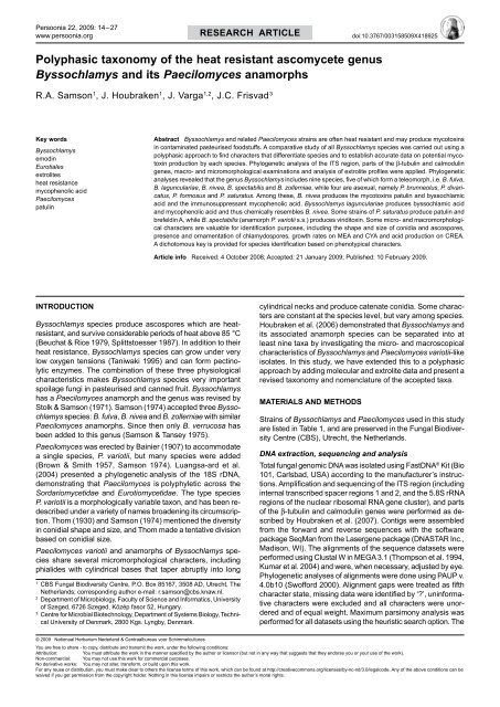
<strong>Polyphasic</strong> <strong>taxonomy</strong> <strong>of</strong> <strong>the</strong> <strong>heat</strong> <strong>resistant</strong> <strong>ascomycete</strong> genus<br />
Byssochlamys and its Paecilomyces anamorphs<br />
R.A. Samson 1 , J. Houbraken 1 , J. Varga 1,2 , J.C. Frisvad 3<br />
Key words<br />
Byssochlamys<br />
emodin<br />
Eurotiales<br />
extrolites<br />
<strong>heat</strong> resistance<br />
mycophenolic acid<br />
Paecilomyces<br />
patulin<br />
Abstract Byssochlamys and related Paecilomyces strains are <strong>of</strong>ten <strong>heat</strong> <strong>resistant</strong> and may produce mycotoxins<br />
in contaminated pasteurised foodstuffs. A comparative study <strong>of</strong> all Byssochlamys species was carried out using a<br />
polyphasic approach to find characters that differentiate species and to establish accurate data on potential mycotoxin<br />
production by each species. Phylogenetic analysis <strong>of</strong> <strong>the</strong> ITS region, parts <strong>of</strong> <strong>the</strong> β-tubulin and calmodulin<br />
genes, macro- and micromorphological examinations and analysis <strong>of</strong> extrolite pr<strong>of</strong>iles were applied. Phylogenetic<br />
analyses revealed that <strong>the</strong> genus Byssochlamys includes nine species, five <strong>of</strong> which form a teleomorph, i.e. B. fulva,<br />
B. lagunculariae, B. nivea, B. spectabilis and B. zollerniae, while four are asexual, namely P. brunneolus, P. divaricatus,<br />
P. formosus and P. saturatus. Among <strong>the</strong>se, B. nivea produces <strong>the</strong> mycotoxins patulin and byssochlamic<br />
acid and <strong>the</strong> immunosuppressant mycophenolic acid. Byssochlamys lagunculariae produces byssochlamic acid<br />
and mycophenolic acid and thus chemically resembles B. nivea. Some strains <strong>of</strong> P. saturatus produce patulin and<br />
brefeldin A, while B. spectabilis (anamorph P. variotii s.s.) produces viriditoxin. Some micro- and macromorphological<br />
characters are valuable for identification purposes, including <strong>the</strong> shape and size <strong>of</strong> conidia and ascospores,<br />
presence and ornamentation <strong>of</strong> chlamydospores, growth rates on MEA and CYA and acid production on CREA.<br />
A dichotomous key is provided for species identification based on phenotypical characters.<br />
Article info Received: 4 October 2008; Accepted: 21 January 2009; Published: 10 February 2009.<br />
Introduction<br />
Byssochlamys species produce ascospores which are <strong>heat</strong><strong>resistant</strong>,<br />
and survive considerable periods <strong>of</strong> <strong>heat</strong> above 85 °C<br />
(Beuchat & Rice 1979, Splittstoesser 1987). In addition to <strong>the</strong>ir<br />
<strong>heat</strong> resistance, Byssochlamys species can grow under very<br />
low oxygen tensions (Taniwaki 1995) and can form pectinolytic<br />
enzymes. The combination <strong>of</strong> <strong>the</strong>se three physiological<br />
characteristics makes Byssochlamys species very important<br />
spoilage fungi in pasteurised and canned fruit. Byssochlamys<br />
has a Paecilomyces anamorph and <strong>the</strong> genus was revised by<br />
Stolk & Samson (1971). Samson (1974) accepted three Byssochlamys<br />
species: B. fulva, B. nivea and B. zollerniae with similar<br />
Paecilomyces anamorphs. Since <strong>the</strong>n only B. verrucosa has<br />
been added to this genus (Samson & Tansey 1975).<br />
Paecilomyces was erected by Bainier (1907) to accommodate<br />
a single species, P. variotii, but many species were added<br />
(Brown & Smith 1957, Samson 1974). Luangsa-ard et al.<br />
(2004) presented a phylogenetic analysis <strong>of</strong> <strong>the</strong> 18S rDNA,<br />
demonstrating that Paecilomyces is polyphyletic across <strong>the</strong><br />
Sordariomycetidae and Eurotiomycetidae. The type species<br />
P. variotii is a morphologically variable taxon, and has been redescribed<br />
under a variety <strong>of</strong> names broadening its circumscription.<br />
Thom (1930) and Samson (1974) mentioned <strong>the</strong> diversity<br />
in conidial shape and size, and Thom made a tentative division<br />
based on conidial size.<br />
Paecilomyces variotii and anamorphs <strong>of</strong> Byssochlamys species<br />
share several micromorphological characters, including<br />
phialides with cylindrical bases that taper abruptly into long<br />
1<br />
CBS Fungal Biodiversity Centre, P.O. Box 85167, 3508 AD, Utrecht, The<br />
Ne<strong>the</strong>rlands; corresponding author e-mail: r.samson@cbs.knaw.nl.<br />
2<br />
Department <strong>of</strong> Microbiology, Faculty <strong>of</strong> Science and Informatics, University<br />
<strong>of</strong> Szeged, 6726 Szeged, Közép fasor 52, Hungary.<br />
3<br />
Centre for Microbial Biotechnology, Department <strong>of</strong> Systems Biology, Technical<br />
University <strong>of</strong> Denmark, 2800 Kgs. Lyngby, Denmark.<br />
cylindrical necks and produce catenate conidia. Some characters<br />
are constant at <strong>the</strong> species level, but vary among species.<br />
Houbraken et al. (2006) demonstrated that Byssochlamys and<br />
its associated anamorph species can be separated into at<br />
least nine taxa by investigating <strong>the</strong> micro- and macroscopical<br />
characteristics <strong>of</strong> Byssochlamys and Paecilomyces variotii-like<br />
isolates. In this study, we have extended this to a polyphasic<br />
approach by adding molecular and extrolite data and present a<br />
revised <strong>taxonomy</strong> and nomenclature <strong>of</strong> <strong>the</strong> accepted taxa.<br />
MaterialS and Methods<br />
Strains <strong>of</strong> Byssochlamys and Paecilomyces used in this study<br />
are listed in Table 1, and are preserved in <strong>the</strong> Fungal Biodiversity<br />
Centre (CBS), Utrecht, <strong>the</strong> Ne<strong>the</strong>rlands.<br />
DNA extraction, sequencing and analysis<br />
Total fungal genomic DNA was isolated using FastDNA ® Kit (Bio<br />
101, Carlsbad, USA) according to <strong>the</strong> manufacturer’s instructions.<br />
Amplification and sequencing <strong>of</strong> <strong>the</strong> ITS region (including<br />
internal transcribed spacer regions 1 and 2, and <strong>the</strong> 5.8S rRNA<br />
regions <strong>of</strong> <strong>the</strong> nuclear ribosomal RNA gene cluster), and parts<br />
<strong>of</strong> <strong>the</strong> β-tubulin and calmodulin genes were performed as described<br />
by Houbraken et al. (2007). Contigs were assembled<br />
from <strong>the</strong> forward and reverse sequences with <strong>the</strong> s<strong>of</strong>tware<br />
package SeqMan from <strong>the</strong> Lasergene package (DNASTAR Inc.,<br />
Madison, WI). The alignments <strong>of</strong> <strong>the</strong> sequence datasets were<br />
performed using Clustal W in MEGA 3.1 (Thompson et al. 1994,<br />
Kumar et al. 2004) and were, when necessary, adjusted by eye.<br />
Phylogenetic analyses <strong>of</strong> alignments were done using PAUP v.<br />
4.0b10 (Sw<strong>of</strong>ford 2000). Alignment gaps were treated as fifth<br />
character state, missing data were identified by ‘?’, uninformative<br />
characters were excluded and all characters were unordered<br />
and <strong>of</strong> equal weight. Maximum parsimony analysis was<br />
performed for all datasets using <strong>the</strong> heuristic search option. The<br />
© 2009 Nationaal Herbarium Nederland & Centraalbureau voor Schimmelcultures<br />
You are free to share - to copy, distribute and transmit <strong>the</strong> work, under <strong>the</strong> following conditions:<br />
Attribution:<br />
You must attribute <strong>the</strong> work in <strong>the</strong> manner specified by <strong>the</strong> author or licensor (but not in any way that suggests that <strong>the</strong>y endorse you or your use <strong>of</strong> <strong>the</strong> work).<br />
Non-commercial: You may not use this work for commercial purposes.<br />
No derivative works: You may not alter, transform, or build upon this work.<br />
For any reuse or distribution, you must make clear to o<strong>the</strong>rs <strong>the</strong> license terms <strong>of</strong> this work, which can be found at http://creativecommons.org/licenses/by-nc-nd/3.0/legalcode. Any <strong>of</strong> <strong>the</strong> above conditions can be<br />
waived if you get permission from <strong>the</strong> copyright holder. Nothing in this license impairs or restricts <strong>the</strong> author’s moral rights.
R.A. Samson et al.: <strong>Polyphasic</strong> <strong>taxonomy</strong> <strong>of</strong> Byssochlamys<br />
15<br />
Table 1 Byssochlamys and Paecilomyces isolates examined in this study.<br />
Species Accession No. Source and notes<br />
B. fulva CBS 132.33 Bottled fruit, UK; ex-type <strong>of</strong> Paecilomyces fulvus<br />
CBS 146.48 T<br />
Bottled fruit, UK<br />
CBS 135.62<br />
Fruit juice, Switzerland; ex-type <strong>of</strong> Paecilomyces todicus<br />
CBS 604.71<br />
Unknown source<br />
CBS 113954 Unknown source, patulin producer acc. to Rice et al. (1977)<br />
B. lagunculariae CBS 373.70 T Wood <strong>of</strong> Laguncularia racemosa (Mangue), Brazil<br />
CBS 696.95<br />
Pasteurized strawberries, <strong>the</strong> Ne<strong>the</strong>rlands<br />
CBS 110378<br />
Unknown source, France<br />
B. nivea CBS 100.11 T Unknown source<br />
CBS 133.37<br />
Milk <strong>of</strong> cow, USA; ex-type <strong>of</strong> Arachniotus trisporus<br />
CBS 271.95<br />
Mushroom bed, China<br />
CBS 102192<br />
Pasteurized drink yoghurt, Belgium<br />
CBS 113245<br />
Pasteurized fruit juice, Switzerland<br />
B. spectabilis CBS 338.51 Fruit juice, Switzerland<br />
CBS 102.74<br />
Unknown source; ex-type <strong>of</strong> Paecilomyces variotii<br />
CBS 101075 T<br />
Heat processed fruit beverage, Japan<br />
CBS 121581<br />
Spoiled sweetened tea, USA<br />
B. verrucosa CBS 605.74 T Nesting material <strong>of</strong> Leipoa ocellata, Australia<br />
B. zollerniae CBS 374.70 T Wood <strong>of</strong> Zollernia ilicifolia and Protium heptaphyllum, Brazil<br />
P. brunneolus CBS 370.70 T Non fat dry milk, Canada<br />
P. divaricatus CBS 284.48 T Mucilage bottle with library paste, USA<br />
CBS 110429<br />
Pectin, Mexico<br />
P. formosus CBS 628.66 Quebracho-tanned sheep lea<strong>the</strong>r, France<br />
CBS 371.70<br />
Annona squamosa, Brazil; ex-type <strong>of</strong> Paecilomyces maximus<br />
CBS 990.73B T<br />
Unknown source<br />
CBS 296.93<br />
Man, bone marrow <strong>of</strong> patient, Uzbekistan<br />
CBS 113247<br />
Soil, Thailand<br />
CBS 372.70<br />
Lecythis unsitata (Lecythidaceae), wood, Brazil; ex-type strain <strong>of</strong> P. lecythidis<br />
P. saturatus CBS 323.34 T Unknown source; ex-type <strong>of</strong> Paecilomyces mandshuricus var. saturatus<br />
CBS 368.70<br />
Medicine containing quinine, UK<br />
CBS 251.55 T<br />
Acetic acid, Brazil; ex-type <strong>of</strong> P. dactylomorphys<br />
CBS 990.73A<br />
Unknown source; ex-type <strong>of</strong> Penicillium viniferum<br />
CBS 492.84<br />
Lepidium sativum, Denmark<br />
Talaromyces byssochlamydoides CBS 413.71 T Dry soil under Pseudotsuga menziesii, USA<br />
Talaromyces emersonii CBS 393.64 T Compost, Italy<br />
Thermoascus crustaceus CBS 181.67 T Par<strong>the</strong>nium argentatum (Compositae), decaying plant, USA<br />
robustness <strong>of</strong> <strong>the</strong> most parsimonious trees was evaluated with<br />
1 000 bootstrap replications (Hillis & Bull 1993). O<strong>the</strong>r statistics,<br />
including tree length, consistency index, retention index and<br />
rescaled consistency index (CI, RI and RC) were calculated.<br />
Monascus pilosus (GenBank accession AY629427) was used<br />
as an outgroup in <strong>the</strong> analyses <strong>of</strong> <strong>the</strong> ITS dataset; Thermoascus<br />
crustaceus was used as an outgroup for <strong>the</strong> β-tubulin and calmodulin<br />
data. Newly generated sequences were deposited in<br />
GenBank with accession numbers FJ389920–FJ390009. The<br />
alignments generated and <strong>the</strong> most parsimonious trees were<br />
deposited in TreeBase under accession numbers S2199 and<br />
M4168–M4170.<br />
Morphological characterisation<br />
The media used for macro-morphological examination included<br />
Czapek yeast autolysate (CYA) agar, malt extract autolysate<br />
(MEA) agar, yeast extract sucrose (YES) agar and creatinesucrose<br />
(CREA) agar (media compositions were according to<br />
Samson et al. 2004). Isolates were incubated at 25, 30 and<br />
37 °C. Micromorphological characterisation <strong>of</strong> <strong>the</strong> asexual<br />
Paecilomyces state was carried out on MEA, hay agar (HAY)<br />
and YES agars. The latter was exclusively used to determine<br />
<strong>the</strong> presence <strong>of</strong> chlamydospores. For <strong>the</strong> analyses <strong>of</strong> <strong>the</strong> features<br />
<strong>of</strong> <strong>the</strong> sexual Byssochlamys state, <strong>the</strong> media oatmeal<br />
agar (OA) and potato-dextrose agar (PDA) were used (for<br />
media formulations, see Samson et al. 2004). The surfaces<br />
<strong>of</strong> conidia, chlamydospores and ascospores were examined<br />
after prolonged incubation (up to 70 d). Conditions and media<br />
for measuring <strong>the</strong> microaerophily, growth on 0.5 % acetic acid<br />
and <strong>the</strong> resistance to propionic acid are described by Frisvad<br />
& Samson (2004).<br />
Extrolite analysis<br />
Strains studied (Table 1) were three-point inoculated on MEA,<br />
YES, PDA, OA and CYA agars. All isolates were analysed for<br />
extrolite metabolites after 2 wk growth at 30 °C. The cultures<br />
were extracted according <strong>the</strong> method <strong>of</strong> Smedsgaard (1997)<br />
and analysed with high performance liquid chromatography<br />
(HPLC) with diode array detection (DAD) (Frisvad & Thrane<br />
1987, 1993). The metabolites found were compared with a<br />
spectral UV library derived from au<strong>the</strong>ntic standards, including<br />
patulin, viriditoxin, mycophenolic acid, byssochlamic acid and<br />
physcion, using <strong>the</strong> same conditions (<strong>the</strong> maximum similarity<br />
is a match <strong>of</strong> 1 000). The retention indices were compared with<br />
those <strong>of</strong> standards.<br />
Results<br />
DNA sequencing<br />
The trees constructed by maximum parsimony analysis <strong>of</strong> <strong>the</strong><br />
datasets <strong>of</strong> <strong>the</strong> protein coding genes β-tubulin and calmodulin<br />
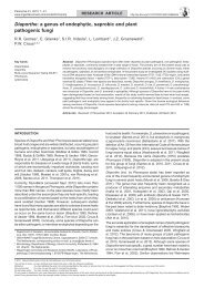
and <strong>the</strong> ITS region exhibited similar topologies (Fig. 1–3).<br />
Molecular analyses revealed that in <strong>the</strong> genus Byssochlamys<br />
nine taxa can be recognised. Five taxa form a teleomorph,<br />
namely B. fulva, B. lagunculariae, B. nivea, B. spectabilis and<br />
B. zollerniae, while four are strictly anamorphic, i.e. P. brunneolus,<br />
P. divaricatus, P. formosus and P. saturatus. Analyses<br />
<strong>of</strong> <strong>the</strong> ITS showed that B. verrucosa is not a member <strong>of</strong> <strong>the</strong><br />
genus Byssochlamys and is related to Thermoascus (Fig. 1).<br />
This was also confirmed by analyses <strong>of</strong> <strong>the</strong> partial β-tubulin<br />
and calmodulin sequence data (data not shown).<br />
The basal nodes were also similar for all three parsimony<br />
trees (Fig. 1–3). Byssochlamys nivea and B. fulva were sister
16 <strong>Persoonia</strong> – Volume 22, 2009<br />
1<br />
ITS<br />
CBS 289.34 Monascus pilosus AY629427<br />
CBS 374.70 B. zollerniae<br />
90 CBS 371.70 P. formosus<br />
79 CBS 113247 P. formosus<br />
100 CBS 372.70 P. formosus<br />
99<br />
CBS 628.66 P. formosus<br />
CBS 296.93 P. formosus<br />
86<br />
CBS 990.73B P. formosus<br />
CBS 370.70 P. brunneolus<br />
100<br />
CBS 121581 B. spectabilis/ P. variotii<br />
99<br />
CBS 338.51 B. spectabilis / P. variotii<br />
73 CBS 102.74 B. spectabilis / P. variotii<br />
CBS 101075 B. spectabilis / P. variotii<br />
CBS 100.11 B. nivea<br />
CBS 133.37 B. nivea<br />
CBS 113245 B. nivea<br />
CBS 271.95 B. nivea<br />
74 CBS 606.71 B. nivea<br />
CBS 132.33 B. fulva<br />
CBS 146.48 B. fulva<br />
CBS 604.71 B. fulva<br />
80 NRRL 32567 B. fulva<br />
100<br />
CBS 135 62 B. fulva<br />
CBS 373.70 B. lagunculariae<br />
CBS 696.95 B. lagunculariae<br />
89 CBS 110378 B. lagunculariae<br />
CBS 251.55 P. saturatus<br />
CBS 323.34 P. saturatus<br />
67<br />
CBS 368.70 P. saturatus<br />
CBS 492.84 P. saturatus<br />
CBS 990.73A P. saturatus<br />
100 CBS 284.48 P. divaricatus<br />
CBS 110429 P. divaricatus<br />
99 CBS 393.64 Talaromyces emersonii<br />
71<br />
CBS 413.71 Talaromyces byssochlamydoides<br />
100 CBS 605.74 Byssochlamys verrucosa<br />
CBS 181.67 Thermoascus crustaceus<br />
100<br />
99<br />
CBS 100.11 B. nivea<br />
CBS 133.37 B. nivea<br />
100 CBS 606.71 B. nivea<br />
CBS 271.95 B. nivea<br />
CBS 113245 B. nivea<br />
94<br />
CBS 132.33 B. fulva<br />
CBS 135.62 B. fulva<br />
NRRL 32567 B. fulva<br />
CBS 604.71 B. fulva<br />
CBS 146.48 B. fulva<br />
Calmodulin<br />
CBS 251.55 P. saturatus<br />
CBS 492.84 P. saturatus<br />
100<br />
CBS 368.70 P. saturatus<br />
CBS 990.73A P. saturatus<br />
99 CBS 323.34 P. saturatus<br />
CBS 373.70 B. lagunculariae<br />
99 CBS 696.95 B. lagunculariae<br />
CBS 110378 B. lagunculariae<br />
CBS 374.70 B. zollerniae<br />
100CBS 296.93 P. formosus<br />
CBS 990.73B P. formosus<br />
88<br />
CBS 371.70 P. formosus<br />
98<br />
100<br />
CBS 113247 P. formosus<br />
CBS 372.70 P. formosus<br />
100<br />
CBS 628.66 P. formosus<br />
100 CBS 338.51 B. spectabilis / P. variotii<br />
CBS 102.74 B. spectabilis / P. variotii<br />
96 CBS 121581 B. spectabilis / P. variotii<br />
99 CBS 101075 B. spectabilis / P. variotii<br />
Fig. 1 One <strong>of</strong> three equally parsimonious trees <strong>of</strong> <strong>the</strong> analysed ITS region<br />
(55 <strong>of</strong> <strong>the</strong> 629 characters were parsimony informative; tree length = 294,<br />
CI = 0.738, RI = 0.855, RC = 0.631, HI = 0.262).<br />
100<br />
CBS 370.70 P. brunneolus<br />
CBS 284.48 P. divaricatus<br />
CBS 110429 P. divaricatus<br />
10<br />
CBS 181.67 Thermoascus crustaceus<br />
species, as were B. lagunculariae and P. saturatus. These<br />
four species form a distinct clade (group I). Byssochlamys<br />
spectabilis and P. brunneolus also clustered toge<strong>the</strong>r and form,<br />
toge<strong>the</strong>r with P. formosus, ano<strong>the</strong>r distinct clade in Byssochlamys<br />
(group II). Paecilomyces divaricatus was basal to<br />
<strong>the</strong>se groups in all analyses, while <strong>the</strong> position <strong>of</strong> <strong>the</strong> single<br />
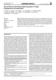
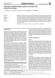
strain <strong>of</strong> B. zollerniae varied. Partial β-tubulin data showed that<br />
B. zollerniae is related to group I with a high bootstrap support<br />
(92 %; Fig. 3). The partial calmodulin data also showed that<br />
B. zollerniae is related to group I (Fig. 2), while analyses <strong>of</strong> <strong>the</strong><br />
ITS regions revealed that it is more related to group II (Fig. 1),<br />
but its placement was supported only by low bootstrap values<br />
on <strong>the</strong>se trees (Fig. 1, 2).<br />
Morphological characterisation<br />
The genus Byssochlamys is morphologically well-defined and<br />
characterised by almost naked ascomata in which croziers<br />
and globose asci are formed with ellipsoidal ascospores. The<br />
ascomatal initials consist <strong>of</strong> swollen an<strong>the</strong>ridia and coiled<br />
ascogonia. All Byssochlamys species have a Paecilomyces<br />
anamorph. The Byssochlamys anamorphs belong to Paecilomyces<br />
sect. Paecilomyces, containing mesophilic, <strong>the</strong>rmotolerant<br />
and <strong>the</strong>rmophilic species (Samson 1974). As discussed by<br />
Fig. 2 One <strong>of</strong> eight equally parsimonious trees <strong>of</strong> <strong>the</strong> analysed partial<br />
calmodulin gene sequences (204 <strong>of</strong> <strong>the</strong> 607 characters were parsimony informative;<br />
tree length = 328, CI = 0.708, RI = 0.889, RC = 0.630, HI = 0.292).<br />
Luangsa-ard et al. (2005), o<strong>the</strong>r species previously classified in<br />
Paecilomyces sect. Isarioidea are now mainly accommodated<br />
in Isaria or newly described genera.<br />
Various microscopic characters are valuable for identification<br />
purposes, including <strong>the</strong> shape and sizes <strong>of</strong> conidia and ascospores,<br />
and presence and ornamentation <strong>of</strong> chlamydospores.<br />
Useful macroscopical features are growth rates on MEA and<br />
CYA, and acid production on CREA. An overview <strong>of</strong> various<br />
macro- and microscopical features is presented in Table 2. The<br />
ratio between <strong>the</strong> colony diameters on MEA incubated at 30 °C<br />
or 37 °C is also diagnostic.<br />
Extrolite analysis<br />
The extrolite pr<strong>of</strong>iles <strong>of</strong> <strong>the</strong> taxa are listed in Table 3. Most<br />
strains <strong>of</strong> B. fulva produced byssochlamic acid, but B. fulva<br />
could be subdivided into two chemically different groups: one<br />
group <strong>of</strong> isolates produced meriditin (a compound produced by<br />
Eupenicillium meridianum) and a peptide with a cycloaspeptide
R.A. Samson et al.: <strong>Polyphasic</strong> <strong>taxonomy</strong> <strong>of</strong> Byssochlamys<br />
17<br />
10<br />
100<br />
100<br />
99<br />
100<br />
83<br />
92<br />
100<br />
100<br />
CBS 271.95 B. nivea<br />
CBS 133.37 B. nivea<br />
CBS 100.11 B. nivea<br />
CBS 113245 B. nivea<br />
CBS 606.71 B. nivea<br />
CBS 146.48 B. fulva<br />
CBS 135.62 B. fulva<br />
CBS 132.33 B. fulva<br />
CBS 113954 B. fulva<br />
CBS 604.71 B. nivea<br />
CBS 368.70 P. saturatus<br />
CBS 323.34 P. saturatus<br />
CBS 990.73A P. saturatus<br />
100<br />
CBS 492.84 P. saturatus<br />
CBS 696.95 B. lagunculariae<br />
CBS 374.70 B. zollerniae<br />
98<br />
CBS 371.70 P. formosus<br />
92<br />
78 CBS 113247 P. formosus<br />
CBS 990.73B P. formosus<br />
100<br />
CBS 296.93 P. formosus<br />
100 CBS 628.66 P. formosus<br />
CBS 372.70 P. formosus<br />
CBS 338.51 B. spectabilis / P. variotii<br />
89 CBS 102.74 B. spectabilis / P. variotii<br />
CBS 121581 B. spectabilis / P. variotii<br />
100<br />
CBS 101075 B. spectabilis / P. variotii<br />
CBS 110429 P. divaricatus<br />
CBS 284.48 P. divaricatus<br />
CBS 181.67 Thermoascus crustaceus<br />
98 CBS 251.55 P. saturatus<br />
CBS 373.70 B. lagunculariae<br />
100<br />
CBS 110378 B. lagunculariae<br />
CBS 370.70 P. brunneolus<br />
β-tubulin<br />
Fig. 3 One <strong>of</strong> 36 equally parsimonious trees <strong>of</strong> <strong>the</strong> analysed partial β-tubulin<br />
gene sequences (156 <strong>of</strong> <strong>the</strong> 494 characters were parsimony informative; tree<br />
length = 304, CI = 0.704, RI = 0.913, RC = 0.643, HI = 0.296).<br />
chromophore (CBS 132.33, CBS 135.62 and CBS 604.71),<br />
while <strong>the</strong> o<strong>the</strong>r group produced a series <strong>of</strong> alkaloids, some<br />
with UV spectra similar to that <strong>of</strong> paspaline (named ‘OLK’ in<br />
Table 2) and some with verrucologen-like UV spectra (called<br />
‘URT’ in Table 2) (CBS 113225, CBS 113246, CBS 113954 and<br />
TM 03.048). The ex-type culture <strong>of</strong> B. fulva was deteriorated<br />
and only produced byssochlamic acid, and none <strong>of</strong> <strong>the</strong> o<strong>the</strong>r<br />
mentioned extrolites.<br />
Some strains <strong>of</strong> B. nivea produced <strong>the</strong> full pr<strong>of</strong>ile <strong>of</strong> extrolites:<br />
patulin, mycophenolic acid, byssochlamic acid and metabolite<br />
‘OLK’ (CBS 900.70, CBS 271.95 and CBS 102192). Mycophenolic<br />
acid, patulin and byssochlamic acid and some <strong>of</strong> <strong>the</strong>ir<br />
precursors were also found by Puel et al. (2005). Byssochlamys<br />
lagunculariae produces <strong>the</strong> same overall extrolite pr<strong>of</strong>ile as<br />
B. nivea, except that patulin has not been detected in <strong>the</strong><br />
former species. Byssochlamys spectabilis and isolates with<br />
only anamorphs (P. variotii) consistently produced viriditoxin,<br />
which was earlier reported from an isolate identified as Spicaria<br />
divaricata (Jiu & Mizuba 1974). O<strong>the</strong>r unique extrolites were<br />
also produced but <strong>the</strong> structures <strong>of</strong> <strong>the</strong>se have not yet been<br />
elucidated.<br />
Table 2 Production <strong>of</strong> extrolites by Byssochlamys and Paecilomyces<br />
species 1 .<br />
Species<br />
All strains <strong>of</strong> P. divaricatus produced anthraquinones including<br />
emodin. However CBS 110429 only produced ano<strong>the</strong>r type <strong>of</strong><br />
anthraquinone, CBS 110428 and CBS 110430 produced asc<strong>of</strong>uranone.<br />
Only <strong>the</strong> ex-type culture <strong>of</strong> B. zollerniae was available<br />
for study and some unique unknown extrolites were found in this<br />
isolate. In this study, we could not confirm viriditoxin production<br />
in P. divaricatus and assume that <strong>the</strong> strain studied by Jiu &<br />
Mizuba (1974) was probably a B. spectabilis isolate.<br />
Our phylogenetic analysis showed that B. verrucosa is related to<br />
Thermoascus. Byssochlamys verrucosa produced cornexistin<br />
and/or byssochlamic acid, and also some specific unidentified<br />
extrolites only seen in this species. Thermoascus aurantiacus<br />
also produces compounds with chromophores (UV spectra)<br />
consistent with cornexistin, byssochlamic acid or similar tropolones<br />
(Frisvad unpubl. data). The production <strong>of</strong> cornexistin and<br />
byssochlamic acid demonstrates that <strong>the</strong> species chemically resemble<br />
B. fulva, B. laguncularia, B. nivea and P. divaricatus.<br />
Paecilomyces saturatus is chemically somewhat diverse. Group I<br />
produced at least one <strong>of</strong> <strong>the</strong> following extrolites: patulin, mycophenolic<br />
acid and a compound which chemically resembles<br />
aspergillic acid (CBS 251.55 T , CBS 223.52, CBS 492.84 and<br />
IBT 21716), while group II produced brefeldin A (CBS 323.34 T<br />
and CBS 990.73A) and may fit with P. mandshuricus var. saturatus.<br />
Paecilomyces formosus consistently produced variotin<br />
and related compounds, except CBS 296.93, which may be<br />
chemically close to P. saturatus (group I).<br />
Discussion<br />
Extrolites<br />
B. fulva Group I: byssochlamic acid, meriditin, ‘cycloaspeptide-like<br />
compound’<br />
Group II: byssochlamic acid, ‘OLK’, ‘URT’<br />
B. lagunculariae Byssochlamic acid, mycophenolic acid, ‘OLK’<br />
B. nivea Byssochlamic acid, mycophenolic acid, patulin, ‘OLK’<br />
B. spectabilis Viriditoxin and o<strong>the</strong>r compounds with characteristic UV<br />
spectra<br />
B. verrucosa Cornexistin and/or byssochlamic acid<br />
B. zollerniae No known extrolites, though several compounds characterized<br />
by a characteristic UV spectrum are present<br />
P. brunneolus ‘Asc<strong>of</strong>uranone-like compound’, ‘tetracycline-like compounds’<br />
P. divaricatus Cornexistin and/or byssochlamic acid, asc<strong>of</strong>uranone, emodin<br />
and o<strong>the</strong>r anthraquinones<br />
P. formosus Variotin<br />
P. saturatus Group I: patulin, mycophenolic acid or ‘aspergillic acid-like<br />
compound’<br />
Group II: brefeldin A<br />
1<br />
Byssochlamic acid, mycophenolic acid, patulin, and viriditoxin were available as au<strong>the</strong>ntic<br />
standards. Evidence for production <strong>of</strong> asc<strong>of</strong>uranone, cornexistin, meriditin and variotin is<br />
based on similar UV spectra as those reported in <strong>the</strong> literature, and <strong>the</strong>ir occurrence in<br />
species already known to produce <strong>the</strong>m. Metabolites in inverted commas have UV spectra<br />
that indicate a chemical relationship to <strong>the</strong> compounds mentioned. For example <strong>the</strong> ‘tetracycline-like<br />
compounds’ in P. brunneolus have UV spectra similar to those <strong>of</strong> tetracycline and<br />
viridicatumtoxin. ‘OLK’ and ‘URT’ are apolar indole-terpene compounds, but <strong>the</strong> chemical<br />
structure is as yet unknown. ‘OLK’ has a paspaline UV spectrum.<br />
Byssochlamys isolates were included in this study to verify <strong>the</strong><br />
connection between <strong>the</strong> anamorphic P. variotii complex and<br />
holomorphic Byssochlamys species. The genus Paecilomyces<br />
was monographed by Samson (1974) who recognised 31 species<br />
divided into two sections, Paecilomyces and Isarioidea.<br />
However, <strong>the</strong> phylogenetic analysis <strong>of</strong> <strong>the</strong> 18S rDNA demonstrates<br />
that Paecilomyces is polyphyletic across two subclasses,<br />
Sordariomycetidae and Eurotiomycetidae (Luangsa-ard et al.<br />
2004).
18 <strong>Persoonia</strong> – Volume 22, 2009<br />
Table 3 Macro-and microscopical features <strong>of</strong> Byssochlamys and Paecilomyces isolates.<br />
Species Conidial length (µm) Conidial shape (predominant) Chlamydospores Ascospore length (µm) Ascospore Colony diam. Degree <strong>of</strong> Acid proornamentation<br />
(mm) on CYA growth on CYA duction<br />
7 d, 30 °C 7 d, 30 °C on CREA<br />
7 d, 30 °C<br />
B. fulva 3.7–7.5 × 1.4–2.5 Cylindrical with truncate ends Absent; in some isolates after prolonged 5.3–7.1 × 3.3–4.3 Smooth 50–90 Good +<br />
incubation present (40 d)<br />
B. lagunculariae 2.7–4.5 × 2.2–3.3 Globose with flattened base Present, smooth 3.8–5 × 3–3.9 Smooth 45–55 Good –<br />
B. nivea 3–4.7 × 2.3–4 Globose to ellipsoidal with flattened base Present, smooth to finely rough 4.1–5.5 × 2.9–3.9 Smooth (8–)28–50 Weak – (+)<br />
B. spectabilis 3.3–6.1 × 1.5–4.4 Predominantly ellipsoidal and ellipsoidal Present, smooth to finely rough 5.2–6.8 × 3.5–4.5 Almost smooth, 30 – 45(– 55) Good –<br />
with truncate ends slightly roughened<br />
B. verrucosa 6.3–13.1 × 1.6–4.7 Cylindrical with truncate ends Absent 6.6–8.4 × 4–6.1 Rough 25–40 Good –<br />
B. zollerniae 2.5–4 × 1.5–3 Globose to ellipsoidal, apiculate Present, warted 3–4.5 × 2.5–3 Smooth 30–35 Weak –<br />
P. brunneolus 3.7–5.5 × 1.8–3.4 Ellipsoid to broadly cylindrical with truncate ends Present, smooth No ascospores detected Not relevant 20–30 Good –<br />
P. divaricatus 3.2–4.6 × 1.6–2.5 Ellipsoidal to cylindrical with truncate ends Absent; in some isolates after prolonged 5.3–7 × 3.8–4.9, smooth 10–17 Moderate –<br />
incubation present (40 d) rarely produced<br />
P. formosus 3–10 × 1.8–3.5 Ellipsoidal to cylindrical with truncate ends Present, smooth and <strong>of</strong>ten pigmented No ascospores detected Not relevant 18–90 Good +<br />
P. saturatus 2.3–7 × 1.7–3.4 Predominantly cylindrical and ellipsoidal Present, smooth No ascospores detected No ascospores 22–55 Good –<br />
without truncate ends detected<br />
Therefore, Paecilomyces is only monophyletic within <strong>the</strong> order<br />
Eurotiales and characterised by a Byssochlamys teleomorph.<br />
In <strong>the</strong> present study <strong>the</strong> P. variotii complex could be divided<br />
into four species, P. divaricatus, P. formosus, P. saturatus and<br />
P. variotii.<br />
Udagawa & Suzuki (1994) described Talaromyces spectabilis,<br />
but phylogenetic analysis <strong>of</strong> <strong>the</strong> 18S rDNA clarified that this species<br />
belongs to <strong>the</strong> genus Byssochlamys (Luangsa-ard et al.<br />
2004). Recently, Houbraken et al. (2008) showed that this<br />
species is heterothallic and that it is linked to <strong>the</strong> anamorphic<br />
species P. variotii. This was confirmed in <strong>the</strong> present study by<br />
morphological features and extrolite data. The presence <strong>of</strong> a<br />
teleomorph with <strong>heat</strong>-<strong>resistant</strong> ascospores for P. variotii explains<br />
<strong>the</strong> ability <strong>of</strong> this fungus to spoil <strong>heat</strong> treated fruit juices<br />
(Piecková & Samson 2000).<br />
Short descriptions <strong>of</strong> B. lagunculariae, B. spectabilis, P. brunneolus,<br />
P. divaricatus, P. formosus and P. saturatus are presented.<br />
The concepts <strong>of</strong> B. fulva, B. nivea and B. zollerniae are<br />
unchanged since <strong>the</strong> descriptions by Stolk & Samson (1971),<br />
with <strong>the</strong> remark that B. nivea var. langunculariae is elevated to<br />
species level. Byssochlamys verrucosa is not discussed here,<br />
because <strong>the</strong> taxon probably belongs to Thermoascus; it was<br />
fully described by Samson & Tansey (1975). Microscopic dimensions<br />
and cultural characters are summarised in Table 3.<br />
Discussion <strong>of</strong> accepted taxa<br />
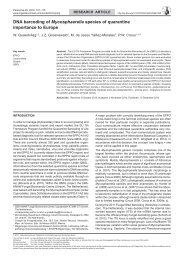
Byssochlamys lagunculariae (C. Ram) Samson, Houbraken<br />
& Frisvad, comb. nov. — MycoBank MB512557; Fig. 4a–f<br />
Basionym. Byssochlamys nivea Westling var. lagunculariae C. Ram,<br />
Nova Hedwigia 16: 311. 1968.<br />
Byssochlamys lagunculariae strains grow fast on MEA, covering<br />
<strong>the</strong> dish within 7 d at 30 °C. Depending on <strong>the</strong> isolate, it<br />
predominantly forms conidia (CBS 373.70 T ) or ascomata (CBS<br />
696.95). Colonies 25–55 mm on MEA at 37 °C after 7 d <strong>of</strong><br />
incubation. Growth occurs under microaerophilic conditions, in<br />
<strong>the</strong> presence <strong>of</strong> 0.5 % acetic acid or 1 000 ppm propionic acid<br />
(pH 3.8). No growth is observed on CYA with 5 % NaCl. Poor<br />
growth and no acid production on CREA.<br />
Morphologically, B. lagunculariae is similar to B. nivea and<br />
shares various characters such as fast growth rate on MEA at<br />
30 °C and globose (to ellipsoidal) conidia with a flattened base.<br />
Chlamydospores are present, uncoloured and smooth-walled.<br />
Though similar in shape to those <strong>of</strong> B. nivea, <strong>the</strong> conidia and<br />
ascospores <strong>of</strong> B. lagunculariae are generally smaller in size.<br />
Ano<strong>the</strong>r difference is that B. lagunculariae grows well on CYA<br />
while B. nivea grows ra<strong>the</strong>r poorly.<br />
Our molecular studies and morphological examinations both<br />
revealed that B. lagunculariae is clearly distinct from B. nivea.<br />
In <strong>the</strong> original description by Ram (1968), B. lagunculariae was<br />
described as a variety <strong>of</strong> B. nivea, distinguished by its smaller<br />
conidia and ascospores. Stolk & Samson (1971) synonymised<br />
it with B. nivea, but <strong>the</strong> present study showed that <strong>the</strong> growth<br />
rate on CYA, and smaller conidial and ascospore sizes are<br />
constant characters that can be used to differentiate between<br />
<strong>the</strong>se species.<br />
This species produces a similar range <strong>of</strong> extrolites to B. nivea,<br />
but patulin has not been detected in B. lagunculariae. Differentiation<br />
between B. nivea and B. lagunculariae, based on<br />
extrolite pr<strong>of</strong>iles, is not possible.<br />
The ex-type culture was isolated from wood <strong>of</strong> Laguncularia<br />
racemosa (mangue) in Brazil and o<strong>the</strong>r strains identified as<br />
this species were found in soil, and pasteurised strawberries<br />
and aloe juice. The occurrence <strong>of</strong> this species in <strong>heat</strong> treated<br />
products makes it an important food spoilage organism.
R.A. Samson et al.: <strong>Polyphasic</strong> <strong>taxonomy</strong> <strong>of</strong> Byssochlamys<br />
19<br />
a<br />
b<br />
c<br />
d<br />
e<br />
f<br />
i<br />
g<br />
h<br />
Fig. 4 Byssochlamys lagunculariae. a–d. Conidiophores; e. conidia; f. asci and ascospores. — Byssochlamys zollerniae. g, h. Phialides; i. chlamydospores;<br />
j. conidia. — Scale bars = 10 µm.<br />
j
20 <strong>Persoonia</strong> – Volume 22, 2009<br />
a<br />
b<br />
c<br />
d<br />
e<br />
f<br />
g h i<br />
Fig. 5 Byssochlamys spectabilis. a–e. Conidiophores; f. conidia; g. ascomata; h, i. asci and ascospores. — Scale bars = 10 µm.
R.A. Samson et al.: <strong>Polyphasic</strong> <strong>taxonomy</strong> <strong>of</strong> Byssochlamys<br />
21<br />
Byssochlamys spectabilis (Udagawa & Shoji Suzuki) Houbraken<br />
& Samson, Appl. Environm. Microbiol. 74: 1618.<br />
2008. — Fig. 5<br />
Basionym. Talaromyces spectabilis Udagawa & Shoji Suzuki, Mycotaxon<br />
50: 82. 1994.<br />
Anamorph. Paecilomyces variotii Bainier, Bull. Trimestriel Soc. Mycol.<br />
France 23: 26. 1907.<br />
≡ Penicillium variotii (Bainier) Sacc., Syll. Fung. 22: 1273. 1913.<br />
Colonies spreading rapidly on MEA at 30 °C and covering <strong>the</strong><br />
Petri dish within 7 d. Similar or higher growth rate at 37 °C<br />
than at 30 °C. Good growth under microaerophilic conditions,<br />
on MEA with 0.5 % acetic acid and on CYA with 1 000 ppm<br />
propionic acid (pH 3.8). No or weak growth on CYA with 5 %<br />
NaCl (0–10 mm). Since B. spectabilis forms its ascospores in<br />
a heterothallic manner, only <strong>the</strong> anamorph is usually produced.<br />
The conidiophores are irregularly branched and ellipsoidal<br />
and/or cylindrical; truncate conidia are formed, which are <strong>of</strong>ten<br />
pale yellow brown. Chlamydospores present, smooth-walled,<br />
in some isolates finely roughened. Often broad, thick-walled<br />
hyphae are present in fresh isolates. Ascospores are formed<br />
when strains <strong>of</strong> opposite mating types are grown toge<strong>the</strong>r.<br />
Our recent study (Houbraken et al. 2008) showed that strains<br />
originating from <strong>heat</strong> treated products are mostly frequently<br />
capable <strong>of</strong> producing fertile progeny. The ascospores are ellipsoidal,<br />
smooth to finely roughened, 5.5–6.5 × 3.5–4.5 µm.<br />
Colonies on MEA agar growing rapidly, attaining a diameter <strong>of</strong><br />
7 cm within 7 d at 30 °C. Poor growth and no acid production<br />
on CREA.<br />
The extrolite viriditoxin was produced by all investigated isolates.<br />
Whe<strong>the</strong>r this extrolite is also produced in foodstuffs is<br />
unknown.<br />
Byssochlamys spectabilis commonly occurs in air, compost,<br />
infected humans and various foodstuffs (including pasteurised<br />
fruit juices, rye bread). It is frequently found in <strong>heat</strong> treated products,<br />
although after isolation only <strong>the</strong> anamorph is produced.<br />
Paecilomyces brunneolus (N. Inagaki) Samson & Houbraken,<br />
comb. nov. — MycoBank MB512559; Fig. 6g–i<br />
Basionym. Paecilomyces variotii Bainier var. brunneolus N. Inagaki, Trans.<br />
Mycol. Soc. Japan 4: 4. 1962.<br />
The type strain <strong>of</strong> P. brunneolus on MEA in 7 d forms colonies<br />
<strong>of</strong> 35–45 mm diam (30 °C) with well-defined margins. Colonies<br />
on MEA 20–30 mm diam in 7 d at 37 °C. Growth occurs<br />
under microaerophilic conditions and in <strong>the</strong> presence <strong>of</strong> 0.5 %<br />
acetic acid or 1 000 ppm propionic acid (pH 3.8). No growth<br />
is observed on CYA with 5 % NaCl. Poor growth and no acid<br />
production on CREA. Microscopical examination showed<br />
short, irregular branched conidiophores (2–3 × 15–25 µm).<br />
Conidia ellipsoidal to broadly cylindrical with truncate ends<br />
(1.5–)2–3(–3.5) × (3.5–)4–5(–5.5) µm. Chlamydospores<br />
present, smooth and hyaline.<br />
Paecilomyces brunneolus is only known from its type culture,<br />
CBS 370.70. Analyses <strong>of</strong> a part <strong>of</strong> <strong>the</strong> β-tubulin, calmodulin<br />
gene and <strong>the</strong> ITS regions showed that this strain is closely<br />
related to B. spectabilis (P. variotii). However, extrolites and<br />
morphological data support that this is a distinct species. Viriditoxin<br />
is consistently produced by B. spectabilis, while P. brunneolus<br />
produces an asc<strong>of</strong>uranone like compound and tetracyclic<br />
compounds. Paecilomyces brunneolus colonies are more<br />
restricted than strains <strong>of</strong> B. spectabilis (P. variotii) on MEA.<br />
Ano<strong>the</strong>r difference is that <strong>the</strong> growth rate <strong>of</strong> P. brunneolus at<br />
37 °C is slower than at 30 °C, while this is <strong>the</strong> opposite for<br />
B. spectabilis.<br />
This species has only been isolated from non-fat, dry milk from<br />
Canada in <strong>the</strong> 1960s.<br />
Paecilomyces divaricatus (Thom) Samson, Houbraken &<br />
Frisvad, comb. nov. — MycoBank MB512561; Fig. 6a–f<br />
Basiomym. Penicillium divaricatum Thom, Bull. Bur. Anim. Ind. U.S. Dep.<br />
Agric. 118: 92. 1910.<br />
≡ Spicaria divaricata (Thom) J.C. Gilman & E.C. Abbott, Iowa State Coll.<br />
J. Sci. 1: 301. 1929.<br />
≡ Spicaria divaricata (Thom) R.M. Ma, Lingnan Sci. J. (Suppl.) 12: 115.<br />
1933.<br />
The growth rate <strong>of</strong> P. divaricatus on MEA is restricted, compared<br />
with o<strong>the</strong>r members <strong>of</strong> <strong>the</strong> investigated Byssochlamys clade,<br />
and colonies <strong>of</strong> 30–40 mm are attained after 7 d <strong>of</strong> incubation at<br />
30 °C. Only P. brunneolus has colony diameters <strong>of</strong> comparable<br />
size. Grows at 37 °C, although slower at 30 °C. Weak growth<br />
and no acid production on CREA. Paecilomyces divaricatus is<br />
characterised by its ellipsoidal to cylindrical, truncate conidia,<br />
measuring 3.5–4.5 × 1.5–2 µm and <strong>the</strong> absence <strong>of</strong> chlamydospores.<br />
Ascomata absent on agar media, though ascomatal<br />
initials, arising as coils, can be observed. Smooth ellipsoidal<br />
ascospores were once observed in a fresh isolate (5.3–7 ×<br />
3.8–4.9 µm) but have not been seen since.<br />
Anthraquinones including emodin are produced by all investigated<br />
isolates. The production and presence <strong>of</strong> emodin, a<br />
genotoxic and diarrheagenic mycotoxin, in foods and feeds<br />
is unknown.<br />
Thom (1910) described <strong>the</strong> species Penicillium divaricatum<br />
but placed this species later in synonymy with P. variotii (Thom<br />
1930). We examined <strong>the</strong> ex-type isolate and conclude that<br />
P. divaricatus is a distinct species. Microscopical analyses <strong>of</strong><br />
<strong>the</strong> original type isolate (CBS 284.48) showed a few structures<br />
resembling ascoma initials. Using a <strong>heat</strong> treatment, strains <strong>of</strong><br />
P. divaricatus could be isolated from various food products. The<br />
survival <strong>of</strong> a <strong>heat</strong> treatment suggests <strong>the</strong> presence <strong>of</strong> a Byssochlamys<br />
teleomorph (ascospores), and many Byssochlamys<br />
initials were present (croziers) in <strong>the</strong>se strains; however, no<br />
ascomata were detected even after prolonged incubation.<br />
This species was isolated from a bottle with mucilage library<br />
paste, <strong>heat</strong> treated pectin and fruit concentrates. Its presence<br />
in <strong>heat</strong> treated products and <strong>the</strong> absence <strong>of</strong> thick-walled<br />
chlamydospores, suggests that this species is able to form <strong>heat</strong><br />
<strong>resistant</strong> ascospores.<br />
Paecilomyces formosus (Sakag., May. Inoue & Tada) Houbraken<br />
& Samson, comb. nov. — MycoBank MB512562;<br />
Fig. 7g–i<br />
Basionym. Monilia formosa Sakag., May. Inoue & Tada, Zentralbl. Bakteriol.,<br />
2. Abt. 100: 302. 1939.<br />
= Paecilomyces maximus C. Ram, Nova Hedwigia 16: 306. 1968.<br />
= Paecilomyces lecythidis C. Ram (as lecythisii), Nova Hedwigia 16: 307.<br />
1968.<br />
Fast growth on MEA at 30 °C and covering <strong>the</strong> dish within 7 d.<br />
Ratio between growth rates at 30 and 37 °C variable; most<br />
strains have slower or similar growth rates (< 1), though CBS<br />
371.70 (ex-type strain <strong>of</strong> P. maximus) and CBS 113247 are<br />
exceptions and have a faster growth rate at 37 °C. Growth on<br />
MEA with 0.5 % acetic acid varying from absence <strong>of</strong> growth to<br />
more than 80 mm after 1 wk <strong>of</strong> incubation. Variable growth patterns<br />
on CYA with 5 % NaCl, 0–25 mm. All investigated strains<br />
have poor growth on CREA and acid production under colonies.<br />
Conidiophores irregularly branched, with olive-brown conidia.<br />
Chlamydospores present (<strong>of</strong>ten on small stalks), smooth walled,<br />
globose and (weakly) pigmented. The conidia <strong>of</strong> this species<br />
are variable, varying from ellipsoidal to cylindrical; all with<br />
truncate ends. In some isolates conidial shape might vary from<br />
ellipsoidal to cylindrical.
22 <strong>Persoonia</strong> – Volume 22, 2009<br />
a<br />
b<br />
c<br />
d<br />
e<br />
f<br />
g h i<br />
Fig. 6 Paecilomyces divaricatus. a–c. Conidiophores; d. conidia; e. ascoma initials; f. ascospores. — Paecilomyces brunneolus. g, h. Conidiophores;<br />
i. conidia. — Scale bars = 10 µm.
R.A. Samson et al.: <strong>Polyphasic</strong> <strong>taxonomy</strong> <strong>of</strong> Byssochlamys<br />
23<br />
a<br />
b<br />
c<br />
d<br />
e<br />
f<br />
g h i<br />
Fig. 7 Paecilomyces saturatus. a–e. Conidiophores; f. conidia. — Paecilomyces formosus. g, h. Conidiophores; i. conidia. — Scale bars = 10 µm.
24 <strong>Persoonia</strong> – Volume 22, 2009<br />
Ram (1968) described two Paecilomyces species, P. lecythidis<br />
and P. maximus, based on <strong>the</strong>ir cultural characters and <strong>the</strong>ir<br />
large-sized conidia.<br />
The results <strong>of</strong> <strong>the</strong> sequencing <strong>of</strong> <strong>the</strong> ITS region, and parts <strong>of</strong><br />
<strong>the</strong> protein coding genes β-tubulin and calmodulin, showed that<br />
P. formosus may consist <strong>of</strong> three taxa, P. formosus, P. lecythidis<br />
and P. maximus. However, <strong>the</strong> three species could not be<br />
distinguished by microscopical examination. One difference<br />
between strains belonging to <strong>the</strong> ‘P. maximus-clade’ and <strong>the</strong><br />
o<strong>the</strong>r members <strong>of</strong> this diverse group, is <strong>the</strong> faster growth rate<br />
<strong>of</strong> this species at 37 °C than at 30 °C. The sequence and morphological<br />
diversity was not detected by <strong>the</strong> extrolite analyses:<br />
<strong>the</strong> ex-type cultures <strong>of</strong> P. lecythidis and P. maximus produced<br />
similar extrolite pr<strong>of</strong>iles, while <strong>the</strong> ex-type culture <strong>of</strong> P. formosus<br />
is degenerated and is a weak producer <strong>of</strong> extrolites.<br />
For a more detailed conclusion and delimitation <strong>of</strong> <strong>the</strong>se three<br />
groups more strains should be studied, particularly emphasising<br />
conidial shape and extrolites production. For <strong>the</strong> time being<br />
we propose to place P. lecythidis and P. maximus in synonymy<br />
with P. formosus.<br />
This species is morphologically similar to P. variotii and <strong>the</strong> main<br />
difference is <strong>the</strong> consistent acid production on CREA.<br />
Paecilomyces formosus has been isolated from tropical and<br />
subtropical soils, wood, sponge, man (bone marrow, blood),<br />
air in a bedroom (Denmark) and pot plant soil <strong>of</strong> Senseviera<br />
trifasciata (Denmark).<br />
Paecilomyces saturatus (Nakaz., Y. Takeda & Suematsu)<br />
Samson & Houbraken, comb. nov. — MycoBank MB512560;<br />
Fig. 7a–f<br />
Basionym. Paecilomyces mandshuricus (Saito) Thom var. saturatus<br />
Nakaz., Y. Takeda & Suematsu, J. Agric. Chem. Soc. Japan 10: 102. 1934.<br />
= Penicillium viniferum Sakag., May. Inoue & Tada, Zentralbl. Bakteriol.,<br />
2. Abt. 100: 303. 1939.<br />
= Paecilomyces dactylethromorphus Bat. & H. Maia, Anais Soc. Biol.<br />
Pernambuco 15: 152. 1957.<br />
The oldest basionym available for this taxon is P. mandshuricus<br />
var. saturatus and <strong>the</strong>refore <strong>the</strong> name <strong>of</strong> this taxon is derived<br />
from this varietal name.<br />
Isolates growing on MEA at 30 °C cover <strong>the</strong> Petri dish within<br />
7 d, with strong olive-brown sporulation. Good growth at 37 °C,<br />
though slower at 30 °C. Good growth on MEA with 0.5 % acetic<br />
acid and CYA with 1 000 ppm propionic acid (pH 3.8). No<br />
growth observed on CYA with 5 % NaCl. Poor growth and no<br />
acid production on CREA.<br />
Morphological examination <strong>of</strong> various strains showed that this<br />
species forms fairly regularly branched, penicillium-like conidiophores,<br />
with ellipsoidal and/or cylindrical conidia without a distinct<br />
truncation. Chlamydospores present, hyaline and smooth<br />
walled. No teleomorph observed.<br />
The production <strong>of</strong> extrolites depends very much on <strong>the</strong> growth<br />
medium; patulin or brefeldin A can be produced.<br />
Paecilomyces saturatus is an easily recognisable species<br />
with its ellipsoidal and/or cylindrical conidia and its fairy regularly<br />
branched, penicillium-like conidiophores. Sakaguchi et al.<br />
(1939) described Penicillium viniferum and this species was<br />
subsequently placed in Paecilomyces by Raper & Thom (1949).<br />
In retrospect, <strong>the</strong> placement in Paecilomyces was correct, although<br />
this species could be interpreted, with its penicillium-like<br />
conidiophores and <strong>the</strong> presence <strong>of</strong> chlamydospores, as an intermediate<br />
form between Penicillium and Paecilomyces. Molecular<br />
studies now show that this species belongs to <strong>the</strong> Byssochlamys<br />
clade and is different from o<strong>the</strong>r olive-brown coloured species<br />
such as Hamigera avellanea (Luangsa-ard et al. 2004), Penicillium<br />
digitatum and P. cylindrosporum (unpubl. data).<br />
This species has been isolated from a variety <strong>of</strong> substrates, e.g.<br />
acetic acid, lea<strong>the</strong>r, medicine containing quinine, a dispersion<br />
<strong>of</strong> fenylacetate and dibutylmaleinate and Lepidium sativum.<br />
Notes on <strong>the</strong> ecology and extrolites production<br />
Byssochlamys and Paecilomyces species are <strong>of</strong>ten found<br />
in acidic habitats such as silage (Scurti et al. 1973, Escoula<br />
1975a, b, Anderson et al. 1979), and in common with Penicillium<br />
series Roqueforti species (Frisvad & Samson 2004) can also<br />
tolerate microaerophilic conditions (Escoula 1975a, b, Taniwaki<br />
1995). Byssochlamys nivea was initially reported to produce<br />
patulin under <strong>the</strong> name Gymnoascus sp. (Karow & Forster<br />
1944, Kuehn 1958), later confirmed by Kis et al. (1969), Scurti<br />
et al. (1973), Rice et al. (1977) and Draughon & Ayres (1980).<br />
Byssochlamys fulva was also reported to produce patulin, albeit<br />
by few strains (Escoula 1975a, b, Percebois et al. 1975, Rice et<br />
al. 1977). Besides <strong>the</strong>ir presence in pasteurised fruit, B. fulva<br />
and B. nivea also form toxic extrolites, such as byssotoxin A<br />
and byssochlamic acid (Kramer et al. 1976, Rice et al. 1977).<br />
Besides mycotoxins, also an antitumor metabolite, byssochlamysol,<br />
a steroid against IGF-1 dependent cancer cells, is<br />
produced by B. nivea (Mori et al. 2003).<br />
Paecilomyces variotii s.l. also produces mycotoxins (Scott<br />
1965), such as patulin (Escoula 1975a, b), sphing<strong>of</strong>ungin E<br />
and F (Frommer et al. 1992) and viriditoxin, reported originally<br />
from an isolate named Spicaria divaricata (Jiu & Mizuba 1974).<br />
Apart from being reported as being a potential mycotoxin,<br />
viriditoxin has also been reported to be a candidate for treatment<br />
<strong>of</strong> antibiotic <strong>resistant</strong> bacteria (Wang et al. 2003). Among<br />
<strong>the</strong> known extrolites are <strong>the</strong> antifungal drug variotin (Takeuchi<br />
et al. 1959, 1964, Suzuki et al. 1990, Omolo et al. 2000), and<br />
o<strong>the</strong>r drug candidates such as cornexistins (Nakajima et al.<br />
1991, Fields et al. 1996), SCH 643432 (Hegde et al. 2003)<br />
and a penicillin-like compound (Burton 1949). The sideramins<br />
ferrirubrin and fusigen (Diekmann 1967, Domsch et al. 1980)<br />
and <strong>the</strong> organic acids 3-indole-acetic acid (Bakalinerov 1968,<br />
Voinova-Raikova et al. 1969), citric acid (Loesecke 1945),<br />
ethyleneoxide-α, β-dicarboxylic acid (Sakaguchi et al. 1939),<br />
(3Z,5E)-octa-3,5-diene -1,3,4-tricarboxylic acid 3,4-anhydride<br />
(Aldridge et al. 1980) have also been reported. The possible<br />
mycotoxins and/or potential drugs byssotoxin A, sphing<strong>of</strong>ungin<br />
E and F, SCH 643432 and byssochlamysol were not available to<br />
us as standards. Given <strong>the</strong> taxonomic revision presented here,<br />
it remains to be seen which species produce <strong>the</strong>se extrolites.<br />
Some connections between species and bioactive extrolites<br />
were confirmed or established here and several species had a<br />
high consistent extrolite pr<strong>of</strong>ile. However, <strong>the</strong> chemo<strong>taxonomy</strong><br />
<strong>of</strong> P. saturatus is unresolved, because <strong>the</strong>re appear to be two<br />
chemotypes, which may or may not indicate that <strong>the</strong>re are two<br />
species ra<strong>the</strong>r than one. Likewise, B. fulva appears to have<br />
two chemotypes, with only byssochlamic acid as a common<br />
extrolite in all isolates examined.<br />
Key to Byssochlamys and related<br />
PaecilomYces anamorphs*<br />
1. Conidia with conspicuously truncate ends or a flattened<br />
base . . . . . . . . . . . . . . . . . . . . . . . . . . . . . . . . . . . . . . . . . 2<br />
1. Conidia ellipsoidal or cylindrical with inconspicuously truncate<br />
ends . . . . . . . . . . . . . . . . . . . . . . . . . . . . P. saturatus<br />
2. Conidia predominantly ellipsoidal and/or cylindrical chlamydospores<br />
absent or present; Byssochlamys teleomorph absent<br />
or present . . . . . . . . . . . . . . . . . . . . . . . . . . . . . . . . . . . . 3<br />
2. Conidia predominantly globose to subglobose, chlamydospores<br />
present; Byssochlamys teleomorph present . . . . 8
R.A. Samson et al.: <strong>Polyphasic</strong> <strong>taxonomy</strong> <strong>of</strong> Byssochlamys<br />
25<br />
a<br />
b<br />
c<br />
d<br />
e<br />
f<br />
g h i<br />
Fig. 8 Byssochlamys fulva. a. Conidiophores; b. conidia; c. asci and ascospores. — Byssochlamys nivea. d. Conidiophores; e. conidia; f. ascospores. — Byssochlamys<br />
verrucosa. g, h. Conidiophores and conidia; i. asci and ascospores. — Scale bars = 10 µm.
26 <strong>Persoonia</strong> – Volume 22, 2009<br />
3. Conidia cylindrical and/or ellipsoidal; measuring 3.4–4.2 ×<br />
1.7–2.1 µm, colonies on MEA restricted, attaining a diameter<br />
<strong>of</strong> less than 45 mm in 7 d at 30 °C . . . . . . . . . . . . . . . . . 4<br />
3. Conidia larger 2.3–8(–13) × 1.5–4.5 µm, cylindrical or ellipsoidal,<br />
colonies on MEA larger than 45 mm after 7 d at<br />
30 °C . . . . . . . . . . . . . . . . . . . . . . . . . . . . . . . . . . . . . . . . 5<br />
4. Chlamydospores present, ratio between diameters on MEA<br />
at 37:30 °C between 0.25 and 0.45, colonies with welldefined<br />
margins . . . . . . . . . . . . . . . . . . . . . . P. brunneolus<br />
4. Chlamydospores absent, ratio between diameters on MEA<br />
at 37:30 °C between 0.60 and 0.75, colonies with more or<br />
less fea<strong>the</strong>ry margins . . . . . . . . . . . . . . . . P. divaricatus**<br />
5. Conidia predominantly ellipsoidal with truncate ends, chlamydospores<br />
present . . . . . . . . . . . . . . . . . . . . . . . . . . . . . . 6<br />
5. Conidia predominantly cylindrical, chlamydospores absent<br />
. . . . . . . . . . . . . . . . . . . . . . . . . . . . . . . . . . . . . . . . . . . . . 7<br />
6. Conidia measuring 3.7–5.6 × 2.4–3.6 µm, no acid production<br />
on CREA; Byssochlamys teleomorph produced in a<br />
heterothallic manner . . . . . . . . . . . . . . . . . . B. spectabilis<br />
6. Conidia measuring 3.2–5.7(–10) × 2–2.9(–3.4) µm, acid<br />
production under colony on CREA; Byssochlamys teleomorph<br />
absent . . . . . . . . . . . . . . . . . . . . . . . . P. formosus<br />
7. Conidia measuring 4.5–6 × 1.7–2.2 µm; ascospores, smoothwalled,<br />
5.5–6.5 × 3.5–4.1 µm, acid production under colony<br />
on CREA . . . . . . . . . . . . . . . . . . . . . . B. fulva (Fig. 8a–c)<br />
7. Conidia measuring 9–11.8 × 2.5–4 µm; ascospores, verrucose<br />
and large, 7.1–8.1 × 5–5.7 µm, no acid production<br />
on CREA . . . . . . . . . . . . . . . . . . B. verrucosa (Fig. 8g–i)*<br />
8. Chlamydospores distinctly rough-walled . . . . . . . . . . . . . .<br />
. . . . . . . . . . . . . . . . . . . . . . . . . . . B. zollerniae (Fig. 4g–j)<br />
8. Chlamydospores smooth walled or finely roughened . . . 9<br />
9. Conidia measuring 3.1–3.8 × 2.5–3.2 µm, good growth on<br />
CYA . . . . . . . . . . . . . . . . . . . . . . . . . . . . . B. lagunculariae<br />
9. Conidia measuring 3.1–4.3 × 2.6–3.4 µm, moderate growth<br />
on CYA . . . . . . . . . . . . . . . . . . . . . . . B. nivea (Fig. 8d–f)<br />
* Byssochlamys verrucosa is phylogenetically unrelated to <strong>the</strong> true Byssochlamys<br />
spp., but <strong>the</strong> taxon is included in <strong>the</strong> key because <strong>of</strong> its striking<br />
morphological similarity to o<strong>the</strong>r Byssochlamys species.<br />
** Smooth ellipsoidal ascospores once observed in a fresh isolate (5.3–7 ×<br />
3.8–4.9 µm).<br />
References<br />
Aldridge DC, Carman RM, Moore RB. 1980. A new tricarboxylic acid anhydride<br />
from Paecilomyces variotii. Journal <strong>of</strong> <strong>the</strong> Chemical Society Perkin<br />
Transactions 1: 2134–2135.<br />
Anderson MS, Dutton MF, Harding K. 1979. Production and degradation <strong>of</strong><br />
patulin by Paecilomyces species, a common contaminant <strong>of</strong> silage. Journal<br />
<strong>of</strong> <strong>the</strong> Science <strong>of</strong> Food and Agriculture 30: 229–232.<br />
Bainier G. 1907. Mycothèque de l’école de Pharmacie XL Paecilomyces,<br />
genre nouveau de Mucédinées. Bulletin Trimestrielle de la Societe de<br />
Mycologie Française 23: 26–27.<br />
Bakalinerov D. 1968. Indole derivatives in soil microscopic fungi. Pochvozniki<br />
Agrokhimika 3: 81–86.<br />
Beuchat LR, Rice SL. 1979. Byssochlamys spp. and processed fruits. Advances<br />
in Food Research 25: 237–288.<br />
Brown AH, Smith G. 1957. The genus Paecilomyces Bainier and its perfect<br />
stage Byssochlamys Westling. Transactions <strong>of</strong> <strong>the</strong> British Mycological<br />
Society 40: 17–89.<br />
Burton HS. 1949. Antibiotics from Penicillia. British Journal <strong>of</strong> Experimental<br />
Pathology 30: 151–158.<br />
Diekmann H. 1967. St<strong>of</strong>fwechselprodukte von Microorganismen; 56 Mitteilung<br />
Fusigen – ein neues Sideramin aus Pilzen. Archive für Mikrobiologie<br />
58: 1–5.<br />
Domsch KH, Gams W, Anderson TH. 1980. Compendium <strong>of</strong> soil fungi.<br />
Academic Press, London, UK.<br />
Draughon FA, Ayres JC. 1980. Insecticide inhibition <strong>of</strong> growth and patulin<br />
production in Penicillium expansum, Penicillium urticae, Aspergillus clavatus,<br />
Aspergillus terreus, and Byssochlamys nivea. Journal <strong>of</strong> Agricultural<br />
and Food Chemistry 28: 1115–1117.<br />
Escoula L. 1975a. Toxigenic moulds in silage II In vitro kinetics <strong>of</strong> patulin and<br />
byssochlamic acid biosyn<strong>the</strong>sis by Byssochlamys nivea Westling in liquid<br />
media. Annales de Recherches Veterinaire 6: 155–163.<br />
Escoula L. 1975b. Toxigenic moulds in silage III Patulin and byssochlamic<br />
acid production by Byssochlamys nivea Westling on a laboratory silage<br />
model. Annales de Recherches Veterinaire 6: 219–226.<br />
Fields SC, Mireles-Lo L, Gerwick BC. 1996. Hydroxycornexitin: a new<br />
phytotoxin from Paecilomyces variotii. Journal <strong>of</strong> Natural Products 59:<br />
698–700.<br />
Frisvad JC, Samson RA. 2004. <strong>Polyphasic</strong> <strong>taxonomy</strong> <strong>of</strong> Penicillium subgenus<br />
Penicillium: A guide to identification <strong>of</strong> food and air-borne terverticillate<br />
Penicillia and <strong>the</strong>ir mycotoxines. Studies in Mycology 49: 1–173.<br />
Frisvad JC, Thrane U. 1987. Standardized high-performance liquid chromatography<br />
<strong>of</strong> 182 mycotoxins and o<strong>the</strong>r fungal metabolites based on<br />
alkylphenone indices and UV-VIS spectra diode-array detection. Journal<br />
<strong>of</strong> Chromatography 404: 195–214.<br />
Frisvad JC, Thrane U. 1993. Liquid column chromatography <strong>of</strong> mycotoxines.<br />
In: Betina V. (ed), Chromatography <strong>of</strong> mycotoxines, Techniques and<br />
applications. Journal <strong>of</strong> Chromatography Library 54: 253–372. Elsevier,<br />
Amsterdam.<br />
Frommer BR, Thornton RA, Mandela SM. 1992. Sphing<strong>of</strong>ungin E and sphing<strong>of</strong>ungin<br />
F – novel serinepalmitoyl transferase inhibitors from Paecilomyces<br />
variotii. Journal <strong>of</strong> Antibiotics 45: 1692–1696.<br />
Hedge VR, Silver J, Patel M, Gullo VP, Puar MS, Das PR, Loebenberg D.<br />
2003. Novel fungal metabolites as cell wall active antifungals: fermentation,<br />
isolation, physico-chemical properties, structure and biological activity.<br />
Journal <strong>of</strong> Antibiotics 56: 437–447.<br />
Hillis DM, Bull JJ. 1993. An empirical test <strong>of</strong> bootstrapping as a method for<br />
assessing confidence in phylogenetic analysis. Systematic Biology 42:<br />
182–192.<br />
Houbraken J, Due J, Varga J, Meijer M, Frisvad JC, Samson RA. 2007.<br />
<strong>Polyphasic</strong> <strong>taxonomy</strong> <strong>of</strong> Aspergillus section Usti. Studies in Mycology 59:<br />
107–128.<br />
Houbraken J, Samson RA, Frisvad JC. 2006. Byssochlamys: significance<br />
<strong>of</strong> <strong>heat</strong> resistance and mycotoxin production. Advances in Experimental<br />
Medicine and Biology 571: 211–224.<br />
Houbraken J, Varga J, Rico-Munoz E, Johnson S, Samson RA. 2008. Sexual<br />
reproduction as <strong>the</strong> cause <strong>of</strong> <strong>heat</strong> resistance in <strong>the</strong> food spoilage fungus<br />
Byssochlamys spectabilis (anamorph: Paecilomyces variotii). Applied and<br />
Environmental Microbiology 74: 1613–1619.<br />
Jiu J, Mizuba S. 1974. Metabolic products <strong>of</strong> Spicaria divaricata NRRL 5771.<br />
Journal <strong>of</strong> Antibiotics 27: 760–765.<br />
Karow EO, Forster JW. 1944. An antibiotic substance from a species <strong>of</strong><br />
Gymnoascus and Penicillium. Science 99: 265–266.<br />
Kis Z, Furger P, Sigg HP. 1969. Über die Isolierung von Pyrenophenol.<br />
Experientia 25: 123.<br />
Kramer RK, Davis ND, Diener UL. 1976. Byssotoxin A, a secondary metabolite<br />
<strong>of</strong> Byssochlamys fulva. Applied and Environmental Microbiology<br />
31: 249–253.<br />
Kuehn HH. 1958. A preliminary survey <strong>of</strong> <strong>the</strong> Gymnoascaceae I. Mycologia<br />
50: 417–439.<br />
Kumar S, Tamura K, Nei M. 2004. MEGA3: Integrated s<strong>of</strong>tware for molecular<br />
evolutionary genetic analysis and sequence alignment. Briefings in Bioinformatics<br />
5: 150–163.<br />
Loesecke HW von. 1945. A review <strong>of</strong> information on mycological citric acid<br />
production. Chemical Engineering News 23: 1952–1959.<br />
Luangsa-ard J, Hywel-Jones NL, Manoch L, Samson RA. 2005. On <strong>the</strong><br />
relationships <strong>of</strong> Paecilomyces sect. Isarioidea species. Mycological Research<br />
109: 581–589.<br />
Luangsa-ard J, Hywel-Jones NL, Samson RA. 2004. The polyphyletic nature<br />
<strong>of</strong> Paecilomyces sensu lato based on 18S-generated rDNA phylogeny.<br />
Mycologia 96: 773–780.<br />
Mori T, Shin-ya K, Takatori K, Aihara M, Hayakawa Y. 2003. Byssochlamysol,<br />
a new antitumor steroid against IGF-1-dependent cells from Byssochlamys<br />
nivea, II Physico-chemical properties and structure elucidation. Journal <strong>of</strong><br />
Antibiotics 56: 6–8.<br />
Nakajima M, Itoi K, Takamatsu Y, Sadao S, Furukawa Y, Furuya K, Honma T,<br />
Kadotani J, Kozasa M, Haneishi T. 1991. Cornexitin: a new fungal metabolite<br />
with herbicidal activity. Journal <strong>of</strong> Antibiotics 44: 1065–1072.<br />
Omolo JO, Anke H, Chhabra S, Sterner O. 2000. New variotin analogues from<br />
Aspergillus viridinutans. Journal <strong>of</strong> Natural Products 63: 975–977.<br />
Perecebois G, Basile A-M, Schwertz A. 1975. Existance commune, sur<br />
les fraises, d’ascospores de Byssochlamys nivea pouvant produire de la<br />
patuline. Mycopathologia 57: 109–111.<br />
Piecková ES, Samson RA. 2000. Heat resistance <strong>of</strong> Paecilomyces variotii<br />
in sauce and juice. Journal <strong>of</strong> Industrial Microbiology and Biotechnology<br />
24: 227–230.
R.A. Samson et al.: <strong>Polyphasic</strong> <strong>taxonomy</strong> <strong>of</strong> Byssochlamys<br />
27<br />
Puel O, Tadrist S, Galtier P, Oswald IP, Delforge M. 2005. Byssochlamys<br />
nivea as a new source <strong>of</strong> mycophenolic acid. Applied and Environmental<br />
Microbiology 71: 550–553.<br />
Ram C. 1968. Timber-attacking fungi from <strong>the</strong> state <strong>of</strong> Maranhao, Brazil;<br />
Some new species <strong>of</strong> Paecilomyces and its perfect state Byssochlamys<br />
Westl. VIII. Nova Hedwigia 16: 305–314.<br />
Raper K, Thom C. 1949. A manual <strong>of</strong> Penicillia. Williams & Wilkins, Baltimore,<br />
MD, USA.<br />
Rice SL, Beuchat LR, Worthington RE. 1977. Patulin production by Byssochlamys<br />
spp. in fruit juices. Applied and Environmental Microbiology<br />
34: 791–796.<br />
Sakaguchi K, Inoue T, Tada S. 1939. On <strong>the</strong> production <strong>of</strong> ethyleneoxide-α,<br />
β-dicarboxylic acid by moulds. Zentralblatt fur Bakteriologie und Parasiten-<br />
Kunde, Abteilung 2, 100: 302–307.<br />
Samson RA. 1974. Paecilomyces and some allied hyphomycetes. Studies<br />
in Mycology 6: 1–119.<br />
Samson RA, Hoekstra ES, Frisvad JC. 2004. Introduction to food- and<br />
airborne fungi. 7th ed. Centraalbureau voor Schimmelcultures, Utrecht,<br />
<strong>the</strong> Ne<strong>the</strong>rlands.<br />
Samson RA, Tansey MR. 1975. Byssochlamys verrucosa sp. nov. Transactions<br />
<strong>of</strong> <strong>the</strong> British Mycological Society 65: 512–514.<br />
Scott B de. 1965. Toxigenic fungi isolated from cereal and legume products.<br />
Mycopathologia et Mycologia Applicata 25: 213–222.<br />
Scurti JC, Cadignola A, Nobile G, Caputo O. 1973. Un ceppo di Byssochlamys<br />
nivea Westl., isolato da insilato di mais integrale, producente patulina.<br />
Allionia 19: 39–42.<br />
Smedsgaard J. 1997. Micro-scale extraction procedure for standardized<br />
screening <strong>of</strong> fungal metabolite production in cultures. Journal <strong>of</strong> Chromatography<br />
A 760: 264–270.<br />
Splittstoesser DF. 1987. Fruits and fruit products. In: Beuchat LR. (ed), Food<br />
and beverage mycology, 2nd ed.: 101–128. Van Nostrand Reinhold, New<br />
York, USA.<br />
Stolk AC, Samson RA. 1971. Studies on Talaromyces and related genera. 1.<br />
Hamigera gen. nov. and Byssochlamys. <strong>Persoonia</strong> 6: 341–357.<br />
Suzuki K, Nozawa K, Nakajima S, Kawai K. 1990. Structure revision <strong>of</strong><br />
mycotoxin, viriditoxin, and its derivatives. Chemical and Pharmaceutical<br />
Bulletin 38: 3180–3181.<br />
Sw<strong>of</strong>ford T. 2000. PAUP*: Phylogenetic analysis using parsimony. version<br />
4.0. Sunderland, Sinauer Associates.<br />
Takeuchi S, Yonehara H, Shoji H. 1964. Crystallized variotin and its revised<br />
chemical structure. Journal <strong>of</strong> Antibiotics Tokyo, Ser. A 17: 267–268.<br />
Takeuchi S, Yonehara H, Umezawa H. 1959. Studies on variotin, a new<br />
antifungal antibiotic 1. Preparations and properties <strong>of</strong> variotin. Journal <strong>of</strong><br />
Antibiotics Tokyo, Ser. A 12: 195–200.<br />
Taniwaki MH. 1995. Growth and mycotoxin production by fungi under<br />
modified atmospheres. PhD <strong>the</strong>sis, Kensington, NSW: University <strong>of</strong> New<br />
South Wales, Australia.<br />
Thom C. 1910. Cultural studies <strong>of</strong> species <strong>of</strong> Penicillium. Bureau <strong>of</strong> Animal<br />
Industry, United States Department <strong>of</strong> Agriculture, Bulletin No. 118.<br />
Thom C. 1930. The Penicillia. Williams & Wilkins Co., Baltimore, USA.<br />
Thompson JD, Higgins DG, Gibson TJ. 1994. CLUSTAL W: improving <strong>the</strong><br />
sensitivity <strong>of</strong> progressive multiple sequence alignment through sequence<br />
weighting, positions-specific gap penalties and weight matrix choice.<br />
Nucleic Acids Research 22: 4673–4680.<br />
Udagawa S, Suzuki S. 1994. Talaromyces spectabilis, a new species <strong>of</strong><br />
food-borne <strong>ascomycete</strong>s. Mycotaxon 50: 91–88.<br />
Voinova-Raikova Z, Bakalivanov D, Chanova D, Stratieva A. 1969. β-indoleacetic<br />
acid in some soil microorganisms. Pochozniki Agrokhimika 4:<br />
85–90 (in Russian).<br />
Wang J, Galgoci A, Kodali SK, Herath KB, Jayasutiya H, Dorso K, Vivente<br />
F, Gonzáles A, Cully D, Bramhill D, Singh S. 2003. Discovery <strong>of</strong> a small<br />
molecule that inhibits cell division by blocking FtsZ, a novel <strong>the</strong>rapeutic<br />
target <strong>of</strong> antibiotics. Journal <strong>of</strong> Biological Chemistry 278: 44424–44428.