Six Species of the Lernanthropidae - Zoological Studies
Six Species of the Lernanthropidae - Zoological Studies
Six Species of the Lernanthropidae - Zoological Studies

Create successful ePaper yourself
Turn your PDF publications into a flip-book with our unique Google optimized e-Paper software.
<strong>Zoological</strong> <strong>Studies</strong> 50(5): 611-635 (2011)<strong>Six</strong> <strong>Species</strong> <strong>of</strong> <strong>the</strong> <strong>Lernanthropidae</strong> (Crustacea: Copepoda) Parasitic onMarine Fishes <strong>of</strong> Taiwan, with a Key to 18 <strong>Species</strong> <strong>of</strong> <strong>the</strong> Family Knownfrom TaiwanJu-shey Ho 1 , Wei-Cheng Liu 2 , and Ching-Long Lin 2, *1Department <strong>of</strong> Biological Sciences, California State University, Long Beach, CA 90840-3702, USA2Department <strong>of</strong> Aquatic Sciences, National Chiayi University, Chiayi 600, Taiwan(Accepted April 12, 2010)Ju-shey Ho, Wei-Cheng Liu, and Ching-Long Lin (2011) <strong>Six</strong> species <strong>of</strong> <strong>the</strong> <strong>Lernanthropidae</strong> (Crustacea:Copepoda) parasitic on marine fishes <strong>of</strong> Taiwan, with a key to 18 species <strong>of</strong> <strong>the</strong> family known from Taiwan.<strong>Zoological</strong> <strong>Studies</strong> 50(5): 611-635. <strong>Six</strong> species <strong>of</strong> copepods belonging to <strong>the</strong> <strong>Lernanthropidae</strong> Kabata, 1979were found parasitic on <strong>the</strong> gill filaments <strong>of</strong> 6 species <strong>of</strong> marine fishes <strong>of</strong> Taiwan. They are: Lernanthropodeschorinemi Pillai, 1962 on Scomberoides commersonnianus Lacepède (Carangidae); Lernanthropodes trachinotiPillai, 1962 on Trachinotus blochii (Lacepède) (Carangidae); Lernanthropus incilis sp. nov. on Evoxymetoponpoeyi Gün<strong>the</strong>r (Trichiuridae); Mitrapus heteropodus (Yü 1933) on Nematalosa nasus (Bloch) (Clupeidae);Sagum epinepheli (Yamaguti et Yamasu, 1960) on Epinephelus awoara (Temminck et Schlegel) (Serranidae);and Sagum folium sp. nov. on Paracaesio caerulea (Katayama) (Lutjanidae). Aside from <strong>the</strong> 2 new species, <strong>the</strong>o<strong>the</strong>r 4 known species were recorded for <strong>the</strong> 1st time from Taiwan. A key to <strong>the</strong> 18 species <strong>of</strong> lernanthropidsoccurring on marine fishes <strong>of</strong> Taiwan is provided. In this paper we propose treating Mitrapus rubiginosus (Redkar,Rangnekar et Murti 1949) as a junior synonym <strong>of</strong> M. heteropodus.http://zoolstud.sinica.edu.tw/Journals/50.5/611.pdfKey words: <strong>Lernanthropidae</strong>, Parasitic copepods, Taiwan, Marine fish.<strong>Lernanthropidae</strong> Kabata, 1979 is a largefamily <strong>of</strong> siphonostomatoid copepods comprisingover 150 species. They are exclusively parasiticon gill filaments <strong>of</strong> marine teleosts. They use<strong>the</strong>ir prehensile antennae and maxillipeds toattach tenaciously to a host’s gill filaments. Theattachment is assisted, in <strong>the</strong> case <strong>of</strong> <strong>the</strong> female,by leg 3 which is modified into a pair <strong>of</strong> large,folded lamellae designed for clamping onto a host’sgill filaments. Thus, lernanthropids can <strong>of</strong>ten causepathological effects like desquamation, erosion,and necrosis <strong>of</strong> <strong>the</strong> host’s gill filaments (Maneraand Dezfuli 2003) and, in cases <strong>of</strong> heavy infection,may lead to asphyxiation, anemia, and secondarybacterial infections (Tokşen et al. 2006).Lernanthropids are largely parasites <strong>of</strong>warm-water fishes. Thus, while 44 species areknown from India (Pillai 1985), only 9 species areknown from Japan (Ho and Do 1985). So far,we have discovered and reported 12 species <strong>of</strong>lernanthropids from Taiwan (Ho et al. 2008, Liu etal. 2009a b). In this paper we add 6 more species,which means that 18 species <strong>of</strong> lernanthropidsin 7 genera are known from Taiwan. Since wehave examined about 20% <strong>of</strong> <strong>the</strong> marine fishes<strong>of</strong> Taiwan, we firmly believe <strong>the</strong>re are morespecies <strong>of</strong> lernanthropids waiting to be discoveredfrom Taiwan. Therefore, a key to <strong>the</strong> species <strong>of</strong>lernanthropids from Taiwan is provided at <strong>the</strong> end<strong>of</strong> this report to facilitate species identification <strong>of</strong>this group.*To whom correspondence and reprint requests should be addressed. Tel and Fax: 886-5-2783065. E-mail:cllin@mail.ncyu.edu.tw611
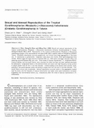
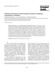
612<strong>Zoological</strong> <strong>Studies</strong> 50(5): 611-635 (2011)MATERIALS AND METHODSFish caught and landed at various fishingports in Taiwan in <strong>the</strong> last 10 yr were purchasedand transferred in an icebox to <strong>the</strong> laboratory forexamination <strong>of</strong> parasitic copepods. Parasites werecarefully removed from <strong>the</strong> host’s gill filamentsunder a dissection microscope, and preservedin 70% ethanol. Selected specimens werelater cleared in 85% lactic acid overnight beforedissection <strong>of</strong> <strong>the</strong> appendages and examinationunder a compound microscope with a series <strong>of</strong>magnifications up to 1500x. All drawings weremade with <strong>the</strong> aid <strong>of</strong> a drawing tube attached to acompound microscope. Measurements <strong>of</strong> bodyparts were taken after specimens were soaked inlactic acid overnight. The mean value is given with<strong>the</strong> range following in paren<strong>the</strong>ses.Below, a full description is given <strong>of</strong> <strong>the</strong>female, and if <strong>the</strong> male is available, only sexuallydimorphic characters are mentioned for <strong>the</strong> male.RESULTSOrder Siphonostomatoida Thorell, 1859Family <strong>Lernanthropidae</strong> Kabata, 1979Genus Lernanthropodes Bere, 1936Lernanthropodes chorinemi Pillai, 1962(Figs. 1, 2)Material examined: 2on 1 (<strong>of</strong> 9) talangqueenfish, Scomberoides commersonnianusLacepsède, 1801, landed at Dong-shi Fishing Porton 13 Jan. 2000.Female: Body (Fig. 1A-C) cylindrical, 3.56(3.50-3.62) mm long (from anterior rim <strong>of</strong> head totip <strong>of</strong> caudal ramus), comprising a subtriangularhead, cylindrical trunk, and small urosome. Head0.98 (0.88-1.08) mm long and 0.95 (0.92-0.98) mmwide, with broadly protruding posterolateral cornersand narrowed anterior end. Trunk cylindrical,narrower than head, only 0.82 (0.80-0.84) mmwide, and without a dorsal plate. Posterior part<strong>of</strong> trunk with ventrally fused lamellae <strong>of</strong> leg 3appearing wider than head, 1.28 (1.24-1.32) mmwide. Genital complex (Fig. 1D) longer than wide,401 (324-478) × 324 (316-332) μm, with a laterallyprotruding egg sac attachment area. Abdomen(Fig. 1D) also with laterally protruding sides, longerthan wide, 223 (211-235) × 215 (194-235) µm,with distinct anal slit. Caudal ramus (Fig. 1D) along distally attenuated process, 324 (284-365) ×97 (89-105) µm, carrying 2 dorsal setae in basalregion and 2 setae at distal end. Egg sac long andstraight.Antennule (Fig. 1E) filiform and 7-segmented;armature formula: 0, 2, 1, 3, 1, 4, and 4.Parabasal process (Fig. 1E) present. Antenna(Fig. 1F) 2-segmented; corpus about 2.5-timeslonger than claw, bearing 1 broad basal seta onmedial surface; claw bearing 2 similar basal setaeand terminal striations. Mandible comprising 2sections; with 8 teeth on terminal blade. Maxillule(Fig. 1G) bilobate, smaller outer lobe tipped with1 spiniform element and larger inner lobe with 3unequal elements. Maxilla (Fig. 1H) 2-segmented,lacertus unarmed; brachium with denticlesscattered on medial surface and bearing 1 bifidspiniform element subterminally and 1 elementdistally; terminal claw fringed with row <strong>of</strong> largerdenticles along both edges. Maxilliped (Fig. 2A)2-segmented; corpus unarmed; shaft longer thanclaw, with broad seta on medial margin close todistal end; claw with striations as in antenna.Ventral surface <strong>of</strong> leg 1 (Fig. 2B) ornamentedwith denticles; both outer and inner setae<strong>of</strong> protopod with large basal papilla; exopod1-segmented, large, and tipped with 5 robustspines; endopod a smaller lobe with long terminal,blunt process. Leg 2 (Fig. 2C) with inconspicuousprotopod carrying a short, blunt inner element andwithout outer seta; exopod tipped with 5 spiniformelements; endopod with 1 long, setiform element.Leg 3 with lamelliform rami completely fused t<strong>of</strong>orm a broad plate entirely covering urosomeventrally (Fig. 1B) and leaving narrow gap dorsally(Fig. 1A). Leg 4 a pair <strong>of</strong> long bilobate processesprotruding out <strong>of</strong> ventral lamella formed by leg 3(Fig. 1A-C). Leg 5 missing.Male: Not collected.Remarks: This is <strong>the</strong> 1st report <strong>of</strong> Les.chorinemi outside <strong>of</strong> India. The 1st report <strong>of</strong>this species was made by Pillai (1962) from gills<strong>of</strong> a doublespotted queenfish, Scomberoideslysan (Forsskål, 1775) [named Chorinemuslysan (Forsskål) in <strong>the</strong> original report], caught <strong>of</strong>fTrivandrum, India. Although both specimens fromTaiwan generally fit <strong>the</strong> description given by Pillai(1962 1985), some differences in fine structureswere noticed. For instance, <strong>the</strong> brachium <strong>of</strong><strong>the</strong> maxilla in <strong>the</strong> Indian specimen is equippedsubterminally with a single (instead <strong>of</strong> double)seta, and leg 2 has an outer (instead <strong>of</strong> inner) seta.There is a remarkable difference in <strong>the</strong> size <strong>of</strong> <strong>the</strong>specimens from <strong>the</strong> 2 places; as <strong>the</strong> specimenfrom India is 8.2 mm long, while <strong>the</strong> one from
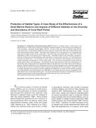
Ho et al. – Copepods Parasitic on Marine Fishes <strong>of</strong> Taiwan 613(A)(C)(F)(H)(G)p4(B)(E)(D)p4crFig. 1. Lernanthropodes chorinemi Pillai, 1962, female. (A) Habitus, dorsal view; (B) habitus, ventral view; (C) habitus, lateral view;(D) urosome with basal part <strong>of</strong> leg 4, dorsal view; (E) antennule and parabasal process, ventral view; (F) antenna, medial view; (G)maxillule, lateral view; (H) maxilla, medial view. Scale bars: A-C = 0.5 mm; D = 0.2 mm; E and G = 20 μm; F and H = 30 μm. p4: leg 4;cr: caudal ramus.
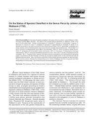
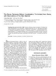
614<strong>Zoological</strong> <strong>Studies</strong> 50(5): 611-635 (2011)Taiwan is only 3.56 mm long on average.Four species <strong>of</strong> Lernanthropodes arecurrently known, namely Les. cucullus Bere, 1936,Les. natalensis Kensley et Grindley, 1973, and Les.trachinoti Pillai, 1962, in addition to Les. chorinemi.Of <strong>the</strong>se Les. chorinemi is most similar to Les.natalensis in having a triangular-shaped headand without a deep, central notch on <strong>the</strong> posteriormargin <strong>of</strong> <strong>the</strong> fused leg 3 lamellae. Never<strong>the</strong>less,Les. chorinemi differs from this closest congenerby possessing a well-developed parabasal processat <strong>the</strong> base <strong>of</strong> <strong>the</strong> antennules and <strong>the</strong> structures<strong>of</strong> <strong>the</strong> maxillule (inner lobe with 3, instead <strong>of</strong> 2,terminal setae) and maxilla (with denticles and bifidspiniform element on <strong>the</strong> brachium).When Pillai (1985) gave <strong>the</strong> 2nd report <strong>of</strong>Les. chorinemi from India, <strong>the</strong> hosts were listed asChorinemus sanctipetri and C. lysan. However,according to Froese and Pauly (2011), both <strong>of</strong><strong>the</strong>m are synonyms <strong>of</strong> Scomberoides lysan. InPillai’s (1985) 2nd report on Les. chorinemi fromIndia, <strong>the</strong> male was described. It looks like <strong>the</strong>male <strong>of</strong> Lernanthropinus sphyraenae (Yamagutiet Yamasu 1959) previously reported by us fromTaiwan (see Ho et al. 2008) in having both legs 3and 4 comprising a single process.Lernanthropodes trachinoti Pillai, 1962(Figs. 3, 4)Material examined: 1 on 1 (<strong>of</strong> 4) snubnosepompano, Trachinotus blochii (Lacepède, 1801),landed at Dong-shi Fishing Port on 19 Jan. 2008.Female: Body (Fig. 3A-C) cylindrical, 5.46mm long (from anterior rim <strong>of</strong> head to posteriormargin <strong>of</strong> fused leg 3 lamellae), comprising head,trunk, and small urosome. Head squarish, 1.36× 1.18 mm, with anterolateral corners protrudingventrally into rounded knob (Fig. 3C). Trunkcylindrical, slightly wider (1.26 mm wide) thanhead, without dorsal plate. Posterior part <strong>of</strong> trunkwith ventrally fused lamellae <strong>of</strong> leg 3 appearingwider (2.02 mm wide) than head. Genital complex(Fig. 3D) slightly wider than long, 478 × 494 µm,with laterally protruding egg sac attachment area.Abdomen (Fig. 3D) also with laterally protrudingsides and wider than long, 267 × 308 µm. Caudalramus (Fig. 3D) elongate, 356 × 162 µm, carrying2 dorsal setae in basal region and 2 setae at distalend. Egg sac long and straight (not illustrated).Antennule (Fig. 3E, F) filiform and indistinctly7-segmented; armature formula: 1, 2, 1, 2, 1, 3 +(A)(B)(C)Fig. 2. Lernanthropodes chorinemi Pillai, 1962, female. (A) Maxilliped, medial view; (B) leg 1, ventral view; (C) leg 2, ventral view.Scale bars: A = 40 μm; B = 20 μm; C = 10 μm.
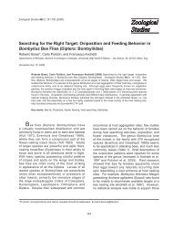
Ho et al. – Copepods Parasitic on Marine Fishes <strong>of</strong> Taiwan 615(A)(B)(C)(H)(I)p4(G)(E)(D)p4(F)crFig. 3. Lernanthropodes trachinoti Pillai, 1962, female. (A) Habitus, dorsal view; (B) habitus, ventral view; (C) habitus, lateral view; (D)urosome and leg 4, dorsal view; (E) antennule, ventral view; (F) tip <strong>of</strong> antennule, ventral view; (G) antenna, medial view; (H) mandible; (I)maxillule, lateral view. Scale bars: A-C = 1 mm; D = 0.5 mm; E = 50 μm; F = 10 μm; G = 0.1 mm; H and I = 30 μm. p4: leg 4; cr: caudalramus.
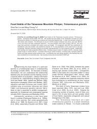
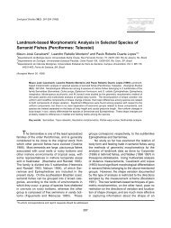
616<strong>Zoological</strong> <strong>Studies</strong> 50(5): 611-635 (2011)1 aes<strong>the</strong>tasc, and 8 + 1 aes<strong>the</strong>tasc. Parabasalprocess absent. Antenna (Fig. 3G) 2-segmented;corpus about twice as long as claw, former bearing1 broad basal seta on medial surface; claw bearingsimilar basal seta and terminal striations. Mandible(Fig. 3H) and maxillule (Fig. 3I) essentiallyas in previous species. Maxilla (Fig. 4A, B)2-segmented; lacertus unarmed; brachium bearing1 subterminal seta on medial margin, 1 blunt,terminal element, and large patch <strong>of</strong> denticles onouter surface; terminal claw fringed with rows <strong>of</strong>denticles along both edges. Maxilliped (Fig. 4C)2-segmented; corpus with fine denticles scatteredin myxal region; shaft longer than claw, with 1subterminal seta on medial margin; claw withstriations as in antenna.Ventral surface <strong>of</strong> leg 1 (Fig. 4D) with denticlesscattered on protopod and endopod;outer protopodal seta simple and thin, but innerprotopodal seta spiniform and arising from largepapilla; exopod 1-segmented and large, tipped with5 robust spines, inner 2 <strong>of</strong> which bear denticles onboth sides; endopod smaller than exopod, carrying1 long seta terminally. Leg 2 (Fig. 4E) protopodinconspicuous, without inner and outer setae;exopod armed as in leg 1, but seta on endopodbilaterally denticulate. Leg 3 with lamelliform ramicompletely fused to form a long plate completelycovering urosome ventrally (Fig. 3B) and leavinglarge gap dorsally (Fig. 3A); posterior edge <strong>of</strong>ventral lamella with 3 indentations (Fig. 3B). Leg 4a pair <strong>of</strong> long bilobate processes (Fig. 3A) arising(A)(B)(D)(C)(E)Fig. 4. Lernanthropodes trachinoti Pillai, 1962, female. (A) Maxilla, medial view; (B) tip <strong>of</strong> maxilla, medial view; (C) maxilliped, medialview; (D) leg 1, ventral view; (E) leg 2, ventral view. Scale bars: A = 50 μm; B = 10 μm; C = 0.1 mm; D = 30 μm; E = 20 μm.
Ho et al. – Copepods Parasitic on Marine Fishes <strong>of</strong> Taiwan 617from basal region <strong>of</strong> urosome (Fig. 3D). Leg 5absent.Male: Not collected.Remarks: Lernanthropodes trachinoti is s<strong>of</strong>ar known to occur on pompanos (Trachinotus)from India and Australia. In India, like in Taiwan, itwas taken from a snubnose pompano (Pillai 19621985), but in Australia, it was reported from ano<strong>the</strong>rspecies <strong>of</strong> pompano, Tra. botla (Shaw, 1803)(see Kabata 1979b). It was intriguing to note <strong>the</strong>specimen <strong>of</strong> Les. trachinoti in Pillai’s (1985) 2ndreport differed from his original report (Pillai 1962)in having a triangular head (cephalothorax) and alarge fused lamellae <strong>of</strong> leg 3 completely concealing<strong>the</strong> bifid leg 4 and urosome in ventral view <strong>of</strong> <strong>the</strong>animal. In o<strong>the</strong>r words, it may represent a differentspecies <strong>of</strong> Lernanthropodes. In fact, Pillai (1985)remarked in his 2nd report <strong>of</strong> Les. trachinoti that“This species closely resembles L. cuculus Bereand distinguished by only minor differences. Theymay turn out to be <strong>the</strong> same.” Inasmuch as <strong>the</strong>original description <strong>of</strong> Les. cuculus is sketchy, n<strong>of</strong>ur<strong>the</strong>r comment can be made at this point. Bere’s(1936) specimens <strong>of</strong> Les. cuculus were found onTra. carolinus (Linnaeus, 1766) and Tra. falcatus(Linnaeus, 1758) from <strong>the</strong> Gulf <strong>of</strong> Mexico.Specimens <strong>of</strong> Les. trachinoti from Taiwan fitwell with <strong>the</strong> original report <strong>of</strong> <strong>the</strong> species givenby Pillai (1962). The male <strong>of</strong> this species is notknown from India nor Taiwan, but Kabata (1979b)found it on Tra. botla from Australia.Genus Lernanthropus de Blainville, 1822Lernanthropus incilis sp. nov.(Figs. 5-7)Material examined: 4 and 2 found on gill filaments <strong>of</strong> Poey’s scabbardfish,Evoxymetopon poeyi Gün<strong>the</strong>r, 1887, landed atCheng-gong Fishing Port: 3 and 1 from 3(<strong>of</strong> 3) E. poeyi on 11 Feb. 2009, and 1 and 1 from 1 (<strong>of</strong> 1) E. poeyi on 25 Mar. 2009. Femaleholotype (USNM 1131890) and male allotype(USNM 1131891) were deposited in <strong>the</strong> NationalMuseum <strong>of</strong> Natural History, Smithsonian Institution,Washington, DC.Female: Body (Fig. 5A-C) large, 7.63 (7.50-7.76) mm long (from anterior rim <strong>of</strong> head to end <strong>of</strong>caudal ramus), divisible into head, neck, trunk, andurosome. Head nearly squarish, 1.95 (1.92-1.98)× 2.01 (1.80-2.22) mm, with narrowed antennalarea. Neck (1st pediger) short and wide, bearinglarge dorsal lobe. Remaining pedigers fused intotrunk, with pedigers 2 and 3 protruding out to forma lateral lobe and pediger 4 expanded posteriorlyinto a large subcircular dorsal plate that is deeplyemarginated in center. Genital complex andabdomen (Fig. 5D) wider than long, 0.40 (0.38-0.42) × 0.89 (0.84-0.94) and 0.48 (0.46-0.50) × 0.62(0.62-0.62) mm, respectively. Caudal ramus (Fig.5D) transformed into a long process, 2.21 (2.04-2.38) × 0.42 (0.40-0.44) mm, bearing 2 basal setaeon ventral surface (Fig. 5E), 1 subterminal seta onouter margin, and 2 small setae at tip. Egg saclong and straight.Antennule (Fig. 5F, G) stocky, indistinctly5-segmented; armature formula: 0, 0, 0, 0, and 9 +2 aes<strong>the</strong>tascs. Parabasal process (Fig. 5F) short.Antenna (Fig. 5H) robust, 2-segmented; corpusunarmed; claw armed with basal seta. Mandible(Fig. 6A) and maxillule (Fig. 6B) essentially as inprevious species. Maxilla (Fig. 6C) 2-segmented,with unarmed lacertus larger and longer thanbrachium; latter subterminally bearing 1 short,spiniform process and patch <strong>of</strong> denticles on medialsurface (usual terminal seta missing); terminalclaw (Fig. 6D) fringed with row <strong>of</strong> denticles onmedial surface. Maxilliped (Fig. 6E) 2-segmented;corpus robust and unarmed; subchela comprisingsmall, seta-bearing shaft and striated claw.Leg 1 (Fig. 6F) with protopod protruding outinto a process which carries an outer seta at itsbase; protopod also with inner conical process;exopod tipped with 5 stocky spines and endopodwith 1 blunt seta (Fig. 6G). Leg 2 (Fig. 6H) morereduced than leg 1, with inconspicuous protopodand weakly armed exopod (Fig. 6I). Leg 3 (Fig.5B) greatly modified, comprising large fleshy,folded lamella splayed ventrally at posterolateralcorners <strong>of</strong> trunk (Fig. 5C). Leg 4 (Fig. 5B) a pair<strong>of</strong> long, bifid processes with round, blunt tip. Leg5 (Fig. 5D) modified into a unilobate, long, obtuseprocess.Male: Body (Fig. 7A, B) smaller than femaleand without dorsal plate on trunk, measuring4.58 mm long (from tip <strong>of</strong> head to end <strong>of</strong> caudalramus). Head (cephalosome) wider than long, 1.64× 1.88 mm, with antennal region set apart fromrest <strong>of</strong> head. First 2 pedigers identifiable by <strong>the</strong>irlateral swellings, wider than long, measuring 0.24× 1.00 and 0.40 × 1.08 mm, respectively. Genitalcomplex indistinguishably fused to trunk. Caudalramus (Fig. 7A, B) long, slender, 745 × 186 μm,and armed as in female.Antennule (Fig. 7C) stocky as in female, butunsegmented and terminally armed with 3 moresetae (Fig. 7D). Parabasal process with basal
618<strong>Zoological</strong> <strong>Studies</strong> 50(5): 611-635 (2011)papilla arising near base <strong>of</strong> antennule (Fig. 7C).Leg 1 (Fig. 7E) with rows <strong>of</strong> denticles on outermargin <strong>of</strong> protopod and medial margin <strong>of</strong> endopod.Leg 2 (Fig. 7F) carrying a process lateral toexopod, subterminally bearing 1 seta-bearingpapilla and terminally 1 smaller seta-bearingpapilla; exopod with dense patch <strong>of</strong> denticlesterminally in addition to bearing 4 spiniformelements; endopod with dense patch <strong>of</strong> denticleson medial surface. Leg 3 (Fig. 7A) modified intopair <strong>of</strong> long, thin, bifid processes. Leg 4 (Fig. 7A)constructed as in leg 3, but longer and armedwith bifid denticles on distal 1/2 <strong>of</strong> exopod. Leg 5absent.Etymology: The species name incilis means“cut in” in Latin. It alludes to <strong>the</strong> possession <strong>of</strong> a(A)(B)(E)(D)p3p5crp4(C)(F)(H)(G)Fig. 5. Lernanthropus incilis sp. nov., female paratype. (A) Habitus, dorsal view; (B) habitus, ventral view; (C) habitus, lateral view;(D) urosome and leg 5, ventral view; (E) basal part <strong>of</strong> caudal ramus circled in D; (F) antennule and parabasal process, dorsal view;(G) tip <strong>of</strong> antennule, ventral view; (H) antenna, medial view. Scale bars: A-C = 1 mm; D = 0.5 mm; E = 25 μm; F = 0.1 mm; G = 20 μm;H = 0.2 mm. p3: leg 3; p4: leg 4; p5: leg 5; cr: caudal ramus.
Ho et al. – Copepods Parasitic on Marine Fishes <strong>of</strong> Taiwan 619(A)(B)(C)(D)(E)(F)(G)(H)(I)Fig. 6. Lernanthropus incilis sp. nov., female paratype. (A) Mandible; (B) maxillule, lateral view; (C) maxilla, medial view; (D) tip<strong>of</strong> maxilla, medial view; (E) maxilliped, medial view; (F) leg 1, ventral view; (G) rami <strong>of</strong> leg 1, ventral view; (H) leg 2, ventral view;(I) exopod <strong>of</strong> leg 2, ventral view. Scale bars: A and D = 20 μm; B = 50 μm; C, F, and H = 0.1 mm; E = 0.2 mm; G and I = 30 μm.
620<strong>Zoological</strong> <strong>Studies</strong> 50(5): 611-635 (2011)deep, central incision on <strong>the</strong> posterior rim <strong>of</strong> <strong>the</strong>dorsal plate.Remarks: More than 100 species are currentlyclassified under Lernanthropus. All <strong>of</strong> <strong>the</strong>mare characteristic in having a large dorsal platecoming <strong>of</strong>f <strong>the</strong> posterior border <strong>of</strong> <strong>the</strong> trunk. Theposterior margin <strong>of</strong> this dorsal plate is entire (wi<strong>the</strong>ven margin) in most cases. It is only in <strong>the</strong>following 3 species that we see, like Lus. incilis sp.nov., <strong>the</strong> presence <strong>of</strong> a deep, central incision in<strong>the</strong> posterior rim <strong>of</strong> <strong>the</strong> dorsal plate: Lus. barnardiCapart, 1959; Lus. monodi Delamare-Debouttevilleet Nunes-Ruivo, 1954; and Lus. obscurus Wilson,1913. Never<strong>the</strong>less, <strong>the</strong> new species from(A)(E)p3p4cr(F)(C)(B)(D)Fig. 7. Lernanthropus incilis sp. nov., male paratype. (A) Habitus, dorsal view; (B) habitus, ventral view; (C) antennule and parabasalprocess, dorsal view; (D) tip <strong>of</strong> antennule, dorsal view; (E) leg 1, ventral view; (F) leg 2, dorsal view. Scale bars: A and B = 1 mm;C = 0.1 mm; D = 20 μm; E and F = 40 μm. p3: leg 3; p4: leg 4; cr: caudal ramus.
Ho et al. – Copepods Parasitic on Marine Fishes <strong>of</strong> Taiwan 621Taiwan is distinguished from Lus. barnardi andLus. monodi by <strong>the</strong> structure <strong>of</strong> <strong>the</strong> trunk (with 2lateral indentations). It fur<strong>the</strong>r differs from Lus.barnardi by <strong>the</strong> shape <strong>of</strong> <strong>the</strong> dorsal plate (largeand comprising 2 subcircular plates) and from Lus.monodi by <strong>the</strong> short neck and structure <strong>of</strong> legs 3and 4 (without pointed tip). Both Lus. barnardi andLus. monodi were reported <strong>of</strong>f <strong>the</strong> west coast <strong>of</strong>Africa (Delamare-Deboutteville and Nunes-Ruivo1954, Capart 1959).As far as <strong>the</strong> general appearance is concerned,Lus. incilis sp. nov. resembles Lus.obscurus <strong>the</strong> most. Both <strong>of</strong> <strong>the</strong>m are also unusualfor Lernanthropus in carrying a pair <strong>of</strong> plumpantennules. However, Lus. incilis sp. nov. differsfrom Lus. obscurus in <strong>the</strong> structure <strong>of</strong> leg 5 (notfoliaceous) and in bearing a large dorsal outgrowthin <strong>the</strong> neck region. Lernanthropus obscurus is s<strong>of</strong>ar known only from <strong>the</strong> West Indies (Wilson 1913).Genus Mitrapus Song et Chen, 1976Mitrapus heteropodus (Yü, 1933)(Figs. 8-10)Material examined: 4 and 1 on 3 (<strong>of</strong>45) Bloch’s gizzard shad, Nematalosa nasus(Bloch, 1795), landed at Sheng-dah Fishing Porton 14 Jan. 1999.Female: Body (Fig. 8A-C) small, but shortand broad, 1.87 (1.62-2.12) mm long (from anteriorrim <strong>of</strong> head to end <strong>of</strong> caudal ramus), divisible intohead, short neck, broad trunk, and small urosome;posterior part <strong>of</strong> trunk (4th pediger) carrying alarge, semicircular dorsal plate. Head slightlylonger than wide, 0.63 (0.58-0.68) × 0.61 (0.60-0.62) mm, with rounded sides. Trunk widestpart <strong>of</strong> body, with forward protruding shouldersand ano<strong>the</strong>r forward protrusion found in front <strong>of</strong>leg 2 (Fig. 8B). Genital complex and abdomenindistinguishably fused into 1 unit (Fig. 8D).Caudal ramus (Fig. 8D, E) longer than wide, 69(65-73) × 45 (41-49) μm, bearing 3 subterminaland 2 terminal setae. Egg sac straight (notillustrated).Antennule (Fig. 8F) filiform, indistinctly6-segmented; armature formula: 3, 0, 2, 1, 4 +1 aes<strong>the</strong>tasc, and 8 + 1 aes<strong>the</strong>tasc. Parabasalprocess absent. Antenna (Fig. 8G) robust,2-segmented; corpus unarmed; claw long andsharply pointed, armed with 2 medial setae inbasal region. Mandible (Fig. 8H) and maxillule (Fig.8I) generally as in previous species. Maxilla (Fig.9A) 2-segmented; lacertus larger than brachiumbut unarmed; latter armed subterminally with 2short setae on medial surface in addition to 1larger terminal seta; terminal claw fringed withrow <strong>of</strong> denticles around medial margin. Maxilliped(Fig. 9B) 2-segmented; corpus robust, with 1small papilla on myxal surface followed by row <strong>of</strong>denticles; subchela comprising small seta-bearingshaft and long, curved claw.Leg 1 (Fig. 9C) protopod with simple outerseta and apically blunt, spiniform inner seta;1-segmented exopod with 5 robust, denticulatedterminal spines; endopod an inflated lobe bearingdenticles on ventral surface and tipped with ashort, spiniform seta. Leg 2 (Fig. 9D) protopodlacking outer and inner seta; 1-segmented exopodwith 3 denticles scattered on ventral surface andarmed with 4 terminal spines, <strong>of</strong> which only inner2 are fringed with denticles; endopod armedgenerally as in leg 1. Leg 3 (Fig. 8A-C) greatlymodified, comprising large fleshy, folded lamellasplayed outward at posterolateral corners <strong>of</strong>trunk. Leg 4 (Fig. 8A, B) a pair <strong>of</strong> greatly unequal,bilobate processes, with exopod about 6 times aslong as endopod. Leg 5 missing.Male: Attached to basal region <strong>of</strong> female leg4 exopod (see Fig. 8B, C). Body (Fig. 10A, B)653 μm long (from tip <strong>of</strong> head to end <strong>of</strong> caudalramus). Head (cephalosome) oblong, 338 ×207 μm, with narrowed antennal region. Trunksubrectangular, narrower than head; each <strong>of</strong>4 pedigers identifiable by its lateral swellings.Genital complex wider than long, 72 × 80 μm,indistinguishably fused to trunk anteriorly and toabdomen posteriorly. Caudal ramus longer thanwide, 34 × 19 μm, shaped and armed as in female.Antennule (Fig. 10C, D) filiform, indistinctly6-segmented; armature formula: 1, 3, 2, 1, 2 + 1aes<strong>the</strong>tasc, and 9 + 1 aes<strong>the</strong>tasc. Legs 1 (Fig.10E) and 2 (Fig. 10F) similar to those in female,except for carrying denticles on ventral surface <strong>of</strong>protopod and equipped with longer terminal setaon endopod. Legs 3 and 4 (Fig. 10A, B) eachrepresented by 1 short outer seta in basal region<strong>of</strong> small knob on posterolateral corners <strong>of</strong> trunk.Leg 5 absent.Remarks: When Song and Chen (1976)created Mitrapus to accommodate Lus.heteropodus Yü, 1933, 3 o<strong>the</strong>r species <strong>of</strong>Lernanthropus were included, namely Lus.rubiginosus Redkar, Rangnekar et Murti, 1949,Lus. engraulis Tripathi, 1962, and Lus. oblongusPillai, 1964. Our specimens from Taiwan areidentifiable with ei<strong>the</strong>r Lus. heteropodus or Lus.rubiginosus. However, we formally consider Lus.
622<strong>Zoological</strong> <strong>Studies</strong> 50(5): 611-635 (2011)(A)(G)(B)p3(D)(E)p4(I)cr(C)(F)(H)Fig. 8. Mitrapus heteropodus (Yü, 1933), female. (A) Habitus, dorsal view; (B) habitus, ventral view; (C) habitus, lateral view; (D)urosome, dorsal view; (E) caudal ramus, ventral view; (F) antennule, dorsal view; (G) antenna, ventral view; (H) mandible; (I) maxillule;lateral view. Scale bars: A-C = 0.5 mm; D and G = 50 μm; E, H, and I = 20 μm; F = 30 μm. p3: leg 3; p4: leg 4; cr: caudal ramus.
Ho et al. – Copepods Parasitic on Marine Fishes <strong>of</strong> Taiwan 623rubiginosus reported by Redkar et al. (1949) tobe conspecific with Lus. heteropodus reported byYü (1933), because <strong>of</strong> <strong>the</strong> resemblance between<strong>the</strong>m in <strong>the</strong> gross morphology <strong>of</strong> both sexes and<strong>the</strong> general structure <strong>of</strong> <strong>the</strong> female appendages.Fur<strong>the</strong>rmore, according to Redkar et al. (1949)<strong>the</strong>ir specimens <strong>of</strong> Lus. rubiginosus (6 females and6 males) were obtained from Chatoessus nasusDay [= Nematalosa nasus (Bloch)], <strong>the</strong> samespecies <strong>of</strong> gizzard shad from which we found ourspecimens <strong>of</strong> M. heteropodus.The 3 species <strong>of</strong> Mitrapus show differencesin <strong>the</strong> ratio <strong>of</strong> <strong>the</strong> endopod to <strong>the</strong> exopod in <strong>the</strong>female leg 4. It is about 1: 16 in M. engraulis,1: 6 in M. heteropodus, and 1: 2 in M. oblongus.Ano<strong>the</strong>r distinguishing point <strong>of</strong> <strong>the</strong>se 3 speciesis seen in <strong>the</strong> structure <strong>of</strong> <strong>the</strong>ir shoulders. Theshoulder <strong>of</strong> M. engraulis is rounded without ananterior protrusion, with a small anterior protrusionin M. oblongus, and bearing a large anteriorprotrusion in M. heteropodus. It is interesting tonote that <strong>the</strong> species with <strong>the</strong> rounded shoulder(M. engraulis) is a parasite <strong>of</strong> <strong>the</strong> anchovy(Engraulidae), and <strong>the</strong> 2 o<strong>the</strong>r species withprotruding shoulders (M. heteropodus and M.oblongus) are parasitic on herrings (Clupeidae).Genus Sagum C. B. Wilson, 1913Sagum epinepheli (Yamaguti et Yamasu 1960)(Figs. 11, 12)Material examined: 1 on 1 (<strong>of</strong> 27) yellowgrouper, Epinephelus awoara (Temminck etSchlegel, 1842), landed at Dong-gang on 27 Dec.2003.Female: Body (Fig. 11A, B) globular, coveredwith denticles on dorsal surface, 3.86 mm long(A)(B)(C)(D)Fig. 9. Mitrapus heteropodus (Yü, 1933), female. (A) Maxilla, medial view; (B) maxilliped, medial view; (C) leg 1, ventral view; (D) leg 2,dorsal view. Scale bars: A = 40 μm; B = 50 μm; C and D = 20 μm.
624<strong>Zoological</strong> <strong>Studies</strong> 50(5): 611-635 (2011)(from tip <strong>of</strong> head to posterior margin <strong>of</strong> dorsalplate), comprising large head, short neck (1stpediger), rectangular trunk with a large subcirculardorsal plate, and minute, concealed urosome.Head bearing beak-like lateral protrusions, 1.24× 1.82 mm, with both sides turned ventrally (Fig.11C). Neck carrying globular swellings (Fig. 11B)on ventral surface lateral to leg 1 (Fig. 12D).Trunk (Fig. 11A, C) with smooth, shoulder-likeanterolateral corners and posterolateral cornersprotruding to rear along lateral sides <strong>of</strong> dorsalplate. Components <strong>of</strong> urosome fused into 1 shortunit (Fig. 11D) and entirely concealed under dorsalplate in dorsal view. Genital complex wider thanlong, 340 × 486 µm. Abdomen also wider thanlong, 146 × 186 µm. Caudal ramus (Fig. 11E) along attenuated process carrying 1 seta and 2knobs in swollen, basal region and 2 setae in distalregion. Egg sac (not shown in Fig. 11) long andcoiled underneath dorsal plate.Antennule (Fig. 11F, G) indistinctly 7-segmented,with armature <strong>of</strong> 0, 0, 1, 1, 0, 2 and10 + 2 aes<strong>the</strong>tascs. Antenna broken (see Fig.11B). Mandible (Fig. 11H) as in previous species.Maxillule broken and lost during dissection.Maxilla (Fig. 12A) 2-segmented, with unarmedlacertus; brachium distally bearing 1 patch<strong>of</strong> denticles and 1 small, blunt element (Fig.12B); terminal claw armed with row <strong>of</strong> denticlesaround margin. Maxilliped (Fig. 12C) indistinctly3-segmented; corpus unarmed; subchela with 1small subterminal seta on shaft; terminal claw withstriations.Leg 1 (Fig. 12D) protopod missing outer setaand with inner element appearing as a spiniformseta; exopod 1-segmented, tipped with 5 stockyspines; endopod reduced to a simple lobe. Leg2 (Fig. 12E) protopod protruding laterally into(A)(B)(E)p3(D)(F)p4cr(C)Fig. 10. Mitrapus heteropodus (Yü, 1933), male. (A) Habitus, dorsal view; (B) habitus, ventral view; (C) antennule, dorsal view; (D)tip <strong>of</strong> antennule, dorsal view; (E) leg 1, ventral view; (F) leg 2, ventral view. Scale bars: A and B = 0.1 mm; C, E, and F = 20 μm;D = 10 μm. p3: leg 3; p4: leg 4; cr: caudal ramus.
Ho et al. – Copepods Parasitic on Marine Fishes <strong>of</strong> Taiwan 625(A)(D)(E)cr(G)p4(F)(C)(B)(H)p3p4Fig. 11. Sagum epinepheli (Yamaguti et Yamasu, 1960), female. (A) Habitus, dorsal view; (B) habitus, ventral view; (C) habitus, lateralview; (D) urosome and leg 4, ventral view; (E) caudal ramus, dorsal view; (F) antennule, dorsal view; (G) tip <strong>of</strong> antennule, dorsal view; (H)mandible. Scale bars: A-C = 1 mm; D = 0.5 mm; E = 50 μm; F = 0.1 mm; G = 20 μm; H = 30 μm. p3: leg 3; p4: leg 4; cr: caudal ramus.
626<strong>Zoological</strong> <strong>Studies</strong> 50(5): 611-635 (2011)large, setulate process; exopod a lobe tipped with3 small spiniform elements; endopod reduced toseta-bearing papilla. Leg 3 (Fig. 11B, C) greatlymodified into fleshy, bent lamella; protopod foldedand protruding ventrally (see Fig. 11C); exopodlarger than endopod, expanded posteriorlyinto a large lamella with dorsal side fused toposteroventral protrusion <strong>of</strong> trunk (see Fig. 11C);endopod a long lamella concealing urosome inventral view <strong>of</strong> animal (see Fig. 11B). Leg 4 (Fig.11D) protopod with outer seta; exopod larger thanendopod, but both rami with foliaceous basal partand long, filiform distal part. Leg 5 missing.Male: Not collected.Remarks: The present species was reportedfrom Japan (Yamaguti and Yamasu 1960) andIndia (Pillai and Sebastian 1967). In all instances,just like from Taiwan, <strong>the</strong> parasites were foundparasitic on gill filaments <strong>of</strong> groupers belonging to<strong>the</strong> genus Epinephelus.Although 11 species <strong>of</strong> Sagum are listed in<strong>the</strong> World <strong>of</strong> Copepods by Walter (2010), many<strong>of</strong> <strong>the</strong>m are so poorly known that a meaningfulcomparison <strong>of</strong> <strong>the</strong> morphology between congenersis impossible. Exceptions to this fact are <strong>the</strong>following 4 species: S. flagellatum Wilson, 1913;S. foliaceum (Richiardi, 1880); S. petersi (vanBeneden, 1852); and S. vespertilio Kabata, 1979.Sagum epinepheli can easily be separated fromS. foliaceum and S. petersi by <strong>the</strong> presence <strong>of</strong>(A)(B)(C)(D)(E)Fig. 12. Sagum epinepheli (Yamaguti et Yamasu, 1960), female. (A) Maxilla, medial view; (B) tip <strong>of</strong> maxilla, medial view; (C)maxilliped, medial view; (D) leg 1, ventral view; (E) leg 2, ventral view. Scale bars: A and C = 50 μm; B = 20 μm; D and E = 30 μm.
Ho et al. – Copepods Parasitic on Marine Fishes <strong>of</strong> Taiwan 627a pair <strong>of</strong> lateral horns on <strong>the</strong> head, and fromS. flagellatum by not having posteroventralprotrusions <strong>of</strong> <strong>the</strong> trunk “prolonged backward andoutward like <strong>the</strong> skirts <strong>of</strong> a long military cloak”(Wilson 1913).Ho et al. (2008) reported <strong>the</strong> occurrence <strong>of</strong>S. vespertilio on Lethrinus nebulosus (Forsskål)collected from Penghu, Taiwan. In that reportS. tuberculatum Pillai, 1985 was proposed tobe relegated to <strong>the</strong> synonym <strong>of</strong> S. vespertilio.Sagum epinepheli can be distinguished from S.vespertilio by having a pair <strong>of</strong> smaller lateral hornson <strong>the</strong> head, lacking a lateral process on <strong>the</strong> neck,carrying a relatively longer, terminal filament oneach ramus <strong>of</strong> leg 4, and <strong>the</strong> absence <strong>of</strong> leg 5.Sagum folium sp. nov.(Figs. 13-15)Material examined: 15 and 3 foundon gill filaments <strong>of</strong> Japanese snapper, Paracaesiocaerulea (Katayama 1934): 2 on 1 (<strong>of</strong> 4)P. caerulea, landed at Dong-gang Fishing Porton 10 Oct. 2003; 13 and 3 on 5 (<strong>of</strong> 6)P. caerulea landed at Cheng-gong Fishing Porton 23 Sept. 2004. Female holotype (USNM1131888) and male allotype (USNM 1131889)were deposited in <strong>the</strong> National Museum <strong>of</strong> NaturalHistory, Smithsonian Institution, Washington, DC.Female: Body (Fig. 13A-C) globular, 4.13(4.04-4.22) mm long (from tip <strong>of</strong> head to posteriormargin <strong>of</strong> dorsal plate), comprising large head,short neck (1st pediger), semi-rectangular trunkwith a large dorsal plate, and minute urosome.Head slightly longer than wide, 1.25 (1.06-1.44)× 1.19 (1.16-1.22) mm, with both sides turnedventrally. Trunk with sclerites on dorsal andlateral sides and anteriorly protruding shoulders.Urosomal somites fused into 1 short unit (Fig. 13D)and entirely concealed under dorsal plate in dorsalview. Genital complex wider than long, 316 (292-340) × 458 (446-470) µm. Abdomen also widerthan long, 174 (162-186) × 279 (251-308) µm.Caudal ramus (Fig. 13E) leaf-like, inserted intoposterolateral corner <strong>of</strong> abdomen, carrying 3 short,naked setae in distal 1/2 <strong>of</strong> dorsal surface andano<strong>the</strong>r 2 setae at distal end. Egg sac (not shownin Fig. 13) long and coiled underneath dorsal plate.Antennule (Fig. 13F, G) indistinctly 7-segmented,with armature formula <strong>of</strong> 4, 1, 1, 0, 1,2 and 8 + 2 aes<strong>the</strong>tascs. Antenna (Fig. 14A)2-segmented; corpus carrying 1 small, basalpapilliform element on medial surface; terminalclaw stocky, also carrying similar basal elementon medial surface and apical surface striations.Mandible (Fig. 14B) composed <strong>of</strong> 2 sections; with8 teeth on terminal blade. Maxillule (Fig. 14C)bilobate; smaller outer lobe tipped with 1 element;larger inner lobe fringed with spinules on distal1/2 <strong>of</strong> medial margin in addition to carrying 3unequal, terminal elements. Maxilla (Fig. 14D, E)2-segmented, with unarmed lacertus; brachiumbearing 1 subterminal and 1 terminal blunt elementand row <strong>of</strong> denticles around margin <strong>of</strong> terminalclaw. Maxilliped (Fig. 14F) 2-segmented; corpuscarrying 1 papilliform element in myxal area;subchela with 1 small subterminal seta on shaftand 1 basal blunt element, median row <strong>of</strong> minutedenticles and apical striations on terminal claw.Leg 1 (Fig. 14G) with inconspicuous protopodcarrying 1 slender outer seta and 1 spiniform,pinnate inner element; exopod 1-segmented,fringed with setules on outer margin and tippedwith 5 stocky spines; endopod reduced to a lobetipped with a small, blunt element. Leg 2 (Fig.14H) more reduced than leg 1, without protopod;exopod a lobe tipped with 5 blunt elements andendopod with1 blunt element. Leg 3 (Fig. 13B,C) with both rami greatly modified into foliaceousstructure; exopod larger than endopod, occupyingmajor portion <strong>of</strong> lateral part <strong>of</strong> trunk (see Fig.13C). Leg 4 (Fig. 13B, D) rami subcylindrical,with setulate basal papilla on outer surface <strong>of</strong>protopod. Leg 5 (Fig. 14I) represented by a bent,blunt process near posterolateral corner <strong>of</strong> genitalcomplex; carrying 1 setulate papilla subterminallyon medial surface.Male: Body (Fig. 15A, B) smaller than female,1.81 (1.68-1.94) mm long (from tip <strong>of</strong> head to end<strong>of</strong> caudal ramus), without dorsal plate on trunk.Head (cephalosome) shaped like a piece <strong>of</strong> toast,slightly wider than long, 0.89 (0.86-0.92) × 0.91(0.74-1.08) mm. First pediger forming a short neckand remaining pedigers fused to form a rectangulartrunk with conical posterolateral protrusion.Genital complex and abdomen indistinguishablyfused to each o<strong>the</strong>r. Caudal ramus (Fig. 15C) alobe measuring 89 (81-97) µm long and 49 (41-57) µm wide, armed as in female.Antennule (Fig. 15D, E) filiform and indistinctly6-segmented, with armature formula <strong>of</strong> 1, 3,2, 0, 1, and 11 + 2 aes<strong>the</strong>tascs. Leg 2 (Fig. 15F)protopod with simple outer seta; exopod armed indistal region with 4 stocky spines and 3 patches <strong>of</strong>spinules; endopod with single subterminal setuleand tuft <strong>of</strong> terminal setules. Legs 3 and 4 (Fig.15A, B) represented by pair <strong>of</strong> bifid cylindrical
628<strong>Zoological</strong> <strong>Studies</strong> 50(5): 611-635 (2011)processes coming <strong>of</strong>f both sides <strong>of</strong> trunk. Leg 5absent.Etymology: The species name foliummeans “leaf” in Latin. It alludes to <strong>the</strong> unusualtransformation <strong>of</strong> <strong>the</strong> caudal rami appearing like apair <strong>of</strong> leaves at <strong>the</strong> end <strong>of</strong> <strong>the</strong> urosome.Remarks: Without <strong>the</strong> presence <strong>of</strong> a pair<strong>of</strong> lateral horns on <strong>the</strong> head, <strong>the</strong> new speciesis closer to S. foliaceum and S. petersi than to<strong>the</strong> remaining 3 well-described congeners <strong>of</strong> S.epinepheli, S. flagellatum, and S. vespertilio.Never<strong>the</strong>less, S. folium sp. nov. can be easilyseparated from those 2 similar species by <strong>the</strong>structure <strong>of</strong> <strong>the</strong> caudal ramus (being leaf-like) andleg 5 (a bent, blunt, short process subterminallycarrying a medial setulate papilla).Among <strong>the</strong> 5 well-described species <strong>of</strong>Sagum, <strong>the</strong> male is known for 2 species, S.epinepheli and S. foliaceum. The male <strong>of</strong> <strong>the</strong>new species can be easily distinguished from that<strong>of</strong> S. foliaceum in having its leg 3 constructed <strong>of</strong>bilobate (vs. unilobate) cylindrical processes andfrom that <strong>of</strong> S. epinepheli in having its large head(cephalosome) shaped like a piece <strong>of</strong> toast anddistinctly wider than <strong>the</strong> trunk (fused pedigeroussomites and urosome).(A)(B)(D)p4p5crp3p4(F)(C)(E)(G)Fig. 13. Sagum folium sp. nov., female paratype. (A) Habitus, dorsal view; (B) habitus, ventral view; (C) habitus, lateral view; (D)urosome, showing leg 4, leg 5, and caudal rami, dorsal view; (E) caudal ramus, dorsal view; (F) antennule, ventral view; (G) tip <strong>of</strong>antennule, ventral view. Scale bars: A-C = 1 mm; D = 0.5 mm; E = 0.1 mm; F = 50 μm; G = 20 μm. p3: leg 3; p4: leg 4; p5: leg 5; cr:caudal ramus.
Ho et al. – Copepods Parasitic on Marine Fishes <strong>of</strong> Taiwan 629(D)(A)(B)(E)(G)(F)(C)(H)(I)Fig. 14. Sagum folium sp. nov., female paratype. (A) Antenna, medial view; (B) mandible; (C) maxillule, lateral view; (D) maxilla,medial view; (E) tip <strong>of</strong> maxilla, medial view; (F) maxilliped, medial view; (G) leg 1, dorsal view; (H) leg 2, dorsal view; (I) leg 5. Scalebars: A = 0.2 mm; B and G = 40 μm; C and I = 50 μm; D and F = 0.1 mm; E = 20 μm; H = 30 μm.
630<strong>Zoological</strong> <strong>Studies</strong> 50(5): 611-635 (2011)DISCUSSIONT h e L e r n a n t h r o p i d a e , f o l l o w i n g t h eLernaeopodidae and Caligidae, is <strong>the</strong> 3rd-largestfamily <strong>of</strong> fish-parasitizing Siphonostomatoida. Thefamily contains over 150 species, with a greatmajority <strong>of</strong> <strong>the</strong>m occurring in tropical waters.Thus, in <strong>the</strong> 3 countries <strong>of</strong> India, Japan, and <strong>the</strong>UK, where <strong>the</strong> fauna <strong>of</strong> fish-parasitizing copepodsare best known, we see that India stands outas having <strong>the</strong> most lernanthropid species, at 44species (Pillai 1985), whereas Japan is surroundedby colder water and has only 8 species (Hoand Do 1985) as is <strong>the</strong> UK with only 5 species(Kabata 1979a). However, in consideration <strong>of</strong> <strong>the</strong>number <strong>of</strong> genera, Taiwan contains 7 genera <strong>of</strong><strong>the</strong> <strong>Lernanthropidae</strong> (Lernanthropinus Do in Hoand Do, 1985, Lernanthropodes, LernanthropsisDo in Ho and Do, 1985, Lernanthropus, Mitrapus,Norion von Nordmann, 1864, and Sagum). Indiais known to have representatives from 6 genera(Aethon, Lernanthropinus, Lernanthropsis,Lernanthropodes, Lernanthropus, and Sagum)and <strong>the</strong> o<strong>the</strong>r 2 aforementioned countries have5 genera in each, namely Lernanthropinus,Lernanthropsis, Lernanthropus, Mitrapus, andSagum in Japan (Ho and Do 1985) and Aethon,Lernanthropodes, Lernanthropus, Norion, andSagum in <strong>the</strong> UK (Kabata 1979a).A c c o r d i n g t o t h e c o p e p o d d a t a b a s eproduced by Boxshall (2011), Lernanthropus is<strong>the</strong> largest genus <strong>of</strong> lernanthropids comprising111 species. As a matter <strong>of</strong> fact, more than 3/4(75.5% or 111/147) <strong>of</strong> lernanthropids belong toLernanthropus. This striking uneven composition<strong>of</strong> species number is also evident in <strong>the</strong>(A)(D)(E)(F)(B)(C)p4crp3Fig. 15. Sagum folium sp. nov., male paratype. (A) Habitus, dorsal view; (B) habitus, ventral view; (C) caudal ramus, dorsal view; (D)antennule, dorsal view; (E) tip <strong>of</strong> antennule, dorsal view; (F) leg 2, dorsal view. Scale bars: A and B = 0.5 mm; C = 25 μm; D = 50 μm;E = 20 μm; F = 30 μm. p3: leg 3; p4: leg 4; cr: caudal ramus.
Ho et al. – Copepods Parasitic on Marine Fishes <strong>of</strong> Taiwan 631Table 1. Hosts and localities <strong>of</strong> 18 species <strong>of</strong> <strong>the</strong> <strong>Lernanthropidae</strong> known from Taiwan. Data in this tableare taken from Bassett-Smith (1898), Boxshall and Montú (1997), Byrnes (1988), Cressey and Collette(1970), Delamare-Deboutteville and Nunes-Ruivo (1954), Gusev (1951), Ho and Do (1985), Ho and Kim(2004), Ho and Sey (1996), Ho et al. (2008), Kabata (1962 1979b), Kensley and Grindley (1973), Kim (1998),Kirtisinghe (1937 1964), Krøyer (1863), Leong (1986), Liu et al. (2009a b), Pillai (1962 1963 1985), Pillaiand Sebastian (1967), Shiino (1955), Shishido (1898), Tripathi (1962), Yamaguti (1936 1954), Yamaguti andYamasu (1959 1960), and Yü (1933). The host names are valid ones following Froese and Pauly (2011).For those articles where a synonym was used for <strong>the</strong> host, <strong>the</strong>y are identified with a number and noted at<strong>the</strong> bottom <strong>of</strong> <strong>the</strong> tableParasite Host LocalityLernanthropinus sphyraenae (Yamaguti et Yamasu, 1959) Mene maculata (Bloch et Schneider) TaiwanSphyraena obtusata CuvierSri LankaSphyraena pinguis Gün<strong>the</strong>rJapanLernanthropodes chorinemi Pillai, 1962 Scomberoides commersonnianus Lacepéde TaiwanScomberoides lysan (Forsskål)IndiaLernanthropodes trachinoti Pillai, 1962 Trachinotus blochii (Lacepéde) India, TaiwanTrachinotus botla (Shaw)AustraliaLernanthropsis mugilii (Shishido, 1898) Acanthopagrus schlegelii schlegelii (Bleeker) 2) IndiaMugil cephalus LinnaeusJapan, India, Korea,Taiwan, AustraliaMugil soiuy BasilewskyChina, Sri Lanka, RussiaPagrus major (Temminck et Schlegel) 3) IndiaLernanthropus cadenati Delamare-Deboutteville et Acanthopagrus berda (Forsskål)KuwaitNunes-Ruivo, 1954Elops machnata (Forsskål)TaiwanElops senegalensis ReganSenegalMegalops cyprinoides (Broussonet)India, TaiwanSparidentex hasta (Valenciennes) 4)KuwaitLernanthropus chrysophrys Shishido, 1898 Acanthopatrus australis (Gün<strong>the</strong>r) AustraliaAcanthopagrus berda (Forsskål) 5)Japan, India, Australia,TaiwanAcanthopagrus latus (Houttuyn)TaiwanAcanthopagrus schlegelii schlegelii (Bleeker) 2) Japan, TaiwanLernanthropus corniger Yamaguti, 1954 Alepes djedaba (Forsskål) 6) South AfricaMegalaspis cordyla (Linnaeus)Indonesia, India, China,Malaysia, Thailand,TaiwanMene maculata (Bloch et Schneider)TaiwanMyripristis vittata ValenciennesTaiwanRastrelliger kanagurta (Cuvier)IndiaLernanthropus cornutus Kirtisinghe, 1937 Ablennes hians (Valenciennes) US east coast, Haiti,Mexico, Panama, Brazil,Peru, Hawaii, Taiwan,Mauritius,<strong>the</strong> PhilippinesPlatybelone argalus argalus (Lesueur) Gulf <strong>of</strong> GuineaStrongylura anastomella (Valenciennes) 7) Japan, KoreaStrongylura exilis (Girard)PeruStrongylura incisa (Valenciennes) 8)Gilbert Is., AustraliaStrongylura leiura (Bleeker) 9)Sri Lanka, Taiwan,<strong>the</strong> PhilippinesStrongylura marina (Walbaum)British HondurasStrongylura strongylura (Hasselt)Malay Peninsula
632<strong>Zoological</strong> <strong>Studies</strong> 50(5): 611-635 (2011)Table 1. (continued)Parasite Host LocalityStrongylura timucu (Walbaum)Strongylura urvillii (Valenciennes)Tylosurus acus acus (Lacepède)Tylosurus acus melanotus (Bleeker)Tylosurus choram (Rüppell)Tylosurus crocodilus crocodilus (Péron etLesueur)Tylosurus punctatus (Gün<strong>the</strong>r)Brazil<strong>the</strong> PhilippinesPuerto Rico, Angola,Panama, Mexico,Java, <strong>the</strong> PhilippinesJapan, TaiwanRed SeaUS east coast,Venezuela, Mexico,Panama, Kenya,Zanzibar, Senegal,Madagascar, Red Sea,Mauritius, Seychelles,Gulf <strong>of</strong> Aden, Senegal,Java, Arabian Gulf,Gulf <strong>of</strong> Thailand,Borneo, Taiwan,<strong>the</strong> Philippines, Japan,Hawaii<strong>the</strong> PhilippinesLernanthropus giganteus Krøyer, 1863 Carangoides ferdau (Forsskål) IndiaCarangoides praecustus (Bennett)ChinaCaranx crysos (Mitchill)JamaicaCaranx hippos (Linnaeus) 11)Jamaica, BrazilCaranx ignobilis (Forsskål)Sri Lanka, TaiwanCaranx leptolepis CuvierKuwaitCaranx melampygus CuvierAdenCaranx sansun (Forsskål)Sri LankaCaranx senegallus CuvierSenegalCaranx sexfasciatus Quoy et Gaimard TaiwanLernanthropus incilis sp. nov. Evoxymetopon poeyi Gün<strong>the</strong>r TaiwanLernanthropus otolithi Pillai, 1963 Otoli<strong>the</strong>s ruber (Bloch et Schneider) 12) IndiaPennahia pawak (Lin)TaiwanPterotolithus maculatus (Cuvier) 13)IndiaLernanthropus pomadasysis Rangnekar and Murti, 1961 Pomadasys kaakan (Cuvier) TaiwanPomadasys maculatus (Bloch)IndiaLernanthropus pristipomoides Kirtisinghe, 1937 Kyphosus vaigiensis (Quoy et Gaimard) TaiwanMitrapus heteropodus (Yü, 1933) Konosirus punctatus (Temminck et Schlegel) China, JapanNematalosa nasus (Bloch)TaiwanNorion priacanthi (Kirtisinghe, 1956) Priacanthus macracanthus Cuvier TaiwanSagum epinepheli (Yamaguti et Yamasu, 1960) Epinephelus akaara (Temminck et Schlegel) JapanEpinephelus awoara (Temminck et Schlegel) TaiwanEpinephelus sp.IndiaSagum folium sp. nov. Paracaesio caerulea (Katayama) TaiwanSagum vespertilio Kabata, 1979 Lethrinus laticaudis (Alleyne et Macleay) 14) AustraliaLethrinus nebulosus (Forsskål)TaiwanSynonyms used in <strong>the</strong> original publications are: 1) Chorinemus lysan in Pillai (1962); 2) Sparus macrocephalus in Shishido (1898),Shiino (1955), Song and Chen (1976), and Pillai (1985); 3) Pagrosomus major in Pillai (1985); 4) Acanthopagrus cuvieri in Ho and Sey(1996); 5) Sparus longispinis in Yamaguti (1936); 6) Caranx djedaba in Kensley and Grindley (1973); 7) Ablennes anastomella in Hoand Do (1985); 8) Tylosurus incisus in Kabata (1962); 9) Tylosurus leisurus in Kirtisinghe (1964); 10) Strongylura crocodile in Delamare-Deboutteville and Nunes-Ruivo (1954); 11) Caranx carangus in Krøyer (1863); 12) Otolithus argenteus in Pillai (1963); 13) Otolithusmaculates in Pillai (1963); and 14) Lethrinus fletus in Kabata (1979b).
Ho et al. – Copepods Parasitic on Marine Fishes <strong>of</strong> Taiwan 633lernanthropids <strong>of</strong> Taiwan, where nine <strong>of</strong> <strong>the</strong> 18known species are in Lernanthropus (see Table 1).As stated in <strong>the</strong> “Introduction”, we believe thatmany more species <strong>of</strong> lernanthropids are yet tobe discovered from <strong>the</strong> marine fishes <strong>of</strong> Taiwan,because so far fewer than 20% <strong>of</strong> <strong>the</strong> availablespecies <strong>of</strong> marine fish from Taiwan have beenexamined for parasitic copepods. Ano<strong>the</strong>rreason why we speculate that more species <strong>of</strong>lernanthropids will be found from Taiwan is <strong>the</strong> factthat a large number <strong>of</strong> lernanthropids reported from<strong>the</strong> Indo-West Pacific region are parasitic on fishesthat are also known to occur in waters <strong>of</strong> Taiwan.As shown in table 2, as far as we are aware, asmany as 25 species <strong>of</strong> such lernanthropids in4 genera are yet to be found from <strong>the</strong> fishes <strong>of</strong>Taiwan.To encourage future work on lernanthropids<strong>of</strong> Taiwan, a key to <strong>the</strong> 18 known species <strong>of</strong> <strong>the</strong>family in Taiwan is provided below. Since <strong>the</strong> male<strong>of</strong> many species <strong>of</strong> <strong>Lernanthropidae</strong> are unknownand <strong>the</strong> taxonomy <strong>of</strong> <strong>the</strong> family is largely basedon <strong>the</strong> morphology <strong>of</strong> <strong>the</strong> female, <strong>the</strong> followingkey is, consequently, applicable only to femalelernanthropids. The publication containing <strong>the</strong> besttaxonomic description for each species is providedin paren<strong>the</strong>ses following each species name inthis key to facilitate a more-rapid verification <strong>of</strong> <strong>the</strong>species identification.1. Fourth pediger without dorsal plate ................................... 2- Fourth pediger with a single, large dorsal plate ................ 4- Fourth pediger with a pair <strong>of</strong> long, widely separated dorsalplates .......................................................................................... Lernanthropinus sphyraenae (Ho et al. 2008: 252-257)2. Fourth pediger with a pair <strong>of</strong> round, dorsal knobs ..................................Lernanthropsis mugilii (Ho et al. 2008: 257-261)- Fourth pediger without outgrowth; rami <strong>of</strong> leg 3 fused t<strong>of</strong>orm a ventral plate (Lernanthropodes) ..............................33. Head triangular; parabasal process present ................................. Lernanthropodes chorinemi (present report: 612-614)- Head rectangular; parabasal process absent ................................ Lernanthropodes trachinoti (present report: 614-617)4. Egg strings coile..................................................................5Table 2. <strong>Species</strong> <strong>of</strong> lernanthropids reported from <strong>the</strong> Indo-West Pacific region with hosts that also occur inwaters <strong>of</strong> TaiwanParasite Fish host Locality, reported byLernanthropinus decapteri Decapterus russelli (Carangidae) India, Pillai (1985)Lernanthropinus forficatus Lepturacanthus savala (Trichiuridae) a India, Pillai (1985)Lernanthropinus gibbosus Saurida tumbil (Synodontidae) India, Pillai (1985)Lernanthropinus sauridae Saurida elongata (Synodontidae) Japan, Ho and Do (1985)Lernanthropus abitocephalus Pomadasys maculatus (Haemulidae) India, Pillai (1985)Lernanthropus atrox Acanthopagrus schelegelii (Sparidae) Japan, Ho and Do (1985)Lernanthropus brevicephalus Lutjanus malabaricus (Lutjanidae) India, Pillai (1985)Lernanthropus breviculus Cheilinus chlorourus (Labridae) Australia, Kabata (1979b)Lernanthropus chlamydotus Strongylura strongylura (Belonidae) India, Cressey and Collette (1970)Lernanthropus chirocentrosus Chirocentrus dorab (Chirocentridae) India, Pillai (1985)Lernanthropus dussumieri Dussumieria acuta (Clupeidae) India, Pillai (1985)Lernanthropus gisleri Argyrosomus japonicus (Sciaenidae) Australia, Kabata (1979b)Lernanthropus koenigii Parastromateus niger (Carangidae) India, Pillai (1985)Lernanthropus latis Lates calcarifer (Latida) India, Pillai (1985)Lernanthropus lappaceus Eleu<strong>the</strong>ronema tetrad actylum (Polynemiae) b India, Pillai (1985)Lernanthropus lativentris Lethrinus harak (Lethrinidae) c India, Pillai (1985)Lernanthropus leiognathi Secutor ruconius (Leiognathidae) India, Pillai (1985)Lernanthropus nemipteri Nemipterus furosus (Nemipteridae) Thailand, Ho and Kim (2004)Lernanthropus opisthopteri Opisthopterus tardoore (Clupeidae) India, Pillai (1985)Lernanthropus secutoris Secutor insidiator (Leiognathidae) India, Pillai (1985)Lernanthropus sillaginis Sillago sihama (Sillaginidae) India, Pillai (1985)Lernanthropus triangularis Gerres filamentosus (Gerridae) India, Pillai (1985)Lernanthropus trifoliatus Polydactylus sextarius (Polynemidae) d Sri Lanka, Kirtisinghe (1964)Mitrapus oblongus Sardinella fimbriata (Clupeidae) India, Pillai (1985)Norion tayenus Priacanthus tayenus (Priacanthidae) Thailand, Ho and Kim (2004)aHost was named Trichiurus savala in Pillai’s report (1985: 550). b Host was named Polynemus tetradactylum in Pillai’s report (1985:565). c Host was named Lethrinus rhodopterus in Pillai’s report (1985: 572). d Host was named Polynemus sextarius in Pillai’s report(1985: 603).
634<strong>Zoological</strong> <strong>Studies</strong> 50(5): 611-635 (2011)- Egg strings linear................................................................85. Leg 2 present (Sagum) ......................................................6- Leg 2 absent.....Norion priacanthi (Ho et al. 2008: 270-274)6. Rami <strong>of</strong> leg 4 with lamelliform basal part and filiform distalpart......................................................................................7- Rami <strong>of</strong> leg 4 subcylindrical.................................................................................Sagum folium (present report: 627-630)7. Neck region (1st pediger) smooth, without outgrowth; leg 5missing............ Sagum epinepheli (present report: 623-627)- Neck region (1st pediger) with a pair <strong>of</strong> small lateralprocesses; leg 5 present .............................................................................. Sagum vespertilio (Ho et al. 2008: 274-278)8. Endopod <strong>of</strong> leg 4 as long as, or longer than, exopod(Lernanthropus)...................................................................9- Endopod <strong>of</strong> leg 4 shorter than exopod.................................................... Mitrapus heteropodus (present report: 621-624)9. Posterolateral corner <strong>of</strong> head protruding out into a process...........................................................................................10- Head without such a process............................................ 1110. Leg 4 concealed by dorsal plate in dorsal habitus view;caudal ramus a swollen process............................................................Lernanthropus cornutus (Liu et al. 2009a: 40-45)- Leg 4 exposed in dorsal habitus view; caudal ramus acylindrical process............................................................................ Lernanthropus chrysophrys (Liu et al. 2009a: 34-40)11. Head with a pair <strong>of</strong> large, forwardly protruding horns ....................... Lernanthropus corniger (Liu et al. 2009b: 120-124)- Head without such outgrowths .........................................1212. Dorsal plate on 4th pediger with a deep, central incision .........................Lernanthropus incilis (present report: 617-621)- Dorsal plate on 4th pediger entire, without such an incision ...........................................................................................1313. Head wider than long, triangular or trapezoidal ...............14- Head longer than wide, not shaped as above ..................1514. Antennule with parabasal process; caudal ramus cylindrical.........Lernanthropus pomadasysis (Ho et al. 2008: 266-270)- Antennule without parabasal process; caudal ramus withswollen base............................................................................ Lernanthropus pristipomoides (Liu et al. 2009b: 128-131)15. Dorsal plate large, concealing urosome entirely in dorsalhabitus view.....................................................................................Lernanthropus giganteus (Liu et al. 2009b: 124-128)- Dorsal plate not entirely covering urosome, at leastexposing caudal rami in dorsal habitus view ....................1616. Antennule with parabasal process; leg 5 long, reaching tip<strong>of</strong> caudal ramus....................................................................................... Lernanthropus otolithi (Ho et al. 2008: 261-266)- Antennule without parabasal process; leg 5 absent ..............................Lernanthropus cadenati (Liu et al. 2009a: 30-34)Acknowledgments: We thank J.H. Hwang <strong>of</strong><strong>the</strong> Mi-Tuo Fishing Port, F.Y. Lee <strong>of</strong> <strong>the</strong> Sheng-Dah Fishing Port, Ching-Kuo Chang <strong>of</strong> <strong>the</strong> Dong-Shi Fishing Port, Dr. S.H. Cheng <strong>of</strong> <strong>the</strong> TaiwanFisheries Research Institute in Dong-Gang, andDr. W.C. Chiang <strong>of</strong> <strong>the</strong> Eastern Marine BiologyResearch Center <strong>of</strong> Taiwan Fisheries ResearchInstitute in Cheng-Gong Fishing Port for <strong>the</strong>irkindness in making <strong>the</strong> necessary arrangementsfor us to purchase fish landed at those fishingports. We would also like to thank Dr. W.Y. Chen,Director <strong>of</strong> <strong>the</strong> Eastern Marine Biology ResearchCenter <strong>of</strong> Taiwan Fisheries Research Institute,for allowing us to use <strong>the</strong> laboratory at his centerfor fish examinations. Thanks are also due to 2anonymous reviewers for <strong>the</strong>ir comments andsuggestions to improve <strong>the</strong> quality <strong>of</strong> this paper.Our appreciation is also given to Y.F. Bai, J.L.Hwang, S.W. Yang, S.C. Hwang, I.C. Ho, M.J.Shih, Y.R. Cheng, Y.Y. Chiang, Y.J. Lin, H.Y. Lin,and M.D. Yü <strong>of</strong> National Chiayi Univ. for <strong>the</strong>irassistance in transportation and examination <strong>of</strong>fishes. The field and laboratory work <strong>of</strong> this projectwas made possible through grants (NSC87-2311-B-002-035, NSC88-2313-B-021-018,NSC89-2313-B-021-011, NSC89-2313-B-415-001,NSC90-2313-B-415-014, NSC91-2313-B-415-010,NSC92-2311-B-415-001, NSC93-2311-B-415-001,NSC94-2311-B-415-004, NSC96-2313-B-415-005,and NSC97-2313-B-415-004-MY3) from <strong>the</strong>National Science Council <strong>of</strong> Taiwan to C.L. Lin.Completion <strong>of</strong> this work was aided by ano<strong>the</strong>rgrant from <strong>the</strong> Paramitas Foundation to J.s. Ho.REFERENCESBassett-Smith PW. 1898. Some new or rare parasiticcopepods found on fish in <strong>the</strong> Indo-tropical region. Ann.Mag. Nat. Hist. 2: 357-372.Bere R. 1936. Parasitic copepods from Gulf <strong>of</strong> Mexico fish.Am. Midl. Nat. 17: 577-625.Boxshall GA. 2011. <strong>Lernanthropidae</strong>. In TC Walter, GBoxshall, eds. World Copepoda database. Availableat http://www.marinespecies.org/copepoda/aphia.php?p=taxdetails&id=135526 Accessed 22 Apr. 2011.Boxshall GA, MA Montú. 1997. Copepods parasitic onBrazilian coastal fishes: a handbook. Nauplius RioGrande 5: 1-225.Byrnes T. 1988. Lernanthropids and lernaeopodids (Copepoda)parasitic on Australian bream (Acanthopagrus spp.).Publ. Seto Mar. Biol. Lab. 33: 97-120.Capart A. 1959. Copepodes parasites. Result. sci. Expéd.Ocěanogr. belg. Eaux côt. afr. Atlant. sud (1948-1949) 3ser. A, p.p. 55-126.Cressey RF, BB Collette. 1970. Copepods and needlefishes:a study in host-parasite relationships. Fish. Bull. 68: 347-432.Delamare-Deboutteville C, L Nunes-Ruivo. 1954. Parasites depoisons de mer Ouest-africain récoltés par M. J. Cadenat.II: Copépodes (premiére note). Genres Lernanthropus,Sagum, Paeon, Pennella. Bull. Inst. Fr. Afriq. Noire. Ser.A 16: 139-166.Froese R, D Pauly. 2011. FishBase. World Wide Webelectronic publication. Available at www.fishbase.org.version Accessed Feb. 2011.Gusev AV. 1951 Parazitičheskie Copepoda s nekotoryhmorskih ryb. Parazitol. Sbornik 13: 394-463.Ho Js, TT Do. 1985. Copepods <strong>of</strong> <strong>the</strong> family <strong>Lernanthropidae</strong>parasitic on Japanese marine fishes, with a phylogeneticanalysis <strong>of</strong> <strong>the</strong> lernanthropid genera. Rep. Sado mar.
Ho et al. – Copepods Parasitic on Marine Fishes <strong>of</strong> Taiwan 635biol. St. Niigata Univ. 15: 31-76.Ho Js, IH Kim. 2004. Lernanthropid copepods (Siphonostomatoida)parasitic on fishes <strong>of</strong> <strong>the</strong> Gulf <strong>of</strong> Thailand.Syst. Parasitol. 58: 17-21.Ho Js, WC Liu, CL Lin. 2008. <strong>Six</strong> species <strong>of</strong> lernanthropidcopepods (Siphonostomatoida) parasitic on marine fishes<strong>of</strong> Taiwan. J. Fish. Soc. Taiwan 35: 251-280.Ho Js, O Sey. 1996. Parasitic Copepoda <strong>of</strong> marine fishes fromKuwait: a preliminary report. Kuwait J. Sci. Engin. 23: 61-69.Kabata Z. 1962. A Pacific record for Lernanthropus cornutusKirtisinghe, 1937, a parasitic copepod. Crustaceana 4:320-321.Kabata Z. 1979a. Parasitic Copepoda <strong>of</strong> British fishes.London: Ray Society.Kabata Z. 1979b. Copepoda <strong>of</strong> Australian fishes, XII. Family<strong>Lernanthropidae</strong>. Crustaceana 37: 198-213.Kensley B, JR Grindley. 1973. South African parasiticCopepoda. Ann. S. Afr. Mus. 62: 69-130.Kim IH. 1998. Illustrated encyclopedia <strong>of</strong> fauna and flora<strong>of</strong> Korea. Vol. 38. Cirripedia, Symbiotic Copepoda,Pycnogonida. Seoul, Korea: Ministry <strong>of</strong> Education.Kirtisinghe P. 1937. Parasitic copepods <strong>of</strong> fish from Ceylon.Parasitology 29: 435-452.Kirtisinghe P. 1964. A review <strong>of</strong> <strong>the</strong> parasitic copepods <strong>of</strong> fishrecorded from Ceylon with description <strong>of</strong> additional forms.Bull. Fish. Res. St. Ceylon 17: 45-132.Krøyer H. 1863. Bidrag til kundskab om snyltekrebsene.Naturhist. Tidsskr. 2: 75-320.Leong TS. 1986. Four parasitic copepods (families<strong>Lernanthropidae</strong> and Lernaeopodidae) <strong>of</strong> Malaysianfishes. Trop. Biomed. 3: 147-155.Liu WC, Js Ho, CL Lin. 2009a. Three species <strong>of</strong> Lernanthropusde Blainville, 1822 (Copepoda, <strong>Lernanthropidae</strong>) parasiticon marine fishes <strong>of</strong> Taiwan. J. Fish. Soc. Taiwan. 36: 29-48.Liu WC, Js Ho, CL Lin. 2009b. Ano<strong>the</strong>r three species<strong>of</strong> Lernanthropus de Blainville, 1822 (Copepoda,<strong>Lernanthropidae</strong>) parasitic on marine fishes <strong>of</strong> Taiwan,with a key to species <strong>of</strong> this genus found in Taiwan. J.Fish. Soc. Taiwan 36: 119-134.Manera M, BS Dezfuli. 2003. Lernanthropus kroyeri infectionsin farmed sea bass Dicentrachus labrax: pathologicalfeatures. Dis. Aquat. Org. 57: 177-180.Pillai NK. 1962. Three new species <strong>of</strong> anthosomid copepodsparasitic on South Indian fishes. J. Parasitol. 48: 613-617.Pillai NK. 1963. Copepods parasitic on South Indian fishes.Family Anthosomidae I. J. Bombay nat. Hist. Soc. 60:655-570.Pillai NK. 1985. The fauna <strong>of</strong> India. Copepod parasites <strong>of</strong>marine fishes. Calcutta: <strong>Zoological</strong> Society <strong>of</strong> India.Pillai NK, MJ Sebastian. 1967. Redescription <strong>of</strong> Sagumepinepheli (Yamaguti & Yamasu) with commentson <strong>the</strong> validity <strong>of</strong> Pseudolernanthropus (Copepoda,Anthosomatidae). Crustaceana 13: 73-80.Redkar M, PG Rangnekar, NN Murti. 1949. Four new species<strong>of</strong> parasitic copepods from <strong>the</strong> marine fishes <strong>of</strong> Bombay. J.Univ. Bombay N.S. 18: 36-50.Shiino SM. 1955. Copepods parasitic on Japanese fishes. 8.The Anthosomidae. Rep. Faculty Fish. Pref. Univ. Mie 2:50-68.Shishido I. 1898. Parasitic copepod, Lernanthropus. Zool.Mag. 10: 120-126.Song D, G Chen. 1976. Some parasitic copepods from marinefishes <strong>of</strong> China. Acta Zool. Sin. 22: 406-424.Tokşen E, H Çağirgan, TT Tanrikul, H Saygi. 2006. The effect<strong>of</strong> emamectin benzoate in <strong>the</strong> control <strong>of</strong> Lernanthropuskroyeri (van Beneden, 1851) (<strong>Lernanthropidae</strong>)investigation in cultured sea bass, Dicengtrarchus labrax(Linnaeus, 1758). Turk. J. Vet. Anim. Sci. 30: 405-409.Tripathi YR. 1962. Parasitic copepods from Indian fishes III.Family Anthosomatidae and Dichelesthidae. Proc. 1st All-India Cong. Zool. 2: 191-217.Walter TC. 2010. Sagum Wilson C.B., 1913. In TC Walter,G Boxshall, eds. World Copepoda database. Availableat http://www.marinespecies.org/copepoda/aphia.php?p=taxdetails&id=347960 Accessed 22 Apr. 2011.Wilson CB. 1913. Crustacean parasites <strong>of</strong> West Indian fishesand land crabs, with descriptions <strong>of</strong> new genera andspecies. Proc. US Natl. Mus. 44: 189-277.Yamaguti S. 1936. Parasitic copepods from fishes <strong>of</strong> Japan.Part 3. Caligoida 2: 1-21.Yamaguti S. 1954. Parasitic copepods from fishes <strong>of</strong> Celebesand Borneo. Publ. Seto Mar. Biol. Lab. 3: 375-398.Yamaguti S, T Yamasu. 1959. Parasitic copepods from fishes<strong>of</strong> Japan with descriptions <strong>of</strong> 26 new species and remarkson two known species. Biol. J. Okayama Univ. 5: 89-165.Yamaguti S, T Yamasu. 1960. New parasitic copepods fromJapanese fishes. Publ. Seto Mar. Biol. Lab. 8: 141-152.Yü SC. 1933. Chinese parasitic copepods collected by H. W.Wu, with descriptions <strong>of</strong> new genera and species. Bull.Fan Mem. Inst. Biol. 4: 117-139.