7thISS - Book - Porifera Research.pdf - Porifera Brasil - UFRJ
7thISS - Book - Porifera Research.pdf - Porifera Brasil - UFRJ
7thISS - Book - Porifera Research.pdf - Porifera Brasil - UFRJ
- No tags were found...

Create successful ePaper yourself
Turn your PDF publications into a flip-book with our unique Google optimized e-Paper software.
Universidade Federal do Rio de JaneiroReitor – Aloísio TeixeiraISBN 978-85-7427-023-4Museu NacionalDiretor – Sérgio Alex K. AzevedoComissão de Publicações do Museu NacionalEditores – Miguel Angel Monné Barrios, Ulisses CaramaschiEditores de Área – Adriano Brilhante Kury, Alexander Wilhelm Armin Kellner, Andrea Ferreira da Costa, Cátia Antunes de Mello Patiu,Ciro Alexandre Ávila, Débora de Oliveira Pires, Guilherme Ramos da Silva Muricy, Izabel Cristina Alves Dias, João Alves de Oliveira, JoãoWagner de Alencar Castro, Marcela Laura Monné Freire, Marcelo de Araújo Carvalho, Marcos Raposo, Maria Dulce Barcellos Gaspar deOliveira, Marília Lopes da Costa Facó Soares, Rita Scheel Ybert, Vânia Gonçalves Lourenço EstevesNormalização – Vera de Figueiredo BarbosaMUSEU NACIONAL – Universidade Federal do Rio de JaneiroQuinta da Boa Vista, São Cristóvão, 20940-040Rio de Janeiro, RJ, <strong>Brasil</strong>Revisão – Fernando Moraes, Raphael Augusto Sims Belleza, Gisele Lôbo-Hajdu, Guilherme MuricyDiagramação e arte-final – Márcio Reis Custódio e Beatriz WallerCapa – Beatriz WallerComissão Editorial do VolumeMárcio Reis CustódioGisele Lôbo-HajduEduardo HajduGuilherme MuricyPatrocinadores – FAPERJ, CNPq, CAPESApoio – Universidade Federal do Rio de Janeiro, Universidade do Estado do Rio de Janeiro, Universidade de São Paulo, PETROBRASImpressão: IMOS Gráfica e EditoraImpresso no <strong>Brasil</strong> / Printed in BrazilFicha catalográficaP836<strong>Porifera</strong> research : biodiversity, innovation and sustainability / editorsMárcio Reis Custódio ... [et al.]. – Rio de Janeiro : MuseuNacional, 2007694 p. ; il. ; 28 cm. – (Série Livros ; 28)Inclui bibliografiaISBN 978-85-7427-023-41. Esponjas. I. Custódio, Márcio Reis. II. Museu Nacional (<strong>Brasil</strong>).III. Série.CDD 593.4
PrefaceThis book began to be assembled in the frame of the 7 thInternational Sponge Symposium, held in Armação dosBúzios (Rio de Janeiro, Brazil) in May 2006. Under differentnames, and with a history almost four decades long now, thisseries of meetings started in London (1968), followed by Paris(1978), Woods Hole (1985), Amsterdam (1993), Brisbane(1998), Rapallo (2002) and Armação dos Búzios. These arethe world’s main international scientific events centered on<strong>Porifera</strong>.The 7 th ISS was the first of this series held in Latin America,and the Museu Nacional of the Universidade Federal do Rio deJaneiro (MN-<strong>UFRJ</strong>) and the Sociedade dos Amigos do MuseuNacional (SAMN) were honored to organize it. In what seemsto be a welcome trend in these meetings, this seventh editionwas also the largest so far. Almost 260 participants from 35countries contributed with 308 presentations, distributed in 14sessions, and covering all aspects of sponge basic and appliedresearch. But we sincerely hope that these numbers will besuperseded in 2010, when the sponge research community isexpected to meet again in Spain.Following the lead of our fellow Italians, Maurizio Pansini,Roberto Pronzato, Giorgio Bavestrello and Renata Manconi,editors of the previous volume Sponge Science in the NewMillenium (2002), we decided not to divide the book by subjectmatters. This organization reflects the current tendency ofmultidisciplinary work in biological sciences, in which evermore studies use different approaches, drawn from severaldisciplines, to address their hypotheses. Like in all previousvolumes, the data presented here will be a valuable, updatedreference of the present knowledge for those working withthis still largely unknown and fascinating group of animals.Titles bridging a variety of disciplines tend to be unattractiveto those conducting sharply focused research. Nevertheless,the focus of ISS meetings has to adjust to a time whereborders between disciplines become more and more blurred,not to speak of borders between Phyla! Nonetheless, <strong>Porifera</strong>constituted the magnet for all the contributions presented inthe 7 th ISS, and so it is with those published in the followingpages of this book. <strong>Porifera</strong> <strong>Research</strong> alone would notconvey the excitement of organizing the meeting and editingthe book, neither would it be fair to all of you who packedyour back-packs and suitcases on every continent on Earthto travel to the far meeting ground at Búzios. To reflect this,and in respect to Brazil´s nearly synonymous significance,first subtitle emerged – Biodiversity. But, nature’s treasuresalone are no guarantee of wealth to any nation, and amongthe many biodiverse countries represented by those whoparticipated in the 7 th ISS, most are developing and struggle togenerate wealth to their peoples. Accordingly, an unavoidabletarget became second subtitle - Innovation. Hoping we can alladjust to an era of growing environmental concern, partly as aconsequence of fear of the global consequences of increasedwarming of the planet, our third subtitle wishes to convey theideals of the editors, as far as exploration of natural resourcesare concerned – Sustainability. In this way, we reached ourmotto – Biodiversity, innovation and sustainability.The book <strong>Porifera</strong> research: biodiversity, innovationand sustainability begins with a series of twelve invitedcontributions, not meant to match the book´s motto, andspanning most fields of research on <strong>Porifera</strong>, from thepaleontology of old Pre-Cambrian rocks to DNA barcodingof recent sponges and its potential effects on the classificationof the Phylum. Following, 61 articles are listed in alphabeticalorder of first author´s family name, again spanning a vastspectrum of disciplines.Differently from the other books in the series, this isnot strictly a Proceedings volume. We decided to open thepossibility for those who were unable to attend the 7 th ISS,as well as those who participated, to publish results otherthan those presented in the meeting. In this way, we expect topresent a broad perspective of the contemporary knowledgeand future research trends in the group. The 73 manuscriptspublished in this book contain the work of over 230 coauthors,and were evaluated by more than 100 anonymouspeer reviewers, in a process that took over a year. Duringthese procedures, we attempted to assure that contrastingideas and opinions could be published. To provide a widedistribution of all articles, the whole book is available in PDFformat for download without restrictions from the <strong>Porifera</strong><strong>Brasil</strong> website: www.poriferabrasil.mn.ufrj.br.It would have been impossible to organize this bookwithout the help of many persons and sponsors. Our deepestacknowledgements go to the authors of the articles publishedalong these pages, for their scientific contributions, which arethe heart of the book. We are also grateful to all reviewers,who spent their time and experience correcting or makingclearer the rationales (and often the language) of submittedmanuscripts. Their work greatly improved the quality of thebook, and we thank you all for your cooperation and patience,and hope that you enjoy the final product as we did.The financial support by some sponsors was essential forthe realization of the 7 th ISS and publication of this book.Special thanks go to FAPERJ, CAPES, CNPq and CENPES/PETROBRAS. The logistic support of some institutions wasalso pivotal. Thanks here go to Museu Nacional (UniversidadeFederal do Rio de Janeiro), Universidade do Estado doRio de Janeiro (UERJ), Universidade de São Paulo (USP),Sociedade dos Amigos do Museu Nacional, Hotel PérolaBúzios and ABVTUR. We also warmly thank all members ofthe steering committee: Antônio Solé-Cava, Beatriz Mothes,Carla Silva, Carla Zilberberg, Cecília Volkmer-Ribeiro, CléaLerner, Cristiano Coutinho, Michelle Klautau, Radovan
Borojevic, Roberto Berlinck and Solange Peixinho; as wellas Andrezj Pisera, Marinella Laport and Sally Leys foradditional support. Finally, we could not possibly forget theseveral volunteers who helped us in preparing and running theconference: André Rossi, Barbara Andrea, Carla Zilberberg,Daniela Batista, Daniela Lopes, Emiliano Calderon, EmílioLanna, Fernanda Azevedo, Fernanda Cavalcanti, Fernandode Moraes, Guilherme Maia, Gustavo da Silva, Karina Hajdu,Leandro Monteiro, Maíra de Oliveira, Mariana Carvalho,Maurício de Campos, Suzi Ribeiro and Viviane Santos.Looking back in time, two moments were crucial for themaking of the 7 th ISS, and consequently for the publicationof this book. First of these, the gathering in Rio de Janeiro,back in 1987, of Professors Nicole Boury-Esnault, SolangePeixinho, Radovan Borojevic and Antônio Solé-Cava,to teach a course on sponge biology to a group of youngundergraduates of Universidade Federal do Rio de Janeiro.Among these, three editors of this book. Remembering thesefirst steps is a much deserved honor we are obliged to render.Secondly, the many demands for a meeting in Rio de Janeiro,starting in Amsterdam, at the occasion of the 4 th ISS in 1993,a time when the four editors of this volume were still half wayin their PhDs.We hope that this book will not only provide an update ofachievements in most fields of inquiry regarding sponges, butalso be a fertile ground for the birth of new questions, debatesand ideas for future endeavours on “<strong>Porifera</strong> <strong>Research</strong>”spread worldwide.The EditorsMárcio Reis CustódioGisele Lôbo-HajduEduardo HajduGuilherme Muricy
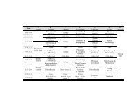
ivEduardo Hajdu, Daniela A. LopesChecklist of Brazilian deep-sea sponges................................................................................... 353-359Isabel Heim, Michael Nickel, Franz BrümmerMolecular markers for species discrimination in poriferans: a case study on species of thegenus Aplysina.......................................................................................................................... 361-371Isabel Heim, Jörg U. Hammel, Michael Nickel, Franz BrümmerSalting sponges: a reliable non-toxic and cost-effective method to preserve poriferans inthe field for subsequent DNA-work.......................................................................................... 373-377Friederike Hoffmann, Eberhard Sauter, Oliver Sachs, Hans Røy, Michael KlagesOxygen distribution in Tentorium semisuberites and in its habitat in the Arctic deep sea....... 379-382Valeria Itskovich, Sergey Belikov, Sofia Efremova, Yoshiki Masuda, Thierry Perez,Eliane Alivon, Carole Borchiellini, Nicole Boury-EsnaultPhylogenetic relationships between freshwater and marine Haplosclerida (<strong>Porifera</strong>,Demospongiae) based on the full length 18S rRNA and partial COXI gene sequences.......... 383-391Michelle Kelly, Michael Ellwood, Lincoln Tubbs, John BuckeridgeThe ‘Lithistid’ Demospongiae in New Zealand waters: species composition anddistribution................................................................................................................................ 393-404Anne Kuusksalu, Madis Metsis, Tõnu Reintamm, Merike KelveConstruction and characterization of a cDNA library from the marine spongeChondrosia reniformis.............................................................................................................. 405-412Emilio Lanna, Leandro C. Monteiro, Michelle KlautauLife cycle of Paraleucilla magna Klautau, Monteiro and Borojevic, 2004 (<strong>Porifera</strong>,Calcarea)................................................................................................................................... 413-418Nathan Lemoine, Nicole Buell, April Hill, Malcolm HillAssessing the utility of sponge microbial symbiont communities as models to studyglobal climate change: a case study with Halichondria bowerbanki........................................ 419-425Daniela Marques, Marise Almeida, Joana Xavier, Madalena HumanesBiomarkers in marine sponges: acetylcholinesterase in the sponge Cliona celata................... 427-432Diana M. Márquez, Sara M. Robledo, Alejandro MartínezAntileishmanial epidioxysterols from extracted sterols of the Colombian marine spongeIrcinia campana........................................................................................................................ 433-437Mirna Mazzoli-Dias, Suzi M. Ribeiro, Patricia Oliveira-SilvaForaminifera associated to the sponge Mycale microsigmatosa in Rio de Janeiro State,southeastern Brazil - an initial approach................................................................................... 439-442Elizabeth L. McLean, Paul M. YoshiokaAssociations and interactions between gorgonians and sponges.............................................. 443-448Larisa L. Menshenina, Konstantin R. Tabachnick, Daniela A. Lopes, Eduardo HajduRevision of Calycosoma Schulze, 1899 and finding of Lophocalyx Schulze, 1887 (sixnew species) in the Atlantic Ocean (Hexactinellida, Rossellidae)............................................ 449-465
viHeinz C. Schröder, Anatoli Krasko, David Brandt, Matthias Wiens, Muhammad NawazTahir, Wolfgang Tremel, Werner E.G. MüllerSilicateins, silicase and spicule-associated proteins: synthesis of demosponge silicaskeleton and nanobiotechnological applications....................................................................... 581-592Carla M.M. da Silva, Meiryelen V. da Silva, Bruno CosmeRedescription of the Brazilian endemic sponge Geodia glariosa (Demospongiae:Geodiidae), with new records on its geographic and bathymetric distribution........................ 593-602Antonio M. Solé-Cava, Gert WörheideThe perils and merits (or The Good, the Bad and the Ugly) of DNA barcoding ofsponges – a controversial discussion........................................................................................ 603-612Frank Spetland, Hans Tore Rapp, Friederike Hoffmann, Ole Secher TendalSexual reproduction of Geodia barretti Bowerbank, 1858 (<strong>Porifera</strong>, Astrophorida)in two Scandinavian fjords........................................................................................................ 613-620Robert W. Thacker, Maria Cristina Diaz, Klaus Rützler, Patrick M. Erwin, Steven J.A.Kimble, Melissa J. Pierce, Sandra L. DillardPhylogenetic relationships among the filamentous cyanobacterial symbionts of Caribbeansponges and a comparison of photosynthetic production between sponges hostingfilamentous and unicellular cyanobacteria................................................................................ 621-626Carsten Thoms, Peter J. SchuppChemical defense strategies in sponges: a review.................................................................... 627-637Laura Valisano, Attilio Arillo, Giorgio Bavestrello, Marco Giovine, Carlo CerranoInfluence of temperature on primmorph production in Petrosia ficiformis(<strong>Porifera</strong>: Demospongiae)......................................................................................................... 639-643Rob W.M. van Soest, Fleur C. van Duyl, Connie Maier, Marc S.S. Lavaleye, Elly J.Beglinger, Konstantin R. TabachnickMass occurrence of Rossella nodastrella Topsent on bathyal coral reefs of RockallBank, W of Ireland (Lyssacinosida, Hexactinellida)................................................................. 645-652Eduardo Vilanova, Carla Zilberberg, Michele Kochem, Márcio R. Custódio, Paulo A.S.MourãoA novel biochemical method to distinguish cryptic species of genus Chondrilla(Chondrosida, Demospongiae) based on its sulfated polysaccharides..................................... 653-659Author index.................................................................................................................................. 661-663Subject index.................................................................................................................................. 665-679Participants list.............................................................................................................................. 680-684
INVITED ARTICLES
<strong>Porifera</strong> <strong>Research</strong>: Biodiversity, Innovation and Sustainability - 2007Reading the code of coral reef sponge communitycomposition and structure for environmental biomonitoring:some experiences from CubaPedro M. AlcoladoInstituto de Oceanología, Ave. 1ra, No. 18406, Playa, La Habana, Cuba. alcolado@ama.cuAbstract: The structure of exposed (non-cryptic) coral reef sponge communities could be considered as a potentially readablecoded message reflecting their physical environment. The present paper describes explorations in Cuba of the potential use ofsponge communities as bio-indicators. Clathria venosa is the sponge that most consistently has proved to be a bioindicator ofurban based pollution in Cuban coral reefs due to its stenotopic character with regard to this stress source. Iotrochota birotulataforma musciformis was abundant close to the polluted Havana Bay, but not in other polluted sites, making it inconsistent asindicator. It has been quite rare in non-polluted waters. Cliona delitrix was represented in an area with great sewage influence.However it did not appear in some polluted sites probably because corals were extremely scarce and small. Scopalina ruetzleriwas well represented close to bays with different degrees of urban based pollution. Cliona varians was well represented onlyin one polluted place. Multivariate analyses (cluster analysis, non-metric multidimensional scaled analysis) have proved to bevery useful tools to clearly segregate sites with regard to level of pollution, and to identify factors and interactions determiningcommunity structure and composition. Abundance or dominance of Tectitethya crypta and Cliona vesparia (alpha stage) weretypical of heavy sedimentation conditions; while Aplysina cauliformis tended to dominate in sites affected by both hurricanesand sedimentation (abundance increased by fragmentation). Meta-analysis of Shannon’s heterogeneity index H’ and Pielou’sequitability index J’ is proposed as a useful tool to classify and compare sites with regard to the way that sponges interprettheir environment (degree of severity and predictability). Meta-analysis by means of a scatter graph with ranges of H’ atdifferent depths provides a spatial framework for comparing and classifying sponge communities with regard to environmentseverity.Keywords: sponges, bio-indicators, coral reefs, CubaIntroductionMany papers have dealt with the factors and interactions thatdetermine sponge distribution and community characteristics(partly reviewed by Sarà and Vacelet 1973, Bergquist1978, Wulff 2006), but few have been explicitly devoted toexploring the potential usefulness of sponge communities asbio-indicators for environmental bio-monitoring purposes.In the last few decades, the search for bio-indicatorshas become an urgent need in a world environment that ischanging at an unprecedented rate. According to Alcolado(1984; with some added arguments), sessile taxa are suitableas potential environmental bio-indicators because:- They must be adapted to the environment due to theirimmobility. Thus, their abundance or their presence (or evenabsence) must reflect the average ecological conditions, orvery recent strong stressful events.- Their composition and community structure are not affectedby migrations or local displacements.- The exposed (non-cryptic) sponge communities, havingpassed the fish predation filter thanks to deterrence (Wulff1997), are influenced more by the physical environmentthan by ecological interactions within themselves (sensuBradbury 1977). Cooperation rather than competitionseems to be the rule among sponge populations (Sarà 1970)and, according to Rützler (1970), sponges are able to solvecompetition by entering into complex epizoic relationships,without detriment to their pumping and filtering activities.Reiswig (1973) adds that small sponge individuals (duringthe first year after settlement) are subject to severe mortalityby competition with other sessile organisms, but whensponges reach greater volume competitors have little furthereffect. On the other hand, sponges overgrow corals muchmore frequently than the reverse, although when the reverseoccurs, the sponge tissue shows no adverse effect (Jacksonand Buss 1975).- The absence of food partitioning mechanisms influencingcommunity structure.Such features favor sponges over many other zoologicalgroups as potential indicators. That does not mean thatthere could not be some degree of influence of biologicalinteractions, but apparently to a much lower extent than thephysical environment (light, waves, sediments, pollution) inbuilding up the community structure and composition. This
also makes community structure and composition easier toanalyze and to understand in a bio-monitoring context.For these reasons, the structure of exposed (non-cryptic)coral reef sponge communities could be considered as apotentially readable coded message reflecting how spongesinterpret their physical environment. Indeed, sponges havebeen suggested as potential environmental bio-indicators byAlcolado (1984, 1985, 1990, 1992, 1994, 1999), Alcolado andHerrera (1987), Muricy (1989, 1991), Zea (1994), Alcolado etal. (1994), Carballo et al. (1994, 1996), Carballo and Naranjo(2001), and Vilanova et al. (2004). Some attempts andsuccesses in Cuba and other countries exploring the potentialuse of sponge communities as simpler, faster and lower costbio-indicators (from a sponge life perspective) are discussedbelow. The results compiled in this review come from a greatnumber of coral reef sites sampled around Cuba since 1976.DiscussionIndicator speciesA few sponge species have been found to be associatedwith polluted or relatively unpolluted conditions in coral reefs(Table 1). Particularly, Clathria venosa (Alcolado, 1984)and Iotrochota birotulata forma musciformis (Duchassaingand Michelotti, 1864) have only been observed dominatingin fore-reefs (10-20 m deep) affected by organic pollution(Alcolado and Herrera 1987) (Table 1; Fig. 1). The firstspecies appeared to be markedly stenotopic of enrichedinshore and coral reef waters and its occurrence has beenvery consistent in all polluted reefs evaluated or visited innorthwestern Cuba (close to Havana Bay, Almendares andQuibú rivers, and the town of Santa Fé), and according toZea (1994), also in Santa Marta, Colombia. This specieswas previously reported by Hechtel (1965) on shells andpiling in the enriched waters of Port Royal (southern shoreof Kingston Harbor), Jamaica (as Microciona microchelan. sp.); and by van Soest (1984) in the fouling communityon dead corals and gorgonians at the bay and Hilton HotelLanding of Curaçao (as Rhaphidophlus raraechelae n. sp.).It was also found in the fouling communities of the concretedock of Marina Barlovento (organically enriched site) andthe seawall of a small organically polluted cove (Rada delInstituto de Oceanología), both in the western Havana City,Cuba. However, I. birotulata forma musciformis was notconsistently dominant or abundant in the visited Cubanpolluted sites.Mycale microsigmatosa Arndt, 1927, which has beenfound dominating under domestic sewage stress in Brazil(Muricy 1989), was also found in a very polluted coastallagoon at Jaimanitas Town, west of Havana city (muddy/algal bottom) together with well developed Suberitesaurantiaca (Duchassaing and Michelotti, 1964), Chondrillaaff. nucula Schmidt, 1862 and Halichondria melanadocia deLaubenfels, 1936. Both S. aurantiaca and C. aff. nucula are“bacteriosponges” (Rützler 2002), which could explain theirabundance in this lagoon.Holmes (1997, 2000), Holmes et al. (2000) and Rützler(2002), comment on the increased abundance and activity ofboring sponges in areas affected by urban based pollution.Indeed, Cliona delitrix Pang, 1973, a species reported asabundant in areas submitted to sewage pollution (Rose andRisk 1985, Chávez-Fonnegra and Zea 2006), was observedduring four years by Marcos and Alcolado (unpublishedobservations) with significant relative abundance (%) at afore-reef site close to both the polluted Quibú River and anearby sewage outfall (western Havana City). However, itwas not found by Alcolado and Herrera (1987) at stationsnear Havana Bay, maybe due to the scarcity and small size ofcorals (dominated by Siderastrea radians Pallas, 1766).Another boring sponge, Cliona varians (Duchassaing andMichelotti, 1864), was well represented only in a pollutedfore-reef close to both the Quibú River and a nearby sewageoutfall at western Havana City (Marcos and Alcoladounpublished observations). However, it was also commonTable 1: Potential indicator species and their respective inferred condition according to authors. D and M = Duchassaing and Michelotti.Dominant or abundant species Indicated condition AuthorClathria venosa (Alcolado, 1984) Organic pollution Alcolado and Herrera (1987)Iotrochota birotulata f. musciformis (D. and M., 1864) Organic pollution Alcolado and Herrera (1987)Scopalina ruetzleri (Wiedenmayer, 1977) Moderate organic pollution Alcolado and Herrera (1987);Muricy (1989); Zea (1994)Sewage pollution Muricy (1989)Cliona delitrix Pang, 1973 Sewage (bacterial) pollution Rose and Risk (1985)Mycale microsigmatosa Arndt, 1927 Sewage pollution Muricy (1989)Amphimedon viridis D. and M., 1864 Sewage pollution Muricy (1989)Aplysina fistularis (Pallas, 1766) Comparatively non-polluted Alcolado (present paper, Fig. 1)Cliona caribbaea D. and M., 1864 Comparatively non-polluted López-Victoria and Zea (2004)Cliona vesparia (Lamarck, 1815) (alpha stage) Sedimentation plus wave stress Alcolado (present paper)Tectitethya crypta (de Laubenfels, 1949) Sedimentation stress Alcolado and Gotera (1985)Aplysina fulva (Pallas, 1766) Strong waves Wulff (1995)Aplysina cauliformis (Carter, 1882) Eventual strong waves and sedimentation Alcolado (present paper)
Fig. 1: Relative abundances ofpotential pollution bioindicatorsspecies, presented as percentagesof total sponge abundance (numberof individuals), at stations locatedat different distances from twomain pollution sources in thenorth-western Cuba (Havana Bayand Almendares River). MarielBay is not significantly polluted.in non-polluted reef areas, which makes it inconsistent as apotential bio-indicator.López-Victoria and Zea (2004) showed that the abundanceof Cliona caribbaea is not related to pollution in San AndrésArchipelago, Colombia. Indeed, this species did not occur atsites close to the organically polluted Quibú River and thenearby sewage outfall, but only in more distant sites (Marcosand Alcolado, unpublished observations).Other sponge species have been associated with factorsother than pollution, namely sedimentation and wave stress(Table 1). Particularly, Aplysina cauliformis is apparentlytolerant to strong waves, as can be deduced from its dominancein coral reef sites exposed to more frequent tropical storms(keys Juan García and Cantiles, southwestern Cuba). This canbe due to its branching morphology, flexibility and elasticity,similar to what was suggested by Wulff (1995) for Aplysinafulva, also branching and with rather similar consistency.The suggested usefulness of the presence or abundanceof some sponges as environmental indicators has beenbased much on expert observation and on inferencesrelated to distance from known pollution sources, waveand wind exposure, visual evidence of varying intensity ofsedimentation, etc. For that reason, to validate these resultsand make further progress, more evidence is necessary,obtained both from well designed experiments and frommultivariate analysis in which factors are directly measuredon appropriate temporal and spatial scales. Additionally,more sites in the Wider Caribbean, suffering various degreesof pollution, tropical storm frequency, exposure to waves anddominant winds, etc., are worth being researched to test thegenerality of the mentioned findings. It would be of particularinterest to determine if Clathria venosa feeds on bacteria withemphasis on enteric taxa, as does Clathria prolifera (Ellis andSolander, 1786) according to Claus et al. (1967).Community indicesIn agreement with other authors (Muricy 1989, Carballo etal. 1996, among others), Alcolado and Herrera (1987) foundthat species richness and Shannon´s heterogeneity index H’were lower at more polluted sites (Fig. 2). Pielou’s equitabilityindex J’ was also lower in the more polluted sites close to themouth of Almendares River (Fig. 2).Given that a condition of significant stress can be inferredonly when the dominance of some of the mentioned indicatorspecies (Table 1) is coupled with low values of speciesrichness or species heterogeneity (Alcolado et al. 1994),these univariate indices have to be taken into account as animportant complement for environmental monitoring.The summing up of the numerical percentages ofindividuals belonging to species that are tolerant to the samekind of stressor (e.g., pollution, sedimentation, turbulence,etc.) could be useful as another potential community indexfor monitoring purposes, as done by Alcolado (1981) withgorgonians to infer relative turbulence intensity, and byHerrera-Moreno (1991), also with gorgonians, to infer relativeorganic pollution level.The usefulness and conceptual validity of diversity indiceshas been controversial (Hurlbert 1971, Peet 1974). However(without disregarding potential pitfalls), the herein exploreddiversity indices can be used and tested pragmatically andheuristically for bio-monitoring purposes in the context ofenvironmental management, not specifically for advancing
(e.g., Cliona aprica, among sponges, Gorgonia flabellumLinnaeus, 1758, among gorgonians, and Acropora palmataLamarck, 1816, among scleractinians). For that reason, bothJ’ an H’ show extremely low values. This combination, withinPreston and Preston’s (1976) original scheme, would suggestan unpredictable environment (with its constant componentomitted).Alcolado’s (1992) inference diagram also differentiatesthe very favorable and constant environments of the deepreefs (e.g., at 20-30 m) within the rank 11, from those that aresimply favorable and quasi-constant (rank 10). Nevertheless,it is advisable to be aware of specific situations of very longtermenvironmental stability where some species can escapefrom demographic control and become excessively dominant,and consequently diminishing H’ and J’. This situation iscommon at reef sites deeper than 25 m. This phenomenon ofcommunity senescence is not contemplated in either of thetwo mentioned inference methods and has to be taken intoaccount in supposedly extremely constant environments (e.g.,deep reef zones, and reefs where hurricanes are very rare, asthose of Bonaire and Tobago).The author’s scale is proposed as an alternative reference(among other possible ones) and could be tested and improvedwith further research. More sites across the Wider Caribbeanshould be studied and included in the scatter graph to refineits spatial contour.Fig. 3: Inference diagram reflecting eleven ways in which spongesinterpret their physical environment, derived from a meta-analysiswith 112 coral reef stations. 1 = extremely severe with mixtureof constant and unpredictable environment; 2 = very severeand unpredictable; 3 = severe and unpredictable; 4 = quasiconstantlysevere; 5 = constantly severe; 6 = moderately severeand unpredictable; 7 = moderately severe and quasi-constant; 8 =moderately severe and constant; 9 = favorable and quasi-constant;10 = constantly favorable; and 11 = very favorable and constant.(H’ = 1.3-2.0 natural bells; J’ = 0.8-1); 6 = moderately andunpredictably severe environment (H’ = 2.0-2.5 natural bells;J’ = 0.5-0.69); 7 = moderately and almost constantly severeenvironment (H’ = 2.0-2.5 natural bells; J’ = 0.7-0.8); 8 =moderately and constantly severe environment (H’ = 2.0-2.5natural bells; J’ = 0.8-1); 9 = favorable and almost constantenvironment (H’ = 2.5-2.9 natural bells; J’ = 0.7-0.8); 10 =favorable and constant environment (H’ = 2.5-2.9 naturalbells; J’ = 0.8-1); and 11 = very favorable and constantenvironment (H’ >2.9 natural bells; J’ = 0.8-1).Rank 1 shows a (qualitative) situation that is notconsidered by Preston and Preston (1975). This is the caseof the surf zones of some Cuban reefs, which have constantaverage (basal or chronic) conditions of fairly strong waveaction, but which are also unpredictably affected by severeimpacts of tropical storms. Under these circumstances, thereis only one predominant species within each sessile taxonScatter graphs for comparing community indices atdifferent depthsScatter graphs of variability of sponge diversity indices,population density and cover with regard to depth wereobtained for many Cuban reef sites (Alcolado 1994, 1999).These graphs, which display the area (range) of variation ofthose indices with regard to depth, can be used as a referencepattern to infer in a comparative way the community conditionwithin stress gradients, taking into account site depth, giventhat such indices do not necessarily behave in the same wayalong depth gradients. What is normally a moderate value ofH’ for a given depth could be considered a high value for alower depth.The upper border of the variation area (an ascending convexline with a slight diminution at depths greater than 25 m)reflects the best conditions registered at different depths forsponge species richness and species heterogeneity (Fig. 4),while the lower border (an asymptotically ascending curve)shows the worst environmental conditions at different depths(Alcolado 1994).Care must be taken at deep reef stations (about 30 m depthor more), as lower diversities can be caused by extremelyconstant and favorable conditions that lead to the dominanceof competitively stronger species, and not by any stressor. Thesame recommendations given for the environmental severityand predictability inference graph are applicable here.Classification and ordinationClassification (Fig. 5) and ordination (Fig. 6) analyseshave proved to be useful when using sponge communitiesto separate sites with regard to degree of pollution and
Fig. 4: Example of meta-analysis as a scatter graph of H’ values atdifferent depths, with classification bands of inferred environmentalconditions (from 112 coral reef sites of Cuba). Care must be takenat stations about 30 m depth or more, as lower diversities can bedetermined by extremely constant and favorable conditions leadingto dominance of very competitive species, and not by severeenvironmental conditions. Arrows indicate more polluted stations(10 m depth) and stations affected by sediments (deeper stations).Fig. 6: MDS analysis segregating stations located at differentdistances from two main pollution sources (Havana Bay andAlmendares River) with regard to degree of pollution (from left toright: very polluted, polluted, and little polluted). This analysis wasdone with quadratic transformation of sponge densities and BrayCurtis similarity Index.Fig. 5: Cluster analysis segregating stations located at differentdistances from two main pollution sources (Havana Bay andAlmendares River) with regard to degree of pollution (goingdownwards: very polluted, polluted, and little polluted). This analysiswas done with quadratic transformation of sponge densities, BrayCurtis similarity Index, and un-weighted paired average clustering.to explore the factors impinging on their structure andcomposition (Alcolado and Herrera 1987, Muricy 1989,1991, Carballo et al. 1994, 1996, Carballo and Naranjo2001, Bell and Barnes 2003, Vilanova et al. 2004, Marcosand Alcolado unpublished observations). Particularly, theMultidimensional Scaled analysis (MDS) has providedclear results (PRIMER version 5). Multivariate techniquesare useful tools for identifying factors and interactions, andas such have to be applied complementarily with simpler,faster and lower cost univariate inference approaches inenvironmental monitoring. Multivariate analyses have toserve also to strengthen the validation and to reduce the pitfallsof potential indicator species and ecological indices that havebeen proposed, to a great extent based on observational andinference approaches.In the context of the application of the suggested biomonitoringmethods, an aspect that deserves future effort isto assess the convenience of using sponge cover instead ofsponge density, both from practical and scientific points ofview.Finally, another matter of concern could be the needof sponge taxonomy skills for the implementation of theproposed bio-monitoring methods. In this sense, the potentialindicator species are easy to identify in situ, and samplingfor calculation of community indices would only requiredifferentiation of species, and not necessarily identification tospecies level. With some practice, the identification of mostcommon species can be learned and the sampling work canbecome even easier.AcknowledgementsI would like to thank the National Museum of Rio deJaneiro, PETROBRAS, the UNDP/GEF Project Sabana-Camagüey and Dr. Robert N. Ginsburg (Ocean <strong>Research</strong> andEducation Foundation) for making possible my participationin the 7 th International Sponge Symposium. I am grateful toDr. Janie Wulff, Dr. Georgina Bustamante and Marta Riverofor their valuable comments to the manuscript.ReferencesAlcolado PM (1981) Zonación de los gorgonáceos someros deCuba y su posible uso como indicadores comparativos de tensión
hidrodinámica sobre los organismos del bentos. Inf Cient-Téc InstOceanol Acad Cien Cuba 187: 1-43.Alcolado PM (1984) Utilidad de algunos índices ecológicosestructurales en el estudio de comunidades marinas de Cuba. CienBiol 11: 61-77Alcolado PM (1985) Estructura ecológica de las comunidades deesponjas de Punta del Este, Cuba. Rep Invest Inst Oceanol AcadCien Cuba 38: 1-65Alcolado PM (1990) General features of Cuban sponge communities.In: Rützler K (ed). New perspectives in sponge biology. SmithsonianInstitution Press, Washington DC. pp. 351-357Alcolado PM (1992) Sobre la interpretación del medio marinomediante el empleo de los índices de diversidad y equitatividad.Cien Biol 24: 124-127Alcolado PM (1994) General trends in coral reef sponge communitiesof Cuba. In: van Soest RWM, van Kempen TMG, Braekman JC(eds). Sponges in time and space: biology, chemistry, paleontology.Balkema, Rotterdam. pp. 251-255Alcolado PM (1999) Comunidades de esponjas de los arrecifes delArchipiélago Sabana-Camagüey, Cuba. Bol Invest Mar Cost 28:95-124Alcolado PM, Gotera GG (1985) Estructura de las comunidadesde esponjas en arrecifes cubanos. Contrib Simp Cien Mar, VIIJornada Cient Inst Oceanol, XX Aniv 1: 11-15Alcolado PM, Herrera A (1987) Efectos de la contaminación sobrelas comunidades de esponjas en el Litoral de La Habana, Cuba.Rep Invest Inst Oceanol Acad Cien Cuba 68: 1-17Alcolado PM, Herrera-Moreno A, Martínez-Estalella N (1994)Sessile communities as environmental bio-monitors in Cubancoral reefs. In: Ginsburg RN (ed). Proceeding of the Colloquiumon “Global aspects of coral reefs: health hazards and history,1993” RSMAS, University of Miami. pp. 27-33Bell JJ, Barnes DK (2003) Effect of disturbance on assemblages: anexample using <strong>Porifera</strong>. Biol Bull 3(1): 144-159Bergquist PR (1978) Sponges. Hutchinson & Co., LondonBradbury RH (1977) Independent lies and holistic truths: towards atheory of coral reefs communities as complex systems. Proc 3 rd IntCoral Reef Symp, Miami 1: 2-17Carballo JL, Sánchez-Moyano JE, García-Gómez JC (1994)Taxonomic and ecological remarks on boring sponges (Clionidae)from the Strait of Gibraltar (southern Spain): tentative bioindicators?Zool J Linnean Soc 112: 407-424Carballo JL, Naranjo SA, García-Gómez JC (1996) Use of marinesponges as stress indicators in marine ecosystems at Algeciras Bay(southern Iberian Peninsula). Mar Ecol Progr Ser 135: 109-122Carballo JL, Naranjo S (2001) Environmental assessment of a largeindustrial marine complex based on a community of benthic filterfeeders. Mar Poll Bull 44: 605-610Claus G, Madri P, Kunen S (1967) Removal of microbial pollutantsfrom waste effluents by the redbeard sponge. Nature 216: 712-714Chávez-Fonnegra A, Zea S (2006) Observation of reef coralundermining by the Caribbean excavating sponge Cliona delitrix(Demospongiae, Hadromerida). In: Custódio MR, Lôbo-Hajdu G,Hajdu E, Muricy G (eds). 7 th International Sponge Symposium -<strong>Book</strong> of Abstracts (Armação dos Búzios, Brazil). Museu Nacional,Série Livros, vol. 16. p. 324Hechtel G (1965) A systematic study of the Demospongiae of PortRoyal, Jamaica. Bull Peabody Mus nat Hist 20: 1-94Herrera-Moreno A (1991) Efectos de la contaminación sobrela estructura ecológica de los arrecifes coralinos en el litoralHabanero. PhD Thesis. Instituto de Oceanología, La HabanaHolmes KE (1997) Eutrophication and its effect on bioerodingsponge communities. Proc 8 th Int Coral Reef Symp, Balboa 2:1411-1416Holmes KE (2000) Effects of eutrophication on bioeroding spongecommunities, with description of new West Indian sponges Clionaspp. (<strong>Porifera</strong>, Hadromerida: Clionaidae). Invert Biol 119: 125-138Holmes KE, Edinger EN, Hariyadi, Limmon GV, Risk MJ (2000)Bioerosion of Live Massive Corals and Branching Coral Rubbleon Indonesian Coral Reefs. Mar Poll Bull 40(7): 606-617Hurlbert SH (1971) The nonconcept of species diversity: A critiqueand alternative parameters. Ecology 52: 577-586Jackson JBC, Buss L (1975) Allelopathy and spatial competitionamong coral reef invertebrates. Proc Natl Acad Sci USA 72: 5160-5163López-Victoria M, Zea S (2004) Current trends of space occupationby encrusting excavating sponges on Colombian coral reefs. MarEcol 26: 33-41Margalef R (1963) On certain unifying principles in ecology. AmNat 97: 357-374Margalef R (1968) Perspectives in ecological theory. University ofChicago Press, ChicagoMuricy G (1989) Sponges as pollution bio-monitors at Arraial doCabo, Southeastern Brazil. Rev Bras Biol 49(2): 347-354Muricy G (1991) Structure des peuplements de spongiaires autourde l’égout de Cortiou (Marseille, France). Vie et Milieu 41(4): 205-221Odum EP (1969) The strategy ecosystem development. Sciences164: 262-270Peet RK (1974) The measurement of species diversity. Ann Rev EcolSyst 5: 285-307Preston EM, Preston JL (1975) Ecological structure in a West Indiangorgonian fauna. Bull Mar Sci 252: 248-258Reiswig HM (1973) Population dynamics of three JamaicanDemospongiae. Bull Mar Sci 23: 191-226Rose CS, Risk MG (1985) Increase in Cliona delitrix infestation ofMontastraea cavernosa heads on an organically polluted portionof the Grand Cayman fringing reef. Mar Ecol 6(4): 345-363Rützler K (1970) Spatial competition among <strong>Porifera</strong>: solution byepizoism. Oecologia 5: 85-95Rützler K (2002) Impact of crustose clionid sponges on Caribbeanreef corals. Acta Geo Hisp 37(1): 61-72Sanders HL (1969) Benthic marine diversity and the stability-timehypothesis. Brookhaven Symp Biol 22: 71-81Sarà M (1970) Competition and cooperation in sponge populations.Symp Zool Soc Lond 25: 273-284Sarà M, Vacelet J (1973) Écologie des Demosponges. In: Grassé PP(ed). Traité de Zoologie, Vol. 3, Masson et Cie, Paris. pp. 462-576Slobodkin LB, Sanders HL (1969) On the distribution ofenvironmental predictability to species diversity. BrookhavenSymp Biol 22: 82-93van Soest RWM (1984) Marine sponges from Curacao and otherCaribbean localities. Part. III. Poecilosclerida. Stud Fauna CuracaoCaribb Isl 66(199): 1-167Vilanova E, Mayer-Pinto M, Curbelo-Fernandez MP, Gonçalves daSilva SH (2004) The impact of a nuclear power plant discharge onthe sponge community of a tropical bay (SE Brazil). In: Pansini M,
10Pronzato R, Bavestrello G, Manconi R (eds). Sponge science in thenew millennium. Boll Mus Ist Biol Univ Genova 68: 647-644Wulff JL (1995) Hurricane effects on survival and orientation oflarge erect coral reef sponges. Coral Reefs 14: 55-61Wulff JL (1997) Parrotfish predation on cryptic sponges of Caribbeancoral reefs. Mar Biol 129(1): 41-52Wulff JL (2006) Ecological interactions of marine sponges. Can JZool 84(2): 146-166Zea S (1994) Patterns of coral and sponge abundance in degradedversus still healthy coral reefs at Santa Marta ColombianCaribbean. In: van Soest RWM, van Kempen TMG, Braekman JC(eds). Sponges in time and space: biology, chemistry, paleontology.Balkema, Rotterdam. pp. 257-264
<strong>Porifera</strong> <strong>Research</strong>: Biodiversity, Innovation and Sustainability - 200711Fossil sponges of Argentina: a reviewMatilde Sylvia BeresiCONICET-CRICYT: Ianigla, Dto. de Geología y Paleontología, Avda Ruiz Leal s/n, 5500 Mendoza, Argentina.mberesi@lab.cricyt.edu.arAbstract: This is a review on fossil sponges and sponge spicules reported from several regions in Argentina and in strataranging in age from Early Cambrian to Tertiary. Sponges have been collected from marine sediments of the Puna, CordilleraOriental and Sierras Subandinas basins, northern Argentina; Famatina Range; Precordillera terrane, San Rafael block,Neuquén basin and from lacustrine deposits of the North Patagonian Massif. Knowledge of the sponge fossil record is basedon whole relatively rigid skeletons, fragments of skeletal nets and spicules seen in thin sections or recovered from acetic acidresidues. Early to Middle Cambrian <strong>Porifera</strong> and Chancelloriids are known from the carbonate platform and slope faciesof the Precordillera terrane. Specimens with body preservation of Protospongia, Diagoniella, Kiwetinokia, fragments ofhexactinellid, and anthaspidellid sponges and sclerites of Chancelloria had been reported from Cambrian of the Precordillera.Remains of hexactinellid sponges, Pelicaspongiidae and Protospongiidae, have been found in Ordovician rocks of the Punaand of the Famatina System, western margin of Gondwana. Protospongia sp. and hexactinellid mesh were reported from UpperCambrian-Lower Ordovician siliciclastic sediments in the Cordillera Oriental and Sierras Subandinas. The most significantfossil record of Lower-Middle Ordovician sponge faunas is from the carbonate platform of the San Juan Precordillera. Spongefaunas are dominated by orchoclad lithistid demosponge genera, although hexactinellids are known from loose spicules androot tufts, and calcareous heteractinid sponges are known from isolated octactine spicules and only one genus. Hexactinellid,calcarean and demosponge spicules were reported from diverse localities of the Precordillera. A Late Jurassic (Oxfordian)carbonate complex of the Neuquén Basin, west-central Argentina, contains siliceous sponges dominated by hexactinellids(Hexactinosa and Lyssakinosa). Palaeospongilla chubutensis, a fresh water sponge, was described from lacustrine Cretaceousdeposits of the Chubut River valley. Oxeas and strongyles, belonging to the Family Spongillidae, have been mentioned fromTertiary sediments of the Paraná basin, northeastern Argentina.Keywords: biostratigraphy, fossil sponges, Argentina.IntroductionThe actual knowledge of the record fossil in Argentinais based on relatively rigid whole skeletons, fragments ofskeletal nets and spicules seen in thin sections or recoveredfrom acid residues for obtaining conodonts. The fossilrecord of sponges comes from several geological provincesof Argentina with different lithologic, paleontologic andenvironmental characteristics.Sponge faunas were collected from marine sediments ofthe Puna, Cordillera Oriental, Sierras Subandinas basins ofnorthern Argentina; Famatina Range, Precordillera Terrane,San Rafael Block, Neuquén Basin (western Argentina), fromlacustrine deposits of the North Patagonian Massif, and fromthe Chaco-Paranaense Basin. Occurrences of sponge faunashave been reported from the Lower Paleozoic (upper-LowerCambrian) to the Cenozoic (Tertiary). Only a few publicationshave dealt with sponge and sponge spicules in Argentina.Most of them concern the fauna from the Cambrian andOrdovician rocks of the Precordillera terrane.Associated with protospongiid spicules, well-preservedchancelloriid sclerites occur in the Cambrian carbonateplatform of the Precordillera. Chancelloriids are Cambrianenigmatic organisms constituting the monophyletic taxonCoeloscleritophora (Bengtson and Missarzhevsky 1981).Sclerites of chancelloriids (Family Chancelloridae) were firstdescribed from the Burgess Shale by Walcott (1920), whointerpreted them as heteractinid sponges. This traditionalview of the fossil group as sponges was accepted for morethan 50 years.In the Precordillera, chancelloriid sclerites associatedwith spicules are a common and distinguishing features ofthe Lower to Middle Cambrian fossil fauna. Protospongiaand Chancelloria crucensis (Rusconi, 1955) were the firstCambrian species of the Mendoza Precordillera.Cambrian Protospongia Salter, 1864 and hexactinellidspicules were mentioned by Pernas (1964), Devizia (1973),Bordonaro and Martos (1985), Heredia et al. (1987) andBeresi and Heredia (1995) from the San Isidro area inthe Precordillera of Mendoza. Afterwards, assemblagestentatively identified as Kiwetinokia utahensis Walcott, 1920,protospongiid skeletal nets and Chancelloria eros Walcott,1920 were reported from early-Middle Cambrian carbonatesblocks of San Juan and Mendoza, Precordillera, by Beresiand Rigby (1994). Two small associated specimens assignedto Protospongia sp. occur in the western Precordillera of San
12Juan (Beresi and Banchig 1997). A synthesis of the Cambriansponge occurrence in the Argentine Precordillera was givenby Beresi (2003a).Silicified sponge spicules from residues of conodonts fromdiverse Cambrian and Ordovician sections in the Precordillera,were described by Mehl and Lehnert (1997). Anthaspidellidskeletal fragments from the Middle Cambrian rocks of thePrecordillera, in the province of San Juan, document the onlyknown occurrence from South America (Beresi and Rigby1994). Protachileum kayseri Zittel, 1877 from the San Juanprovince, was the first report of a Precordilleran sponge.Taxonomic studies have been concentrated in the rich spongefauna of the warm carbonate platform (San Juan Formation)from the Lower-Middle Ordovician of the Precordillera basin(Beresi and Rigby 1993, Carrera 1996a, 1996b).The purpose of this paper is to review the occurrence offossil sponges and their biostratigraphic distribution in thediverse geological provinces of Argentina.Cambrian and Ordovician fauna from theNorthwest Argentinian regionSiliciclastic sediments with minor carbonates dominatethe Cambrian and Ordovician of Northern Argentina andthe Central Andean Basin of South America. Cambrian andOrdovician sponges and spicules from Northwest Argentinaprovide additional paleontological data from the siliciclasticplatforms of western Gondwana.PunaIn the Puna region (Fig. 1A-B), a single specimen (Fig.2M-N) of a complete Ordovician hexactinellid sponge wasdiscovered. It has been collected from volcaniclastic rocksof the Las Vicuñas Formation (Tremadocian) in Lari Creek,southwest of the Salar del Rincon area, Salta province,Argentina. The material was assigned to the new genus andspecies Larispongia magdalenae (Carrera, 1998), that belongto the family Pelicaspongiidae, and it is the first record of thefamily in western Gondwana.Subandean RangesThe first report on the occurrence of one completelypreserved hexactinellid sponge is a part and counterpart of around small sponge described as Diagoniella sp. (Beresi et al.2006). This sponge (Fig. 2H) was reported for the first timefrom the Upper Cambrian-Lower Ordovician siliciclasticrocks of the Orcomato Formation, in the Candelaria Range,Sub-Andean Ranges, in Salta Province (Fig. 1B).The fossiliferous record of the unit suggests a Cambrian-Ordovician age. The sponge is preserved in greenishyellowishshales, as dark stained flattened rounded bodies(somewhat deformed and fragmentary).Fig. 1: A. South America showing the position of Argentina. B.Location of the Basins of Argentina with sponge fauna: (Pu) Puna;(CO) Cordillera Oriental, (SS) Sierras Subandinas, (F) Famatina,(Pa) Paraná basin, (P) Precordillera, (SR) San Rafael Block, (N)Neuquén basin, (MP) North Patagonian Massif. C. Map of Argentineshowing the San Juan and Mendoza provinces. D. Sponge localitieswithin the Precordillera Precordillera of San Juan and northernMendoza provinces.Eastern CordilleraHexactinellid meshes of Protospongia (Beresi et al. 2006)were recently reported from Lower Tremadocian siliciclasticsediments in the Eastern Cordillera, Salta Province (Fig. 1).The hexactinellid spicules were recovered from the lower
13Fig. 2: A. Cambrian anthaspidellidsponge fragment, genus andspecies indeterminant fromSan Isidro, Mendoza. B-G.Coeloscleritophora: Chancelloriaeros Walcott, 1920, scleritesfrom the upper Lower–MiddleCambrian of Zonda Range andSan Isidro area. H. Diagoniellasp. from Salta province, Sub-Andean ranges. I. Cambrianfragment of bioclastic limestoneshowing sclerites of ChancelloriaWalcott, 1920, San Isidro. J.Root tuft, large monaxons ormonactine-like spicules, in bundlewith small stauracts or hexacts(Tontal Range). K. Protospongiasp. showing stauractine-basedskeleton and long rayed hexactinesforming marginal spines alongthe left margin (Tontal Range).L. Protospongia sp. Esteban andRigby, 1998, specimen PIL 14.192from Peña Negra section, Famatinaregion. M-N. Larispongiamagdalenae Carrera, 1998,holotype CEGH-UNC 17365 fromLari Creek, Puna region. N. Detailof the same specimen showingdermal hexactines surroundingmajor gaps.levels of the Saladillo Formation, at the Angosto de laQuesera section, Eastern Cordillera.Spicules of Protospongia (Fig. 2K) are preserved on uppersurfaces of yellow-brownish shales and sandstones belongingto the lower section of the unit, sharing the stratigraphicposition with Tremadocian graptolites. The sedimentationof the greenish and yellowish shales and sandstones ofthe Saladillo Formation indicates a transition to an upperoffshore-lower offshore environmental setting.Famatina systemCatamarca ProvinceThe only discovery of Ordovician sponges in thisprovince corresponds to isolated hexactinellid spicules,which have been described by Aceñolaza and Toselli (1977)for the Chaschuil region. The material appears dispersed incarbonate concretions of the Suri Formation (Arenig).La Rioja ProvinceThe sponge material came from black siliceous graptoliticshales of the upper part of the Volcancito Formation of LowerTremadocian age, outcropping in the Peña Negra locationin the Famatina range (Fig. 1B). Fragments of a reticulatedskeletal net of Protospongia species (A and B) were describedby Esteban and Rigby (1998), in the siliclastic Famatinabasin, western margin of Gondwana (Fig. 2L).The sponges are associated with planktonic graptolites andthis level can be assigned to the Lower Tremadoc (Estebanand Gutierrez-Marco 1997).
14Precordillera (Cuyania) Terrane, WesternArgentinaThe Argentine Precordillera is situated along the forefrontof the high Andes at approximately 28º to 37º S, and it isa major geologic province in northwestern Argentina. Itcontains a complete thick sequence of Early Paleozoic rocks.The Precordillera, as part of the Cuyania Terrane, wasformed during the Andean (Tertiary) crustal shortening. Thisdistinctive terrane can be recognized mainly on the basis ofits key stratigraphic composition, involving biostratigraphic,sedimentary and magmatic events; its boundaries withadjacent geologic regions are abrupt (Ramos et al. 1986).In accordance with the Terrane concept, the presentPrecordillera, plus the San Rafael Block and San JorgeLimestones, integrate a unique geologic entity, the so-calledPrecordillera Terrane or Cuyania Terrane.Two hypotheses exist regarding the origin of thePrecordillera: 1) the Precordillera represents a terrane ofLaurentian origin that became attached to Gondwana (westernArgentina) already during Ordovician times (Thomas andAstini 2003). It includes either the classical Precordillera aswell as the San Rafael Block, to the south in the provinceof Mendoza, and the San Jorge Limestones cropping out inthe Province of La Pampa, within the Sierras Pampeanasstructural setting as an allochthonous terrane “Cuyania”,accreted to Gondwana during the lower Paleozoic. 2) thePrecordillera is considered as an autochthonous Gondwananfragment (Baldis et al. 1989, Aceñolaza et al. 1999, 2002)displaced by simple transcurrence mechanics, from ahypothetical intermediate sector between South America,Africa, and Antarctica. Recently, U-Pb geochronologyof detrital zircons indicated a Gondwanan provenancefor Lower Cambrian and Upper Ordovician sandstonesof the Precordillera of western Argentina, supporting theautochthonous Gondwanan nature of the PrecordilleraTerrane (Finney et al. 2003a, 2003b).Cambrian <strong>Porifera</strong>The Cambrian system of the Argentinian Precordillerais represented by a carbonate platform, in the east, and ofa continental slope, in the west. Cambrian platform andslope facies, containing spicules and chancelloriids scleritesare located in the Precordillera of San Juan and northernMendoza provinces, western Argentina (Fig. 1C-D).Cambrian sponges are known mainly from fragmentsof skeletal nets and dissociated spicules from the shallowcarbonate platform sequences of the upper-Lower to MiddleCambrian in the eastern and central belts, and from the slopeolistostromic sequences with allochthonous blocks in thewestern part of the Precordillera of San Juan Province. Thespicules from the Upper Cambrian were collected in TontalRange, San Juan Precordillera (Beresi and Banchig 1997)and in the La Cruz Olistolith, San Isidro area (Beresi andHeredia 1995), Precordillera of Mendoza Province.Sclerites of Chancelloria (Coeloscleritophora) occur inshallow carbonate platforms and allochthonous blocks inthe Precordillera of San Juan and Mendoza. Well preservedsclerites are associated with protospongiid spicules (Fig. 2B-G).Cambrian assemblagesTwo spicule assemblages occur in the Cambrian facies(Beresi and Rigby 1994, Beresi 2003a). The autochthonousassemblage corresponding to material collected from theupper Lower to Middle Cambrian platform sequence of theeastern Precordillera of San Juan. This assemblage consistsof a variety of stauractines and sclerites of Chancelloria eros(Walcott, 1920). The Protospongiidae are represented bytriradiate prodianes, pentactines and hexactines, all belongingto Kiwetinokia utahensis Walcott, 1920, Protospongia andanthaspidellid fragments. This fossil fauna represents theoldest assemblages known of Argentina.The allochthonous assemblage proceeds from the diverseCambrian carbonate olistoliths of slope sequences of thewestern San Juan Precordillera and from the classical area ofSan Isidro, Mendoza Province. The assemblages consist ofthe first precordilleran Protospongia with body preservation,Diagoniella Rauff, 1894, Kiwetinokia Walcott, 1920 andChancelloria and skeletal net with hexactines and monaxons(Beresi and Banchig 1997).Demosponges have a limited record in the Cambrianof the Precordillera. Typically anthaspidellid fragmentswith dendroclones (Fig. 2A) have been reported from thecarbonate platform and slope sequences of the San Juan andMendoza Precordillera (Beresi and Rigby 1994).Ordovician <strong>Porifera</strong>Deposits of Ordovician carbonate basins occur in thecentral and eastern Precordillera. The Lower-MiddleOrdovician sediments of the Precordillera represent adrowning carbonate platform with a diverse and relativelycomplete fossil record.Well-preserved and diverse faunas of sponges have beencollected from limestones of the San Juan Formation (UpperTremadoc-Early Llanvirn) in the Precordillera basin of SanJuan province (Fig. 1C-D). This fauna represents the mostsignificant Ordovician sponge fauna known from SouthAmerica and provides the first extensive record of spongesderived from a stable carbonate platform, constituting one ofthe most important Early Ordovician sponge associations ofthe world.Precordilleran sponge faunas are dominated by orchocladlithistid demosponge genera, although hexactinellids areknown from loose spicules and root tufts, and calcareousheteractinid sponges are known from isolated octactinespicules and only one genus. Spicules assemblages werereported from diverse localities of the Precordillera (Beresiand Esteban 2003, Carrera 2003).The San Juan Formation was deposited on an opencarbonate shelf, bounded to the west by continental slopeand oceanic basin deposits. The diverse marine fauna andthe lack of specific structures indicative for shallow water
15conditions point to low energy, subtidal conditions within anopen-platform environment during the entire sedimentationinterval. The age of the San Juan Formation is wellconstrained by conodonts (Sarmiento 1985, Albanesi andOrtega 2002) spanning from the late Tremadocian (Paltodusdeltifer Zone) to the early Darriwilian (Lenodus variabilis– Eoplacognathus suecicus zones).The first report of a Precordilleran sponge wasProtachileum kayseri Zittel, 1877 from the Talacasto Gulch,Precordillera of San Juan Province (Fig. 3J).Anthaspidellid genera first appear in the basal beds ofthe San Juan Formation (Upper Tremadoc) at the NiquivilRange, Eastern Precordillera of San Juan, associated withreef-mound (Cañas and Carrera 1993). The cosmopolitanArchaeoscyphia Hinde, 1889 and Rhopalocoelia Raymondand Okulitch, 1940 are the predominant genera in thissponge-algal association (Fig. 3H, 3E).Diverse and well-preserved sponge faunas are from themiddle and upper part of the San Juan Formation (Arenig-Lower Llanvirn). In this carbonate platform the fauna isdominated by orchocladine lithistid demosponges. Theirfirst taxonomic study was made by Beresi and Rigby (1993)and afterwards by Carrera (1996a, 1996b, 1998). Apart fromorchocladine demosponges there are hexactine spicules,hexactinellid root tufts, and isolated octactine spicules thatdocument the presence of the Heteractinida.Fig. 3: Early and MiddleOrdovician orchoclad lithistidsponges from the Precordillera ofSan Juan. (A-B-C-D-G-K; Beresiand Rigby 1993). A. Talacastoniachela Ianigla PI T-2. B. Tangentialthin section. C. Anthaspidellaannulata Ianigla PI T-49. D.Calycocoelia perforata IaniglaPI VI-2. E. Rhopalocoelia clarkiiRaymond and Okulitch, 1940,Ianigla PI T-22. F. Hudsonospongiacyclostoma Raymond andOkulitch, 1940, Ianigla PI VI-2.G. Hudsonospongia talacastensis.Ianigla PI T-32. H. Archaeoscyphiaminganensis Billings, 1859 IaniglaPI T-47. I. Incrassospongia ramisCarrera, 1996b, CEGH-UNC9308. J. Protachileum kayseriZittel, 1877, Ianigla PI H-43. K.Aulocopium sanjuanensis IaniglaPI VI-13.
16The greatest generic and specific diversity of lithistidsponges occurs during the Lower Llanvirnian (Darriwilian)in the upper most part of the San Juan Formation. A total of15 demospongiid genera and 20 species are described. Thisfauna shows a variety of external morphologies and bodyplan.Many new Ordovician species were described for the SanJuan Formation by Beresi and Rigby (1993): Anthaspidellainornata, A. annulata (Fig. 3C), and A. alveola,Archaeoscyphia nana, Aulocopium sanjuanensis (Fig.3K), Calycocoelia perforata (Fig. 3D), Hudsonospongiatalacastensis (Fig. 3G), H. cyclostoma (Fig. 3F),Patellispongia robusta, Psarodictyum magna, Rhopalocoeliarama, among other species. The new species Talacastoniachela (Fig. 3A-B) was described from the classicalOrdovician Talacasto section, Central Precordillera (Fig.1B). Root tufts of hexactinellids also occur (see Table 1).New megamorinid genera as Rugospongia viejoensis(family Saccospongidae) (Carrera, 1996a) and thetricranocladine sponge Eoscheiella concave (FamilyHindiidae Rauff, 1893) have been recovered from the top ofthe San Juan Formation in the Cerro Viejo, Huaco (Carrera2007). Nexospongia sillaensis (family NexospongiidaeCarrera, 1996a) was described from the Cerro La Silla,Niquivil Range, Eastern Precordillera.In the upper levels of the San Juan Formation from theEarly Llanvirnian, at the Cerro La Chilca section, thecalcareous heteractinid Chilcaia bimuralis (Carrera, 1994)and a lithistid species Incrassospongia ramis (Carrera,1996a) were described (Fig. 3I).Endemic genera such as Protachileum and TalacastoniaBeresi and Rigby, 1993, from the Talacasto Gulch andRugospongia Carrera, 1996 and Chilcaia from differentlocalities of the San Juan Formation occur in thePrecordillera.Ordovician sponges from the Precordillera show changesfrom algal-sponge (reef ecosystems) in the Early Arenigto stromatoporid associations in the Middle Arenig toanthaspidellid demosponge dominated associations in theUpper Arenig to Lower Llanvirn. From the Llanvirn up tothe Upper Ordovician, the effects of diverse abiotic factorssuch as volcanic activity, sea level fluctuations and finallythe global climatic cooling, could have contributed to thedecrease of the sponges diversity.The diversification of the orchoclad demosponges in theLower Ordovician carbonate platform of the Precordillerawas similar to worldwide radiation pattern (Carrera andRigby 1999).Ordovician sponge spiculesPrecordillera of San Juan ProvinceThe oldest spicule assemblage comes from the LowerOrdovician limestones (Oepikodus intermedius Zone) ofthe San Juan Formation. The Arenigian silicified spiculeassemblages were documented by Gnoli and Serpagli (1980)in the Pachaco section, western Precordillera.Calcarean and demosponge spicules assemblagesrecovered from residues of conodont samples of severalLower to Middle Ordovician sections of the San JuanPrecordillera were described by Mehl and Lehnert (1997). Thewell preserved silicified spicules include: Polyactinellidae,Heteractinellidae (Calcarea) and hexactinellid anddemosponge spicules. The species Dodecaactinella onceraMehl and Lehnert, 1997, Sardospongia cynodonta Mehl andLehnert, 1997, Praephobetractinia sp. and Eiffelia sp. werereported from Lower Ordovician (Arenig) strata of the SanJuan Formation (Fig. 4A-E).These spicule assemblages were collected from reef-moundhorizons and biostromes with sponges, stromatoporoidsand receptaculitids of the San Juan Formation (LowerArenig-Lower Llanvirn) and from the Gualcamayo and LasAguaditas formations (Lower Llanvirn to Caradocian).San Rafael BlockSponge spicules are derived from residues of conodontsamples from Middle Ordovician strata, in the geologicalprovince of the San Rafael Block, southern MendozaProvince, Argentina (Fig. 1C-D). Spicules (Fig. 4F-Q) havebeen recovered from the Ponón Trehué Formation, a clasticcarbonatesequence. <strong>Porifera</strong>n taxa (Beresi and Heredia2000) include two spicule assemblages: 1) associations ofexclusively heteractinellid spicules (sexiradiates) restricted toArenigian allochthonous blocks of the Oepikodus evae Zone(Heredia 2001) from the shallow platform of the San JuanFormation; and 2) associations of hexactinelliid spicules,calcarean triaene and monaxons, from Upper Llanvirnianautochthonous limestones and carbonate sandstones of thePigodus serra Zone and the P. anserinus Zone (Heredia2001) from the outer platform and slope.The spicule associations of the Ponón Trehué Formationrepresent the most austral Ordovician assemblage describedin the context of the Precordillera (Cuyania) Terrane.Jurassic sponges from the Neuquén BasinA late Jurasic (Oxfordian) carbonate complex wasdeveloped on the foreland side of the Neuquén Basin (Fig.1B), west- central Argentina and form part of the CordilleraPrincipal. Shelf carbonates facies are exposed throughoutMendoza and Neuquén provinces.One of these facies consists of small siliceous spongebuildups of the La Manga Formation (Plicatilis Zone), welldeveloped at the Río Poti Malal section, in southern MendozaProvince. The siliceous sponges with moderate diversityare fossilized in their original shape and exhibit calcareouspreservation.Sponge fauna is dominated by hexactinellids(Hexactinosida and Lyssacinosida, 95%) and lithistiddemosponges (5%). Up to now, approximately 20% of thematerial has been preliminary determinated (Beresi 2003b).
17Table 1: Biogeographic distribution of sponge taxa from the Cambrian and Ordovician Argentine Basins.Sponge taxaPrecordilleraSan JuanMendozaNorthern RegionFamatina SystemPaleoenvironmentDEMOSPONGIAEFamily AnthaspidellidaeAllosacus sp. Carrera, 1994Anthaspidella alveola Beresi and Rigby, 1993Anthaspidella annulata Beresi and Rigby, 1993Anthaspidella inornata Beresi and Rigby, 1993Archaeoscyphia minganensis Beresi and Rigby, 1993Archaeoscyphia nan Beresi and Rigby, 1993Archaeoscyphia pulchra Bassler, 1941Aulocopium sanjuanensis Beresi and Rigby, 1993Calycocoelia perforata Beresi and Rigby, 1993Hudsonospongia cyclostoma Raymond and Okulitch, 1940Hudsonospongia talacastensis Beresi and Rigby, 1993Incrassospongia ramis Carrera, 1996Patellispongia argentina Carrera, 1994Patellispongia occulata Bassler, 1941Patellispongia robusta Beresi and Rigby, 1993Patellispongia magna sp.Protachilleum kayseri Zittel, 1877Psarodictium magna Beresi and Rigby, 1993Rhopalocoelia clarikii Raymond and Okulitch, 1940Rhopalocoelia rama Beresi and Rigby, 1993Rhopalocoelia regularis Raymond and Okulitch, 1940Rhopalocoelia tenuis Carrera, 1994Talacastonia chela Beresi and Rigby, 1993XXXXXXXXXXXXXXXXXXXXXXXCarbonate platformFamily HindiidaeEoscheiella concave Carrera, 2007 X Carbonate platformFamily NexospongiidaeNexospongia sillaensis Carrera, 1996 X Carbonate platformFamily SaccospongiidaeRugospongia viejoensis Carrera, 1996 X Carbonate platformHEXACTINELLIDAFamily PelicaspongiidaeLarispongia magdalenae Carrera, 1998 X Marine volcaniclasticFamily ProtospongiidaeProtospongia sp. A X Silicoclastic platformProtospongia sp. B X Silicoclastic platformDiagoniella sp. X X X Carbonate platformProtospongia sp. X X X Carbonate platformKiwetinokia utahensis Walcott, 1920 X X Carbonate platformFamily uncertainRoot tuft X Carbonate platformHETERACTINIDAChilcaia bimuralis Carrera, 1994XDodecaactinella oncera Mehl and Lehnert, 1997XSardospongia cynodonta Mehl and Lehnert, 1997XPraephobetractinia sp.XEiffelia sp. X XOctactine spiculesXTriactine spiculesXCarbonate platform
18Fig. 4: Ordovician spongespicules. A-E. Calcarean anddemospongid spicules from theLower-Middle Ordovician of theSan Juan Precordillera (Mehland Lehnert 1997). CEGH-UNC 15951. A. Sardospongiacynodonta. B. Calcarean triactine.C. Demospongid oxea. D-E.Dodecaactinella oncera. F-Q.Spicules from the Ponón TrehuéFormation, San Rafael Block(Beresi and Heredia 2000). F.Octactine spicule shows the distaland two of the tangential raysbroken. G. Octactine spicule.H. The proximal-distal verticalray shows a prominent node.I. Monaxon spicule appears tohave been sheared diagonally bydiagenetic processes. J. Calcareantriactine. K-Q. Scanningelectron microscope (SEM)photomicrographs. K, M, N, P.Octactine spicules. L. monaxonspicule shows the central circularcanal. O, Q. Hexactine spicules.The following species have been identified: Laocoetis sp.,Laocoetis procumbens and Laocoetis parallela (HexactinosidaSchrammen, 1903, Family Craticulariidae Rauff, 1893) (Fig.5C-J); Cribrospongia sp., Cribrospongia clathrata andCribrospongia cucullata (family Cribrospongiidae Roemer,1864) (Fig. 5A-B) and Polygonatium sp. (LyssacinosidaZittel, 1877).Sponges belonging to the Family Cribrospongiidae arecup-shaped (Cribrospongia reticulata), tubular and conical.Only a few specimens are triangular in shape and compressed(Cribrospongia cucculata). Fragments of cylindrical totubular sponges belong to the genus Laocoetis (=CraticulariaZittel, 1877; emend. Schrammen, 1937).Cretaceous freshwater sponge from the NorthPatagonian MassifThe only freshwater sponge was described from LowerCretaceous lacustrine sediments of the Chubut River Valley,in the Chubut Province, North Patagonian Massif (Fig. 1B).The sponge was determined as Palaeospongilla chubutensisby Ott and Volkheimer (1972). The encrusting sponge belongsto the monogeneric family Palaeospongillidae Volkmer-
19Fig. 5: Oxfordian hexactinellidsponges at the Río Potimalalsection, Neuquén Basin (Beresi1997) A. Cribrospongia cuccullataQuenstedt, 1878, lateral view. B.Dermal surface with craticulariiddiplorhysis. C. Laocoetisprocumbens Goldfuss, 1826,lateral view. D. Upper view of theosculum sponge showing in H. E.Upper view of the narrow osculumand folded wall in a cribrospongiidsponge. F. Lateral view of acylindrical sponge. G. Laocoetisclathrata Goldfuss, 1833, lateralview. H. Longitudinal section ofa tubular cribrospongiid sponge. I.Longitudinal section of a tubularcribrospongiid sponge. J. Upperview of the same sponge, showedin I. K-M. Palaeospongillachubutensis Ott and Volkheimer,1972. K. Megasclere withcentral canal. L. Gemmules. M.Gemmule and spicular texture ofthe sponge skelton. N-O. Tertiarymegascleres, Paraná basin.Ribeiro and Reitner, 1991. It is characterized by acanthoxeasto acanthostrongyles gemmoscleres (Fig. 5K-O).Tertiary spicules of the Paraná basinIsolated oxeas and strongyles, possibly belonging to thespecies Trochospongilla repens (family Spongillidae) werecollected from Tertiary (Miocene) pelitic sediments of theParaná basin, northeast of Argentina (Fig. 1).Borings of Cliona entrerriana and C. ameghinoi oncalcareous shells of Ostrea patagonica have also been foundin Tertiary sediments.RemarksFossil sponges are known from several geologic basinswith different lithologic, sedimentologic and environmentalcharacteristics (Table 2). There are sponges and loose spiculesrepresentative of the Classes: Hexactinellida, Demospongidaand Calcarea.The oldest sponge fauna known in Argentina is from theupper Lower Cambrian of the Precordillera, the youngest oneoccurs in the Middle Tertiary of the Paraná Basin.Protospongiids characterized the western old Gondwanacontinent and are known from the Cambrian and Lower
21Beresi MS, Esteban SB (2003) Sponge fauna and spiculesassemblages from the Ordovician of Argentina: a review. In:Aceñolaza FG (ed). Aspects of the Ordovician System in Argentina,Serie Correlación Geológica 16. INSUGEO, Tucumán. pp. 71-85Beresi MS, Heredia SE (1995) Asociación de espículas de poríferoscámbricos en la Precordillera de Mendoza. Ameghiniana 32: 401-405Beresi MS, Heredia SE (2000) Sponge spicule assemblages fromthe Middle Ordovician of Ponón Trehué, southern Mendoza,Argentina. Rev Esp Paleontol 15(1): 37-48Beresi MS, Rigby JK (1993) The Lower Ordovician sponges of SanJuan, Argentina. Geol Stud 39: 1-64Beresi MS, Rigby JK (1994) Sponges and Chancelloriids from theCambrian of western Argentina. J Paleontol 68(2): 208-217Beresi MS, Banchig AL (1997) First hexactinellid spicules of theLate Cambrian Olistolith, of the Los Sombreros Formation, TontalRange, San Juan Precordillera, Argentina. Rev Esp Paleontol12(2): 141-150Bonetto AA, Ezcurra de Drago LD (1969) Systematics notes aboutthe genus Uruguaya Carter (<strong>Porifera</strong>, Spongillidae). Physis28(77): 351-358Bordonaro OL, Martos L (1985) El género Chancelloriaen el Cámbrico de San Juan. Reunión de ComunicacionesPaleontológicas, Asociación Paleontológica Argentina, San Juan:6-7Cañas F, Carrera MG (1993) Early Ordovician microbial-spongereceptaculitidbioherms of the Precordillera basin, WesternArgentina. Facies 29: 169-178Carrera MG (1994) An Ordovician sponge fauna from San JuanFormation, Precordillera basin, Western Argentina. Neues JahrbGeol Paläontol Abhand 191(2): 201-220Carrera MG (1996a) Ordovician Megamorinid sponges from SanJuan Formation, Precordillera, Western Argentina. Geobios 29(6):643-650Carrera MG (1996b) Nuevos poríferos de la Formación San Juan(Ordovícico), Precordillera Argentina. Ameghiniana 33(3): 335-342Carrera MG (1998) First ordovician sponge from the Puna region,northwestern Argentina. Ameghiniana 35(2): 205-210Carrera MG (2007) The oldest hindiid demosponge from theDarriwilian (Middle Ordovician) of the Argentine Precordillera:evolutionary implications for the Tricranocladines. J Paleontol81(4): 754-759Carrera MG, Rigby JK (1999) Biogeography of the Ordoviciansponges. J Paleontol 73: 26-37Devizia C (1973) Estudio geológico del sector de San Isidro,Departamento Las Heras Mendoza. PhD Thesis, Museo de LaPlata. La PlataEsteban SB, Gutiérrez-Marco JC (1997) Graptolitos del Tremadocdel Sistema de Famatina (Argentina). V Reunión InternacionalProyecto 351 PICG “Paleozoico Inferior del Noroeste deGondwana”, Libro de Resúmenes. pp. 59-63Esteban SB, Rigby JK (1998) Hexactinellid sponges from theLower Tremadocian Volcancito Formation, Famatina Range,Northwestern Argentina. Geol Stud 43: 1-7Finney SC, Gleason J, Gehrels G, Peralta S, Aceñolaza G (2003)Early Gondwanan connection for three Argentine Precordilleraterrane. Earth Planet Sci Lett 205: 349-359Finney SC, Gleason J, Gehrels GE, Peralta SH, Vervoort JD (2003b)U/Pb geochronology of detrital zircons from Upper OrdovicianLas Vacas, La Cantera, and Empozada formations, NW Argentina.In: Albanesi GL, Beresi MS, Peralta SH (eds). Ordovician fromthe Andes. Serie Correlación Geológica 17. INSUGEO, Tucumán.pp. 191-196Gnoli M, Serpagli E (1980) A sponge spicula assemblage fromLower Ordovician of Precordilleran Argentina. Riv Ital Paleontol86: 267-272Goldfuss A (1826-1833) Petrefacta Germaniae tam ea, quaein museum Universitatis Regiae Borussicae FredericaeWilhelminae Rhenanae servantur quam ali quaecunque in MuseisHoeninghausiano Muensteriano aliisque extant. IconibusetDescriptionibus Illustrata Erster Theil. Arnz & Comp, DüsseldorfHeredia SE (2001) Late Llanvirnian conodonts from the PonónTrehué Formation, Mendoza, Argentina. GAIA 16: 101-117Mehl D, Lehnert O (1997) Cambro-Ordovician sponge spiculesassemblages in the Ordovician of the Argentine Precordillera andpaleoenvironmental ties. Neues Jahrbuch Geol Palaontol Abhand204: 204-246Ott E, Volkheimer W (1972) Palaeospongilla chubutensis n.g etn.sp. ein Süsswasserschwamm der Kreide Patagoniens. NeuesJahrbuch Mineral Geol Paläontol 140(1): 49-63Pernas RD (1964) El género Protospongia en el Cámbrico deMendoza. Miscelánea 1, Consejo Investigaciones Científicas: 3-4.La PlataQuenstedt FA (1876-1878) Die Schwämme In: PetrefactenkundeDeutschlands. Der Ersten Abtheilung, Fünfter Band. Korallen(Schwämmen). LeipzigRauff H (1893-1894) Palaeospongiologie. Palaeontographica 40:1-346Ramos VA, Jordan TA, Allmendinger R, Mpodozis C, Kay SM,Cortés J, Palma M (1986) Paleozoic terranes of the CentralArgentine-Chilean Andes. Tectonics 5(6): 855-880Raymond PE, Okulitch VJ (1940) Some Chazyan sponges. BullMus Comp Zool 86(5): 197-214Roemer FA (1864) Die Spongitarien des norddeutschenKreigebirges. Palaeontographica 8: 1-64Rusconi C (1955) Fósiles cámbricos y ordovícicos al oeste de SanIsidro, Mendoza. Rev Mus Hist Nat Mendoza 8: 3-64Sarmiento GN (1985) La biozona de Amorphognathus variabilis-Eoplacognathus pseudoplanus (Conodonta), Llanvirniano inferior,en el flanco oriental de la Sierra de Villicúm. 1º Jornadas sobreGeología de la Precordillera, Actas 1: 119-123Schrammen A (1903) Zur Systematik der Kieselspongien.Mittelungen aus dem Roemer-Museum, Hildescheim 19: 1-21Schrammen A (1937) Die Kieselspongien des Oberen Jura vonSüddeutschland, B, Besonderer teil. Palaeontographica 85: 1-114Thomas WA, Astini RA (2003) Ordovician accretion of theArgentine Precordillera terrane to Gondwana: a review: J S AmEarth Sci 16: 67-79Volkmer-Ribeiro C, Reitner J (1991) Renewed study of the typematerial of Palaeospongilla chubutensis Ott and Volkheimer(1972). In: Reitner J, Keupp H (eds). Fossil and recent sponges.Springer, Berlin. pp.121-133Walcott CD (1920) Middle Cambrian Spongiae. Cambrian geologyand paleontology. Smithsonian Misc Collect 67(6): 261-364Zittel KA (1877) Protachileum Kayseri: in Uber Primordiale unduntersilurische fossilien aus der Argentine Republik. BeiträgeGeologie und Paläontologie der Argentine Republik, 2: 22-23
<strong>Porifera</strong> <strong>Research</strong>: Biodiversity, Innovation and Sustainability - 200723Morphological and cytological descriptions of a newPolymastia species (Hadromerida, Demospongiae)from the North-West Mediterranean SeaNicole Boury-Esnault (1*) , Chantal Bézac (2)(1)Aix-Marseille Université, CNRS UMR-6540 DIMAR, Centre d’Océanologie de Marseille. Station Marine d’Endoume.Rue Batterie des Lions, 13007. Marseille, France. nicole.boury_esnault@univmed.fr(2)Aix-Marseille Université, CNRS UMS-2196, Centre d’Océanologie de Marseille. Station Marine d’Endoume. RueBatterie des Lions, 13007. Marseille, FranceAbstract: A new species of Polymastia (Hadromerida, Polymastiidae), P. harmelini is described from the coast of Provence(NW Mediterranean). Although this region has been intensively studied, new species are regularly found there. Its descriptionincludes morphological, anatomical and cytological features and the species is compared to the Polymastia species from theAtlanto-Mediterranean area.Keywords: Hadromerida, Polymastia, skeleton, anatomy, cytology, taxonomyIntroductionEven in the new era of “bar-coding” precise morphologicaland anatomical descriptions of organisms are still necessaryfor an unassailable systematics. As Jenner (2006) stressed“The study of morphology needs no excuse. It is theuncontested and irreplaceable documentation of life’sdiversity”. This is particularly essential in animals such assponges. The framework of the existing classification for<strong>Porifera</strong> has been recently revisited in a collective book“Systema <strong>Porifera</strong>” (Hooper and van Soest 2002). In the last20 years the taxonomy of the genus Polymastia Bowerbank,1864 (Demospongiae, Hadromerida, Polymastiidae) has beenimproved by taking into account the precise organisationof the skeleton in the main body and in the papilla (Boury-Esnault 1987, Kelly-Borges and Bergquist 1997, Morrow andBoury-Esnault 2000, Boury-Esnault 2002). It has been shownalso the importance of cytological criteria as discriminatingcharacters (Boury-Esnault 1974, Boury-Esnault et al. 1994).In a survey of the sponge fauna from the caves of the NWMediterranean coast, Jean-Georges Harmelin has discoveredat the entrance of the 3PP cave a new species belonging to thegenus Polymastia (Fig. 1).Materials and methodsThe specimens were collected by SCUBA diving during asurvey of “La Ciotat 3PP cave” (43°09’N, 5°36’E). The 3PPcave (Vacelet et al. 1994) is a long term biodiversity researchfocal site of the NW Mediterranean (Warwick et al. 2003).The specimens were collected in 1999, 2002 and 2004 fixed inbuffered formalin 4% and then transferred to alcohol 70%. Tostudy the shape and size of spicules, dissociation of siliceousskeleton in HNO 3was done using routine procedures (Boury-Esnault and Rützler 1997), and then mounted on a slide in adrop of epoxy resin.For the skeleton thin sections were made after inclusionin epoxy resin of small pieces of the specimen followingBoury-Esnault et al. (2002). Sections of about 1 mm weremade with an 11-1180 Isomet low speed saw (Buehler). Thesections were then adhered to a slide, ground and polishedwith a polisher (ESCIL 200 GTL) to a thickness of 15 µm.The finishing touches were done by hand with abrasive papersn° 600 and n° 1200, and 8 and 3 µm alumina powder. The thinsection was then coloured with toluidine blue under heat forseveral seconds. A coverslip with a small drop of resin wasplaced on the thin slides for observation.For cytology in light and transmission electron microscopy(TEM), the specimens were fixed in glutaraldehyde 2.5% in amixture of 0.4 M cacodylate buffer and seawater (4 vol: 5 vol)(Boury-Esnault et al. 1984). They were postfixed during 1hin 2% osmium tetroxide in seawater, dehydrated through analcohol series, and embedded in Araldite. Semi-thin sectionswere stained with toluidine blue. Thin sections, contrastedwith uranyl acetate and lead citrate, were observed under aZEISS EM912 transmission electron microscope.ResultsPolymastia Bowerbank, 1864Polymastia Bowerbank, 1864: p. 177; type species Halichondria(Spongia) mamillaris by original designation.Pencillaria Gray, 1867: p. 527; type species Spongia mamillarisby original designation
24Fig. 1: A. Polymastia harmelinisp. nov. Living specimen photographedin situ. The specimenwas covered by sediments, scalebar: 0.8 cm. B. Detail of an exhalantpapilla of P. harmelini sp.nov. Note the dark ring below theoscule, scale bar: 0.4 cm. (photosRoland Graille). C. Type specimenin alcohol, scale bar: 0.5 cm.Rinalda Schmidt, 1870: p. 51; type species Rinalda uberrimaby original designation.Diagnosis: Polymastiidae, thickly encrusting, spherical orcushion-shaped, always with papillae. Skeleton composed ofradial tracts of principal spicules with free spicules scatteredin between. Cortex composed of at least two layers, the superficiallayer is a palisade of small tylostyles, the lower layeris made of intermediary spicules, tangential, semi-tangentialor perpendicular to the surface. Exotyles echinating the surfacemay be present. The principal spicules can be tylostyles,subtylostyles, styles, or strongyloxeas, intermediary spiculesare most often tylostyles, and ectosomal spicules are alwaystylostyles.Polymastia harmelini sp. nov.Material examined: 5.08.1999 type specimen (Fig. 1C);13.09.1999; 6.11.2002; threshold of the 3PP cave in LaCiotat (Provence coast). Type specimen deposited in the“Museum national d’Histoire naturelle de Paris” (MNHN-DNBE.1562).Type locality: on the threshold of the 3PP cave(43°09’N/5°36’E) at the basis of the west wall of the entranceat about 18 m deep.DescriptionExternal characters (Fig. 1): The specimens are cushionshaped and cover a surface of about 100 cm 2 . The thickness isabout 3-5 mm. In situ (Fig. 1A) the papillae only are visible.The body is covered by sediments and particles trapped bythe hispid surface. The colour of the papillae is brown, as wellas the surface. The choanosome has a deep yellow colour inlife. The cortex is difficult to tear but it is easily detachablefrom the choanosome. There are about 28 inhalant papillaeand 1 exhalant papilla bearing an oscule per specimen. A darkring followed by a white one surrounds the oscule (Fig. 1B).The length of the inhalant papilla is 4-10 x 1.5-3 mm and thatof the exhalant ones is 8-12 x 4 mm.
25Fig. 2: Polymastia harmelini sp. nov. Organisation of the main bodyskeleton. A. General view, scale bar: 175 µm. B. Detail of the cortex,scale bar: 115 µm. C. Detail of the cortex, scale bar 115 µm. D.detail of the base, scale bar: 115 µm.Skeleton (Fig. 2-3): The ectosomal skeleton is about 320-370 µm thick and composed of three layers: the upper oneis a dense palisade (150-170 µm) of tylostyles which lie ona layer of collagen (90-120 µm) (Fig. 2A). The basal layer ofthe cortex is a tangential layer (50-80 µm) made of intermediaryspicules (Figs 2A-C). Right below the surface is a layerof cells with granular inclusions (25 µm) which is responsiblefor the brown colour of the ectosome (Fig. 2B-C). The basalpart in contact with the substratum is constituted by the tangentiallayer of intermediary spicules (Figs 2A and 2D). Thepalisade is absent and the sponge is fixed to the substratum bya collagen layer (Fig. 2D).Choanosomal tracts of principal spicules can reach 340µm in thickness at the basis. These tracts are divided into twoor three smaller ones (170 µm) below the ectosome (Fig. 2A).They cross the ectosome and echinate the surface at distancesof approximately 400-500 µm (Fig. 2C). Ectosomal andintermediary spicules are scattered between the choanosomaltracts (Fig. 2).The skeleton of the papilla consists of ascendingmultispicular tracts running through the length of the papillae(Fig. 3). About 25 to 35 tracts are present in a papilla andeach tract has a diameter of 50-100 µm. The central exhalantcanal is about 160 µm in diameter. It is surrounded in theexhalant papilla by about 10 inhalant canals 80 to 150 µmin diameter. The septa between the canals are strengthenedby intermediary spicules (Fig. 3). The ectosomal skeletonof the papilla is about 260-300 µm thick and composed oftwo layers (Fig. 3). Towards the periphery there is a layerof tangentially arranged intermediary spicules (50 µm) andfollowed by a palisade of ectosomal spicules (180-290 µm).Towards the surface, the extremities of the ectosomal spiculesform a regular hispidation of about 100 µm in height. Belowthe cell surface there is a layer of spherulous cells of about50 µm thick.Spicules (Fig. 4): Ectosomal spicules are tylostyles with awell-marked head 122-239 x 1.7-5.2 µm (mean = 193 x 2.8µm) straight or slightly bent (Fig. 4C). Intermediary spiculesFig. 3: Polymastia harmelini sp.nov. Organisation of the papillaskeleton. A. Exhalant papilla,scale bar: 100 µm. B. Detail ofthe inhalant part of a papilla. Thearrow indicates inhalant opening,scale bar: 100 µm. Abbreviations:C: cortex; E: exhalant canal; F:transversal section of fascicleof principal spicules. I: inhalantcanal.
26Fig. 4: Polymastia harmelini sp. nov. SEM views of spicules. A.Principal spicules, scale bar: 48 µm. B. Intermediary spicules, scalebar: 38 µm. C. Ectosomal spicules, scale bar: 15 µm. Abbreviations:i: intermediary spicules; e: ectosomal spicules.Fig. 5: A Polymastia harmelini sp. nov. anatomy of the main body,semithin sections. A. General view, scale bar: 100 µm. B. Detail ofthe limit between ectosome and choanosome, scale bar: 50 µm. C.Detail of the upper part of the ectosome, scale bar: 25 µm. D. Detailof the choanosome, scale bar: 25 µm. Abbreviations: C: cortex; Ch:choanosome; cc: choanocyte chamber; S: location of spicules.are styles, subtylostyles or tylostyles straight 370-583 x 5.3-11 µm (mean = 456 x 6.5 µm) (Fig. 4B). Principal spiculesare styles, subtylostyles or tylostyles straight 646-837 x 8-16µm (mean = 745 x 11 µm) (Fig. 4A).Anatomy (Fig. 5-6): The cortex, 430-600 µm thick, is collagenouswith few cells present except close to the surface(Fig. 5). The choanosome has a higher density of cells. Onsemi-thin sections, choanocyte chambers have a diameter ofabout 15-25 µm which correspond to an estimated volumeof 1750-8120 µm 3 . The number of choanocytes is 8-18 on asection of a choanocyte chamber. Using the indirect methodof Rasmont and Rozenfeld (1981) the estimated number ofchoanocytes is 75-120 per chamber. The choanocytes havea periflagellar sleeve between the flagellum and the collar ofmicrovillies.Oocytes are visible in the semithin sections in the specimencollected in August 1999. The oocytes are ovoid or sphericalin shape. The size is about 32 x 12 µm and the nucleus 8.5 x5 µm. They have a homogeneous content.The papillae have a higher cell density than the cortexespecially close to the surface where cells with inclusionsconstitute a layer of about 30 µm (Fig. 6A-B). The exhalantcanal is surrounded by a sphincter of contractile cells, absentaround the inhalant canals (Fig. 6A and 6C).Cytology (Figs. 7-9): The most abundant cells are the cellswith granular inclusions, which constitute a layer close to thesurface (Figs. 2, 5A, 6A, 7) and which confer a brown colourto the cortex. These cells are dispersed in the mesohyl. Theyhave an ovoid to spherical shape (Fig. 7A-B) and the sizeis about 6.3-11.6 x 2.8-7.9 µm. The cytoplasm is reduced tosmall strands and the nucleus is distorted by the abundance ofgranular inclusions the diameter of which varies from 0.8 to4.9 µm (Fig. 7A-B). In some cells the inclusions seem to havecompletely fused and the cell has a granular appearance (Fig.7B). The distorted nucleus has a diameter from 1.6 to 2.6 µmand is often smaller than the inclusions.Cells with a cytoplasmic paracrystalline inclusion arepresent in the mesohyl (Fig. 8). The cells are ovoid in shapeand are 5.6-6.9 µm in length and 3.2-5.9 µm in thickness.The cytoplasm is reduced to thin strands due to the presenceof vacuoles (0.9-1.7 µm in diameter) with a heterogenouscontent and a paracrystalline rod of 2.4-5.6 µm in length to1.6-2.4 µm in thickness (Fig. 8A). The crystalline structure
has a mesh of 0.03 µm in diameter (Fig. 8B). The nucleus is1.7-1.9 µm in diameter.Spiculocytes are present in the mesohyl of the choanosome.They are elongated cells which contain a vacuole with atriangular axial filament around which a spicule is secreted.The nucleus is often nucleolated and numerous mitochondriaare present in the cytoplasm.The contractile cells present around the exhalant canalsand the oscule have a length which can reach more than 20µm for a thickness of about 3 µm at the level of the nucleus(Fig. 9A). The nucleus is ovoid (3 x 1.6 µm). All along thelength of the cell, contractile filaments of about 0.025 µmthick are aligned (Fig. 9B).Archaeocytes are present in the mesohyl (Fig. 9C). Theyare ovoid cells about 6 x 3.5 µm and a nucleus of 3.2 x 2.6µm. The nucleus is nucleolated and the diameter of thenucleolus is about 0.5 µm. A very active Golgi apparatus isalways present. A variable number of phagosomes (about 1µm in diameter) is observed in the cytoplasm.Rare glycocytes which possess small osmiophilic inclusionsand rosettes α of glycogen are also present in the mesohyl(Fig. 9D). They measure 5.4-8.6 x 2-4.6 µm; the nucleus isabout 2 µm in diameter and the osmiophilic inclusions about0.2-0.3 µm.Discussion27Fig. 6: Polymastia harmelini sp. nov. Anatomy of the papillae,semithin section. A. General view, scale bar: 70 µm. B. Detail ofthe external part of the ectosome, scale bar: 25 µm. C. Detail ofthe internal part and of the sphincter of contractile cells around theexhalant canal, scale bar: 25 µm. Abbreviations: E: exhalant canal;Ec: ectosome; I: inhalant canal.In the Atlanto-Mediterranean area three Polymastia specieshave a cortex made of three layers: an external palisade oftylostyles, an intermediary layer of collagen, and an internallayer of tangential intermediary spicules: P. mamillaris(Müller, 1806), P. arctica (Merejkowsky, 1878) and P. grimaldi(Topsent, 1913) and these species have been often mixed upTable 1: Comparison of P. harmelini sp. nov. with the three species of the Atlantic area sharing a cortex of three layers as recentlyredescribed in Boury-Esnault (1987) for P. grimaldi, Morrow and Boury-Esnault (2000) for P. mamillaris and Plotkin and Boury-Esnault(2004) for P. arctica (measures in μm).Characters P. harmelini sp. nov P. mamillaris P. arctica P. grimaldiCortex Thickness 350 400 560 650Number of layers 3 3 3 3Palisade layer 170 300 235 250Collagenous layer 100 20 130 150Tangential layer 80 80 200 250Choanosome Subcortical layer of free absent 500 560 absentspiculesFree spicule type ectosomal and ectosomal ectosomal ectosomalintermediaryPapillae Number/specimen >10 >10 >100 >100Budding absent absent present absentSpicules Ectosomal tylostyles fusiform tylostyles fusiform tylostyles tylostyleSize 190 x 3 170 x 12 170 x 5 220 x 7Intermediary tylostyles subtylostyles styles fusiform tylostylesSize 456 x 6.5 445 x 13 410 x 10 440 x 14Principal tylostyles fusiformfusiform tylostyles fusiform strongyloxeastrongyloxeaSize 745 x 11 1052 x 24 800 x 14 1800 x 23Exotyles absent absent absent presentSize - - - 4000 x 10Distribution NW Mediterranean Swedish west coast White and Barrents Sea Boreal AtlanticDepth range 18 m 76-225 m 4-109 m 70-650 m
28Fig. 7: Polymastia harmelini sp.nov. TEM micrographs of cellswith inclusions. A. Cell withindividualized granular inclusions,scale bar: 1.5 µm. B. Cell withfused granular inclusions, scalebar: 1.5 µm. Abbreviations: n:nucleus; g: granular inclusion.Fig. 8: Polymastia harmelini sp.nov. TEM micrographs. A. Cellwith a paracrystalline inclusion,cell with granular inclusion,scale bar: 1.6 µm B. Detail of aparacrystalline inclusion, scalebar: 0.3 µm. Abbreviations: c:paracrystalline inclusion; g:granular inclusion; n: nucleus.Table 2: Comparison of the cytology of P. harmelini sp. n with the three species of Polymastia for which we have cytological data.Cell types P. harmelini sp. nov P. penicillus P. robusta P. janeirensisExopinacocytes T-shaped T-shaped T-shaped T-shapedCells with intranuclearparacrystalline inclusions- endopinacocyte - collencytes, glycocytesCells with paracrystallineinclusions in the cytoplasm+ + - -Spherulous cells - + - -Vacuolar cells - - several vacuoles 1 vacuoleBacteriocyte - - + -Glycocytes + + + +Contractile cells around exhalantcanals and oscule+ + + and at the limit ectosome/choanosomePeriflagellar sleeve + + + +until the recent redescription of their type specimens (Table1) (Boury-Esnault 1987, Morrow and Boury-Esnault 2000,Plotkin and Boury-Esnault 2004). Polymastia harmelinisp. nov. shares with these three species a cortex constitutedby three layers. Polymastia grimaldi differs from the threeother species by the presence of a fringe of exotyles at thelimit between the upper and the lower surface. Polymastiamamillaris (type species of the genus) differs from the otherspecies by the shape and size of ectosomal spicules, and thethinness of the collagenous layer. Polymastia mamillaris andP. arctica share the presence of a layer of groups of ectosomalspicules at the limit of the choanosome (Morrow and Boury-
29Fig. 9: Polymastia harmelinisp. nov. TEM micrographs. A.Elongated contractile cells in thesphincter around the exhalantcanal, scale bar: 3 µm. B. Detailof the contractile filaments ofa contractile cell, scale bar: 0.3µm. C. Archaeocyte with a largenucleolated nucleus, scale bar:0.7 µm. D. Glycocyte with smallosmiophilic inclusions and cellwith a paracrystalline inclusion,scale bar: 1 µm. Abbreviations:c: paracrystalline inclusion; co:collagen; f: contractile filaments; i:osmiophilic inclusion; n: nucleus.Esnault 2000, Plotkin and Boury-Esnault 2004) absent in P.harmelini sp. nov. and P. grimaldi. Polymastia arctica is theonly species of Polymastia known so far which show buds atthe extremity of the papillae.The cytology is known only in three species: P. penicillus(Montagu, 1818) [under the name P. mamillaris], P. robusta(Bowerbank, 1861) [Boury-Esnault 1974, Boury-Esnault1976] and P. janeirensis (Boury-Esnault, 1973) [Boury-Esnault et al. 1994]. The four species show identicalcytological characters such as T-shaped exopinacocytes asit is general in Demospongiae, the presence of contractilecells around exhalant canals and oscules and, at the limit ofectosome and choanosome in P. janeirensis, of a periflagellarsleeve around the flagella, a character of Hadromerida. Thevolume of the choanocyte chamber is in the same range asthat known for P. janeirensis (3400-7800 µm 3 ) and moregenerally in Hadromerida (Boury-Esnault 2006). Glycocytesare present in the four species even if they are less abundantin P. harmelini sp. nov. The cells with inclusions are themost characteristic features of the four species. Spherulouscells are present in P. penicillus, cells with paracrystallineinclusions in the cytoplasm and cells with granular inclusionsin P. harmelini sp. nov., endopinacocytes with intranuclearparacrystalline inclusion in P. penicillus and collencytes withintranuclear paracrystalline inclusion in P. janeirensis andvacuolar cells in P. robusta and P. janeirensis.BiogeographyIn the Mediterranean Sea six Polymastia species havebeen recorded: P. mamillaris, P. robusta, P. inflata Cabioch,1968, P. polytylota Vacelet, 1969, P. tissieri (Vacelet,1961) [Uriz and Rosell 1990], and P. sola Pulitzer-Finali,1983. The specimens under the name P. mamillaris areprobably P. penicillus (Morrow and Boury-Esnault 2000).Thanks to the precise drawing it is possible to reassign thespecimens collected by Uriz (1983) to P. penicillus but sucha reassignment is difficult in many other cases (Sarà 1958,Carballo and Gómez 1994).The Polymastia species collected in the MediterraneanSea so far are bathyal or circalittoral species and are alsopresent in the nearby Atlantic (Boury-Esnault et al. 1994).Polymastia harmelini sp. nov. has been collected on thethreshold of a cave at 18 m. Sarà (1958) has collected a “P.mamillaris” from littoral cave of the Italian coast. Howeverthe description of Sarà is not sufficiently precise to understandto which species the specimens collected belong. Carballoand Garcia-Gómez (1994) have also collected specimens ofPolymastia in a littoral cave of the Gibraltar strait. There is
30no description in the paper and it is impossible to understandto which species the specimens belong. Polymastia sola isinsufficiently described and the type specimen is not available.In conclusion with this new species six species have beenfound in NW Mediterranean: P. penicillus [under the nameP. mammillaris], P. robusta, P. inflata, P. tissieri, P. polytylotaand this new species P. harmelini sp. nov. which is for thetime being the only Mediterranean endemic species.AcknowledgementsWe would like to thank our friend Jo Harmelin who discovered thefirst specimen of this sponge, Roland Graille for in situ photographsand Christian Marschal for his ability to make the sections of theskeleton. We thank also the “service de microscopie électronique” ofthe IBDM and Jean-Paul Chauvin to have given access to the TEM.ReferencesBoury-Esnault N (1973) Campagnes de la Calypso au large des côtesatlantiques de l’Amérique du Sud (1961-1962). 29. Spongiaires.Rés Sci Camp Calypso 10: 263-295Boury-Esnault N (1974) Structure et ultrastructure des papillesd’éponges du genre Polymastia Bowerbank. Arch Zool exp gén115: 141-165Boury-Esnault N (1976) Morphogénèse expérimentale des papillesinhalantes de l’éponge Polymastia mamillaris (Müller). Arch Zoolexp gén 117 : 181-196Boury-Esnault N (1987) The Polymastia species (Demosponges,Hadromerida) of the Atlantic area. In: Vacelet J, Boury-EsnaultN (eds). Taxonomy of <strong>Porifera</strong> from the N.E. Atlantic andMediterranean Sea, vol. 13. Springer-Verlag, Berlin Heidelberg.pp. 29-66Boury-Esnault N (2002) Family Polymastiidae Gray, 1867. In:Hooper JNA, van Soest RWM (eds). Systema <strong>Porifera</strong>: a guideto the classification of sponges, vol. 1. Kluwer Academic/PlenumPublishers, New York. pp. 201-219Boury-Esnault N (2006) Systematics and evolution of Demospongiae.Can J Zool 84: 205-224Boury-Esnault N, Rützler K (1997) Thesaurus of sponge morphology.Smithsonian Press, WashingtonBoury-Esnault N, De Vos L, Donadey C, Vacelet J (1984) Comparativestudy of the choanosome of <strong>Porifera</strong>: I. The Homoscleromorpha. JMorphol 180: 3-17Boury-Esnault N, Hajdu E, Klautau M, Custódio M, Borojevic R(1994) The value of cytological criteria in distinguishing spongesat the species level: the example of the genus Polymastia. Can JZool 72: 795-804Boury-Esnault N, Marschal C, Kornprobst J-M, Barnathan G(2002) A new species of Axinyssa Lendenfeld, 1897 (<strong>Porifera</strong>,Demospongiae, Halichondrida) from the Senegalese coast.Zootaxa 117: 1-8Bowerbank JS (1861) List of British sponges. In: McAndrew R (ed).List of the British marine invertebrate fauna. Rep Br Ass Advmt Sci30: 235-236Bowerbank JS (1864) A monograph of the British Spongiadae.Robert Hardwicke, LondonCabioch L (1968) Contribution à la connaissance de la faune desSpongiaires de la Manche occidentale. Démosponges de la régionde Roscoff. Cah Biol mar 9: 211-246Carballo JL, García-Gómez JC (1994) Esponjas del Estrecho deGibraltar y áreas próximas, con nuevas aportaciones para la faunaiberica. Cah Biol mar 35: 192-211Gray JE (1867) Notes on the arrangement of sponges, with thedescription of some new genera. Proc Zool Soc London 1867(2):492-558Hooper JNA, van Soest RWM (2002) Systema <strong>Porifera</strong>: a guideto the classification of sponges, vol. 1. Kluwer Academic/PlenumPublishers, New YorkJenner RA (2006) Challenging received wisdoms: Some contributionsof the new microscopy to the new animal phylogeny. Integr CompBiol 46: 93-103Kelly-Borges M, Bergquist PR (1997) Revision of SouthwestPacific Polymastiidae (<strong>Porifera</strong>: Demospongiae: Hadromerida)with descriptions of new species of Polymastia Bowerbank,Tylexocladus Topsent, and Acanthopolymastia gen. nov. fromNew Zealand and the Norfolk Ridge, New Caledonia. N Z J MarFreshwat Res 31: 367-402Merejkowsky C de (1878) Les éponges de la mer Blanche. MémAcad imp Sci, St Petersburg 26: 1-51Montagu G (1818) An essay on sponges, with descriptions of all thespecies that have been discovered on the Coast of Great Britain.Mem Werner Nat Hist Soc 2: 67-121Morrow CC, Boury-Esnault N (2000) Redescription of the typespecies of the genus Polymastia Bowerbank, 1864 (<strong>Porifera</strong>,Demospongiae, Hadromerida). Zoosystema 22: 327-335Müller OF (1806) Zoologia danica. Havniae, CopenhagenPlotkin AS, Boury-Esnault N (2004) Alleged cosmopolitanism insponges: the example of a common Arctic Polymastia (<strong>Porifera</strong>,Demospongiae, Hadromerida). Zoosystema 26: 13-20Pulitzer-Finali G (1983) A collection of Mediterranean Demospongiae(<strong>Porifera</strong>) with, in appendix, a list of the Demospongiae hithertorecorded from the Mediterranean Sea. Ann Mus Civ Stor NatGenova 84: 445-621Rasmont R, Rozenfeld F (1981) Etude micro-cinématographique dela formation des chambres choanocytaires chez une éponge d’eaudouce. Ann Soc R Zool Belg 111: 33-44Sarà M (1958) Studio sui Poriferi di una grotta di marea del Golfo diNapoli. Arch Zool Ital 43: 203-280Schmidt O (1870) Grundzüge einer Spongien Fauna des AtlantischenGebietes. Wilhelm Engelmann, LeipzigTopsent E (1913) Spongiaires provenant des campagnes scientifiquesde la «Princesse Alice» dans les mers du Nord (1898-1899 - 1906-1907). Imprimerie de Monaco, MonacoUriz MJ (1983) Contribución a la fauna de esponjas (Demospongia)de Catalunya. An Sec Cienc Col Univ Gerona 7: 1-220Uriz MJ, Rosell D (1990) Sponges from bathyal depths (1000-1750m) in the western Mediterranean Sea. J Nat Hist 24: 373-391Vacelet J (1961) Quelques éponges remarquables de Méditerranée.Rec Trav Inst Pêches maritimes 25: 351-354Vacelet J (1969) Eponges de la Roche du Large et de l’étage bathyalde Méditerranée (récoltes de la Soucoupe plongeante Cousteau etdragages). Mém Mus natl Hist nat 59: 145-219Vacelet J, Boury-Esnault N, Harmelin J-G (1994) Hexactinellidcave, a unique deep-sea habitat in the scuba zone. Deep-Sea Res41: 965-973Warwick RM, Emblow C, Féral J-P, Hummel H, Avesaath P van,Heip C (2003) European marine biodiversity research sites. NIOO-CEME, Yerseke, the Netherlands
<strong>Porifera</strong> <strong>Research</strong>: Biodiversity, Innovation and Sustainability - 200731Two new haplosclerid sponges from CaribbeanPanama with symbiotic filamentous cyanobacteria,and an overview of sponge-cyanobacteriaassociationsMaria Cristina Diaz (1,2*) , Robert W. Thacker (3) , Klaus Rützler (1) , Carla Piantoni (1)(1)Invertebrate Zoology, National Museum of Natural History, Smithsonian Institution, Washington, D.C. 20560-0163, USA.ruetzler@si.edu(2)Museo Marino de Margarita, Blvd. El Paseo, Boca del Río, Margarita, Edo. Nueva Esparta, Venezuela.crisdiaz@ix.netcom.com(3)Department of Biology, University of Alabama at Birmingham, Birmingham, AL 35294-1170, USA. thacker@uab.eduAbstract: Two new species of the order Haplosclerida from open reef and mangrove habitats in the Bocas del Toro region(Panama) have an encrusting growth form (a few mm thick), grow copiously on shallow reef environments, and are ofdark purple color from dense populations of the cyanobacterial symbiont Oscillatoria spongeliae. Haliclona (Soestella)walentinae sp. nov. (Chalinidae) is dark purple outside and tan inside, and can be distinguished by its small oscules with radial,transparent canals. The interior is tan, while the consistency is soft and elastic. The species thrives on some shallow reefs,profusely overgrowing fire corals (Millepora spp.), soft corals, scleractinians, and coral rubble. Xestospongia bocatorensissp. nov. (Petrosiidae) is dark purple, inside and outside, and its oscules are on top of small, volcano-shaped mounds andlack radial canals. The sponge is crumbly and brittle. It is found on live coral and coral rubble on reefs, and occasionallyon mangrove roots. The two species have three characteristics that make them unique among the families Chalinidae andPetrosiidae: filamentous, multicellular cyanobacterial symbionts rather than unicellular species; high propensity to overgrowother reef organisms and, because of their symbionts, high rate of photosynthetic production. These are the first descriptionsof West Atlantic haplosclerid species associated with an Oscillatoria-type symbiont; all previous records of haploscleridcyanobacteriaassociations were of symbioses with unicellular cyanobacteria. High rates of photosynthetic production ofOscillatoria spongeliae could explain the abundance and overgrowth capability of the two host sponges in the region’s reefenvironments. An overview of associations between sponges and cyanobacteria is presented.Keywords: Haplosclerida, new species, cyanobacteria, PanamaIntroductionThe marine subtidal habitats of the Bocas del Toro region(coral reef, mangrove, and sea grasses) are abundantlycolonized by marine sponges. A recent survey of spongesfrom non-cryptic habitats in this region reports 120 describedspecies (Diaz 2005). The Haplosclerida Topsent, 1928represent the most diverse sponge order at Bocas del Toro,with thirty five species spread across five sponge families:Chalinidae (12 spp.), Petrosiidae (8 spp.), Niphatidae (7 spp.),Callyspongiidae (4 spp.), and Phloeodictydae (4 spp.). Twoundescribed species were encountered during this survey, onebelonging to Haliclona Grant 1835, sub-genus Soestella deWeerdt, 2000, family Chalinidae Gray, 1867, and the secondone to Xestospongia de Laubenfels, 1932, family Petrosiidaevan Soest, 1980. Both are thin to thickly encrusting speciescopiously packed with filamentous cyanobacteria identified asOscillatoria spongeliae (Thacker et al. 2007). The presenceof filamentous cyanobacteria as symbionts in these spongesconstitutes a unique occurrence, both phylogenetically andgeographically. To date, 100 sponge species in 29 familiesare known to harbor cyanobacteria (Table 1). The orderHaplosclerida contains the highest number of species withthis type of association (25, in 11 genera). Of these, 24species support unicellular cyanobacteria, while only oneundescribed Caribbean species in the family Niphatiidaevan Soest, 1980 is reported to have filamentous symbionts(Diaz 1996). This unique association seems to have twostriking ecological consequences: a competitive advantageover other reef organisms through overgrowth, includingeven aggressive reef species such as Millepora (Hydrozoa,Cnidaria) and Neofibularia Hechtel, 1965 (Demospongeae,<strong>Porifera</strong>), and high photosynthetic rates which characterizethese two species as phototrophic sponges (Thacker et al.2007). The present paper describes the morphology andecological features of both new species and discusses theirsystematic affinities with close relatives in the Caribbean.
32Table 1: Sponge species with cyanobacterial symbionts, modified from Diaz (1996). Families assigned to orders: 1. Homoclerophorida; 2.Astrophorida; 3. Halichondrida (sensu van Soest et al., 1989); 4. Poecilosclerida; 5. ‘Lithistida’; 6. Hadromerida; 7. Haplosclerida (sensude Weerdt, 1985); 8. Dictyoceratida; 9. Dendroceratida; 10. Verongida; 11. Clathrinida; 12. Leucettida; 13. Sycettida; 14. Spirophorida.Symbionts (SYM) include the unicellular Aphanocapsa feldmanni-like (Af), A. raspaigella-like (Ar), Prochloron spp. (Pro), Synechococcusspongiarum (S.spo), Synechocystis trididemni-like (St), Synechocystis spp.-like (Sy), the filamentous Oscillatoria spp.-like (O.sp),Oscillatoria spongeliae-like (O.spo), ? = uncertain status and * = only cyanobacterial pigments detected with thin layer chromatography.Some species have more than one described symbiont; others may contain synonymous Aphanocapsa and Synechococcus symbionts. Theregions surveyed: Australia (AUS), Bahamas (BAH), Belize (BEL), Great Barrier Reef (GBR), Guam (GU), Japan (JP) Mediterranean(MED), North and South Baja California (NBC, SBC), Palau (PAL), Papua New Guinea (PNG), Puerto Rico (PR), Red Sea (RS), Sulawesi(SUL) and Zanzibar (ZZ).Family Taxa Sym Region SourcePlakinidae 1 Oscarella sp. Ar MED Wilkinson 1980Plakinidae 1 Placinolopha mirabilis O.spo SUL Díaz 1996Ancorinidae 2 Penares aff. schulzei Af SUL Díaz 1996Ancorinidae 2 Jaspis stellifera Af GBR Wilkinson 1979Ancorinidae 2 Stelletta clavosa Af PNG Díaz 1996Ancorinidae 2 Stelletta kallitetilla S.spo BAH Steindler et al. 2005Ancorinidae 2 Stelletta pudica S.spo BAH Steindler et al. 2005Geodiidae 2 Geodia papyracea Af BEL Rützler 1990Geodiidae 2 Geodia neptuni Af BEL Rützler 1990Geodiidae 2 Geodia sp. 1 Af BEL Rützler 1990Axinellidae 3 Cymbastela sp. ? PNG Díaz 1996Axinellidae 3 Pseudaxinella tubulosa S.spo BAH Steindler et al. 2005Halichondriidae 3 Axinyssa aplysinoides ? PNG Díaz 1996Halichondriidae 3 Halichondria sp. Ar MED Wilkinson 1980Halichondriidae 3 Pseudaxinyssa sp. Sy GBR Larkum et al. 1988Dictyonellidae 3 Dictyonella funicularis Ar BEL Rützler 1981Dictyonellidae 3 Svenzea zeai S.spo BAH Steindler et al. 2005Desmacellidae 4 Neofibularia irata Af GBR Wilkinson 1980Desmacellidae 4 Neofibularia notilangere Af BEL Rützler 1990Chondropsidae 4 Batzella melanos St GBR Larkum et al. 1988Crambeidae 4 Crambe sp. Af MED Wilkinson 1980Hymedesmiidae 4 Phorbas sp. Af, Ar MED Wilkinson 1980Isodictyidae 4 Coelocarteria singaporense ? PNG Díaz 1996Microcionidae 4 Clathria sp. Af MED Wilkinson 1980Mycalidae 4 Mycale hentscheli Sy NZ Webb and Maas 2002Rhabderemidae 4 Rhabderemia sorokinae * PNG Díaz 1996Rhabderemidae 4 Rhabderemia sp. Af SUL Díaz 1996Theonellidae 5 Discodermia dissoluta Af BEL Díaz 1996Theonellidae 5 Theonella conica Af, O.sp,S.spoSULZZDíaz 1996Steindler et al. 2005Theonellidae 5 Theonella swinhoei Pro JP Hentschel et al. 2002Theonellidae 5 Theonella swinhoei AfS.spoRS Wilkinson 1978Steindler et al. 2005Theonellidae 5 Theonella sp. 1 Af SUL Díaz 1996Theonellidae 5 Theonella sp. 2 Af SUL Díaz 1996Theonellidae 5 Theonella sp. 3 O.sp SUL Díaz 1996Siphonidiidae 5 Leiodermatium sp. * PNG Díaz 1996Alectonidae 6 Neamphius huxleyi Af PNG, SUL Díaz 1996Chondrosiidae 6 Chondrilla australiensis S.spo AUS Usher et al. 2004a, 2004bChondrosiidae 6 Chondrilla nucula Af, S.spo MED Sarà 1966Clionaidae 6 Spheciospongia florida S.spo ZZ Steindler et al. 2005Clionaidae 6 Spheciospongia sp. Af BEL Rützler 1990Clionaidae 6 Cliona sp. Af MED Sarà 1966Tethyidae 6 Tethya sp. O.sp MED Sarà 1966Spirastrellidae 6 Spirastrella sp. St GBR Cox et al. 1985Latrunculiidae 6 Latrunculia sp. Af SUL Díaz 1996Callyspongiidae 7 Callyspongia sp. Af GBR Wilkinson 1980Callyspongiidae 7 Siphonochalina sp. Af RS Wilkinson 1978
33Table 1 (cont.)Chalinidae 7 Haliclona sp. * RS Wilkinson 1978Chalinidae 7 Haliclona (Reniera) sp. Ar MED Wilkinson 1978Niphatidae 7 Amphimedon sp. 1 Ar SUL Díaz 1996Niphatidae 7 Amphimedon sp. 2 Af SUL Díaz 1996Niphatidae 7 Cribrochalina dura Af BEL Rützler 1990Niphatidae 7 Cribrochalina vasculum Af BEL Rützler 1990Niphatidae 7 Niphates sp. O.sp BAH, BEL Díaz 1996Petrosiidae 7 Neopetrosia exigua Af, S.spo PNG, SUL, PAL Díaz 1996Thacker 2005Petrosiidae 7 Neopetrosia subtriangularis Af, S.spo BEL Rützler 1990Petrosiidae 7 Petrosia ficiformis Af MED Sarà 1966Petrosiidae 7 Petrosia pellasarca Af PR Vicente 1990Petrosiidae 7 Petrosia sp. S.spo ZZ Steindler et al. 2005Petrosiidae 7 Xestospongia muta Af, S.spo BEL Rützler 1990Steindler et al. 2005Petrosiidae 7 Xestospongia proxima S.spo BAH Steindler et al. 2005Petrosiidae 7 Xestospongia rosariensis Af PR Vicente 1990Petrosiidae 7 Xestospongia sp. Af SUL Díaz 1996Petrosiidae 7 Xestospongia testudinaria Af PNG, SUL Díaz 1996Petrosiidae 7 Xestospongia wiedenmayeri Af BEL Rützler 1990Phloeodictyidae 7 Calyx podatypa Af BEL Rützler 1990Phloeodictyidae 7 Oceanapia sp. * PNG, SUL Díaz 1996Phloeodictyidae 7 Oceanapia ambionensis Ar SUL Díaz 1996Phloeodictyidae 7 Pellina semitubulosa Af MED Sarà 1966Dysideidae 8 Dysidea granulosa O.spo GU Thacker and Starnes 2003Dysideidae 8 Dysidea sp. O.spo GBR Larkum et al. 1987Dysideidae 8 Dysidea sp. 1 O.spo PNG, SUL Díaz 1996Dysideidae 8 Dysidea sp. 2 O.spo PNG, SUL Díaz 1996Dysideidae 8 Dysidea sp. 3 O.sp PNG, SUL Díaz 1996Dysideidae 8 Lamellodysidea chlorea O.spo PNG, SUL Díaz 1996Dysideidae 8 Lamellodysidea herbacea O.spo GBR Larkum et al. 1987Irciniidae 8 Ircinia campana Af, S.spo BEL Rützler 1990Steindler et al. 2005Irciniidae 8 Ircinia felix Af, S.spo BEL Rützler 1990Steindler et al. 2005Irciniidae 8 Ircinia ramosa * GBR Wilkinson 1983Irciniidae 8 Ircinia variabilis Af, Ar, S.spo MED Sarà 1971Steindler et al. 2005Irciniidae 8 Psammocinia sp. * PNG Díaz 1996Spongiidae 8 Coscinoderma sp. Af GBR Wilkinson 1980Spongiidae 8 Phyllospongia alcicornis Af GBR Wilkinson 1992Spongiidae 8 Phyllospongia foliacens Af GBR Wilkinson 1978Spongiidae 8 Phyllospongia papyracea Af GBR Wilkinson 1992Spongiidae 8 Spongia sp. ? MED Wilkinson 1980Thorectidae 8 Carteriospongia foliascens S.spo ZZ Steindler et al. 2005Thorectidae 8 Carteriospongia sp. Af SUL, PNG Díaz 1996Thorectidae 8 Carteriospongia sp. Af GBR Wilkinson 1992Thorectidae 8 Dactylospongia elegans * PNG, SUL Díaz 1996Thorectidae 8 Hyrtios violaceus O.spo BEL Rützler 1990Thorectidae 8 Lendenfeldia frondosa Ar SUL, PNG Díaz 1996Thorectidae 8 Lendenfeldia dendyi Pro, O.spo ZZ Steindler et al. 2005Aplysillidae 9 Aplysilla sp. Ar MED Wilkinson 1980Darwinellidae 9 Darwinella sp. 1 Af SUL Díaz 1996Aplysinellidae 10 Suberea azteca Af SBC Díaz 1996Aplysinellidae 10 Suberea mollis O.sp RS Wilkinson 1978Aplysinidae 10 Aplysina aerophoba Af, S.spo MED Sarà 1966Hentschel et al. 2002Aplysinidae 10 Aplysina archeri Af, S.spo BEL Rützler 1990Steindler et al. 2005
34Table 1 (cont.)Aplysinidae 10 Aplysina cauliformis Af, S.spo BEL Rützler 1990Steindler et al. 2005Aplysinidae 10 Aplysina fistularis Af, S.spo BEL Rützler 1990Steindler et al. 2005Aplysinidae 10 Aplysina fulva Af, S.spo BEL Rützler 1990Steindler et al. 2005Aplysinidae 10 Aplysina gerardogreeni Af, S.spo SBC Díaz 1996Steindler et al. 2005Aplysinidae 10 Aplysina lacunosa Af, S.spo BEL Rützler 1990Steindler et al. 2005Aplysinidae 10 Aplysina sp. Af BEL Díaz 1996Aplysinidae 10 Verongula rigida Af BEL Rützler 1990Aplysinidae 10 Verongula gigantea Af BEL Rützler 1990Aplysinidae 10 Verongula reiswigi Af BEL Rützler 1990Clathrinidae 11 Clathrina sp. Ar MED Wilkinson 1980Leucettidae 12 Pericharax heteroraphis Af GBR Wilkinson 1979Leucettidae 12 Leucetta sp. ? PNG Díaz 1996Sycettidae 13 Sycon sp. Ar MED Feldmann 1933Tetillidae 14 Cinachyrella australiensis * PNG Díaz 1996Tetillidae 14 Tetilla arb Af BCN Díaz 1996Materials and methodsSpecimens were collected during field work in 2003 and2005, using snorkel and SCUBA equipment while exploringtwo reefs (Swan Cay and Crawl Cay Canal) between 0-15m deep in the Bocas del Toro region. Sponges were fixedin 10% formalin in seawater and preserved in 70% ethanol.Skeletal and histological preparations for light microscopyand scanning electron microsopy (SEM) followed standardmethodology (Rützler 1978). The skeletal arrangement wasdescribed, and the length and width of each spicule type weremeasured in each specimen. Type material is deposited in the<strong>Porifera</strong> collection of the Smithsonian Institution’s NationalMuseum of Natural History, Washington, DC (USNH), and inthe Snithsonian Tropical <strong>Research</strong> Institute (STRI) laboratoryat Bocas del Toro, Panama (BT).ResultsSystematic descriptionsClass Demospongiae Sollas, 1885.Order Haplosclerida Topsent, 1928Family Chalinidae Gray, 1867Genus Haliclona Grant, 1835Sub-Genus Soestella de Weerdt, 2000Haliclona (Soestella) walentinae sp. nov.(Figs. 1-3; Table 2)Material examined. Holotype: USNM 1106220, CrawlCay Canal (9 o 15’050”N, 82 o 07’631”W), 5-10 m deep,covering top and sides of Acropora cervicornis on a shallowreef where Millepora, and Porites were the dominant coralspecies, collectors M.C. Díaz and R. Thacker, 21-06-05.Paratypes: USNM 1106221, Crawl Cay Canal (9 o 15’050”N,82 o 07’631”W), 5 m, on top and along sides of Acroporacervicornis, collectors M.C. Díaz and R. Thacker, 21-06-05.BT-045, Swan Cay (9 o 27’198”N, 82 o 18’024”W), 5 m deep,between fire coral (Millepora sp), and lettuce coral (Agariciasp.) on a shallow reef with strong surge and currents, collectorM.C. Díaz, 08-2003.DescriptionShape and size: Thin encrusting sheets (1-2 mm thick)covering patches ranging from five to a few hundred cm 2(Fig. 1A, B).Surface: Smooth to irregularly rugose to the naked eye, porousunder a microscope. Small oscules (1-2 mm in diameter)with transparent membranes, regularly distributed over thesponge surface. Radial canals converging toward oscules.Spicule tracts piercing through the skin (ectosome) createa microhispid appearance, only visible under a microscope(Fig. 1B).Colour: In live, deep dark- brown to purple outside, taninside. Cream to white in alcohol. External color due to thephotosynthetic cyanobacteria.Fig. 1: In situ morphology and skeleton arrangement of the newspecies: A. Haliclona walentinae sp. nov. habit (scale: 6 cm); B.detail showing bumpy surface, radial canals, and oscules (1-2 mm)with white oscular membranes (scale: 5 mm); C. cross sectionthrough the choanosome with Soestella-type arrangement of subanisotropicchoanosomal skeleton of ill defined paucispicularprimary lines connected by paucispicular secondary ones (scale:100 μm); D. Xestospongia bocatorensis sp. nov. habit (scale: 2cm); F. isotropic unispicular to paucispicular reticulation (2-3spicules across) forming polygonal-shaped meshes (scale: 120μm).
36Table 2: Spicule measurements of specimens of Haliclonawalentinae sp. nov. [max.-min. length (mean±SD) x max.-min.width (mean±SD)] in µm.Material studiedOxeaUSNM 1106220 130–161 (140±9.3) x 6–9 (7.6±0.9)USNM 1106221 130–160 (140±9.2) x 3–9 (4.8±1.6)BT-045 100–180 (132±19) x 3–8 (5±1)Consistency: soft, compressible, and resilient, easy to peeloff the substrate.Ectosomal skeleton: Poorly developed, some paucispicularspicule tracts and loosely strewn spicules (Fig. 1C). Ectosomenot peelable. The ectosome on the underside of the spongeaccumulates sand.Choanosomal skeleton: Paucispicular, loosely organizedprimary skeleton tracts (20-40 µm in diameter), and mostlyunispicular tracts or single spicules connecting them. Spiculetracts densely enveloped by filamentous cyanobacteria (Fig.3A, B). Spongin scarce, barely discernable.Spicules: Hastate to fusiform oxea, straight or slightly curved(100-180 x 3-9 µm). (Table 2, Fig. 2A).Ecology: The species was found thriving on a shallow reef,profusely overgrowing fire corals (Millepora spp.), softcorals, scleractinians, and other sponges, such a Neofibularianolitangere (Duchassaing and Michelotti, 1864). It appearedto be a rather aggressive species, dominating all neighboringsessile invertebrates.Remarks: This species is here assigned to the subgenusSoestella, following the definition by de Weerdt (2000)where “ill defined paucispicular primary lines, irregularllyconnected by unispicular secondary lines” characterize theskeletal architecture.Eight additional species in this subgenus occur in theCaribbean: H. (Soestella) caerulea (Hechtel, 1965), H.(S.) lehnerti de Weerdt (2000), H. (S.) luciencis de Weerdt(2000), H. (S.) melana Muricy and Ribeiro (1999), H. (S.)piscaderaensis (van Soest, 1980), H. (S.) smithsae de Weerdt(2000), H. (S.) twincayensis de Weerdt et al. (1991), and H. (S.)vermeuleni de Weerdt (2000). Four of these, H. (S). caerulea,H. (S.) piscaderaensis, H. (S.) twincayensis, and H. (S.)vermeuleni are among common species in the region of Bocasdel Toro (Diaz 2005). None of these, nor any other species ofChalinidae, are known to be associated with cyanobacteria(Table 1). Two species in Soestella (melana, and luciencis)are black to dark brown color, but only darkly pigmentedcells are reported, at least for the former (de Weerdt 2000).Distinct morphological and ecological differences separateH. (S.) walentinae from the other Haliclona (Soestella)Caribbean species. Among them a thinly encrusting growthhabit, soft but resilient consistency, characteristic osculemorphology, and possession of cyanobacterial symbionts.The filamentous cyanobacteria turn out to be a branch ofOscillatoria spongeliae, with genetic affinities to certainPacific sponge symbionts (Thacker et al. 2007), makingHaliclona walentinae a very interesting subject for bothecological and evolutionary studies.Etymology: The species is named after Dr. Walentina deWeerdt (University of Amsterdam) whose work with theHaplosclerida has been essential in our understanding of thegroup.Family Petrosiidae van Soest, 1980Genus Xestospongia de Laubenfels, 1932Xestospongia bocatorensis sp. nov.(Figs. 1-3; Table 3)Material examined. Holotype: USNM 1106222, Crawl CayCanal (9 o 15’050”N, 82 o 07’631”W), 12 m, top of Acroporacervicornis on a shallow reef where Millepora and Poriteswere the dominant coral species, collectors M.C. Díaz andR. Thacker, 21-06-05. Paratypes: BT-019, Crawl Cay Canal(9 o 15’050”N, 82 o 07’631”W), 6 m, between fire coral, andAgaricia spp. colonies, on a shallow reef, collector: M.C.Díaz, 08-2003; BT-163, same data as holotype.DescriptionShape and size: Thinly encrusting species (2-5 mm thick), inpatches from five to a few hundred cm 2 .Surface: Smooth. Oscules (1-2 mm diameter) on top of lowvolcano-shaped mounds (1-2 mm of height).Consistency: Crumbly and brittle.Colour: In live, dark purple, inside and out (Fig. 1D). Creamto white in alcohol.Ectosomal skeleton: No organization, spicules strewntangentially (Fig. 1E).Choanosomal skeleton: Isotropic unispicular to paucispicularreticulation forming polygonal meshes (100-320 µm indiameter), and paucispicular primaries (2-3 spicules across)200-300 µm apart. Filamentous cyanobacteria densely packedaround the skeleton (Fig. 3C, D).Spicules: Fusiform to slightly hastate stout oxeas in onesize class (230-320 x 8-15 µm) (Table 3), with pointed ends.Sigmas, c-shaped (10-26 x
37Fig. 2: SEM photomicrographs of spicules: A. Haliclona walentinae sp. nov. (USNM 1106220) oxeas; B. Xestospongia bocatorensis sp.nov. (USNM 1106222), oxeas and sigmas.Table 3: Spicule measurements of specimens of Xestospongia bocatorensis sp. nov. [max-min. length (mean±SD-) x max.- min. width(mean ± SD)] in µm.Material studied Oxea Sigma (length in µm)USNM 1106222 280–320 (302±11.5) x 12–15 (13±0.9) 20–25 (22±1.6)BT-019 230–260 (248±11.2) x 8–12 (11.8±0.7) 10–12 (11.8±0.7)BT-163 270–305 (293±12.4) x 8–12 (10.6±1.2) 10–26 (19±1.22)material. Seven other Xestospongia species are recognized inthe Caribbean: X. arenosa van Soest and de Weerdt (2001), X.caminata Pulitzer-Finali (1986), X. deweerdtae Lehnert andvan Soest (1999), X. muta (Schmidt, 1870), X. portoricensisvan Soest (1980), X. proxima (Duchassaing and Michelotti,1864), X. rosariensis Zea and Rützler (1983), none of thesehas the thinly encrusting morphology of X. bocatorensis sp.nov. Three are very common inhabitants of Bocas del Tororeefs: X. proxima, X. muta, X. rosariensis. Even though all ofthese species harbor symbiotic cyanobacteria, Xestospongiabocatorensis sp. nov. is unique for its possession of a hostspecificclade of filamentous Oscillatoria spongeliae, ratherthan the more typical unicellular symbionts, CandidatusSynechococcus spongiarum (Usher et al. 2004a, 2004b,Thacker et al. 2007).Etymology: The species is named after the Bocas del Tororegion, an extensive system of islands with well developedmangrove communities and patchy reefs in northeasternPanama where the new species was found.Discussion and conclusionsTo evaluate the relative frequency of associations betweencyanobacterial symbionts and marine sponges, we compileddata from morphological and phylogenetic studies of spongesand their symbionts (Table 1, Diaz 1996, Steindler et al. 2005).Prior to genetic studies, many unicellular cyanobacterialsymbionts were classified as Aphanocapsa feldmanni Fremy,1933; some of these have subsequently been recognized asmembers of the genus Synechococcus (Usher et al. 2006),including a proposed species of sponge-specific unicellularcyanobacteria, Candidatus Synechococcus spongiarumUsher, 2004. Here, we present symbiont names as givenby the authors of each study, and recognize that some of
38Fig. 3: Filamentous cyanobacteria (Oscillatoria spongeliae) and choanocyte chambers shown in sections of the new species: A, B. Haliclonawalentinae sp. nov.; C, D. Xestospongia bocatorensis sp. nov.these names may be synonyms (Table 1). Clearly, combinedmorphological and genetic studies are needed to resolve someof these issues.Symbiosis of sponges and filamentous (Oscillatoriatype)cyanobacteria is a common occurrence in the Indo-Pacific region where at least 10 common species areknown for this association. The families concerned arePlakinidae (Homosclerophorida), Theonellidae (“Lithistida”),Dysideidae and Spongiidae (Dictyoceratida), and Aplysinidae(Verongida). In the much better studied Mediterranean Sea,only one Tethya (Tethyidae, Hadromerida) is known with thiskind of symbiont. Until our discovery of Haliclona walentinaesp. nov. and Xestospongia bocatorensis sp. nov., only tworecords of sponges with Oscillatoria-type symbionts existedin the tropical western Atlantic. One is the common “bleedersponge” Hyrtios violaceus (Duchassaing and Michelotti,1864) (Thorectidae, Dictyoceratida), of which the symbiontfine-structure was studied (Rützler 1990). The other is anundescribed species of Niphates (Niphatidae, Haplosclerida),which was recorded from the Bahamas and Belize (Diaz1996). The phototrophic properties of the new species, thenature of the cyanobacterial symbionts, and the phylogeneticaffinities of the symbionts to those hosted by Pacific sponges(Thacker et al. 2007) lends these biological assemblages aunique ecological and evolutionary significance. An unsolvedissue remains about the origin of the two new species: arethey systematic and ecological oddities among Caribbeansponges, or are they invasive species that originated in thetropical Pacific?AcknowledgmentsWe thank Dr. Walentina (“Wallie”) de Weerdt (Amsterdam) whokindly examined fragments of the specimens and commented on theiridentification. This is Caribbean Coral Reef Ecosystems (CCRE)contribution number 798, supported in part by the HunterdonOceanographic <strong>Research</strong> Fund.
ReferencesCox GC, Hiller RG, Larkum AWD (1985) An unusual cyanophytecontaining phycourobilin and symbiotic with ascidians andsponges. Mar Biol 89: 149-163de Weerdt WH (2000) A monograph of the shallow-water Chalinidae(<strong>Porifera</strong>, Haplosclerida) of the Caribbean. Beaufortia 50: 1-67de Weerdt WH, Ruetzler K, Smith KP (1991) The Chalinidae(<strong>Porifera</strong>) of Twin Cays, Belize, and adjacent waters. Proc BiolSoc Wash 104(1): 189-205Diaz MC (1996) Molecular and ecological studies of spongemicrobialassociations. PhD Thesis. University of California,Santa CruzDiaz MC (2005) Common sponges from shallow marine habitatsfrom Bocas del Toro region, Panama. Carib J Sci 41(3): 466-475Duchassaing de Fonbressin P, Michelotti G (1864) Spongiaires de lamer Caraibe. Natuurk Verh Holl Mij Wetensch Haarlem (2) 21(3):1-124Feldmann J (1933) Sur quelques cyanophycées vivant dans le tissuedes éponges. Arch Zool Exp Gén 75: 331-404Hechtel GJ (1965) A systematic study of the Demospongiae of PortRoyal, Jamaica. Bull Peobody Mus Nat Hist 20: 1-94Hentschel U, Hopke J, Horn M, Friedrich AB, Wagner M, HackerJ, Moore BS (2002) Molecular evidence for a uniform microbialcommunity in sponges from different oceans. Appl EnvironMicrobiol 68: 4431-4440Larkum AWD, Cox GC, Hiller RG, Dibbayawan TP (1988)Prokaryotic algal symbionts of coral reef sponges. Proc 6 th IntCoral Reef Symp, Townsville 3: 163-169Larkum AWD, Cox GC, Hiller RG, Parry DL, Dibbayawan TP(1987) Filamentous cyanophytes containing phycouribilin and insymbiosis with sponges and an ascidian of coral reefs. Mar Biol95: 1-13Lehnert H, van Soest RWM (1999) More North Jamaican deep forereefsponges. Beaufortia 49(12): 141-169Muricy G, Ribeiro SM (1999) Shallow water Haplosclerida (<strong>Porifera</strong>,Demospongiae) from Rio de Janeiro State, Brazil (southwesternAtlantic). Beaufortia 49(9): 83-108Pullitzer-Finalli G (1986) A collection of West Indian Demospongiae(<strong>Porifera</strong>). In appendix, a list of Demospongiae hitherto recordedfrom the West Indies. Ann Mus Civ Storia Nat Genova 86: 65-216Rützler K (1978) Sponges in coral reefs. In: Stoddart DR, JohannesRF (eds). Coral reefs: research methods. Monographs onOceanographic Methodologies (UNESCO) 5: 299-314Rützler K (1981) An unusual bluegreen alga symbiotic with twonew species of Ulosa (<strong>Porifera</strong>:Hymeniacidonidae) from CarrieBow Cay, Belize. Mar Ecol 2: 35-50Rützler K (1990) Associations between Caribbean sponges andphotosynthetic organisms. In: Rützler K (ed). New perspectivesin sponge biology. Smithsonian Institution Press, Washington DC.pp. 455-466Sarà M (1966) Associazioni fra <strong>Porifera</strong> e alghe in acque superficialidel litorale marino. Ric Sci 36: 277-282Sarà M (1971) Ultrastructural aspects of the symbiosis between twospecies of the genus Aphanocapsa (Cyanophyceae) and Irciniavariabilis (Demospongiae). Mar Biol 11: 214-221Schmidt O (1870) Grundzüge einer Spongien-Fauna des AtlantischenGebietes. Wilhelm Engelmann, LeipzigSteindler L, Huchon D, Avni A, Ilan M (2005) 16S rRNA phylogenyof sponge-associated cyanobacteria. Appl Environ Microbiol71(7): 4127-4131Thacker RW (2005) Impacts of shading on sponge-cyanobacteriasymbioses: a comparison between host-specific and generalistassociations. Int Comp Biol 45: 69-376Thacker RW, Diaz MC, Rützler K, Erwin PM, Kimble SJA, PierceMJ, Dillard SL (2007) Phylogenetic relationships among thefilamentous cyanobacterial symbionts of Caribbean sponges anda comparison of photosynthetic production between spongeshosting filamentous and unicellular cyanobacteria. In: CustódioMR, Lôbo-Hajdu G, Hajdu E, Muricy G (eds). <strong>Porifera</strong> research:biodiversity, innovation and sustainability. Série Livros 28. MuseuNacional, Rio de Janeiro. pp. 621-626Thacker RW, Starnes S (2003) Host specificity of the symbioticcyanobacterium Oscillatoria spongeliae in marine sponges,Dysidea spp. Mar Biol 142: 643-648Usher KM, Fromont J, Sutton DC, Toze S (2004a) The biogeographyand phylogeny of unicellular cyanobacterial symbionts in spongesfrom Australia and the Mediterranean. Microb Ecol 48: 167-177Usher KM, Kuo J, Fromont J, Toze S, Sutton DC (2006) Comparativemorphology of five species of symbiotic and non-symbioticcoccoid cyanobacteria. Eur J Phycol 41(2): 179-188Usher KM, Toze S, Fromont J, Kuo J, Sutton DC (2004b) A newspecies of cyanobacterial symbiont from the marine spongeChondrilla nucula. Symbiosis 36: 183-192van Soest RWM (1980) Marine sponges from Curaçao and otherCaribbean localities. Part II: Haplosclerida. Stud fauna CuracaoCaribb Isl 62(191): 1-173van Soest RWM, de Weerdt WH (2001) New records of Xestospongiaspecies (Haplosclerida: Petrosiidae) from the Curaçao reefs, with adescription of a new species. Beaufortia 51: 109-117Vicente V (1990) Response of sponges with autotrophic symbiontsduring the coral bleaching episode in Puerto Rico. Coral Reefs8(1): 99-202Webb VL, Maas EW (2002) Sequence analysis of 16S rRNA gene ofcyanobacteria associated with the marine sponge Mycale (Carmia)hentscheli. FEMS Microbiol Lett 207(1): 43-47Wilkinson CR (1978) Microbial associations in sponges. I. Ecology,physiology, and microbial populations of coral reefs sponges. MarBiol 49: 161-167Wilkinson CR (1979) Nutrient translocation from symbioticcyanobacteria to coral reef sponges. In: Lévi C, Boury-EsnaultN (eds). Biologie des spongiaires. Colloques Internationaux duCNRS, vol. 291. Éditions du CNRS, Paris. pp. 373-380Wilkinson CR (1980) Cyanobacteria symbiotic in marine sponges.In: Schwemmler W, Schenck HEA (eds). Endocytobiology,Endosymbiosis and Cell Biology. De Gruyter, Berlin. pp. 993-1002Wilkinson CR (1983) Net primary productivity in coral reef sponges.Science 219: 410-412Wilkinson CR (1992) Symbiotic interactions between marinesponges and algae. In: Reiser W (ed.) Algae and symbioses.Biopress Ltd, Bristol. pp.111-151Zea S and K Rützler (1983) A new species of Xestospongia (<strong>Porifera</strong>,Demospongiae), from the Colombian Caribbean. Caldasia 13:817-83139
<strong>Porifera</strong> <strong>Research</strong>: Biodiversity, Innovation and Sustainability - 200741Sponge embryology: the past, the present and thefutureAlexander V. EreskovskyAix-Marseille Université, CNRS UMR-6540 DIMAR, Centre d’Océanologie de Marseille. Station Marine d’Endoume. RueBatterie des Lions, 13007. Marseille, France (corresponding address); and Department of Embryology, Biological Faculty,Saint-Petersburg State University, Universitetskaya nab. 7/9, 199034 Saint-Petersburg, Russia.aereskovsky@mail.ruAbstract: Developmental biology of sponges has a 140-year-old history. It made important contributions to spongiologyin general: the creation of the subkingdom Enantiozoa, the separation of the Calcinea and the Calcaronea in the Calcarea,and the separation of the Tetractinomorpha and the Ceractinomorpha within the Demospongiae. Nevertheless, embryonicdevelopment has been studied in only 93 sponge species. This review must be restricted primarily to embryonic developmentand metamorphosis of sponges, because full modern information on its normal development is limited to only a few studies.Mechanisms of morphogenetic movements emerged in the course of evolution prior to the separation of the covering cell layeras ectoderm and the internal digestive cell mass as endoderm. Therefore, it is incorrect to apply the term “gastrulation” tosponge development. It is difficult to use comparative embryological data on sponges for phylogenetic interpretations becausetheir development is highly polymorphic. The same cleavage pattern and blastula type may be characteristic of different larvaltypes. On the other hand, the same larval types may develop from different cleavage patterns and types of morphogenesis.However, embryological data do indicate groups or ‘types’ of sponge development. The observed variety of developmentalpatterns indicates that no linear ways of developmental evolution are common for all <strong>Porifera</strong>. It testifies to an early divergenceof sponge macrogroups or, probably, paraphyly and their long parallel evolution. The basal phylogenetic position of the<strong>Porifera</strong> among the Metazoa and its suggested paraphyly make new investigations on embryology and larvae especiallyimportant. Relatively few homologues of developmental genes are known in the <strong>Porifera</strong>. Comparison of transcription factorsthat regulate genes expression during sponge morphogenesis will provide an evolutionary perspective to relationships amongbasal metazoan phyla.Keywords: development, evolution, morphogenesis, phylogeny, spongesThe pastDevelopmental studies of sponges have a 140-year-oldhistory. Ever since the work of Ernst Haeckel (1866) (Fig.1), they have inspired, enriched and modified evolutionarythought. Altogether, about 540 articles concerning spongeembryology have been published. They involve approximately36 species of Calcarea, 140 species of Demospongiae, and aslittle as 3 Hexactinellida species.The first period of sponge development studies falls onthe last third of the 19 th century. It was the “Golden Age”of sponge embryology. About 110 articles on this topic werepublished (Fig. 2). Uncontested leadership in this researchfield belonged to German zoologists: Schulze, Maas,Schmidt, Keller (Fig. 3) and others.The basis of sponge comparative embryology was laid atthat time. Haeckel (1874) admitted that embryological studiesof calcareous sponges (Haeckel 1872) were the starting pointfor his ideas about the origin of Metazoa, later formalizedas the Gastraea theory of ontogeny recapitulating phylogeny,in which the gastrula is viewed as the recapitulation of agastraean ancestor that evolved via selection on a simple,planktonic hollow ball of cells to develop the capacity to feed(Haeckel 1874). On the basis of comparative embryologicaldata of some demosponges, Delage (1892, 1899) (Fig. 4)discovered that during a metamorphosis of parenchymellalarvae external flagellated cells migrate inward to form thechoanoderm of the adult sponge. These observations haveallowed Delage to propose a hypothesis of “inversion of thegerm layers”. Being based on this hypothesis, he separatedsponges from Metazoa into a special group, Enantiozoa thatsignified “inside out animals”. Bidder (1898), followingMinchin (1896), subdivided Calcarea into two subclasses,Calcinea and Calcaronea, distinguished deep embryologicaldifferences (e.g., coeloblastula in Calcinea, amphiblastula inCalcaronea), and the position of the nucleus in the choanocytes(with nucleus basal in choanocyte independent of flagellumin Calcinea and with nucleus apical in choanocyte linked tothe flagellum in Calcaronea).The second period falls on the first half of the 20 thcentury (1900-1960), when interest in sponge developmentdeclined (Fig. 2). Almost the only active researches weremade in Belgium by Brien (Fig. 5), Meewis, Leveaux, andin France by Tuzet, Duboscq and Lévi (Fig. 6). Claude Lévi
42Fig. 1: Ernst Haeckel (1834-1919) and Nicolas Miklucho-Maclay(1846-1888) during the expedition to the Red Sea in 1866 (From.I.I. Kanaev, 1966).in his famous work (1956) was the first to use embryologicalcharacters of sponges in systematics.However, this period was marked by the emergence of amajor branch in developmental biology of sponges. Wilson(1907) pioneered the use of sponges as model animalsfor cell adhesion research. He described species-specificreaggregation of mechanically dissociated sponge cells.His works provided an impulse for studies of behaviour ofseparate cells and regeneration in sponges.The third period started with the application of electronmicroscopy and new optical and experimental methods tosponge studies (Fig. 2). Spermatogenesis and oogenesis,fertilization (in Calcaronea) and larval structure (in allporiferan classes) were investigated ultrastructurally.The results of these works were extensively applied toevolutionary and phylogenetic constructions concerning both<strong>Porifera</strong> and Metazoa in general. At the same time, completedevelopment from egg to juvenile was investigated at theultrastructural level only in some species, including someSpongillidae (see: Weissenfels 1989), Halisarca dujardiniJohnston, 1842 (Demospongiae, Halisarcida) (Ereskovskyand Gonobobleva 2000, Ereskovsky 2002, Gonoboblevaand Ereskovsky 2004a, 2004b, Ereskovsky et al. 2005,2007a, Mukhina et al. 2006), some species of Oscarella(Ereskovsky and Boury-Esnault 2002, Boury-Esnault etal. 2003, Ereskovsky 2005, Ereskovsky et al. 2007b) andAmphimedon queenslandica Hooper and van Soest, 2006 (asReniera sp.) (Leys and Degnan 2002, Degnan et al. 2005,Larroux et al. 2006).Looking back, we can see that out of the 540 articleson sponge embryology, only 93 are devoted to embryonicdevelopment in the strict sense. They deal with 21 speciesof Calcarea, 2 species of Hexactinellida and about 70 speciesof Demospongiae (Fig. 7). Strangely enough, there is onlytwo publications (Hill et al. 2004, Laroux et al. 2006) onthe development of sponges during sexual reproductionwhere molecular-biological methods were used. <strong>Research</strong>ersonly start to decode complex embryonic morphogenesis atthe ultrastructural level (Boury-Esnault et al. 1999, 2003,Ereskovsky and Gonobobleva 2000, Ereskovsky and Boury-Fig. 2: The trend in general spongedevelopment publications between1870 and 2006.
43Fig. 3: German spongiologsportraits. A. Frans Eilhard Schulze(1840-1921) ZM B IX-608; B.Otto Maas (1867-1916); C. OscarSchmidt (1823-1886); D. ConradKeller (1848-1930) ZM B I/1753.(The photos are kindly given by C.Eckert).Esnault 2002, Leys and Degnan 2002, Gonobobleva andEreskovsky 2004a, Usher and Ereskovsky 2005, Leys etal. 2006) and investigate developmental genes expressionduring embryonic development (Hill et al. 2004, Laroux etal. 2006).The presentGastrulation: verbal or real problem in sponges?Applicability of the term “gastrulation” to spongedevelopment is one of the sore points in our discussion (see:Efremova 1997, Leys 2004, Ereskovsky and Dondua 2006).There are two principal definitions of this term.The first is used by most, but not all developmentalbiologists: Gastrulation is the process in embryonicdevelopment in the course of which three primary germlayers are formed and the gut is formed through complexcell migrations (Technau and Scholz 2003, Stern 2004,Keller 2005, Martindale 2005). The second definition israre: Gastrulation is the process that results in a multilayeredorganism during embryonic development (Efremova1997, Leys and Degnan 2002, Maldonado 2004, Leys2004). According to these authors, the formation of amultilayered embryo during embryogenesis in spongesshould be considered as gastrulation, since mechanismsof cell reorganization in the blastula are similar with thoserecognized as gastrulation in cnidarians. This contradictionstems from the absence of a generally accepted point ofview on the homology of embryonic processes and theirderivatives in sponges and other animals.Despite recent impressive progress in morphogeneticresearch in general, works on sponge embryonicmorphogenesis are very rare. Therefore, investigations ofmechanisms of sponge embryonic development are currently
44Fig. 4: Yves Delage (1854-1920) at the Roscoff Marine laboratory,1905 (From: Beetscen and Fischer, 2004).much more important than terminological discussions.Morphogenesis is the mechanism responsible for creation ofbody plan during embryonic development, metamorphosis,asexual reproduction and regeneration. Morphogeneticinvestigations are a promising branch in developmentalbiology of sponges.Formation of multilayer embryos in Metazoa is achievedeither by the migration of individual cells, or, by movementsof cell sheets (Keller et al. 2003, Keller 2005). The formermorphogenetic movements are known as mesenchymalmorphogenesis or epithelial–mesenchymal transitions. Onesuch example is multipolar ingression (Shook and Keller2003). The latter is epithelial morphogenesis and invaginationis such an example (Keller et al. 2003, Gilbert 2003).Morphogenetic cell movements are determined by complexand specific gene systems. Their origin and evolution resultedin the diversity of metazoan developmental types. Apparently,they are involved in multicellular embryos’ formation in allanimals, including sponges.For instance, formation of sponge larvae is accompaniedby almost all types of cell movements, characteristic ofEumetazoa (Efremova 1997, Leys 2004, Maldonado 2004,Ereskovsky 2005, Leys and Ereskovsky 2006, Ereskovsky andDondua 2006): cell delamination (Hexactinellida – Oopsacasminuta Topsent, 1927) (Fig. 8A), morula delamination(Demospongiae: Dendroceratida, Dictyoceratida,Halichondrida, Haplosclerida) (Fig. 8B), invagination,Fig. 5: Paul Brien (1894-1975), Brussels, 1968. (The photo is kindlygiven by Ph. Willenz).unipolar and multipolar ingression (Demospongiae:Halisarcida – Halisarca dujardini) (Fig. 8C, D). At thesame time, some unique morphogeneses, not found in othermulticellular animals, have been described in sponges. Theyare, for example, multipolar egression in Homoscleromorpha(Demospongiae) (Fig. 8E), polarized delamination(Demospongiae: Poecilosclerida and Halichondrida)(Fig. 8F), excurvation in Calcaronea (Calcarea) (Fig.8G), formation of blastula (pseudoblastula) by means ofingression of maternal cells into the embryo in Chondrosiareniformis Nardo, 1833 (Demospongiae: Chondrosida) (Fig.8H), and unipolar proliferation (Demospongiae: Verticillitida– Vaceletia crypta (Vacelet, 1977) (Fig. 8I).According to comparative embryological data on <strong>Porifera</strong>and Cnidaria, ancestors of Metazoa must have been ableto form epithelial layers and to disaggregate these layersinto individual cells. They were capable of epithelialmorphogenesis and also had regulatory mechanismscontrolling cell ingression and ensuring directed movementof cell masses. It may be therefore concluded thatmechanisms of morphogenetic movements emerged in thecourse of evolution prior to the separation of the coveringcell layer as ectoderm and the internal digestive cell massas endoderm. This testifies to the independence of processesof spatial distribution of cells and their specification in theforming embryo as ectodermal and endodermal tissues(Ereskovsky and Dondua 2006). So, it is incorrect to applythe term “gastrulation” to sponge development.
larvae have a strongly pronounced anterior-posterior polarity,distinct photoreception and other kinds of taxis (Maldonado2004). Some demosponge larvae have desmosome-likecell junctions (Fig. 9). Finally, it has been shown thatparenchymella of A. queenslandica possesses some of thetranscription factor genes that appear to be characteristic ofMetazoa. They are expressed during the development of thisspecies (Larroux et al. 2006).Cellular and molecular basis of embryonicmorphogenesis in spongesCellular basis of embryonic morphogenesisDuring metazoan embryonic development, the cells canundergo changes either autonomously or in conjunction withtheir neighbors to form an embryo. Most of morphogeneticmovements require that a subset of cells detach from theirneighbors and acquire properties allowing them to migrateto new position. Obviously, the consequences of changesin cell shape and motility will be quite different if cellsare joined in an epithelium or if they are unconstrained byneighbors. Cell motility is generated by contractile elementsof the cytoskeleton. The following question requiring ananswer arises: What is the cytoskeleton dynamics duringembryonic morphogenesis in sponges?Cell-extracellular matrix adhesionOne of the main molecules that mediate cell anchorageto the substratum during the morphogenesis is integrin,which are key molecules during early animal development(Darribere et al. 2000). (Integrins, members of thetransmembrane linker proteins family, traverse the cellmembrane, anchoring the actin microfilaments on the insideand may bind to the fibronectin and in other extracellularmatrix proteins). Integrins were shown to be present in someadult demosponges: Ophlitaspongia tenuis and Microcionaprolifera Ellis and Solander, 1786 (Brower et al. 1997, Kuhnset al. 2001, Sabella et al. 2004), Geodia cydonium (Jameson,1811) (Pancer et al. 1997, Müller 1997) and in Suberitesdomuncula (Olivi, 1792) (Wimmer et al. 1999). The followingquestions arise: Are integrins involved in embryonicdevelopment of sponges? Is their morphogenetic role thesame in sponges and in other animals?45Fig. 6: A. Odette Tuzet (1903-1976) and O. Duboscq (1868-1943),Banuls-sur-Mer Marine laboratory, 1937; B. Claude Lévi, Paris,2000 (The photo is kindly given by J. Vacelet).Evolutionary importance of larvaeEvolutionary importance has been attached to larvae ofBilateria since A. Kowalevsky’s studies on ascidia (1866).This idea has recently received molecular-biological support(Raff 1994, Peterson and Davidson 2000). Indeed, spongeIntercellular adhesionCell-cell interactions are also important for tissueformation during development. A remarkable feature ofsponges is that when dissociated to single cells they canundergo species-specific reaggregation (Wilson 1907).This is mediated by an extracellular proteoglycan complex,known as aggregation factor (AF) that acts as a bridgebetween receptor proteins on neighboring cells (Schutze etal. 2001). The AF receptor also possesses an RGD (Arg-Gly-Asp attachment site) integrin-binding motif. RGD containingpeptides will block AF-mediated aggregation. Both the RGDpeptide and AF stimulate a range of intracellular responses(Wimmer et al. 1999). It was proposed that binding of AFpromotes interaction between the RGD of the AF receptor
46Fig. 7: The trend in sponge embryonicdevelopment publicationsbetween 1870 and 2006.Fig. 8: Different types ofmorphogenesis in spongesresulting in larva formation: A.Cell delamination (Hexactinellida– Oopsacas minuta); B. Moruladelamination (Demospongiae:Dendroceratida, Dictyoceratida,Halichondrida, Haplosclerida); C.Invagination (Halisarca dujardini,Demospongiae); D. Multipolaringression (H. dujardini,Demospongiae); E. Multipolaregression (Homoscleromorpha,Demospongiae); F. Polarizeddelamination (Demospongiae:Poecilosclerida and Halichondrida);G. Excurvation (Calcaronea,Calcarea); H. Formation of blastula(pseudoblastula) by means ofingression of maternal cells into theembryo (Chondrosia reniformis,Demospongiae: Chondrosida);I. Unipolar proliferation(Demospongiae: Verticillitida– Vaceletia crypta). (From:Ereskovsky and Dondua 2006).
47Fig. 9: Semi-thin micrographsof demosponges larvae with thedesmosom-like cell junctions(insets). A. Parenchymella ofIrcinia oros (Dictyoceratida); B.Disphaerula of Halisarca dujardini(Halisarcida); C. Cinctoblastula ofCorticium candelabrum Schmidt,1862 (Homoscleromorpha); D.Paren-chymella of Pleraplysillaspinifera (Schulze, 1879)(Dictyoceratida). Abbreviations:AP – anterior pole, PP – posteriorpole. Scale bar, A – 100 µm; Inset– 0,2 µm; B - 50 µm; Inset – 25nm; C - 50 µm; Inset – 0,2 µm; D– 50 µm; Inset – 0,2 µm.and sponge integrin proteins (Harwood and Coates 2004).Many excellent studies dealt with the fine mechanisms ofcell-cell interactions in sponge cell cultures (see: Fernandez-Busquets et al. 2002, Misevic et al. 2004). Nevertheless, nocadherin, catenin or related proteins have been identified insponges (Harwood and Coates 2004).However, there is not a single work demonstratingeither specific or differential cellular adhesiveness insponge embryonic development.Developmental genes in spongesThe presence of metazoan developmental genes indemosponge genomes has been shown (e.g., Degnan etal. 1993, 1995, Coutinho et al. 1994, 2003, Seimiya et al.1994, 1997, Hoshiyama et al. 1998, Richelle-Maurer et al.1998, Manuel and Le Parco 2000, Adell et al. 2003, Perovicet al. 2003, Wiens et al. 2003a, b, Adell and Muller 2004,Hill et al. 2004, Manuel et al. 2004, Richelle-Maurer et al.2006, Larroux et al. 2006, 2007). However, their roles inembryogenesis and metamorphosis are unknown. To date,our understanding of sponge gene expression is restrictedchiefly to asexual reproductive processes, such as gemmulegermination, cell aggregation, and primorphs formation.Hill et al. (2004) followed the expression of non-HoxAntp-class Bar-/Bsh-like gene during larva releasing,larva swimming and metamorphosis. Laroux et al.(2006) demonstrated that an extensive range of metazoantranscription factor genes, including members of the ANTPclass (outside Hox, ParaHox, and extended-Hox clades),Pax, POU, LIM-HD, Sox, nuclear receptor (NR), Fox(forkhead), T-box, Mef2, and Ets gene classes are expressedduring A. queenslandica (Haplosclerida) development.These data combined with developmental gene expressionpatterns from other animals suggest that these genes mayhave played important regulatory roles in the embryos of thefirst metazoans. These works will probably now trigger anexplosion of studies on the role of developmental genes insponge development.
48Fig. 10: The basal-apical andposterior-anterior axis of spongeson different stage of its life cycle.A. Haliclona sp. from WhiteSea. B. Diagram of demospongesorganization. C, D. Semi-thinmicrograph (C) and diagram(D) of Halisarca dujardini(Halisarcida) rhagon. E, F. SEMmicrograph (E) and diagram (F) ofHalisarca dujardini (Halisarcida)disphaerula larva. The arrowsindicate the basal-apical (A – D)and posterior-anterior (E, F) axis.Abbreviation: AP – anterior pole,CC – choanocyte chamber; Ep –exopinacoderm; O – osculum; PP– posterior pole. A – 2 cm; B –250µm; C - 50 µm; D - 50 µm; E - 50µm; F - 50 µm.Axis formationAn important characteristic distinguishing sponges fromhigher metazoans is the nature of body symmetry. Higheranimals have two obvious body axes, anterior-posterior anddorsal-ventral, and are therefore bilaterally symmetrical(Bilateria). All young (rhagon or olynthus) and monooscularsponges, by contrast, have a single overt axis (apical-basal)defined by the presence of an osculum at one end (Fig. 10 A-D). The question is: Does the apical-basal axis of an adultsponge correspond to the posterior-anterior axis of higheranimals? Since the larvae of all the sponges investigated alsopossess an apical-basal axis, the answer to this question maybe yes (Ereskovsky 2005) (Fig. 10E, F).Development: phylogeny and evolutionThe following question is very important in this respect:Can embryological data be applied to sponge phylogenyand evolution?According to the paraphyletic hypotheses, based onmolecular data, <strong>Porifera</strong> consists of four groups: Calcarea,Hexactinellida, Demospongiae and Homoscleromorpha(Borchiellini et al. 2001, 2004). I proposed sevendevelopmental types in recent <strong>Porifera</strong>: I - “trichimella”(Hexactinellida); II - “calciblastula” (Calcinea); III- “amphiblastula” (Calcaronea); IV - “cinctoblastula”(Homoscleromorpha); V - “disphaerula” (Halisarcida);VI - direct development (Tetilla, Spirophorida); VII -“parenchymella” (Ereskovsky 2004).There are four principal cleavage patterns in sponges:incurvational, polyaxial, radial and chaotic (Fig. 11: 1-4).They result in formation of three main types of blastula:stomoblastula, coeloblastula and stereoblastula (Fig. 11: 5-7). The latter two types are derived from different cleavagepatterns. Different embryonic morphogeneses lead to 8 or 9larval types (Fig. 11: 8-22).Difficulties of using embryological data for phylogeneticinterpretation of <strong>Porifera</strong> are associated with a high degreeof polymorphism of their development. The same cleavagepattern and blastula type may be characteristic of severaldifferent larval types. For example, radial cleavage, resultingin coeloblastula and stereoblastula, leads to parenchymella,trichimella, direct development, and coeloblastula (Fig. 11:3-6 2 -13, 14, 15; 7 1 -16, 17, 18).On the other hand, the same larval type may be the resultof different cleavage patterns and modes of morphogenesis.
Fig. 11: Diagram of spongescleavage and morphogenesis,leading to the larvae. 1–4 -Cleavage patterns in sponges:incurvational (1), polyaxial (2),radial (3), and chaotic (4). Threemain form of sponges blastula:stomoblastula (5), coeloblastula(6), and stereoblastula (7).Different larval types of sponges:amphiblastula of Calcaronea(Calcarea) (8); calciblastulaof Calcinea (Calcarea) (9);coeloblastula (10), parenchymella(11), and disphaerula (12)of Halisarca (Halisarcida);parenchymella of Vaceletia crypta(Verticillitida) (13); pseudoblastulaof Chondrosia reniformis(Chondrosida) (14); trichimella ofOopsacas minuta (Hexactinellida)(15); juvenile of Tetilla under directdevelopment (16); parenchymellaof Tethya aurantium (Pallas, 1766;Hadromerida) (17); coeloblastulaof Polymastia robusta Bowerbank,1866 (Hadromerida) (18);parenchymella of Dictyoceratida(19); parenchymella offreshwater Haplosclerida (20);parenchymella of Poecilosclerida(21); cinctoblastula ofHomoscleromorpha (22).49
50For example, parenchymella may originate from polyaxial,radial and chaotic cleavage, using different modes ofmorphogenesis (Fig. 11: 11, 13, 17, 19-21). Coeloblastulalarva can result from polyaxial (Halisarcida and Calcinea) aswell as radial cleavage (Hadromerida), through coeloblastulaor stereoblastula stages (Fig. 11: 9, 10, 18).The fact that similar characters can result from differentdevelopmental pathways means that ontogenetically earlierstages can be evolutionarily altered. The opposite caseshowing early similarity with later occurring differences ismore common. However, both aspects taken together revealthat in the course of evolution developmental stages may bealtered at all levels, from the molecular to the morphogenetic,regardless of whether a stage occurs early or late during theontogenetic process.Results of the comparative analysis of the cleavage andembryonic morphogenesis testify that these characters takenseparately cannot form a basis for phylogenetic constructionswithin the <strong>Porifera</strong>. For example, Calcinea (Calcarea)embryogenesis (Fig. 11: 2-6 1 -9) is much closer to thedevelopment of Halisarcida (Demospongiae) (Fig. 11: 2-6 1 -10) than to Calcaronea (Fig. 11: 1-5-8).Thus, a variety of cleavage patterns, types of blastulae andmorphogenesis, leading to larvae formation in sponges, doesnot allow making a conclusion about certain linear ways ofdevelopmental evolution for all <strong>Porifera</strong>. It testifies to anearly divergence of sponge macrogroups or, more likely,paraphyly and their long parallel evolution.The futureWe are now on the threshold of the fourth period ofsponge developmental studies. To enhance our knowledge onthis topic, the following steps are currently necessary:- To select some model sponge species with different typesof development;- To investigate their development from egg to juvenile atultrastructural level;- To decode morphogenetic mechanisms of developmentusing ultrastructural and molecular methods.Studies of cellular and molecular basis of embryonicmorphogenesis in sponges will provide answers to thefollowing important questions:- What is the role of intercellular contacts andcytoskeleton dynamics in embryonic and postembryonicmorphogenesis?- What is the role of cell-cell and cell-extacellular matrixinteractions?- Are integrins, laminins, and signaling molecules involvedin development the same in sponges and other metazoans?- Which “developmental genes” work during spongeembryonic development?- Does the apical-basal axis of adult sponge correspond to theanterior-posterior axis of higher animals?AcknowledgmentsI thank Prof. Galina Korotkova, Prof. Archil Dondua (Saint-Petersburg State University), Dr. Nicole Boury-Esnault, Dr.Jean Vacelet, Dr. Carole Borchiellini, Dr. Thierry Perez (Centred’Océanologie de Marseille, France) for helpful discussions, CarstenEckert (Museum für Naturkunde, Zentralinstitut der Humboldt,Universität zu Berlin, Germany) and Dr. Philippe Willenz (RoyalBelgian Institute of Natural Sciences, Brussels) and J. Vacelet forproviding spongiologs’ portraits, Daria Tokina (Zoological Instituteof RAS, Saint-Petersburg, Russia) for technical assistance, andNatalia Lentsman for improving the English. This work was fundedby the program RFBR N 06-04-48660 and 06-04-58573.ReferencesAdell T, Grebenjuk VA, Wiens M, Müller WEG (2003) Isolationand characterization of two T-box genes from sponges, thephylogenetically oldest metazoan taxon. Dev Genes Evol 213:421-434Adell T, Müller WEG (2004) Cloning and characterization of T-boxand forkhead transcription factors from <strong>Porifera</strong>. In: Pansini M,Pronzato R, Bavestrello G, Manconi R (eds). Sponge science in thenew millennium. Bull Mus Ist Biol Univ Genova 68: 163-173Bidder GP (1898) The skeleton and classification of calcareoussponge. Proc Roy Soc 64: 61-76Borchiellini C, Manuel M, Alivon E, Boury-Esnault N, Vacelet J.Le Parco Y (2001) Sponge paraphyly and the origin of Metazoa. JEvol Biol 14(1): 171-179Borchiellini C, Chombard C, Manuel M, Alivon E, Boury-EsnaultN, Vacelet J (2004) Molecular phylogeny of Demospongiae:implications for classification and scenarios of character evolution.Mol Phyl Evol 32: 823-837Boury-Esnault N, Efremova SM, Bézak C, Vacelet J (1999)Reproduction of a hexactinellid sponge: first description ofgastrulation by cellular delamination in the <strong>Porifera</strong>. Invertebr RepDev 35: 187-201Boury-Esnault N, Ereskovsky AV, Bezac C, Tokina D (2003) Larvaldevelopment in Homoscleromorpha (<strong>Porifera</strong>, Demospongiae)first evidence of basal membrane in sponge larvae. Invertebr Biol122: 187-202Brower DL, Brower SM, Hayward DC, Ball EE (1997) Molecularevolution of integrins - genes encoding integrin beta subunits froma coral and a sponge. Proc Natl Acad Sci 94(17): 9182-9187Coutinho CC, Vissers S, van de Vyver G (1994) Evidence ofhomeobox genes in the freshwater sponge Ephydatia fluviatilis. In:van Soest RWM, van Kempen TMG, Braekman JC (eds). Spongesin time and space: biology, chemistry, paleontology. Balkema,Rotterdam. pp. 385-388Coutinho CC, Fonseca RN, Mansure JJ, Borojevic R (2003) Earlysteps in the evolution of multicellularity: deep structural andfunctional homologies among homeobox genes in sponges andhigher metazoans. Mech Dev 120: 429-440Darribere T, Skalski M, Cousin H, Gaultier A, Montmory C,Alfandari D (2000) Integrins: regulators of embryogenesis. BiolCell 92: 5-25Degnan BM, Degnan SM, Naganuma T, Morse DE (1993) The etsmultigene family is conserved throughout the Metazoa. Nucl AcidsRes 21(15): 3479-3484Degnan BM, Degnan SM, Giusti A, Morse DE (1995) A Hox/Homhomeobox gene in sponges. Gene 155(2): 175-177
51Degnan BM, Leys SP, Larroux C (2005) Sponge development andantiquity of animal pattern formation. Integr Comp Biol 45: 335-341Delage Y (1892) Embryogenie des éponges. Arch Zool Expér Gén10: 345-498Delage Y (1899) On the position of sponges in the Animal Kingdom.In: Sedgwick A (ed). Proceedings of the 4 th International Congressof Zoology. Clay & Sons, Cambridge. pp 57-62Efremova SM (1997) Once more on the position among Metazoa- Gastrulation and germinal layers of sponges. In: Ereskovsky AV,Keupp H, Kohring R (eds). Modern problems of poriferan biology.Freie Universitat, Berlin. pp. 7-15Ereskovsky AV, Gonobobleva EL (2000) New data on embryonicdevelopment of Halisarca dujardini Johnston, 1842(Demospongiae: Halisarcida). Zoosystema 22: 355-368Ereskovsky AV, Boury-Esnault N (2002) Cleavage pattern inOscarella species (<strong>Porifera</strong>, Demospongiae, Homoscleromorpha),transmission of maternal cells and symbiotic bacteria. J Nat Hist36: 1761-1775Ereskovsky AV (2002) Polyaxial cleavage in sponges (<strong>Porifera</strong>): anew pattern of Metazoan cleavage. Dokl Biol Sci 386: 472-474Ereskovsky AV (2004) Comparative embryology of sponges andits application for poriferan phylogeny. In: Pansini M, PronzatoR, Bavestrello G, Manconi R (eds). Sponge science in the newmillennium. Bull Mus Ist Biol Univ Genova 68: 301-318Ereskovsky AV (2005) Comparative embryology of sponges. St.Petersburg State Univ. Press. St. PetersburgEreskovsky AV, Gonobobleva EL, Vishnyakov AE (2005)Morphological evidence for vertical transmission of symbioticbacteria in the viviparous sponge Halisarca dujardini Johnston(<strong>Porifera</strong>, Demospongiae, Halisarcida). Mar Biol 146(5): 869-875Ereskovsky AV, Dondua AK (2006) The problem of germ layers insponges (<strong>Porifera</strong>) and some issues concerning early metazoanEvolution. Zool Anzeiger 245: 65-76Ereskovsky AV, Konjukov P, Willenz P (2007a) Experimentalmetamorphosis of Halisarca dujardini larvae (Demospongiae,Halisarcida): evidence of flagellated cell totipotentiality. J Morphol268(6): 529-536Ereskovsky AV, Tokina DB, Bézac C, Boury-Esnault N (2007b)Metamorphosis of cinctoblastula larvae (Homoscleromorpha,<strong>Porifera</strong>). J Morphol 268(6): 518-528Fernandez-Busquets XK, William J, Simpson TL, Ho M, GerosaD, Grob M, Burger MM (2002) Cell adhesion-related proteinsas specific markers of sponge cell types involved in allogeneicrecognition. Dev Comp Immunol 26(4): 313-323Gilbert SF (2003) Developmental biology. Sinauer Associates Inc.SunderlandGonobobleva EL, Ereskovsky AV (2004a) Polymorphism infree-swimming larva of Halisarca dujardini (Demospongiae,Halisarcida). In: Pansini M, Pronzato R, Bavestrello G, ManconiR (eds). Sponge science in the new millennium. Bull Mus Ist BiolUniv Genova 68: 349-356Gonobobleva EL, Ereskovsky AV (2004b) Metamorphosis of thelarva of Halisarca dujardini (Demospongiae, Halisarcida). BullInst roy Sci Nat Belgique Biol 74: 101-115Haeckel E (1866) Generelle Morphologie der Organismen. Berlin,George ReismerHaeckel E (1872) Die Kalksschwämme. Eine Monographie in zweiBänden. Georg Reimer, BerlinHaeckel E (1874) Die Gastrae Theorie, die phylogenetischeClassification des Thierreichs und die Homologie der Keimblatter.Jena Ztschr Naturwiss 8: 1-55Harwood A, Coates JC (2004) A prehistory of cell adhesion. CurOpinion Cell Biol 16: 470-476Hill A, Tetrault J, Hill M (2004) Isolation and expression analysis ofa poriferan Antp-class Bar-/Bsh-like homeobox gene. Dev GenesEvol 214: 515-523Hoshiyama D, Suga H, Iwabe N, Koyanagi M, Nikoh N, Kuma K,Matsuda F, Honjo T, Miyata T (1998) Sponge pax cDNA related topax-2/5/8 and ancient gene duplications in the pax family. J MolEvol 47(6): 640-648Keller R, Davidson LA, Shook DR (2003) How we are shaped: thebiomechanics of gastrulation. Differentiation 71: 171-205Keller R (2005) Cell migration during gastrulation. Curr Opin CellBiol 17: 533-541Kowalevsky A.O. (1866) Entwiklungsgeschichte der einfachenAscidien. Mém Acad Sci St Petersbourg 10(15): 1-16Kuhns WJ, Rusciano D, Kaltenbach J, Ho M, Burger MM, Fernandez-Busquets X (2001) Up-regulation of integrins alpha(3) beta(1) insulfate-starved marine sponge cells: functional correlates. BiolBull 201(2): 238-239Larroux C, Fahey B, Liubicich D, Hinman VF, Gauthier M,Gongora M, Green K, Wörheide G, Leys SP, Degnan BM (2006)Developmental expression of transcription factor genes in ademosponge: insights into the origin of metazoan multicellularity.Evol Dev 8: 150-173Larroux C, Fahey B, Degnan SM, Adamski M, Rokhsar DS, DegnanBM (2007) The NK homeobox gene cluster predates the origin ofHox genes. Curr Biol 17: 1-5Lévi C (1956) Étude des Halisarca de Roscoff. Embryologie etsystematique des Démosponges. Trav Stat Biol Roscoff NS 7: 3-181Leys SP (2004) Gastrulation in sponges. In: Stern CD (ed).Gastrulation. From cells to embryo. Cold Spring Harbor LabPress, New York. pp. 23-32Leys SP, Degnan BM (2002) Embryogenesis and metamorphosis ina haplosclerid demosponge: gastrulation and transdifferentiation oflarval ciliated cells to choanocytes. Invertebr Biol 121: 171-189Leys SP, Cheung E, Boury-Esnault N (2006) Embryogenesis in theglass sponge Oopsacas minuta: formation of syncytia by fusion ofblastomeres. Integr Compar Biol 46(2): 104-117Leys SP, Ereskovsky AV (2006) Embryogenesis and larvaldifferentiation in sponges. Can J Zool 84: 262-287Maldonado M (2004) Choanoflagellates, choanocytes, and animalmulticellularity. Invertebr Biol 123: 1-22Manuel M, Le Parco Y (2000) Homeobox gene diversification in theCalcareous sponge, Sycon raphanus. Mol Phylog Evol 17: 97-107Manuel M, Le Parco Y, Borchiellini C (2004) Comparative analysisof Brachyury T-domains, with the characterization of two newsponge sequences, from a hexactinellid and a calcisponge. Gene340: 291-301Martindale MQ (2005) The evolution of metazoan axial properties.Nature Rev Genetics 6: 917-927Minchin EA (1896) Suggestions for a natural classification of theAsconidae. Ann Mag Nat Hist 18: 349-362Misevic GN, Guerardel Y, Sumanovski LT, Slomianny MC, DemartyM, Karamanos Y, Maes E, Popescu O, Strecker G (2004) Molecularrecognition between glyconectins as an adhesion self-assemblypathway to multicellularity. J Biol Chem 279: 15579-15590
52Mukhina YI, Kumeiko VV, Podgornaya OI, Efremova SM (2006)The fate of larval flagellated cells during metamorphosis of thesponge Halisarca dujardini. Int J Dev Biol 50: 533-541Müller WEG (1997) Origin of metazoan adhesion molecules andadhesion receptors as deduced from cDNA analyses in the marinesponge Geodia cydonium: a review. Cell Tissue Res 289(3): 383-395Pancer Z, Kruse M, Müller I, Müller WEG (1997) On the originof metazoan adhesion receptors: cloning of integrin alpha subunitfrom the sponge Geodia cydonium. Mol Biol Evol 14: 391-398Pavans de Ceccatty M (1986) Cytosceletal organisation and tissuepatterns of epithelia in the sponge Ephydatia muelleri. J Morphol189: 45-65Perovic S, Schröder HC, Sudek S, Grebenjuk VA, Batel R, MauroTifani IMM, Müller WEG (2003) Expression of one spongeIroquois homeobox gene in primmorphs from Suberites domunculaduring canal formation. Evol Devel 5(3): 240-250Peterson KJ, Davidson EH (2000) Regulatory evolution and theorigin of the bilaterians. Proc Natl Acad Sci 97: 4430-4433Raff R (1994) Developmental mechanisms in the evolution of animalform. In: Bengdson S (ed). Early life of earth. Nobel symposium. N84. Columbia University Press, New York. pp. 489-500Richelle-Maurer E, van de Vyver G, Vissers S, Coutinho CC(1998) Homeobox-containing genes in freshwater sponges:characterization, expression, and phylogeny. In: Müller WEG(ed). Molecular evolution: evidence for monophyly of Metazoa.Springer Verlag, vol. 19. Berlin. pp. 157-175Richelle-Maurer E, Boury-Esnault N, Itskovich VB, ManuelM, Pomponi SA, van de Vyver G (2006) Conservation andphylogeny of a novel family of non-Hox genes of the Antp class inDemospongiae (<strong>Porifera</strong>). J Mol Evol 63: 222-230Sabella CA, Faszewski EE, Kaltenbach JC, Kuhns WJ, Burger MM,Fernandez-Busquets X (2004) Immunocytochemical detection ofintegrins alpha(3) and beta(1) in allografts of the marine sponge,Microciona prolifera. Biol Bull 207: 162Schutze J, Krasko A, Diehl-Seifert B, Müller WEG (2001) Cloningand expression of the putative aggregation factor from the marinesponge Geodia cydonium. J Cell Sci 114: 3189-3198Seimiya M, Ishiguro H, Miura K, Watanabe Y, Kurosawa Y (1994)Homeobox-containing genes in the most primitive Metazoa, thesponges. Europ J Biochem 221: 219-225Seimiya M, Watanabe Y, Kurosawa Y (1997) Identification of POUclasshomeobox genes in a freshwater sponge and the specificexpression of these genes during differentiation. Europ J Biochem243: 27-31Shook D, Keller R (2003) Mechanisms, mechanics and function ofepithelial–mesenchymal transitions in early development. MechDevel 120: 1351-1383Stern C (ed) (2004). Gastrulation: from cells to embryo. Cold SpringHarbor Press, Cold Spring HarborTechnau U, Scholz CB (2003) Origin and evolution of endodermand mesoderm. Int J Dev Biol 47: 531-539Usher K, Ereskovsky AV (2005) Larval development, ultrastructureand metamorphosis in Chondrilla australiensis Carter, 1873(Demospongiae, Chondrosida, Chondrillidae). Invert Rep Dev 47:51-62Weissenfels N (1989) Biologie und Mikroskopische Anatomie derSüßwasserschwämme (Spongillidae). Gustav Fischer Verlag.Stuttgart, New YorkWiens M, Mangoni A, D’Esposito M, Fattorusso E, KorchaginaN, Schroder HC, Grebenjuk VA, Krasko A, Batel R, Müller IM,Müller WEG (2003) The molecular basis for the evolution of themetazoan bodyplan: extracellular matrix-mediated morphogenesisin marine demosponges. J Mol Evol 57: S60-S75Wiens M, Batel R, Korzhev M Müller WEG (2003) Retinoid Xreceptor and retinoic acid response in the marine sponge Suberitesdomuncula. J Exp Biol 206: 3261-3271Wilson HV (1907) On some phenomena of coalescence andregeneration in sponges. J Exp Zool 5: 245-258Wimmer W, Perovic S, Kruse M, Schroder HC, Krasko A, BatelR, Müller WEG (1999) Origin of the integrin-mediated signaltransduction -functional studies with cell cultures from the spongeSuberites domuncula. Europ J Biochem 260: 156-165
<strong>Porifera</strong> <strong>Research</strong>: Biodiversity, Innovation and Sustainability - 200753Sponge coordination, tissues, and the evolution ofgastrulationSally P. LeysUniversity of Alberta. CW 405, Biological Sciences. Edmonton, Alberta, T6G 2E9. Phone: (780) 492-6629. Fax: (780) 492-9234. sleys@ualberta.caAbstract: One of the unifying features of animals is that they carry out rapid, coordinated movement. This ability resultsfrom the early evolution of tissues that can both conduct signals and contract. The origin of tissues is thus intimately tiedto the origin of nerves and muscle, which have long been considered the major innovations of cnidarians (anemones andjellyfish). However, the hypothesis that muscle may have conferred important selective advantages in preying and escapein the cnidarian ancestor suggests that this ancestor also had nerves, or at least the ability to coordinate contractions of itsmuscle. These ideas are supported by a considerable body of work showing that the genes which in triploblasts are involved inmuscle specification and differentiation are expressed during the development of medusa and polyp in three cnidarian modelspecies. Recent data suggest that sponges are paraphyletic, implying that cnidarians and sponges share a common ancestorthat had a sponge-like body plan. Here I re-examine evidence for a coordinated contraction system in modern cellular spongesas evidence for functional tissues. I suggest that coordination of the animal is evidence that sponges do possess tissues whicharise by gastrulation-like processes during embryogenesis as is the case in other metazoans. The sponge peristaltic contractilesystem may represent the foundations of coordinating tissues and have set the stage for the innovation of nerves and musclein later animals.Keywords: evolution of nerves, coordination, evolution of tissues, <strong>Porifera</strong>Introduction:Although the <strong>Porifera</strong> have long been regarded as anevolutionary side-branch of metazoans (Parazoans), recentmolecular phylogenies propose that sponges are paraphyletic– that calcareous sponges (Calcispongia) and possiblyHomoscleromorphs (Peterson, personal communication) aremore closely related to cnidarians and other metazoans thanthey are to other sponges (Borchiellini et al. 2001, Medina etal. 2001). A paraphyletic <strong>Porifera</strong> suggests that the ancestralmetazoan was a sponge-like organism, a suggestion thatmay not be as revolutionary as it first appears. The spongebody plan is only difficult to grasp in the light of workwhich suggests that sponges are colonies of a few types ofcells organized around a system of water canals (Simpson1984), animals that lack true epithelia (Mackie 1984, Tyler2003), and the ability to coordinate activity at the level ofthe whole organism (Mackie 1979). In contrast, recent workshows that sponge epithelia are sealed units (Gonoboblevaand Ereskovsky 2004) with tight junction proteins (Adellet al. 2004) and basement membranes (Boute et al. 1996,Boury-Esnault et al. 2003, Maldonado 2004). Sponges lackneurons (Pavans de Ceccatty 1989) but syncytial spongetissues are nonetheless excitable and propagate electricalsignals that control the feeding current (Leys and Mackie1997). In cellular sponges external stimuli (such as contact byamphipods) trigger waves of contraction (Nickel 2004), andsimilar contractile waves are known to be widespread amongall cellular sponges. Though slow, the contractions in cellularsponges illustrate the components of peristaltic waves seenin animals with a nervous system. Peristalsis is an efficientmechanism for controlling fluid movement through a tube,and typically consists of a series of motor patterns that controlrelaxation in front of and contraction behind the object beingmoved by the fluid. The same pulsating movement occurs inthe hearts of the invertebrate chordate Amphioxus (Holland etal. 2003) and ascidians, as well as in the gut and circulatorysystems of insects and molluscs. In the sea pansy Renilla, amember of the most basal cnidarian group the Pennatulacea,peristaltic contractions of the gastrovascular cavity (GVC)– a tube-like gut that pervades the entire animal – controlthe movement of fluid for feeding and respiration as well asgamete release (Mechawar and Anctil 1997).I suggest that the sponge body represents the firstelaboration of a peristaltic contractile system in Metazoa, asystem that was later adapted for locomotion, digestive andcirculatory activity, and which gave rise to the hydrostaticskeleton. It is likely that elements of signaling used in theseactivities in higher animals may be found in extant sponges.I further suggest that coordinated contractions in sponges isevidence that these animals do possess tissues, and that thesemust have arisen during embryogenesis via gastrulation-likeprocesses as in other animals. It is our challenge to understandwhat elements of tissues known in higher animals are used insponges.
54Propagated contractions in spongesCellular sponges have long been known to contractinhalant and exhalent openings (ostia and oscula), andportions of their canal system (Mackie 1979). The slow ratesof propagation (4-400 µm s -1 ) and difficulty of observation inlarge animals have led to the conclusion that events are localand decremental. But at least three species show propagatedcontractions that are involved in expulsion of sediment(Nickel 2004) or gametes (Reiswig 1970), and rhythmic,diurnal pulses that may assist water flow through the animal(Weissenfels 1990, Nickel 2004). Given the slowness ofthe events, electrical signaling is not likely to be involved.Although glass sponges can propagate electrical signals,they do this through an uninterrupted giant syncytium (Leysand Mackie 1997). So far there is no convincing evidence ofgap junctions (or other communicating junctions) in cellularsponges that would allow equivalent, rapid signalling (seeLeys and Meech 2006). The slowness of the events suggests aslower mechanism of signaling via extracellular molecules ispossible. Neurotransmitter molecules have been localized intissues of calcareous sponges (Lentz 1966) and demospongelarvae (Weyrer et al. 1999), and many of these chemicalshave been shown to affect the contraction of ostia and oscula(Parker 1910, Prosser et al. 1962, Emson 1966, Prosser 1967).Fascinating videos showing rhythmic contractions of theasconoid calcareous sponge Leucosolenia (C. Bond, personalcommunication) and time-lapse photographs of contractionsin leuconoid calcareous sponges (Gaino et al. 1991) suggestthe habit is widespread among cellular sponges.Mechanism(s) of coordination of contractionsVideo microscopy and image analysis of contractilebehaviour show that both marine and freshwater sponges(de Vos and van de Vyver 1981, Weissenfels 1990, Nickel2004) control the movement of water through a singleaquiferous system (one osculum) in a manner similar toperistaltic contractions in the pennatulacean anthozoanRenilla. However, whereas most adult sponges are opaqueto microscopy, juvenile demosponges are usually transparentso that individual cells crawling and cells forming epitheliallinings to canals can be observed in vivo. Freshwater spongeshave the added advantage that they can be readily grown invitro from gemmules (overwintering cysts), thereby providinga relatively easy preparation for analysis of the mechanismof signaling. Experiments can be carried out on 7 day-oldjuvenile sponges hatched at room temperature using wellestablishedprotocols (de Vos 1971, de Vos and van de Vyver1981, Francis and Poirrier 1986, Elliott 2004). Contractionsbegin after mechanical or chemical stimuli, and kinetics ofcontractions can be determined by computer assisted imageanalysis (1 image/10s) (Fig. 1, Elliott and Leys 2007).After each treatment the sponge carries out a stereotypicalbehaviour involving dilation and contraction, effectivelyexpelling water (and any particulates) from the canal system.How are contractions propagated? The problem has beenconsidered in depth by Jones (1962) who suggested thefollowing possibilities: local changes in pressure that inducecontractions some distance away; stretch receptors actingsequentially in adjacent cells; release of an aqueous hormoneinto the water or of a chemical messenger into the mesohyl;and local (non-propagating) action potentials that functionto excite adjacent cells. Although recent work has focusedon the secreted hormone hypothesis (Ellwanger et al. 2004,Ellwanger and Nickel 2006), it is most likely that severalmechanisms interact. For example, a local change in pressurecould activate stretch receptors, which in turn could trigger therelease of a locally acting messenger. In Ephydatia muelleriresponses to mechanical stimuli involve a peristaltic-likewave of dilation and contraction (Leys and Meech 2006), yetspasms also occur simultaneously on either side of a singlesponge (Ellwanger et al. 2004, Elliott and Leys 2007) – howare these triggered? Perhaps minute changes in pressurestretch cell membranes at a distant location and trigger anapparently simultaneous contraction. Waves of contractioncan also be seen to travel down (along) a canal and at the sametime across canals. While a pressure wave could precede thecontraction in both directions, evidence that amoeboid cellsin the mesohyl cease crawling as the contraction passes (Fig.2) point to a secreted messenger.Many molecules have been shown to trigger oscular andostia closure, and to intiate contractions (reviewed by Jones1962, Lawn 1982, Leys and Meech 2006). Widespreadevidence for glutamate in signaling in plants (Demidchiket al. 2004), Paramecium (Yang et al. 1997) and astrocytes(Nedergaard et al. 2002), and evidence for metabotropicGlu/GABA receptors in sponges (Perovic et al. 1999),makes this molecule an especially good candidate for asignaling molecule. Indeed, recent experiments in bothTethya (Ellwanger and Nickel 2006) and Ephydatia (Elliottand Leys 2007) suggest that contractions can be triggered byapplication of glutamate in a concentration dependent manner.The working hypothesis is that contractions propagate viacalcium waves as in mammalian astrocytes (Nedergaard1994) and mast cells (Osipchuk and Cahalan 1992), eithervia the direct action of stretch receptors or by the release oflocally acting chemicals, much as outlined by Jones (1962):a stimulus (pressure/mechanical) causes stretch receptors totrigger a rise in intracellular calcium, causing contraction ofthe cell, tension on adjacent cells, and stimulating releaseof a secreted messenger (such as glutamate), which in turnstimulates contraction of nearby cells, and so on.The slow rates of contraction – around 20 µm s -1 – lendsupport to this hypothesis. However, some contractions, likethat which travels up the osculum of E. muelleri at up to 375µm s -1 (McNair, 1923), are much faster. Since gap junctioncoupling is enhanced in the presence of glutamate (Enkvistand McCarthy 1994), it is possible that cells may be coupledby ‘almost gap junctions’, which connect cells as glutamatelevels rise. During peristalsis in cnidarians and higher animals,nitric oxide signaling allows relaxation prior to contraction(Moroz et al. 2004, Anctil et al. 2005). Preliminary resultsshow that nitric oxide (NO) synthase staining in E. muelleritissues fixed for NADPH-diaphorase (Elliott and Leys 2007),and experiments by Ellewanger and Nickel (2006) suggestthat NO modulates contractions in Tethya. Thus waves ofcontraction along the aquiferous canals in sponges may bemodulated much in the same way they are in cnidarians
55Fig. 1: Dilation and contraction of the aquiferous system of a 7-day old juvenile Ephydatia muelleri. White arrows point to the osculum,which increases in diameter by frame C as the canals dilate in A and B, and contract in C and D. The white regions between the dilatedexhalent canals in (B) are the compressed inhalant canals (Elliott and Leys 2007).Fig. 2: Cells crawling through the mesohyl stop when a wave of contraction passes by. A, B. India ink added to a dish with a sandwichpreparation is taken into the chambers (black oblique lines). When all chambers are filled a wave of contraction propagates along thecanals (white arrow pointing left) and across canals (white arrow pointing across). There is a slight delay before the contraction is seenmoving along the second canal (2). C. Plot of the track (diamond) of a single cell crawling in the mesohyl of the first canal during the firstcontraction. As the canal diameter narrows during a contraction (10 minutes, 600 s after addition of dye), the cell ceases forward movement(long arrow and bar). Crawling commences once the canal is fully contracted (10 minutes later). Forward motion is slowed a second time(small arrows) when the incurrent canal (1) begins to dilate once more (Modified from Elliott and Leys, 2007).– even though the events occur at a much slower rate thanallowed by nerves and true muscle.Histology and ontogeny of contractile and signalingtissuesCoordination of contractions in cellular sponges remainsa difficult problem because the concept of the sponge asan animal (Eumetazoa) is controversial. If sponges havea cellular level of organization (Parazoa) as is traditionallythought, there are no tissues: thus along what structure docontractions propagate and how does the animal maintainintegrity among the cells during contractions?Most modern texts suggest that during the evolution ofbasal metazoans there was a graded acquisition of structuredtissues (Gilbert 2003), culminating in the invention ofmesoderm by primitive bilaterians. Cnidarians are usuallyregarded as diploblastic animals with only endoderm andectoderm; however genes whose bilaterian homologs areimplicated in mesodermal specification and differentiationare expressed during development of the anthozoan cnidarianNematostella (Martindale et al. 2004) as well as in a tissuethat gives rise to striated muscle in the hydrozoan jellyfishPodocoryne (Spring et al. 2002). It has therefore beensuggested that mesoderm in triploblasts may have arisenfrom the endoderm of diploblastic animals, or, alternatively,that cnidarians arose from a triploblastic ancestor and thatdiploblasty is a secondary simplification (Martindale et al.2004, Siepel and Schmid 2005).
56TissuesUnder the hypothesis of sponge paraphyly (see above), asponge-like animal is presumed to have given rise to earlycnidarians. The leap is large. Sponges are filter feedersthat lack nerves and muscle. Furthermore, sponges are notgenerally considered to have tissues, yet to some extentthis last point may be a problem of definition. For example,transport strands in the tropical sponge Aplysina function asa distinct tissue, carrying phagocytosed material to the tip ofthe sponge presumably for growth (Leys and Reiswig 1998);regional concentrations of symbiont-containing cells thatprovide nutrition to sponges may do the same (e.g., Yahel etal. 2003). Tissues arise from germ layers during ontogenyand are mesenchymal or epithelial in nature. While currenttheory holds that epithelia preceded mesenchyme both duringevolution and in development (Hay 1968, Pérez-Pomares andMuñoz-Chàpuli 2002, Price and Patel 2004), extant spongesare considered to be largely mesenchymal animals. The ideathat their covering layer is not a true epithelium comes from theabsence of a well-defined basement membrane (Woollacott andPinto 1995). Strictly speaking, epithelia are considered to besheets of cells with apical-basal polarity, cell-cell junctions andan extracellular matrix – ECM, cuticle or equivalent apically,and basement membrane basally – that maintains cell polarity(Tyler 2003, Cereijido et al. 2004). Other authors however,consider epithelia as “physical barriers between two differentextracellular environments, with or without the presence ofa basal lamina” (Pérez-Pomares and Muñoz-Chàpuli 2002).Athough homoscleromorph sponges are considered to have a‘true’ basement membrane, the condensation of ECM belowchoanocyte chambers is extremely slight (Boury-Esnaultet al. 2003), and readily can be confused with the mucouscoat on the apical surface of cells. An image showing similarcondensation of ECM under the epithelium of the larva ofthe demosponge Crambe crambe (Maldonado 2004), coupledwith evidence that other sponges have a type of collagenfunctionally equivalent to Type IV collagen (a typicalconstituent of basement membranes) (Aouacheria et al.,2006) suggests that a comprehensive morphological study ofsponge epithelia with a focus on seeking structures equivalentto a basal lamina is well-warranted.One practical tool for studying sponge epithelia is simplelabeling of the actin cytoskeleton in epithelia (Pavans deCeccatty 1986). Using modern fluorescent labels for actinmicrofilaments (Bodipy-phallacidin fluorescein, MolecularProbes, OR) the surface ‘epithelium’ of 6-day old juvenilefreshwater sponges shows remarkably extensive tractsof actin (Fig. 3). Bundles run from cell to cell formingcontinuous paths from the choanosome to the edge of theanimal, a distance of several hundred microns. In fact,continuous tracts can traverse over 1 mm through the apicalpinacoderm of these sponges. Where cells adjoin one-anotherthere are dense plaques of actin reminiscent of adhesionplaques in smooth muscle, as shown in freeze fracture andthin section by Pavans de Ceccatty (1986) (Fig. 3C, D). Theapical pinacoderm is a tri-layered structure formed by twoepithelial sheets that sandwich a very thin collagenous (andcellular) mesohyl (Elliott and Leys 2007). Like the transportpathways in Aplysina (Leys and Reiswig 1998), the apicalpinacoderm of the juvenile sponge is designed to function asan epithelium.GastrulationIf sponges can be considered to possess functionaltissues, how do these tissues arise? The problem of theorigin of tissues and germ layers in sponges has been a longstandingdebate with growing disagreement as to whethergastrulation-like processes occur (Efremova 1997, Leys2004, Ereskovsky and Dondua 2006, reviewed in Leys andEreskovsky 2006). Most studies of sponge development arelargely descriptive. Given that the vast majority of spongesare ovoviviparous (brood their young), development cannotbe readily followed in vitro as it can in many other phyla.Furthermore, mechanisms of development (cleavage patternsand segregation of cells to form layers) are diverse (reviewedin Leys and Ereskovsky 2006), and the lack of uniformity hasled to a great disparity in views on comparative development.Experimental data is now needed to test the hypothesis thatsponges undergo gastrulation-like processes during ontogeny(Leys and Degnan 2002, Leys 2004, Leys and Eerkes-Medrano 2005). Homologs of genes known to be involvedin the formation of mesoderm in other animals have beensequenced from a calcareous sponge (Manuel et al. 2004) andfrom demosponges (Adell et al. 2003, Bebenek et al. 2004,Hill et al. 2004), but expression patterns in early gastrula-likestages are only just being examined (Larroux et al. 2006). Ofspecific interest should be the expression pattern in calcareoussponges, a group in which embryogenesis involves distinctlyepithelial movements and invagination of the larva to form ablastopore-like structure (Leys and Eerkes-Medrano 2005).Speculations and considerationsIt is valuable to remember that “all is possible withsponges” (Boury-Esnault 2006). These animals are specializedfor suspension feeding on bacteria and ultraplankton (Pileet al. 1996, 1997, Ribes et al. 1999), but this poses certainproblems: the filter may occasionally become clogged, and theflow bringing in food may not be sufficient for gas exchangein certain habitats. The idea that peristaltic-like contractions(condensation contractions) may have arisen to assist thefiltering mechanism in sponges is not new (Weissenfels 1990).However, with increasing knowledge of the physiology anddevelopment of other basal metazoans, it is now interesting tonote how similar the rhythmic contractions in sponges are tothose in pennatulaceans such as Renilla (Anctil 1989, 1991).Apparently the absence of nerves and true muscle in spongesis no handicap given the power of a hydrostatic skeleton.Seen in this light, the sponge body plan with its aquiferoussystem could be considered to contain the underpinnings forthe evolution of peristaltic contractile systems found in lateranimals.
57Fig. 3: A, B. The actin cytoskeleton in the apical pinacoderm (exopinacoderm) of a 6-day old juvenile sponge (E. muelleri). In A, arrowsindicate actin tracts labeled with Bodipy phallacidin-fluorescein, and arrowheads mark points of contact of individual cells. In B, arrowsindicate dense plaques of actin at points of contact of cells. C. Freeze fracture preparation showing a desmosome-like region on the basalepithelial membrane in E. muelleri. D. Thin section (TEM) showing actin microfilaments (mf) and a dense plaque at the point of contactbetween two cells (C and D from Pavans de Ceccatty, 1986).AcknowledgementsI thank G. Elliott and G. Tompkins-MacDonald for interestingdiscussions and G. Elliott and I. Ijieke for contributions to figures1 and 2. The foundations for these ideas derive from the researchof M. Pavans de Cecatty and G.O. Mackie. Funding was kindlyprovided by NSERC.ReferencesAdell T, Gamulin V, Perovic-Ottstadt S, Wiens M, Korzhev M,Müller IM, Müller WEG (2004) Evolution of metazoan cell junctionproteins: the scaffold protein MAGI and the transmembranereceptor tetraspanin in the demosponge Suberites domuncula. JMol Evol 59(1): 41-50Adell T, Grebenjuk VA, Wiens M, Müller WEG (2003) Isolationand characterization of two T-box genes from sponges, thephylogenetically oldest metazoan taxon. Dev Genes Evol 213:421-434Anctil M (1989) Modulation of a rhythmic activity by serotonin viacyclic AMP in the coelenterate Renilla koellikeri. J Comp PhysiolB 159: 491-500Anctil M (1991) Modulation of rhythmic contractions by melatoninvia cyclic GMP in the coelenterate Renilla koellikeri. J CompPhysiol B 161: 569-575Anctil M, Poulain I, Pelletier C (2005) Nitric oxide modulatesperistaltic muscle activity associated with fluid circulation in thesea pansy Renilla koellikeri. J Exp Biol 208: 2005-2017Aouacheria, A, Geourjon, C, Aghajari, N, Navratil, V, Deléage, G,Lethias, C Exposito, J-Y (2006) Insights into early extracellularmatrix evolution: spongin short chain collagen-related proteins arehomologous to basement membrane Type IV collagens and forma novel family widely distributed in invertebrates. Mol Biol Evol23: 2288-2302Bebenek I, Gates R, Morris J, Hartenstein V, Jacobs D (2004) sineoculis in basal Metazoa. Dev Genes Evol 214: 342-351Borchiellini C, Manuel M, Alivon E, Boury-Esnault N, Vacelet J,Le Parco Y (2001) Sponge paraphyly and the origin of Metazoa. JEvol Biol 14 (1): 171-179Boury-Esnault N (2006) Systematics and evolution of Demospongiae.Can J Zool 84: 205-224
58Boury-Esnault N, Ereskovsky A, Bezac C, Tokina D (2003) Larvaldevelopment in the Homoscleromorpha (<strong>Porifera</strong>, Demospongiae).Invertebr Biol 122 (3): 187-202Boute N, Exposito JY, Boury-Esnault N, Vacelet J, Nor N, MiyazakiK, Yoshizato K, Garrone R (1996) Type IV collagen in sponges,the missing link in basement membrane ubiquity. Biol Cell 88: 37-44Cereijido M, Contreras RG, Shoshani L (2004) Cell adhesion,polarity, and epithelia in the dawn of metazoans. Physiol Rev 84(4): 1229-1262de Vos L (1971) Etude ultrastructurale de la gemmulogenèse chezEphydatia fluviatilis. J Microsc 20: 15-20de Vos L, van de Vyver G (1981) Etude de la contraction spontanéechez l’éponge d’eau douce Ephydatia fluviatilis cultivée in vitro.Ann Soc R Zool Belg 111: 21-31Demidchik V, Adobea Essah P, Tester M (2004) Glutamate activatescation currents in the plasma membrane of Arabidopsis root cells.Planta 219: 167-175Efremova SM (1997) Once more on the position among Metazoa -Gastrulation and germinal layers of sponges. Berl geowiss Abhand20: 7-15Elliott GRD (2004) The contractile apparatus of the sneezingfreshwater sponge Ephydatia muelleri. Newsletter Am MicroscrSoc November: 5-7Elliott GRD, Leys SP (2007) Coordinated contractions effectivelyexpel water from the aquiferous system of a freshwater sponge. JExp Biol (in press)Ellwanger K, Brümmer F, Nickel M (2004) Glutamate, GABAand serotonin induce contractions in the sponge Tethya wilhelma(<strong>Porifera</strong>: Demospongiae). In: Kinzelbach R (ed). Jahrestagungder Deutschen Zoologischen Gesellschaft, Abstractband, Rostock.pp. 157Ellwanger K, Nickel M (2006). Neuroactive substances specificallymodulate rhythmic body contractions in the nerveless metazoonTethya wilhelma (Demospongiae, <strong>Porifera</strong>). Front Zool 3: 7Emson RH (1966) The reactions of the sponge Cliona celata toapplied stimuli. Comp Biochem Physiol 18: 805-827Enkvist MOK, McCarthy KD (1994) Astroglial gap junctioncommunication is increased by treatment with either glutamate orhigh K + concentration. J Neurochem 62: 489-495Ereskovsky AV, Dondua AK (2006) The problem of germ layers insponges (<strong>Porifera</strong>) and some issues concerning early metazoanevolution. Zoologischer Anzeiger 245: 65-76Francis JC, Poirrier MA (1986) Particle uptake in two fresh-watersponge species, Ephydatia fluviatilis and Spongilla alba (<strong>Porifera</strong>:Spongillidae). Trans Am Microsc Soc 105(1): 11-20Gaino E, Pansini M, Pronzato R, Cicogna F (1991) Morphologicaland structural variations in Clathrina clathrus (<strong>Porifera</strong>,Calcispongiae). In: Reitner J, Keupp H (eds). Fossil and recentsponges. Springer-Verlag, Berlin. pp. 360-371Gilbert R (2003) Developmental Biology. 8 th Edition. SinauerAssociates Inc. SunderlandGonobobleva EL, Ereskovsky AV (2004) Metamorphosis of thelarva of Halisarca dujardini (Demospongiae, Halisarcida). BullInst R Sci Nat Belg 74: 101-115Hay ED (1968) Organization and fine structure of epithelium andmesenchyme in the developing chick embryo. In: Fleischmajer R,Billingham R (eds). Epithelial-mesenchyme interactions. Williamsand Wilkins, Baltimore. pp. 31-55Hill A, Tetrault J, Hill M (2004) Isolation and expression analysis ofa poriferan Antp-class Bar-/Bsh-like homeobox gene. Dev GenesEvol 214: 515-523Hille B (2002) Ionic channels of excitable membranes. Sinauer,SunderlandHolland ND, Venkatesh TV, Holland LZ, Jacobs DK, Bodmer R(2003) AmphiNK2-tin, an amphioxus homeobox gene expressedin myocardial progenitors: insights into evolution of the vertebrateheart. Dev Biol 255: 128-137Jones WC (1962) Is there a nervous system in sponges? Biol RevCamb Philos Soc 37: 1-50Larroux C, Fahey B, Liubicich D, Hinman V, Gauthier M, GongoraM, Green CR, Woerheide G, Leys SP, Degnan BM (2006)Developmental expression of transcription factor genes in ademosponge: insights into the origin of metazoan multicellularity.Evol Dev 8(2): 150-173Lawn ID (1982) <strong>Porifera</strong>. In: Shelton GAB (ed). Electricalconduction and behaviour in ‘simple’ invertebrates. ClarendonPress, Oxford. pp. 49-72Lentz TL (1966) Histochemical localization of neurohumors in asponge. J Exp Zool 162: 171-180Leys SP (2004) Gastrulation in sponges. In: Stern C (ed).Gastrulation: from cells to embryo. Cold Spring Harbor Press. pp.23-31Leys SP, Degnan BM (2002) Embryogenesis and metamorphosis ina haplosclerid demosponge: gastrulation and transdifferentiationof larval ciliated cells to choanocytes. Invertebr Biol 121 (3): 171-189Leys SP, Eerkes-Medrano DI (2005) Gastrulation in calcareoussponges: in search of Haeckel’s gastraea. Integr Comp Biol 45(2):342-351Leys SP, Ereskovsky A (2006) Embryogenesis and larvaldifferentiation in sponges. Can J Zool 84: 262-287Leys SP, Meech RW (2006) Physiology of coordination in sponges.Can J Zool 84: 288-306Leys SP, Mackie GO (1997) Electrical recording from a glasssponge. Nature 387: 29-30Leys SP, Reiswig HM (1998) Nutrient transport pathways in theneotropical sponge Aplysina. Biol Bull 195: 30-42Mackie GO (1979) Is there a conduction system in sponges? In: LéviC, Boury-Esnault N (eds). Biologie des spongiaires. ColloquesInternationaux du CNRS, vol. 291. Éditions du CNRS, Paris. pp145-152Mackie GO (1984) Introduction to the diploblastic level. In:Bereiter-Hahn J, Matoltsy JAG, Richards KS (eds). Biology of theintegument. Springer-Verlag, Berlin. pp. 43-46Mackie GO (1990) The elementary nervous system revisited. AmerZool 30: 907-920Maldonado M (2004) Choanoflagellates, choanocytes, and animalmulticellularity. Invertebr Biol 123: 1-22Manuel M, Le Parco Y, Borchiellini C (2004) Comparative analysisof Brachyury T-domains, with the characterization of two newsequences, from a hexactinellid and a calcisponge. Gene 340: 291-301Martindale MQ, Pang K, Finnerty JR (2004) Investigating the originsof triploblasty: ‘mesodermal’ gene expression in a diploblasticanimal, the sea anemone Nematostella vectensis (phylum, Cnidaria;class, Anthozoa). Development 131: 2463-2474McNair GT (1923) Motor reactions of the fresh-water spongeEphydatia fluviatilis. Biol Bull 44: 153-166
59Mechawar N, Anctil M (1997) Melatonin in a primitive metazoan:seasonal changes in levels and immunohistochemical visualizationin neurons. J Comp Neurol 387: 243-254Medina M, Collins AG, Silberman J, Sogin ML (2001) Evaluatinghypotheses of basal animal phylogeny using complete sequencesof large and small subunit rRNA. Proc Natl Acad Sci USA 98:9707-9712Moroz LL, Meech RW, Sweedler JV, Mackie GO (2004) Nitricoxide regulates swimming in the jellyfish Aglantha digitale. JComp Neurol 471: 26-36Nedergaard M (1994) Direct signaling from astrocytes to neurons incultures of mammalian brain cells. Science 263: 1768-1771Nedergaard M, Takano T, Hansen AJ (2002) Beyond the role ofglutamate as a neurotransmitter. Nat Rev Neurosci 3: 748-755Nickel M (2004) Kinetics and rhythm of body contractions in thesponge Tethya wilhelma (<strong>Porifera</strong>: Demospongiae). J Exp Biol207: 4515-4524Osipchuk Y, Cahalan M (1992) Cell-to-cell spread of calcium signalsmediated by ATP receptors in mast cells. Nature 359: 241-246Parker GH (1910) The reactions of sponges with a consideration ofthe origin of the nervous system. J Exp Zool 8: 765-805Pavans de Ceccatty M (1986) Cytoskeletal organization and tissuepatterns of epithelia in the sponge Ephydatia mulleri. J Morphol189: 45-65Pavans de Ceccatty M (1989) Les éponges, à l’aube descommunications cellulaires. Science 142: 64-72Pérez-Pomares JM, Muñoz-Chàpuli R (2002) Epithelialmesenchymaltransitions: a mesodermal cell strategy for evolutiveinnovation in metazoans. Anat Rec 268: 343-351Perovic S, Krasko A, Prokic I, Müller IM, Müller WEG (1999) Originof neuronal-like receptors in Metazoa: cloning of a metabotropicglutamate/GABA-like receptor from the marine sponge Geodiacydonium. Cell Tissue Res 296: 395-404Pile AJ, Patterson MR, Savarese M, Chernykh VI, Fialkov VA(1997) Trophic effects of sponge feeding with lake Baikal’s littoralzone. 2. Sponge abundance, diet, feeding efficiency, and carbonflux. Limnol Oceanogr 42: 178-184Pile AJ, Patterson MR, Witman JD (1996) In situ grazing on plankton
<strong>Porifera</strong> <strong>Research</strong>: Biodiversity, Innovation and Sustainability - 200761Gemmules as a key structure for the adaptiveradiation of freshwater sponges: a morphofunctionaland biogeographical studyRenata Manconi (1) , Roberto Pronzato (2)(1)Dipartimento di Zoologia e Genetica Evoluzionistica. Università di Sassari. Via Muroni 25, I-07100 Sassari, Italy.r.manconi@uniss.it(2)Dipartimento per lo Studio del Territorio e delle sue Risorse. Università di Genova. Corso Europa 26, I-16132 Genova,Italy. pronzato@dipteris.unige.itAbstract: This paper concerns an excursus on morphology, diversity and biogeography of freshwater sponges to test ifgemmules could be considered a strategic successful device in the natural history of Spongillina. Taxa richness of the six familiesbelonging to the suborder Spongillina (Demospongiae, Haplosclerida) is notably high if compared to the other sessile filterfeeder benthic taxa living in freshwater such as Cnidaria and Bryozoa. Although extremely conservative for some charactersthe plastic bauplan of <strong>Porifera</strong> favoured the adaptive radiation of Spongillina in inland waters worldwide and producedboth structural and functional evolutionary novelties mainly at the level of resting bodies (gemmules). Clonation, modulararchitecture and cryptobiosis represent successful adaptive strategies to support survival and dispersal of freshwater sponges atall latitudes from the Arctic Circle to Patagonia in a wide variety of extremely discontinuous freshwater habitats. A comparativeanalysis on morpho-functional diversity of asexual resting bodies based on literature data vs. new investigations by scanningelectron microscopy highlighted some trends in the evolution of the genera belonging to Metaniidae, Potamolepidae, andSpongillidae that display typical gemmular architectures. Other endemic sponges from ancient lakes, namely Lubomirskiidae,Malawispongiidae and Metschnikowiidae, are exclusively sexual and share this reproductive strategy by swimming larvae(parenchymellas) with the gemmule-producing families. The evolutionary success of Spongillina is not easy to be interpreted.In some cases a dispersalist model explains better the natural history of the taxon mainly for species till now consideredcosmopolitan as in the case of Ephydatia fluviatilis. In other cases, e.g. the genus Corvospongilla, the high level of speciosityand endemicity seems to match the vicariance model. The distributive pattern of genera/families from ancient lakes representsa peculiar case; these taxa share both morphological (spiculation, skeletal architecture, absence of gemmules) and biological(perennial life cycle, absence of cryptobiosis) traits in spite of their extremely disjunct geographic distribution and high levelsof endemism.Keywords: Biodiversity, <strong>Porifera</strong>, taxonomic richness, morpho-functional traits, cryptobiosis, distribution, ecology,discontinuous habitatsIntroductionThe wide adaptive radiation of sponges has resulted inthe colonization of continental waters at all latitudes fromcold deserts to equatorial rainforests and hot deserts; fromthe coast line to high plains and mountains and subterraneanenvironments. An extremely wide variety of lotic and lentichabitats have been colonised ranging from springs, watercourses, and brackish waters in estuaries and enclosed seas,to thermal vents, caldera lakes, alpine lakes, and caves.Ephemeral pools, marshes, swamps, billabongs, ouedsand pans, are also suitable habitats together with oceanicislands and man-made basins, such as pools in gardens andarchaeological sites, reservoirs, water tanks and pipelines (seeManconi and Pronzato 2002, Pronzato and Manconi 2002).Recorded bathymetric distribution ranges from hundredsof meters in some lakes (Crane 1991, de Ronde et al. 2002)to the surface exposed to sunlight during low-water levels(Manconi and Pronzato 1994, 2002). Freshwater sponges areable to survive extreme environmental conditions rangingfrom ice, hot waters, dry-up, anoxy, eutrophy, high levelsof chemicals, hydrocarbons and heavy metals (Old 1932,Jewell 1935, 1939, Sarà and Vacelet 1973, Harrison 1974,1977, Rader 1984, Rader and Winget 1985, van Soest andVelikonja 1986, Willenz et al. 1986, Francis and Harrison1988, Ricciardi and Reiswig 1993, Richelle-Maurer et al.1994a, 1994b, Vacelet 1994, de Ronde et al. 2002, Rota andManconi 2004).Body shape of sponges from inland waters range from thinwhitish crusts to dark brown massive cushion, brilliant greenbranching or erect growth forms (Manconi and Pronzato1991). In some seasons, according to the climate, mostfreshwater sponges are represented on the substratum onlyby small spherules or resting stages, known as gemmules
62(Simpson and Fell 1974, Manconi and Pronzato 1991, 1994,2002, Corriero et al. 1993, Pronzato et al. 1993, Pronzato andManconi 1994, 1995, 2002).Sponges represent a natural resource for their functionalrole in auto-depurative processes of water bodies (Manconiand Desqueyroux-Faundez 1999, Manconi et al. 1999)playing a key-role in the re-cycling of organic matter. Theircontribution to the energetic equilibrium of freshwaterecosystems by pumping rate is conspicuous and a fingersizedSpongilla lacustris can filter more than 125 litres perday (Frost 1978, 1980, 1991).Sponges are also centres of biological association,representing a suitable but selective refuge microhabitat.They host a notably diverse assemblage of organisms rangingfrom animals and protists to bacteria and algae relatedby endocellular symbiosis, inquinilism, commensalisms,and highly specialized predation as in the case of Insectabelonging to the orders Neuroptera, Tricoptera, and Diptera.Most freshwater invertebrate taxa have been recordedin sponges, namely Hydrozoa, Nematoda, Oligochaeta,Polychaeta, Gastropoda, Bivalvia, Isopoda, Amphipoda,Ostracoda, Hydracarina, and Bryozoa, to several families ofinsects encompassing the typical and exclusive spongillafliesSisyridae (Berg 1948, Brown 1952, Brønsted and Brønsted1953, Brønsted and Loevtrup 1953, Parfin and Gurney 1956,Volkmer-Ribeiro and de Rosa-Barbosa 1974, Resh 1976,Resh et al. 1976, Steffen 1967, Frost and Williamson 1980,Kahl and Konopacha 1981, Konopacha and Socinski 1985,Kamaltynov et al. 1993, Weissmair and Mildner 1995, Gugel1996, Oliveira Roque et al. 2004, Rota and Manconi 2004,Weinberg et al. 2004). Sponges host fishes and amphibiansalso to nest fertilized eggs (Kilian and Campos 1969,Manconi and Pronzato 2002), represent one of the food itemsfor fishes, freshwater turtles and ducks (Dominey and Snyder1988, McCauley and Longcore 1988, Seigel and Brauman1994, Kennett and Tory 1996, Armstrong and Booth 2005),and are reported as a refuge for millipedes during inundationof Amazonian floodplains (Adis 1992).Freshwater sponges have been used since ancient times alsoby humans, and it is known that some African and Amazonianpopulations produce ceramics strengthened by spongespicules (Linnè 1925, Serrano 1933, Brissaud and Houdayer1986, Adamson et al. 1987, McIntosh and MacDonald 1989).Other practical uses are in the field of cosmesis as in the caseof dried spongillids used in the 19 th century by Russian youngladies to scrub their faces to have rosy cheeks (Kuznetzow1898), and at present some cosmetics are based on the actionof siliceous spicule powder. In the 17 th century SamuelHahnemann enclosed freshwater sponges in his MateriaMedica as a homeopathic remedy for psoriasis with thecommon pre-linnean Russian name of Badiaga (Allen 1986)although a notable confusion exists on the identification atthe genus and species level of the used material, reported asSpongilla fluviatilis and Spongia palustris.Freshwater sponges are also useful indicators ofpalaeoenvironmental changes by the analysis of spicularremains in sediments (Harrison et al. 1979, Harrison andWarner 1986, Harrison 1988, Volkmer-Ribeiro and Turcq1996, Candido et al. 2000).No sponges are currently listed on the IUCN Red List,although official threatened species lists of few countriesreport on freshwater sponges (e.g. Brazil, Norway); in somecases they are indirectly protected being sympatric with“umbrella species” such as fishes and amphibians. Manconiand Desqueyroux-Faundez (1999) suggest that conservationof freshwater sponge fauna would represent a sustainableapproach to maintain biodiversity and to improve the rationalmanagement of freshwater natural resources. It is provedalso that freshwater sponges could control the presence ofthe bivalve Dreissena polymorpha (Ricciardi et al. 1995,Swierczynski 1996, Lauer and Spacie 2004).Recent freshwater sponges: gemmular structureand biogeographyRecent freshwater sponges are ascribed to 45 genera in6 families, namely Lubomirskiidae, Malawispongiidae,Metaniidae, Metschnikowiidae, Potamolepidae, andSpongillidae. Species richness, 217 species (Table 1), is highif compared to that of the other freshwater sessile invertebratesbelonging to Cnidaria and Bryozoa (Manconi and Pronzato,in press a).Freshwater sponges occur worldwide, except for theAntarctic region and the North Pole. Geographic distributionis related of course to the geological and climatic history ofthe continents and to the long-term dynamics of hydrographicbasins. The highest taxonomic diversity at the biogeographicscale is recorded from the Neotropical (63 species), Palaearctic(59 species), and Afrotropical (49 species) regions. Speciesrichness is lower in the other zoogeographic regions: Oriental(37 species), Australasian (33 species), Nearctic (32 species),and Pacific Oceanic Islands (5 species) (Fig. 1; Table 1)(Manconi and Pronzato, in press a).The oldest fossils of sponges from inland water are veryfew and dated back to the Mesozoic Era (Ott and Volkheimer1972, Dunagan 1999, Richter and Wuttke 1999). Eospongillamorrisonensis Dunagan, 1999 is known from the ColoradoUpper Jurassic, while Palaeospongilla chubutensis Ott andVolkheimer, 1972 was recorded together with Spongillapatagonica Volkmer-Ribeiro and Reitner, 1991 from thePatagonian Lower Cretaceous in the Chubut Valley. Morerecent taxa are known from the European Eocene as in thecase of Lutetiospongilla heily Richter and Wuttke, 1999.The presence of resistant bodies sharing most traits withrecent Spongillidae in one of the best preserved fossil,Palaeospongilla chubutensis, indicates that gemmules havebeen extremely conservative structures since the Cretaceous(Manconi and Pronzato 2002).The process of freshwater colonization seems to bestrictly related to the cryptobiosis phenomenon and to theevolutionary novelty represented by resistant bodies, thegemmules. Different approaches exist to the problem ofinland water colonization and phylogeny of freshwatersponges (Brien 1966, 1969, Volkmer-Ribeiro 1979, 1986,1990, Volkmer-Ribeiro and de Rosa Barbosa 1979, Volkmer-Ribeiro and Watanabe 1983, Pronzato and Manconi 2002).Our opinion is that only Haplosclerida, belonging to thesuborder Spongillina, colonised inland waters. At present,we do not know, however, how many colonization processes
64Table 1 (cont.)Heteromeyenia Potts, 1881 (7 species) NA-PA-NT-AUH. baileyi (Bowerbank, 1863) (North and Central America, Argentina, Europe) type species NA-PA-NTH. horsti Ezcurra de Drago, 1988 (Argentina) NTH. insignis Weltner, 1895 (Brazil) NTH. latitenta (Potts, 1881) (NE-USA, Mexico) NAH. stepanowii (Dybowsky, 1884) (Europe, Russia, China, Korea, Japan, Australia, Brazil, Argentina) PA-AU-NTH. tentasperma (Potts, 1880) (NE-USA) NAH. tubisperma (Potts, 1881) (NE-America) NAHeterorotula Penney and Racek, 1968 (7 species) AU-NTH. capewelli (Bowerbank, 1863) (Australia) type species AUH. contraversa (Racek, 1969) (E-Central Australia) AUH. fistula Volkmer and Motta 1995 (South America) NTH. kakauensis (Traxler, 1896) (New Zealand) AUH. multidentata (Weltner, 1895) (E-Australia, Tasmania) AUH. multiformis (Weltner, 1910) (W-Australia) AUH. nigra (von Lendenfeld, 1887) (E-Australia) AUNudospongilla Annandale, 1918 (6 species) PA-AT-AUN. coggini (Annandale, 1910) (Yunnan Lakes, W-China ) type species PAN. cunningtoni (Kirkpatrick, 1906) (L. Tanganyika, Africa) ATN. ehraiensis Lizhen, 1998 (Yunnan, W-China) PAN. moorei (Evans, 1899) (L. Tanganyika) ATN. vasta (Weltner, 1901) (Sulawesi, Indonesia) AUN. yunnanensis (Annandale, 1910) (Yunnan, W-China) PAPachyrotula Volkmer-Ribeiro and Rützler, 1997 (1 species) PACP. raceki (Rützler, 1968) (New Caledonia) monotypic PACPectispongilla Annandale, 1909 (4 species) OL-AU-PAP. aurea Annandale, 1909 (India) type species OLP. botryoides Haswell, 1882 (Australia) AUP. stellifera Annandale, 1915 (S-India) OLP. subspinosa Annandale, 1911 (India, Japan, Korea) OL-PARacekiela Bass and Volkmer-Ribeiro, 1998 (4 species) PA-NA-NTR. ryderi (Potts, 1882) (Amphiatlantic in the N-Hemisphere, Belize) type species PA-NA-NTR. biceps (Lindenschmidt, 1950) (Michigan, USA) NAR. pictovensis (Potts, 1885) (from E-Canada to New York) NAR. sheilae (Volkmer-Ribeiro, de Rosa Barbosa and Tavares, 1988) (South America) NTRadiospongilla Penney and Racek, 1968 (16 species) AU-PAC-NT-PA-AT-OL-NAR. sceptroides (Haswell, 1882) (E-Australia, New Zealand, New Guinea, New Caledonia) type species AU-PACR. amazonensis Volkmer-Ribeiro and Maciel, 1983 (Brazil) NTR. cantonensis (Gee, 1929) (China) PAR. cerebellata (Bowerbank, 1863) (trop. Africa, Ind.-Pak., Indon., Philipp., Japan, N. Guinea, China, Russia?, SE-Eur.) AU-AT-OL-PAR. cinerea (Carter, 1849) (Bombay, Himalayas, Pakistan) OLR. crateriformis (Potts, 1882) (USA, Can., Mexico, W. Indies, Suriname, Brazil, China, Japan, S-Asia, Australia) NA-NT-PA-AU-OLR. hemephydatia (Annandale, 1909) (India, New Guinea, E-Australia) OL-AUR. hispidula (Racek, 1969) (Australia) AUR. hozawai (Sasaki, 1936) (Japan) PAR. indica (Annandale, 1907) (India, Indonesia, Philippines, New Guinea?) OL-AUR. multispinifera (Gee, 1933) (E-Australia) AUR. philippinensis (Annandale, 1909) (Philippines to N-Australia) OL-AUR. sansibarica (Weltner, 1895) (Zanzibar, L. Upemba, L. Sonfon, Sierra Leone, Zambia, Congo basin) ATR. sendai (Sasaki, 1936) (Japan, Korea) PAR. sinoica (Racek, 1969) (Australia) AUR. streptasteriformis Stanisic, 1978 (N. Territory, Australia) AUSanidastra Volkmer-Ribeiro and Watanabe, 1983 (1 species) PAS. yokotonensis Volkmer-Ribeiro and Watanabe, 1983 (Japan, Sardinia and Corsica) monotypic PASaturnospongilla Volkmer-Ribeiro, 1976 (1 species) NTS. carvalhoi Volkmer-Ribeiro, 1976 (R. Juruá, Brazil, Venezuela) monotypic NTSpongilla Lamarck, 1816 (15 species) PA-NA-OL-AT-AU-NTS. lacustris (Linnaeus, 1759) (Palaearctic, Nearctic) type species PA-NAS. alba Carter, 1849 (Ind., SE-Asia, Turk., Afr., Madag., Australia, Papua-N. Guin., USA, S. Salvador, S. Am.) OL-AT-AU-NT-PA-NAS. aspinosa Potts, 1880 (E-Canada, USA, China) NA-PAS. cenota Penney and Racek, 1968 (Yucatan, Costa Rica, Florida) NT-NAS. chaohuensis Cheng, 1991 (Hebei, N-China) PAS. heterosclerifera Smith, 1918 (N-America) NAS. inarmata Annandale, 1918 (Japan) PAS. jiujiangensis Cheng, 1991 (Hebei, N-China) PAS. mucronata Topsent, 1932 ( Diennè-Timbuctù, R. Niger) AT
65Table 1 (cont.)S. permixta Weltner, 1885 (Bibisande, central Africa) ATS. prespensis Hadzische, 1953 (L. Prespa, Macedonia) PAS. shikaribensis Sasaki, 1934 (Japan) PAS. spoliata Volkmer-Ribeiro and Maciel, 1983 (Venezuela, Brazil) NTS. stankovici Arndt, 1938 (L. Ohrid, Macedonia) PAS. wagneri Potts, 1889 (SE-USA, Florida, South Carolina) NAStratospongilla Annandale, 1909 (9 species) OL-AT-PA-AU-NAS. bombayensis (Carter, 1882) (Mumbay, Natal) type species OL-ATS. africana Annandale, 1914 (Victoria Falls, Zambesi) ATS. akanensis (Sasaki, 1934) (Japan) PAS. clementis (Annandale, 1909) (Philippines, China, Japan, tropical W-Africa) OL-PA-ATS. gravelyi (Annandale, 1912) (Mumbay, India) OLS. indica (Annandale, 1908) (Thailand, India, Africa) OL-ATS. lanei Racek, 1969 (Australia) AUS. penney (Harrison, 1979) (Florida) NAS. sumatrana (Weber, 1890) (Indonesia, India, Africa) OL-ATTrochospongilla Vejdowsky, 1883 (14 species) PA-NA-NT-OL-AU-ATT. horrida (Weltner, 1893) (Holarctic) type species PA-NAT. delicata Bonetto and de Drago, 1967 (Argentina, Brazil) NTT. gregaria Bowerbank, 1863 (Venezuela) NTT. lanzamirandai Bonetto and Ezcurra de Drago, 1964 (Brazil) NTT. latouchiana Annandale, 1907 (India, Burma, China, SE-Australia, SW-Africa) OL-PA-AU-ATT. leidii (Bowerbank, 1863) (Florida, Panama) NA-NTT. minuta (Potts, 1881) (Argentina, Bolivia, Venezuela, E-Brazil) NTT. paulula (Bowerbank, 1863) (R. Amazon, Venezuela, Suriname, Argentina) NTT. pennsylvanica (Potts, 1882) (North America) NAT. petrophila Racek, 1969 (E-Australia) AUT. philottiana Annandale, 1907 (India, S-China, Philippines, Africa) OL-ATT. tanganyikae Evans, 1899 (L. Tanganyika) ATT. singpuensis Chen, 1991 ((Hebei, N-China) PAT. variabilis Bonetto and Ezcurra de Drago, 1973 (Argentina, Brazil) NTUmborotula Penney and Racek, 1968 (1 species) PA-OL-AUU. bogorensis (Weber, 1890) (N-China, Korea, SE-Asia, Andaman Islands, E-Australia) monotypic PA-OL-AUUruguayella Bonetto and Ezcurra de Drago, 1969 (5 species) NTU. repens (Hinde, 1888) (R. Uruguay, upper R. Paraná, Argentina) type species NTU. amazonica (Weltner, 1895) (R. Amazon) NTU. macandrewi (Hinde, 1888) (R. Paraguay, R. Paraná) NTU. pygmea (Hinde, 1888) (R. Paraguay, R. Uruguay) NTU. ringueleti (Bonetto and Ezcurra de Drago, 1969) (upper R. Paraná, R. Uruguay) NTFamily Lubomirskiidae Rezvoi, 1936 (4 genera, 10 species) PABaikalospongia Annandale, 1914 (4 species) PAB. bacillifera Dybowsky, 1880 (L. Baikal) type species PAB. intermedia Dybowsky, 1880 (L. Baikal) PAB. dzhegatajensis Rezvoi, 1927 (L. Djegataj Kul, Urianhajskaja Region) PAB. erecta Efremova, 2004 (L. Baikal) PALubomirskia Dybowsky, 1880 (4 species) PAL. baikalensis (Pallas, 1776) (L. Baikal) type species PAL. abietina Swartschewsky 1901 (L. Baikal) PAL. fusifera Soukatschoff, 1895 (L. Baikal) PAL. incrustans Efremova, 2004 (L. Baikal) PARezinkovia Efremova, 2004 (1 species) PAR. echinata Efremova, 2004 (L. Baikal) monotypic PASwartschewskia Makushok, 1927 (1 species) PAS. papiracea (Dybowsky, 1880) (L. Baikal) monotypic PAFamily Malawispongiidae Manconi and Pronzato, 2002 (5 genera, 6 species) PA-AT-AUCortispongilla (Annandale, 1918 (1 species) PAC. barroisi (Topsent, 1892) (L. Tiberiade, R. Jordan) monotypic PAMalawispongia Brien, 1972 (1 species) ATM. echinoides Brien, 1972 (L. Malawi) monotypic ATOchridaspongia Arndt, 1937 (2 species) PAO. rotunda Arndt, 1937 (L. Ohrid, Macedonia) type species PAO. interlithonis Gilbert and Hadzische, 1984 (L. Ohrid) PA
66Table 1 (cont.)Pachydictyum Weltner, 1901 (1 species) AUP. globosum Weltner, 1901 (L. Posso, Sulawesi) monotypic AUSpinospongilla Brien, 1974 (1 species) ATS. polli Brien, 1974 (L. Tanganyika) monotypic ATFamily Metaniidae Volkmer-Ribeiro, 1986 (5 genera, 22 species) NA-NT-AT-OL-AUAcalle Gray, 1867 (1 species) NTA. recurvata (Bowerbank, 1863) (Brazil) monotypic NTCorvomeyenia Weltner, 1913 (4 species) NA-NTC. everetti (Mills, 1884) (NE-USA, S-Canada) type species NAC. carolinensis Harrison 1971 (South Carolina, NE-USA) NAC. epilithosa Volkmer-Ribeiro, de Rosa Barbosa and Machado, 2005 (Brazil) NTC. thumi (Traxler, 1895) (Brazil) NTDrulia Gray, 1867 (5 species) NTD. browni (Bowerbank, 1863) (R. Amazon, Rio Negro in Brazil, Rio Beni in Bolivia, Venezuela) Type species NTD. conifera Bonetto and Ezcurra de Drago, 1973 (Rio Orinoco, Venezuela) NTD. cristata (Weltner, 1895) (R. Amazon, Tapajos) NTD. ctenosclera Volkmer-Ribeiro and Mothes de Moraes, 1981 (Rio Negro, Amazonia) NTD. uruguayensis Bonetto and Ezcurra de Drago, 1969 (R. Uruguay, R. Paraná, Argentina, Suriname) NTHoussayella Bonetto and Ezcurra de Drago, 1966 (1 species) NTH. iguazuensis Bonetto and Ezcurra de Drago, 1966 (R. Iguazú) monotypic NTMetania Gray, 1867 (11 species) NT-AT-AU-OLM. reticulata (Bowerbank, 1863) (Brazilian and Venezuelan Amazonian basin) type species NTM. fittkaui Volkmer-Ribeiro, 1979 (Amazonian Basin) NTM. godeauxi Brien, 1968 (Central Africa) ATM. kiliani Volkmer-Ribeiro and Costa, 1992 (Brazil) NTM. ovogemata Stanisic, 1979 (N-Australia) AUM. pottsi (Weltner, 1895) (Congo basin, Angola, Borneo, Indonesia) AT-OLM. rhodesiana Burton, 1938 (SE-Africa, Congo basin) ATM. spinata (Carter, 1881) (Amazonian basin) NTM. subtilis Volkmer-Ribeiro, 1979 (Amazonian basin) NTM. vesparia (von Martens, 1868) (Borneo, Indonesia, Australia) OL-AUM. vesparioides (Annandale, 1908) (Tenasserim, Burma, Australia) OL-AUFamily Metschnikowiidae Czerniawsky, 1880 (1 genus, 1 species) PAMetschnikowia Grimm, 1876 (1 species) PAM. tuberculata Grimm, 1876 (Caspian Sea) monotypic PAFamily Potamolepidae Brien, 1967 (6 genera, 29 species) NT-AT-PACEchinospongilla Manconi and Pronzato, 2002 (1 species) ATE. brichardi Brien, 1974 (L. Tanganyika) monotypic ATOncosclera Volkmer-Ribeiro, 1970 (14 species) NT-PAC-ATO. jewelli Volkmer-Ribeiro, 1963 (Rio Grande do Sul, Brazil) type species NTO. atrata (Bonetto and Ezcurra de Drago, 1973) (R. Apurá, Argentina) NTO. diahoti (Rützler, 1968) (New Caledonia) PACO. gilsoni (Topsent, 1912) (Fiji) PACO. intermedia (Bonetto and Ezcurra de Drago, 1973) (R. Orinoco, Venezuela) NTO. navicella (Carter, 1881) (Brazil, Argentina) NTO. petricola (Bonetto and Ezcurra de Drago, 1973) (R. Uruguay, Argentina) NTO. ponsi (Bonetto and Ezcurra de Drago, 1973) (R. Uruguay, Argentina) NTO. rousseletii (Kirkpatrick, 1906) (R. Zambezi, Africa) ATO. schubarti (Bonetto and Ezcurra de Drago, 1973) (R. Uruguay, Argentina, Brazil) NTO. schubotzi Weltner, 1913 (Aruwimi, Congo basin) ATO. spinifera (Bonetto and Ezcurra de Drago, 1973) (R. Orinoco, Venezuela) NTO. stolonifera (Bonetto and Ezcurra de Drago, 1973) (R. Pirú, Argentina) NTO. tonolli (Bonetto and Ezcurra de Drago, 1968) (Parami, Brazil) NTPotamolepis Marshall, 1883 (7 specie) ATP. leubnitziae Marshall, 1883 (Congo basin, L. Mweru, R. Niger, L. Tanganyika) type species ATP. belingana Lévi, 1965 (Cameroon, R. Ivindo Gabon) ATP. chartaria Marshall, 1883 (Isangila Congo basin, R. Luapula, L. Tanganyika, R. Niger) ATP. marshalli Burton, 1938 (Matadi Congo basin) ATP. micropora Burton, 1938 (Matadi Congo basin) ATP. pechuelii Marshall, 1883 (Matadi-Matemba Congo basin, L. Tanganyika) ATP. weltneri Moore, 1903 (L. Tanganyika, Zimbabwe) AT
67Table 1 (cont.)Potamophloios Brien, 1970 (5 species) ATP. stendelli (Jaffe, 1916) (L. Mweru, L. Luapula, L. Tanganyika) type species ATP. gilberti Brien, 1969 (L. Mweru, R. Luapula) ATP. hispida Brien, 1969 (L. Mweru, R. Luapula) ATP. songoloensis Brien, 1969 (L. Mweru, R. Luapula) ATP. symoensi (Brien, 1967) (Luapula basin) ATSterrastrolepis Volkmer- Ribeiro and de Rosa Barbosa, 1978 (1 species) NTS. brasiliensis Volkmer-Ribeiro and de Rosa Barbosa, 1978 (R. Turvo, Brazil) monotypic NTUruguaya Carter, 1881 (1 species) NTU. corallioides (Bowerbank, 1863) (Brazil, Uruguay, Venezuela) monotypic NTIncertae sedis (3 genera, 3 species) NT-AT-PABalliviaspongia Boury-Esnault and Volkmer-Ribeiro, 1992 (1 species) NTB. wirrmanni Boury-Esnault andVolkmer-Ribeiro, 1992 (L. Titicaca, Peru-Bolivia) monotypic NTMakedia Manconi, Cubeddu and Pronzato, 1999 (1 species) ATM. tanensis Manconi, Cubeddu and Pronzato, 1999 (L. Tana, Ethiopia) monotypic ATOhridospongilla Gilbert and Hadzische, 1984 (1 species) PAO. stankovici Gilbert and Hadzische, 1984 (L. Ohrid, Macedonia) monotypic PAFig. 1: Taxonomic diversityof freshwater sponges at thezoogeographic scale recordedfrom the Neotropical (63 species,22 genera, 3 families), Palaearctic(59 species, 21 genera, 4 families),Afrotropical (49 species, 18genera, 4 families), Oriental (37species, 11 genera, 2 families),Australasian (33 species, 13genera, 3 families), Nearctic (32species, 13 genera, 2 families), andOceanic Pacific Islands (5 species,4 genera, 2 families).occurred in continental waters. It is proved that Spongillinashare the general structure of both eggs and sperms, andthe larval stage parenchymella type III sensu Ereskovsky(1999, 2004) with the other Haplosclerida suborders; theparenchymella of Spongillina shows however exclusive traitssuch as choanocyte chambers and canals.The key evolutionary novelty, represented by peculiarresistant bodies is known among marine Haplosclerina inthe genus Haliclona (de Weert 2002), while resistant bodiesare absent in the suborder Petrosina (Desqueyroux-Faundezand Valentine 2002). Among the recent Spongillina, the trait“resistant body” is shared only by the families Metaniidae,Potamolepidae, and Spongillidae, but it is absent in theother families Lubomirskiidae, Malawispongiidae, andMetschnikowiidae (Manconi and Pronzato 2002) (Fig. 2).Freshwater sponges producing gemmules display a pluriannuallife cycle characterised by four steps: vegetative growthphase, gemmulation/sexual reproduction, cryptobiosis,hatching of gemmules and regeneration (Fig. 3). The lowmetabolism of gemmules allows sponges to survive extremeenvironmental conditions and to re-establish an active spongeby the rapid proliferation of totipotent cells (Weissenfels1989, Pronzato and Manconi 1995).The production of gemmules is a trait shared by mostfreshwater sponges but gemmules display different levels ofmorphological complexity. The functional role of gemmulesis double as propagules and as resting bodies. These evidencessuggest testing the working hypothesis “the evolutionarysuccess of freshwater sponges – at the level of geographicrange, species richness, and abundance – is related to theefficiency of gemmules as dispersal devices”. Moreover the
68Fig. 2: Schematic reconstructionof the morpho-functional evolutionarytrend of dormant bodies insponges: a. Totipotent cell (several<strong>Porifera</strong>); b. Gemmule with a simplestructure (few Haplosclerina*and few Spongillina**); c. Gemmulewith tri-layered pneumatictheca (several Spongillina**); d.Spiny gemmulosclere radially arranged(several Spongillina**).All sponge species are characterizedby the presence of totipotentcells able to regenerate or recoverthe sponge body. Only few speciesof Haplosclerina*, belongingto the genus Haliclona, are able toproduce over wintering gemmules.Metaniidae and Spongillidae,among Spongillina**, bear complexgemmules with multilayeredtheca, pneumatic layer and spinygemmuloscleres frequently radiallyarranged.Fig. 3: Life cycle phases of Ephydatia fluviatilis recorded in a Sardinian stream by means of time lapse photography: a. Hatching ofgemmules and growth; b. Vegetative phase; c. Sexual reproduction, gemmulation and degeneration; d. Cryptobiosis. The presence of theactive sponge and gemmules are indicated by dotted and solid strips respectively.structure of gemmules and their morphological traits are, atpresent, diagnostic at the family, genus and species level.The families Lubomirskiidae and Metschnikowiidae showan extremely restricted geographic range in the Baikal Lakeand the Caspian Sea, respectively (Fig. 4). A peculiar caseis represented by Malawispongiidae with a discontinuousdistribution in ancient lakes from the Great African RiftValley (Tanganyika and Malawi) to the Syrian-PalestinianJordan Rift Valley (Lake Tiberias), and from the Balkanianarea (Ohrid Lake), to the extremely distant Wallacea in theSulawesi Island (Poso Lake) (Fig. 4). These three familiesshare the trait “absence of gemmules” being their reproductivemode exclusively sexual or by fragmentation (Manconi andPronzato 2002).Potamolepidae show a circumtropical range ofGondwanian origin, in rainforests with 29 species (Fig. 4).Their “reproductive mode is both sexual and asexual” andresistant bodies are “gemmules with a simple architecture”(Fig. 5).
69Fig. 4: Geographic distribution ofthe seven recent families belongingto the suborder Spongillina. Thetotal number of species and generaare reported for each family onmaps.Spongillidae are cosmopolitan with 146 species, whilethe Gondwanian Metaniidae are Circumtropical with 22species (Fig. 4). The two families share the traits “sexual andasexual reproductive mode” and “gemmules with a complexarchitecture” (Fig. 5). These complex gemmules with an“armed gemmular theca” to protect totipotent cells, seems tobe perfect dispersal propagules (Fig. 5).Several species of Spongillidae are characterised by agemmule with a “well developed pneumatic layer“ to float andto perform dispersal downstream, spicules “from tangentiallyto radially arranged” to strengthen the gemmular theca, and”spiny spicules” are able to hook efficiently onto the carrier.In some genera, as Stratospongilla, a “double spicularlayer” protects the gemmule more efficiently (Fig. 5),and the genus Saturnospongilla shows the “displacementof the pneumatic layer” with the result of a “ring-shapedpneumatic layer” possibly to increase the performances foranemophilous-hydrochorous dispersal (Fig. 5).A comparative analysis of the gemmular architecture clearlyshows that several species of the two families Metaniidae andSpongillidae share most gemmular diagnostic traits. Spiculesare “radially arranged in the gemmular theca”, the “pneumaticlayer is well developed”, “gemmuloscleres are spiny”, and a“cage of megascleres protects the gemmule” (Fig. 5).A first evidence of this analysis is that the workinghypothesis “freshwater sponge success is strictly related tothe efficiency of gemmules as dispersal devices” does notwork. In fragmented-discontinuous habitats it appears that
70Fig. 5: Gemmular structurein different families offreshwater sponges. a-b.Gemmules of Potamolepidae(a, gemmular theca ofPotamophloios stendelli;b, Gemmular cage ofUruguaya corallioides). d-f. Gemmules of Metaniidaeare more complex, witha pneumatic layer almostalways present, and radiallyarranged gemmuloscleresbearing hooks in mostspecies (c-e, Metaniareticulata); in some speciessan external cage addsprotection to the gemmulartheca (f, Drulia browni).g-j. The general structureof gemmules is shared bySpongillidae and Metaniidae:developed pneumaticlayer, spiny gemmuloscleresradially arranged(g-i, Anheteromeyeniaargyrosperma), outer spicularcage (j, Stratospongillabombayensis). k.Saturnospongilla carvalhoi,among Spongillidae, showsa gemmular structure witha peculiar ring-shapedpneumatic layer (arrows).
71Fig. 6: The gemmule structureof Spongillidae. Umborotulabogorensis shows spiny gemmuloscleresradially arranged (a-c)within the pneumatic layer (c).Species belonging to the genusEunapius display a peculiarpneumatic layer (d, E. carteri)with spiny gemmuloscleres notradially arranged (e, E. nitens).Some gemmules of Spongillalacustris lack gemmuloscleres (f)and pneumatic layer (g).the absence of gemmules determines the absence of dispersalat a large-scale and therefore a condition of endemism asin sponges from ancient lakes (Fig. 4). On the other hand,dispersal power would be low for simple gemmules (“absenceof spiny gemmuloscleres”, “absence of pneumatic layer”)with consequently a relatively restricted geographic range asin the case of Potamolepidae (Fig. 4-5). Finally, taxa bearingcomplex gemmules would show a high dispersal powerwith a tendency to cosmopolitism. This might happen forMetaniidae and Spongillidae.Biogeographic data on Metaniidae show however that ahighly complex gemmular architecture does not support theirrelatively restricted distribution and low species richness.Although the gemmular complexity in Spongillidae andMetaniidae is comparable, a notable divergence existsbetween the worldwide distribution of the former and theCircumtropical pattern of the latter. The geographic rangeand the species richness of Metaniidae are, on the other hand,similar to that of Potamolepidae characterised by a simplegemmule (Figs 4-5).
72pneumatic layer is absent (Figs 6-7) suggesting that the trait“complexity of gemmular architecture” does not support therelatively restricted distribution of the genus Umborotula vs.Eunapius and Spongilla.At the species level within the genus Spongilla, differentbiogeographic patterns are evident, some species being verycommon and widespread - as in the case of the HolarcticSpongilla lacustris - while other species are rare andmonotopic - as for Spongilla prespensis Hadzische, 1953and Spongilla stankovici Arndt, 1938 from the Balkanianarea, respectively endemic to Lake Prespa and Lake Ochrid(Pronzato and Manconi 2002) (Fig. 8). It is consequentlyevident that it is not true that taxa with complex gemmulesas perfect dispersal devices show a wider geographic range;this statement is valid at the family, the genus and the specieslevel.We can consider a further condition among Spongillidae.Spongilla lacustris displays two gemmular types (Manconiand Desqueyroux-Faundez 1999) and its widespreaddistribution, with a Holarctic range, could be related to thispeculiar condition.Also, species belonging to the genus Corvospongilla, one ofthe most speciose within Spongillidae, display two gemmulartypes diverging in their morpho-functional roles. A first type,the “sessile gemmule”, is strictly adherent to the substratum,with absence of pneumatic layer and well developed spicularcage; the supposed functional role of this “heavy gemmule”is to persist in situ. A second type is a “free gemmule” in thesponge skeleton, with a well developed pneumatic layer andwithout spicular cage; the supposed functional role of this“light gemmule” is dispersal (Fig. 9) (Manconi et al. 2004).The genus Corvospongilla shows at the biogeographic scalea notably disjunct range with 17 species extremely rare andknown only from restricted areas. This biogeographic patternmatches better the vicariance model versus the dispersalone. The evolutionary novelty of two diverging gemmulararchitectures seems not to favour the wide spreading of thegenus Corvospongilla, being most species endemic and rare(Manconi and Pronzato 2004).Approaching conclusionsFig. 7: Geographic distribution of the genera Umborotula, Eunapiusand Spongilla. The latter two genera are cosmopolitan although theirgemmules are simple (see Fig. 6), while the former is restricted tothe Oriental Region.The analysis of gemmular complexity at the genus levelwithin Spongillidae indicates that the genus Umborotula,with a highly complex gemmule (Fig. 6), is monotypic (only1 species) and restricted to E-Asia and Australia (Manconiand Pronzato 2002) (Fig. 7). On the other hand the genusEunapius is cosmopolitan with a moderately complexgemmular architecture (“spicules not radially arranged”,“spicules without hooks”) (Figs 6-7). This condition isalso shared by the genus Spongilla, where sometimes theDifferent possible problems could affect our workinghypothesis; they could be: i) errors in the systematics;ii) a high richness in species complexes with the result ofmisleading geographic ranges; iii) a diversified physiologicalcontrol of gemmular dormancy.Another approach to explain the relationships between thestructure of gemmules and the distribution of freshwater spongetaxa could be to consider the old axiom of Biogeography:“species distribution and richness is frequently strictly relatedto the distribution and number of taxonomists“.If we focus on the species richness of African freshwatersponges, since the first record by Carter in 1881, the temporaltrend shows clearly that taxonomic richness increased slowlyin more than one century, with a present maximum of 56species at the continental scale (other 6 taxa are reported atthe genus, family and suborder level) out of 175 findings(Manconi and Pronzato in press b, unpublished). Moreoverendemicity s.s. is notably high (78.5%), 23 species are known
73Fig. 8: Pattern of geographicdistribution in the genus Spongilla.Holarctic range of S. lacustris vs. S.prespensis and S. stankovici eachstrictly endemic to two differentBalkanian lakes.only from the holotype, and the knowledge on some areas isscattered and fragmentary, if not lacking, as for Madagascar.All these data strongly suggest that taxonomic richness ofAfrican freshwater sponges is underestimated and speciesdistribution is probably mislead (Manconi and Pronzato inpress b).A historical analysis of the whole knowledge on freshwatersponges in the field of taxonomy and biogeography - basedon the number of papers per year - shows that only 3 specieshave been described before the erection of the genus Spongillaby Lamarck (1816). The following 60 years are characterisedby long-lasting periods of inactivity (no papers) or very lowactivity (1-2 paper/year). Successively a positive trend isevident, with peaks corresponding both to the most productiveauthors, and to the periodical maxima of taxonomic richnessincrease (Fig. 10). Annandale, for example, described 29 newspecies in 10 years (1907-1918), Potts described 16 speciesin 10 years (1880-1889), Weltner described 15 species in 21years (1893-1913), Bonetto and Ezcurra de Drago described17 species in 8 years (1966-1973) always together, Volkmer-Ribeiro described 13 species in 43 years (1963-2005) withdifferent co-authors, Brien described 11 species in 8 years(1967-1974) while Bowerbank described 11 species in asingle year (1863).We can generalize that species richness of freshwatersponges seems, at present, to be underestimated and destinedto increase with further researches based on a criticalanalysis and a synthesis on all materials in collections andon all taxonomic data from the literature, to have a strongmorphological basis to compare and support the results ofmolecular biology. New sampling campaigns are also needed,as suggested by the recent new findings on the freshwaterssponge fauna. For example, one out of five samplings in theAustralian Kakadu park resulted in the discovery of newmorpho-traits for the genus Pectispongilla, and additionalones for the whole Spongillidae (Manconi et al. 2006). If wemove to the Caribbean area, 12 samplings in the West Indiesresulted in the discovery of a notably rich Spongillino-faunain the island of Cuba (Manconi and Pronzato 2005).Finally, from a total of 209 contributions reported in theVI th Sponge Conference abstract book, ten concern freshwatersponges but only two focus on the field of systematics
74Fig. 9: Corvospongillamesopotamica is characterizedby the presence of two gemmularmorpha. The sessile heaviergemmule, strictly adherent to thesubstratum, lacks pneumatic layerand is protected by a reinforcedspicular cage (a); the free lightergemmule shows a well developedpneumatic layer (b) and fewgemmuloscleres; c. Schematicreconstruction of the life cycle ofC. mesopotamica to evidence thedouble role of gemmules divergingin their morpho-functional role.Circles indicate dispersal (lightgemmules), squares indicatepersistence (heavy gemmules).Fig. 10: Yearly production oftaxonomic and biogeographicpapers on freshwater sponges aftertheir actual separation from othersponge higher taxa. Data source:Zoological Record.
75(Pansini and Pronzato 2002). This trend is confirmed bycontents of the 7 th International Sponge Symposium abstractbook: only 16 contributions, out of 308, concern Spongillina(10 on systematics, faunistic and distribution) (Custódio et al.2006). At present the number of taxonomists on freshwatersponges is dramatically low (less than ten) and withoutabsolutely necessary new recruitments they are on the brinkof extinction.AcknowledgementsThis paper is dedicated to the memory of the Brazilian specialist offreshwater sponges Rosaria de Rosa-Barbosa. R. Manconi is gratefulto the organizers of the 7 th International Sponge Symposium fortheir kind invitation and financial support. <strong>Research</strong> supported bythe Italian Ministero dell’Istruzione, dell’Università e della RicercaScientifica e Tecnologica (MIUR-PRIN 2004057217 ‘Zoogeographyof Mediterranean-Southern African disjunct distributions by amultimethod approach’), the European program INTERREGSardinia-Corsica-Tuscany on Biodiversity, the Università di Sassariand Università di Genova.ReferencesAdamson DA, Clark JD, Williams MAJ (1987) Pottery temperedwith sponge from the White Nile, Sudan. Afr Arch Rev 5: 115-127Adis J (1992) On the survival strategy of Mesostoma hylaeicumJeekel, a millipede from Central Amazonian floodplains. Ber NatmedVer Innsbruck, suppl. 10: 183-187Allen TF (1986) Encyclopaedia of pure materia medica, Vol. II, B.Jain Publishers, New DelhiArmstrong G, Booth DT (2005) Dietary ecology of the Australianfreshwater turtle (Elseya sp.: Chelonia: Chelidae) in the BurnettRiver, Queensland. Wildlife Res 32: 349-353Berg K (1948) Biological studies on the river Susaa. Faunistic andbiological investigations. <strong>Porifera</strong>. Folia Limnol Scand 4: 10-23Brien P (1966) Le poliphiletisme des éponges d’eau douce.L’embryogénèse et la larve chez Potamolepis stendelli (Jaffé). C RAcad Sci Paris 263: 649-652Brien P (1969) Les Potamolepides africaines. Polyphyletisme deséponges d’eau douce. Arch Zool Exp Gen 110: 527-561Brissaud I, Houdayer A (1986) Sponge spicules as a characteristic ofancient African pottery from Mali. J Fld Arch 13: 357-358Brønsted A, Brønsted HV (1953) The effect of symbioticzoochlorellae on the germination rate of gemmules of Spongillalacustris (L.). Vidensk Medd Dan Naturhist Foren 115: 133-144Brønsted HV, Louvtrup E (1953) The respiration of sponge gemmuleswithout and with symbiotic unicellular algae. Vidensk Medd DanNaturhist Foren 115: 145-157Brown HP (1952) The life history of Climacia areolaris (Hagen),neuropterous “parasite” of freshwater sponges. Amer Mid Nat 47:130-160Candido JL, Volkmer-Ribeiro C, Filho FLS, Turcq BJ, DesjardinsT, Chauvel A (2000) Microsclere variations of Dosilia pydanieli(<strong>Porifera</strong>, Spongillidae) in Caracaranã lake (Roraima - Brazil).Palaeoenvironmental implication. Biociencias 8(2): 77-92Carter HJ (1881) History and classification of the known species ofSpongilla. Ann Mag Nat Hist 7: 77-107Corriero G, Vaccaro P, Manconi R, Pronzato R (1993) Life strategiesof Ephydatia fluviatilis (L., 1759) in two different environments.In: van Soest RWM, van Kempen TMG, Braekman JC (eds).Sponges in time and space. Balkema, Rotterdam. pp. 321-326Crane K (1991) Hydrothermal vents in Lake Baikal. Nature350(6316): 281Custódio MR, Lôbo-Hajdu G, Hajdu E, Muricy G (eds.) (2006) 7 thInternational Sponge Symposium - <strong>Book</strong> of Abstracts (Armaçãodos Búzios, Brazil). Museu Nacional, Série Livros 16, Rio deJaneiroDesqueyroux-Faundez R, Valentine C (2002) Phloeodictyidae,Petrosiidae. In: Hooper JNA, van Soest RWM (eds). Systema<strong>Porifera</strong>: a guide to the classification of sponges. KluwerAcademic/Plenum Publishers, New York. pp. 893-917Dominey J, Snyder AM (1988) Kleptoparasitism of freshwater crabsby cichlid fishes endemic to Lake Barombi Mbo, Cameroon, WestAfrica. Env Biol Fish 22(2): 155-160Dunagan SP (1999) A North American freshwater sponge(Eospongilla morrisonensis new genus and species) from theMorrison Formation (Upper Jurassic), Colorado. J Paleontol73(3): 389-393Ereskovsky AV (1999) Development of sponges of the orderHaplosclerida. Russian J Mar Biol 25(5): 361-371Ereskovsky AV (2004) Comparative embryology of sponges andits application for poriferan phylogeny. In: Pansini M, PronzatoR, Bavestrello G, Manconi R (eds). Sponge science in the newmillennium. Boll Mus Ist Biol Univ Genova 68: 301-318Francis JC, Harrison FW (1988) Copper and zinc toxicity inEphydatia fluviatilis (<strong>Porifera</strong>: Spongillidae). Trans Am MicroscSoc 107(1): 67-78Frost TM (1978) In situ measurements of clearance rates for thefreshwater sponge Spongilla lacustris. Limnol Oceanogr 23(5):1034-1039Frost TM (1980) Clearance rate determinations for the freshwatersponge Spongilla lacustris: effect of temperature, particle type andconcentration, and sponge size. Arch Hydrobiol 90(3): 330-356Frost TM (1991) <strong>Porifera</strong>. In: Thorp JH, Covich AP (eds). Ecologyand classification of North American freshwater invertebrates.Academic Press, New York. pp. 95-124Frost TM, Williamson CE (1980) In situ determination of theeffect of symbiotic algae on the growth of the fresh-water spongeSpongilla lacustris. Ecology 61: 1361-1370Gugel J (1996) The sponge mite Unionicola minor (Acari,Hydrachnellae, Unionicolidae) in a pond at the spring of theDarmbach near Darmstadt. Hessische Faunistische Briefe 15(1):11-16Harrison FW (1974) <strong>Porifera</strong>. In: Hart CW, Fullers SLH (eds).Pollution ecology of freshwater invertebrates. Academic Press,New York. pp. 29-66Harrison FW (1977) The taxonomic and ecological states of theenvironmentally restricted spongillid species of North America. IIICorvomeyenia carolinensis Harrison, 1971. Hydrobiologia 56(2):187-190Harrison FW (1988) Utilization of freshwater sponges inpaleolimnological studies. In: Gray J (ed). Paleolimnology:aspects of freshwater paleoecology and biogeography. Elsevier,Amsterdam. pp. 387-397Harrison FW, Warner BG (1986) Fossil freshwater sponges(<strong>Porifera</strong>: Spongillidae) from Western Canada: an overlookedgroup of Quaternary paleoecological indicators. Trans Am MicroscSoc 105(2): 110-120
76Harrison FW, Gleason PJ, Stone PA (1979) Paleolimnology of LakeOkeechobee, Florida: an analysis utilizing spicular components offreshwater sponges (<strong>Porifera</strong>: Spongillidae). Not Nat Acad Nat SciPhiladelphia 454: 1-6Jewell ME (1935) An ecological study of the fresh-water sponges ofnortheastern Wisconsin. Ecol Monogr 5: 461-504Jewell ME (1939) An ecological study of the freshwater sponges ofWisconsin. II. The influence of calcium. Ecology 20: 11-28Kahl K, Konopacka A (1981) Oligochaeta inhabiting the coloniesof the sponge Spongilla lacustris (L.) in the River Gac. ActaHydrobiol 23(3): 243-249Kamaltynov RM, Chernykh VI, Slugina ZV, Karabanov EB (1993)The consortium of the sponge Lubomirskia baikalensis in LakeBaikal, east Siberia. Hydrobiologia 271(3): 179-189Kennett R, Tory O (1996) Diet of two Freshwater turtles, Chelodinarugosa and Elseya dentata (Testudines: Chelidae) from the wetdrytropics of Northern Australia. Copeia 2: 409-419Kilian EF, Campos H (1969) Freshwater sponge as sites for hatchingof the brood of a fish. Naturwissenschaften 5(6): 333Konopacka A, Socinski J (1985) Macrofauna inhabiting the coloniesof the sponge Spongilla lacustris (L.) in the River Gac. XIII.Ecology of aquatic organisms. 3. Animals. Verh Int Verein Limnol22: 2968-2973Kuznetzow J (1898) Fischerei und thierebentung in den GewassernRusslands. Ministerium fur landwirthschaft und reichsolomanen.Department fur landwirthschaft, St. PetersburgLamarck JBPA (1816) Histoire naturelle des animaux sans vertèbres.Verdière, ParisLauer TE, Spacie A (2004) Space as a limiting resource in freshwatersystems: competition between zebra mussels (Dreissenapolymorpha) and freshwater sponges (<strong>Porifera</strong>). Hydrobiologia517(1): 137-145Linnè S (1925) The technique of the South American ceramics.Göteb K Vetensk Vitter Hets-Samh-Handl Sjatte Foljden Ser. B29(5): 1-199Manconi R, Desqueyroux-Faundez R (1999) Freshwater sponges(<strong>Porifera</strong>, Spongillidae) from the Lake of Geneva, Switzerland.Rev Suisse Zool 106(3): 571-580Manconi R, Pronzato R (1991) Life cycle of Spongilla lacustris(<strong>Porifera</strong>, Spongillidae): a cue for environmental dependentphenotype. Hydrobiologia 220: 155-160Manconi R, Pronzato R (1994) Spongillids of Mediterranean islands.In: van Soest RWM, van Kempen TMG, Braekman JC (eds).Sponges in time and space. Balkema, Rotterdam. pp. 333-340Manconi R, Pronzato R (2002) Spongillina subord. nov.Lubomirskiidae, Malawispongiidae n. fam., Metaniidae,Metschnikowiidae, Potamolepidae, Spongillidae. In: HooperJNA, van Soest RWM (eds). Systema <strong>Porifera</strong>: a guide to theclassification of sponges. Kluwer Academic/Plenum Publishers,New York. pp. 921-1019Manconi R, Pronzato R (2004) The genus Corvospongilla Annandale(Haplosclerida, Spongillina, Spongillidae) with description ofa new species from eastern Mesopotamia, Iraq. Arch Hydrobiolsuppl Mon Stud 151(1-2): 161-189Manconi R, Pronzato R (2005) Freshwater sponges of the WestIndies: discovery of Spongillidae (Haplosclerida, Spongillina) fromCuba with biogeographic notes and a checklist for the Caribbeanarea. J Nat Hist 39(36): 3235-3253Manconi R, Pronzato R (in press a) Global diversity of sponges(<strong>Porifera</strong>: Spongillina) in freshwater. In: Balian E, MartensK, Leveque C (eds). Freshwater animal diversity assessment.HydrobiologiaManconi R, Pronzato R (in press b) Sponges from Africaninland waters: the genus Eunapius (Haplosclerida, Spongillina,Spongillidae). Arch Hydrobiol suppl Mon StudManconi R, Cubeddu T, Corriero G, Pronzato R (1999) Commercialsponges farming as natural control of floating cages pollution.In: Enne G, Greppi F (eds). New species for Mediterraneanaquaculture. Biofutur Elsevier, Paris. pp 269-274Manconi R, Saadalla HAA, Cubeddu T, Ferretti C, Pronzato R(2004) Resting stages of Corvospongilla sp. (Demospongiae,Spongillidae) from Mesopotamia, Iraq. In: Pansini M, PronzatoR, Bavestrello G, Manconi R (eds) Sponge science in the newmillennium. Boll Mus Ist Biol Univ Genova 68: 441-448Manconi R, Cubeddu T, Ferretti C, Pronzato R (2006) A newmorphological trait for northern Australian pectispongillas.In: Custódio MR, Lôbo-Hajdu G, Hajdu E, Muricy G (eds). 7 thInternational Sponge Symposium - <strong>Book</strong> of Abstracts (Armaçãodos Búzios, Brazil). Museu Nacional, Série Livros 16, Rio deJaneiro. pp. 225McCauley DG, Longcore JR (1988) Foods of juvenile ring-neckedducks: relationship to wetland pH. J Wildl Manage 52(2): 177-185McIntosh SK, MacDonald KC (1989) Sponge spicules in pottery:new data from Mali. J Fld Arch 16: 489-494Old MC (1932) Environmental selection of the fresh-water sponges(Spongillidae) of Michigan. Trans Amer Microsc Soc 51: 129-136Oliveira Roque de F, Trivinho-Strixino S, Marques Couceiro SR,Hamada N, Volkmer-Ribeiro C, Messias MC (2004) Speciesof Oukuriella Epler (Diptera, Chironomidae) inside freshwatersponges in Brazil. Rev Bras Entomol 48(2): 291-292Ott E, Volkheimer W (1972) Palaeospongilla chubutensis n.g. etn.sp. ein susswasserschwamm aus der Kreide Patagoniens. N JbGeol Palaont Abh 140: 49-63Pansini M, Pronzato R (eds) (2002) VI th International SpongeConference, <strong>Book</strong> of Abstracts. Boll Mus Ist Biol Univ Genova66-67: 1-706Parfin SI, Gurney AB (1956) The spongillaflies with special referenceto those of the Western Hemisphere (Sisyridae, Neuroptera). ProcUnit States Natl Mus 105: 421-529Pronzato R, Manconi R (1994) Life history of Ephydatia fluviatilis:a model for adaptive strategies in discontinuous habitats. In: vanSoest RWM, van Kempen TMG, Braekman JC (eds). Sponges intime and space. Balkema, Rotterdam. pp. 327-331Pronzato R, Manconi R (1995) Long term dynamics of a freshwatersponge population. Freshwat Biol 33: 485-495Pronzato R., Manconi R (2002) Atlas of European freshwatersponges. Ann Mus Civ St Nat Ferrara 4: 3-64Pronzato R, Manconi R, Corriero G (1993) Biorhythm andenvironmental control in the life history of Ephydatia fluviatilis(Demospongiae Spongillidae). Boll Zool 60: 63-67Rader RB (1984) Factors affecting the distribution of a freshwatersponge. Freshwat Invertebr Biol 3: 86-97Rader RB, Winget RN (1985) Seasonal growth rate and populationdynamics of a freshwater sponge. Hydrobiologia 123: 171-176Resh MA (1976) Life cycles of invertebrate predators of fresh-watersponge. In: Harrison FW, Cowden RR (eds). Aspects of spongebiology. Academic Press, New York. pp. 299-354
77Resh VH, Morse JC, Wallace JD (1976) The evolution of the spongefeeding habit in the caddis fly genus Ceraclea (Trichoptera:Leptoceridae). Ann Entomol Soc Amer 69(5): 937-941Ricciardi A, Reiswig HM (1993) Freshwater sponges (<strong>Porifera</strong>,Spongillidae) of eastern Canada: Taxonomy, distribution, andecology. Can J Zool 71(4): 665-682Ricciardi A, FL Snyder, DO Kelch, HM Reiswig (1995) Lethal andsublethal effects of sponge overgrowth on introduced dreissenidmussels in the Great Lakes - St. Lawrence River system. Can JFish Aquat Sci 52: 2695-2703Richelle-Maurer E, Degoudenne Y, van de Vyver G, DejingheL (1994a) Some aspects of the ecology of Belgian freshwatersponges. In: van Soest RWM, van Kempen TMG, Braekman JC(eds). Sponges in time and space. Balkema, Rotterdam. pp. 341-350Richelle-Maurer E, Degoudenne Y, van de Vyver G, DejingheL (1994b) Some aspects of heavy metal tolerance in freshwatersponges. In: van Soest RWM, van Kempen TMG, Braekman JC(eds). Sponges in time and space. Balkema, Rotterdam. pp. 351-354Richter G, Wuttke M (1999) Lutetiospongilla heili n. gen. n. sp. unddie eozäne Spongillidenfauna von Messel. Cour ForschungsinstSenckenb 216: 183-195Ronde De CEJ, Stoffers P, Garbe-Schonberg D, Jones B, ManconiR, Christenson BW, Browne PRL., Hissman K, Botz R, Davy BW,Schmitt M, Battershill CN (2002) Discovery of active hydrothermalventing in Lake Taupo, New Zealand. J Volcan Geotherm Res 115:257-275Rota E, Manconi R (2004) Taxonomy and ecology of spongeassociateMarionina spp. (Clitellata, Enchytraeidae) from theHoromatangi Geothermal System of Lake Taupo, New Zealand.Int Rev Hydrobiol 89: 58-67Sarà M, Vacelet J (1973) Écologie des Démosponges: influence desfacteur physicochimiques. In: Grassé PP (ed). Traité de Zoologie,Anatomie, Systematique, Biologie. 3(l) Masson et C ie , Paris. pp.462-576Serrano A (1933) Observaciónes sobre la àlfareria de los-medanosde Colón. Mem Mus Parana 6: 1-2Seigel RA, Brauman RJ (1994) Food habits of the yellow-blotchedmap turtle (Graptemys flavimaculata). Mus Tech Rep No. 28,Mississippi Museum of Natural Science, JacksonSimpson TL, Fell PE (1974) Dormancy among the <strong>Porifera</strong>:gemmule formation and germination in fresh-water and marinesponges. Trans Am Microsc Soc 93(4): 544-577Steffen AW (1967) Ectosymbiosis in aquatic insects. Symbiosis 2:207-209Swierczynski M (1996) A cooccurrence of sponges (<strong>Porifera</strong>) andDreissena polymorpha (Pall.) Przegl Zool 40(1-2): 119-121Vacelet J (1994) <strong>Porifera</strong>. In: Juberthie C, Decu V (eds).Encyclopaedia Biospeologica, Société Internationale deBiospéologie. Pub. 1. Moulis, Bucarest. pp. 35-38van Soest RWM, Velikonja M (1986) <strong>Porifera</strong>. In: Botosaneanu L(ed). Stygofauna Mundi. Brill, LeidenVolkmer-Ribeiro C (1979) Evolutionary study of the freshwatersponge genus Metania Gray, 1867: I. The new species. Amazoniana6(4): 639-649Volkmer-Ribeiro C (1986) Evolutionary study of the freshwatersponge genus Metania Gray, 1867: III. Metaniidae, new family.Amazoniana 9(4): 493-509Volkmer-Ribeiro C (1990) A new insight into the systematics,evolution and taxonomy of freshwater sponges. In: Rützler K (ed).New perspectives in sponge biology. Smithsonian Institution Press,Washington DC. pp. 323-33 lVolkmer-Ribeiro C, Turcq BJ (1996) SEM analysis of siliceousspicules of a freshwater sponge indicates paleoenvironmentalchanges. Acta Microsc 5B: 186-187Volkmer-Ribeiro C, de Rosa-Barbosa R (1974) A freshwater spongemolluskassociation in Amazonian waters. Amazoniana 5(2): 285-291Volkmer-Ribeiro C, de Rosa-Barbosa R (1979) Neotropicalfreshwater sponges of the family Potamolepidae Brien, 1967. In:Lévi C, Boury-Esnault N (eds). Biologie des spongiaires. Coll IntCNRS 291. pp. 503-511Volkmer-Ribeiro C, Watanabe Y (1983) Sanidastra yokotonensis, n.gen. and n. sp. of freshwater sponge from Japan. Bull Nat Sci MusTokyo Ser A 9(4): 151-159Weinberg I, Glyzina O, Weinberg E, Kravtsova L, Rozhkova N,Sheveleva N, Natyaganova A, Bonse D, Janussen D (2004) Typesof interactions in consortia of Baikalian sponges. In: Pansini M,Pronzato R, Bavestrello G, Manconi R (eds) Sponge science in thenew millennium. Boll Mus Ist Biol Univ Genova 68: 655-663Weert de WH (2002) Chalinidae. In: Hooper JNA, van Soest RWM(eds). Systema <strong>Porifera</strong>: a guide to the classification of sponges.Kluwer Academic/Plenum Publishers, New York. pp. 852-873Wells A (1995) New Caledonian Hydroptilidae (Trichoptera), withnew records, descriptions of larvae and a new species. AquatInsects 17(4): 223-239Weissenfels N (1989) Biologie und mikroskopische anatomie dersusswasserschwamme (Spongillidae). Fischer, Stuttgart, NewYorkWeissmair W, Mildner P (1995) On the study of Sisyridae(Neuroptera), their hosts and their aquatic habitat in Carinthia.Carinthia II 105(2): 535-552Willenz P, Vray B, Maillard MP, van de Vyver G (1986) A quantitativestudy of the retention of radioactively labeled E. coli by the freshwatersponge Ephydatia fluviatilis. Physiol Zool 59(5): 495-504
<strong>Porifera</strong> <strong>Research</strong>: Biodiversity, Innovation and Sustainability - 200779Evolution of multicellularity in <strong>Porifera</strong> via selfassemblyof glyconectin carbohydratesGradimir N. Misevic (1*) , Camille Ripoll (1) , Jonathan Norris (1) , Vic Norris (1) , Yann Guerardel (2) , EmmanuelMaes (2) , Gerard Strecker (2) , Pascal Ballet (3) , Yannis Karamanos (4) , Lazar T. Sumanovski (5) , OctavianPopescu (6) , Nikola Misevic (7)(1)Laboratoire “Assemblages Moléculaires: Modélisation Imagerie et SIMS”, FRE CNRS 2829, Faculté des Sciences del’Université de Rouen, 76 821 Mont Saint Aignan Cedex, France, gradimir@gradimir.com(2)Unité de Glycobiologie Structurale et Fonctionnelle, Université des Sciences et Technologies de Lille, UMR 8576 CNRS,59655 Villeneuve D’Ascq, France(3)University of Brest, Brest, France(4)Laboratoire de Biochimie Moléculaire et Cellulaire, Université d’Artois, Faculté J Perrin, rue J. Souvraz, SP18, 62307Lens, France(5)Department of <strong>Research</strong>, University Hospital of Basel, CH-4058 Basel, Switzerland,(6)Molecular Biology Center and Institute for Interdisciplinary Experimental <strong>Research</strong>, Babes-Bolyai University, 400006Cluj-Napoca, Rumania(7)University of Bremen, 28359 Bremen, GermanyThis work is dedicated to the memory of Maurice DemartyAbstract: <strong>Research</strong> done in the last century on <strong>Porifera</strong> has provided insights into the molecular mechanism of the biologicalprocesses of cell adhesion, innate immunity, and self-recognition. Evidence that this mechanism is based on glyconectin selfassemblyis shown by the structure to function relationships deduced from studies of carbohydrates isolated from three differentsponge species. The structural studies were performed on purified glyconectin carbohydrates from Microciona prolifera,Halichondria panicea and Cliona celata using nuclear magnetic resonance and mass spectrometry. Seventeen novel, speciesspecificcarbohydrate sequences were revealed that belong to the <strong>Porifera</strong> glyconectin family. The functional, cell recognitionanalyses of carbohydrate self-association were performed by measuring binding forces between individual glycan moleculesunder physiological conditions; the results show that the association strength between homotypic pairs of glycans (400 pN)are higher than those between heterotypic pairs (20 pN). This difference is sufficient to explain the species-specific separationof glycan-coated beads in vitro and the sorting of cells in nature. We propose that the glyconectin carbohydrates, which are theconstituents on the cell surface that are the most exposed to the environment, were responsible for the molecular recognitionprocesses that underpinned the emergence of multi-cellularity.Keywords: evolution of multicellularity, <strong>Porifera</strong>, cell recognition and adhesion, glyconectin carbohydrates and proteoglycans,atomic force microscopyAn approach to study the evolution ofmulticellularityWhy?Changing patterns of matter and energy achieved inmulticellular life their most sophisticated form so far known.During biological evolution, two essential steps occurred: thefirst was the emergence of cells and the second subsequentone was the development of multicellular assemblies. Inan effort to understand the emergence and evolution of thecomplex and versatile multicellular life forms present todayon earth, as well as to satisfy our curiosity of whether otherkinds of similarly hierarchically organized life could exist inthe universe, whether it be in the past, present or future, twocommon, related questions may be formulated in the followingway: what were the molecular mechanisms that enabled thecreation and persistence of multicellular (or multi unit) life?And, can the similar or alternative mechanisms be predictablein both time and space? In order to devise experiments thatcould generate data to provide, in part, the answer to thesequestions, we begin with the following logical statement: theevolution of multicellular forms of life required the emergenceof cellular self-non-self discrimination and adhesion, wherethe sensor molecules guiding such recognition and adhesionshould be present at the outermost cell surface (althoughmany processes based on novel as yet imaginary physico-
80chemical principles might be invoked in extraterrestrial lifeforms). By gaining recognition and adhesion properties, theprimordial multicellular organisms could preserve functionaland morphological identity throughout their life cycles.How?In our experimental study of the molecular bases guidingthe evolution of multicellularity, it was imperative to selectthe most appropriate model systems and organisms. We havechosen xenogeneic cell recognition and adhesion of severalsponge species from the phylum <strong>Porifera</strong> because they representtoday the simplest multicellular life forms, closest in termsof evolution to primordial multicellular life. In consequence,the molecular mechanism of self-non-self discrimination andadhesion in sponges should be most similar to the mechanismthat operated during the emergence of multicellularity. At thebeginning of the last century, fundamental phenomenologicalexperiments showed that the xenogeneic re-aggregation ofdissociated sponge cells is promoted by extracts made fromthe surfaces of their cells (Wilson 1907, Galstoff 1925, Curtis1962, Moscona 1968). Numerous repetitions and variationsof these experiments and characterization of aggregationpromotingextracts, called at the time aggregation factors, havebeen done (Humphreys 1963, Cauldwell et al. 1973, Mullerand Zahn 1973, Jumblatt et al. 1980). More sophisticatedpurification and characterization of aggregation-promotingextracts have shown that they contain a new class of largecell surface proteoglycan-like molecules, heavily coveredby long glycan chains, named by G.N. Misevic glyconectins(GNs, derived from glyco connecting, connecting cells viaglycans; Papakonstantinou and Misevic 1993, Dammer etal. 1995, Misevic and Popescu 1995, Guerardel et al. 2004,Misevic et al. 2004). Large GN glycans are the outermostmacromolecules on the cell surface and display an enormousvariability despite their similar structures (Fig. 1). Theseglycans should therefore be considered as the primarycandidates for sensing the environment and performingthe self/non-self recognition and adhesion essential for theevolution of the multicellularity. In the first part of this reportwe describe the key experiments performed to identify, isolateand sequence functional glyconectin glycan molecules. In thesecond part we explain our novel experimental approach andshow quantitative measurements of recognition and adhesionphenomenon on the molecular and cellular level. Finally,in the last part we present a few thoughts and facts aboutthe value of the <strong>Porifera</strong> model system to basic research.Emergence of complexityThe emergence of more complex multicellular organismswas based on the appearance of higher degrees of complexityand the multistep nature of cell recognition and adhesionsystems. These can be related to 1) allogeneic self-non-selfdiscrimination in the divergence of species and 2) syngeneicorgan and tissue specificity during morphogenesis.Structural analyses of glyconectin glycan self-nonselfrecognition moleculesStructural analyses of glyconectins (GNs) isolatedfrom three sponge species Microciona prolifera (GN1),Halichondria panicea (GN2) and Cliona celata (GN3),used as a experimental model system to study evolutionof the multicellularity, will be reviewed here. Generalapproach, depicted in Fig. 1, consists of four steps rangingfrom the release of glycans of their protein core towardsglycan fragmentation, fingerprinting and sequencing usingnuclear magnetic resonance (NMR) and mass spectrometry(MS). In the first step protein free polysaccharide chainsfrom purified GNs were prepared by extensive pronasedigestion of the protein part (Misevic et al. 1982, Misevicand Burger 1993). This was followed by separation of intactglycans from free amino acids by gel filtration and ionexchangechromatography. In the second step glycans wereseparated and isolated by column chromatography and/or gelelectrophoresis (Fig 1). GN1 was found to have two majorglycans with molar masses 200 x 10 3 and 6 x 10 3 , as previouslyreported (Misevic et al. 1982, Misevic and Burger 1986,1990a, 1990b, 1993, Spillmann et al. 1993, Spillmann et al.1995), GN2 had one major glycan with molar mass of 180 x10 3 kD, representing more then 60% of the total carbohydratecontent, and GN3 contained also one major glycan species(50% of total carbohydrates) with molar mass of 110 x 10 3kD (Fig. 1). The third step in our analyses was chemical andenzymatic fragmentation of GN glycans. The results obtainedrevealed that each species has its own fingerprint signature.The last step was sequencing of each GN glycan fragmentby combination of two dimensional COSY90 high resolutionNMR and three types of MS: EI-MS - Electronic ImpactMass Spectrometry, CI-MS - Chemical Ionization MassSpectrometry and MALDI-TOF MS - Matrix Assisted LaserDesorption Ionization Time of Flight Mass Spectrometry(Fig 1). Complex analyzes of NMR and MS fingerprintingdata revealed that four large glycans g200 and g6, g180, andg110 of GN1, GN2 and GN3 respectively, are built by novelrepetitive units (Misevic et al. 1982, 1987, Misevic 1989,Spillmann et al. 1993, 1995, Misevic and Burger 1986, 1990a,1990b, 1993, Popescu and Misevic 1997) (Fig. 2). As shownin Table 1 four short sulfated and one pyruvilated unit inGN1, eight larger and branched pyruvilated oligosaccharidesin GN2 which represent heterogeneous but related family ofstructures, and four sulfated units in GN3 were sequenced(Guerardel et al. 2004, Misevic et al. 2004).We propose two possible models of organization for GN1and GN3 carbohydrate moieties within the glycan chain(Fig. 2A and B). The first model represents a high molecularweight, linear, acid sensitive polysaccharide connectedto an acid resistant domain. This polysaccharide may becomposed of either heterogeneous short repetitive units or alarge homogeneous repetitive unit comprising acidic labileglycosidic bonds (Fuc/Ara). The actual size of a homogeneousrepetitive unit is difficult to assess since most Fuc- and Araglycosidiclinkages are cleaved in mild acidic conditions. Thesecond model represents a mixed ramified polysaccharidecomposed of an acid resistant core connected through Fuc/
81Fig. 1: The fist panel shows EM, AFM and X-ray images of glycans dimensions at cellular, molecular and atomic level. EM; the ElectronMicroscope image of cells stained for acidic polysaccharides. These glycans are the most peripheral molecules (over 200 nm) from the cellsurface with very high density and abundance. AFM; Atomic Force microscope image of GN1 with g200 glycan arms of 180 nm. X-ray;model of protein on plasma membrane in blue with small glycans in yellow and large glycan in green which is an order of magnitude longerthen presented if the real length of g200 glycan would be taken in account. In the second panel as the example of the second step of structuralanalyses, a polyacrylamide gel electrophoresis of purified glyconectin glycan fraction is presented. Electrophoresis of glyconectin glycanswas performed on a polyacrylamide gradient gel (7.5-15%). Gels were stained with 0.3% alcian blue in 3% acetic acid in aqueous 25%isopropanol. Lane a, 20 µg of GN1 glycans; lane b, 20 µg of GN2 glycans; lane c, 20 µg of GN3 glycans. The third panel shows the thirdstep of structural analyses of glycans by fingerprinting with trifluoroacetic acid hydrolyses. TLC analysis of hydrolyzed fractions of GN1and GN2 stained by sulfuric orcinol. Lane 1, standard Glc degrees of polymerization (DP); lane 2, 0.1 M trifluoroacetic acid hydrolysis ofGN1; lane 3, 0.1 M trifluoroacetic acid hydrolysis of GN2; lane 4, 1 M hydrolysis of GN2. The forth step in structural analyses using NMRand MS sequencing are shown in the forth panel. Complex sequencing procedure in combined NMR and MS complementary approachrequires sophisticated instrumentation and high skills.Fig. 2: A and B. Two putativemodels of ultrastructural organizationof GN1 and GN3 glycans withlinear and/or ramified repetitiveunits (blue circles symbolize Hexoseand/or GlcNAc, blue stripedsquares Fucose). C. Model of GN2highly ramified repetitive glycanstructure (blue circle symbolizeHexose and/or GlcNAc, yellowtriangles Py(4,6)Gal), blue stripedsquares Fucose.Ara residues to small oligosaccharides that are released bymild acidic hydrolysis.In contrast with GN1 and GN3, mild hydrolysis of GN2released large oligosaccharides that were further fragmentedin smaller units using stronger acidic conditions. Analysisof both fractions revealed that the acid labile carbohydratemoiety of GN2 comprised a highly ramified polysaccharidebackbone. It is constituted by an extremely heterogeneousmixture of hexose (mannose and galactose) oligomeres allterminated by Py(4,6)Gal residues and randomly interrupted
82Table 1: Glycan structures obtained by NMR and MS after mild hydrolyses of isolated GN1, GN2 and GN3 polysaccharides.GN1 GN2 GN3GlcNAc-Fuc-(SO 3)GlcNAc-Fuc Py(4,6)Gal-(Hex) 0-1-Fuc HexNAc-(SO 3)Ara/Fuc-FucGlcNAc-(SO 3)Gal-Fuc Py(4,6)Gal-(Hex) 0-3-GlcNAc Hex-HexNAc-(SO 3)Ara/Fuc-HexNAc-FucGal-(SO 3)Gal-GlcNAc-Fuc Py(4,6)Gal-(Hex) 1-5Py(4,6)Gal-GlcNAc-Fuc(SO 3)GlcNAc-[Fuc]FucPy(4,6)Gal-(Hex) 0-2[Hex]HexPy(4,6)Gal-[Hex]Hex-HexPy(4,6)Gal-Hex-[Py(4,6)Gal]Hex(Hex) 4-[Py(4,6)Gal]HexPyGal-Hex-[Hex]HexNAcby Fuc and GlcNAc residues. The observed heterogeneity ofreleased oligosaccharides did not permit definitive conclusionsabout the ultrastructural organization of repetitive motifs.In conclusion, structural analyses of GN1, GN 2 and GN3isolated from three different sponge species revealed thattheir carbohydrate content ranges between 40–60% of theirtotal mass thus characterized them as a heavily glycosilatedmacromolecules. The physico-chemical properties of eachof four major GN glycan (GM1 g200 and g6, GN2 g189and GN3 g110) such as size, composition, high content ofanionic groups (carboxyl and/or pyruvate and/or sulfate),resistance to most glycosaminoglycan degrading enzymes,monoclonal antibodies mapping and their highly repetitivenew type of sequences characterized them as novel classof acidic glyconectin type of glycans. Using the aboveinterdisciplinary approach and technologies we have foundthat also higher invertebrates like sea urchins as well asvertebrates like mammals (rodents and humans) have similartype of glyconectin glycan structures (Papakonstantinouand Misevic 1993, Misevic and Popescu 1995). Therefore,glyconectin carbohydrates can be considered as a new familyof species-specific glycans containing different classes ofmolecules present in Metazoans.Functional measurements of glyconectin glycansself-non-self recognition properties by AtomicForce Microscopy and color coded cell-beadadhesionFor complete understanding of molecular mechanismsguiding evolution of multicellularity, it is necessary tocomplement structural studies on glyconectin glycans withquantitative functional measurements of cell adhesion andrecognition. Such structure to function relationship in <strong>Porifera</strong>experimental model system was established by taking twocomplementary approaches. The first one was Atomic ForceMicroscopy (AFM) measurements of intermolecular bindingstrength between individual glyconectin glycans underphysiological conditions (Dammer et al. 1995). The secondone was quantification of color coded cell-cell, bead-bead andbead-cell recognition and adhesion mediated by glyconectinglycans (Popescu and Misevic 1997, Misevic et al. 2004).AFM measurements of intermolecular binding strengthIntermolecular binding forces between cell surfacemolecules are keeping cells together in multicellularorganisms. To provide direct and quantitative evidence thatglyconectin carbohydrates can indeed support cell adhesion,in 1993 we have developed a novel technology based onAFM measurements of binding strength between glyconectincarbohydrates under the physiological conditions (Dammeret al. 1995). Interactions between individual adhesionmolecules (immunoglobulin, selectin, cadherin, integrins andextracellular matrix adhesions) were usually investigated bykinetic binding studies, calorimetric methods, x-ray diffraction,nuclear magnetic resonance and other spectroscopic analyses.These methods do not provide direct measurement of theintermolecular binding forces, which are fundamental forligan-receptor association related to cell adhesion andrecognition. To measure glyconectin to glyconectin interactionforces, we covalently attached glyconectins via their proteinpart to an AFM sensor tip and a flat mica surface (Fig. 3).The attachment process involved only glyconectin proteinsbut did not modify functional carbohydrate adhesion sites. Asshown in the schematic presentation in Fig. 3 the cantilever tiphaving attached glyconectin molecules was carefully movedtoward the substrate surface and a series of approach-andretractcycles were collected in physiological liquid medium.GN-GN binding was characterized by measuring both theforce necessary to separate the GN-functionalized sensor tipfrom the GN surface (final jump-off) and the percentage ofinteraction events under different ionic conditions (Dammeret al. 1995). These two indicators of GN activity variedreversibly with the Ca 2+ concentration, in agreement withGN-promoted cell adhesion and GN-coated bead aggregation(shown in the following section of functional analyses). At aCa 2+ concentration of 10 mM, the average force between GNswas 125 pN, ranging up to 400 pN, with high probability ofbinding (60 ± 10%). At a Ca 2+ concentration of 2 mM, celladhesion and GN bead aggregation were sharply reduced,and the force (40 ± 15 pN) and probability of binding (12 ±5%) were also reduced (Dammer et al. 1995). The interactionbetween GNs is Ca 2+ -selective, as reported with a cellaggregation assay. Indeed, 10 mM Mg 2+ could not replaceCa 2+ in AFM experiments or in adhesion of glyconectincoatedbeads (Dammer et al. 1995).
83Fig. 3: Schematic presentation ofAFM measurements of intermolecularbinding strength between glyconectin1 carbohydrates.Use of the monoclonal antibody (mAb) block 2 provideda third line of evidence that the AFM-measured interactionsoriginate from binding between glyconectin glycans. Thisantibody recognizes a carbohydrate epitope of GN1 (see Table1) and specifically inhibits GN1-promoted cell adhesion andGN1-coated bead aggregation (Dammer et al. 1995). In AFMexperiments, block 2 Fab fragments in 10 mM Ca 2+ SWT(Sea Water buffered with 20 mM Tris pH 7.4) reduced theinteractive force to approximately the level measured at 2 mMCa 2+ . A control mAb did not prevent GN1-GN1 binding underequivalent conditions. Thus, during AFM measurements inall tested experimental conditions, GN1-GN1 interactionsresemble cell-cell adhesion events observed in vivo.The shape of the approach-and-retract cycles showsthat string-like structures were responsible for GN1-GN1interactions. These strings are likely to be the GN1 armscomposed of glycans with a relative molecular weightof 200 x 10 3 (g200), which have been shown to mediatepolyvalent GN1-GN1 binding (Fig. 3). This possibility isfurther supported by the fact that the length (180 nm) and thenumber (20 copies) of the g200 glycan per GN1 molecule aresimilar to the length and number of GN1 arms as measuredby AFM and electron microscopy (Dammer et al. 1995).Finally, the inhibitory mAb block 2 is directed against a selfassociationepitope located on the g200 glycan. The shapeof the approach-and-retract curves between glyconectinssuggested the presence of long-range interactions, interpretedas the lifting and extension of string-like glyconectin glycans,followed by further stretching until the elastic force of thecantilever equals the strength of the binding and the lever“jumps off”. At a physiological Ca 2+ concentration of 10 mMin seawater multiple jump-offs were frequently observed,indicating polyvalent binding with an average adhesive forceof 40 ± 15 pN per release (Dammer et al. 1995).AFM measurements of intermolecular binding betweenhomotypic pairs of GN2 and GN3 showed that intermolecularbinding forces per pair of molecules are, as in GN1, in therange of 400 pN. Heterotypic combination of GNs revealedintermolecular binding strength of 20 pN (detailed results tobe published). Similar results were obtained with purifiedGN glycans confirming the results of intact GNs where onlycarbohydrate moieties were available for interaction whilethe protein part was used for immobilization to surfaces (seeFig. 3). Therefore, carbohydrate to carbohydrate interactionis responsible for GN-GN self-non-self recognition andadhesion. In conclusion, measurement of binding forcesintrinsic to adhesion molecules is necessary to assess theircontribution to the maintenance of the anatomical integrity ofmulticellular organisms. Our atomic force microscopy resultsof the binding strength between cell adhesion glyconectinglycans under physiological conditions showed thathomotypic adhesive force of 400 piconewtons per molecular
84Fig. 4: Glyconectin glycoconjugatesare cell adhesion and recognitionmolecules. Ca 2+ -dependentglyconectin to glyconectin interactionsmediate species-specific cellcellrecognition and adhesion. A.M. prolifera, B. H. panicea, and C.C. celata living sponges. Shownare self- and non-self-discriminationand adhesion in the suspensionof mixed M. prolifera (orange),H. panicea (white), and C.celata (brown) live cells bearingglyconectins. D and E) ACMFSW(Artificial Calcium and MagnesiumFree Sea Water) without 10mM Ca 2+ (D) and ACMFSW with10 mM Ca 2+ at 0°C after 20 minof rotation (E). The microscopicallyobserved color of the cellsis somewhat different from that ofthe whole sponge. Early cell sortingexperiments were usually donewith binary sponge combinationsat room temperature without rotation.The sorting is thus dependenton the presence of recognitionmolecules at the cell surface, cellmotility, and speed of new synthesisand/or secretion of additionalrecognition molecules. Our rotaryassays using either metabolicallyattenuated or fixed cells reduce thenumber of variable parameters.
85pair could hold the weight of 1600 cells assuring the integrityof the multicellular sponge organism (Dammer et al. 1995).Interaction forces between heterotypic molecules were 20times lower and are thus not sufficient to sustain existenceof heterotypic aggregates under physiological hydrodynamicconditions of natural sea environment. Furthermore, this dataalso explain why small and loose unspecific aggregation wassometimes observed during the initial stage of heterotypicmixing under mild agitation.Glyconectin glycans mediate color coded cell andbead adhesionThe cell adhesive function of three sponge glyconectinspurified from Microciona prolifera (GN1), Halichondriapanicea (GN2) and Cliona celata (GN3) was tested in arotary reaggregation assay with live metabolically attenuatedand/or fixed cells depleted of endogenous GNs. All threeglyconectins, at concentrations mimicking in vivo conditions,mediated cell adhesion in the presence of physiologicalsea water with 10 mM CaCl 2, and not below 1 mM CaCl 2(Guerardel et al. 2004, Misevic et al. 2004). In the absence ofGNs, independently of CaCl 2concentration, no aggregationcould be observed. Magnesium ions could not replace Ca 2titration experiments of Ca 2+ concentration dependence ofsponge glyconectin self-interactions revealed a transition at5mM and 100% interactions at physiological 10 mM CaCl 2,identical to that of Ca 2+ dependent glyconectin promoted celladhesion (Jumblatt et al. 1980, Dammer et al. 1995). Theseexperiments indicated that a Ca 2+ dependent glyconectin toglyconectin interactions play a pivotal role in cell adhesion ofthe three selected marine sponge species. The specificity ofadhesion of GNs bearing cells was tested in a trinary speciescombination (Microciona prolifera, Halichondria paniceaand Cliona celata) with living dissociated and metabolicallyattenuated cells in artificial sea water at 0°C in the presence,and absence of 10 mM Ca 2+ (physiological concentration inseawater). In a rotary assay, species-specific recognition andadhesion occurred only with 10 mM Ca 2+ within 5-15 min.(Fig. 4). Upon removal of GNs from cell surface by repetitivewashing none of the three species displayed aggregation in thepresence of 10 mM Ca 2+ . Adding back the purified GNs to thesame live cells at 0°C completely restored species-selectivecohesion. Similar results were obtained with non-living fixedcells. These experiments indicated that glyconectins andCa 2+ mediate the initial steps of xenogeneic cell recognitionand adhesion of the three selected sponge species andwere extending previously reported phenomenological andbiochemical studies about the role of proteoglycan-likeglycoconjugates in binary assays of dissociated sponge cells(Wilson 1907, Galstoff 1925 , Curtis 1962, Humphreys 1963,Moscona 1968, Cauldwell et al. 1973 , Müller and Zahn 1973,Jumblatt et al. 1980).In the second type of recognition assay, we reconstitutedthe observed cell recognition by using artificial system ofglyconectin color coated beads. Glyconectin 1 was attachedvia its protein part to fluorescent pink, glyconectin 2 tofluorescent green, and glyconectin 3 to fluorescent blue latexamidinebeads leaving glycan molecules free for interactions.Unlabeled glyconectins were immobilized on a nitrocelluloseFig. 5: Simultaneous species-specific glyconectin to glyconectinrecognition in suspension and blotting assay. Letters were drawn using4 µl of 1.5 mg/ml glyconectins on a Hybond-C extra nitrocellulosemembrane (Amersham Biosciences) and probed in SWT with pink,green, and blue fluorescent beads coated with glyconectin 1, 2, and3, respectively. A. SWT without 10 mM Ca 2+ . B. SWT with 10 mMCa 2+ . All photographs were taken after 30 min of mixing.membrane in such a manner that the three molecules were usedto draw the subsequent letters of the words GLYCONECTINRECOGNITION. The three bead types were mixed and addedto the coated membrane in the presence of 10 mM CaCl 2orabsence of calcium ions. As shown in Fig. 5, within 5-15 min ofconstant rotation species-specific bead-bead aggregation andhomophilic recognition between membrane-bound and beadboundglyconectins were observed through three separatecolored aggregates and selective staining of each letter onlywith 10 mM CaCl 2. Both processes occurred at apparentlysimilar rates for each of the three glyconectins. In controlexperiments with glyconectin 1 separately attached to pink,yellow and white beads, as expected, mixed color aggregateswere formed upon addition of 10 mM CaCl 2. In the absenceof 10 mM CaCl 2, bead aggregation did not occur either in themixture of three glyconectins or of one glyconectin coated tothree color beads. Ex vivo color coded cell-bead experiments
86Fig. 6: Species-specific glyconectin to glyconectin interactions mediate bead-cell recognition and adhesion. Xenogeneic glyconectin selfrecognitionin a mixture of glutaraldehyde-fixed cells and glyconectin-coated beads in SWT in the presence of 10 mM Ca 2+ . M. proliferacells bearing glyconectin 1 were incubated with: glyconectin 1 (pink beads) (A), glyconectin 2 (yellow beads) (D), and glyconectin 3(white beads) (G). H. panicea cells bearing glyconectin 2 were incubated with: glyconectin 1 (B), glyconectin 2 (E), and glyconectin 3 (H)color-coded beads. C. celata cells bearing glyconectin 3 were incubated with: glyconectin 1 (C), glyconectin 2 (F), and glyconectin 3 (I)color-coded beads (glutaraldehyde fixation changes cell colors, i.e. M. prolifera, orange to yellowish white; H. panicea, white to yellowishbrown; and C. celata, brown to brownish orange. We did not observe differences in adhesion properties between fixed and live metabolicallyattenuated cells in a rotary assay.showed that artificial beads covered with glyconectin glycanswill co-aggregating in the species-specific manner only withhomotypic cells having same glyconectin glycans (Fig. 6).Similar types of experiments were also done with purifiedglycans from all three species. Results obtained confirmedthat self-non-self discrimination of GNs is based on selectivecarbohydrate to carbohydrate self-assembly (Misevic et al.1987, Misevic and Burger 1993) which represents a novelmechanism complementary to well studied protein to proteinand protein to carbohydrate interactions of adhesion andrecognition molecules.Color coded bead experiment was also performed byoverlaying agarose gel containing electrophoreticallyseparated three glyconectins with color coated glyconectinbeads. As shown in Fig. 7, after overnight incubation at roomtemperature, in the presence of 10 mM CaCl 2under gentleagitation, species specific staining of gel glyconectin bandsidentical to ones stained with Toluidine blue and Amido blackshowed that glyconectin to glyconectin interactions are highlyspecies-specific (Guerardel et al. 2004, Misevic et al. 2004).The combinations of the above described experimentsdemonstrate species-specific molecular self-recognition ofglyconectins in an elementary reconstituted bead adhesionsystem which fully resembles glyconectin mediated cellcellrecognition and adhesion. Thus, glyconectin glycansmediate self and non-self discrimination via selective glycanto glycan assembly in the initial step of sponge cell adhesionand xenogeneic recognition.“Evolution” of the <strong>Porifera</strong> model system inresearchIn the second part of the past century, zoology and ecologyresearch on <strong>Porifera</strong> was highly considered. Unfortunately,the same sponge model system was often neglected in thefield of biochemistry and molecular biology. This researchwas put to the bottom of the list of priory and was classifiedas risky, marginal and not serious (e.g. comments that “ifpossible this research should be avoided for the sake of thescientists and institutions involved”). Fortunately, and in
References87Fig. 7: Electrophoretic separation of sponge glyconectins. A. 0.75%agarose gel stained with 0.02% toluidine blue followed by 0.1%Amido Black 10B. a-c, GNs from M. prolifera GN1, H. paniceaGN2, and C. celata GN3, respectively (10 µg each). B. 0.75%agarose gel stained with color-coded fluorescent beads coated withGN1 (pink) (a), GN2 (green) (b), and GN3 (blue) (c) in the presenceof SWT with 10 mM CaCl 2.contrast to the expectations of the official representatives ofthe scientific community, molecular- and cellular-orientedfundamental research on sponges - exemplified in this articleby evolution of multicellularity, as well as by other reportsin this book - have generated a vast body of knowledgeof new structures, novel molecular mechanisms and newnanotechnologies. Consequently this interdisciplinaryresearch on sponges, which integrates biology, physics,chemistry and mathematics, starts to gain deserved respectas measured by the appearance of publications in journalswith high impact factors and the citations of these papers,and the level of attendance at the international conferences inthe now clearly established interdisciplinary sponge field. Inconclusion we are arguing that any model system is valuableif competent scientists can use it to develop and test originalideas to help solve fundamental scientific questions.AcknowledgmentsThis work is supported by private funds of Gradimir Misevic, SwissNational Science Foundation, European Union 6 th FrameworkNetwork of Excellence Nanobeams, CNRS France, University ofRouen France, Région Normandy France, University of Lille France,and region Nord pas de Calais.Cauldwell CB, Henkart P, Humphreys T (1973) Physical propertiesof sponge aggregation factor. A unique proteoglycan complex.Biochemistry 12: 3051-3055Curtis ASG (1962) Pattern and mechanism in the reaggregation ofsponges. Nature 196: 245-248Dammer U, Popescu O, Wagner P, Anselmetti D, Guntherodt HJ,Misevic GN (1995) Binding strength between cell adhesionproteoglycans measured by atomic force microscopy. Science 267:1173-1175Galstoff PS (1925) Regeneration after dissociation (an experimentalstudy on sponges) I. Behavior of dissociated cells of Microcionaprolifera under normal and altered conditions. J Exp Zool 42: 183-221Guerardel Y, Czeszak X, Sumanovski LT, Karamanos Y, PopescuO, Strecker G, Misevic GN (2004) Molecular fingerprinting ofcarbohydrate structure phenotypes of three porifera proteoglycanlikeglyconectins, J Biol Chem 279: 15591-15603Humphreys T (1963) Cell aggregation: chemical dissolution andin vitro reconstruction of sponge cell adhesions: I. Isolation andfunctional demonstration of the components involved. Dev Biol8: 27-47Jumblatt JE, Schlup V, Burger MM (1980) Cell-cell recognition:specific binding of Microciona sponge aggregation factor tohomotypic cells and the role of calcium ions. Biochemistry 19:1038-1042Misevic GN (1989) Immunoblotting and immunobinding of acidicpolysaccharides separated by gel electrophoresis. MethodsEnzymol 179: 95-104Misevic GN, Burger, MM (1986) Reconstitution of high cell bindingaffinity of a marine sponge aggregation factor by cross-linking ofsmall low affinity fragments into a large polyvalent polymer. J BiolChem 261: 2853-2859Misevic GN, Burger MM (1990a) Involvement of a highly polyvalentglycan in the cell-binding of the aggregation factor from the marinesponge Microciona prolifera. J Cell Biochem 43: 307-314Misevic GN, Burger MM (1990b) The species-specific cell-bindingsite of the aggregation factor from the sponge Microciona proliferais a highly repetitive novel glycan containing glucuronic acid,fucose, and mannose. J Biol Chem 265: 20577-20584Misevic GN, Burger, MM (1993) Carbohydrate-carbohydrateinteractions of a novel acidic glycan can mediate sponge celladhesion, J Biol Chem 268: 4922-4929Misevic GN, Popescu, O (1995) A novel class of embryonic celladhesion glycan epitopes is expressed in human colon carcinomas.J Mol Recognit. 8: 100-105Misevic GN, Finne J, Burger MM (1987) Involvement ofcarbohydrates as multiple low affinity interaction sites in theself-association of the aggregation factor from the marine spongeMicrociona prolifera. J Biol Chem 262: 5870-5877Misevic GN, Guerardel Y, Sumanovski LT, Slomianny MC, DemartyM, Ripoll C, Karamanos Y, Maes E, Popescu O, Strecker G (2004)Molecular recognition between glyconectins as an adhesion selfassemblypathway to multicellularity. J Biol Chem 279: 15579-15590Misevic GN, Jumblatt JE, Burger MM (1982) Cell binding fragmentsfrom a sponge proteoglycan-like aggregation factor. J Biol Chem257: 6931-6936
88Moscona AA (1968) Cell aggregation: properties of specific cellligandsand their role in the formation of multicellular systems.Dev Biol 18: 250-277Muller WEG, Zahn RK (1973) Purification and characterization ofa species-specific aggregation factor in sponges. Exp Cell Res 80:95-104Papakonstantinou E, Misevic GN (1993) Isolation andcharacterization of a new class of acidic glycans implicated in seaurchin embryonal cell adhesion. J Cell Biochem 53: 98-113Popescu O, Misevic GN (1997) Self-recognition by proteoglycans.Nature 386: 231-232Spillmann D, Hard K, Thomas-Oates J, Vliegenthart JF, Misevic G,Burger MM, Finne J (1993) Characterization of a novel pyruvylatedcarbohydrate unit implicated in the cell aggregation of the marinesponge Microciona prolifera. J Biol Chem 268: 13378-13387Spillmann D, Thomas-Oates JE, van Kuik JA, Vliegenthart JF,Misevic G, Burger MM, Finne J (1995) Characterization of anovel sulfated carbohydrate unit implicated in the carbohydratecarbohydrate-mediatedcell aggregation of the marine spongeMicrociona prolifera. J Biol Chem 270: 5089-5097Wilson HV (1907) On some phenomena of coalescence andregeneration in sponges. J Exp Zool 5: 245-258
<strong>Porifera</strong> <strong>Research</strong>: Biodiversity, Innovation and Sustainability - 200789<strong>Porifera</strong>: an enigmatic taxon disclosed by molecularbiology/cell biologyWerner E.G. Müller (*) , Isabel M. MüllerInstitut für Physiologische Chemie, Abteilung Angewandte Molekularbiologie, Universität, Duesbergweg 6, D-55099Mainz; Germany. tel.: +49-6131-39-25910; fax.: +49-6131-39-25243. wmueller@uni-mainz.deAbstract: It was a long and painful scientific process to position the most enigmatic metazoan phylum, the <strong>Porifera</strong>, intothe correct phylogenetic place among the eukaryotes in general and the multicellular animals in particular. This well studiedtaxon, the sponges, was first grouped to the animal-plants or plant-animals then to the Zoophyta or Mesozoa and finally to theParazoa. Only after molecular biological techniques became available, was it possible to place the <strong>Porifera</strong> monophyleticallywith the other metazoan phyla, justifying a unification of all multicellular animals to only one kingdom, the Metazoa. Thefirst strong support came from the discovery that cell-cell and cell-matrix adhesion molecules cloned from sponges (the mainwork was performed with the demosponges Suberites domuncula and Geodia cydonium) and subsequently expressed, sharehigh sequence similarity/functional identity with the corresponding molecules of other metazoans. Besides these evolutionarynovelties for Metazoa, the sponges possess also morphogens and transcription factors in common with the other metazoanphyla. Surprising was the fact that even those elements exist in <strong>Porifera</strong> which are characteristic for pattern and axis formation(e.g. Frizzled receptor). The cell culture system from sponges, the primmorphs, provided a suitable tool for the understandingof morphogenetic events. Furthermore, stem cell marker genes could be isolated, underscoring that sponge cells have thecapacity to differentiate. Hence, we can conclude that the body plan of the Urmetazoa must have been constructed by similargenomic regulatory systems as found now in higher Metazoa. In a relatively short period, within approximately 200 millionyears, from 700 to 500 million years ago, the basic pathways are established which allowed the transition to complex,individual metazoan systems through the formation of adhesion molecules; based on the development of a complex immunesystem and of the apoptotic machinery an integrated system, the Urmetazoa, could be reached. Hence, from the insights intothe genome repertoire of some representative species within the <strong>Porifera</strong> it became overt that the ancestor of all multicellularanimals, the Urmetazoa, was provided with a surprisingly high complexity of structural and regulatory molecules. These datacaused a paradigmatic change; the <strong>Porifera</strong> are complex and simple but by far not primitive. The different hurdles which hadto be taken to state that the <strong>Porifera</strong> are not Parazoa [“alongside” animals] or Mesozoa [“middle” animals], but Metazoa arereviewed here with special emphasis on the contributions coming from molecular biological approaches.Keywords: Evolution; Metazoa; molecular phylogeny; UrmetazoaIntroductionSponges are sessile filter-feeding organisms with anextremely effective and complex network of water-conductingchannels and choanocyte chambers lined with flagellatedchoanocyte cells. Until not too long ago the “ground” space,the mesohyl, between the external pinacoderm and theinternal choanoderm [endopinacoderm], the two cell layersthat surround sponges, was thought to consist of mostlyfunctionally independent cells (Pechenik 2000). Such a setup would result in the formation of amorphous, unorganizedcreatures (Pechenik 2000). However, during the last fewyears the existence of cell surface-bound receptors and theirextracellular as well as intracellular segments could be provedand it was concluded that the sponges possess moleculesthat allow the establishment of a distinct body plan. Thediscovery of the metazoan novelties first developed duringevolution in sponges which comprise the cell-cell/-matrix,signal transduction-, immune-, neuronal- and morphogeneticmolecules helped to overcome the long-standing debatewhether sponges are specialized protists or true Metazoa(Hyman 1940).The phylum <strong>Porifera</strong> is subdivided into three classes, theHexactinellida, the Demospongiae and the Calcarea. Until veryrecently, the phylogenetic positions of these classes remainedunresolved. Like any other metazoan also sponges have adefined Bauplan; this has most artistically been illustratedby Haeckel (1872). Unlike other Metazoa adult sponges areconsidered to have no pronounced anterior/posterior polarity;surely they do not have a dorsal ventral axis. In highermetazoans the patterning along the anterior-posterior axis isregulated among others by the “famous” family of homeoboxgenes. Homeobox-related genes that have been identifiedin sponges display, however, a more general function astranscription factors active in all sponge cells (Seimiya et al.1998).The body plan of metazoans is defined and its orientationis fixed by the inorganic skeleton. In most sponges this
90solid support, the spicules, is composed of hydrated,amorphous, noncrystalline silica [SiO 2/H 2O] as in theclasses of Demospongiae and Hexactinellida, or of calciumcarbonate [CaCO 3] as in the class of Calcarea. The secretionof spicules occurs in specialized cells, the sclerocytes. Whilein Demospongiae silica is deposited around an organicfilament, no organic axial structure is found in the spiculesfrom Calcarea. Exciting new data on spicule formation and theenzymatic silicatein-mediated biosynthesis of silica has beensummarized recently (Müller et al. 2005, 2006).In the following, the different steps which led to anunderstanding of the genetic repertoire of sponges and to thetuned expression of structural and morphogenetic moleculesthat form the basis for sponge individuals and their body plans(Bauplan) are discussed.Position of the <strong>Porifera</strong> among the metazoansThe presently used phylum name <strong>Porifera</strong> dates back to1836 (Grant 1836) and was originally “Poriphera/Poriphora”.The impact of sponges in biology was always remarkable,because they are the (major) taxon which can shed lighton the evolution and the origin of the Metazoa. There was,however, one obstacle; before 1850 hardly any scientist hadseen sponges in their natural environment, especially not inthe ocean. After the first extensive expeditions at the end ofthe 19 th century, e.g. the ‘Challenger’ expedition, this situationchanged. In 1876 Campbell (Campbell 1876), wrote in hislog-letters, “those beautiful ‘glass-rope sponges’, Hyalonemaetc., have been found by our researchers to be ‘the mostcharacteristic inhabitants of the great depths all over the world,and with them ordinary siliceous sponges, some of which rivalHyalospongiae in beauty‘”. Then Sollas (1888), who workedon the demosponge collection from the ‘Challenger’ expeditionwrote “Now that von Lendenfeld has pointed it out nothing canbe clearer, and no one, as he remarks, ‘will raise any objectionto the statement’ ‘that the sponges are evidently Metazoa…’”.But nevertheless many spongiologists were still under theimpression of earlier works, which claimed that sponges areseparate independent entities in systematics and should belooked upon as e.g. plant-animals (Esper 1794), animal-plants(Pallas, 1787) or Mesozoa (DeLage and Hérouard 1899). Todayit should be accepted that this phylum must be grouped to theMetazoa, being qualitatively identical to the “higher” Metazoa[or, if pressed, to the Eumetazoa] and having only quantitativelydifferent characters as other metazoan phyla (Müller 2001). Inthe Proceeding of the Second Sponge Symposium from 1978,Rasmont (1979) wrote “At least five main differences can berecognized between the development of sponges and that ofother Metazoa; among them ”morphogenetic interactionsbetween sponge cells depend largely on long-ranged chemicalmessengers and the genetic individuality … appears to be verydifferent of that of other Metazoa”.Around the year 1990 it became obvious that a furtherclarification of the phylogenetic position and the (potential)richness of the sponge morphological and functional characterswould not be possible unless the basic genomic regulatorysystems were known. As a consequence, sequencing ofribosomal genes was introduced to obtain more phylogeneticinformation. The 5S rRNA proved to be little reliable (Doolittleand Brown 1994), while data from 18S rRNA as well as 28SrRNA molecules allowed phylogenetic inference (Halanych1991). However, this approach revealed conflicting results.Based on sequence data of 18S rRNA alone both a polyphyletic(Field et al. 1988) and a monophyletic origin of Metazoacould be concluded (Christen et al. 1991). As reasons for thiscontroversy, the following possibilities were claimed; (i) themolecular phylogenetic methods, basing on sequence data ofrRNA, have reached their limits, (ii) there is a hidden paralogy,and/or (iii) lateral gene transfer is responsible.The introduction of molecular cloning of genes coding forinformational proteins, increased in a rapid, self-acceleratingmanner our knowledge on the evolution to the <strong>Porifera</strong>; itfurthermore supported the subdivision of this phylum into thethree classes, Hexactinellida, Demospongiae and Calcarea. Itis now clear that the <strong>Porifera</strong> must be grouped together withthe other metazoan phyla into one monophyletic unit (Mülleret al. 1994a, Müller 1995). Consequently the hypotheticalancestor of all metazoan phyla was named Urmetazoa (Müller2001, Müller et al. 2001). Even though the present-day dataon the strikingly high similarity/homology of sponge proteinswith related molecules from evolutionary younger metazoanphyla are overwhelming, the earlier view of the sponge origincan still be found in the literature (e.g. Pechenik 2000).In the following, the different steps in the elucidation of thebeauty of the sponge body plan, based on a complex geneticnetwork, with the main focus on demosponges (mainly byusing Suberites domuncula (Olivi, 1792) and Geodia cydonium(Jameson, 1811)) are outlined. The discussion is restrictedprimarily to results which are based on molecular and cellbiological studies, because data obtained with cytological/morphological techniques proved to be insufficient. The basicturning point to the modern integrating view of sponge biology/body plan started with the paper of Müller et al. (1994a [FourthSponge Symposium in 1993]), describing the molecule [lectin]that for the first time indicated a monophyly of all metazoananimals, including the <strong>Porifera</strong>. Another publication of Mülleret al. (1994b [3 rd Brazilian Symposium on Extracellular Matrix,Angra dos Reis (Brazil), 1994]) formulated this hypothesis.Cell-cell and cell-matrix adhesion in sponges (1973)Sponges have long been used as classic model for basicstudies to understand metazoan cell-cell adhesion. Wilson(1907) introduced this system to experimental biology whereit then became a traditional model to study both cell-cell andcell-matrix adhesion (reviewed in: Burger and Jumblatt 1977,Müller 1982, Müller et al. 1988). The marine demospongesMicrociona prolifera Bowerbank, 1862, G. cydonium and S.domuncula have been most thoroughly studied.In 1973 the first extracellular particle, termed aggregationfactor [AF], which promotes the species-specific aggregationof sponge cells, had been described from G. cydonium (Müllerand Zahn 1973) (Fig. 1). Shortly after the second AF wasidentified in M. prolifera (Henkart et al. 1973). The AF wascharacterized as high-molecular weight complexes with asedimentation coefficient of 90S which are assembled from aseries of proteins that are covalently and noncovalently boundto the core structure (summarized in: Müller 1982) (Fig. 1Aand B). In its “native form” the AF appears as a sphere with
localized on the cell periphery. These systems are prerequisitesfor the establishment and stabilization of the functionalarrangement of cells in the organism” (Müller 1982).Monophyletic origin of all metazoan phyla (1993)91Fig. 1: Cell adhesion system in sponges. The aggregation factor[AF] from G. cydonium. A. Electron micrograph of the nativeAF; preparation shadowed with platinum. B. Electron micrographof the core structure AF; shadowed with platinum. C. Workingmodel in 1976 for the AF-aggregation receptor (AR)-mediated cellrecognition in G. cydonium. The sunburst structure comprises thecore of the AF (after Müller 1976).a diameter of 1000 Å and a concave cup structure (Fig. 1A).Treatment of the AF with detergents yields a core structure thatappears as “sunburst” with a circular center (diameter 1,000Å) and 25 radiating arms (Fig. 1B). In the presence of Ca 2+ theAF interacts with a membrane component, termed aggregationreceptor [AR]. A 140 kDa polypeptide was found to participatein the AF-mediated reaggregation process. This polypeptideinteracts with a galectin that links individual AF molecules tothe AR at the plasma membrane and consequently bridges twocells together (Fig. 1C).The major obstacle to the identification of the moleculesparticipating in the very complex and dynamic cell-celland cell-matrix recognition in sponges was the fact that theunderlying molecules involved had not been obtained bymolecular cloning. Even until 1994 it remained uncertain if thesponge adhesion molecules display high sequence relationshipto functionally related molecules in higher Metazoa (Gamulinet al. 1994). With cloning of a galectin sequence (Pfeifer etal. 1993, Müller et al. 1997) as the first cell-cell adhesionmolecule, and the integrin sequence as the first cell-matrixadhesion receptor in G. cydonium (Pancer et al. 1997, Wimmeret al. 1999a), it became overt that sponges contain moleculeshighly related to those known to also promote adhesion inProtostomia and Deuterostomia (Fig. 2). These molecularbiological data confirmed earlier cell biological observationsthat “sponges have developed a number of recognition systemsPhylogenetic relationships especially with regard to thephylum <strong>Porifera</strong> had been formulated, but often uncertaintiesremained because of difficulties to distinguish betweenconvergent or divergent features.During the 4 th International <strong>Porifera</strong> Congress in 1993, wedescribed the first protein-coding sequence, a galectin from thesponge G. cydonium; galectins exist only in Metazoa (Müller atal. 1994a). In parallel, it was stated/reported that “sequencingof the 18S rRNA gene is not suitable to resolve deep branchesin the phylogenetic tree” and “… the data (rRNA-sequencingdata) fail to support the monophyly of sponges and otherMetazoa stronger than the monophyly of sponges and plants”(Rodrigo et al. 1994). Our approach relied on amino acid [aa]sequence data obtained from genes identified mainly fromthe marine sponges G. cydonium and S. domuncula. We haveselected mainly genes that code for proteins which are featuresof multicellularity, such as (i) extracellular adhesion molecules(galectins) with their typical carbohydrate binding site [LH(F)NPR~(G)~V~NT~(G)~W~(T)E~FPF], found in all vertebrateS-type lectins, and also found in the sponge lectin (Pfeifer etal. 1993), (ii) cell surface receptors, like integrin (Pancer et al.1997) and speract receptors – Scavenger Receptor, Cysteine-Rich domain (SRCR) containing receptors (Blumbach et al.1998), (iii) signal transduction molecules, like the receptortyrosine kinase [RTK] (Schäcke et al. 1994a), the serine/threonine [Ser/Thr] kinases (Kruse et al. 1997, 1998), or (iv)transcription factors, the serum response factor (Scheffer et al.1997). These molecular biological and cell biological findingshave been taken as one major clue for the, now established,view that Metazoa, including <strong>Porifera</strong>, are of monophyleticorigin, with the Urmetazoa as the hypothetical ancestor (Fig.3).Based on these sequence data it was reasonable to placethe <strong>Porifera</strong> into the kingdom “Animalia” together withthe (Eu)Metazoa (Gamulin et al. 1994, Müller et al. 1994b,Müller 1995, Müller [ed.] 1998a, 1998b, Müller 1998c). Thismonophyletic origin of all metazoan phyla was subsequentlyoften confirmed. Especially by taking sponge genes that codefor RTK it is now established that modular proteins that werecomposed by exon-shuffling, are common to all metazoanphyla.Evolutionary age of the <strong>Porifera</strong> (1994)Also in 1994 (Schäcke et al. 1994b) it was possible, basedon the number of non-synonymous substitutions in spongesequences, to estimate the time when the sponges divergedfrom the common ancestor of all metazoans; approximately650-665 million years ago (MYA). Although the <strong>Porifera</strong> arethe oldest Metazoa by fossil documentation, their earliest recordis only from the Vendian about 600 MYA (reviewed in: Mehlet al. 1998). Hence, it still leaves us with a large stratigraphicalgap down to the assumed age of the monophylum Metazoa,
92Fig. 2: Different stages of morphogenesis in sponges (schematic model based on studies with the demosponges G. cydonium and S. domuncula).First, cell-cell and second, cell-matrix adhesion molecules allow the mechanical interaction between adjacent cells or cells and extracellularmatrix molecules. AF in the extracellular space interacts with the AR and mediate the first mechanical contact. The AF-mediated cell-cellrecognition is species-specific and very likely controlled by the complex SRCR/SCR-AR, that is embedded in the plasma membrane. Third,after this primary AF-AR-AF contact intracellular signal transduction pathways are activated, resulting in a selective gene expression. Newinsertion of adhesion receptors into the plasma membrane follows; e.g. integrin and other receptors, involved in tissue and skeleton formation.Together with these receptors their ligands are synthesized, e.g. collagen, molecules containing the fibronectin FN 3modules, or mucin-likemolecules, which establish the cell-matrix adhesion system. During this phase growth factors are synthesized, e.g. the precursor and the matureepidermal growth factor (EGF), which interact with the newly synthesized receptors. Fourth, solute molecules are released which initiate axisformation. Fifth, after completion of these phases pattern formation can start, a process which is controlled by “morphogenetic” cell-surfacereceptors, like Frizzled, and by transcription factors, e.g. Forkhead. Also LIM-class homeodomain transcription factors are activated.800 MYA, from which the <strong>Porifera</strong> are supposed to be the firstgroup that split off.In molecular evolution an unsolved question is the validityof the molecular clock hypothesis which implies that the rateof molecular evolution is nearly constant per year amongdifferent evolutionary lineages. Consequently, this rate shouldbe linked with the mutation rate and hence is closer correlatedwith the number of generations per unit time than to timeitself. However, the standard value for the average rate of nonsynonymoussubstitutions in DNA can vary between 0.6 and0.9 x 10 -9 per site and year. Therefore, the calculated time ofdivergence of the different phyla might vary as well. Applyingthese rates of non-synonymous substitutions to the conservedamino acid (aa) stretches in the tyrosine kinase domains ofthe RTK and the sequences of two sponge S-type lectins, itcould be estimated that the sponge molecules diverged fromthe common ancestral gene approximately 800 MYA. Thisfigure is in good agreement with the generally accepted timeof divergence found by traditional methods (Reitner andWörheide 2002).
93Fig. 3: Phylogenetic position of the <strong>Porifera</strong> between the Urmetazoa and the Urbilateria. The major evolutionary novelties which have to beattributed to the Urmetazoa are those molecules which mediate apoptosis and control morphogenesis, the immune molecules and primarily thecell adhesion molecules. The siliceous sponges with the two classes Hexactinellida and Demospongiae emerged first and finally the Calcarea,which possess a calcareous skeleton, appeared. These three classes of <strong>Porifera</strong> are living fossils that provide a reservoir for molecular biologicalstudies. The Archaeocyatha, sponge related animals with a calcareous skeleton, became extinct. The Calcarea are very likely a sister group ofthe Cnidaria. From the latter phylum the Ctenophora evolved which comprise not only an oral/aboral polarity but also a biradial symmetry.Finally the Urbilateria emerged from which the Protostomia and the Deuterostomia originated. Very likely the Urmetazoa emerged betweenthe two major “snowball earth events”, the Sturtian glaciation (710 to 680 MYA) and the Varanger-Marinoan ice ages (605 to 585 MYA). Inthe two poriferan classes Hexactinellida and Demospongiae the skeleton is composed of amorphous and hydrated silica, while the spiculesof Calcarea are composed of Ca-carbonate. The latter biomineral is also prevalent in Protostomia and also in Deuterostomia. In vertebratesthe bones are composed of Ca-phosphate [apatite]. The autapomorphic character for the Demospongiae is the spicule-synthesizing enzymesilicatein.Intracellular signal transductions (2003)With the increasing awareness of monophyly of allmetazoans several pressing questions arose. The major enigmain development is pattern formation. Detailed investigationsled to an understanding of the genetic networks constructingand controlling body plan formation in metazoan “crownspecies” and recently also answers for the body plan of spongescould be given (see: Müller 2005); most of these studies wereperformed with G. cydonium and S. domuncula. The structuraland functional molecules were cloned and expressed. The cellcelland cell-matrix adhesion molecules found in sponges shareamazingly high sequence and functional similarity to those of“higher” Metazoa (Fig. 2). The extracellular binding sites tothe ligands as well as the intracellular domains of these cellmembrane receptors remained conserved throughout the animalkingdom. Functional studies proved that the receptors areprovided with the properties of outside-in signaling (Wimmeret al. 1999b). One major step forward was the developmentof the primmorph system, a technique to grow sponge cells inculture (Müller et al. 1999a). With application of this system,solute morphogenic factors (e.g. myotrophin; Schröder et al.2000), or secreted molecules (e.g. epidermal growth factor;Perović-Ottstadt et al. 2004), as well as their receptors, whichare involved in axis formation (Frizzled receptor; Adell et al.2003b), and transcription factors that are required for polarityformation (e.g. the organizer-specific factor LIM homeobox
94protein; Wiens et al. 2003b, or Forkhead; Adell et al. 2003a)were discovered.The phylogenetic relationship of the sponge classes(1997)It is generally agreed that multicellularity in Plantae, Fungiand Metazoa arose in the Proterozoic approximately 1,000MYA. As the earliest Metazoa the <strong>Porifera</strong> evolved with themajor taxa Hexactinellida, Demospongiae and Calcarea. Theskeletal elements of the sponges, the spicules are composedin Demospongiae and Hexactinellida of hydrated, amorphous,noncrystalline silica while those of the class Calcarea areformed from calcium carbonate. The Demospongiae andHexactinellida are the only two taxa within the kingdom ofMetazoa which utilize silica instead of calcium in their mineralskeleton; calcium is otherwise the dominant inorganic skeletalcomponent.According to fossil records, the Hexactinellida appeared firstwhile Demospongiae and Calcarea developed later (Reitnerand Mehl 1995). It can be argued that <strong>Porifera</strong> might not havebeen the first metazoan phylum which evolved; however,they are the only still extant witnesses of an evolutionary stepthat occurred during the maturation of the Metazoa, near theProterozoic-Phanerozoic boundary close to 800 MYA. In thisrespect they have been considered as living fossils (Müller1998c).Our group has analyzed genes of sponges, the DemospongiaeS. domuncula and G. cydonium, the Calcarea, Sycon raphanus(Schmidt, 1862), as well as from the Hexactinellida,Aphrocallistes vastus (Schulze, 1887) and Rhabdocalyptusdawsoni (Lambe, 1892) in order to obtain an insight into thegenome organization as well as the function of genes codingfor functional proteins (Fig. 3). Hexactinellids are syncytialrather than cellular; approximately 75% of the tissue forms amultinucleate syncytium while the remaining tissue consistsof uninucleate ‘cells’ which are connected to the syncytium byperforated (aqueous) plugged junctions (Leys 1995). In twoearly approaches to resolve the phylogenic relationships ofthe three sponge classes (Schulze 1887, von Lendenfeld 1889)the Calcarea were considered to form the basis of the spongetaxon. While Schulze (1887) suggested that the Hexactinellidarepresent together with the Calcarea the earliest classes, vonLendenfeld (1889) proposed that the Demospongiae grouptogether with the Calcarea and the Hexactinellida appearedlater in evolution. More recent cladistic analyses includingmorphological data suggested that <strong>Porifera</strong> are monophyletic.However, for respect to the phylogenetic relationships amongthe sponge classes two contrary views have been presented.One considers cellular sponges, the Calcarea and theDemospongiae, as the sister group of the syncytial sponges, theHexactinellida (Mehl and Reiswig 1991), while in the oppositeview the siliceous sponges, Hexactinellida and Demospongiae,form the sister group to the calcareous sponges, the Calcarea(Böger 1988). Again, the sequencing data from rDNA werenot conclusive enough to solve this question (Medina et al.2001).On the other hand, studies on several sequences ofinformative molecules from the three poriferan classes, as wellas detailed analyses of housekeeping proteins, e.g. heat shockprotein (Koziol et al. 1997) or ß-tubulin (Schütze et al. 1999),and of proteins involved in signal transduction, e.g. Ser/Thrkinases (Kruse et al. 1997 and 1998) or calmodulin (Schütze etal. 1999), revealed that among the three classes of <strong>Porifera</strong> theHexactinellida are the phylogenetically oldest taxon, while theCalcarea is the class closest related to higher metazoan phyla(Fig. 4).Furthermore, data especially from studies with a series of Ser/Thr kinases supported the position of Calcarea as sister groupto higher metazoan phyla (Müller et al. 1998). The branchingorder originating from ancestral unicellular eukaryotes viaViridiplantae-Fungi to <strong>Porifera</strong>, the simplest metazoans,follows both the published fossil data and the sequence dataobtained (Fig. 4). Since the Hexactinellida are syncytialanimals and since our analyses indicate that they branched offfrom a common ancestor earlier than other sponges, this couldimply that multicellularity came about by the division of amultinucleate syncytium rather than by aggregation of formerlysingle cells. However, it is also possible that the syncytialnature of the Hexactinellida reflects a reduction of a previouslymulticellular stage by fusion to form a syncytium. Additionalsupport for an early origin of the Hexactinellida comes frompaleontological evidence, which shows that the Hexactinellidawere present in the Early Cambrian while the Calcarea and theDemospongiae arose later, in the Middle Cambrian (Reitnerand Mehl 1995). The oldest sponge spicules found to date(approximately 600 MYA) come from China, and are mainlymonaxonal spicules, but some also have definite ”crosses”,which are typical of triaxones from hexactinellids.These data show that within the phylum <strong>Porifera</strong>, the classHexactinellida diverged first from a common ancestor; whileCalcarea and Demospongiae appeared later; the Calcareaare hence the sister group to the Cnidaria [paraphyleticrelationship].Primmorphs – the sponge in vitro cell culturesystem (1998)Sponge cells can be cultivated in vitro as primmorphs(Custódio et al. 1998, Müller et al. 1999a); they can be definedas a special form of in vitro cell culture, which allows theformation of a three-dimensional organization of aggregatesthat contain proliferating and differentiating cells. We havefocused on the formation of primmorphs from the demospongesS. domuncula, Dysidea avara (Schmidt, 1862) (Müller etal. 2000) and occasionally G. cydonium. For this technique,sponge tissue samples are separated into single cells undershaking in Ca 2+ and Mg 2+ -free artificial seawater containingEDTA. The cells are transferred after washing into medium,supplemented with Ca 2+ , Mg 2+ and antibiotics. Immediatelyafter transfer, the single cells form small ~20 cells containingaggregates which grow in size during the subsequent threedays to 1,000 µm large cell clumps. After usually five daysprimmorphs are formed. As the basal medium natural seawateris enriched to 0.2% with RPMI 1640–medium. This innovativestep has been patented (granted US patent 6,664,106).Primmorphs are characterized by the presence of proliferatingcells and a distinct histology. The amount of DNA-synthesizing/proliferating cells present in primmorphs reaches 20% to 30%depending on the age of the primmorphs. The diameter of the
Also the iron concentration, as Fe (+++) , should be increased to30 µM for optimal growth conditions of primmorphs. Onefurther growth promoting protein has been isolated from S.domuncula which was shown to stimulate proliferation ofsponge cells; the myotrophin-like polypeptide (Schröder etal. 2000). Recombinant sponge myotrophin stimulated theoverall protein synthesis by 5-fold (Schröder et al. 2000).Besides these chemical factors physical factors such as watercurrent are critically important for primmorph survival and cellgrowth (reviewed in: Schröder et al. 2003). Incubation of theprimmorphs under such conditions resulted in the formation ofcanals in the primmorphs and the expression of the homeoboxgene Iroquois (Perović et al. 2003).Immune system (1998)95Fig. 4: Branching order of the three poriferan taxa at the basis of theMetazoa. During the transition from the common ancestor with theFungi, the Urmetazoa evolved from which the Hexactinellida branchedoff first, followed by the Demospongiae. The third sponge taxon, theCalcarea, appeared which forms the sister group to the Urbilateria.This branching order, now well established, was first elucidated onmolecular terms using representative protein sequences encoding theprotein kinase C (PKC; the catalytic domain has been used for theanalysis; Kruse et al. 1997). Sequences from the following organismshave been used: Metazoa cPKC from the deuterostomes Xenopuslaevis [frog - cPKC_XL] and Lytechinus pictus [sea urchin - cPKC_LP], from the protostomes cPKC from Drosophila melanogaster [fruitfly - cPKC_DM] and Aplysia californica [mollusc, cPKC_AC], alsofrom Caenorhabditis elegans [cPKC_CE] and those from the spongesof the classes (i) Demospongiae, G. cydonium [cPKC_GC] and S.domuncula [cPKC_SD], (ii) Calcarea, S. raphanus [cPKC_SR], and(iii) the Hexactinellida, R. dawsoni [cPKC_RD] as well as from theyeast Saccharomyces cerevisiae [PKC_SC]. The phylogenetic treewas constructed on the basis of aa sequence alignment. Computing ofthe sequences by using the procedure of neighbour-joining applyingthe “Neighbor” program from the PHYLIP package PROTPARS[Protein-Parsinomy] (Felsenstein 1993) as described (Kruse et al.1997).cell aggregates increases steadily after approximately threedays incubation; after a total treatment/incubation for fivedays primmorphs are formed from cell aggregates. Duringthe phase of primmorph formation the aggregates contract toround 1 to 5 mm large bodies, leaving behind detritus and deadcells. In the initial phase the primmorphs remain round-shapedbut after incubation of longer than three to four weeks manyof them attach to the bottom of the culture dish. Microscopicanalysis of the sections through primmorphs revealed thatthe cells present in the interior are surrounded by an almostcomplete single-cell layer of epithelial-like cells. The cellsthat compose the squamous epithelium of the primmorphs arepinacocytes as judged from their flattened, fusiform extensionsand their prominent nucleus. The cells inside the primmorphsare primarily spherulous cells while the others may be termedamoebocytes and archaeocytes.Growth conditions could be optimized by supplementingthe natural seawater/0.2% of RPMI1640–medium withsilicate. Natural seawater contains < 5 µM silicate; however,the optimal concentration of silicate for cell proliferationand spicule formation is 60 µM (Krasko et al. 2000, 2002).The important contribution of Metchnikoff (1892) wasthe description of the phagocytotic activity of sponge cells,archaeocytes, as a mechanism to eliminate non-self particlesand, even more advanced, to encapsulate the foreign materialwithin cell aggregates of the sponge prior to the eliminationby “ablation”. These abilities of sponges were discussedby Metchnikoff in the context of inflammation processes,proceeding in Metazoa during infection. The major stepin the elucidation of the cellular mechanisms by whichthe sponges eliminate non-self and accept self came fromelegant experimental transplantation studies. Smith andHildemann in their extensive review (Smith and Hildemann1986) have grouped sponge alloimmune responses into twomajor rejection processes. Some species may form barriers toseparate from non-self tissue; e.g. the marine sponge Axinellaverrucosa (Esper, 1794) (Buscema and van de Vyver 1983)or the freshwater sponge Ephydatia muelleri Lieberkühn,1855, while others may react by cytotoxic factors whichdestroy the transplant; e.g. the marine sponges Callyspongiadiffusa Sollas, 1885 (Hildemann et al. 1979) or G. cydonium(Pfeifer et al. 1992). The breakthrough in the discovery thatimmune mechanisms in sponges are highly similar to those,found in other metazoan phyla, came again after application ofmolecular biological techniques (see: Müller et al. 1999b).Defense against microbes/parasites: Almost all marinedemosponges contain bacteria. Until now no conclusive dataare available to say which bacterial strains are symbiotic andwhich are parasitic. One report at least suggests that the numberof bacterial strains that are symbiotic or commensalic is limited(Althoff et al. 1998). All specimens of the marine demospongeHalichondria panicea (Pallas, 1766) collected from the BalticSea, the North Sea, as well as in the Mediterranean Sea werefound to harbor one defined bacterium which belongs to thetaxon of Roseobacter/Rhodobacter. Based on this finding it wassuggested that this bacterial strain accepts at least a commensalrelationship with the host.First data on the molecular mechanism by which thehost (sponge) might discriminate between symbiotic orcommensalic and parasitic bacteria have been obtained.We could demonstrate that defined bacterial strains can beengulfed by specific sponge cells, the bacteriocytes (Böhmet al. 2001). Furthermore, protein synthesis in tissue fromS. domuncula is inhibited after incubation with the bacterialendotoxin lipopolysaccharide (LPS; Böhm et al. 2001). Since
96Ser/Thr directed mitogen-activated protein (MAP) kinases areessential components of the LPS-mediated pathway, evidenceof activation of these kinases in response to LPS was sought.Molecular biological and immunological studies confirmedthat these pathways also exist for the <strong>Porifera</strong>, indicating thatsuch defense pathways are highly conserved between spongesand humans (Böhm et al. 2001).One powerful tool to eliminate microbes is intracellulardigestion. This cellular defense mechanism against foreigninvaders is well developed from <strong>Porifera</strong> to insects and humans.Sponges possess specialized amoeboid cells, the archaeocytes(Metchnikoff 1892), which have in the past been consideredas macrophages of sponges. Mammalian macrophages arethe first cells to encounter non-self material. They expressseveral receptors, scavenger receptors, that bind to bacteria ortheir constituents, and hence act as key molecules in innateimmunity. Among them is the type I macrophage scavengerreceptor which comprises highly conserved SRCR domains.With regard to sponges, molecules comprising SRCR domainshave been discovered first in G. cydonium (Blumbach et al.1998). Data strongly suggest that sponges comprise SRCRdomaincontaining cell-surface molecules which might beinvolved in the recognition of bacteria. In addition, it is likelythat the ingested “non-self” bacteria are killed by both anoxidative and a nonoxidative (enzymatic) mechanism. SeveralcDNAs coding for lysosomal enzymes, have been isolatedfrom sponges as well.Very recently, a further (putative) defense system againstinvading bacteria and/or viruses has been detected inDemospongiae: the (2’-5’)oligoadenylate synthetase [(2-5)Asynthetase] system. In mammalian organisms, the (2-5)Asynthetase(s) catalyzes the synthesis of a series of 2’-5’-linkedoligoadenylates, termed (2-5)A [= pppA(2’p5’A)n [pnAn]]from ATP (Hovanessian 1991). In turn, (2-5)A acts as anallosteric activator of a latent endoribonuclease, the RNaseL, which degrades single-stranded viral or cellular RNA.In mammalian organisms the (2-5)A system is activated byinterferons. The first sponge species studied that was foundto display higher levels of (2-5)A oligoadenylate synthetaseand its products than vertebrate cells (Kuusksalu et al. 1995)was G. cydonium. The sponge (2-5)A synthetase was cloned(Wiens et al. 1999). This enzyme as well as its products arepresent in sponges and in the deuterostome lineage, but absentin protostomes. Recently, functional assays elucidated the roleof the (2-5)A synthetase in sponges, especially with respect toa potential infection with foreign, pathogenic microorganisms.The sponge cellular system, which proved to be suitable forthis approach, are the sponge primmorphs. The experimentsshowed that primmorphs synthesized (2-5)A in larger amountsif they were incubated with LPS, suggesting an activation ofthe synthetase through a LPS-initiated pathway (Grebenjuk etal. 2002).Histo(in)compatibility responses in sponges on tissuelevel: Studies of histo(in)compatibility response in spongeshave been performed for 30 years. Initially it was reportedthat sponges have only a low capacity for allorecognition(Moscona 1963). However, after defining the system, itbecame apparent that sponges have a very high degree ofprecision when discriminating between self/self and self/nonself.We applied two transplantation techniques for our studies:the insertion technique for G. cydonium and the parabiosismethod. From G. cydonium tissue, pieces were removed witha cork borer from one specimen and were inserted into holesin the recipients (insertion technique) (Pancer et al. 1996;reviewed in: Müller and Müller 2003). All autografts fused andeventually no boundary line was seen; in contrast allograftsinitially fused together, but after approximately 3 to 5 daysthe rejected graft tissue formed a pronounced demarcationboundary and underwent apoptotic/necrotic degeneration andfinally resorption.Histo(in)compatibility responses in sponges on cellularlevel: A cellular assay was developed to allow analysis of thehisto(in)compatibility reactions on a cellular level (Müller etal. 2002). The basis of the assay was developed following theestablishment of the primmorph system. In the mixed spongecell reaction (MSCR) assay dissociated cells either fromthe same individual (autogeneic MSCR) or from differentindividuals (allogeneic MSCR) were mixed at equal cellconcentrations. If cells from the same individual were mixed,autogeneic MSCR, 2 mm large aggregates were formed duringthe initial two days of incubation, which finally became 5 to10 mm large primmorphs. In assays using cells from differentspecimens, they did not form single primmorphs but separatedafter two days, indicating that during the allogeneic MSCRthe cells recognize non-self and form individual-specificaggregates.Molecules involved in histocompatibility response ofsponges: Using transplantation models from both G. cydoniumand S. domuncula (Müller et al. 1999b) it was established thatmacrophage-derived cytokine-like molecules are activatedduring allograft rejection. Among those sponge cytokinesactivated is the allograft inflammatory factor 1, a factor whichhas been described in rats and was identified as a cytokineresponsivemacrophage molecule. The cDNA encoding theputative allograft inflammatory factor 1 (AIF-1) like moleculefrom S. domuncula has been cloned (Kruse et al. 1999). Astrong upregulation has been determined in the rejectionzone from allografts (Kruse et al. 1999). In parallel withthis change in expression, a second characteristic moleculewas identified which resulted in increased expression ofthe Tcf-like transcription factor (TCF) after transplantationin S. domuncula (Müller et al. 2006). Also the sponge TCFpolypeptide shares highest similarity to those protostome anddeuterostome transcription factors that are involved in diversedevelopmental processes.Further molecules/factors very likely involved inhisto(in)compatibility reactions are the glutathione peroxidaseand the endothelial-monocyte-activating polypeptide(EMAP). In vertebrates EMAP (type II) causes cell activationand expression of adhesion molecules in endothelial cellsas well as in monocytes and granulocytes from human andmouse resulting in angiogenesis. The putative EMAP-relatedpolypeptide was cloned from the marine sponge G. cydonium;it has a deduced molecular mass of 16 kDa and shows highsequence similarity (again) to the human and murine EMAP(Pahler et al. 1998).The glutathione peroxidase (GPX) is activated in humans/vertebrates during the early phases of inflammation that occurduring graft recognition or during wound healing in mammalswhen reactive oxygen species (ROS) are formed. It is the
97major enzyme involved in the detoxification of ROS duringthese processes. The cDNA encoding the putative sponge GPXis known from S. domuncula (Kruse et al. 1999). As in theprevious experiments using the AIF-1 like molecule from S.domuncula, the expression of the gene encoding the GPXrelatedprotein was low in the controls. However in the zonesbetween grafts (the attachment zones), the expression of the S.domuncula GPX increased gradually with time, and reacheda maximal level of 6.5-fold. This finding suggested again thatduring graft fusion and rejection in sponges, ROS are generatedwhich amplify the immune response, as they do in cytokineactivatedmacrophages in vertebrates.Finally a pre-B-cell colony-enhancing factor was found inS. domuncula (Müller et al. 1999b). In the primmorph systemof S. domuncula, the expression of the gene encoding thiscytokine-like molecule increased after exposure of the cellsto membranes from another species, such as those from G.cydonium. This indicated further that sponges have a molecularmechanism for the recognition of non-self.Molecules in sponges comprising polymorphic Ig-likedomains: The most striking similarity between moleculesinvolved in the human adaptive immunity and sequencesisolated from G. cydonium are among those which containimmunoglobulin (Ig)-like domains, the receptor tyrosinekinase (RTK) and the sponge adhesion molecules (SAM).The G. cydonium RTK molecule possesses in the deducedpolypeptide structure two complete Ig-like domains (Müllerand Schäcke 1996). Two other SAM species have been foundwhich do not encode a tyrosine kinase but also contain in theextracellular part two Ig-like domains (GC-SAM) (Blumbachet al. 1999). The Ig-like domains found in GC-SAM long form(L) and GC-SAM short form (S) as well as in the RTK displayhigh sequence similarity to the V domain of mammalianimmunoglobulin domains (Blumbach et al. 1999). Studies withthe two G. cydonium genes GC-SAML and GC-SAMS wereperformed during auto- and allografting; the results revealedthat those genes undergo a differential expression (Blumbachet al. 1999).Apoptosis (2000)Apoptosis represents the morphological manifestation ofprogrammed cell death and paradoxically it is a prerequisitefor metazoan life. Thus, apoptosis is responsible for the demiseof cells during many physiological processes. Obviouslyapoptosis must be regulated by a complex network of variousmolecular signaling pathways. <strong>Research</strong> during the past 20years has led to the identification of major functional groupsof molecules involved in apoptotic pathways. These includemembers of the Bcl-2 superfamily, members of the TNFfamily, caspases and their activators. Yet, the evolutionaryconservation of those elements of the apoptotic machinery wasonly established from nematode to man.Recently the present day knowledge on apoptosis in spongewas compiled (Wiens and Müller 2006). The key moleculesare: The poriferan Bcl-2 homologues (Wiens et al. 2000a,2001a, 2004), the death domain proteins, which are inducers ofapoptotic cell death, belonging to the family of Fas, TRADD(Wiens et al. 2000b) as well as the the poriferan caspases thathave been identified in G. cydonium and/or S. domuncula(Wiens et al. 2002, 2003b).Stem cells (1966/2003)By definition, stem cells (i) have the capacity of selfreplicationand (ii) give rise to more than one type of maturedaughter cells. In general, three levels of stem cells can bedistinguished, (i) totipotent stem cells that can rise to an intactorganism, including the germinal tissues, (ii) pluripotentstem cells are capable to give rise to cells, derived from thegerm layers, and (iii) multipotent stem cells that give riseto a single organ/tissue system. In sponges, the plasticity ofthe differentiation states of cells dominates over a “pointof-no-return”differentiation. While Borojevic (1966)provided experimental data indicating that the origin of thedifferentiation paths to the terminally differentiated cells startswith archaeocytes, Diaz (1977) proposed that the choanocyteshave the capacity to differentiate to the archaeocytes. Thepresently accepted hypothesis is that the archaeocytes, presentin early embryos or gemmules, are the totipotent stem cells.In the freshwater sponge Ephydatia fluviatilis Linné 1758the skeletal cells, sclerocytes, very likely originate fromarchaeocytes (Weissenfels 1989). The fate of the sclerocytes isunknown; degeneration as well as a movement, away from thespicule, has been discussed (Bergquist 1978, Simpson 1984).For a series of sponge species it could be shown that oocytesoriginate from archaeocytes (see: Witte and Barthel 1994).These findings imply that both types of cells, the archaeocytesand the germ cells have the same stem cell propensity; they aretotipotent stem cells. For this reason we group here the spongecells only to totipotent stem cells, including the pluripotentstem cells, and the multipotent progenitor cells. Throughthe multipotent stem cell stages the sponge cells proceed tothe terminally differentiated, somatic cells (see: Fig. 5). Theterminally differentiated cells, the collencytes/sclerocytes andthe myocytes, very likely undergo cell death, after formationof the structural elements they produce the spicules and thefibrils (Müller et al. 2005).At present the study of embryonic stem cells in spongesis limited, since no technique to induce mass production ofembryos under controlled conditions has yet been successful.As a substitution, the three-dimensional cell culture has beenestablished for S. domuncula. Under suitable conditionsdissociated, single cells form special types of cell aggregates,the primmorphs. They contain cells of high proliferation anddifferentiation capacity.Gene expression pattern of archaeocytes (stem cells);“reproductive” cells: Until recently it was not possible todefine cell types in sponges in a strict manner. Now molecularmarkers have been worked out, allowing also the distinction ofthe different levels of stem cells. Stem cells are self-renewingpopulations of cells that undergo symmetric and/or asymmetricdivisions either to self-renew or to differentiate. This minimaldefinition does not allow a clear distinction of stem cells fromother dividing and differentiating cells. Recently geneticexpression markers have been identified, which can be appliedfor the identification of “embryonic” cells and tissue insponges.
98Fig. 5: Sponge stem cell system. Schematic outline of the postulated development of the toti-/pluri-/multipotent sponge embryonic stem cells,the archaeocytes, to the germ cells on one side and to the three major differentiated cell types, the epithelial-, the contractile- and the skeletalcells. It is indicated that during these transitions progenitor cells characteristic for these lineages have to be passed. The (potential) factors,e.g. noggin and the mesenchymal stem cell-like protein (MSCP) on the path to the skeletal cells, which trigger the differentiation are shown.In addition it is outlined that committed progenitor cells are formed which respond to the silicate/Fe (+++) stimulus through differentiation toskeletal cells, the sclerocytes (=skeletal cells). Embryonal stem cells are present in the blastocyst embryo, the blastomeres, and also in thegemmules, there as the thesocytes.Like in any other metazoan, also in sponges fertilizedeggs develop during a series of cell divisions to morulae,blastulae and perhaps even to gastrulae. Subsequently theembryos develop to mature larvae, which can be classified intoseveral types (Leys 2003). The first study, using molecularmarkers to determine the restriction of gene expression duringembryogenesis in a sponge appeared recently (Perović-Ottstadtet al. 2004). We found that in oocytes, morulae and blastulae/larvae from S. domuncula, distinct genes are expressed, amongthem a sponge-specific receptor tyrosine kinase (RTKvs). Inaddition, the sex-determining protein FEM1 and the spermassociated antigen-related protein are highly expressed; inadult animals the levels of expression of these genes are verylow (Perović-Ottstadt et al. 2004) (Fig. 6).Marker genes for totipotent cells: Using the biologicalsystems of larvae and gemmules, the question for molecularmarkers of stem cells according to the stringent definitioncould be approached by screening for genes which areexpressed in “dormant” germ cells and in “dormant” cells ofgemmules. Two candidate genes have been identified, whichare highly expressed either in oocytes or in cells of gemmules,the receptor tyrosine kinase RTKvs (oocytes and early larvae;Perović-Ottstadt et al. 2004) and the embryonic developmentprotein EED (gemmules; Müller 2006). In tissue of adult S.domuncula both genes are expressed only in scattered cells ofthe pinacoderm.In situ hybridization demonstrated that RTKvs_SUBDOis highly expressed in eggs and early stages of embryos in S.domuncula. In the adults RTKvs_SUBDO-expressing cells aredetectable only rarely around the aquiferous canals. EED2_SUBDO is highly expressed in gemmules, while only a fewisolated cells are found in the other tissue. In view of thesedata, we suggest that the cells expressing these two genesrepresent archaeocytes, which are in the “functional” state ineither fertilized eggs or cells constituting early embryos (asin RTKvs_SUBDO), or form the gemmules (EED2_SUBDO).Future studies must show if precursor archaeocyte-cells existwhich express RTKvs_SUBDO and EED2_SUBDO. To datewe propose that these two genes are expressed in primordial,perhaps totipotent, cells of S. domuncula.Gene expression pattern of archaeocytes (stem cells) -sclerocyte lineage [skeletal cells]: Sclerocytes are the cells whichproduce the siliceous spicules, the skeletal elements of sponges.During the progression from the totipotent archaeocytes, viathe multipotent cells to the “terminally differentiated” somaticsclerocytes, marker genes are expressed. The first cDNAs
99identified in S. domuncula whose deduced proteins sharesequence similarity to mammalian stem cell markers werethe mesenchymal stem cell-like protein (MSCP-l) and noggin(Müller et al. 2003a). They can be considered as marker genesfor multipotent stem cells. The mesenchymal stem cell-likeprotein (MSCP) is expressed in vertebrates in mesenchymalstem cells (van den Bos et al. unpublished [see: accessionnumber MN_016647]). These authors provided evidence thatMSCP is expressed in osteogenic mesenchymal stem cells.The S. domuncula gene MSCP encoding the mesenchymalstem cell-like protein was isolated by PCR (Müller et al.2003a). Functional studies revealed that the expression of thisgene is under positive control of the morphogenetic inorganicelements silicon and ferric iron (Krasko et al. 2002).The other potential gene involved in the differentiation ofstem cells in sponges is noggin. Noggin is a glycoprotein thatbinds bone morphogenetic proteins selectively and opposestheir effects. The noggin-like protein from S. domuncula wasdeduced from the SDNOGG-l cDNA (Müller et al. 2003b).Expression studies revealed a higher level of the SDNOGG-ltranscripts in primmorphs in the presence than in the absenceof silicate/Fe (+++) .Finally, a deduced protein with similarity to the vertebrate“glia maturation factor” should be mentioned. This protein isrestricted in vertebrates to the nervous system. The spongerelated protein is again closer related to the human moleculethan to the corresponding molecules from D. melanogasteror S. cerevisiae (Müller et al. 2003b). Northern blot studies,supported by in situ hybridization analyses, revealed that theexpression of these genes follows a sequential order, afterexposure of the animals/primmorphs to silicate/Fe (+++) . At firstnoggin is expressed, followed by silicatein and finally the gliamaturation factor-like molecule. At present no molecules havebeen identified, which might act as cis- or trans regulators forthis sequential expression, and control the temporal and spatialexpression of these genes.Gene expression pattern of archaeocytes (stem cells)- pinacocyte lineage [epithelial layer]: The surface layerconstituted of pinacocytes can be looked upon as anepithelium. One marker gene for the differentiation of thearchaeocytes/stem cells to the pinacocytes has been isolated;the Iroquois (marker gene for the pinacocyte lineage) thatcodes for a putative homeobox gene has been isolated fromS. domuncula (Perovic et al. 2003). Expression of the putativeIroquois transcription factor was found in these cells adjacentto the canal system; it is upregulated in primmorphs whichare cultivated in strong water current (Perovic et al. 2003).The restriction of the Iroquois expression to a specific tissueregion, here the epithelial layer of the aquiferous system, addsa further piece to the understanding of the complexity of tissueorganization in sponges.Gene expression pattern of archaeocytes (stem cells)- myocyte lineage: Myocytes in sponges are functionallycharacterized as cells which synthesize the organic skeletalelements, e.g. collagen. During the progress of archaeocytes tomyocytes, myotrophin is expressed in S. domuncula (Schröderet al. 2000). Myotrophin was first found in mammaliansystems; in cardiac myocytes myotrophin stimulates proteinbiosynthesis, suggesting a crucial role in the formation ofcardiac hypertrophy. The closest similarity of the spongeFig. 6: Sequential expression of (putative) stem cell marker genes in S.domuncula. In primmorphs as well as in germ cells a high expressiontwo genes can be identified, the sponge-specific receptor tyrosinekinase (RTKvs) and the embryonic ectoderm development protein(EED). They might be considered as markers for totipotent stem cells.At exposure of the primmorphs to a water current, the transcriptionfactor Iroquois is expressed; this process is seen primarily in epithelialcells. Noggin as well as silicatein gene expression is provoked afteraddition of silicate/Fe (+++) to the culture medium; the expression isprominent in the skeletal (spicule)-forming cell lineage. In contractilecells (myocytes), myotrophin is expressed. As underlay of the bars,early drawings of a larva of Aplysilla sulfurea Schulze, 1878. (above;DeLage 1892) and a cross section through an entire sponge (Craniellaschmidtii Sollas, 1886), showing embryos within the parent (Sollas1888), are given.molecule is with the human sequence (identity 50% / similarity72%; Schröder et al. 2000). Recombinant sponge myotrophinstimulates protein synthesis by 5-fold (Schröder et al. 2000).Since myotrophin is neither expressed during embryogenesisnor in gemmules it might be characterized as a marker genefor the myocyte lineage. After incubation of single cells withmyotrophin the primmorphs show an unusual elongated, ovalshape. Furthermore, in the presence of myotrophin spongecells up-regulate collagen gene expression. We assume thatthe sponge myotrophin causes in homologous cells the same/similar effect as the cardiac myotrophin in mammalian cells,where it is also involved in initiation of cardial ventricularhypertrophy.Focusing on the stem cell system in sponges the mainlessons are; (i) sponge cells progress from a primordial stageto terminally differentiated stages, (ii) they contain totipotentstem cells, (iii) during the progression from stem cells todifferentiated cells genes are expressed, among which some
100share high sequence similarity to those identified in vertebrates(Fig. 6). At present it is the notion that the plasticity of stemcells is high, because of the high regeneration/repair capacityof somatic sponge cells.Axis formation in sponges (2003)Pattern formation requires the definition of the main axes(reviewed in: Müller 2005). One basic requirement for ametazoan body plan is the close interactions between adjacentcells via junctions. Our screening for a gene encoding a tightjunction scaffold protein from a sponge, here S. domuncula,was successful; the scaffold protein membrane-associatedguanylate kinase with inverted arrangement (MAGI) hadbeen identified (Adell et al. 2004). The sponge MAGIscaffold protein comprises the characteristic six PDZdomains that are involved in protein:protein interaction, twoWW domains that bind to proline-rich peptide motifs and theconserved guanylate kinase motif. In addition, the existenceof one tetraspan receptor, tetraspanin, in S. domunculahas been reported (Müller et al. 1999d). The tetraspaninsbelong to a group of hydrophobic proteins, comprising fourtransmembrane domains with a series of conserved aa residuesin the extracellular loops (Fig. 7B). By in situ hybridizationit is shown that the MAGI gene is highly expressed in theepithelial cell layer and in the cells surrounding the canals.Focusing of the axis formation, diploblastic animals,<strong>Porifera</strong> and Cnidaria, are characterized by one main body axis[apical/oscular-basal], while the triploblasts have two axes [inaddition to the antero-posterior axis, the dorsal-ventral]. Manysignaling pathways are involved in those polarity formingprocesses. Two large groups can be distinguished; pathwayswhich originate from secreted signaling molecules, and thosewhich are controlled by transcription factors.Signaling molecules: Wnt pathway: The Wnt signalingpathway is a cell communication system which regulates cellfatedecisions, tissue polarity and morphogenesis (Fig. 7A).The Frizzled protein is the membrane receptor for the Wntsecreted glycoproteins. Through the canonical Wnt signalingpathway, the activated Frizzled binds to Dishevelled (Dsh),which leads to the stabilization and accumulation of ß-cateninin the nucleus, where it activates the TCF/lymphoid enhancerfactor (LEF) transcription factor. Besides this canonical Wntsignaling pathway two further related pathways have beenidentified. A signaling downstream of Dsh which includesthe Rho and JNK cascade, the non canonical Wnt signalingpathway. And, the Wnt/calcium pathway that stimulatesintracellular calcium release in a G-protein-dependentmanner. In vertebrates the canonical Wnt signaling pathwayis involved in axis specification, non canonical Wnt signalingin the formation of cell polarity and convergent extension,and the Wnt/calcium pathway in tissue separation. Recentstudies demonstrated that genes encoding the Frizzled receptor(Adell et al. 2003a), as well as genes expressing proteinsdownstream of this receptor are present already in sponges.In situ hybridization analysis in adult S. domuncula specimensshowed expression in the cortex region and in the epitheliallayer of the aquiferous canals. Furthermore, Northern blotanalysis revealed an upregulation of its levels of expressionduring the formation of sponge primmorphs.Transcription factors: T-box – Forkhead: Duringdevelopment sets of genes, most of them transcription factors,that are responsible for cell fate and pattern determination, areexpressed. Among them are T-box (Adell et al. 2002, 2003b),Forkhead and Homeobox gene families; they have been foundto be extremely conserved on sequence and functional level inall metazoans. Members of the T-box family, e.g. Brachyury,are involved in the formation and differentiation of the thirdgerm layer, the mesoderm, in triploblastic animals. Recently,two T-box genes have been isolated from the sponge S.domuncula; a Brachyury gene and a homologue of the Tbx2-3-4-5 genes from chordates, which in the latter taxa are involvedin the formation of the limbs.Since the larvae of S. domuncula cannot be routinelycultivated it is – at present – not possible to conduct experimentswith them. Therefore, studies are restricted to adult animalsand cultured sponge primmorphs. Interestingly it could bedemonstrated that the expression of the Brachyury gene isupregulated in differentiating sponge cells during formationof canal-like structures, suggesting that already in <strong>Porifera</strong>the primordial axis is genetically fixed. This assumption wassubsequently confirmed by the isolation and phylogeneticcharacterization of five members of the winged-helix/Forkheadgene family from the sponge S. domuncula. Forkhead proteinsform a subfamily within the large group of helix-turn-helixproteins. They are responsible for a wide range of functionsand key roles in early developmental processes, duringorganogenesis and also for the function of the major organsand tissues in the adult animals. HNF3β, the founding memberof this gene family, is responsible for the formation of terminalstructures that develop into the gut.The expression patterns of Forkhead and T-box/Brachyurygenes during late blastulae and early gastrulae, support theassumption that these two sets of genes are required forthe morphogenetic movements occurring during processesidentical or phylogenetically preceding gastrulation. Moreover,the overexpression of Brachyury is seen from <strong>Porifera</strong> andCoelenterata onwards to the triploblasts in distinct regions ofthe body, usually adjacent to the organizers.In sponges the water enters the animals through manyporocytes on their surface, the inhalant openings, into thecanals formed by the endopinacoderm and then passes to thechoanocyte chambers from where the water is driven to thecentral atrium and finally pressed through the oscule backinto the environment. Generally, the number of oscules isrestricted and many species comprise only one major oscule.Several lines of morphological/cytological and molecularbiological evidence indicate that the aquiferous canal systemin sponges represents the organizational center of the sponges.During embryogenesis amphiblastula or parenchymella larvaeare formed (see: Leys 2003) that have in their interior cavitychoanocyte chambers; these chambers are the central organlikestructure which directs and controls the water current.Subsequently canals are formed which finally fuse with theouter pinacoderm layer. 3D-cell culture experiments likewiserevealed that an increase of the water current in the culture fluidresults in canal formation, controlled by the homeodomainprotein Iroquois (Perović et al. 2003). In order to determineif the oscule, the morphologically most prominent regionin the sponge acts also as an organizer-like region ablation/
101Fig. 7: A. Wnt signaling pathway. The extracellular Wingless (Wg)/Wnt ligand binds to the Frizzled receptor (Fz) which regulates cell-fatedecisions through the Dishevelled (Dsh) molecule that is composed of three functional domains (DIX, PDZ, DEP). From there two pathwayseither lead to the activation of the TCF/LEF transcription factor or to the JNK kinase cascade. The resulting selective gene expression causesa planar cell polarity. Those molecules which have been already identified in sponges are highlighted in bold. B. Tight junction proteins in S.domuncula; schematic representation of the molecules involved. Tight junctions are sealing the epithelial layer of metazoan organisms tocontrol the lateral-extracellular transport in the aqueous milieu. By the formation of tight junctions cells undergo a polarization. The tightjunctional cell membrane-spanning receptors, here tetraspanin, associate with the scaffold protein, the PDZ protein MAGI. The scaffoldprotein MAGI plays a crucial role in the organization of the membrane receptor molecules and the effector molecules; the latter composethe cytoskeleton and the signal transduction molecules.transplantation studies were performed with S. domuncula; thespecimens comprise only one oscule and histological sectionsshow a large atrium in which the exhaling canals end. Theregeneration capacity of the oscule region is different from thatof other regions of the body. After removal of the oscule thisarea regenerates and is sealed only by an intact epithelium witha double pinacoderm layer but no new oscule is formed evenat other parts of the surface. If however, the oscule is excisedand transplanted to another site an intact oscule with an atriumis formed (reviewed in: Müller 2005).It could also be demonstrated that the oscule regioncomprises the highest level of expressed genes indicative fororganizer regions. Among the overexpressed genes in the tissuesurrounding the oscule are the LIM-homeodomain protein,Brachyury, Frizzled receptor and the secreted Frizzled-relatedproteins, noggin or Iroquois. These data are strong argumentsfor the assumption that regionalized organizer centers arepresent from <strong>Porifera</strong> to the crown triploblastic species and arelocalized close to the openings to the body cavity (see: Müller2005).After accepting the monophyletic origin of all metazoans,including <strong>Porifera</strong>, and that all animals have common basicelements of the immune system and the body plan, it was nottoo surprising that also ancestral homeobox genes are presentin sponges. The developmental processes resulting in theformation of a body axis require a head center; e.g. in bilaterians,the Spemann’s organizer (Spemann 1936). The genes whichare involved in the establishment of the head organizer duringembryogenesis have been grouped into three classes ofhomeobox genes, the Paired-class, the Antennapedia-class andthe LIM-class genes. In this area a rapid progress was made insponges in the last few years. A paired-class (Pax-2/5/8)-genehad been isolated from the freshwater sponge E. fluviatilis,which encodes a complete although substantially degeneratedhomeodomain (Seimiya et al. 1998). In S. domuncula acDNA encoding a LIM/homeobox protein has been isolatedwhich comprises high sequence similarity to the related LIMhomeodomain proteins in other animals (Wiens et al. 2003a):its potential function was elucidated in the primmorph model.If they are cultured on a homologous galectin matrix on whichthey form canal-like structures, morphogenetic processesare triggered that also involve the LIM/homeobox protein/transcription factor.In addition, retinoic acid plays an important role in localsignaling of homeodomain factor-mediated vertebratedevelopment. As the first metazoan nuclear hormone receptor
102the retinoid X receptor (RXR) was identified in <strong>Porifera</strong> and itsrole was studied in the primmorph system. Like in vertebratesor in Drosophila also in S. domuncula retinoic acid is formedfrom ß-carotene via cleavage by the ß,ß-carotene-15,15’-dioxygenase to retinal and further oxidization to retinoic acid.Retinoic acid (9-cis-retinoic acid) binds to RXR resulting in aregulation of transcriptional activity of morphogenetic genes,including also a homeobox gene (Wiens et al. 2003c).In <strong>Porifera</strong> the zygote increases in size and develops flagellaeafter fertilization. Depending on the taxon morphologicallyslightly different types of larvae are formed and released fromthe maternal body. During this phase the embryo polarizes,a process which is morphologically primarily obvious in thelocalization of the cilia and the formation of the body cavity.Then gastrulation takes place driven by an asymmetric andtangentially arranged cleavage. The epithelia within the bodycavity invaginate under formation of the choanocyte chambers.The embryos become sessile and the young sponge forms anoscule, through which the body cavity opens to the externalmilieu; with this process the young sponge forms a oscular/apical-basal axis.The oscule region – as mentioned above – can be consideredas an organizer, since there the characteristic vertebrate organizergenes are expressed. In addition, this region is provided witha regeneration capacity which is distinguished from the rest ofthe animal. There are especially the organizer-specific genes,Lim-homeodomain, Brachyury, Frizzled receptor, noggin andIroquois, which are expressed along the aquiferous systemand the oscule. This water canal system is the combined routefor feeding, secretion and gas exchange. Strong evidence hasbeen collected in the last two years which indicate that these“organizer” genes are not only expressed around the canalsand the oscule but also display morphogenetic activity.X-ray analyses of the skeleton of the Lake Baikalsponge Lubomirskia baikalensis (Pallas, 1776) reveal thatthe architecture of the specimens is supported by a highlyordered arrangement of the spicules within the body (Fig. 8);Kaluzhnaya et al. (2005a, 2005b). Lubomirskia baikalensisfollows an organized growing pattern, reflecting a radiateaccretive growth process. This pattern is maintained throughoutthe body of the animals and can be seen at the tips, the growingparts of the specimens, and at the basis. During growth newlayers/rings of new longitudinal spicule bundles are added atthe tip of the branches. A similar highly regular arrangementof the spicules can also be seen in the hexactinellid Euplectellaaspergillum (Owen, 1841).There is no doubt that such highly ordered growth processesin the <strong>Porifera</strong> are genetically controlled. The discovery thatorganizer-specific genes are present at the tips of the branches,around the oscules, and the fact that growth along the oscular/apical-basal axis proceeds in a radiate accretive pattern, suggestthat at the “growth lines”, which are readily seen betweenthe preceding and the succeeding growth layers, genes areexpressed which form serial modules along this oscular/apicalaxis (Fig. 8). Such serial modules should, however, not betermed segments, because this would imply that they werebuilt by a more complex genetic network, as seen in insects.The power of genomicsThere was never a faster progress in the understanding ofthe differentiation capacity of sponge cells than during the last10 years when the power of molecular biology was developedand applied. In the past biochemical data were not sufficientto provide solid evidence for the phylogenetic position of the<strong>Porifera</strong> within the multicellular animals. With the applicationof molecular biological techniques it became clear that duringthe transition from the fungal state to the colonial state ofmulticellular animals two sets of innovative molecules hadto be developed, the cell-cell and the cell-matrix adhesionsystems. Surprisingly the receptors and ligands involved inthese processes and so far identified from the <strong>Porifera</strong> to thecrown taxa of Metazoa share no similarity to the Fungi or thePlantae. These evolutionary novelties allowed the cells to formcomplex aggregates which made interaction in a tuned mannerpossible. The basis for a specialized integration of the cellswithin these aggregates was an efficient signal transductionthat allowed extracellular ligands to cause a modulation of thecell metabolism after complex interaction with membraneassociatedreceptors. These processes affected either theintermediate metabolism or the gene replication/expressionmachinery.Only operationally separated from these adhesion/signaltransduction systems are the two further qualities of themulticellular animals systems, the immune system andthe programmed cell death/apoptosis. These achievementsallowed the aggregates to form individuals which have thecapability to form functional units composed of cells withdifferent properties. Having reached this evolutionary step themulticellular animals could distinguish between self-self andself/non-self and were able to adjust their cellular homeostasis.In addition, data suggest that the transcription factors alreadyexisted in the Urmetazoa which allowed to establish a simpleform of body axis. These evolutionary novelties had to becreated within a period of approximately only 200 millionyears, from the common ancestor of Fungi and Metazoa tothe appearance of the Urmetazoa as the common ancestor ofall metazoan phyla. This time period is comparatively shortwith respect to the subsequent 600 million years of evolutionof Metazoa. The finding that the basic molecular strategies toform the integrated Urmetazoa have not changed qualitativelyduring this time, but were only prone to a processes ofspecialization is surprising. Until 10 years ago the oppositeview was dominating, stating that the <strong>Porifera</strong> are only Parazoa[“alongside” animals] or Mesozoa [“middle” animals]. Thelatter terms imply qualitative assessments that – in view ofthe molecular data compiled – proved not to be helpful for afurther understanding of the evolution of Metazoa.Taken together, the hypothetical ancestor of Metazoa, theUrmetazoa (Müller 2001, Müller et al. 2001), was alreadyprovided with the basic regulatory gene repertoire, like celladhesion molecules [aggregation factor or galectin] andcell differentiation factors [involved in collagen synthesis]as well as with a highly elaborated immune system (Mülleret al. 1999b) which allowed a pattern formation due to anexpression of morphogens, a Pax-A like homeodomain protein,Hox-like molecules or a Lim-class homeodomain protein. Inaddition, recent studies in our laboratory demonstrated that
103Fig. 8: Schematic view of pattern formation in <strong>Porifera</strong>. During embryogenesis the fertilized egg develops to a larva with a central cavity(ca). During and after settlement of the larva first choanocyte chambers (cc) are formed and the body cavity opens up to the external milieuthrough an oscule (os). The growth of the adult proceeds along the apical-basal axis in a compartmental manner; next to the photo of a branch,a schematic outline is given. The data suggest that the growth of those sponges which display an arborescent morphology, like the freshwatersponge L. baikalensis, proceeds by addition of serial modules along the body axis. The expression levels of those genes that code for structuralproteins (the four silicateins and the MBL) as well as the mago nashi follow an apical-basal gradient; while the expressions of the catabolicenzyme cathepsin L and the house-keeping gene tubulin follows an opposite direction or remains constant. Interestingly enough, the inhibitorof the Wnt-pathway, the soluble frizzled molecule, comprises a higher level of expression at the basis, compared to the top of the branches.Finally, it is proposed that the body axis is controlled by paired-class and LIM-class homeoproteins (HP).S. domuncula expresses Forkhead/T-box genes (Adell et al.2003a) and also a retinoic acid receptor (Wiens et al. 2003c).Furthermore, apoptotic genes are expressed (e.g. Wiens et al.2001) which are considered to be involved in the formation ofthe sponge body cavities. Based on these data it can be deducedthat sponges are provided with amazingly rich and diversifiedregulatory molecules that allow pattern formation.ReferencesAdell T, Wiens M, Müller WEG (2002) Sd-Bra and SD-Tbx: firstT-box transcription factors from <strong>Porifera</strong>. Boll Mus Ist Biol UnivGenova 66-67: 5Adell T, Nefkens I, Müller WEG (2003a) Polarity factor “Frizzled”in the demosponge Suberites domuncula: identification, expressionand localization of the receptor in the epithelium/pinacoderm.FEBS Letters 554: 363-368Adell T, Grebenjuk VA, Wiens M, Müller WEG (2003b) Isolationand characterization of two T-box genes from sponges, thephylogenetically oldest metazoan taxon. Dev Genes Evol 213:421-434Adell T, Gamulin V, Perović-Ottstadt S, Wiens M, Korzhev M,Müller IM, Müller WEG (2004) Evolution of metazoan cell junctionproteins: The scaffold protein MAGI and the transmembranereceptor tetraspanin in the demosponge Suberites domuncula. JMol Evol 59: 41-50Althoff K, Schütt C, Krasko A, Steffen R, Batel R, Müller WEG(1998) Evidence for a symbiosis between bacteria of the genusRhodobacter and the marine sponge Halichondria panicea: harboralso for putatively-toxic bacteria? Mar Biol 130: 529-536Bergquist PR (1978) Sponges. Hutchinson, LondonBlumbach B, Pancer Z, Diehl-Seifert B, Steffen R, Münkner J,Müller I, Müller WEG (1998) The putative sponge aggregationreceptor: isolation and characterization of a molecule composedof scavenger receptor cysteine-rich domains and short consensusrepeats. J Cell Sci 111: 2635-2644Blumbach B, Diehl-Seifert B, Seack J, Steffen R, Müller IM, MüllerWEG (1999) Cloning and expression of new receptors belonging tothe immunoglobulin superfamily from the marine sponge Geodiacydonium. Immunogenetics 49: 751-763Böger H (1988) Versuch über das phylogenetische System der<strong>Porifera</strong>. Meyniana 40: 67-90Böhm M, Hentschel U, Friedrich A, Fieseler L, Steffen R, GamulinV, Müller IM, Müller WEG (2001) Molecular response of thesponge Suberites domuncula to bacterial infection. Mar Biol 139:1037-1045Borojevic R (1966) Étude expérimentale de la différentiation descellules de l’ éponge au cours de son développement. Dev Biol 14:130-153Burger MM, Jumblatt J (1977) Membrane involvement in cellcellinteractions: a two component model system for cellularrecognition that does not require live cells. In: Lash JW, Burger
104MM (eds). Cell and tissue interactions. Raven Press, New York.pp. 155-172Buscema M, van de Vyver G (1983) Variability of allograft rejectionprocesses in Axinella verrucosa. Dev Comp Immunol 7: 613-616Campbell LG (1876) Log Letters from ‘The Challenger’. MacMillan,LondonChristen R, Ratto A, Baroin A, Perasso R, Grell KG, Adoutte A(1991) An analysis of the origin of metazoans, using comparisonsof partial sequences of the 28S RNA, reveals an early emergenceof triploblasts. EMBO J 10: 499-503Custódio MR, Prokic I, Steffen R, Koziol C, Borojevic R, BrümmerF, Nickel M, Müller, WEG (1998) Primmorphs generated fromdissociated cells of the sponge Suberites domuncula: A modelsystem for studies of cell proliferation and cell death. Mech AgeingDevelop 105: 45-59Delage Y (1892) Embryogénie des éponges. Arch Zool Exp 2: 345-498DeLage Y, Hérouard E (1899) Traité de zoologie concrète. LeSoudier, ParisDiaz JP (1977) Transformation histologiques et cytologiques posttraumatiqueschez la demosponge Suberites massa Nardo. BullMus Natl Hist Nat (Paris) 445: 375-396Doolittle WF, Brown JR (1994) Tempo, mode, the progenote, andthe universal root. Proc Natl Acad Sci USA 91: 6735-6742Esper EJC (1794) Die Pflanzentiere. Raspesche Buchhandlung.NürnbergFelsenstein J (1993) PHYLIP, ver. 3.5. University of Washington,SeattleField KG, Olsen GJ, Lane DJ, Giovannoni SJ, Ghiselin MT, RaffEC, Pace NR, Raff RA(1988) Molecular phylogeny of the animalkingdom. Science 239: 748-753Gamulin V, Rinkevich B, Schäcke H, Kruse M, Müller IM, MüllerWEG (1994) Cell adhesion receptors and nuclear receptors arehigly conserved from the lowest Metazoa (marine sponges) tovertebrates. Biol Chem Hoppe-Seyler 375: 583-588Grant RE (1836) Animal kingdom. In: Todd RB (ed). Thecyclopaedia of anatomy and physiology, vol 1. Sherwood-Gilbert-Piper, LondonGrebenjuk VA, Kuusksalu A, Kelve M, Schütze J, Schröder HC,Müller WEG (2002) Induction of (2’-5’)oligoadenylate synthetasein the marine sponges Suberites domuncula and Geodia cydoniumby the bacterial endotoxin lipopolysaccharide. Europ J Biochem269: 1382-1392Haeckel E (1872). Atlas der Kalkschwämme. Verlag von GeorgReimer, BerlinHalanych KM (1991) 5S ribosomal RNA sequences inappropriatefor phylogenetic reconstruction. Mol Biol Evol 8: 249-253Henkart P, Humphreys S, Humphreys T (1973) Characterizationof sponge aggregation factor. A unique proteoglycan complex.Biochemistry 12: 3045-3050Hildemann WH, Johnston IS, Jokiel PL (1979) Immunocompetencein the lowest metazoan phylum: transplantation immunity insponges. Science 204: 420-422Hovanessian AG (1991) Interferon-induced and double-strandedRNA-activated enzymes: a specific protein kinase and 2’,5’-oligoadenylate synthetases. J Interferon Res 11: 199-205Hyman LH (1940) The Invertebrates; vol. 1. McGraw Hill, NewYorkKoziol C, Leys SP, Müller IM, Müller WEG (1997) Cloning ofHsp70 genes from the marine sponges Sycon raphanus (Calcarea)and Rhabdocalyptus dawsoni (Hexactinellida). An approach tosolve the phylogeny of sponges. Biol J Linn Soc 62: 581-592Kaluzhnaya OV, Belikov SI, Schröder HC, Rothenberger M, ZapfS, Kaandorp JA, Boreiko A, Müller IM, Müller WEG (2005a)Dynamics of skeleton formation in the Lake Baikal spongeLubomirskia baicalensis. Part I. Biological and biochemicalstudies. Naturwissenschaften 92: 128-133Kaluzhnaya OV, Belikov SI, Schröder HC, Wiens M, Giovine M,Krasko A, Müller IM, Müller WEG (2005b) Dynamics of skeletonformation in the lake baikal sponge Lubomirskia baicalensis. PartII. Molecular biological studies. Naturwissenschaften 92: 134-138Krasko A, Batel R, Schröder HC, Müller IM, Müller WEG (2000)Expression of silicatein and collagen genes in the marine spongeSuberites domuncula is controlled by silicate and myotrophin.Europ J Biochem 267: 4878-4887Krasko A, Schröder HC, Batel R, Grebenjuk VA, Steffen R,Müller IM, Müller WEG (2002) Iron induces proliferation andmorphogenesis in primmorphs from the marine sponge Suberitesdomuncula. DNA Cell Biol 21: 67-80Kruse M, Müller IM, Müller WEG (1997) Early evolution ofMetazoan serine/threonine- and tyrosine kinases: Identification ofselected kinases in marine sponges. Mol Biol Evol 14: 1326-1334Kruse M, Leys SP, Müller IM, Müller WEG (1998) Phylogeneticposition of the Hexactinellida within the phylum <strong>Porifera</strong> based onamino acid sequence of the protein kinase C from Rhabdocalyptusdawsoni. J Mol Evol 46: 721-728Kruse M, Steffen R, Batel R, Müller IM, Müller WEG (1999)Differential expression of allograft inflammatory factor 1 andof glutathione peroxidase during auto- and allograft response inmarine sponges. J Cell Sci 112: 4305-4313Kuusksalu A, Pihlak A, Müller WEG, Kelve M (1995) The (2’-5’)oligoadenylate synthetase is present in the lowest multicellularorganisms, the marine sponges: demonstration of the existence andidentification of its reaction products. Eur J Biochem 232: 351-357Leys SP (1995) Cytoskeletal architecture and organelle transport ingiant syncytia formed by fusion of hexactinellid sponge tissues.Biol Bull 188: 241-254Leys SP (2003) Comparative study of spiculogenesis in demospongeand hexactinellid larvae. Micr Res Tech 62: 300-311Medina M, Collins AG, Silberman JD, Sogin ML (2001) Evaluatinghypothesis of basal animal phylogeny using complete sequences oflarge and small subunit rRNA. Proc Natl Acad Sci USA 98: 9707-9712Mehl D, Reiswig HM (1991) The presence of flagellar vanesin choanomeres of <strong>Porifera</strong> and their possible phylogeneticimplications. Z zool Syst Evolut-Forsch 29: 312-319Mehl D, Müller I, Müller WEG (1998) Molecular biological andpalaeontological evidence that Eumetazoa, including <strong>Porifera</strong>(sponges), are of monophyletic origin. In: Watanabe Y, Fusetani N(eds). Sponge science - multidisciplinary perspectives. Springer-Verlag, Tokyo. pp 133-156Metchnikoff É (1892) Leçons sur la pathologie comparée deinflammation. G. Masson, ParisMoscona AA (1963) Studies on cell aggregation: demonstration ofmaterials with selective cell-binding activity. Proc Natl Acad SciUSA 49: 742-747Müller WEG (1982) Cell membranes in sponges. Intern Rev Cytol77: 129-181
106Rasmont R (1979) Les éponge: des metazoaires et des societesde cellules. In: Lévi C, Boury-Esnault N (eds). Biologie desspongiaires. Centre National de la Recherche Scientifique, Paris.pp. 21-29Reitner J, Mehl D (1995) Early Paleozoic diversification of sponges:new data and evidences. Geol Paläont Mitt Innsbruck 20: 335-347Reitner J, Wörheide G (2002) Non-lithistid Demospongiae – originsof their palaeobiodiversity and highlights in history of preservation.In: Hooper JNA, van Soest RWM (eds). Systema <strong>Porifera</strong>: aguide to the classification of sponges. Kluwer Academic/PlenumPublishers, New York. pp 52-70Rodrigo AG, Bergquist PR, Bergquist PL, Reeves RA (1994) Aresponges animals? An investigation into the vagaries of phylogeneticinference. In: van Soest RWM, van Kempen TMG, Braeckman JC(eds). Sponges in time and space: biology, chemistry, paleontology.Balkema, Rotterdam. pp. 389-393Schäcke H, Schröder HC, Gamulin V, Rinkevich B, Müller IM,Müller WEG (1994a) Molecular cloning of a receptor tyrosinekinase from the marine sponge Geodia cydonium: a new memberof the receptor tyrosine kinase class II family in invertebrates. MolMembr Biol 11: 101-107Schäcke H, Müller IM, Müller WEG (1994b) Tyrosine kinase fromthe marine sponge Geodia cydonium. In: Müller WEG (ed). Useof aquatic invertebrates as tools for monitoring of environmentalhazards. Gustav Fischer verlag, Stuttgart. pp 201-211Scheffer U, Krasko A, Pancer Z, Müller WEG (1997) Highconservation of the serum response factor within metazoa: cDNAfrom the sponge Geodia cydonium. Biol J Linn Soc 61: 127-137Schröder HC, Krasko A, Batel R, Skorokhod A, Pahler S, Kruse M,Müller IM, Müller WEG (2000) Stimulation of protein (collagen)synthesis in sponge cells by a cardiac myotrophin-related moleculefrom Suberites domuncula. FASEB J 14: 2022-2031Schröder HC, Brümmer F, Fattorusso E, Aiello A, de Rosa S, AmmarMSA, Müller WEG (2003) Sustainable production of bioactivecompounds from sponges: primmorphs as bioreactors. In: MüllerWEG (ed). Marine molecular biotechnology, vol. 1: sponges(<strong>Porifera</strong>). Springer, Berlin. pp. 163-197Schulze FE (1887) Zur Stammesgeschichte der Hexactinelliden.Akademie der Wissenschaften, BerlinSchütze J, Custódio MR, Efremova SM, Müller IM, Müller WEG(1999) Evolutionary relationship of Metazoa within the eukaryotesbased on molecular data from <strong>Porifera</strong>. Proc R Society Lond 266:63-73Seimiya M, Naito M, Watanabe Y, Kurosawa Y (1998) Homeoboxgenes in the freshwater sponge Ephydatia fluviatilis. Progr MolecSubcell Biol 19: 132-155Simpson TL (1984) The cell biology of sponges. Springer-Verlag,New YorkSmith LC, Hildemann W H (1986) Allogeneic cell interactions duringgraft rejection in Callyspongia diffusa (<strong>Porifera</strong>; Demospongia); astudy with monoclonal antibodies. Proc R Soc Lond 226: 465-377Sollas WJ (1888) Report on the Tetractinellida. Rep Sci Res VoyH.M.S. ‘Challenger’, Zool 25(63): 1-458Spemann H (1936) Experimentelle Beiträge zu einer Theorie derEntwicklung. Springer Verlag, Berlinvon Lendenfeld R (1889) A monograph of the horny sponges. RoyalSociety, LondonWeissenfels N (1989) Biologie und Mikroskopische Anatomie derSüßwasserschwämme (Spongillidae). Gustav Fischer Verlag,StuttgartWiens M, Müller WEG (2006) Cell death in <strong>Porifera</strong>: molecularplayers in the game of apoptotic cell death in living fossils. Can JZool 84: 307-321Wiens M, Kuusksalu A, Kelve M, Müller WEG (1999) Origin ofthe interferon-inducible (2’-5’)oligoadenylate synthetases: cloningof the (2’-5’)oligoadenylate synthetase from the marine spongeGeodia cydonium. FEBS Letters 462: 12-18Wiens M, Krasko A, Müller CI, Müller WEG (2000a) Molecularevolution of apoptotic pathways: cloning of key domains fromsponges (Bcl-2 homology domains and death domains) and theirphylogenetic relationships. J Mol Evol 50: 520-531Wiens M, Krasko A, Müller IM, Müller WEG (2000b) Increasedexpression of the potential proapoptotic molecule DD2 andincreased synthesis of leukotriene B4 during allograft rejection ina marine sponge. Cell Death Diff 7: 461-469Wiens M, Diehl-Seifert B, Müller WEG (2001) Sponge Bcl-2homologous protein (BHP2-GC) confers selected stress resistanceto human HEK-293 cells. Cell Death Diff 8: 887-898Wiens M, Luckas B, Brümmer F, Ammar MSA, Steffen R, Batel R,Diehl-Seifert B, Schröder HC, Müller WEG (2002) Okadaic acid:a potential defense molecule for the sponge Suberites domuncula.Mar Biol 142: 213-223Wiens M, Mangoni A, D’Esposito M, Fattorusso E, KorchaginaN, Schröder HC, Grebenjuk VA, Krasko A, Batel R, Müller IM,Müller WEG (2003a) The molecular basis for the evolution of themetazoan bodyplan: extracellular matrix-mediated morphogenesisin marine demosponges. J Mol Evol 57: 1-16Wiens M, Krasko A, Perovic S, Müller WEG (2003b) Caspasemediatedapoptosis in sponges: cloning and function of thephylogenetic oldest apoptotic proteases from metazoa. BiochimBiophys Acta 1593: 179-189Wiens M, Batel R, Korzhev M, Müller WEG (2003c) Retinoid Xreceptor and retinoic acid response in the marine sponge Suberitesdomuncula. J Exp Biol 206: 3261-3271Wiens M, Perovic-Ottstadt S, Müller IM, Müller WEG (2004)Allograft rejection in the mixed cell reaction system of thedemosponge Suberites domuncula is controlled by differentialexpression of apoptotic genes. Immunogenetics 56: 597-610Wilson HV (1907) On some phenomena of coalescence andregeneration in sponges. J Exp Zool 5: 245-258Wimmer W, Blumbach B, Diehl-Seifert B, Koziol C, Batel R,Steffen R, Müller IM, Müller WEG (1999a) Increased expressionof integrin and receptor tyrosine kinase genes during autograftfusion in the sponge Geodia cydonium. Cell Adhes Commun 7:111-124Wimmer W, Perovic S, Kruse M, Krasko A, Batel R, Müller WEG(1999b) Origin of the integrin-mediated signal transduction:functional studies with cell cultures from the sponge Suberitesdomuncula. Europ J Biochem 178: 156-165Witte U, Barthel D (1994) Reproductive cycle and oogenesis ofHalichondria panicea (Pallas) in Kiel bight. In: van Soest RWM,van Kempen TMG, Braeckman JC (eds). Sponges in time andspace: biology, chemistry, paleontology. Balkema, Rotterdam. pp.297-305
<strong>Porifera</strong> <strong>Research</strong>: Biodiversity, Innovation and Sustainability - 2007107Diversity and evolution of deep-sea carnivorousspongesJean VaceletAix-Marseille Université, CNRS UMR-6540 DIMAR, Centre d’Océanologie de Marseille. Station Marine d’Endoume. RueBatterie des Lions, 13007. Marseille, France. jean.vacelet@univmed.frAbstract: The carnivorous habit of feeding that has been discovered in a cavernicolous species of Cladorhizidae is probablygeneral for all the representatives of this deep-sea family, which numbered approximately 90 species at the end of the 20 thcentury. Recent reports have shown that the number of species is actually considerably higher and that carnivory probably alsooccurs in several representatives of other Poecilosclerida families. A few specimens collected by trawling in the Pacific andAtlantic oceans have been described as new species. A larger sample collected from manned submersibles on rocky substratesnear active hydrothermal sites in the south Pacific has provided a remarkably high proportion of new species. However, itis at present difficult to determine whether the abundance and diversity of carnivorous sponges in this collection is linkedto the vicinity of hydrothermal sites, which provides solid substrata and general organic enrichment, and also stimulates aspecial sampling effort by direct methods. Carnivorous sponges cannot be considered as true members of the hydrothermalfauna, as they are apparently absent from the rich animal communities that thrive in the immediate environment of activesmokers. The new species from the South Pacific include several representatives of Abyssocladia, previously synonymizedwith Phelloderma (Myxillina), increasing the microsclere heterogeneity of carnivorous sponges. Moreover, some other deepseapoecilosclerids, Euchelipluma spp. (Guitarridae) and some Esperiopsis spp. (Esperiopsidae) also appear to be carnivorous.This may suggest that carnivory appeared independently in several evolutionary lines of poecilosclerids. Conversely, however,the polyphyly of carnivorous sponges is not supported by a number of shared characters. The two hypotheses are discussed,but it is suggested, given the important morphological adaptations of these sponges, their ambiguous relationships with extantfamilies of poecilosclerids and our rapidly increasing knowledge regarding their diversity, that it would be premature todrastically change the classification before having more information, especially of reproduction and molecular characters.Keywords: Cladorhizidae, Carnivorous sponges, Evolution, Deep Sea, PoeciloscleridaIntroductionThe discovery that a representative of the Cladorhizidae,Asbestopluma hypogea Vacelet and Boury-Esnault, 1996was carnivorous (Vacelet and Boury-Esnault 1995) has ledto renewed interest in these strange deep-sea sponges thatwere previously only studied from a taxonomic point of view.Several lines of evidence suggest that this unexpected feedinghabit is general in the Cladorhizidae as defined in Systema<strong>Porifera</strong> (Hajdu and Vacelet 2002) with three valid genera,Cladorhiza Sars, 1872, Asbestopluma Topsent, 1901 andChondrocladia Thompson, 1873. It has been shown that inaddition to A. hypogea several cladorhizids contain crustaceandebris in the course of being digested (Kübler and Barthel1999, Vacelet and Boury-Esnault 2002, Reiswig and Lee2007). Furthermore, all the cladorhizids display morphologicalcharacters that are seemingly related to carnivory. Theydisplay a peculiar symmetrical shape, generally stipitate withlateral processes lined by hook-shaped microscleres. Most ofthem seem to be devoid of the sponge diagnostic attributes,i.e. an aquiferous system with canals, ostia, osculum andchoanocyte chambers. An aquiferous system is present onlyin the genus Chondrocladia, in which, however, it is modifiedand apparently not used for water filtration, but for the inflationof turgescent spheres at the surface of which prey capture isperformed. Furthermore, recent observations suggest that thisfeeding regime also occurs in some other poecilosclerids thatmay have other family level affinities.These carnivorous sponges, which do not concur with theconventional definition of the phylum as given by Bergquist(1978) “a sedentary, filter-feeding metazoan which utilizesa single layer of flagellated cells (choanocytes) to pumpa unidirectional water current through its body”, havedeveloped an organization that is unique in the Metazoa,feeding on macro-prey by cells acting individually, withoutany digestive cavity (Vacelet and Duport 2004). The evolution,most likely from “normal sponges”, biology, ecology anddiversity of such a remarkable derivation from a taxon that isconsidered as the most basal in the evolution of Metazoa, arefascinating new topics of research. The aim of this paper is toexamine what is known to date of the diversity, classificationand ecology of the carnivorous poecilosclerid sponges.How many taxa there are, whether they are monophyletic,or polyphyletic as carnivorous plants, and whether they aresignificant components of the deep-sea ecosystems, will bethe main questions addressed.
108Biodiversity of carnivorous spongesAt the end of the 20 st century the Cladorhizidae numberedapproximately 90 species. Most of them were describedwithout any reference to their histological organization. Thislack of information could be due to the poor preservationusual for deep-sea animals collected by dredging or trawling.It may be stressed too that the describers of cladorhizids weresponge taxonomists expecting a system of apertures, canalsand choanocyte chambers, which in this case was absent orsignificantly modified. Several of them, however, such asLundbeck (1905, p. 47), expressed their surprise that neitherpores nor oscula have ever been mentioned. Careful observerssuch as Ridley and Dendy (1887) wrote “ The Crinorhizaforms appear to be without oscula and pores, nor have wesucceeded in finding flagellated chambers, although someof the specimens were in very fair condition. It seems justpossible, therefore, that, as originally suggested by Sars inthe case of the first known species of the genus, Cladorhizaabyssicola, these sponges have some method of obtaining theirsupplies of nutriment which is quite different from that foundin other sponges; this is, however, extremely unlikely”. Thesame authors interpreted the lining of hook-like microscleres,which are now understood to be trapping devices for prey, asan “efficient protection against parasites and other enemies”.Recent observations on species that are definitely carnivorous,Asbestopluma hypogea and Chondrocladia gigantea (Hansen,1885) have provided new information on their histology andorganization. Moreover, a few recent taxonomic studies andstudies in progress indicate that the diversity of cladorhizidsponges, and more generally of carnivorous sponges, in thedeep sea is much higher than previously assumed.Since the beginning of the 21 st century, 15 species have beendescribed as new (Vacelet and Boury-Esnault 2002, Cristoboet al. 2005, Lehnert et al. 2005, Vacelet 2006, Reiswig andLee 2007), increasing significantly the number of knownspecies and resurrecting the genus Abyssocladia Lévi, 1964which has been tentatively transferred from Phellodermidaeto Cladorhizidae. The new species were collected in the deepsouth Pacific and south Atlantic, with a very high ratio of newspecies in the various collections, indicating that these largeareas certainly still contain a large number of undescribedspecies. The study that I recently published on specimens fromthe deep Pacific (Vacelet 2006) is particularly indicative of thepoor knowledge that we have of this fauna. This collectionincludes 9 species, of which 9 are new, although this area– admittedly very large – has been explored for sponges bythe ‘Challenger’, ‘Vitiaz’ and ‘Galathea’ expeditions (Ridleyand Dendy 1887, Koltun 1958, 1959, 1970, Lévi 1964).Deep-sea sponges have often been described from very fewspecimens, so that their intraspecific variability is poorlyknown, possibly wrongly resulting in an exaggerated splittingof species. This does not appear to be the case, as the newspecies are significantly different from any known species,and variability is low when several specimens are present. Anexample is Abyssocladia agglutinans Vacelet, 2006, which isknown by two specimens that are exactly similar althoughdistant by 557 km.In this study, such a high proportion of new species couldbe ascribed to the collection of the specimens by mannedsubmersibles on submarine ridges, in deep-sea environmentswhere active hydrothermal vents favour general faunaenrichment and where hard substrates are relatively common.This could provide a higher diversity than the methods thatwere used in the deep Pacific by the ‘Challenger’, ‘Vitiaz’and ‘Galathea’ expeditions during which the specimenswere collected by blind trawling generally on mud bottoms.However, a preliminary study of cladorhizids collected bytrawling off New Zealand (Kelly and Vacelet, in progress),including the Kermadec Trench which has been previouslythoroughly explored by the ‘Galathea’ expedition (Lévi1964), also reveals a high ratio of undescribed species.Similar results were obtained by Cristobo et al. (2005) forthe genus Chondrocladia in the south Atlantic. A collectionpresently under study (Fourt, Vacelet and Boury-Esnault,unpublished report to IFREMER) from the deep NorthAtlantic, an area which has been more thoroughly exploredthan the deep Pacific, also contains an unexpectedly highproportion of new species of Cladorhizidae, although not ashigh as in the Pacific. It is thus obvious that the diversity ofcladorhizid sponges in the deep sea is far higher than is knownto date and that many species, possibly genera, have yet to bediscovered. It must be stressed, however, that caution must beexercised when describing such fragile sponges that are mostoften incomplete, in which some spicule categories are oftenprecisely located, and which often collect pieces of othersponges due to the adhesive properties of their prey-trappingsurfaces.None of the new species described in the generaAsbestopluma, Cladorhiza and Abyssocladia display anytrace of aquiferous system or choanocyte chamber. The bestpreserved specimens display a regular arrangement of themicroscleres that line the lateral filaments, with the alae ofchelae or the teeth of sigmancistras outwardly directed, anarrangement which allows the capture of the thin setae ofcrustacean prey. Moreover, a few specimens contain crustaceandebris, especially clear in Abyssocladia huitzilopochtliVacelet, 2006, also found in two new Chondrocladia spp.These facts confirm that all the sponges presently classifiedin Cladorhizidae are very likely carnivorous. Indisputablegeneral evidence, however, is difficult to provide in thesefragile deep-sea animals, which easily lose the lateralfilaments or appendages on which the prey are trappedand on which experimentation is not easy. Even in the bestpreserved specimens, prey are rarely visible, which is notsurprising, considering that carnivorous sponges are “sit-andwaitpredators” that very likely do not eat frequently in theoligotrophic deep sea.Furthermore, it appears that Asbestopluma, Chondrocladiaand Cladorhiza spp. are not the only carnivorous sponges.Several deep-sea poecilosclerids that are, or were, classifiedin diverse families due to their microsclere spicules, but thatdisplay morphology similar to that of Cladorhizidae, mayalso be carnivorous. As already pointed out, Abyssocladia,previously synonymized with Phelloderma Ridley and Dendy,1886 (Phellodermidae, Myxillina), is now reconsidered asa valid genus of Cladorhizidae with seven species (Vacelet2006) and several new species in the course of description.A carnivorous feeding habit is highly likely in EucheliplumaTopsent, 1909, which has been classified in the Guitarridae
109due to the presence of placochelae similar to those highlydiagnostic of filter-feeding sponges belonging to GuitarraCarter, 1874. The genus contains three species, E. pristinaTopsent, 1909, E. (Desmatiderma) arbuscula Topsent, 1928and E. elongata Lehnert et al., 2006. All display a pinnateshape, with regularly arranged lateral filaments lined bymicroscleres with the alae and teeth outwardly directed.This skeleton organization, the shape of the sponge andthe seemingly absence of aquiferous system suggest acarnivorous feeding habit, which has been confirmed by theobservation of crustaceans debris in some specimens of E.pristina (Vacelet 1999, Vacelet and Segonzac 2006). Anotherexample may be found in the Esperiopsidae, where deepseaEsperiopsis spp. of the group of E. villosa Carter, 1874,including E. symmetrica Ridley and Dendy, 1886 and E.desmophora Hooper and Lévi, 1989, have a morphology thatis highly suggestive of carnivorous feeding. The similaritiesin shape and skeleton arrangement, including a reinforcementby desmas which is rather unsual in poecilosclerids, betweenEsperiopsis desmophora and the Ordovician spongeSaccospongia baccata Bassler suggest that carnivory may bevery ancient in poecilosclerid sponges.It thus appears that there is in fact a very high biodiversityof carnivorous sponges in the deep sea. There are now morethan one hundred described representatives of Cladorhizidaewhich very likely have this feeding habit, but this number iscertainly much higher, and it appears too that carnivory hasbeen developed in some representatives of other poeciloscleridfamilies as construed in the present consensual classification.So far, however, this feeding habit is restricted to the orderPoecilosclerida. A role of exotyles present in diverse orders ofdemosponges in trapping large particles has been suggestedby Hajdu (1994), but this does not indicate a carnivorousfeeding habit in sponges possessing exotyles, in which anaquiferous system has generally been reported. The highspecific diversity and the biogeographical distribution of thecarnivorous sponges are difficult to correlate with peculiaritiesin the reproduction mechanisms and to the dispersal ability,as the reproduction of these species is poorly known. Inthe Mediterranean, Asbestopluma hypogea has been able tocolonize several littoral caves most probably from deep-seacanyons (Bakran-Petricioli et al. 2007), suggesting relativelyhigh dispersal ability.EcologyAll carnivorous sponges are deep-sea species that werepreviously considered as well adapted to the most foodpoormid-basin areas (Gage and Tyler 1991). They may beconsidered as “sit-and-wait predators”, spending a minimalamount of energy during long periods between rare feedingopportunities. Three species of Cladorhizidae have been foundat more than 8000 m depth and Asbestopluma occidentalisLambe, 1883 is the deepest known sponge, living in hadaldepth at 8840 m (Koltun 1970). A few cladorhizids, however,are able to live at only 100 m depth in high latitudes, and theMediterranean cavernicolous species, Asbestopluma hypogea,lives at a few meters depth in a cold-water littoral cave, butmost likely colonizes this habitat from a deep-sea population.They live either on muddy bottom, where they often developrhizoids as an anchoring base, or on rocky bottom where theirdiversity has certainly been more seriously underestimated.However, it remains unknown which is the more favourabletype of bottom. In a few cases, it seems that the same species,or very close species, may live on both types of substratum,developing either an enlarged fixation base on rocks orrhizoids in mud. The high diversity found in collections takenby manned submersibles from rocky bottom near activehydrothermal vents might suggest that this previously poorlysampled environment is their preferred habitat. It wouldappear that carnivorous sponges, as generally filter-feedingsponges, do not take part in the rich oases of life thriving inthe immediate proximity of active hydrothermal vents. Oneexception is Cladorhiza methanophila (Vacelet et al. 1995,1996) which constitutes unusually large populations nearmethane sources of a mud volcano in the Barbados becauseof symbiosis with methanotroph bacteria. The number anddiversity of carnivorous sponges, however, could be enhancedat a certain distance from such sites both by the unusualprevalence of rocky substrates due to volcanic activity,and by a general enrichment of the deep-sea ecosystem.No quantitative data are available to date in relation to thisquestion. The abundance of Chondrocladia lampadiglobusVacelet, 2006 on the East Pacific Ridge between 2586 and2684 m depth has been estimated at 1-2.6 individuals perkm of path by the manned submersible Nautile, but thisestimation was made some hundreds of meters from activevents and we do not know if this is general on the East PacificRidge. So far hydrothermal sites have been the favoritetargets of exploration from manned submersibles and ROVs,introducing an evident bias. In a Pacific abyssal plain rich inpolymetallic nodules, the density of Cladorhizidae has beenestimated respectively at 16, 4 and 5 individuals per hectarefor Chondrocladia, Cladorhiza and Asbestopluma (Tilot1992). Given their relatively small number and small size, itwould not appear at present that carnivorous sponges play animportant role in the deep-sea food chains.Evolution and classificationThe diversity of microscleres and of organization incarnivorous poecilosclerids raises a puzzling problem ofevolution and classification. It has been pointed out by Hajduand Vacelet (2002) that the family Cladorhizidae as construedin Systema <strong>Porifera</strong> in the suborder Mycalina lacks a clearsynapomorphy and that it could be polyphyletic. However,this has to be reexamined, as several shared charactersof Cladorhizidae were also found in other sponges, suchas Euchelipluma, Abyssocladia and Esperiopsis spp. thatnow appear to belong to a set of carnivorous sponges. Thethree cladorhizid genera Asbestopluma, Cladorhiza andChondrocladia display a special shape, generaly stipitate,pinnate or branching (Table 1), and their megascleres,although differing in size and shape according to theirlocalization in the sponge, are referable to a single category(mycalostyles) with the same skeleton arrangement.Asbestopluma and Cladorhiza share the general organization,with a stipitate, often pinnate shape and absence of aquiferoussystem, but not Chondrocladia, which has kept the spongeaquiferous system although its function and organization are
110Table 1: Characters of the genera of carnivorous sponges (including some unpublished data). +: present in all species. ±: present in somespecies only. * present in a single species.Abyssocladia Asbestopluma Chondrocladia Cladorhiza Esperiopsis (pars) EucheliplumaStipitate, pinnate or branching + + + + +Stipitate, with inflated spheres +Rhizoids + +Aquiferous system +Mycalostyles + + + + + +Basal substrongyles ± ± * ±Basal desmas ± ± *Spinose tylostyles or oxeas ± ± *Microtylostyles ±Surface lining by microscleres + + + + ? +Palmate anisochelae * +Anchorate anisochelae * +Anchorate isochelae +Palmate isochelae ± + +Arcuate isochelae ±Abyssochelae +Placochelae +Sigmas ± ± ± ± + *Sigmancistras + ± ± ± +Forceps ±significantly modified. The chelae microscleres of the threegenera, however, are different, being palmate anisochelaein Asbestopluma, anchorate/unguiferate anisochelae inCladorhiza and anchorate isochelae in Chondrocladia,indicating possible polyphyly according to the presentinterpretation of chelae (Hajdu et al. 1994). The valueof microscleres, and especially of cheloids in spongeclassification has often been the subject of debate. However,the most common opinion today for Poecilosclerida is thatsummarized by van Soest (2002, p. 518): “Because of complexmorphology, chelae are considered to reflect phylogeneticrelationships both at the family and genus level”. Thisseemingly phyletic heterogeneity of sponges conventionallyclassified in Cladorhizidae is even more evident for thewhole set of carnivorous sponges with the recent additions.Carnivorous sponges now very likely include: (i) three speciesof Euchelipluma, presently classified in Guitarridae due to thepresence of the diagnostic placochelae; (ii) several species ofEsperiopsis of the villosa group, currently classified in theEsperiopsidae, with palmate isochelae (Vacelet 2006); (iii)seven described species of Abyssocladia and several newspecies under study from New Zealand and the North Atlantic,currently classified in the Cladorhizidae although they havepalmate or arcuate isochelae. The case of Abyssocladia isparticularly puzzling because the genus was previouslysynonymized, on the basis of possession of special isochelae(abyssochelae), with Phelloderma Ridley and Dendy, 1886in family Phellodermidae van Soest and Hajdu, 2002 in thesuborder Myxillina in which the chelae are not palmate, butarcuate. However, it now appears that the type of isochelae,arcuate or palmate, is rather uncertain in Abyssocladia as thegenus stands now with the inclusion of the newly describedspecies and of species in the process of description, as will beexplained below in greater detail.The heterogeneity of the Cladorhizidae, and more generallyof carnivorous poecilosclerids, could be interpreted in twodifferent ways: (i) carnivory has developed relatively recentlyin several different lines of evolution of Poecilosclerida, someor all of the characters that they share being homoplasies dueto their special mode of life, with the consequence that thevarious genera of carnivorous sponges are to be classifiedin these different lines, matching up several families ofMycalina, possibly of Myxillina; (ii) carnivory developedvery early in Poecilosclerida, possibly before the divergenceof Mycalina and Myxillina, the shared characters beingsymplesiomorphic, with the consequence that they are tobe classified in a single high level taxon, possibly a distinctsuborder. The present set of data, summarized in Table 1, withthe shared and distinctive characters of carnivorous sponges,will be examined and discussed below.Fig. 1: Representatives of four genera of carnivorous spongesillustrating the diversity of microscleres in sponges with a similarmorphology and organization. All have similar megascleres(mycalostyles) with or without addition of strongyles. A.Abyssocladia naudur Vacelet, 2006, paratype, abyssochelae,sigma and sigmancistra. B. Asbestopluma agglutinans Vacelet,2006, holotype and paratype, anisochelae 1, anisochelae 2, andsigmancistra. C. Cladorhiza segonzaci Vacelet, 2006, holotypeand paratypes, anchorate anisochelae, sigma and sigmancistra. D.Euchelipluma pristina Topsent, 1919, specimen from Barbados,isochela, placochela and sigmancistra. A, B and C from Vacelet(2006). D from Vacelet and Segonzac (2006).
111
112All the presumed carnivorous sponges share a certainnumber of morphological characters. They display a specialouter morphology, absence or significant modification of theaquiferous system, an unusual microsclere arrangement andthe same type of megasclere skeleton. A stipitate shape withsymmetrical lateral expansions is found in all genera. Forinstance, the feather-like shape with a more or less laterallycompressed axis bearing symmetrical laterals filaments linedby prey-trapping microscleres is found in all genera exceptChondrocladia, and species classified in different generaor families may be remarkably similar in shape (Fig.1).The absence of canal system and choanocyte chambersalso appears to be general, again with the exception ofChondrocladia. The main skeleton is always composed of moreor less modified mycalostyles, frequently strongly fusiform,building longitudinal axes, showing only size differentiationaccording to their position in the main or secondary axes,without an ectosomal differentiation. In several species ofthe various genera, the base of the main axis is reinforced bymycalostyles modified in short strongyles. In three species,Euchelipluma arbuscula, Asbestopluma (Helophloeina)stylivarians (Topsent, 1928), Esperiopsis desmophora, againbelonging to various genera and families, these strongylesare themselves modified in desmas. As far as may be inferredfrom the description of often poorly preserved specimens,most, possibly all species have on their lateral extensions alining of sigmoid or cheloid microscleres arranged with teethand alae outwardly directed. Carnivorous sponges are theonly poecilosclerids possessing true sigmancistras, which aresometimes difficult to differentiate from sigmas but most oftenclearly distinct. Sigmancistras are recorded in all genera exceptEsperiopsis, although their occurrence is not general in all thespecies. They have been reported for only two Asbestoplumaspp. and four Chondrocladia spp., but they are more frequentin Cladorhiza and occur in all species of Abyssocladia andEuchelipluma. Sigmancistras have been supposed by Hajdu(1994) to be the primitive condition of development ofdiverse poecilosclerid microscleres (cyrtancistra, diancistra,clavidisc), an hypothesis which may underline the possibleantiquity of carnivory in Poecilosclerida.An interesting issue for other shared characters ofcarnivorous sponges could be the reproductive phenomena.Reproduction is very poorly known in these deep-sea sponges,although large embryos have been reported fairly often (forinstance by Lundbeck 1905), but without precise description.These reports and preliminary observations suggest thatcarnivorous sponges could share some peculiarities. In severalgenera the embryos have been described as large, includingfascicles of megascleres and a variety of microscleres, andwith a special envelope which suggested to Topsent (1909)that they were gemmules rather than embryos in Cladorhizaspp. According to personal unpublished observations inAsbestopluma hypogea, young embryos have multiflagellatedcells (Fig. 2), a character which is very unusual in sponges(multiflagellated cells are known only in the trichimellalarva of Hexactinellida). This is confirmed by preliminaryobservations in another species of Asbestopluma (Leys, pers.comm.). The spermatogenesis of A. hypogea also appearsvery unusual (Vacelet 1996) (Fig. 2), possibly in relation withthe absence of choanocyte chambers from which the spermcells of sponges generally derive. Spermatocysts developin the body, and then migrate towards the end of the lateralprocesses, where the mature cysts become free. In the maturecyst, sperm cells are surrounded by two envelopes, the innerone unicellular, the outer one made by closely intertwinedcells. Two tufts of forceps are diametrically protruding onthe mature cyst, and may serve either as flotation devices fordispersal of the whole cyst or for capture by the Velcro-likelining of the filaments of another individual. Preliminary lightmicroscope observations suggest that similar phenomenamay occur in Cladorhiza methanophila (Vacelet and Boury-Esnault 2002), in Chondrocladia gigantea (Hansen, 1885)(Kübler and Barthel 1999), and in Euchelipluma pristina(unpublished).These shared characters of carnivorous sponges, however,are in contrast with the characters of the chelae microscleres,which is an important character for the suborder classificationof Poecilosclerida. The chelae are arcuate in the suborderMyxillina, and palmate in the suborder Mycalina (Hajduet al. 1994, Hooper and van Soest 2002). The presence ofpalmate anisochelae, anchorate anisochelae and anchorateisochelae in the three genera of Cladorhizidae as defined inSystema <strong>Porifera</strong>, was already rather puzzling. The additionof Euchelipluma, Abyssocladia and some Esperiopsis spp.,in which are present placochelae and isochelae grading frompalmate to arcuate, greatly increases the microsclere diversityof carnivorous poecilosclerids. Moreover, most of theabyssochelae and isochelae of Abyssocladia, as well as theisochelae of Euchelipluma are difficult to assign precisely tothe arcuate or palmate type. In several species, isochelae andabyssochelae may be “palmate to arcuate”, or clearly palmate,or clearly arcuate (confirmed by Hajdu, pers. comm.). In afew other Abyssocladia and Asbestopluma, some microsclerecharacters do not agree with the present distinction betweenmicroscleres of Mycalina and Myxillina. Abyssocladiadominalba Vacelet, 2006 has small anisochelae with one endpalmate and the other arcuate, in addition to arcuate isochelaeand “palmate to arcuate” abyssochelae. An Asbestoplumasp. from New Zealand, in the course of description by Kellyand Vacelet, has arcuate, possibly anchorate, anisochelae inaddition to the normal palmate anisochelae. An Abyssocladiasp., which will also be described from New Zealand,has isochelae intermediate between the arcuate and theanchorate types. This supports the hypothesis of polyphyly ofcarnivorous poecilosclerids, which would mean that severalshared characters were obtained by homoplasic evolution inseveral lines of Mycalina, in relationship with carnivorousfeeding. Is that likely?This feeding mode does not in fact need any aquiferoussystem and could induce a certain number of commonfeatures. A plumose or pinnate shape, which is most propitiousfor the passive capture of swimming prey, needs strong axesof fusiform spicules, ideally reinforced at the base of the stalkby intermingled spicules such as a cover of short vermiformstrongyles or of desmas. Prey capture also requires a lining ofthe lateral expansions by hook-like cheloid microscleres, ableto trap the setae or appendages of invertebrate prey. Diverseshapes of chelae and sigmas are propitious for such a role, andchelae, sigmancistras and placochelae appear particularly welladapted. Possible similarities in reproduction processes could
113Fig. 2: Reproductive stages in Asbestopluma hypogea. A. Semi-thin section through an embryo, showing internal cells with yolk inclusions,an outer layer of multiflagellated cells (arrow) and a maternal envelope. B. TEM view of a multiflagellated cell of the same embryo. C.Mature sperm cyst at the tip of a filament, showing sperm cells (Sp), an inner envelope (En) and an outer envelope (Oe) made of closelyintertwined cells, and two tufts of forceps (desilicified) within their sclerocytes (Fo).also be induced by the absence of aquiferous system and lifein the deep sea. Such a hypothesis of convergent evolutionof different characters linked to a carnivorous feeding habitfrom different families of Poecilosclerida cannot be ruled out.However, it is rather unlikely that the microscleres appearedindependently in several lines of evolution, for instance theplacochelae in Euchelipluma and in Guitarridae. Moreover,this interpretation would mean the allocation of each genusto a precise family, which in fact appears difficult. Theaffinities of Asbestopluma with Mycalidae, of Esperiopsis
114with Esperiopsidae and of Euchelipluma with Guitarridaerely only on a single category of microscleres and on similarmegascleres which, however, are very differently arranged infilter-feeding representatives of these families. Furthermore,Cladorhiza, Chondrocladia and Abyssocladia would remainwith uncertain affinities in the present classification system.The second hypothesis is more in agreement with thecharacters shared by carnivorous poecilosclerids, such asspecial morphology, megasclere nature and arrangement,microsclere arrangement, and the absence or modificationof aquiferous system. Only the genus Chondrocladiawould appear to be distinctive by its morphology and thepreservation of an aquiferous system. However, a taxonincluding all the carnivorous sponges, even excludingChondrocladia, would not be in agreement with thepresent classification of Poecilosclerida. The highly diversemicrosclere complement found in the various genera wouldmatch more or less adequately several families of Mycalina,possibly also of Myxillina. This could mean that the presentbasis of the classification of Poecilosclerida in suborderis not appropriate. It could also mean that carnivory isvery ancient in sponges – with a possible support from thesimilarities between the Ordovician Saccospongia baccataand Esperiopsis desmophora – and appeared in an earlyancestor of Poecilosclerida with mycalostyle megascleresand very diverse cheloid microscleres, to be considered assymplesiomorphies. These microscleres could subsequentlyhave evolved differently in the diverse lines of Poecilosclerida,developing a clearer distinction between palmate, arcuateand anchorate types of chelae. It is interesting to note that incarnivorous poecilosclerids the cheloid microscleres have anobvious function, for which both their shape and arrangementare well designed, while in filter-feeding poecilosclerids theyhave no apparent function. Was this function lost in filterfeedingsponges, or was it relatively recently “designed”by carnivorous sponges from preadapted microscleres ofpresently unknown usefulness?I consider that there is presently not enough strongevidence for one or another of these hypotheses, which reston morphological and spicule characters. As suggested by thesurprising specific diversity found in the recent collections, wemay in the near future expect the discovery in the deep sea ofa large number of undescribed species, which will bring newinformation. The data on reproduction are still very incompleteand poorly understood, but the uniqueness of preliminaryobservations in a few species suggests that they will providesignificant information. Furthermore, indications from genesequences will certainly become available soon and will bringnew data on the molecular phylogeny of Poecilosclerida,which is at present insufficiently achieved and includes veryfew exploitable data for carnivorous sponges. My feeling isthat it would be preferable to wait for this new informationbefore offering an interpretation of the phylogeny or a formalproposal for the classification of carnivorous Poecilosclerida.However, in predictive mode, the present set of data appearsto me more in agreement with the hypothesis of monophylyor paraphyly of carnivorous poecilosclerids, possibly to beconsidered as a distinct suborder of Poecilosclerida, ratherthan with a polyphyletic interpretation.AcknowledgementsI am grateful to Michel Segonzac (Ifremer) and Michelle Kelly(NIWA) for entrusting me with the study of deep-sea specimens.I also acknowledge the technical help of Chantal Bézac, Centred’Océanologie de Marseille. This paper benefited from fruitfuldiscussions and comments from Nicole Boury-Esnault and EduardoHajdu.ReferencesBakran-Petricioli T, Vacelet J, Zibrowius H, Petricioli D,Chevaldonné P, Rada T (2007) New data on the distribution of the“deep-sea” sponges Asbestopluma hypogea and Oopsacas minutain the Mediterranean Sea. Mar Ecol Evol Persp 28(suppl. 1): 10-23Bergquist PR (1978) Sponges. Hutchinson & Co, LondonCristobo FJ, Urgorri V, Rios P (2005) Three new species ofcarnivorous deep-sea sponges from the DIVA-1 expedition in theAngola Basin (South Atlantic). Organisms Diversity & Evolution5: 203-213Gage JD, Tyler PA (1991) Deep-sea biology. Cambridge UniversityPress, Cambridge.Hajdu E (1994) A phylogenetic interpretation of hamacanthids(Demospongiae, <strong>Porifera</strong>), with the redescription of Hamacanthapopana. J Zool 232: 61-77Hajdu E, Vacelet J (2002) Family Cladorhizidae. In: Hooper JNA, vanSoest RWM (eds). Systema <strong>Porifera</strong>: a guide to the classificationof sponges, vol. 1. Kluwer Academic/Plenum Publishers, NewYork. pp. 636-641Hajdu E, van Soest RWM, Hooper JNA (1994) Proposal for aphylogenetic subordinal classification of poecilosclerid sponges.In: van Soest RWM, van Kempen TMG, Braekman JC (eds).Sponges in time and space: biology, chemistry, paleontology.Balkema, Rotterdam. pp. 123-140Hooper JNA, Lévi C (1989) Esperiopsis desmophora n. sp. (<strong>Porifera</strong>:Demospongiae): a desma-bearing Poecilosclerida. Memoir QueenslMus 27: 437-441Hooper JNA, van Soest RWM (2002) Order Poecilosclerida Topsent,1928. In: Hooper JNA, van Soest RWM (eds). Systema <strong>Porifera</strong>:a guide to the classification of sponges, vol. 1. Kluwer Academic/Plenum Publishers, New York. pp. 403-408Koltun VM (1958) Cornacuspongida of sea waters washing the SouthSakhalin and the South Kurile Islands region. Issled dalvenost MorSSSR 5: 42-77Koltun VM (1959) Corneosiliceous sponges of the northern and fareastern seas of the USSR. Zoological Institute of the Academy ofSciences of the USSR, Moscow-LeningradKoltun VM (1970) Sponge fauna of the north-western Pacific fromthe shallows to the ultra-abyssal depths. Inst Oceanol Acad SciUSSR 86: 165-221Kübler B, Barthel D (1999) A carnivorous sponge, Chondrocladiagigantea (<strong>Porifera</strong>: Demospongiae: Cladorhizidae), the giant deepseaclubsponge from the Norwegian trench. Memoir Queensl Mus44: 289-298Lehnert H, Watling L, Stone R (2005) Cladorhiza corona sp. nov.(<strong>Porifera</strong>: Demospongiae: Cladorhizidae) from the Aleutian Islands(Alaska). J Mar Biol Assoc UK 85: 1359-1366Lévi C (1964) Spongiaires des zones bathyale, abyssale et hadale.Galathea Rep 7: 63-112
115Lundbeck W (1905) <strong>Porifera</strong>. Pars II: Desmacidonidae (pars).Danish Ingolf-Exp 6(2): 1-219Reiswig HM, Lee WL (2007) A new species of Cladorhiza (<strong>Porifera</strong>:Cladorhizidae) from S. California (USA). In: Custódio MR, Lôbo-Hajdu G, Hajdu E, Muricy G (eds). <strong>Porifera</strong> research: biodiversity,innovation, sustainability. Série Livros 28. Museu Nacional, Riode Janeiro. pp. 517-523Ridley OS, Dendy A (1887) Report on the Monaxonida collected byH.M.S. ‘Challenger’ during the years 1873-1876. Rep Sci Res VoyH.M.S. ‘Challenger’, Zool 20(59): 1-275Tilot V (1992) La structure des assemblages mégabenthiques d’uneprovince à nodules polymétalliques de l’Océan Pacifique tropicalEst. Thesis, Université de Bretagne occidentale, BrestTopsent E (1909) Étude sur quelques Cladorhiza et sur Eucheliplumapristina n. g. et n. sp. Bull Inst océanogr Monaco 151: 1-23Topsent E (1928) Une mycaline productrice de desmes Desmatidermaarbuscula n. g., n.sp. Bull Inst océanogr Monaco 519: 1-8Vacelet J (1996) Deep-sea sponges in a Mediterranean cave. In:Uiblein F, Ott J, Stachowitsch M (eds). Deep-sea and extremeshallow-water habitats: affinities and adaptations, 11. AustrianAcademy of Sciences, Vienna. pp. 299-312Vacelet J (1999) Outlook to the future of sponges. Memoir QueenslMus 44: 27-32Vacelet J (2006) New carnivorous sponges (<strong>Porifera</strong>, Poecilosclerida)collected from manned submersibles in the deep Pacific. Zool JLinn Soc 148: 553-584Vacelet J, Boury-Esnault N (1995) Carnivorous sponges. Nature373: 333-335Vacelet J, Boury-Esnault N (2002) A new species of carnivorousdeep-sea sponge (Demospongiae: Cladorhizidae) associated withmethanotrophic bacteria. Cahiers Biol Mar 43: 141-148Vacelet J, Boury-Esnault N, Fiala-Médioni A, Fisher CR (1995) Amethanotrophic carnivorous sponge. Nature 377: 296Vacelet J, Duport É (2004) Prey capture and digestion inthe carnivorous sponge Asbestopluma hypogea (<strong>Porifera</strong>:Demospongiae). Zoomorphology 123: 179-190Vacelet J, Fiala-Médioni A, Fisher CR, Boury-Esnault N (1996)Symbiosis between methane-oxidizing bacteria and a deep-seacarnivorous cladorhizid sponge. Mar Ecol Prog Ser 145: 77-85Vacelet J, Segonzac M (2006) <strong>Porifera</strong>. In: Desbruyères D, SegonzacM, Bright M (eds). Handbook of deep-sea hydrothermal ventfauna, 18. Denisia. pp. 35-46van Soest RWM, Hajdu E (2002) Family Phellodermidae fam. nov.In: Hooper JNA, van Soest RWM (eds). Systema <strong>Porifera</strong>: a guideto the classification of sponges, vol. 1. Kluwer Academic/PlenumPublishers, New York. pp. 621-624van Soest RWM (2002) Suborder Myxillina Hajdu, van Soest &Hooper, 1994. In: Hooper JNA, van Soest RWM (eds). Systema<strong>Porifera</strong>: a guide to the classification of sponges, vol. 1. KluwerAcademic/Plenum Publishers, New York. pp. 515-520
<strong>Porifera</strong> <strong>Research</strong>: Biodiversity, Innovation and Sustainability - 2007117South American continental sponges: state of theart of the researchCecília Volkmer-RibeiroMuseu de Ciências Naturais, Fundação Zoobotânica do Rio Grande do Sul. Rua Dr. Salvador França, 1427. 90690-000,Porto Alegre, RS, Brazil. <strong>Research</strong> fellow of CNPq. cvolkmer@fzb.rs.gov.brAbstract: An intensive survey and study of the South American freshwater sponges started around four decades ago. A largenumber of results came out which now allows a first overview. An outstanding number of new species of freshwater spongesand even new genera were described. The new materials presented remarkable differences when compared with speciesfrom the other continents. This new sponge continental fauna has ever since been subjected to constant checking due tocontinued and extensive surveys, redescriptions and habitat descriptions. The diversity of the South American sponge faunais remarkable being possibly the richest in the world. All the review efforts dedicated to this branch of Demosponges areenhancing confidence in the perception of how thrustworthy some characters are and how much their degree of variation islinked to specific habitats, thus enabling the application of this taxonomic knowledge.Keywords: Continental sponges, research, South America, state of the artBuild and rebuild your softwareEvolution has been the main word leading the researchof all of us as biologists and naturalists in our efforts tounderstand how living beings have organized themselves andhow living processes of all sorts play around us. Lately, withthe growing success of replicating life in our labs, evolutionincludes the idea of how we are setting up life around theworld and even around the planetary system. One but not theleast of the resulting consequences is that taxonomists arebeing urged to come up with applied propositions stemmingfrom their taxonomic research. This taxonomic perfectioningtakes place and weight as our scientific life goes by andparticularly if it stays long enough as that of a large numberof us has been staying. Looking back and forth becomes adaily routine with special emphasis on ideas, hypotheses andconclusions, challenged by every new evidence, findings andreading. One starts with grain-size evidences, cements themtogether, builds up his/her initial operating system and fromthen on builds and rebuilds oneself’s rationale. Plain commonsense, but a painful process… And here it is when Scienceturns into Art and Philosophy. Into art because a scientist´salways present inquiring impulses are driven by a desire tograsp and reproduce the perfection offered by Nature. Intophilosophy because only this endeavour will heal the humanmind in its failure to reach that goal.The gains of the continental approachA whole new continental fauna of Demosponges, that ofthe South American continent, was disclosed mainly overthe last half of the 20 th century. This continental fauna maybe comparable in importance to the findings of fossil marinesponge faunas preserved in rocky outcrops which latelyemerged as continental areas. The main difference betweenthese lies on the fact that those marine faunas are a snapshot ofthe past and not an ongoing movie as the present continentalsponge faunas are. And, as such, research on continentalsponge faunas benefits from playbacks whenever collectingsites are re-visited to check hypotheses, improve descriptionsand study niches (niche sensu Hutchinson 1967).But more than offering easy access to previous collectingsites, continental sponge faunas offer the chance tostudy biological/ ecological (adaptation),versus geologic(phylogeny) evolution, when continental drift is consideredand continental plates are understood as analogous to hugeislands. A large isolation degree in respect to time and spacecausing easier determination and follow up of geographicbarriers and vicariance effects (Nelson and Platnick 1981)appears then as important aspects to drive the search forevolving characters in these continental Demosponge faunasaiming to the perfection of species identification, the detectionof endemisms and the estimation of the time consumed alongall these processes. All such aspects are obviously harder todetect when marine demosponges are considered.Three main realms should be in fact considered in whatrespects sponges: the marine, the epicontinental and thecontinental one. The first and third ones need no furtherexplanation. The second one has to do with those seas in theprocess of continental enclosure, so turning into brackish andthen fresh water. This epicontinental marine fraction of thepresent world waters has seldom been surveyed for sponges,in spite of the fact that other phyla were studied in detail andseen to pass by a drastic reduction in biodiversity, like, forinstance the Echinoderms in the Baltic Sea (Hutchinson 1967).The presently known rich marine versus poor freshwater
118poriferan biodiversity (Hooper and van Soest 2002) certainlyallows for the expectation that the present epicontinentalsponge faunas may also offer very interesting and intriguingselection processes and local extinctions prior to adapting tofreshwater.Continental surveying of an unknown sponge faunaThe first descriptions of South American continentalsponges were produced in the 19 th century and were basedon a few specimens gathered in the Orinoco, Amazon andUruguay Rivers by foreign explorers and deposited mainlyin English and German Museums. The next main taxonomicefforts date to the middle of the last century, when Bonettoand Ezcurra de Drago started to survey the Argentineancontinental sponges (Ezcurra de Drago 1971) and Volkmer-Ribeiro the Brazilian ones (Volkmer-Ribeiro 1981) resultingat present in a number of twenty nine new species, six newgenera and one new extant family described, plus one newfossil species and family defined. At this time significantfreshwater sponge collections were initiated by these authorsat respectively the Instituto Nacional de Limnologia - INALI,Santa Fé, Argentina and the Museu de Ciências Naturais,Fundação Zoobotânica do Rio Grande do Sul, Rio Grandedo Sul, Brazil.Bonetto and Ezcurra de Drago as well as Volkmer-Ribeiroinitially faced a confusing situation in what respected thetaxonomy of the world’s freshwater sponges long ago splitby Carter´s taxonomic proposals (Jewell 1952). The problemwas however overcome due to the remarkable differencesthat the new materials presented when compared with thedescriptions available for the species already known. In thatway an outstanding number of new species was describedwhich is resisting the continued studies and surveys that arebeing carried out until the present. The appearance of Penneyand Racek’s (1968) comprehensive revision of the world’sfreshwater sponges offered next a sound basis for revisionalstudies of species and genera and proposition of newgenera. The master lines established by Penney and Racekwere followed by both of the authors, Volkmer-Ribeiro andEzcurra de Drago, resulting in the validation and enlighteningof prior proposals for diverse continental sponge genera byGray (1867), according to detailed redescriptions (Volkmer-Ribeiro and De Rosa-Barbosa 1972, Volkmer-Ribeiro 1984,Volkmer-Ribeiro and Costa 1992, Volkmer-Ribeiro andTavares 1995, Tavares and Volkmer-Ribeiro 1997) of newmaterials with South American species, which had been onlybriefly described before.Two neighboring continental platesA milestone mark at this point was the study of the EdwardPotts´ collection of type materials of the species he describedfor the United States and Canada (Potts 1887) and depositedat the Academy of Natural Sciences of Philadelphia (Volkmer-Ribeiro and Traveset 1987). That collection had not beentaken in consideration by Penney and Racek (op. cit.). Theidea was that comparative descriptions of species restrictedto the Nearctic/Neotropical regions would favor any futurevicariance studies due to the vicinity of the North and theSouth American continental plates. In this way genera withexclusive Nearctic-Neotropical distribution came into lightsuch as Corvomeyenia Weltner, 1913 (Volkmer-Ribeiro etal. 2005) and Anheteromeyenia Schröder, 1927 (Volkmer-Ribeiro 1986a), or with predominant occurrence in these twocontinents like Racekiela Bass and Volkmer-Ribeiro, 1998(Bass and Volkmer-Ribeiro 1998).One genus and five continental plates drifted apartThe continued concern with the search for trustworthydiagnostic specific characters present in dried preservedmaterials (so that comparative studies could keep onencompassing old preserved materials) was deepened nextwith the revision of Metania Gray 1867, which has a tropicaldistribution. Such an effort sprung from the large number ofspecimens of this genus collected particularly in the Brazilianand Venezuelan Amazonia. At this time the search for whatwould become trustworthy diagnostic characters for speciesidentification was centered on the large isolated spongefaunas at the plates of South America, Africa, Australia,India and Indonesia split from Gondwana and drifted apart.The resulting study brought to attention the existence of agondwanian fauna of freshwater sponges (Volkmer-Ribeiro1986b, Volkmer-Ribeiro and Costa 1992, 1993, Silva andVolkmer-Ribeiro 2001), which Volkmer-Ribeiro and DeRosa-Barbosa (1979) had already indicated by extending theoccurrence of the family Potamolepidae from the Ethiopianto other gondwanian plates. An array of characteristics cameto light again which confirmed Gray´s (1867) insight ofthe value of gemmoscleres, microscleres and the gemmulestructure for the definition of genera and species.Timing the birth of a continental sponge faunaAlso, from the studies on Metania it was possible for thefirst time to envision the time elapsed in attaining speciationand generic diversification for a group of continental spongesi.e. the Cretaceous drifting apart of the Gondwanian plates.Given the established paradigm that geologic time scales arethose required to produce measurable change, particularlyof gemmosclere’s and microsclere’s shape and size, as wellas on number of spicule categories present, a continuedrevisional effort was extended to all genera of South Americancontinental sponges, allowing a more confident redefinitionof monospecific genera (Acalle, Volkmer-Ribeiro and DeRosa-Barbosa 1972, Uruguaya and Sterrastrolepis, Volkmer-Ribeiro and De Rosa-Barbosa 1979, and the recognition of newgenera (Oncosclera Volkmer-Ribeiro, 1970, SaturnospongillaVolkmer-Ribeiro, 1976, Corvoheteromeyenia, Ezcurra deDrago, 1979, Racekiela Bass and Volkmer-Ribeiro, 1998) aswell as the description of new species, among which thosewhere monospecific genera descriptions had been based on:Saturnospongilla carvalhoi, Sterrastrolepis brasiliensis.At present the largest number of records in the SouthAmerican freshwater sponges are from Brazil and Argentinabut reports have also been produced for Suriname (Ezcurra deDrago 1975), Venezuela (Bonetto and Ezcurra de Drago 1973,Volkmer-Ribeiro and Pauls 2000), Chile (Ezcurra de Drago1974, Kilian and Wintermann-Kilian 1976) and Uruguay
119(Berroa Belén 1968). The South American continental platehas so been crossed from north to south and east to westleading to the idea that a large number of habitats has yetto be surveyed for continental sponges in this remarkablydiverse continent. Now, quite a different picture has emergedof this South American fauna showing that it is one of therichest, if not the richest in the world.Applied sponge taxonomyContinuous checking of habitat characteristics versusspecies occurrence is a tool never discarded by specialists intheir search for the confirmation of a species status and thedescription of ecomorphic variations of characters (Volkmer-Ribeiro 1973, Poirrier 1974). The performance of the abovementionedprocedures, besides submitting species and generato constant revisional efforts generates confidence in speciesdefinitions and provides a series of applications for thistaxonomic knowledge.The first concerns the monitoring of freshwater habitatswith respect to the integrity of their biodiversity or theirrestoration with environmental recovering practices. Theknowledge now available encompasses the detection ofseveral sponge assemblages in some typical South Americanhabitats, such as Coastal ponds, lakes and lagoons (Volkmer-Ribeiro and Machado 2007), Cerrado (Savannah) pondswhere an assemblage of five species thrive or where theyformed spongillite deposits in the past (Volkmer-Ribeiro etal. 1998). Also, particular sponge assemblages have beendetected in large South American rivers, as for instancethe rocky bottoms of the middle Uruguay river (Ezcurra deDrago and Bonetto 1969), the rocky tributaries of the middleParaná river, as well as in the macrophyte stands of its middlefloodplain (Ezcurra de Drago 1993, 2003), in Amazonianriver rocky bottoms and in their marginal seasonally floodedforests (Batista et al. 2003).The application of this taxonomic tool is also provingto be rewarding onto environmental and climaticpaleointerpretations, following the identification of spongespecies based on the spicules detected in columns ofrecovered lake sediments of quaternary age (Siffeddine et al.1994, Volkmer-Ribeiro and Turcq 1996, Turcq et al. 1998,Cândido et al. 2000, Volkmer-Ribeiro et al. 2007, Parolin etal. 2007).While the surveys are being extended across the continentsome restricted local endemisms remain unchallenged. As aresult the first official State and National recognitions and redlistings of sponge species under threat were attained, uponthe following of IUCN standard procedures. Oncosclerajewelli, Anheteromeyenia ornata and Drulia browni integratethe red list of endangered fauna of Rio Grande do Sul State,Brazil (Volkmer-Ribeiro 2003). Oncosclera jewelli, A.ornata, Uruguaya corallioides, Sterrastrolepis brasiliensis,Corvoheteromeyenia australis, Corvoheteromeyeniaheterosclera, Corvospongilla volkmeri, Heteromeyeniainsignis, Houssayella iguazuensis, Racekiela sheilae andMetania kiliani are listed with the Brazilian endangeredfreshwater invertebrates and fishes (<strong>Brasil</strong> 2004). This factoffers support to national and regional policies aiming at thepreservation of particular freshwater habitats and the speciesthey contain.Lately, surveys for the South American continental spongeshave also focused on river, lake, lagoonal and pond waterscontained in preserved areas such as State and NationalParks and Ecological Stations with the aim of establishingparameters for biomonitoring and bioindication, at the sametime improving the knowledge of the species they containand the use of such preserved areas as biodiversity banks(Volkmer-Ribeiro et al. 1988, 1999, 2005, Tavares et al.2005, Volkmer-Ribeiro and Almeida 2005).A further application of this taxonomic tool is within thecontext of archeological studies. Spicules (“cauxi”), presentin archeological Amazonian pottery are revealing unsuspectednative technologies and histories of the sustainablemanagement of natural resources (in this case biosilicaproduced by the sponges), besides allowing the tracing ofcultural trends and past native population migrations withinthe continent (Volkmer-Ribeiro and Gomes 2006, Volkmer-Ribeiro and Viana 2006).What next?Homo sapiens may be producing a more extensivemodification of the Earth´s surface than any other animalspecies did before. One such profound environmental changeis the damming of large rivers in order to produce hydroelectricpower, particularly throughout the Tropical and Sub-tropicalrealms. South America is a continent where the damming oflarge rivers has boomed over the last thirty years. Huge lakeshave been formed in areas where this permanent freshwaterhabitat was previously absent, such as in the AmazonianRegion, famous for its seasonal “várzea” lakes. In regard tothe rich Amazonian sponge fauna, surveys have extended tothis new habitat in order to detect the invasion by sponges andthe exclusion/adaptation forces in action.Results have shown that colonization is being carried bysome species previously detected in the riverine rocky bottomswhen the prior Impact Assessment surveys were done. Allharder substrates located in the lake waters (excluding theanoxic ones), including the trunks of the forest flooded bythe lake, are being used by those sponges that had occupiedmore extensively the original river bottoms (Volkmer-Ribeiroand Hatanaka 1991). The monitoring of the occupation ofthese dammed waters by sponges is being continued bearingin mind to offer taxonomic substrate for further researchpurposes encompassing from basic sponge biology andecology to the production of biosilica or biocompounds bysponges. The mapping of substrates occupied by sponges inthese dammed waters, allied to their continued recruitmentas a consequence of the permanent ingression of upstreamgemmules, renders these living stocks ideal for continuedobservation/monitoring and experimentation. These naturalsystems are better than laboratory aquaria, where freshwatersponge species other than those belonging to Ephydatia arebarely kept alive for a few days.Another area of research being pursued based on thetaxonomic knowledge currently available is the area ofmedicine, as there are several historical and some currentrecords of dermal diseases caused by contact with sponge
120spicules, particularly in the Amazonian Region. The discoveryof freshwater sponge spicules acting as agents of ocularpathology in the Araguaia River area (Brazilian Amazonia)has only recently been reported (Volkmer-Ribeiro et al. 2006,Volkmer-Ribeiro and Batista 2007). Other pathologies relatedto freshwater sponge spicules inferred from archeological workhave also been compiled and discussed in the aforementionedpublications.The spreading of the geographic surveys of the SouthAmerican continental sponges is obviously an ongoingprocess. Hopefully at the same speed as global economicenterprises are reaching them and their habitats. Renewedefforts should, from now on, focus on the aquatic habitatscontained in preserved areas which, as a rule, benefit ofprevious selections aiming the protection of continentalbiomes. The invertebrate faunas of such biomes are yetpoorly known and their study will certainly come up with thedetection of new species.AcknowledgmentsThe author is indebted to the organizers of the 7 th InternationalSponge Symposium (Armação dos Búzios, RJ) for the invitation topresent this opening speech as well as to two anonymous referees forthe suggestions presented. She heartily thanks Dr. Eduardo Hajdu fora minutious reading of the MS and valuable improvements indicated.She acknowledges the continued support CNPq. has provided to theresearch projects proposed along the last three decades.ReferencesBass D, Volkmer-Ribeiro C (1998) Radiospongilla crateriformis(<strong>Porifera</strong>, Spongillidae) in the West Indies and taxonomic notes.Iheringia Sér Zool 85: 123-128Batista TCA, Volkmer-Ribeiro C, Darwich A, Alves LF (2003)Freshwater sponges as indicators of floodplain lake environmentsand of river rocky bottoms in Central Amazonia. Amazoniana17(3/4): 525-549Berroa Belén C (1968) Nomina de las esponjas dulceacuícolas de lafauna del rio Uruguay, América Del Sur. Physis 27(75): 285-289Bonetto AA, Ezcurra de Drago I (1973) Aportes al conocimiento delas esponjas del Orinoco. Physis 32(84): 19-27<strong>Brasil</strong> (2004) Instrução Normativa nº 5, de 21 de maio de 2004.Diário Oficial da Republica Federativa do <strong>Brasil</strong>, Brasília 28 demaio de 2004, Seção 1: 136-142Candido JL, Volkmer-Ribeiro C, Simões Filho FL, Turcq BJ,Chauvel A (2000) Microsclere variations of Dosilia pydanieli(<strong>Porifera</strong>, Spongillidae) in Caracaranã lake (Roraima - Brazil).Palaeoenvironmental implication. Biociências 8(2): 77-92Ezcurra de Drago I (1971) <strong>Porifera</strong>. In: Hulbert SH (ed). Biotaacuática de Sudamérica Austral. San Diego State University, SanDiego. pp. 57-61Ezcurra de Drago I (1974). La presencia de Spongilla (Eunapius)fragilis Leidy en Chile (<strong>Porifera</strong>, Spongillidae). Physis B 33(87):249-252Ezcurra de Drago I (1975) Freshwater sponges of Suriname. StudFauna Suriname 15: 175-183Ezcurra de Drago I (1979) Um nuevo gênero sudamericano deesponjas: Corvoheteromeyenia gen. nov. (<strong>Porifera</strong> Spongillidae).Neotropica 25(74): 109-118Ezcurra de Drago I (1993) Distribución geográfica de las esponjasargentinas (<strong>Porifera</strong>: Spongillidae, Potamolepidae y Metaniidae).Relaciones zoogeográficas, vias de poblamiento. In: BoltovskoyA, López HL (eds). Conferencias de Limnologia, Instituto deLimnologia “Dr. R. A. Ringuelet”. La Plata, Buenos Aires. pp.115-125Ezcurra de Drago I (2003) Biodiversidad de <strong>Porifera</strong> en el litoralargentino. Grado de competência com el bivalvo invasorLimnoperna fortunei (Dunker, 1857) (Bivalvia, Mytilidae).In: Aceñolaza FG (ed). Temas de la Biodiversidad del Litoralargentino, INSUGEO, Miscelâneas 12, Tucumán. pp. 5-12Ezcurra de Drago I, Bonetto AA (1969) Algunas caracteristicas delbentos en los saltos del rio Uruguay, con especial referencia a laEcologia de los Poríferos. Physis 28(77): 359-369Gray JE (1867) Notes on the arrangement of sponges, with thedescription of some new genera. Proc Zool Soc London 1867: 492-558Hooper JNA, van Soest RWM (2002) Systema <strong>Porifera</strong>: a guide tothe classification of sponges. Kluwer Academic/Plenum Publishers,New YorkHutchinson GE (1967) A treatise on limnology. Volume II.Introduction to lake biology and the limnoplancton. John Wiley &Sons, New YorkJewell ME (1952) The genera of North American freshwater sponges;Parameyenia, new genus. Trans Kansas Acad Sci 55: 445-457Kilian E, Wintermann-Kilian G (1976) Die SpongillidenSüdamericas derzeitiger Stand der Kenntniss iher Verbreitung.In: Descimon H (ed). Biogeographie et evolution en Ameriquetropicale. Publications du Laboratoire de Zoologie de l´ÉcoleNormale Superieure 9: 75-97Nelson G, Platnik NI (1981) Systematics and biogeography:cladistics and vicariance. Columbia University Press, New YorkParolin M, Volkmer-Ribeiro C, Stevaux JC (2007) Sponge spiculesin peaty sediments as paleoenvironmental indicators of theholocene in the Upper Paraná River, Brazil. Rev Bras Paleontol10(1): 17-26Penney JT, Racek AA (1968) Comprehensive revision of a worldwidecollection of freshwater sponges (<strong>Porifera</strong>: Spongillidae). Bull U Snatn Mus 272: 1-184Poirrier M (1974) Ecomorphic variation in gemmoscleres ofEphydatia fluviatilis Linnaeus (<strong>Porifera</strong>: Spongillidae) withcomments upon its systematics and ecology. Hydrobiologia 44(4):337-347Potts E (1887) Contributions towards a synopsis of the Americanforms of freshwater sponges with descriptions of those named byother authors and from all parts of the world. Proc Acad Nat SciPhiladelphia 39: 158-270Sifeddine A, Fröhlich F, Fournier M, Martin L, Servant M, SoubièsF, Turcq B, Suguio K, Volkmer-Ribeiro C (1994) La sedimentationlacustre indicateur de changements des paléoenvironments aucours des 30.000 dernières années (Carajás, Amazonie, Brésil). CR Acad Sci Paris 318(II): 1645-1652Silva CMM, Volkmer-Ribeiro C (2001) Key to the Ethiopian speciesof the genus Metania Gray, 1867 (<strong>Porifera</strong>: Metaniidae) withredescription of Metania rhodesiana and M. godeauxi, comb. n.Bull Inst r Sci nat Belg Biologie 71: 127-138Tavares MCM, Volkmer-Ribeiro C (1997) Redescrição dasesponjas de água doce Oncosclera navicella (Carter, 1881)(Potamolepidae) e Spongilla spoliata Volkmer-Ribeiro & Maciel,1983 (Spongillidae). Biociências 5(1): 97-111
121Tavares MCM, Volkmer-Ribeiro C, Hermany G (2005) Seasonalabundance in a sponge assembly at a Southern Neotropical innerdelta. J Coast Res 42: 335-342Turcq B, Sifeddine A, Martin L, Absy ML, Soubies F, Suguio K,Volkmer-Ribeiro C (1998) Amazon Forest fires: a lacustrine reportof 7.000 years. Ambio 27(2): 139-142Volkmer-Ribeiro C (1973) Redescription and ecomorphic variationsof the freshwater sponge Trochospongilla minuta (Potts, 1887).Proc Acad Nat Sci Philadelphia 125(8): 137-144Volkmer-Ribeiro C (1970) Oncosclera - a new genus of freshwatersponges (<strong>Porifera</strong>-Spongillidae) with redescription of two species.Amazoniana 2(4): 435-442Volkmer-Ribeiro C (1976) A new monotipic genus of neotropicalfreshwater sponges (<strong>Porifera</strong>-Spongillidae) and evidence of aspeciation via hybridism. Hydrobiologia 50(3): 271-281Volkmer-Ribeiro C (1981) <strong>Porifera</strong>. In: Hurlbert SH, Rodrigues G,Santos ND (eds). Aquatic biota of tropical South America. Part 2:Anarthropoda. San Diego State University, San Diego. pp. 86-95Volkmer-Ribeiro C (1984) Evolutionary study of the genus MetaniaGray, 1867 (<strong>Porifera</strong>: Spongillidae): II. Redescription of twoNeotropical species. Amazoniana 8(4): 541-553Volkmer-Ribeiro C (1986a) Acanthodiscus new genus and genusAnheteromeyenia redefined (<strong>Porifera</strong>, Spongillidae). Iheringia,Sér Zool 81: 31-43Volkmer-Ribeiro C (1986b) Evolutionary study of the freshwatersponge genus Metania Gray, 1867: III. Metaniidae, new family.Amazoniana 9(4): 493-509Volkmer-Ribeiro C (2003) Poríferos. In: Fontana CS, Bencke GA,Reis RE (orgs). Livro vermelho da fauna ameaçada de extinção noRio Grande do Sul. Edipucrs, Porto Alegre. pp. 43-48Volkmer-Ribeiro C, Almeida FB de (2005) As esponjas do LagoTupé. In: Santos-Silva E, Aprile FM, Scudeller VV, Melo S (orgs).BioTupé: meio físico, diversidade biológica e sociocultural doBaixo Rio Negro, Amazônia Central. INPA, Manaus. pp.123-134Volkmer-Ribeiro C, Batista TCA (2007) Levantamento de cauxi(<strong>Porifera</strong>, Demospongiae), provável agente etiológico de doençaocular em humanos, Araguatins, Rio Araguaia, Estado doTocantins, <strong>Brasil</strong>. Rev Bras Zool 24(1): 133-143Volkmer-Ribeiro C, Correia MMF, Brenha SLA, Mendonça MA(1999) Freshwater sponges from a Neotropical sand dune area.Mem Qld Mus 44: 643-649Volkmer-Ribeiro C, Costa PR (1992) On Metania spinata (Carter,1881) and Metania kiliani n. sp.: <strong>Porifera</strong>, Metaniidae Volkmer-Ribeiro, 1986. Amazoniana 7(1): 7-16Volkmer-Ribeiro C, Costa PR (1993) Redescription of the Orientaland Australian species of the genus Metania Gray, 1867 (<strong>Porifera</strong>:Metaniidae). Iheringia, Sér Zool 74: 81-101Volkmer-Ribeiro C, De Rosa-Barbosa R (1972) On Acalle recurvata(Bowerbank, 1863) and an associated fauna of other freshwatersponges. Rev Bras Biol 32(3): 303-317Volkmer-Ribeiro C, De Rosa-Barbosa R (1978) A new genus andspecies of Neotropical freshwater sponges. Iheringia, Sér Zool 52:103-107Volkmer-Ribeiro C, De Rosa-Barbosa R (1979) Neotropicalfreshwater sponges of the Family Potamolepidae Brien, 1967. In:Lévi C, Boury-Esnault N (eds) Biologie des spongiaires. CentreNational de la Rechereche Scientifique, Paris. pp: 503-511Volkmer-Ribeiro C, De Rosa-Barbosa R, Machado VS (2005)Corvomeyenia epilithosa sp. nov. (<strong>Porifera</strong>, Metaniidae) no ParqueNacional da Serra Geral, Rio Grande do Sul, <strong>Brasil</strong>. Rev Bras Zool22(4): 844-852Volkmer-Ribeiro C, Ezcurra de Drago I, Parolin M (2007) Spiculesof the freshwater sponge Ephydatia facunda indicate lagoonalpaleoenvironments at the Pampas of Buenos Aires Province,Argentina. J Coast Res 50: 449-452Volkmer-Ribeiro C, Gomes DMC (2006) Ferraz Egreja: implicaçõeszooarqueológicas no estudo do antiplástico cerâmico. In: VialouAV (org.) Pré-história do Mato Grosso, Volume 2: Cidade dePedra. EDUSP, São Paulo. pp. 203-206Volkmer-Ribeiro C, Hatanaka T (1991) Nota Cientifica: composiçãoespecífica e substrato da espongofauna (Porífera) no Lago daUsina Hidroelétrica-Tucuruí, Pará, <strong>Brasil</strong>. Iheringia, Sér Zool 71:177-178Volkmer-Ribeiro C, Lenzi HL, Oréfice F, Pelajo-Machado M,Alencar LM de, Fonseca CF, Batista TCA, Manso PPA, CoelhoJ, Machado M (2006) Freshwater sponge spicules: a new agent ofocular pathololgy. Mem Inst Oswaldo Cruz 101(8): 899-206Volkmer-Ribeiro C, Machado VS (2007) Freshwater sponges(<strong>Porifera</strong>, Demospongiae) indicators of some coastal habitats inSouth America: redescriptions and key to identification. Iheringia,Sér Zool 97(2): 157-167Volkmer-Ribeiro C, Motta JFM, Callegaro VLM (1998) Taxonomyand distribution of Brazilian spongillites. In: Watanabe Y, FusetaniN (eds). Sponge sciences: multidisciplinary perspectives. Springer-Verlag, Tokyo. pp. 271-278Volkmer-Ribeiro C, Mansur MCD, Mera PAS, Ross SM (1988)Biological indicators in the aquatic habitats of the Ilha de Maraca.In: Milliken W, Ratter JA (eds). Maracá – The biodiversity andenvironment of an Amazonian rainforest. Chichester, John Willey& SonsVolkmer-Ribeiro C, Parolin M (2005) Segundo registro deSterrastrolepis brasiliensis Volkmer-Ribeiro & De Rosa-Barbosa(Demospongiae, Potamolepidae) com descrição do habitat e deassembléia, Bacia do Rio Paraná, <strong>Brasil</strong>. Rev Bras Zool 22(4):1003-1013Volkmer-Ribeiro C, Pauls SM (2000) Esponjas de agua dulce(<strong>Porifera</strong>, Demospongiae) de Venezuela. Acta Biol Venez 20(1):1-28Volkmer-Ribeiro C, Tavares MCM (1995) Redescrição de Druliauruguayensis Bonetto & Ezcurra de Drago, 1968 com redefiniçãodo gênero Drulia Gray, 1867 (<strong>Porifera</strong>: Metaniidae). Biociências3(1): 183-205Volkmer-Ribeiro C, Traveset A (1987) Annotated catalog of the typespecimens of Potts´ species of freshwater sponges. Proc Acad NatSci Philadelphia 139: 223-242Volkmer-Ribeiro C, Turcq B (1996) SEM analysis of siliceousspicules of a freshwater sponge indicate paleoenvironmentalchanges. Acta Microscop 5(B): 186-187Volkmer-Ribeiro C, Viana SA (2006) Cerâmica arqueológica comcauixi. In: Viana SA (coord). Pré-História no Vale do Rio Manso/MT. Universidade Católica de Goiás, Goiânia. pp. 309-327
<strong>Porifera</strong> <strong>Research</strong>: Biodiversity, Innovation and Sustainability - 2007123The Sponge Barcoding Project: aiding in theidentification and description of poriferan taxaGert Wörheide (1*) , Dirk Erpenbeck (1,2) , Christian Menke (1)(1)Abteilung Geobiologie, Geowissenschaftliches Zentrum, Georg-August Universität Göttingen, Goldschmidtstr. 3, D-37077 Göttingen, Germany. gert.woerheide@geo.uni-goettingen.de, derpenb@gwdg.de, cmenke@davion.de(2)Biodiversity Program, Queensland Museum, South Brisbane, Queensland, AustraliaAbstract: Sponges are among the most ancestral metazoans and are often notoriously difficult to identify due to theirdepauperate suite of complex morphological characters. However, as a group they are highly diverse, ecologically importantand of significant commercial importance to the pharmaceutical and biomaterials industry. Therefore, means of unambiguousidentification are urgently needed. Sponge barcodes promise a set of indispensable tools to aid taxonomists and ecologists inconfirmation of identification of sponge species, and have the potential to enhance the discovery of drug-producing species.Here, we introduce the Sponge Barcoding Project (SBP) and present the structure of the Sponge Barcoding Database (SBD).Keywords: DNA barcoding, DNA taxonomy, spongesIntroductionSponges are among the most ancestral metazoans(e.g., Medina et al. 2001) and may hold many clues toour understanding of the evolution of early animal anddevelopmental processes (Martindale 2005). They are highlydiverse, abundant in nearly every aquatic habitat, somefreshwater and most marine, and play numerous importantecological roles, e.g. in nutrient cycling (Lesser 2006) oras bioeroding organisms in coral reefs (Lopez-Victoria andZea 2005). Their significant commercial importance to thepharmaceutical and biomaterials industry is increasinglybeing recognized, e.g. as producers of highly potent secondarymetabolites (reviewed in e.g. Faulkner 2000) useful for drugdevelopment (Munro et al. 1994).Many sponge species are notoriously difficult to identify,often even by taxonomic experts, because morphologicalcharacters for comparative morphology are scarce and proneto homoplasies, highly variable or otherwise unsuitable forunambiguous identification. In addition, many spongesdiscovered in large scale biodiversity surveys remainundescribed (Hooper and Ekins 2005), partly also due to thelack of skilled taxonomists. As a result of uncertainties inmorphological systematics, sponge species have frequentlybeen regarded as widely distributed (‘cosmopolitan’).However, genetic approaches, mostly using allozymes, haveclearly shown that such cosmopolitan sponge species arerare and appear to result from over-conservative systematics,lumping morphologically similar but evolutionary distinctlineages into one widely distributed morpho-species (e.g.Klautau et al. 1999). The question of how to describe anddistinguish such genetically distinct and reproductivelyisolated lineages remains, due to the difficulty of relating thosegenetic differences to morphological delineation of ‘species’.Secondly, however, what is a species in sponges? Whilethe use of fixed differences in “diagnostic” morphologicalcharacters (e.g. spicules and architecture) is practical andhas served reasonably well to catalogue diversity, it isdoubtful that such a typological system reflects the realbiological diversity. Sponge alpha-taxonomy still is a quiteartificial system solely based on morphological differenceswithout considering evolutionary history and/or reproductiveisolation. However, this issue will not be discussed in depthhere. For further discussion see Solé-Cava and Wörheide(2007). Nonetheless, correctly identifying reproductivelyisolated and evolutionary distinct lineages of sponges remainspertinent for understanding a broad range of subjects such asmarine ecology, biodiversity, dispersal, animal evolution anddiscovery of pharmaceutically / biotechnologically valuabletaxa.Nonetheless, conventional morphological taxonomy aloneclearly is at its limit with the task of distinguishing closelyrelated but evolutionary distinct sponge lineages, especially incharacter poor taxa such as e.g. Halichondrida. The utilizationof additional characters, such as informative signature DNAsequences (also known as DNA barcodes, Hebert et al. 2003a,2003b), and the establishment of a DNA sequence-aidedtaxonomic system might provide an opportunity to overcomethese shortcomings and aid our future endeavours to strive formore comprehensive species discoveries and descriptions aswell as the deeper understanding of evolutionary factors thatshape species distributions in space and time. In any case, aDNA sequence-based taxonomic system should by no meansreplace but rather complement conventional taxonomy basedon comparative morphology – the DNA sequences will beregarded as additional characters to described morphological(and biochemical) features.
124The pitfalls and conceptual weaknesses of DNA barcodinghave been discussed extensively in the literature (e.g. Moritzand Cicero 2004, Meyer and Paulay 2005, Hickerson etal. 2006) and shall not be repeated here as they are alsosummarized by Solé-Cava and Wörheide (2007). Despite allacknowledged shortcomings of DNA barcoding, a group ofsponge biologists convened after a round table discussion atthe 7 th International Sponge Conference and agreed that timewas ripe to act and initiated the Sponge Barcoding Project.This project was proposed by the first author (GW) duringhis talk, with an international steering committee consistingof Gert Wörheide (project coordinator, Germany), AndreaBlanquer (Spain), Paco Cardenas (Norway), Christina MariaDiaz (Venezuela), Sandra Duran (Spain), Dirk Erpenbeck(Australia, Germany), Dennis Lavrov (USA), Jose Lopez(USA), Grace McCormack (Ireland), Shirley Pomponi (USA)and Bob Thacker (USA) (in alphabetical order).The aim of the present contribution is to briefly outlinethe Sponge Barcoding Project (SBP), its recently launchedwebsite (www.spongebarcoding.org) and the SpongeBarcoding Database (SBD).ScopePhylum <strong>Porifera</strong> (sponges) consists of more than 8,000described species, with an estimated species number of 15,000(Hooper and van Soest 2002). The Sponge Barcoding Project(SBP) aims at establishing a DNA sequence-based referencesystem to aid future species discovery and description. It willwork towards covering species from all sponge taxa, fromclasses Demospongiae, Hexactinellida, and Calcarea, rangingin habitat from the marine intertidal to the deep-sea, as wellas freshwater, and from different biogeographic regions. Inthe long term the SBP intends to sequence DNA signaturesequences of about 8,000 taxa, with an initial phase of 3years focusing on 2,000 species covering all genera. This willprovide a platform from which more extensive sampling canbe directed.To establish a solid taxonomic framework, the SBP willstart with recently described type specimens curated inassociated museums and supplement these with unequivocallyidentifiable species. Fresh material of such taxa will becollected by individual groups involved in the SBP and willbe taxonomically identified by an expert before making DNAsignature sequences publicly available via the project’s websiteand database. Ongoing pilot studies will evaluate the efficiacyof the methods using closely related taxa and investigate therelationship between intra- vs. interspecific diversity (i.e., the“Barcoding gap”, Meyer and Paulay 2006).Background and approachThis is the first worldwide barcoding project on anydiploblast taxon, and intends to cover the completetaxonomic range of <strong>Porifera</strong>. Several smaller pilot studieshave recently been conducted independently, with variouslevels of resolution and success (Duran and Rützler 2006,Wörheide 2006). The standard mtDNA cytochrome oxidasesubunit 1 (COI) barcoding fragment, which is used for almostall current (eukaryotic) barcoding initiatives, spans overa ca. 650 nucleotide region close to the 5’ end (Erpenbecket al. 2006). This mitochondrial protein displays sufficientvariability in most bilaterian species. However, in diploblasticanimals mitochondrial proteins display a lower evolutionaryrate (Wörheide et al. 2000, Shearer et al. 2002) and it hasbeen shown, that frequently co-occurring, congeneric siblingsponge species are difficult to separate with COI fragments(Wörheide 2006) due to very low variability. However,a more variable downstream fragment appears to bearadequate resolution (Erpenbeck et al. 2006). Therefore, aconcerted effort is now needed to evaluate the usefulness ofDNA signature sequences for poriferan species discoveryand description, and warrants comprehensive, phylum-widecoverage.Due to the fact that the highly conserved COI-barcodingprimers are prone to amplify sponge commensals and/orsymbionts, the primary task in the first phase of the projectis to optimize sponge-specific primer design. Additionally,to resolve closely related species, we will supplement thestandard ca. 650 bp fragment with 440 bp of downstreamsequence. The addition of an unlinked marker such as eitherrDNA ITS or the C2D2 region of the 28S rDNA gene mightprove pivotal to accomplish the project’s aims. The secondtask will then be, after sufficient initial data have beengathered, to evaluate the potential of those DNA signaturesequences for species distinction, i.e. the error rate associatedwith certain thresholds of genetic distances commonly usedfor species designation (see also Meyer and Paulay 2005,Hickerson et al. 2006). It is imaginable that once a sufficientlyand densely covered reference system has been establishedand evaluated, identification of any given specimen, using thestandard barcoding marker, at least to genus level should bepossible. From there, species designation would be contingenton more variable signature sequences such as rDNA ITS or afragment of the 28S rDNA. However, all this will take placein combination with conventional comparative morphology.Therefore, we will focus the initial phase of the SBP onappropriately identified and curated type specimens to build ataxonomically sound and solid backbone for a DNA sequenceaidedtaxonomy. Samples will be obtained from associatedpartners e.g. at the Zoological Museum in Amsterdam/Netherlands (Dr. Rob van Soest - >18,000 specimens) andfrom the Queensland Museum in Brisbane/Australia (Prof.John Hooper - >34,000 specimens), or from collections of SBPpartners such as the Harbor Branch Oceanographic Institution(Dr. S. Pomponi - >34,000 specimens) in Florida, USA.Initial assessments of DNA quality from these collectionsindicate they are of an adequate suitability. DNA sequencingwill be carried out either by individual SBP partners (allof whom have significant prior expertise in the field andappropriate capacities), or can be pooled at certain DNAsequencing facilities (e.g. at the coordinating institution, theGöttingen Centre for Biodiversity and Ecology, Germany).All efforts will be undertaken to support developing countriesin their efforts to produce DNA barcodes from specimensof their sponge fauna. Sequences and associated data(voucher and taxonomic information) will be made publiclyavailable at the project’s database (see below) on its website(www.spongebarcoding.org) and submitted to the Barcode ofLife Data Systems and Genbank/EMBL databases.
125Such a system could also enable a “reverse” taxonomicsystem, in that in large-scale biodiversity surveys, all collectedsamples are genotyped for DNA signature sequences,followed by pooling all specimens with identical or highlysimilar sequences. This will enable focused morphologicalwork on distinct genetic lineages.The Sponge Barcoding Website(www.spongebarcoding.org)The Sponge Barcoding Website (Fig. 1) has been set up toserve the project’s aims and provide a centralized platformfor data exchange. Information about the approach, progressand the people involved can be found on separate pages, aswell as a list of local (taxonomic or geographic) campaigns,as well as a guide on how to get involved in the project (Fig.1). The central place for data access is the “Data” page, thepoint of entry for the Sponge Barcoding Database, which isoutlined in more detail below.Technical concept and implementation of theSponge Barcoding DatabaseThe Sponge Barcoding Database (SBD) is developedwith the aim to function as the primary access pointfor DNA signature sequences together with providinginformation on conventional morphological taxonomiccharacters to aid species discovery, description andcharacterization. The unique combination of sponge-specificconventional taxonomic information and DNA signaturesequences is the distinguishing feature, in which the SBDdiffers from other database systems, such as Genbank(www.ncbi.nlm.nih.gov/) or the Barcode of Life Data Systems(www.barcodinglife.com/). While records of the SBD will beFig. 1: The entry page of the Sponge Barcoding Project’s website (www.spongebarcoding.org), accessed on 31 May 2007.
126Fig. 2: The structure of the Sponge Barcoding Database. Rectangles represent tables in the database with ovals connected to them denotingtheir attributes (i.e. table columns), diamonds denote relationships between the tables and their multiplicities given as numbering on eachline (1 or n). For example, for any given species, there can be arbitrarily many specimen records, but each specimen is of exactly one species.For each specimen, arbitrarily many sequences can be saved, but each sequence will be obtained from only one specimen.linked with both databases, both do not provide the desiredflexibility and have the desired options available for theSBD, e.g. they do not provide fields to store more detailed(morphological) taxonomic descriptions.An additional backbone for nomenclatorial andtaxonomical entries is the cross-linking to the World <strong>Porifera</strong>Database (WPD, www.marinespecies.org/porifera/). Thisdata base is edited by Rob van Soest, Nicole Boury-Esnault,Dorte Janussen and John Hooper and has gone online in 2005.The WPD will provide the ultimate taxonomic authority withregards to accepted species names.The structure of the SBD (Fig. 2) was developed withflexibility and avoidance of redundancy in mind. For example,if multiple DNA sequences will be provided for one specimen,then the specimen information is saved only once.To achieve this, the database consists of multiple tables(Fig. 2):- A species table containing species names and an ID foreach species. The Species ID (WPD-ID) will directly beobtained and linked to the record in the World <strong>Porifera</strong>Database, which can also directly be accessed through theSBP website.- A specimen table containing all the information relatedto one collected specimen, e.g. collection date, locationand a morphological description. Each specimen recordhas its own, unique record number in the SBD (SBD-ID).Specimen records are linked to the species table by theirspecies IDs. This table also contains fields for filenames ofassociated pictures. The associated images themselves aresaved outside of the database in the file system.- A sequence table containing sequence strings and relatedinformation like sequence type and the Genbank AccessionNumber. Sequence records are linked to the specimentable by the SBD-ID. This table also stores filenames ofsequencing chromatograms and quality values, which willhave to be submitted along with the sequence string to allowquality assessment. Chromatograms can be viewed from thequery results page.- A separate table linked to the specimen table that containsrelevant literature references e.g. for molecular applicationsof the sequence. (References to original descriptions and/orrevisions are found in the WPD).- A user table and an edit history table. For any change madeto a specimen record, an entry containing the specimenrecord number, the ID of the user that committed the changeand a comment is inserted into the edit history. This allowstracking of any changes made to the specimen data.The splitting of the database into multiple tables increasesits flexibility. A specimen record can have arbitrarily manyassociated sequences and reference entries without anyredundant saving of information. A field “submitted as” inthe specimen table contains the genus and species name asvalid at the time of entry into the database. Additionally, theWPD-ID will be stored, and records displayed to the userafter queries will dynamically load the current valid genusand species names from the WPD and display the currentlyaccepted name in addition to the name as submitted. Thisallows records to still be found by their original name evenafter a name has been synonymized e.g. after a taxonomic
Fig. 3: Record #167 from the Sponge Barcoding Database, accessible via the “data” button on www.spongebarcoding.org. This is anexample of a “Reference” record that contains all neccessary information (see text for details). Several subcategories (e.g., morphologicaldescription, reference, and associated DNA sequences can be enlarged to display their full content by clicking on “show/hide”). Accessedon 31 May 2007.127
128revision. The database currently displays two categories ofrecords to the end user (additional maintenance categories areavailable to the editors):• REFERENCE: records from described species with a fulltaxonomic description, DNA signature sequence(s), andverification of voucher material by a recognized taxonomicexpert (Fig. 3).• SUBMITTED: records from described species that eitherlack full taxonomic description or verification by ataxonomic expert, or DNA signature sequences from asyet undescribed and unverified species.Categorical “Submitted” records should only be used forcomparative purposes and NOT for species identification.To avoid database inconsistency, the deletion of recordsneeds to cascade through the database. If a specimen recordis deleted, all associated reference and sequences must inturn also be deleted. The database does this automatically.Likewise, any image or chromatogram files are automaticallydeleted from the file system upon removal of theircorresponding database entry.The databases’ user interface is written in PHP as a website.Web enabled data viewing and editing allows for flexibleaccess to the database from many different platforms. ThePHP interface also takes care of user authentication and errorchecking.AcknowledgmentsWe acknowledge funding by the German <strong>Research</strong> Foundation(DFG). D.E. acknowledges financial support of the European Unionunder a Marie-Curie outgoing fellowship (MOIF-CT-2004 ContractNo 2882).ReferencesDuran S, Rützler K (2006) Ecological speciation in a Caribbeanmarine sponge. Mol Phylogenet Evol 40: 292-297Erpenbeck D, Hooper JNA, Wörheide G (2006) CO1 phylogeniesin diploblasts and the ´Barcoding of Life´ - are we sequencing asuboptimal partition? Mol Ecol Notes 6: 550–553.Faulkner DJ (2000) Highlights of marine natural products chemistry(1972-1999). Nat Prod Rep 17:1-6Hebert PD, Cywinska A, Ball SL, deWaard JR (2003b) Biologicalidentifications through DNA barcodes. Proc Roy Soc Lond B BiolSci 270: 313-321Hebert PD, Ratnasingham S, deWaard JR (2003a) Barcoding animallife: cytochrome c oxidase subunit 1 divergences among closelyrelated species. Proc Roy Soc Lond B 270(Suppl 1): S96-9Hickerson M, Meyer C, Moritz C (2006) DNA barcoding will oftenfail to discover new animal species over broad parameter space.Syst Biol 55: 729-739Hooper JNA, Ekins M (2005) Collation and validation of museumcollection databases related to the distribution of marine sponges innorthern Australia. Report to the National Oceans Office, Australia(Contract number C2004/020). pp. 235Hooper JNA, van Soest RWM (2002) Systema <strong>Porifera</strong>: a guide tothe classification of sponges. Kluwer Academic/Plenum Publishers,New YorkKlautau M, Russo CAM, Lazoski C, Boury-Esnault N, ThorpeJP, Solé-Cava AM (1999) Does cosmopolitanism result fromoverconservative systematics? A case study using the marinesponge Chondrilla nucula. Evolution 53: 1414-1422Lesser MP (2006) Benthic-pelagic coupling on coral reefs: Feedingand growth of Caribbean sponges. J Exp Mar Biol Ecol 328: 277-288Lopez-Victoria M, Zea S (2005) Current trends of space occupationby encrusting excavating sponges on Columbian coral reefs. MarEcol 26: 33-41Martindale MQ (2005) The evolution of metazoan axial properties.Nat Rev Genet 6: 917-927Medina M, Collins AG, Silberman JD, Sogin ML (2001) Evaluatinghypotheses of basal animal phylogeny using complete sequencesof large and small subunit rRNA. Proc Natl Acad Sci USA 98:9707-9712Meyer CP, Paulay G (2005) DNA barcoding: error rates based oncomprehensive sampling. PLoS Biology 3: e422Moritz C, Cicero C (2004) DNA Barcoding: promise and pitfalls.PLoS Biology 2: e354Munro MHG, Blunt JW, Lake RJ, Litaudon M, Battershill CN, PageMJ (1994) From seabed to sickbed: what are the prospects? In:van Soest RWM, van Kempen TMG, Braekman JC (eds). Spongesin time and space: biology, chemistry, paleontology. Balkema,Rotterdam. pp. 473-484.Shearer TL, Van Oppen MJH, Romano SL, Worheide G (2002)Slow mitochondrial DNA sequence evolution in the Anthozoa(Cnidaria). Mol Ecol 11: 2475-2487Wörheide G (2006) Low variation in partial cytochrome oxidasesubunit I (COI) mitochondrial sequences in the corallinedemosponge Astrosclera willeyana across the Indo-Pacific. MarBiol 148: 907-912Wörheide G, Degnan BM, Hooper JNA (2000) Populationphylogenetics of the common coral reef sponges Leucetta spp. andPericharax spp. (Porfera: Calcarea) from the Great Barrier Reefand Vanuatu. In: Hopley D, Hopley P, Tamelander J, Done T (eds).Ninth International Coral Reef Symposium, Bali, vol. Abstracts,p. 21
RESEARCH ARTICLES
<strong>Porifera</strong> <strong>Research</strong>: Biodiversity, Innovation and Sustainability - 2007131Spongivory by juvenile angelfish (Pomacanthidae)in Salvador, Bahia State, BrazilBárbara R. Andréa (1) , Daniela Batista (1,2) , Cláudio L.S. Sampaio (3) , Guilherme Muricy (1*)(1)Departamento de Invertebrados, Museu Nacional, Universidade Federal do Rio de Janeiro. Quinta da Boa Vista, s/no.,São Cristóvão. 20940-040 Rio de Janeiro, RJ, <strong>Brasil</strong>. muricy@acd.ufrj.br(2)Departamento de Biologia Marinha, Universidade Federal Fluminense, Niterói, Rio de Janeiro, <strong>Brasil</strong>(3)Departamento de Sistemática e Ecologia, Universidade Federal da Paraíba, João Pessoa, <strong>Brasil</strong>.Abstract: Adult angelfish of the genera Pomacanthus and Holacanthus (Family Pomacanthidae) are considered the mostimportant spongivorous fishes of the Caribbean, with sponges comprising more than 70% of their gut contents. However,despite their commercial importance as ornamental fish, little is known about the diet of juvenile angelfish, which aregenerally considered to be cleaners. The goal of this study was to identify through gut content analysis the sponge specieseaten by juveniles of the angelfish Pomacanthus paru, Holacanthus ciliaris and Holacanthus tricolor in Salvador, Bahia state,Brazil. We also estimated the frequency of occurrence of each sponge species in the diet of juvenile angelfish, and tested thecorrelation between fish size and number of sponge species preyed. In Salvador, 34 species of sponges were found in the gutcontents of 14 out of 16 specimens of juvenile angelfish. Twenty-two species of sponges were eaten by Holacanthus tricolor,15 by H. ciliaris, and 14 by Pomacanthus paru. There was a significant positive correlation between fish size and numberof preyed sponge species, but the coefficient of determination was low and even the smallest fishes had sponges in their gutcontents. These findings indicate that juveniles of all three species of angelfish are generalists in the consumption of spongesin Brazil, a diet similar to that of the adults. The most frequent sponges in juvenile angelfish gut contents were Tedania ignis,Mycale sp., and Spirastrella sp., all with 37.5% of frequency in the 16 stomachs analyzed. Tedania ignis was not consumed byH. ciliaris in Salvador, but it was consumed by the other fish species, being the most frequent prey of H. tricolor and P. paru.Juvenile angelfish probably adapt to a cleaning behavior or to benthic feeding according to local environmental conditions.The development of sponge-based artificial foods may allow longer maintenance and reproduction of angelfish in aquaria,thus helping to protect their natural populations.Keywords: Holacanthus, Pomacanthus, <strong>Porifera</strong>, Predation, Southwestern AtlanticIntroductionSponges are important structural and functional componentsof coral reefs, where they participate in numerous ecologicalrelationships such as competition, commensalism, symbiosis,bioerosion and predation (reviewed by Diaz and Rützler 2001,Wulff 2001, 2006a). Although reef sponges are abundantand many species live exposed to predators, few animalsare known to feed on them, probably due to their physical(spicules) and chemical (secondary metabolites) defenses(Pawlik et al. 1995, Hill et al. 2005, Wulff 2006a, 2006b).Among invertebrates, spongivory is shown by opisthobranchs(e.g., Glossodoris pallida, Archidoris montereyensis,Peltodoris atromaculata, Tylodina perversa), sea stars (e.g.,Oreaster reticulatus, Echinaster echinophorus), sea urchins(e.g., Eucidaris tribuloides and Lytechinus variegatus) andhermit crabs (Reiswig 1973, Wulff 1995, 2006b, Becerroand Paul 1998, Wadell and Pawlik 2000, Santos et al. 2002,Gemballa and Schermutzki 2004). Some associated copepods,amphipods, isopods and alpheid shrimps also consume theirhost sponges (Pawlik 1983, Ríos and Duffy 1999, Marianiand Uriz 2001). The main vertebrate sponge-feeders are thesea turtle Erethmochelys imbricata (Meyland 1988) and reeffishes belonging to the families Pomacanthidae, Ostraciidae,Tetraodontidae, Ephippidae and Monacanthidae. Amongthese, the family Pomacanthidae contains the most importantsponge-feeding fishes in the Caribbean, particularly in thegenera Pomacanthus and Holacanthus (Randall and Hartman1968, Wulff 1994).The family Pomacanthidae includes 88 species distributedin all tropical seas (Allen et al. 1998, Debelius et al. 2003).Within the family there is a diverse range of feedingspecializations, including herbivory, planktivory, cleaningactivity, and omnivory (Böhlke and Chaplin 1968, Houriganet al. 1989, Allen et al. 1998, Deloach 1999, Bellwood etal. 2004). Ontogenetic variation in diet contents has beenobserved in many pomacanthids (Thresher 1980, Deloach1999) as well as in other reef fish families such as Blenniidae,Kyphosidae and Scaridae (Bellwood 1988, Sturm and Horn1998, Muñoz and Ojeda 2000). In the Caribbean, spongescomprise over 70% of the diet of adults of the commonpomacanthid species Pomacanthus paru (French angelfish),Pomacanthus arcuatus (Gray angelfish), Holacanthus ciliaris(Queen angelfish) and Holacanthus tricolor (Rock beauty)
132(Randall and Hartman 1968, Dunlap and Pawlik 1996). InBrazil, adults of Pomacanthus paru also feed mostly onsponges and algae, in variable proportions according tothe locality studied (Batista 2006). The feeding behaviorof juveniles, however, appears to be different from that ofadults in these species. Juveniles of H. ciliaris, P. paru and P.arcuatus may act as cleaners until they have approximately10-15 cm in total length (Feder 1966, Thresher 1980, Deloach1999, Sazima et al. 1999). The primary food of P. paru andP. arcuatus juveniles was reported to be filamentous algae,with copepods picked from client fishes and few free-livingcopepods making up to 25% of their diet (Deloach 1999).In Abrolhos Archipelago, Brazil, juveniles of the Frenchangelfish P. paru removed ectoparasites of 31 species of reeffishes (Sazima et al. 1999). Food items in their stomachsincluded caligid (15-30%) and harpaticoid copepods (5-10%),together with both red and green algae (30-70%). Juveniles ofH. ciliaris also feed on algae until they reach sexual maturity(Deloach 1999). The diet of H. tricolor juveniles is largelyunknown, but it is assumed that it is a combination of driftingplankton, small benthic invertebrates, and possibly the mucusand parasites of larger fishes (Thresher 1979, 1980, Gaspariniand Floeter 2001).Brazil is one of the five leading exporting countries oftropical aquarium fishes in the world, and the interest inmarine ornamental organisms has increased substantially frommid- to late 1990’s (Gasparini et al. 2005, Floeter et al. 2006).Juvenile angelfish are preferred over the adults for ornamentalpurposes due to their smaller size and beautiful color patterns.In Ceará State, NE Brazil, angelfish juveniles have beencaptured and exported in large numbers: 43,730 specimensof H. ciliaris, 22,969 of P. paru, and 8,757 specimens of H.tricolor between 1995 and 2000 (Monteiro-Neto et al. 2003),although these values may be overestimated (Gasparini et al.2005). Cleaner species play an important ecological role inreef habitats, and their removal may negatively affect otherfish species, including commercially important ones (Sazimaet al. 1999, Monteiro-Neto et al. 2003). Therefore, knowledgeabout the feeding behavior of juvenile angelfish is importantfor the conservation of angelfish species and of coral reefcommunities.In this study we tested the hypothesis that sponges are animportant part of the diet of juvenile angelfish, and not onlyof the adults. We identified the sponge species found in thegut contents of juveniles of Pomacanthus paru, Holacanthusciliaris and H. tricolor in Salvador, Bahia state, Brazil. Wealso estimated the frequency of each sponge species in thegut contents of juvenile angelfish, and tested the correlationbetween fish size and number of prey species. Our resultsmay aid in the development of a better diet for the growthand reproduction of angelfish in captivity, thus helping toprotect angelfish species which are currently threatened bythe increase of marine ornamental fish trade in Brazil.Materials and methodsSixteen specimens belonging to three species of angelfishwere studied: Pomacanthus paru (n=5); Holacanthus ciliaris(n=6) and Holacanthus tricolor (n=5). Fish were collected infour locations in Salvador, Bahia state, Brazil (Fig. 1): PraiaFig. 1: Location of the study area.da Ribeira, in Todos os Santos Bay (12º54’S–38º29’W),Barra Grande, in Itaparica Island (13º03’S–38º38’W), RioVermelho (13º00’S–38º30’W), and Praia da Pituba (13º00’S–38º30’W). The specimens were collected from 18/II/2003 to10/III/2004 by commercial ornamental fisheries (Axé OnlineLtd.) in shallow reefs and rocky bottoms. Only juvenileswere caught, easily recognized by their special color patterns,which change after sexual maturity is reached (Böhlke andChaplin 1968, Thresher 1980). Specimen size varied between5.0-14.5 cm in H. ciliaris, 5.0-14.0 cm in H. tricolor, and3.0-14.5 cm in P. paru. The stomachs were removed andtheir contents were separated based on color, texture andconsistency. Only the sponge fragments were identified tolower rank taxons, and no attempts were made to quantify theabundance of each species consumed in the gut contents ofthe 16 angelfish specimens analyzed. The sponge fragmentswere dehydrated in an alcohol series (50-100%) with a finalxylene step and included in paraffin. Transverse sections weremounted on microscope slides for identification. Free spiculeswere not considered as evidence of predation, only fragmentslarge enough to be sectioned and in which the skeleton couldbe observed. Sponges were deposited in the sponge collectionof Museu Nacional, Universidade Federal do Rio de Janeiro,Brazil. A linear regression between fish size (standard length -SL) and number of prey species was calculated online (http://faculty.vassar.edu/lowry/VassarStats.htm).
ResultsSponges comprised more than 90% of the gut contentsof juvenile angelfish in Salvador, together with a fewunidentified filamentous algae (< 10%). The guts of twojuveniles of H. ciliaris were empty. A total of 34 spongespecies were found in the diet of angelfish in Salvador (Table1). The greatest species richness was observed in gut contentsof Holacanthus tricolor (22 sponge species), followed by H.ciliaris and Pomacanthus paru (15 and 14 sponge species,respectively). Twelve sponge species were only predated byH. tricolor: Acanthanchora sp., Coelosphaeridae unidentified,Desmapsamma anchorata, Lissodendoryx sp., Microcionidaeunidentified 1, Microcionidae unidentified 2, Monanchorasp., Myxillina unidentified, Pachastrellidae unidentified,Phellodermidae unidentified, Plakinastrella sp., and Timea sp.Only three species were predated exclusively by Holacanthusciliaris: Artemisina sp., Chalinidae unidentified, and Tethyasp. Six species were eaten exclusively by P. paru: Chondrillanucula complex, Cyamon sp., Mycale laxissima, Niphatidaeunidentified, Raspaciona sp., and Tetillidae unidentified (Table1). As a whole, the sponge species most frequently predated133by pomacanthids in Salvador were Tedania ignis, Mycale sp.,and Spirastrella sp., all present in 37.5% of the 16 specimensanalyzed. Although Tedania ignis was not consumed by H.ciliaris, it was very frequent in the gut contents of P. paru(80% of the specimens examined) and common in H. tricolor(40%; Table 1). The number of sponge species per stomachvaried from 4-9 in H. tricolor, from 1-7 in P. paru, and from0-8 in H. ciliaris.The curves of accumulated richness of sponge species inangelfish diet did not reach stabilization with the small samplesizes studied here, neither with each fish species consideredseparately nor when they were considered together (Fig. 2).This indicates that the real number of sponge species in thediet of juvenile angelfish in Salvador is probably much higherthan that shown by our results.There was a significant positive correlation (p
134Fig. 2: Curves of accumulated richness of sponge prey species in juvenile angelfish gut contents. A, Holacanthus tricolor; B, Holacanthusciliaris; C, Pomacanthus paru; D, all species pooled together.DiscussionJuvenile angelfish fed largely on sponges in Salvador (>90%), and only a few filamentous algae were also found intheir gut contents. These figures are similar to those of adultangelfish diet in the Caribbean, where sponges comprised74.8% to 97.1% of the diet of Pomacanthus paru andHolacanthus ciliaris, respectively (Randall and Hartman1968). Algae were also the second most common item inthe diet of adult pomacanthids in the Caribbean, varyingfrom 0.8% to 13.4% of the diet of H. tricolor and P. paru,respectively (Randall and Hartman 1968). Other food itemscommon in Caribbean adults such as tunicates, hydroids,zoantharians, and bryozoans were not found in juveniles fromSalvador.Fourteen out of 16 fishes (87.5%) collected in one year hadsponge remains in their stomachs, indicating that spongivoryis not occasional, but frequent and widespread in the juvenileangelfish population in Salvador. Adult angelfish appear tohave a more varied diet, at least in the Caribbean, wheresponges were found in only 12 of 23 stomachs of P. paru(52.1%) and 18 out of 34 specimens of P. arcuatus (52.2%).Adults of H. tricolor are more specialized in sponges, withsponge remains in 22 of 24 specimens (91.7%; Randall andHartman 1968).The diet of juvenile angelfish in Salvador includes at least34 sponge species. All three angelfish species studied here,Pomacanthus paru, Holacanthus ciliaris, and H. tricolorwere generalists in the consumption of sponges, eating 14 to22 different species each. The real number of sponge speciesconsumed by these angelfish is probably higher than that, asindicated by the shape of the accumulated richness curves(Fig. 2). Although there was a significant positive correlationbetween fish size and number of preyed sponge species(Fig. 3), the coefficient of determination was low and eventhe smallest fishes had sponges in their gut contents. Part ofthis correlation may reflect greater gut volume in larger fishindividuals, allowing them to have more bites (and thereforemore species) in their guts at the time they were collected.The diet of pomacanthid juveniles in Salvador is verysimilar to that of the adults, both in the Caribbean and inBrazil: they are “smorgasbord-feeding sponge specialists”,consuming a variety of benthic invertebrates and algae,apparently chosen according partly to prey palatability andpartly to prey availability in the environment (Randall andHartman 1968, Hourigan et al. 1989, Wulff 2006a, Batista2006). This is at odds with some studies which suggest thatjuveniles of H. ciliaris and P. paru are cleaners until they haveapproximately 10-15 cm in total length, when they change tobenthic feeding of a mainly sponge and algae diet (Feder 1966,Böhlke and Chaplin 1968, Hourigan et al. 1989, Sazima et al.1999). Other studies however found little cleaning activity injuveniles of P. paru (Wicksten 1995, 1998). Among TropicalWestern Atlantic angelfish, juveniles of P. paru seem to be themost specialized as cleaners, with P. arcuatus and H. ciliarishaving a mixed diet composed mostly of algae and detritus
135Fig. 3: Linear regression between fish size (standard length incm) and the number of sponge species in gut contents of juvenileangelfish. Slope = 1.1173, intercept = 5.0347, Standard error of theestimate = 3.169.and being only occasional cleaners (Thresher 1980, Deloach1999). The diet of H. tricolor juveniles was hitherto poorlyknown, although cleaning activity has been reported in a fewother studies (Thresher 1979, 1980, Gasparini and Floeter2001).The method of study employed by each author is importantto explain such disagreements. When only direct underwaterobservation is considered (e.g., counting the bites given in eachprey species), there is a trend to increase the relative importanceof algae and of cleaning activity in the diet of pomacanthids(Böhlke and Chaplin 1968, Thresher 1980, Hourigan etal. 1989, Sazima et al. 1999, Batista 2006). Juveniles ofangelfish such as H. tricolor dwell mostly in cryptic habitatsinaccessible for divers (Thresher 1980). These habitats areoften dominated by sponges (Sarà and Vacelet 1973). Otherspecies of angelfish, including adult specimens, also usesmall caves and reef crevices for sheltering and foraging,biasing the direct observation of preyed items towards moreexposed organisms such as algae and gorgonians (Thresher1980, Batista 2006). When only gut contents are analyzed, therelative importance of spongivory seems to increase, both inproportion to algae and benthic invertebrates and in numberof species (e.g., Randall and Hartman 1968, Deloach 1999). Itis possible that sponge fragments remain recognizable in theguts of angelfish longer than copepods and filamentous algae,due to their high collagen and spicule content (Chanas andPawlik 1995). This would artificially increase the proportionof spongivory in studies based only in gut contents andwithout direct observations, such as the present one (see alsoRandall and Hartman 1968). The complementary use of bothfield observations and gut content analysis allows a betterunderstanding of pomacanthid feeding habits (Hourigan et al.1989, Sazima et al. 1999, Batista 2006, Wulff 1994, 2006a).It is possible therefore that juvenile angelfish in Salvador arenot almost exclusively sponge-eaters as shown by our results,but might also consume other benthic organisms and act ascleaners occasionally. This issue can only be solved throughsystematic direct observations on juvenile angelfish, whichcould not be carried out in this study.Another explanation for the variation in the diet ofpomacanthid juveniles in different studies is a high dietaryplasticity of pomacanthid species. Allopatric angelfishpopulations may specialize to use different resources,depending on their availability in each locality. For example,the abundance of reef fish in general is greater in the AbrolhosNational Marine Park, which is a protected area, than inSalvador, which has been subject to heavy fishing (Gaspariniet al. 2005, Floeter et al. 2006). It may thus be easier forjuveniles of pomacanthids such as P. paru to establish acleaning station in Abrolhos than in Salvador, where theyhave to adapt their diet to the available resources, mainlysponges and algae. The presence of a suitable habitat forjuveniles close to a reef fish community also helps to explainthe intense cleaning activity of P. paru juveniles in Abrolhosreefs (Sazima et al. 1999).Although always composed mostly of sponges, the specificcomposition of the diet of adult angelfish varied stronglyin different localities such as the Bahamas, Virgin Islands,and Panama in the Caribbean (Randall and Hartman 1968,Feddern 1968, Hourigan et al. 1989, Wulff 1994), and Atoldas Rocas, Abrolhos and Ilha Grande in Brazil (Batista 2006),irrespective of the study method. In all these locations andin Salvador, each species of angelfish fed on a diverse arrayof sponge species, ranging from 12 in P. paru to 40 in H.ciliaris, both in the U.S. Virgin Islands (Randall and Hartman1968, Hourigan et al. 1989). Together, Pomacanthus paruand P. arcuatus ate 64 sponge species in Panama (Wulff1994). The only exception was H. tricolor, which wasobserved feeding on a single sponge species in Panama andwas therefore considered a possible specialist (Wulff 1994).However, adults of H. tricolor are known to fed on 14 to 28different sponge species in the U.S. Virgin Islands and PuertoRico (Randall and Hartman 1968, Hourigan et al. 1989), andjuveniles in Salvador fed on 22 sponge species. This indicatesthat H. tricolor is also a generalist in the consumption ofsponges, like most other pomacanthids. The number of spongespecies eaten by each individual fish in Salvador varied from0-9 (average 4.5). This tolerance to predate upon diversesponge species may be a strategy of angelfish to overcomethe effects of sponge toxins by eating small amounts of eachtoxic substance from many different sponge species (Wulff1994). It has also be suggested as an adaptation of benthicfish species with pelagic larvae such as angelfish to increasetheir chances of survival in areas with different availabilityof specific food items (Hourigan et al. 1989). The similaritybetween gut contents of different but co-specific specimenswas low in Salvador (maximum 0.273 between two specimensof H. tricolor; data not shown), indicating that individual fishchoose their food independently. This may be related to thevery small home range of angelfish juveniles (around 1-2 m 2 ),much smaller than that of the adults (up to 2,300 m 2 in P.paru; Thresher 1980, Hourigan et al. 1989).All specimens studied here came from the same generalregion in Salvador, but dietary similarity between the threespecies was low (maximum 0.296 between H. ciliaris and H.
136tricolor; data not shown) and in the same range of intraspecificsimilarity. Most of the interspecific variation found inangelfish diet in Salvador thus appears to be random, and dueto the sum of independent choices of individual fishes of thethree different species studied. As most fishes, spongivoreshave great plasticity in their feeding behavior. This variabilitycould have influence from food availability, fish size, speciesand other temporal and spatial factors. However, part ofthe differences in dietary composition between sympatricspecies is probably due to an active choice of preys, possiblyrestrained by the physiological tolerance of each fish species tothe prey’s toxins. A certain degree of choice in pomacanthidsis supported by observations of predation upon relativelyrare sponge species (Hourigan et al. 1989, Wulff 1994).Holacanthus passer often feeds on plankton and fish faecesin the water column in the eastern Pacific (Aburto-Oropeza etal. 2000); there are few exposed sponges on coral reefs there,but when cryptic sponges were exposed the angelfish quicklyswam down out of the water column to feed on them (Wulff1997). Caribbean angelfish also prefer to eat mangrove orcryptic sponge species whenever they are made available inthe reefs (Dunlap and Pawlik 1996, Wulff 2005). In general,spongivorous fish tend to choose the most palatable orundefended prey species available in a given locality (Dunlapand Pawlik 1996), although which prey is more palatablevaries according to the predator species (Wulff 2006a). Forinstance, the fire-sponge Tedania ignis is commonly eaten byH. tricolor and P. paru, but apparently not by H. ciliaris, bothin Brazil and in the Caribbean (Table 1; see also Randall andHartman 1968). The starfish Oreaster reticulatus also feedson Tedania ignis in Belize, but not on the sibling species T.klausi (Wulff 2006b). Also, the dietary similarity betweencongeneric species of both Pomacanthus and Holacanthus isgreater than between species from different genera (Table 1;see also Hourigan et al. 1989). Whether this truly representsa species-specific choice of prey by pomacanthids remains tobe experimentally demonstrated.Both adult and juvenile angelfish present high plasticity,tolerance, and a certain degree of choice in their smorgasbordsponge-feeding specialized strategy. This appears to bea highly derived feeding strategy, which is coupled withmorpho-functional modifications in the teeth, jaws, and gillrakers that make pomacanthids (especially Holacanthus andPomacanthus) more apt to feed on structurally resilient andfirmly attached benthic prey such as sponges, gorgonians andtunicates (Hourigan et al. 1989, Bellwood et al. 2004; Konowand Bellwood 2005). Sponges have been evolving during atleast 580 MY (Li et al. 1998), and they developed chemicaland physical defenses so effective that only very specializedpredators such as angelfish and opisthobranchs are able tofeed on them. This may help to explain why there are so fewpredators of sponges, despite their great abundance in coralreefs.Angelfish are threatened by marine ornamental fish tradein Brazil, and juveniles are the targets preferred by aquarists(Gasparini et al. 2005, Floeter et al. 2006). The maintenanceof angelfish in aquaria is difficult due to their aggressivenessand their diet made up mostly of sponges (Thresher 1980). Thedevelopment of artificial food based on the sponge speciesfound in this study to be eaten by angelfish juveniles in thefield might help their reproduction and survival in captivity,allowing aquarists to enjoy angelfish in their saltwater tankswithout eliminating them from coral reefs, where they playimportant ecological roles.AcknowledgementsWe thank José de Anchieta Nunes, Camilo Ferreira and Ericka Conifor laboratory assistance. We are also grateful to Samuele Clericiof Axé Online Fishes for the kind donation of specimens for study.We thank the two anonymous reviewers for their comments, whichgreatly improved the manuscript. This study was supported by grantsand fellowships from Conselho Nacional de DesenvolvimentoCientífico e Tecnológico (CNPq) and Fundação Carlos Chagas Filhode Amparo à Pesquisa do Estado do Rio de Janeiro (FAPERJ).ReferencesAburto-Oropeza O, Sala E, Sánchez-Ortiz C (2000) Feedingbehaviour, habitat use, and abundance of the angelfish Holacanthuspasser (Pomacanthidae) in the southern Sea of Cortés. EnvironBiol Fish 57: 435-442Allen GR, Steene R, Allen M (1998) A guide to angelfishes andbutterflyfishes. Odyssey Publishing, PerthBatista D (2006) Espongivoria por Pomacanthus paru Bloch, 1787(Perciformes: Pomacanthidae) na costa brasileira. MSc Thesis,Universidade Federal Fluminense, NiteróiBecerro MA, Paul VJ (1998) Intra-colonial variation in chemicaldefenses of the sponge Cacospongia sp. and its consequences ongeneralist fish predators and the specialist nudibranch predatorsGlossodoris pallida. Mar Ecol Prog Ser 168: 187-196Bellwood DR (1988) Ontogenetic changes in the diet of early postsettlementScarus species (Pisces: Scaridae). J Fish Biol 33(2):213-219Bellwood DR, Herwerden LV, Konow N (2004) Evolution andbiogeography of marine angelfishes (Pisces: Pomacanthidae). MolPhylog Evol 33: 140-155Böhlke JE, Chaplin CCG (1968) Fishes of the Bahamas and adjacenttropical waters. Livingston Publications, WynnewoodChanas B, Pawlik JR (1995) Defenses of Caribbean sponges againstpredatory reef fish. II. Spicules, tissue toughness, and nutritionalquality. Mar Ecol Prog Ser 127: 195-211Debelius H, Tanaka H, Kuiter RH (2003) Angelfishes, a comprehensiveguide to Pomacanthidae. TMC Publishing, ChorleyDeloach N (1999) Reef fish behavior – Florida, Caribbean, Bahamas.New World Publications, JacksonvilleDiaz MC, Rützler K (2001) Sponges: an essential component ofCaribbean coral reefs. Bull Mar Sci 69(2): 535-546Dunlap M, Pawlik JR (1996) Video-monitored predation byCaribbean reef fishes on an array of mangrove and reef sponges.Mar Biol 126: 117-123Feddern HA (1968) Systematics and ecology of Western Atlanticangelfishes, family Chaetodontidae, with an analysis ofhybridization in Holacanthus. PhD Thesis, University of Miami,Coral GablesFeder HM (1966) Cleaning symbiosis in the marine environment.In: Henry SM (ed). Symbiosis. Vol. 1. Academic Press, New York.pp. 327-380Floeter SR, Halpern BS, Ferreira CEL (2006) Effects of fishing andprotection on Brazilian reef fishes. Biol Conserv 128: 391-402
137Gasparini JL, Floeter S R (2001) The shore fishes of Trindade Island,southwestern Atlantic. J Nat Hist 35 (11): 1639-1656Gasparini JL, Ferreira CEL, Floeter SR, Sazima I (2005) Marineornamental trade in Brazil. Biodiv Conserv 14: 2883-2899Gemballa S, Schermutzki F (2004) Cytotoxic haplosclerid spongespreferred: a field study on the diet of the dotted sea slug Peltodorisatromaculata (Doroidoidea: Nudibranchia). Mar Biol 144: 1213-1222Hill MS, Lopez NA, Young KA (2005) Anti-predator defencesin Western North Atlantic sponges with evidence of enhanceddefence through interactions between spicules and chemicals. MarEcol Prog Ser 291: 93-102Hourigan TF, Stantion FG, Motta PJ, Kelley CD, Carlson B (1989)The feeding ecology of three species of Caribbean angelfishes.Environ Biol Fishes 24(2): 105-116Konow N, Bellwood DR (2005) Prey-capture in Pomacanthussemicirculatus (Teleostei, Pomacanthidae): functional implicationsof intramandibular joints in marine angelfishes. J Exp Biol 208:1421-1433Li CW, Chen JY, Hua TE (1998) Precambrian sponges with cellularstructures. Science 279: 879-882Mariani S, Uriz MJ (2001) Copepods of the genus Asterocheres(Copepoda: Siphonostomatoida) feeding on sponges: behavioraland ecological traits. Invertebr Biol 120: 269-277Meyland A (1988) Spongivory in hawksbill turtles: a diet of glass.Science 239: 393-395Monteiro-Neto C, Cunha FEA, Nottingham MC, Araújo ME, RosaIL, Barros GML (2003) Analysis of the marine ornamental fishtrade at Ceará sate, northeast Brazil. Biol Conserv 12: 1287-1295Muñoz AA, Ojeda FP (2000) Ontogenetic changes in the diet ofthe herbivorous Scartichthys viridis in a rocky intertidal zone incentral Chile. J Fish Biol 56: 986-998Pawlik JR (1983) A sponge-eating worm from Bermuda:Branchiosyllis oculata (Polychaeta, Syllidae). Mar Ecol 4: 65-79Pawlik JR, Chanas B, Toonen RJ, Fenical W (1995) Defensesof Caribbean sponges against predatory reef fish I. Chemicaldeterrency. Mar Ecol Prog Ser 127: 183-194Randall JE, Hartman WD (1968) Sponge-feeding fishes of the WestIndies. Mar Biol 1(3): 216-225Reiswig HM (1973) Population dynamics of three JamaicanDemospongiae. Bull Mar Sci 23: 191-226Ríos R, Duffy JE (1999) Description of Synalpheus williamsi: anew species of sponge-dwelling shrimp (Crustacea: Decapoda:Alpheidae), with remarks on its first larval stage. Proc Biol SocWash 112: 541-552Santos CP, Coutinho AB, Hajdu E (2002) Spongivory by Eucidaristribuloides from Salvador, Bahia. J Mar Biol Assoc UK 82: 295-297Sarà M, Vacelet J (1973) Écologie des démosponges. In Grassé PP(ed). Traité de Zoologie, vol. 3, Spongiaires. Masson, Paris. pp.462-576Sazima I, Moura RL, Sazima C (1999) Cleaning activity ofjuvenile angelfish, Pomacanthus paru, on the reef of the AbrolhosArchipelago, western South Atlantic. Environ Biol Fishes 56: 399-407Sturm EA, Horn MH (1998) Food habits, gut morphology and pH,and assimilation efficiency of the zebra-perch Hermosilla azurea,an herbivorous kyphosid fish of temperate marine waters. Mar Biol132: 515-522Thresher RE (1979) Possible mucophagy by juvenile Holacanthustricolor (Pisces: Pomacanthidae). Copeia 1979: 160-162Thresher RE (1980) Reef fish: behavior and ecology on the reef andin the aquarium. John Bartholomew and Sons, EdinburgWaddell B, Pawlik JR (2000) Defenses of Caribbean sponges againstinvertebrate predators. II. Assays with sea stars. Mar Ecol Prog Ser195: 133-144Wicksten MK (1995) Associations of fishes and their cleaners oncoral reefs of Bonaire, Netherland Antilles. Copeia 1995: 477-481.Wicksten MK (1998) Behavior of cleaners and their client fishes atBonaire, Netherland Antilles. J Nat Hist 32: 13-30Wulff JL (1994) Sponge feeding by Caribbean angelfishes,trunkfishes and filefishes. In: van Soest RWM, van Kempen TMG,Braekman JC (eds). Sponges in time and space: biology, chemistry,paleontology. Balkema, Rotterdam. pp. 265-271Wulff JL (1995) Sponge-feeding by the starfish Oreaster reticulatus.Mar Biol 123: 313-325Wulff JL (1997) Causes and consequences of differences in spongediversity and abundance between the Caribbean and eastern Pacificat Panama. Proc 8 th Int Coral Reef Symp, Balboa 2: 1377-1382Wulff JL (2001). Assessing and monitoring coral reef sponges: whyand how? Bull Mar Sci 69(2): 831-846Wulff JL (2005) Trade-offs in resistance to competitors andpredators, and their effects on the diversity of tropical marinesponges. J Anim Ecol 74: 313-321Wulff JL (2006a). Ecological interactions of marine sponges. CanJ Zool 84: 146-166Wulff JL (2006b) Sponge systematics by starfish: predatorsdistinguish cryptic sympatric species of Caribbean fire sponges,Tedania ignis and Tedania klausi n. sp. (Demospongiae,Poecilosclerida). Biol Bull 211: 83-94
<strong>Porifera</strong> <strong>Research</strong>: Biodiversity, Innovation and Sustainability - 2007139Growth and morphology of a reef-forming glasssponge, Aphrocallistes vastus (Hexactinellida), andimplications for recovery from widespread trawldamageWilliam C. Austin (1*) , Kim W. Conway (2) , J. Vaughn Barrie (2) , Manfred Krautter (3)(1)Marine Ecology Centre & Khoyatan Marine Laboratory, 9835 Seaport Place, Sidney, B.C., Canada. baustin@mareco.org(2)Pacific Geoscience Centre, Geology Survey of Canada, P.O. Box 6000, Sidney, B.C., Canada. KConway@nrcan.gc.ca,vbarrie@nrcan.gc.ca(3)Institut für Geologie und Paläontologie der Universität Stuttgart, Herdweg 51, D-70174, Stuttgart, Germany.Manfred.krautter@geologie.uni-stuttgart.deAbstract: Living hexactinellid reefs, known only on the western Canadian shelf, are being damaged by dredging and trawling.Recovery of damaged or destroyed hexactinellid reefs depend on many interrelated factors including sponge larval settlementand survival, sponge growth rates and the balance between suspended sediment trapping by the sponges and smothering bysedimentation. In this paper we present our work on one species found on the reefs, Aphrocallistes vastus Schulze 1887,measuring growth rates (approximately 300 cm 2 yr -1 in surface area), the sizes of larger sponges (up to 3.4 m long x 1.1 m highx 0.5 m wide), indicators of successful recruitment (low based on occurrence of only one small individual in study site), andthe form sponges can take under various environmental conditions. A. vastus sponges are very fragile and one was observedto die after breaking in two. Broken sponges have been observed on trawled reefs.Keywords: Aphrocallistes, form, growth, HexactinellidaIntroductionHexactinellid sponge reefs are found widely distributedon the western Canadian shelf in both Georgia and QueenCharlotte Basins of British Columbia. Multibeam surveyinghas provided accurate maps of sponge reef distribution inwater depths of 100 to 240 m. The largest reef complex isroughly 40 kilometers long and has been growing for about9,000 years with bioherms commonly up to 15 m in height(Conway et al. 1991). These reefs form a stable and complexhabitat for many species of invertebrates and fish. However,bottom trawling has damaged reefs in most areas (Conwayet al. 2000, Krautter et al. 2001). Aphrocallistes vastus is adominant species in all sponge reefs surveyed (Conway et al.1991, Conway et al. 2000). However, it also regularly occursin BC fjords at depths as shallow as 25 m (Leys et al. 2004)and in one location at 5 m (Austin 1999, 2003). Determinationof predicted tide levels has shown that some of these spongesactually occurred at only 2 m below 0 datum (Austin 2007).A. vastus together with two other species (Heterochonecalyx, Farrea occa) can be considered as foundation speciesin the sense that they are the reef formers and generate acomplex hard substrate habitat supporting a diverse biotain contrast to the surrounding soft level bottom substrate(Conway et al. 1991, Austin pers. observ.). Given their roleas foundation species, and the demonstrated impacts oftrawling over the reefs (Conway et al. 2001), there has beenmuch interest in assessing growth rates and recruitment asimportant determinants of recovery rates. However, estimatesof minimum growth rates have only been inferred fromobserved sizes of A. vastus on a pipeline, cables, and a sunkenvessel available for settlement for a known number of years.(see discussion for details).Shallow and accessible populations of Aphrocallistesvastus are present in Saanich Inlet, a fjord near Victoria, BC(Austin 1984, Leys et al. 2004) allowing these sponges tobe studied as proxies for the sponges of the vast, but remote,northern reefs. Divers could directly observe, measure andphotograph sponges over time. Here we present the results todate on growth, form, size, and recruitment at Senanus Reefin Saanich Inlet.Material and methodsGrowthIncrease in mass of the sponge wall would be the most directmeasure of growth but is not feasible for in situ measurements.However, the wall thickness is nearly constant for most ofthe sponge so that increase of surface area can approximategrowth. Weighted, numbered floats were placed adjacentto sponges selected for subsequent growth measurements.
140Initially five sponges were selected with a surveyor’s meterrod over time to assess growth; the very irregular form of fourof these precluded accurate measurements of surface area.One small individual was ideal for measurement. It includeda solid non-growing phallus-like cylinder and a growing fairlyflat lobe. Multiple photos were taken while looking at a phototaken previously so that photos could be taken from the sameposition and distance relative to the sponge. A surveyor’s rodwas held at the same position adjacent to the sponge. Thesponge was photographed on eight dates over a period of fouryears. The outline of the sponge was drawn on transparentacetate which was slightly enlarged or reduced as necessaryto fit the outline of the non-growing phallus portion as wellas the decimeter spacing on the surveyor’s rod. The acetatewas overlain on graph paper and the area was calculated bysumming the squares within the outline. The area was alsocalculated from digitized images.Surface area of large spongeDivers harvested one moderate size sponge to determinesurface area of the sponge wall. In A. vastus the wall thicknessremains constant throughout the body except for the edgesof the mittens and of the oscula. Given that the thickness ofthe sponge wall remains constant, the ratio of surface areato weight should be approximately the same throughoutthe sponge. This sponge measured 0.65 m long x 0.48 m indiameter and occupied a space of about 0.12 m 3 . The spongewas air dried for 4 months.FormThe forms of A. vastus sponges were observed andphotographed from a range of habitats.ResultsGrowthThe appearance of the measured sponge over a 4 yearperiod is shown in Fig. 1.The area of the A. vastus sponge when first measured, withadditions one year later and 3 ½ years later are shown in Fig.2. In A. vastus the framework spicules are fused togetheralong the main stem and at least on the sides of mitten shapedlateral projections. The edges of the mittens may be soft asare the edges of the oscula. Increase in surface area onlyoccurred in the soft unfused areas of the sponge. The surfacearea (2 sides) was 369 cm 2 when first measured; 661 cm 2 oneyear later and 1,631 cm 2 3 ½ years later (Fig. 3). No attemptwas made to assess surface area in the sponge photographedafter 4 years as much of the surface was directed obliquely atthe camera. However, the greatest linear dimension increasedfrom 10.2 cm in year 0 to 50.0 cm in year 4Surface area of large spongeThe large sponge collected by the divers weighed 948.4 g.Six 5 cm diam. wall plugs taken from different regions of thesponge were weighed. Weights ranged from 0.44 g to 0.74 g(mean 0.55 g for plugs with a surface area of 19.625 cm 2 . Thetotal surface area is, therefore, approximately 33,840 cm 2 or3.38 m 2 ([948.4 g/0.55 g] x 19.625 cm 2 ).Divers measured some of the larger sponges (Fig. 4, Table1). Most of the sponges in the Senanus Reef populationare on the order of one meter in size and typically have avery convoluted surface which amplifies the surface areaconsiderably. Between 30 and 60 such sponges would beseen on a dive. Divers also looked for sponges smaller thanapproximately 5 cm in height or width. Six divers were ableto find only one small sponge over the course of their dives(about 20 minutes) in March and September 2003.FormAphrocallistes vastus individuals can take many formsin addition to the moderately dense mitten form dominatingSenanus Reef (Fig. 5A). In an area of very high currents(Seymour Narrows) A. vastus has a very dense compact form(Fig. 5B). On Senanus Reef a few sponges form a tall narrowcylinder with sparse lateral mittens (Fig. 5C). This growthform dominates on some sponge reefs along with chalices(Fig 5D). Tubes with no mittens (Fig 5E) infrequently occurin fjords.Divers made counts of the number of sponges with livingand dead bases. Seventy nine percent of 28 sponges over 30cm in height had dead bases (Fig. 6).DiscussionGrowthWe consider that surface area increases are a good measureof biomass changes as the thickness of the fused sponge wall isapproximately the same throughout the sponge. The soft partswhere growth occurred were limited to the ends of branchesor of the main tube. Wulff (1990) reported that growth wasonly at branch apices in 3 demosponges she studied. Themeasurements are a relatively small underestimate as therounded edges would be directed obliquely at the camera. Anadditional underestimate is likely for the surface area of thesponge taken at year 3 ½ as there are some slight bulbouslateral expansions on the edges. The limited data, if reasonablyaccurate, indicate a modest increase in growth rate withincreased size. If the growth rate prior to year 0 approximatesthat measured for year 0 to year 1, then extrapolating back(dotted line in Fig. 3) the sponge first settled about 1 ½ yearsearlier. The straight lines on the graph are for visualizationof overall growth rate. The lines would actually be slightlycurved upward. At this time we only have growth rates forone sponge and for the first 5 years of age.Changes in a linear dimension (height or width) are oftenused as a measure of growth. Given that this dimension maybear little relation to increase in mass (see e.g. the differentgrowth forms in Fig. 5), for purposes of comparison weincluded the length along the greatest axis when we firstmeasured the sponge on year 0 and when we measured itagain 4 years later.This was a difference of 39.8 cm or an average increasein size along one dimension of 10 cm year -1 . Austin (2003)reported on the height of A. vastus sponges that had settled ona gas pipeline that had been in place for 9 years in the Strait of
141Fig. 1: Size of “phallus” form of A. vastus Sept. 12, 2002 (A), Sept. 13, 2003 (B), Feb. 18, 2006 (C), and Sept. 10, 2006 (D).Georgia (Secret Cove). The largest was 64 cm tall, equivalentto an average growth rate of 7 cm year -1 if the sponge settledon the pipe when it was first installed. If this sponge had alinear growth rate comparable to that measured in our taggedsponge (10 cm year -1 ) and our tagged sponge was 1 year old,then the largest pipeline sponge could have settled 3 yearsafter the pipeline was installed, and was 6 years old whenmeasured.Leys et al. (2007) report the linear growth rate of a smallAphrocallistes vastus of approximately 1-3 cm year -1 basedon photographs of the same sponge taken 1 year apart. Theyreport massive changes in shape with growth including the“flanges” or projections and the location of the osculum. Ourobservations as noted above are that no changes occurred inthe fused portions except for repair of damage to small areas ofthe surface. We suggest that some of the changes seen betweenthe two images in Leys et al. (2007) are due to differences inperspective as the two images appear to be rotated about 45 oapart when viewed from above the sponges.Fig. 2: Area of A. vastus sponge when first measured, with additionsone year later and 3 ½ years later.
142Fig. 3: Growth rate of A. vastussponge over 3 ½ years (solid line)with estimate of time when firstsettled (dotted line).Table 1: Sizes of large A. vastusLength Height Width3.4m 1.1m 0.5m2.7m 1.0m 1.4m2.1m 1.0m 0.4m1.4m 2.0m 0.6m1.4m 0.8m 0.8mFig. 4: Typical form of a giant A. vastus sponge.There have been a few studies reported on growth ratesin other species of hexactinellids. Dayton (1979) monitoredAntarctic populations of 3 hexactinellid species over periodsof 3 to 10 years. Scolymastra joubini and Rosella nudashowed no evidence of growth except for slight growth of2-3 cm over 3-10 years in 3 individuals. However, smallindividuals of Rossella racovitzae increased in volume up to292% over 3 years and larger individuals of the same speciesincreased in length by 11 to 16 cm over 10 years. Marliave(1992) measured the length of Rhabdocalyptus dawsoni (anapproximately cylindrical hexactinellid) over a period ofabout 6 months at the entrance to a British Columbia fjord(Howe Sound). He found that large (30-100 cm) sponges grewless than 20% in length while small (2-3.5 cm) sponges grewup to 71% in length. Leys and Lauzon (1998) measured thechange in length and in volume of Rhabdocalyptus dawsoniin the same fjord as that used in the present study. They foundthe average growth in length over 3 years was 1.98 cm year -1with a minimum of 0.76 cm year -1 and a maximum of 5.7 cmyear -1 . Gatti (2002) estimated the age of Rossella spp. basedon respiration rates. She used these rates to model growthin AMIGO [Advanced modeling of invertebrate growth fromOxygen consumption]. The average size Rossella spp. were186 years old and the largest was 1515 years old based on themodel.The hexactinellid sponges studied by the above authorsbelong to a different order from that of Aphrocallistes,have a quite different type of skeleton and generally have acylindrical rather than a branching growth form. Given thesedifferences, the growth rate in greatest dimension for ourindividual Aphrocallistes vastus averaging 10 cm year -1 issignificantly greater than that found in other hexactinellidsto date.The total surface area (3.68 m 2 ) in the harvested sponge isabout 20 times that of our measured sponge when estimatedto be 5 years old. If the growth rate increase in our measuredsponges holds true for large individuals, the harvested spongewould be about 1 century old. The largest sponge measuredwas 30 times the size of our harvested sponge based onoverall height, width and depth. We will not speculate on theage of this sponge, but hope to obtain some age estimatesin the coming year. A 1 m high Rhabdocalyptus dawsoni, inthe same inlet was estimated to be 220 years old based on
Fig. 5: Various forms of A. vastus sponge. Typical in fjord (A); inarea of high water currents (B); atypical “skeleton” sponge (C);on sponge reef (D); atypical form in fjord (E).143
144Fig. 6: Living A. vastus sponge with dead base.measured volume increases for small individuals (Leys andLauzon 1998).Given that divers were able to find only one small (< 5cm)sponges after a total of 2 hours searching, we conclude thatrecruitment was nil or very low over the period of the study.Divers report large numbers of small individuals at anothersite about 9 km away. Leys et al. (2007) report that of themany specimens of NE Pacific Aphrocallistes vastus collectedsince the early 1980s, developing embryos were found onlyonce.FormAphrocallistes vastus is very fragile. Most of the wall hasa texture and friability of a thin slice of toast. An erect growthform is suitable in the deep sea, and in most areas of a fjordwhere currents are minimal. The low compact form shownin Fig. 5B is likely an adaptation to the strong currents in thearea. The mittens in Fig. 5A amplify the surface area and theirlargely vertical orientation minimizes clogging by sedimentas discussed elsewhere (Austin 2003). The tall thin sponge(Fig. 5C) has been dubbed “the skeleton sponge” by divers. Itoccurs in the same habitat as the more typical sponges.Simple tubes (Fig. 5E) and chalices (Fig. 5D) may be aresponse to some environmental factor. For example, theentire inhalant surface would be facing obliquely downin a chalice form where sediment would not accumulate.We can only surmise that sediment would be blown off theupper exhalent surface. Some demosponges vary their formduring growth. Halichondria panicea, for example has alow encrusting form which becomes stiffer and stronger inhigh wave/current energy environments, while in low currentenergy environments it has an erect ramose form and a morepliant, weaker skeleton (Palumbi 1986, Barthel 1991).The number of moderate sized sponges with dead bases(79%) was surprising. One interpretation of the data is thatthose with dead bases are dying which, if correct, indicatesa major die-off of sponges at our study site. Anotherinterpretation is that concentrating growth above a dead basehas adaptive value. The filtering portion of the sponge isabove the substrate so is less subject to clogging from buildupor re-suspension of sediment.We speculate that, perhaps, rather than living materialdying at the base, the syncytium might move apically.Syncytial streaming has been demonstrated in microscopicpreparations of Rhabdocalyptus dawsoni tissue sandwichedbetween slide and coverslip (Leys and Mackie 1994). Therate of streaming was 2 μm sec -1 , which is equivalent to 7 mmhour -1 . Cytoplasmic streaming at a macroscopic level mightbe inferred from the opening and closing of spaces in the softportions of the osculum noted by Austin (2003). The rate orfrequency of apical streaming could be in response to impactsfrom sedimentation. Hence, sponge reefs subject to moderatesedimentation might grow up more slowly than those withhigh sedimentation. Such a mechanism could help explainhow sponges avoid burial but also do not grow high so fastthat they become unstable.For such a strategy to be successful the base and itsattachment must be structurally sound during the life of thesponge. A. vastus may live, at least, many decades. Maldonadoet al. (2005) have demonstrated that unlike diatoms, the silicain sponge spicules (including one hexactinellid species)showed little or no dissolution after acid cleaning andsubmersing in water low in silicates over an 8 month period.There is some support for very slow dissolution in the fieldfrom observations of hexactinellid sponge bases attachedto the wall of Saanich Inlet. These are at a depth where thewater has been anoxic for at least many decades (Levings etal. 1983).ConclusionsWhat are the implications of our observations on impactsof trawling over A. vastus populations? The fragile nature ofthese sponges and their high profile dictates that they wouldmost certainly be broken by a trawl and such damage hasbeen observed on the reefs (e.g., Conway et al. 2001).Once broken, several lines of evidence indicate that theywould likely not survive. When small areas are broken orremoved by a hole saw, regeneration repairs the damage(Austin 2003). However, divers found a sponge sliced intwo (likely by fishing line) (Austin et al. unpublished), andit subsequently died. If a sponge were knocked on its side bya trawl the broad surface of the mittens would be horizontalresulting in accumulation of sediment on the upper surface.Similarly, the tubular form common in sponge reefs, ifknocked over, would be buried in sediment on its lower sideand subject to sedimentation on its upper side.If the growth rate measured in our study is representativeof growth rates on a sponge reef, then juveniles could reacha moderate size in a decade or two. However, many decadesto perhaps a century or more would elapse before an “oldgrowth” reef was fully developed. Recruitment of juveniles isalso a key factor. No small (
AcknowledgementsOur thanks to Paula Romagosa, who worked on the graphics and whowith Katya Austin reviewed the manuscript for spelling, grammaticaland typographical errors, Sherry Ward, Coastal & Ocean ResourcesInc., who helped on digitizing images, Jonathan Grant & FredHolmes vessel operators, and the key contributions by members ofthe Victoria Dive Club: Mike Miles, Neil Lake, Tom Dakin, DougBifford, Parris Champoise, Joe Doiron, Carole Valkenier Pope, IanPope, Al Truby, Andy Murch, Sandie Hankewich, Doug Campbell,Mike Kalina, James Dranchuk, Mark Gottfried, Fred Peters, andDavid Willis. Also, our thanks to two anonymous reviewers for theirhelpful suggestions.ReferencesAustin WC (1984) Underwater birdwatching. In: Juniper SK,Brinkurst RO (eds). Proceedings of a multidisciplinary symposiumon Saanich Inlet. Canadian Tech Rept Hydrogr Ocean Science 38.pp 104Austin WC (1999) The relationship of silicate levels to the shallowwater distribution of hexactinellids in British Columbia. MemoirQueensl Mus (abstract) 44: 44Austin WC (2003) Sponge gardens. A hidden treasure in BritishColumbia. http://mareco.org/khoyatan/spongegardens (accessedon September 23, 2006)Austin WC (2007) Sponge gardens update. A hidden treasure inBritish Columbia. http://mareco.org/khoyatan/spongegardensBarthel D (1991) Influence of different current regimes on thegrowth form of Halilchondria panicea Pallas. In: Reitner J, KeuppH (eds). Fossil and recent sponges. Springer-Verlag, Berlin. pp.387-394Conway KW, Barrie JV, Austin WC, Luternauer JL (1991) Holocenesponge bioherms on the western Canadian continental shelf. ContShelf Res 11: 771-790Conway KW, Krautter M, Barrie JV, Austin WC, Neuweiler M(2000) Extant hexactinellid sponge reefs: our endangered seafloorheritage. Abstracts GeoCanada, May 29-June 2, 2000. CalgaryConway KW, Krautter M, Barrie JV, Neuweiler M (2001)Hexactinellid sponge reefs on the Canadian continental shelf: aunique “living fossil”. Geoscience Canada 28(2): 71-78145Dayton PK (1979) Observations of growth, dispersal and populationdynamics of some sponges in McMurdo Sound, Antarctica.In: Lévi C, Boury-Esnault N (eds). Biologie des spongiaires.Colloques Internationaux du CNRS, vol. 291. Éditions du CNRS,Paris. pp. 271-282Gatti S. (2002) The role of sponges in high-Antarctic carbonand silicon cycling – a modeling approach. Ber PolarforschMeeresforsch 434: 1-102Krautter M, Conway KW, Barrie JV, Neuweiler M (2001) Discoveryof a “living dinosaur”; globally unique modern hexactinellidsponge reefs off British Columbia, Canada. Facies 44: 265-282Levings CD, Foreman RE, Tunnicliffe VJ (1983) Review of thebenthos of the Strait of Georgia and contiguous fjords. Can J FishAq Sci 40: 1120-1141Leys SP, Lauzon NRJ (1998) Hexactinellid sponge ecology: growthrates and seasonality in deep water sponges. J Exp Mar Biol Ecol230: 111-129Leys SP, Mackie GO (1994) Cytoplasmic streaming in thehexactinellid sponge Rhabdocalyptus dawsoni (Lambe 1873). In:van Soest RWM, van Kempen TMG, Braeckman J (eds). Spongesin time and space: biology, chemistry, paleontology. Balkema,Rotterdam. pp. 417-423Leys SP, Wilson K, Holeton C, Reiswig HM, Austin WC, TunnicliffeVJ (2004) Patterns of glass sponge (<strong>Porifera</strong>, Hexactinellida)distribution in coastal waters of British Columbia. Canada. MarEco Prog Ser 283: 133-149Leys SP, Mackie GO, Reiswig HM (2007) The biology of glasssponges. Adv Mar Biol 52: 1-145Marliave JB (1992) Environmental monitoring through naturalhistory research. Canadian Tech Rpt Fish Aq Sci 1879: 199-209Maldonado M, Carmona M, Velásquez Z, Puig A, Cruzado A,López A, Young CM (2005) Siliceous sponges as a silicon sink: anoverlooked aspect of benthopelagic coupling in the marine siliconcycle. Limnol Oceanogr 50(3): 799-809Palumbi SR (1986) How body plans limit acclimation responses of ademosponge to wave force. Ecology 67(1): 208-214Wulff JL (1990) Patterns and processes of size change in Caribbeandemosponges of branching morphology. In: Rűtzler K (ed). Newperspectives in sponge biology. Smithsonian Institution Press,Washington. pp. 425-434
<strong>Porifera</strong> <strong>Research</strong>: Biodiversity, Innovation and Sustainability - 2007147Symbiotic relationships between sponges and otherorganisms from the Sea of Cortes (Mexican Pacificcoast): same problems, same solutionsEnrique Ávila (1,2*) , José Luís Carballo (1) , José Antonio Cruz-Barraza (1,2)(1)Laboratorio de Ecología del Bentos, Instituto de Ciencias del Mar y Limnología. Universidad Nacional Autónoma deMéxico, Estación Mazatlán, Avenida Joel Montes Camarena S/N, Apartado Postal 811, Mazatlán 82000, México. Tel.+52 669 985 28 45. Fax: +52 669 982 61 33. kike@ola.icmyl.unam.mx(2)Posgrado en Ciencias del Mar y Limnología, UNAM, Mazatlán, MéxicoAbstract: This study provides a morphological description of three symbiotic associations between sponges (Haplosclerida)and other macroorganisms from the Sea of Cortes (Mexican Pacific Ocean). These associations include: (1) a two-spongeassociation (Haliclona sonorensis/Geodia media), (2) a sponge-red macroalga association (Haliclona caerulea/Jania adherens)and (3), a sponge-coral association (Chalinula nematifera/Pocillopora spp.). So far these interactions seem to be obligatoryfor the sponges (Haliclona spp. and C. nematifera), since we have never found them living in isolation. Interestingly, similarassociations have been described from other places around the world. Associations quite similar to (1) have been describedfrom the Caribbean, and associations (2) and (3) are comparable to others described from the Western Pacific. Instead ofcomparing these associations with their “sibling” associations worldwide, we discussed the ability of haplosclerid spongesto form symbiotic associations with other organisms, since these sponges pertain to the group of which the most associationshave been described.Keywords: Sea of Cortes, sponge-alga, sponge-coral, symbiotic associations, two-spongeIntroductionSponges are one of the major phyla found in the hardsubstratemarine benthos (Sarà and Vacelet 1973). One of theirmore interesting characteristics is that they are able to establisha great diversity of relationships (mutualism, commensalismand parasitism) with unicellular and multicellular organisms(Palumbi 1985, Rützler 1990, Magnino and Gaino 1998, Ilanet al. 1999, Wulff 1999, Thakur and Müller 2004).Most of these relationships have been reported in theCaribbean (West 1979, Rützler 1990, Wulff 1997a, 1997b,1999, Wilcox et al. 2002), Mediterranean Sea (Uriz et al.1992, Gaino and Sará 1994), Red Sea (Meroz and Ilan 1995,Ilan et al. 1999), Indo-Pacific region (Steindler et al. 2002,Calcinai et al. 2004) and Western Pacific (Vacelet 1981,Trautman et al. 2000). Surprisingly, in a large number ofcases, the same species are involved in similar interactions indifferent oceans (Ilan et al. 1999, Wulff 2006). For example,the sponges Haliclona caerulea Hechtel, 1965, Haliclonacymaeformis Esper, 1794 and Dysidea janiae (Duchassaingand Michelotti, 1864) are species found to establish similarinteractions with red macroalgae (Vacelet 1981, Rützler 1990,Trautman et al. 2000, Carballo and Ávila 2004). However, it isunknown whether this group of “cosmopolitan” associationsoccurs in the East Pacific Ocean.The order Haplosclerida (Demospongiae) is the groupin which the most of the symbiotic relationships have beenregistered with the family Chalinidae Gray, 1867 accountingfor more than 50% of the associations described in thisorder (see discussion). Sponges of this order can establishassociations with microorganisms such as bacteria (Vacelet etal. 2001), cyanobacteria (Steindler et al. 2002), dinoflagellates(Garson et al. 1998) and zoochlorellae (Frost and Williamson1980), and with macroorganisms such as polychaetes (Dauer1974), macroalgae (Vacelet 1981), mangroves (Ellison et al.1996), barnacles (Ilan et al. 1999), hydrozoans (Schuchert2003), anthozoans (West 1979), ophiurids (Henkel andPawlik 2005), insects (Gaino et al. 2004) and other sponges(Wilcox et al. 2002).In the present study, we describe three interactions involvinghaplosclerid sponges from the Sea of Cortes (Mexican PacificOcean). They are a two-sponge association (Haliclonasonorensis-Geodia media), a sponge-alga association(Haliclona caerulea-Jania adherens) and a sponge-coralassociation (Chalinula nematifera-Pocillopora spp.). Wediscuss the surprising parallelism that exists worldwide,given that very similar interactions occur in different oceans.In addition, we comment on the characteristics that this group– in which more symbiotic relationships have been registered –has in order to establish these symbiotic associations.
148Material and methodsSpecimens of the three associations were collected bySCUBA diving from 2 to 12 m depth, in four localities fromthe Sea of Cortes (Eastern Pacific Ocean, Mexico) (Fig. 1).We followed the techniques described by Rützler (1974) forspicule and tissue preparations for light microscopy. Crosssectionsof the specimens were washed in distilled waterand then dried on a cover glass and coated with gold forscanning electron microscope (SEM) observations. A numberof 20 to 50 spicules chosen randomly were measured (lengthx width) from each of the specimens studied. The numberbetween brackets in each description is the average. Afterthe description, the specimens were fixed in formaldehyde4% and after 24 h they were transferred to 70% alcohol fortheir preservation. All the specimens were deposited in theColección de Esponjas of the Instituto de Ciencias del Mary Limnología, UNAM (LEB-ICML-UNAM), in Mazatlán(Mexico).In the Haliclona sonorensis/Geodia media association, thesurface of G. media covered by H. sonorensis was estimated.First we took photographs of the specimens, then wedetermined the covered area (%) using the computer programCoral Point Count with Excel extensions (CPCe) (Kohler andGill 2006). The frequency of the C. nematifera/Pocilloporaspp. association was determined in three transects of 50 mlength at a depth between 4 to 6 m. In each one of thesetransects we chose 20 colonies of coral at random, anddetermined the percentage of these containing sponge. Inthe same area, we also checked if the sponge was on anothertype of substratum. For each sample of the association wedetermined the species of coral and estimated the percentageof the colony overgrown by the sponge as number of brancheswith sponge of the total of branches.ResultsAssociation Haliclona sonorensis Cruz-Barraza andCarballo, 2006 – Geodia media Bowerbank, 1873Material examined. Eleven specimens of the associationwere collected between 2 and 5 m depth in two localitiesfrom the northern Sea of Cortes: Punta Cazón (Bahía Kino,Sonora, 28°52’20” N, 112°02’01” W), and Punta Pinta (LaChoya, Puerto Peñasco, Sonora, 31°18’05” N, 113°59’11”W) (Fig. 1), from August 2000 to April 2005.Description of the species involved in the association. Theepizoic sponge was identified as Haliclona sonorensis Cruz-Barraza and Carballo, 2006, which is a cushion-shaped spongefrom 2 to 5 mm in thickness (Fig. 2A). Consistency is soft,compressible, but fragile and brittle. The surface is smoothand the ectosomic layer is not easily detachable. The color ispinkish violet in life and ocre or light brown in alcohol. Theoscules are scarce, and circular or oval-shaped (from 0.5 to 1mm in diameter), situated at the summits of volcano-shapedelevations. The skeletal material is constituted by oxeas thatmeasure: 77-(112)-150 x 2-(5.6)-10 µm.The supporting sponge was identified as Geodia mediaBowerbank, 1873. This is a massive-incrusting to massiveamorphous sponge (from 3.5 to 8 cm thick). The surface isirregular, smooth to the naked eye, but finely rough to thetouch. The natural color of the surface is from dark-brown towhite. The choanosome is yellowish or beige. Small ostialporesfrom 150 to 300 µm are regularly distributed on thesurface. The oscules are contained in several small, circularor oval-shaped well-defined flattened sieves (containingfrom 7 to more than 100 oscules). The oscules measure from0.22 to 2.5 mm in diameter. Consistency of the ectosome isvery hard due to the cortex of sterrasters. The choanosomeis cavernous and very densely spiculated, with a firm andslightly compressible consistency. The skeletal material isconstituted by megascleres: oxeas, 620-(1430)-1950 x 10-(31)-42 µm; large styles, 620-(1077)-1260 x 22-(36)-45µm; strongyloxeas, 150-(197.2)-292 x 2.5-(4.9)-7.5 µm;plagiotriaenes, 550-(1078)-1700 µm rabdome length; andanatriaenes, 1120-(1410)-2040 µm rabdome length, andmicroscleres: sterrasters, 25-(62.8)-90 µm oxyasters, 20-(27.2)-45 µm; oxyspherasters, 6.3-(9.5)-13 µm.Description of the association. The specimens of theassociation were found attached to the rocky substrata,covering areas from 8 x 6.5 to 20 x 15 cm, approximately.H. sonorensis forms a thin layer that covers up to 57 % ofthe surface of G. media, while the surface that is not coveredby the sponge is occupied by other epibionts (green and redalgae, bryozoans, polychaetes and bivalves). Only the oscularareas (from 1 to 4 cm in diameter) are free of these epibionts(Fig. 2A). In some cases, more than one individual of H.sonorensis was observed on a same specimen of Geodia, whichwas evident by their different tonality of coloration. Despitebeing interwoven, Haliclona was unattached in some areas,where we observed that the external tissue of Geodia did notseem to be damaged by the epizoic sponge (Fig. 2B, C). Thearea of G. media lacking epibionts has a rough texture dueto the external layer of sterrasters (Fig. 2C, D), but the SEMshowed that the megascleres (triaenes and oxeas) of G. mediaprotrude the surface in the areas covered by Haliclona (Fig.2C, F) penetrating in the Haliclona sonorensis tissue (Fig.2D, E). There were spicules (oxeas) of H. sonorensis insidethe ostias and embedded in the choanosome of G. media, andthere were also sterrasters of G. media in the choanosome ofH. sonorensis.Haliclona sonorensis has been invariably found living onthe surface of G. media which suggests that this species needsto live in association with G. media.Association Chalinula nematifera (de Laubenfels, 1954)– Pocillopora spp. Lamarck, 1816Material examined. A total of ten specimens of thisassociation were collected between 3 and 12 m depth inthree sites from Isabel Island (Bahía Tiburones, Playa LasMonas and Playa Iguanas), Nayarit, Mexico (28°52’20” N,112°02’01” W) (Fig. 1), from December 2003 to July 2006.Morphological description of the species in the association.The epizootic sponge has been identified as Chalinulanematifera (de Laubenfels, 1954). This is an encrustingsponge of violet color (1-4 mm thickness). This sponge growsonly on live corals found in Isabel Island (Fig. 3). The surfaceis smooth to the naked eye, but it is punctated and shaggy insome places. Oscules are abundant, circular, from 4 to 6 mm
149Fig. 1: Sampling localities(letters). The numbers indicatethe site where specimens of eachsponge association were collected:(1) two-sponge association, (2)sponge-alga association and (3)sponge-coral association.in diameter, and regularly distributed on the surface. Theyare situated at the summits of volcano-shaped elevations.Consistency is soft and spongy, somewhat elastic and slimy.The skeleton consists of oxeas: 87-(98)-112.5 x 2.5-(4.4)-5µm. Our specimens also show the characteristic pale threadsthrough the body as described by de Laubenfels (1954),which presumably is a symbiotic fungus (WF Prud’hommevan Reine, comments in de Weerdt 2002).The coral species on which C. nematifera was foundwere identified as Pocillopora damicornis Linnaeus, 1758,P. meandrina Dana, 1846, P. capitata Verrill, 1864 and P.verrucosa Ellis and Solander, 1786. In general, these coralspecies have characteristic shape because they form denselyramified colonies. They have calices crowded together overregularly-spaced wart-like projections (verrucae) (P. capitataand P. verrucosa).Description of the association. C. nematifera was foundalways on live ramified corals that live in areas exposed tolight, most frequently with P. verrucosa (67%), and never onanother type of substratum. Nevertheless, it is possible to findthese species of coral (mentioned above) without the sponge.Approximately 17% of the Pocillopora colonies studiedhad C. nematifera in association. In these sponge/coralinteractions, C. nematifera can cover branches partially ortotally (5±5.12% of the total of branches of the colony), andin all these cases we observed that the surface of the coveredcoral has no polyps. This sponge adhered firmly to the coraland is not easy to detach it from the substratum withoutbreaking it. In fact, through the SEM images, we observedthat the skeletal structure of the sponge seems to be cementedto the coral septa by spongin layers (Fig. 3C).Association Haliclona caerulea Hechtel, 1965 – Janiaadherens Lamouroux, 1816Material examined. Sixteen specimens of the sponge-algaassociation were collected from ten sites from the MazatlánBay (23º13’49’’ N, 106º27’43’’ W), Sinaloa, Mexico (Fig. 1),between 2 to 6 m depth, from November 1997 until October2003.Morphological description of the species in the association.Haliclona (Gellius) caerulea is a massive sponge (from
150Fig. 2: Haliclona sonorensis– Geodia media association. A.two-sponge association containingseveral epibionts on its surfaceexcept on the osculate area.B. cross section of a specimenshowing the Geodia surface almosttotally covered by Haliclonasonorensis. D. SEM imageshowing the surface contact of thetwo interacting sponges, and C,E, F. the megascleres of Geodiaprotruding its ectosome, which areused as anchorage for the externalsponge. The arrow in F showsan ostium of the internal sponge.Scale bars: A and B= 2 cm, C= 5mm, D= 500 µm, E= 200 µm, F=500 µm.3 to 13 cm high), white or beige in life and very brittle. Theskeleton is constituted by oxeas (82.5-(177.3)-210 µm) andsigmas (17.5-(21.6)-30 µm). The sponge has an unispicularectosomal skeleton, formed by an isotropic tangential reticulationof oxeas, and the choanosomal skeleton is a somewhatconfused reticulation of uni-multispicular primary and secondarylines that are difficult to appreciate because of theirassociation with the alga (Fig. 4B).The alga was identified as Jania adherens Lamouroux,1816, which is an articulated erect red macroalga. The alga ispink with white joints, repeatedly branched, with a calcifiedthallus (from 0.4 to 0.5 mm diameter) except at the genicula.Description of the association. This sponge lives in intimateassociation with the red calcareous alga Jania adherens. Theassociation consists of a massive and compact form where thesponge completely fills the spaces between the algal branches(Fig. 4A). The sponge generally covers the alga, and the algalbranches very rarely protrude beyond the association surface.The morphology of the association is derived from the growthform of both organisms. Specimens of this association are lo-
151Fig. 3: A. Chalinula nematifera –Pocillopora spp. association. B. C.nematifera tissue in close contactwith the coral polyps (arrows). C.SEM image showing the skeletalstructure of the sponge cementedto the coral septae (arrows). Scalebars A= 1 cm, B= 2000 µm, C =100 µm.cally abundant in rocky substrate environments (2-6 m depth),in areas of strong wave force. However, in these places, it isnot possible to find the two species living in isolation, eventhough J. adherens lives in isolation in the intertidal zone.Jania adherens forms compact turfs approximately 2 cm highin the intertidal zone. However, in the subtidal zone (in associationwith H. caerulea) it reaches up to 13 cm in height. Inthe images obtained by SEM, we observed that the spiculesof H. caerulea adhere firmly to the stalks of J. adherens bymeans of a fine spongin layer (Fig. 4C). We also observed thatthe primary lines of the sponge are partially replaced by themacroalgal thallus.DiscussionTwo-sponge associationInteractions among two or more sponges of differentspecies, including cases of mutualism (Wilcox et al. 2002),parasitism (Wulff 1999) or space competition (Rützler 1970,Wulff 2006) have been previously recorded.A two-sponge association quite similar to ours wasdescribed recently from the Florida Keys (Wilcox et al.2002). In both cases, as it has also been documented for othertwo-sponge associations (Sim 1998, Wilcox et al. 2002),
152Fig. 4: A. Haliclona caerulea– Jania adherens association.B. SEM images showing theskeletal structure of H. caerulea,which are partially substituted bythe J. adherens branches in theassociation, and C. a close up ofa spicule secondary line adheredto the algal thallus by means of afine spongine layer. Scale bars A=1 cm, B= 300 µm, C= 60 µm.Haliclona sp. seems to be anchored on the protruding spiculesof Geodia spp.There are studies that suggest that growth and survivalincreases when certain sponges of different species live inassociation, because they have different susceptibility tofactors such as burial by sediment, fragmentation, predationand pathogens (Wulff 1997a, 1999, Engel and Pawlik 2005,Wulff 2006). However, in the association from the FloridaKeys, it was suggested that it could be a mutualistic andobligatory symbiosis, where the external sponge protectsits host from the predation, while Geodia sp. offers it a suresubstratum for its survival (Wilcox et al. 2002).However, it is interesting to comment that the Caribbeansponge Geodia gibberosa is chemically undefended againstturtle and fish predation (Dunlap and Pawlik 1996, 1998), anduses its chemical products to attract fouling organisms. Thiscould resemble Geodia sp. in association with Haliclona sp.(Wilcox et al. 2002), since one of the benefits acquired by thesponge Geodia cydonium fouled by the red alga Rytyphlöeatinctoria is the protection against the adverse effects ofultraviolet radiation, allowing the specimens to live underilluminated habitats (Mercurio et al. 2006).It is possible that an obligatory relationship also existsbetween H. sonorensis and G. media, at least for H.sonorensis, since it has not been found in isolation within theenvironment where the associations are encountered. In fact,the high specificity of Haliclona to live with Geodia is veryinteresting because this association does not originate in ahighly space-competitive environment like reef ecosystems,and therefore it could not be a simple case of epizoism dueto space limitation (Rützler 1970). In addition, it is importantto mention that there is no evidence of tissue damage on thesurface of G. media covered by H. sonorensis, as it has beendocumented in other two-sponge interactions (Thacker et al.1998). Although we did not make experiments to test whethera benefit exists between G. media and H. sonorensis, bioactivenatural products have been described in G. media from theSea of Cortes (Pettit et al. 1981, Pettit et al. 1990), that couldattract fouling organisms, similar to Geodia gibberosa (Engeland Pawlik 2000, Engel and Pawlik 2005).In the association described here, one or more specimensof H. sonorensis are found attached to the same G. mediaspecimen, establishing contact but without fusing. Theseobservations support the idea that this sponge most likelycolonizes G. media through larval settlement, in order toobtain an appropriate substratum for its survival (Wulff1997b, Wilcox et al. 2002).Sponge-coral associationSponge-coral interactions are common in coral reefs wherestrong competition for space exists and where it is frequentthat aggressive sponges overgrow the coral (Aerts 1998).The sponge Chalinula nematifera has been describedpreviously overgrowing hard corals in reefs from the West-Central Pacific (de Laubenfels 1954). Surprisingly, this speciesalso seems to be specifically attracted to pocilloporid coralsfrom the Sea of Cortes, since it has not been found colonizingother types of substratum. Nevertheless, the species of coralassociated with this sponge can be found in an isolated form.This suggests that it could be an obligatory relationship forthe sponge, and facultative for the host (Pocillopora spp.).We also suggest that this relationship can have negativeoutcome for the coral, since the sponge seems to be killing thecoral tissue, similar to what has been documented for severalsponge/coral interactions (Jackson and Buss 1975, Plucer-Rosario 1987, Macintyre et al. 2000, Aerts 2000, Rützler2002, de Voogd et al. 2004, Coles and Bolick 2006, Gochfeld
153et al. 2006). However, we do not know if C. nematifera useschemical products to kill the coral polyps (Nishiyama andBakus 1999) or if it simply smothers them (Wulff 1999).Some authors have suggested that the ability of spongesto overgrow corals appears to depend on their growth rateand form (Aerts 2000). For example, in the sponge Terpioshoshinota, it was suggested that its success to overgrow largeextensions of live corals is due to a fast spreading rate, aidedby fast asexual propagation (fragmentation) (Plucer-Rosario1987, Rützler and Muzik 1993).Chalinula nematifera possibly finds a substratum thatoffers multiple protected microhabitats in pocilloporid corals,since they are densely ramified species which are often usedas microhabitats by several organisms (Patton 1974). Thus,C. nematifera could likely benefit by finding an appropriatesubstratum that offers protection against environmentalfactors (e.g. UV radiation, sedimentation and/or predation).This is an advantage that it could not find with the othernon-branched coral species (Porites, Psammocora, Pavonaand Fungia) that inhabit the same site as the C. nematifera/Pocillopora spp. association, most likely because they do notoffer this kind of physical protection.Although in most of the sponge-coral interactions thathave been described, it has been found that these relationshipsseem to be negative for the coral (de Voogd et al. 2004, Colesand Bolick 2006, Gochfeld et al. 2006, López-Victoria et al.2006), there are also documented cases of mutualisms; forexample, the sponge Mycale laevis Carter 1882 which isfound encrusting on the lower surfaces of flattened reef corals(mainly on Montastrea annularis) in the Caribbean Sea. M.laevis benefits by colonizing a substrate that is free from othersessile organisms. In turn, the coral benefits from an increasedfeeding efficiency as a result of water currents produced bythe sponge and it is protected from invasion by boring sponges(Goreau and Hartman 1966). In the case of sponge/octocoralassociations, the sponge generally obtains structural supportfrom its partner and the octocoral obtains protection againstpredation (van Soest 1987, Calcinai et al. 2004). In addition,it has been documented that both organisms also benefit byincreasing their dispersal capacity (Calcinai et al. 2004).Sponge-alga associationAssociations between sponges and photosyntheticorganisms are important not only for the partners, but alsofor the ecosystem they inhabit, because they can contributesignificantly to the primary productivity, mainly inoligothrophic ecosystems (Wilkinson 1983, Steindler et al.2002). One of the most-studied sponge/macroalga symbiosesis the Haliclona cymaeformis/Ceratodictyon spongiosumassociation (from the Great Barrier Reef, Australia)(Trautman et al. 2000, Trautman and Hinde 2002, Davy etal. 2002), which we could consider as a “sibling” associationof the H. caerulea/J. adherens association described here.In both cases the mutualistic association derives similarbenefits for the partners (such as protection against physicaldisturbances, structural support, high dispersal capacitythrough fragmentation), and in both cases the sponge is aspecies of Haliclona that lives associated with a red macroalga(Trautman et al. 2000, Trautman and Hinde 2002, Carballoand Ávila 2004, Carballo et al. 2006). However, in spite ofthe similarities of these two interactions, the H. caerulea/J. adherens association does not live in an oligotrophicenvironment in the Bay of Mazatlán (see Carballo 2006).Our investigations into the H. caerulea-Jania adherensassociation suggests that this is an obligatory and mutualisticassociation, where both organisms benefit by increased growth,widespread their distribution area toward an environmentwhere none can inhabit by itself, and by the acquisition ofa structural support that gives them bigger rigidity than theirfree-living form (Ávila and Carballo 2004, Carballo and Ávila2004, Carballo et al. 2006). H. caerulea can also substitutepart of its skeletal structure (mainly primary lines) with thebranches of J. adherens, reducing the investment in spiculeproduction (Carballo et al. 2006), as it has been documentedin other sponge symbioses (de Laubenfels 1950, Vacelet1981, Rützler 1990, Uriz et al. 1992). In addition, it has beendemonstrated that H. caerulea in association with J. adherens(from the Pacific coast of Panama) acquires the benefit ofbeing protected against fish predation (Wulff 1997a).On the other hand, H. caerulea has never been foundin isolated form at the depth range where the sponge-algaassociation lives in the Bay of Mazatlán, because it has a veryfragile structure, and it is unable to live in that environment,which is characterized by strong water movement (Carballoet al. 2006). Although this association reproduces mainly byfragmentation, larval settlements of H. caerulea have beenregistered in the intertidal zone where J. adherens inhabits inisolated form (Ávila and Carballo 2006). In fact, laboratoryexperiments demonstrated that the larvae of H. caerulea canselect J. adherens as substratum while rejecting others (Ávilaand Carballo 2006), probably because the alga fronds offera shady and tangled microrefuge, which could increase thepost-settlement survival (Buss 1979, Maldonado and Uriz1998).On the other hand, it is important to add that this associationhas also been found in other localities in Mexico, such asTuxpan, Veracruz (in the Gulf of Mexico) and Manzanillo,Colima and Huatulco, Oaxaca (in the Mexican Pacific)(personal observations).The diversity of associations among the haploscleridspongesThe three symbiotic sponges (H. sonorensis, C. nematiferaand H. caerulea) described in this study belong to the orderHaplosclerida, which is one of the most diverse groups of thephylum <strong>Porifera</strong> (Hooper and van Soest 2002). Upon revisingthe literature, we have found that most of the sponges (21%of 248 cases) that have been previously recorded establishingassociations with other taxa (see introduction), belong to theorder Haplosclerida, and mostly to the family Chalinidae(50% of 51 registrations) (Fig. 5) (Dauer 1974, Vacelet 1981,Ellison et al. 1996, Garson et al. 1998, Ilan et al. 1999, Vaceletet al. 2001, Steindler et al. 2002, Wilcox et al. 2002, amongothers). These associations have been recorded mainly inshallow tropical and subtropical environments (such as coralreefs), which are characterized by a high competition forspace and food by the organisms that inhabit there. It seemsthat some sponges have probably “learned” to associate with
154Fig. 5: A. Symbiotic associationsbetween sponges and otherorganisms registered in thedifferent orders of the classDemospongiae and B. in the orderHaplosclerida.other organisms that offer them some kind of protectionagainst environmental factors (such as fish predation,sedimentation, UV radiation, tissue resistance). For example,in the Geodia sp./Haliclona sp. association, it has beensuggested that Geodia sp. is chemically defended against fishpredation and protected from sedimentation by the spongepartner (Wilcox et al. 2002). In another haploclerid speciessuch as Haliclona cymaeformis and Haliclona implexiformisthat live associated with photosynthetic organisms (macroalgaand mangrove respectively), it has been demonstrated thatnutrient translocation also exists between the sponge andits host (Ellison et al. 1996, Davy et al. 2002). In the caseof species that shelter cyanobacteria (e.g. Adocia atra andHaliclona debilis) in their tissue, it has been suggested thatthey can benefit by protecting their surface from UV-radiationand/or obtain an alternative source of food (Rützler 1990,Steindler et al. 2002). It has also been suggested that theyreinforce their skeletal structure using their partner to avoidbeing broken into fragments by the current, as is the caseof H. caerulea (Carballo et al. 2006) and H. cymaeformis(Trautman and Hinde 2002).On the other hand, there are many bioactive compoundsknown in haplosclerid sponges (Frincke and Faulkner1982, Isaacs and Kashman 1992, van Soest and Braekman1999), which can also be used for different purposes such asantifoulants (Sera et al. 2002), as antimicrobial compounds(Xue et al. 2004, Ely et al. 2004) or in competition for space(Nishiyama and Bakus 1999).ConclusionsIn general, it seems that in the sponge/sponge andsponge/macroalga associations the relationships are usuallymutualistic, or at least no type of damage has been evidencedamong the associated organisms. In contrast, in most of thenon-boring sponges associated to ramified corals that havebeen described (including C. nematifera/Pocillopora spp.association), the relationship seems to be negative for the coralspecies. Nevertheless, it is necessary to do more experimentaland population dynamics studies of these associations inorder to understand more about the complexity of their lifehistory and their ecological importance in the ecosystem theyinhabit.AcknowledgementsWe are grateful to the following sources of funding: CONABIOFB666/S019/99, CONABIO FB789/AA004/02, CONABIODJ007/26 and CONACYT SEP-2003- C02-42550. This workwas carried out under permission of SAGARPA (Permit numbers:DGOPA.02476.220306.0985 and DGOPA.06648.140807.3121).We thank Clara Ramírez, Pedro Allende and Victoria Montes forhelp with the literature and images, German Ramírez and CarlosSuárez for their computer assistance, Cayetano Robles, GonzaloPérez and Cristina Vega for their assistance in the field sampling,and the Director Parque Nacional Isla Isabel Jorge Castrejón for theavailability and the permission conferred for the collection of thesamples in Isabel island.
ReferencesAerts LAM (1998) Sponge/coral interactions in Caribbean reefs:analysis of overgrowth patterns in relation to species identity andcover. Mar Ecol Prog Ser 175:241-249Aerts LAM (2000) Dynamics behind standoff interactions in threereef sponge species and the coral Montastraea cavernosa. PSZNMar Ecol 21: 191-204Ávila E, Carballo JL (2004) Growth and standing stock biomass ofa mutualistic association between the sponge Haliclona caeruleaand the red alga Jania adherens. Symbiosis 36(3): 225-244Ávila E, Carballo JL (2006) Habitat selection by larvae ofthe symbiotic sponge Haliclona caerulea (Hechtel, 1965)(Demospongiae, Haplosclerida). Symbiosis 41: 21-29Buss LW (1979) Bryozoan overgrowth interactions-theinterdependence of competition for space and food. Nature 281:475-477Calcinai B, Bavestrello G, Cerrano C (2004) Dispersal andassociation of two alien species in the Indonesian coral reefs:the octocoral Carijoa riisei and the demosponge Desmapsammaanchorata. J Mar Biol Assoc UK 84: 937-941Carballo JL (2006) Effect of natural sedimentation on the structureof tropical rocky sponge assemblages. Ecoscience 13: 119-130Carballo JL, Ávila E (2004) Population dynamics of a mutualisticinteraction between the sponge Haliclona caerulea, and the redalga Jania adherens. Mar Ecol Prog Ser 279: 93-104Carballo JL, Ávila E, Enríquez S, Camacho L (2006) Phenotypicplasticity in a mutualistic association between the sponge Haliclonacaerulea and the calcareous macroalga Jania adherens inducedby transplanting experiments. I: morphological responses of thesponge. Mar Biol 148: 467-478Coles SL, Bolick H (2006) Assessment of invasiveness of the orangekeyhole sponge Mycale armata in Kâne’ohe Bay, O’ahu, Hawai’i.7 th Int Sponge Symp (abstract) pp. 110Cruz-Barraza JA, Carballo JL (2006) A new species of Haliclona(Demospongiae: Haplosclerida) living in association with Geodiamedia Bowerbank (Mexican Pacific coast). Zootaxa 1343: 43-54Davy SK, Trautman D, Borowitzka MA, Hinde R (2002) Ammoniumexcretion by a sponge and its ecological importance in a spongerhodophytesymbiosis. J Exp Biol 205: 3505-3511Dauer DM (1974) Polychaete fauna associated with Gulf of Mexicosponges. Florida Scientist 36: 192-196de Laubenfels MW (1950) The sponges of Kaneohe Bay, Oahu. PacSci 4(1): 3-36de Laubenfels MW (1954) The sponges of the west-central Pacific.Oreg St Monogr Zool 7: 1-306de Voogd N, Becking LE, Hoeksema BW, Noor A, van SoestRWM (2004) Sponge interactions with spatial competitors in theSpermonde Archipelago. In: Pansini M, Pronzato R, BavestrelloG, Manconi R (eds). Sponge science in the new millennium. BollMus Ist Biol Univ Genova 68: 253-261de Weerdt WH (2002) Family Chalinidae Gray. In: Hooper JNA, vanSoest RWM (eds). Systema <strong>Porifera</strong>: a guide to the classificationof sponges. Kluwer Academic/Plenum Publishers, New York. pp.852-873Dunlap M, Pawlik JR (1996) Video-monitored predation byCaribbean reef fishes on an array of mangrove and reef sponges.Mar Biol 126: 117-123Dunlap M, Pawlik JR (1998) Spongivory by parrotfish in Floridamangrove and reef habitats. PSZN Mar Ecol 19: 325-337155Ellison AM, Farnsworth EJ, Twilley RR (1996) Facultativemutualism between red mangroves and root-fouling sponges inBelizean mangal. Ecology 77(8): 2431-2444Ely R, Supriya T, Naik CG (2004) Antimicrobial activity of marineorganisms collected off the coast of South East. India. J Exp MarBiol Ecol 309: 121-127Engel S, Pawlik JR (2000) Allelopathic activities of sponge extracts.Mar Ecol Prog Ser 207: 273-281Engel S, Pawlik JR (2005) Interactions among Florida sponges. II.Mangrove habitats. Mar Ecol Prog Ser 303: 145-152Frincke JM, Faulkner DJ (1982) Antimicrobial metabolites of thesponge Reniera sp. J Am Chem Soc 104: 265-269Frost TM, Williamson CE (1980) In situ determination of theeffect of symbiotic algae on the growth of the freshwater spongeSpongilla lacustris. Ecology 61: 1361-1370Gaino E, Lancioni T, La Porta G, Todini B (2004) The consortiumof the sponge Ephydatia fluviatilis (L.) living on the commonreed Phragmites australis in Lake Piediluco (central Italy).Hydrobiologia 520: 175-178Gaino E, Sarà M (1994) Siliceous spicules of Tethya seychellensis(<strong>Porifera</strong>) support the growth of a green alga: A possible lightconducting system. Mar Ecol Prog Ser 108: 147-151Garson MJ, Flowers AE, Webb RI, Charan RD, McCaffrey EJ (1998)A sponge/dinoflagellate association in the haplosclerid spongeHaliclona sp.: cellular origin of cytotoxic alkaloids by Percolldensity gradient fractionation. Cell Tissue Res 293: 365-373Gochfeld D, Harrison L, Olson J, Lesser MP, Ankisetty S, Slattery M(2006) Allelopathic effects of the Caribbean sponge Svenzea zeaion the coral Montastrea annularis. In: Custódio MR, Lôbo-HajduG, Hajdu E, Muricy G (eds). 7 th International Sponge Symposium -<strong>Book</strong> of Abstracts (Armação dos Búzios, Brazil). Museu Nacional,Série Livros, vol. 16. pp. 116Goreau TF, Hartman WD (1966) Sponge: effect on the form of reefcorals. Science 151: 343-344Henkel TO, Pawlik JR (2005) Habitat use by sponge-dwellingbrittlestars. Mar Biol 146: 301-313Hooper NA, van Soest RWM (2002) Systema <strong>Porifera</strong>: a guide to theclassification of sponges. Kluwer Academic, Plenum Publishers,New YorkIlan M, Loya Y, Kolbasov GA, Brickner I (1999) Sponge inhabitingbarnacles on coral reefs. Mar Biol 133: 709-716Isaacs S, Kasbman Y (1992) Shaagrockol B and C; twohexaprenylhydroquinone disulfates from the Red Sea spongeToxiclona toxius. Tetrahedron Lett 33: 2227-2230Jackson JBC, Buss L (1975) Allelopathy and spatial competitionamong coral reef invertebrates. Proc Natl Acad Sci 72: 5160-5163Kohler KE, Gill SM (2006) Coral Point Count with Excel extensions(CPCe): A Visual Basic program for the determination of coral andsubstrate coverage using random point count methodology. CompGeosci 32: 1259-1269López-Victoria M, Zea S, Weil E (2006) Competition for spacebetween encrusting excavating Caribbean sponges and other coralreef organisms. Mar Ecol Prog Ser 312: 113-121Macintyre IG, Precht WF, Aronson RB (2000) Origin of the PelicanCays ponds, Belize. Atoll Res Bull 466: 1-11Magnino G, Gaino E (1998) Haplosyllis spongicola (Grübe)(Polychaeta, Syllidae) associated with two species of sponges fromEast Africa (Tanzania, Indian Ocean). PSZN Mar Ecol 19: 77-87
156Maldonado M, Uriz MJ (1998) Microrefuge exploitation by subtidalencrusting sponges: patterns of settlement and post-settlementsurvival. Mar Ecol Prog Ser 174: 141-150.Mercurio M, Corriero G, Gaino E (2006) Sessile and non-sessilemorphs of Geodia cydonium (Jameson) (<strong>Porifera</strong>, Demospongiae)in two semi-enclosed Mediterranean bays. Mar Biol 148: 489-501Meroz E, Ilan M (1995) Cohabitation of a coral reef sponge and ascyphozoan. Mar Biol 124: 453-459Nishiyama GK, Bakus GK (1999) Release of allelochemicals bythree tropical sponges (Demospongiae) and their toxic effects oncoral substrate competitors. Memoir Queensl Mus 44: 411-417Palumbi SR (1985) Spatial variation in an algal-spongecommensalism and the evolution of ecological interactions. AmNat 126: 267-274Patton WK (1974) Community structure among the animalsinhabiting the coral Pocillopora damicornis at Heron Island,Australia. Bull Mar Sci 14(2): 212-243Pettit GR, Rideout JA, Hasler JA (1981) Isolation of geodiastatins1 and 2 from the marine sponge Geodia mesotriaena. J Nat Prod44(5): 588-92Pettit GR, Rideout JA, Hasler JA (1990) Isolation of geodiatoxins2 and 4 from the marine sponge Geodia mesotriaena. Comp BioPhysiol Part C 96(2): 305-6Plucer-Rosario G (1987) The effect of substratum on the growth ofTerpios, an encrusting sponge which kills corals. Coral Reefs 5(4):197-200Rützler K (1970) Spatial competition among <strong>Porifera</strong>: solution byepizoism. Oecologia 5: 85-95Rützler K (1974) The burrowing sponges of Bermuda. SmithsonContrib Zool 165:1-32Rützler K (1990) Associations between Caribbean sponges andphotosynthetic organisms. In: Rützler K (ed). New perspectivesin sponge biology. Smithsonian Institution Press, Washington DC.pp. 455-466Rützler K (2002) Impact of crustose clionid sponges on CaribbeanReef corals. Acta Geol Hisp 37: 61-72Rützler K, Muzik K (1993) Terpios hoshinota, a newcyanobacteriosponge threatening Pacific reefs. In: Uriz MJ, RützlerK (eds). Recent Advances in Ecology and Systematics of Sponges.Sci Mar 57(4): 395-403Sarà M, Vacelet (1973) Écologie des Démosponges. In: Grassé PP(ed). Spongiaires. Anatomie, Physiologie, Systématique, Écologie.Masson de Cie, Paris. pp. 462-576Schuchert P (2003) Hydroids (Cnidaria, Hydrozoa) of the Danishexpedition to the Kei Islands. Steenstrupia 27: 137-256Sera Y, Adachi K, Fujii K, Shizuri Y (2002) Isolation ofHaliclonamides: New peptides as antifouling substances from amarine sponge species, Haliclona. Mar Biotechnol 3: 441-446Sim CJ (1998) Two-sponge association from Komun Island,Korea. In: Watanabe Y, Fusetani, N (eds). Sponge sciences:multidisciplinary perspectives. Springer-Verlag, Tokyo. pp 109-117Steindler L, Beer S, Ilan M (2002) Photosymbiosis in intertidal andsubtidal tropical sponges. Symbiosis 33:263-273Thacker RW, Becerro MA, Lumbang WA, Paula VJ (1998)Allelopathic interactions between sponges on a tropical reef.Ecology 79(5): 1740-1750Thakur NL, Müller WEG (2004) Biotechnological potential ofmarine sponges. Curr Sci 86(11): 1506-1512Trautman DA, Hinde R, Borowitzka MA (2000) Populationdynamics of an association between a coral reef sponge and a redmacroalga. J Exp Mar Biol Ecol 244: 87-105Trautman DA, Hinde R (2002) Sponge/algal symbioses: a diversityof associations. In: Seckbach J (ed). Symbiosis: Mechanisms andModel Systems. Kluwer Academic Publishers, Netherlands, 796pp.Uriz MJ, Rosell D, Maldonado M (1992) Parasitism, comensalismor mutualism? The case of Scyphozoa (Coronatae) and hornysponges. Mar Ecol Prog Ser 81: 247-255Vacelet J (1981) Algal-sponge symbioses in the coral reefs of NewCaledonia: a morphological study. Proc 4 th Int Coral Reef Symp,Manila 2: 713-719Vacelet J, Sofyani AA, Lihaibi SA, Kornprobst JM (2001) A newhaplosclerid sponge species from the Red Sea. J Mar Biol AssocUK 81: 943-948van Soest RWM, Braekman JC (1999) Chemosystematics of<strong>Porifera</strong>: a review. Memoir Queensl Mus 44: 569-589West DA (1979) Symbiotic zoanthids (Anthozoa: Cnidaria) ofPuerto Rico. Bull mar Sci 29: 253-271.Wilcox TP, Hill M, DeMeo K (2002) Observations on a new twospongesymbiosis from the Florida Keys. Coral Reefs 21: 198-204Wilkinson CR (1983) Net primary productivity of coral reef sponges.Science 219: 410-412Wulff JL (1997a) Causes and consequences of differences in spongesdiversity and abundance between the Caribbean and eastern Pacificat Panama. Proc 8 th Int Coral Reef Symp, Panama 2: 1377-1382Wulff JL (1997b) Mutualisms among species of coral reef sponges.Ecology 78: 146-159Wulff JL (1999) A sponge that cheats on diffuse mutualism amongother sponge species. Mem Qld Mus 44: 686Wulff JL (2006) Ecological interactions of marine sponges. Can JZool 84: 146-166Xue S, Zhanga HT, Wua PC, Zhanga W, Yuan Q (2004) Study onbioactivity of extracts from marine sponges in Chinese Sea. J ExpMar Biol Ecol 298: 71-78
158the Università Politecnica delle Marche and the Hai PhongInstitute of Oceanology (HIO) for the study of biodiversityand conservation in a coastal area of Vietnam.Samples were collected either through SCUBA diving orsnorkelling in 15 sites located in Ha Long Bay Islands (Fig.1). Eight of these sites are salt lakes, while seven are locatedalong the coast surrounding the islands. The latter werechosen among those periodically surveyed for biodiversityassessments by the team of HIO. Sponges were collectedfrom the rocky shores of both the lakes and the islands downto a depth of about 3 m and from the small reefs along theislands, which are only slightly deeper (7-8 m). Lakes wereconsidered as open (Hang Du II, Hang Tham, Hang Luong),semi-enclosed (Dau Be, Cat Ba, Me Cung) and enclosed(Hang Du I, Bui Xam) when they were connected to the seaby large canals, small conduits or through the karst system,respectively. No quantitative sampling was taken, but ineach sampling station all the discrete sponge species werecollected by three divers during 45 minutes of dive. Thesmall-encrusting and cryptic species were not collected.Whenever possible, sponge specimens were photographedin the field, or on board after collection; they were fixed in4% formaldehyde solution in sea water and preserved in 60%ethanol. Some specimens were dried.ResultsDistributionSixty three demosponges have been identified (36 to specieslevel), out of 182 specimens collected from the marine lakesand the coasts of the isles of Ha Long Bay (Table 1). Forty sixspecies were found in the lakes and 40 in the bay; 23 specieswere found only in the lakes and 17 only in the bay (Table1).According to presence/absence data, the most commonspecies in the studied area, including lakes and coastal sites,were Dysidea cinerea Keller, 1889 and Haliclona (Haliclona)sp. 2 (present in 60% of the collecting stations), Tethyaseychellensis (Wright, 1881) (53%), Haliclona (Gellius)cymaeformis (Esper, 1794) and Spheciospongia tentorioides(Dendy, 1905) (46.6%), and Mycale philippensis (Dendy,1896) (40%). However, in the absence of quantitativeFig. 1: Location of the marinelakes and coastal sites surveyed insix island groups (Bo Hon, HangTrai, Dau Be, Cat Ba, Conf andCong Do) in the western part ofHa Long Bay (Vietnam): 1-HangLuong Lake; 2-Me Cung Lake;3-Bui Xam Lake; 4-Hang Du ILake; 5-Hang Du II Lake; 6-DauBe Lake; 7-Cat Ba Lake; 8-HangTham Lake; 9-Coastal site I; 10-Coastal site II; 11-Coastal site III;12-Coastal site IV; 13-Coastal siteV; 14-Hang Toi Dark Cave; 15-Coastal site VI.
Table 1: List of the Demosponge species collected from some marine lakes and coastal sites of Ha Long Bay: 1-Hang Luong Lake; 2-MeCung Lake; 3-Bui Xam Lake; 4-Hang Du I Lake; 5-Hang Du II Lake; 6-Dau Be Lake; 7-Cat Ba Lake; 8-Hang Tham Lake; 9-Coastal siteI; 10-Coastal site II; 11-Coastal site III; 12-Coastal site IV; 13-Coastal site V; 14-Hang Toi Dark Cave; 15- Coastal site VI. The species thatare new records for Vietnam are marked by “*”.SpeciesMarine LakesCoastal sites1 2 3 4 5 6 7 8 9 10 11 12 13 14 15Aaptos cf. pernucleata (Carter, 1870) *●Acanthella hispida Pulitzer-Finali, 1980 * ● ●Aka mucosa (Bergquist, 1965) * ● ● ●Amorphinopsis excavans Carter, 1887 *●Amphimedon sp. ● ● ● ●Aplysina sp. ● ●Biemna megalosigma Hentschel, 1912 * ● ● ●Bubaris sp.●Callyspongia sp.●Chondrilla australiensis Carter, 1873 ● ● ● ● ●Cinachyrella australiensis (Carter, 1886) *●Cladocroce sp. ● ● ● ● ● ● ●Clathria sp.●Cliona aurivilli (Lindgren, 1897) ● ● ●Cliona celata Grant, 1826 ● ● ● ● ●Cliona orientalis Thiele, 1900 * ● ● ● ●Cliona sp.●Cliothosa hancocki (Topsent, 1888) ● ● ● ● ●Desmanthus incrustans (Topsent, 1889) *●Dictyonella sp. 1●Dictyonella sp. 2●Dysidea cinerea Keller, 1889 * ● ● ● ● ● ● ● ● ●Dysidea cf. fragilis (Montagu, 1818) ● ● ● ●Echinodictyum asperum Ridley and Dendy, 1886 ● ● ●Eurypon sp.●Gelliodes fibulatus (Carter, 1881) ● ●Halichondria sp. ● ●Haliclona (Gellius) cymaeformis (Esper, 1794) ● ● ● ● ● ● ●Haliclona (Haliclona) sp. 1 ● ● ●Haliclona (Haliclona) sp. 2 ● ● ● ● ● ● ● ● ●Haliclona (Haliclona) sp. 3●Haliclona (Haliclona) sp. 4●Haliclona (Haliclona) sp. 5●Haliclona (Haliclona) sp. 6●Haliclona (Reniera) sp. 1●Haliclona (Reniera) sp. 2●Haliclona (Reniera) sp. 3●Haliclona (Gellius) sp. ● ● ● ●Hyattella intestinalis (Lamarck, 1814) * ● ● ●Ircinia echinata (Keller, 1889) *●Ircinia sp.●Mycale (Mycale) crassissima (Dendy, 1905)●Mycale (Zygomycale) parishi (Bowerbank, 1875) * ● ● ● ● ●Mycale (Mycale) philippensis (Dendy, 1896) ● ● ● ● ● ●Mycale (Mycale) sp.●Penares cf. sollasi Thiele, 1900 *●Petrosia (Petrosia) nigricans Lindgren, 1897 * ● ● ●Pione carpenteri (Hancock, 1867) * ● ●Suberites sp. 1 ● ●Suberites sp. 2●Protosuberites sp. 1 ● ●Protosuberites sp. 2●Spheciospongia solida Ridley and Dendy, 1886 ● ●Spheciospongia tentorioides (Dendy, 1905) * ● ● ● ● ● ● ●Spirastrella cf. cunctatrix Schmidt, 1868 * ● ●Spirastrella decumbens Ridley, 1884 *●Spongia irregularis (von Lendenfeld, 1885) ● ● ● ● ●Stelletta aruensis Hentschel, 1912 *●Tedania (Tedania) brevispiculata Thiele, 1903 ● ● ● ●Terpios cruciata Dendy, 1905 *●Tethya seychellensis (Wright, 1881) * ● ● ● ● ● ● ● ●Topsentia cavernosa (Topsent, 1897) *●Xestospongia cf. testudinaria (Lamarck, 1815) * ● ● ● ●159
160data but based on field observations, Haliclona (Gellius)cymaeformis, living in symbiotic association with therodophyte alga Ceratodictyon spongiosum Zanardini, 1878,was the most abundant species, thriving with an impressivenumber of specimens on all the horizontal substrates, evenin very shallow waters where it might be exposed to air atlow tide. It was dark green in colour and may vary in shapefrom thickly encrusting to massive or, more often, bushy(Fig. 2A). A remarkable number of boring sponges i.e. Akamucosa (Bergquist, 1965), Cliona celata Grant, 1826, C.orientalis Thiele, 1900, Cliothosa hancocki (Topsent, 1888),Spheciospongia tentorioides, and Cliona aurivilli (Lindgren,1897) – the latter according to Calcinai et al. (2006) – werefound both in the lakes and in the surrounding sea, whilethe distribution of Pione carpenteri (Hancock, 1867) andSpirastrella decumbens Ridley, 1884 seems to be restrictedto the lakes (Table 1).The bayIn the costal stations of the bay we have observed thatsponges settled on two types of substrates: The calcareousrocky shores of the islands, extending down to 3-4 m in depthand the coral gardens which proliferate on the horizontalsurfaces to a depth of about 8 m. On the coast, the suitablesubstrate for sponge settlement was very scarce, due tothe dense belt of bivalve molluscs. Horny sponges such asDysidea cinerea, Dysidea cf. fragilis (Montagu, 1818),Spongia irregularis (von Lendenfeld, 1885), as well asseveral species of Haplosclerida (Haliclona spp., Cladocrocesp.) and Tethya seychellensis were the most common specieson the rocky shores.In the coral gardens, sponges behave as opportunisticspecies, dwelling on corals [e.g. Gelliodes fibulatus (Carter,1881), Amphimedon sp., Ircinia echinata (Keller, 1889)], onpockets of sediment in between corals (Biemna megalosigmaHentschel, 1912), and on coral rubble [Acanthella hispidaPulitzer-Finali, 1980, Spheciospongia tentorioides,Xestospongia cf. testudinaria (Lamarck, 1815)]. These lasttwo species were particularly abundant and seem to contributeto consolidate the coral fragments.Along the coast of the islands one can find some peculiarhabitats represented by semi-dark environments, such as arocky tunnel with a steady current, open at both ends, leadingto Xang Luong cove in the Island of Bo Hon and Hang ToiCave, a cavity with a depth of 1-1.5 m, in the Island of CongDo (Fig. 1). In these habitats, sponges were abundant anddiverse. Cinachyrella australiensis (Carter, 1886), Penarescf. sollasi Thiele, 1900, and Stelletta aruensis Hentschel,1912 have been recorded in Hang Toi Cave only.In the above mentioned tunnel steady water movementsupported numerous colonies of the octocoral Carijoa riisei(Duchassaing and Michelotti, 1860).Several sponge species [Callyspongia sp., Mycalephilippensis, Mycale (Mycale) crassissima (Dendy, 1905),Spirastrella cf. cunctatrix Schmidt, 1868, Tedania (Tedania)brevispiculata Thiele, 1903 and some unidentified Haliclona]were epizoic on Carijoa colonies in the Ha Long Bay. Theyinitially use the octocoral skeleton as support and subsequentlyovergrow it completely. Haliclona (Haliclona) sp. 2 (Fig. 2C)has been observed on a colony already covered by Mycalephilippensis. Carijoa appears to be unharmed by the epizoicsponges because its anthocodia are free to expand and retract(Fig. 2B).The lakesAmong the pool of 23 species recorded only in the lakes,strong differences among the different basins were observed.The only species recorded in 50% of the lakes is Haliclona(Gellius) sp.; four other species i.e. Pione carpenteri (Hancock,1867), Suberites sp. 1, Protosuberites sp. 1, Spirastrella cf.cunctatrix, were present in 25% of the lakes. Each of theother 18 species was recorded only in one lake.The highest number of species (28) was found in theenclosed lake of Bui Xam, which has no detectable connectionto the sea. 70% of these species were found both in the lakesand the coastal stations. Three species, Clathria sp., Euryponsp. and Spirastrella decumbens were found only in this lake.In the enclosed lake of Hang Du I, ten species wereidentified, 7.5% of which were in common with the coastalstations. However, Aaptos cf. pernucleata (Carter, 1870),Dictyonella sp. 1, Haliclona (Haliclona) sp. 3 and Haliclona(Reniera) sp. 2 were found only in this lake. Suberites sp. 1,a very abundant species as regards the number and size ofspecimens, was found just in Hang Du I and in Dau Be, a lakeconnected to the sea by small conduits. Pione carpenteri andSpirastrella decumbens seem to be restricted to the enclosedlakes of Hang Du I and Bui Xam.In three lakes, Hang Luong, Me Cung and Cat Ba, thesponge fauna was completely composed of species that werealso present in the bay, while in Hang Du II, Dau Be and HangTham the overlapping with the bay fauna ranged between 55and 75%.EcologyHang Du I lake was studied in detail because of theextreme variability of its environmental conditions directlyaffecting the sponge fauna. Direct observations of Suberitessp. 1 showed that during the dry season, in winter and earlyspring, numerous healthy specimens thrived close to the lakesurface (Fig. 2D). In late spring and summer, morphologicalrearrangements and degeneration phenomena due to thecombined effect of rainwater stratification with hightemperatures were observed in this upper zone (Fig. 2E).Particularly evident was the case of Protosuberites sp. 1 (Fig.2F, G). As soon as water cooled again, very quick fall andwinter growth followed, but the original conditions did notseem to be restored after a single season.Fig. 2: A. Haliclona (Gellius) cymaeformis: a bushy specimen.B. Sponges associated with colonies of the octocoral Carijoariisei. Polyps continue their filtering activity; the worm-likeorganisms are synaptid holothurians. C. Epizoic sponges onCarijoa colonies: Haliclona (Haliclona) sp. 2 (green) and Mycale(Mycale) philippensis (red). Morphological rearrangements oftwo sponge species of the Hang Du I lake in spring and latesummer: D-E. Suberites sp. 1; F-G. Protosuberites sp. 1.
161
162BiogeographyTwenty-three species, corresponding to 63.8% of the 36fully identified sponges of our collection, are new records forthe coast of Vietnam (Table 1). Most of these species (32 outof 36) are distributed in the Indian Ocean, including the RedSea, and in the West Pacific Ocean, including Australia. Threeof them (Cliona celata, Dysidea fragilis, Tethya seychellensis)are considered cosmopolitan, while a single species, Aaptospernucleata, is known from the West Indies only.DiscussionThe recorded data suggest that the sponge fauna of theTonkin Gulf is similar to that of the adjacent tropical areasof Indonesia and Northern Australia. Both the peculiargeomorphology and oceanography of the bay may representimportant factors negatively affecting the northern expansionof sponges.The number of species found in the lakes (46) is comparableto that found until now in four anchialine lakes of EastKalimantan “which were thought to represent a lagoonal reefof a former barrier reef complex” (de Voogd et al. 2006). Aspecies particularly adapted and restricted to the lakes appearsto be Suberites sp. 1, very likely cospecific with the Suberitessp. reported from Lake Satonda (Palau) and East KalimantanLakes (Indonesia) (de Voogd et al. 2006).The species composition of sponge fauna is very differentin the studied lakes without any apparent relation with theobserved degree of connection to the open sea. This factresults from the comparison of the number of sponge species,28 and 10, respectively, recorded in the two “enclosed” lakesof Bui Xam and Hang Du I. The occurrence of scatteredcolonies of massive corals – which are present in Bui Xamand absent in Hang Du I – suggests the presence of anundetected, large connection between the first lake and theopen sea. Conversely, the high degree of isolation and thepeculiar character of the above mentioned lake are confirmedby the presence of a dense population of the non-stingingjellyfish (Rhizostoma sp.) (Cerrano et al. 2006) and fourspecies of sponges – out of a total of 10 – which are absentelsewhere. The different degree of isolation from the opensea of these two lakes is demonstrated by the severe waterstratification occurring in Hang Du I, while in the Bui Xamlake stratification phenomena are less intense. Due to thestillness of this sheltered basin, a light and cool rainwaterlayer (salinity < 7 ‰) as thick as 150 cm (on a maximumlake depth of about 6 m) stratifies on the lake surface thuspreventing the normal mixing of the water column. Thisresulted in an abnormal rising of the bottom temperature,which was as high as 36°C in September 2003 (Fig. 3). Thisthermal crisis produced a surplus of organic debris, comingfrom vegetable and animal decay, that deposits on the lake’smuddy bottom that became anoxic (Cerrano et al. 2006). Thespring conditions, recorded in April 2003 and 2004, wereeach similar and presumably bound to the climatic pattern ofthe year (Fig. 3).Seasonal variations in temperature and salinity such asthose recorded in Hang Du I remarkably affect the spongefauna. The response to the environmental stress seems tobe related to the sponge position on the bottom, becauseClionaid species and Tethya seychellensis, living in shelteredhabitats, appear unaffected, whereas Suberites sp. 1 andPseudosuberites sp. 1, living in exposed locations, showevident regression. Rapid growth rates as those observedafter the late summer crisis were already recorded in spongesafter negative events (Ayling 1983). These growth rates maybe also supported by increased food abundance (Duckworthet al. 2003) resulting from the restoration of the normalconditions in the water column. Summer temperatures intemperate regions normally cause positive growth rates insponges (Turon et al. 1998, Tanaka 2002) whereas spongeshrinking was associated to colder temperatures (Duckworthand Battershill 2001). However, persistence of high watertemperatures in late summer may stress benthic organisms,sometimes triggering mass casualties (Cerrano et al. 2000).Regression and reorganization of adult sponges – apparentlyneither related to environmental stress nor to a seasonal cycle– were observed in natural conditions (Pansini and Pronzato1990, Bell and Smith 2004).Besides salinity variations, another physical factoraffecting sponge fauna both in lakes and coastal sites appearsto be the high water turbidity. The impact of sedimentation onsponges is well known (Sarà and Vacelet 1973, Bell and Smith2004) as is the defensive reaction set up by sponges in orderto avoid clogging of their aquiferous system (Reiswig 1971,Bell 2004, Cerrano et al. 2004). In a shallow water area suchas Ha Long Bay, tidal range and competition with bivalvemolluscs reduce the space available for sponge settlement onvertical substrates and overhangs, which are protected fromsediment deposition. This may cause an overall reductionof the specific richness of porifera but may also select taxaproducing fistules (e.g. Biemna megalosigma), adapted to livepartially buried by the sediment on horizontal substrates.Symbiosis may be the clue for explaining the greatabundance of Haliclona (Gellius) cymaeformis, associatedwith the rodophyte Ceratodyction spongiosum, in the intertidaland subtidal of the bay. Steindler et al. (2002) suggest that thephotosynthetic activity of the algae may fulfill the energeticneeds of the sponges when they stop filtering, being partiallyemerged at low tide, and that symbionts protect them fromUV radiation, particularly intense in shallow water. Inaddition, Pile et al. (2003) showed that the sponge, feeding onnitrogen-rich bacteria and protozoans of the ultraplankton, canmeet the nitrogen demand of both partners of the symbioticassociation. Therefore, several positive factors could allowHaliclona (Gellius) cymaeformis to thrive in the shallowwater environment even in the presence of rather low watertransparency.Until the present, marine lake biota is poorly known,but they very likely host many species new to science asthe taxonomy of sponges here suggests. As highlighted byDawson and Hamner (2005) marine lakes offer the possibilityto study the founder effect at different stages, in relation totheir age and level of isolation. The lakes of North Vietnamare smaller than Palau and Indonesian ones and host a faunavery interesting in relation to speciation processes and to itsphysiology, being strongly adapted to sudden modificationsof the environmental parameters. These aspects may at leastpartially explain the large variation in qualitative composition
163Fig. 3: Environmental variations in Hang Du I lake.of the sponge fauna between different lakes, and betweenthe lakes and the surrounding marine areas. It is importantto increase the knowledge of these unique habitats beforehuman activities and climate change would irreparablydamage them.AcknowledgementsWe are indebted to Do Co Thung and his team of the Hai PhongInstitute of Oceanology for supporting us in the field and to MassimoSarti of the Università Politecnica delle Marche for his geologicaladvice.ReferencesAyling AL (1983) Growth and regeneration rates in thinly encrustingDemospongiae from temperate waters. Biol Bull 165: 343-352Bell JJ (2004) Evidence for morphology-induced sedimentsettlement prevention on the tubular sponge Haliclona urceolus.Mar Biol 146(1): 29-38Bell JJ, Smith D (2004) Ecology of sponge assemblages (<strong>Porifera</strong>)in the Wakatobi region, south-east Sulawesi, Indonesia: richnessand abundance. J Mar Biol Assoc UK 84: 581-591Calcinai B, Azzini F, Bavestrello G, Cerrano C, Pansini M, ThungDC (2006) Boring sponges from the Ha Long Bay (Tonkin Gulf,Vietnam). Zool Stud 45(2): 201-212Cerrano C, Bavestrello G, Bianchi CN, Cattaneo-Vietti R, Bava S,Morganti C, Morri C, Picco P, Sara G, Schiaparelli S, SiccardiA, Sponga F (2000) A catastrophic mass-mortality episode ofgorgonians and other organisms in the Ligurian Sea (north-westernMediterranean), summer 1999. Ecol Lett 3: 284-293Cerrano C, Pansini M, Valisano L, Calcinai B, Bavestrello G,Sarà M (2004) Lagoon sponges from Carrie Bow Cay (Belize):ecological benefits of selective sediment incorporation. In: PansiniM, Pronzato R, Bavestrello G, Manconi R (eds). Sponge Science inthe new Millenium. Boll Mus Ist Biol Univ Genova 68: 239-252Cerrano C, Azzini F, Bavestrello G, Calcinai B, Pansini M, Sarti M,Thung DC (2006) Marine lakes of karst islands in Ha Long Bay(Vietnam). Chem Ecol 22(6): 489-500Dawydoff C (1952) Inventaire des animaux benthique récoltés parmoi dans le domaine maritime Indochinois. Porifères. Suppl BullBiol France Belgique 37: 46-51Dawson M, Hamner WN (2005) Rapid evolutionary radiation ofmarine zooplankton in peripheral environments. Proc Natl AcadSci USA 102 (26): 9235-9240de Voogd N, de Weerdt WH, van Soest RWM (2006) The spongefauna of the anchialine lakes of Kakaban and Maratua (East,Kalimantan, Indonesia). In: Custódio MR, Lôbo-Hajdu G, HajduE, Muricy G (eds). 7 th International Sponge Symposium - <strong>Book</strong> ofAbstracts (Armação dos Búzios, Brazil). Museu Nacional, SérieLivros, vol. 16. p. 242Duckworth AR, Battershill CN (2001) Population dynamics andchemical ecology of New Zealand Demospongiae Latrunculiasp. nov. and Polymastia croceus (Poecilosclerida: Latrunculiidae:Polymastiidae). N Zealand J Mar Freshw Res 35: 935-949Duckworth AR, Samples GA, Wright AE, Pomponi SA (2003) In vitroculture of the tropical sponge Axinella corrugata (Demospongiae):effect of food cell concentration on growth, clearance rate, andbiosynthesis of stevensine. Mar Biotechnol 5(6): 519-527Hamner WM, Hamner PP (1998) Stratified marine lakes of Palau(Western Caroline Island). Phys Geogr 19: 175-220Hooper JNA, Kennedy JA, van Soest RWM (2000) Annotatedchecklist of sponges (<strong>Porifera</strong>) of the South China Sea region. TheRaffles Bull Zool suppl 8: 125-207Lévi C (1961) Éponges intercotidales de Nha Trang (Viet Nam).Arch Zool Exp Gén 100: 127-150Lindgren NG (1898) Beitrag zur kenntniss der Spongienfauna desMalayischen Archipels und der Chinesischen Meere. Zool JahrbAbt Syst Geogr Biol Thiere 11: 283-378Pansini M, Pronzato R (1990) Observations on the dynamics of aMediterranean sponge community. In: Rützler K, Macintyre VV,Smith KP (eds). New Perspectives in Sponge Biology. SmithsonianInstitution Press, Washington DC. pp. 404-415
164Pile AJ, Grant A, Hinde R, Borowitzka MA (2003) Heterotrophy onultraplankton communities is an important source of nitrogen for asponge–rhodophyte symbiosis J Exp Biol 206: 4533-4538Reiswig HM (1971) In situ pumping activities of tropicalDemospongiae Mar Biol 9: 38-50Sarà M, Vacelet J (1973) Écologie des Démosponges. In: Grassé PP(ed). Traité de Zoologie, Vol. 3, Part. 1. Masson, Paris. pp. 462-576Steindler L, Beer S, Ilan M (2002) Photosymbiosis in intertidal andsubtidal tropical sponges. Symbiosis 33: 263-273Tanaka K (2002) Growth dynamics and mortality of the intertidalencrusting sponge Halichondria okadai (Demospongiae,Halichondrida). Mar Biol 140(2): 383-389Tang VT (2001) The Eastern Sea Resources and Environment. TheGioi Publishers, HanoiTuron x, Tarjuelo I, Uriz, MJ (1998) Growth dynamics and mortalityof the encrusting sponge Crambe crambe (Poecilosclerida) incontrasting habitats: correlation with population structure andinvestment in defence. Funct Ecol 12(4): 631-639
<strong>Porifera</strong> <strong>Research</strong>: Biodiversity, Innovation and Sustainability - 2007165Microbial nitrification in Mediterranean sponges:possible involvement of ammonia-oxidizingBetaproteobacteriaKristina Bayer, Susanne Schmitt, Ute Hentschel (*)<strong>Research</strong> Center for Infectious Diseases, University of Wuerzburg. Roentgenring 11, D-97070 Wuerzburg, Germany. Tel.:+49-931-312581. Fax: +49-931-312578. kristina.bayer@mail.uni-wuerzburg.de, susanne.schmitt@mail.uni-wuerzburg.de,ute.hentschel@mail.uni-wuerzburg.deAbstract: The aim of this study was to assess the potential for nitrification in Aplysina aerophoba Schmidt 1862 using acombined physiological and molecular approach. Whole animals were incubated in experimental aquaria and the concentrationsof ammonia, nitrate and nitrite were determined in the incubation water using colorimetric assays. Nitrate excretion ratesreached values of up to 3.6 µmol g -1 fresh weight day -1 (equivalent to 830 nmol g -1 dry weight h -1 ) and were matched byammonia excretion rates of up to 0.56 ± 0.09 µmol g -1 fresh weight day -1 . An accumulation of nitrite was not detected in anyof the experiments. Control experiments without sponges showed no variation in nitrogen species in the incubation water. Aslight increase in ammonia excretion was observed over 11 days of maintenance in holding tanks that were constantly suppliedwith fresh, untreated Mediterranean seawater. Other sponges from the same habitat (Dysidea avara Schmidt 1862, Tethya sp.,Chondrosia reniformis Nardo 1847) showed high rates of ammonia excretion but nitrate excretion was significantly reducedor absent. Using specific PCR primers, 16S rRNA genes of the betaproteobacterial clade of the Nitrosospira cluster 1 wererecovered from A. aerophoba, D. avara and Tethya sp. tissues. In conclusion, this study provides physiological and molecularevidence for the presence of nitrifying bacteria in A. aerophoba while the potential for nitrification in the other spongesremains to be investigated.Keywords: Ammonia-oxidizing Betaproteobacteria, microbial consortia, nitrification, 16S rRNA gene, spongeIntroductionSponges (<strong>Porifera</strong>) are evolutionarily ancient metazoanswith a fossil record dating back nearly 600 million years intime (Li et al. 1998). Today, an estimated 13,000 species,classified in three classes (Demospongiae, Calcarea,Hexactinellida) populate virtually all benthic marine andfreshwater habitats (Hooper and van Soest 2002). Spongeshave a primitive morphology lacking true organs or tissues.Most metabolic functions are carried out by totipotent,amoeboid cells, termed archaeocytes that move freelythrough the mesohyl matrix. Inhalant and exhalant canalsbuild an aquiferous system through which water is activelypumped by flagellated choanocytes (Brusca and Brusca1990). As filter-feeders, sponges efficiently take up nutrientslike organic particles and microorganisms from the seawater,leaving the expelled water essentially sterile (Reiswig 1974,Pile 1997, Wehrl et al. 2007).Despite the fact that sponges feed on microorganisms,large amounts of extracellular microorganisms populate themesohyl matrix of many demosponges (for recent reviews,see Hentschel et al. 2003, 2006, Imhoff and Stöhr 2003,Hill 2004). Bacterial numbers may constitute as much as40 - 60 % of the total biomass exceeding concentrationsof seawater by two to four orders of magnitude. Moleculardiversity analyses showed that the sponge microbiota isphylogenetically complex, yet highly sponge-specific.Members of eight eubacterial phyla [Proteobacteria(Alpha-, Gamma-, Deltaproteobacteria), Acidobacteria,Actinobacteria, Bacteroidetes, Chloroflexi, Cyanobacteria,Gemmatimonadetes and Nitrospira], members of the recentlydiscovered candidate phylum ‘Poribacteria’ (Fieseler etal. 2004), and of one archaeal phylum (Crenarchaeota) arenumerically abundant and metabolically active in sponges.As none of these sponge-specific microorganisms have beenobtained in pure culture, their function, metabolism, andpossibly nutritional interactions with the host sponge arevirtually unknown.In this study, we aimed to investigate the process ofmicrobial nitrification in Mediterranean demosponges.Nitrification describes the oxidation of ammonia (NH 3)to nitrite (NO 2-) by ammonia-oxidizing bacteria (AOB)and archaea (AOA) and subsequently to nitrate (NO 3-)by nitrite-oxidizing bacteria (NOB) for energy purposes(Kowalchuk and Stephen 2001). Several lines of evidencesuggest that marine sponges are indeed a reservoir for
166nitrifying microorganisms. Firstly, sponges and many othermarine invertebrates release ammonia as a metabolic wasteproduct (Wang and Douglas 1998, Davy et al. 2002) andnitrate excretion has already been documented in Caribbean(Corredor et al. 1988, Pile 1996, Diaz and Ward 1997) andMediterranean (Jimenez and Ribes 2007) sponges. Secondly,16S rRNA gene sequences of several clades of ammoniaoxidizingBeta- and Gammaproteobacteria and nitriteoxidizingNitrospina were recovered from sponge tissues,making microbial nitrification a likely scenario (Hentschel etal. 2002, Diaz et al. 2004). In the present study, a combinationof physiological and phylogenetic approaches was employedto explore the potential of microbial nitrification in Aplysinaaerophoba Schmidt 1862 and several other Mediterraneandemosponges.Materials and methodsAnimal collectionWhole, intact colonies of the sponges A. aerophoba,Dysidea avara Schmidt 1862, Tethya sp. and Chondrosiareniformis Nardo 1847 were collected by SCUBA divingoffshore Banyuls-sur-Mer (France) (42°43’N, 10°08’E) andfrom Rovinj (Croatia) (45°05’N, 13°38’E) at depths from 2-20 m. The animals were in the range of 30-50 g wet weight(about 15 g for D. avara). Small tissue pieces were removedfrom freshly collected animals, immediately frozen in liquidnitrogen and stored at -80°C until use. Whole, intact animalswere maintained in > 1000 L volume, flow-through holdingtanks that were constantly supplied with fresh, untreatedMediterranean seawater prior to the experiments.Sponge incubationsIndividual specimens were placed into aquaria containingthree liters of fresh, untreated Mediterranean seawater. Aconstant water current was generated by small aquariumpumps (Vita Tech 300, Vitakraft, Germany). Only spongesthat were in good physiological condition as judged by theirregular filtration activity were chosen for the experiments.The experiments were performed in triplicate while a fourthaquarium without a sponge served as a control. In timeintervals, 10 ml aliquots were removed from each aquarium,placed on ice and frozen at -20°C until use.Determination of ammonia, nitrate and nitriteconcentrationsThe ammonia concentration was determined with theIndol-phenol-blue reaction (Parsons et al. 1984). Theconcentration of nitrite (NO 2-) was determined by the Griessreaction (Parsons et al. 1984). Nitrate (NO 3-) concentrationswere measured indirectly after conversion to nitrite by the nir -mutant E. coli strain JBC 606 as described in Pospesel et al.(1998). Ammonia and nitrate standard curves ranging from0-100 µM were performed for each measurement series andfresh standards were prepared on a weekly basis.DNA extraction and PCRFor the amplification of 16S rRNA genes from ammoniaoxidizingBetaproteobacteria, the primers AOB189f (5’-GGA GAA AAG CAG GGG ATC G-3’) and AOB1224r(5’-CGC CAT TGT ATT ACG TGT GA-3’) were used, thatoriginally had been designed as the FISH probes NSO190and NSO1224, respectively (Loy et al. 2003). The PCRreaction mix contained 1 x PCR reaction buffer (Qiagen), 2mM of each primer, 0.2 mM dNTPs (Sigma) and 1.25 U TaqPolymerase (Qiagen) in a final volume of 50 µl. The PCRprotocol was as follows: 1 min initial denaturation at 94°Cfollowed by 30 cycles of denaturation at 94°C for 30 sec,primer annealing at 56°C for 30 sec and elongation at 72°Cfor 50 sec. The PCR was terminated with a final elongationstep at 72°C for 5 min.Cloning, RFLP-Analysis and SequencingPurified PCR products (PCR purification kit, Qiagen)were ligated into the pGEMT-easy vector (Promega) andtransformed by electroporation into competent E. coli XL 1-Blue cells. The enzymes Msp I and Hae III were used forrestriction fragment length polymorphism (RFLP) analysis.Plasmid DNA was isolated from selected clones by standardminiprep procedures (Sambrook et al. 1989) and sequencingwas performed on an ABI 377XL automated sequencer(Applied Biosystems).Phylogenetic analysisSequences obtained in this study were checked forchimeras with the program Pintail. Sponge sequencestogether with reference sequences [received from GenBankusing BLAST (http://www.ncbi.nlm.nih.gov/BLAST)] werealigned automatically with ClustalX and the alignment wassubsequently corrected manually in Align. Neighbor Joining(with Jukes-Cantor correction) and Maximum Parsimonytrees were constructed using the ARB software package(Ludwig et al. 2004).Results and discussionIn vivo sponge incubationsFor A. aerophoba, nitrate excretion rates of 3.6 ± 0.27 µmolg -1 fresh weight day -1 (equivalent to 830 nmol g -1 dry weighth -1 ) were determined which corresponded to an ammoniaexcretion of 0.56 ± 0.09 µmol g -1 fresh weight day -1 (n = 4 ±S.E.) (Fig. 1A). The nitrate excretion rate was about six foldhigher than the ammonia excretion rate. Ammonia and nitratedid not appear in the incubation water in sponge-free controlaquaria. Nitrite was not detected in any of the incubations.In order to measure ammonia uptake rates, 100 or 200 µMNH 4Cl final concentrations were added to the incubationwater. Aplysina aerophoba was capable of ammonia uptakewhich corresponded to a nitrate excretion rate of 9.2 and 5.0µmol g -1 fresh weight day -1 (Fig. 1B). Aplysina aerophobawas not capable of taking up nitrate which was tested at aconcentration of 100 µM (data not shown).
167Fig. 1: A. Ammonia and nitrate excretion by A. aerophoba (n = 4± S.E.). B. Ammonia uptake and nitrate excretion (n = 2). Symbolsrepresent ammonia (♦) and nitrate (▲) concentrations.Ammonia and nitrate excretion rates of A. aerophoba weredetermined in correlation to the maintenance time in holdingtanks (Fig. 2). After one day of maintenance, the ammoniaexcretion rate was 0.54 ± 0.07 µmol g -1 fresh weight day -1 .After six and 11 days of maintenance, the ammonia excretionrates were slightly increased (1.04 ± 0.05 and 1.11 ± 0.05µmol g -1 fresh weight day -1 , respectively, Krustal-Wallis Testp=0.051). Nitrate excretion rates were similar over time ofmaintenance, ranging from 0.74 ± 0.15 (Fig. 2A), to 0.94 ±0.36 (Fig. 2B) and 0.87 ± 0.43 µmol g -1 fresh weight day -1(Fig. 2C). Interestingly, a correlation between ammonia andnitrate excretion and sponge pumping activity was evident.While ammonia was excreted at almost double rates in nonpumping sponges, nitrate was not excreted in sponges whoseosculi were closed as judged by visual inspection (data notshown). The observation that the mesohyl of non-pumpingsponges becomes anaerobic within 15 min (Hoffmann etal. unpublished) would be consistent with an inhibitionof the aerobic process of nitrification. However, possibledifferences in the diffusion process of ammonia and nitrate innon pumping sponges cannot be excluded.Fig. 2: Ammonia and nitrate excretion by A. aerophoba over timeof maintenance in flow-through holding tanks (n = 3 ± S.E.). Therates were measured after one day (A), six days (B) and 11 days (C)after collection. Symbols represent ammonia (♦) and nitrate (▲)concentrations.With respect to microbial loads in the mesohyl tissue,sponges are characterized as high and low microbial abundancesponges (Hentschel et al. 2006). Accordingly, ammonia andnitrate excretion rates were determined in the Mediterranean
168weight day -1 (Fig. 3C) respectively, nitrate excretion wasreduced or not detectable in any of the three sponge speciesinvestigated. Furthermore, Tethya sp. and C. reniformis werenot able to take up ammonia (data not shown).Phylogenetic analysisIn total, 200 clones were compared by restriction fragmentlength polymorphism analysis and four major restrictionpatterns were detected. After removal of five chimeras, nine,one, and two sequences from A. aerophoba, D. avara andTethya sp. libraries, respectively, were used for phylogenetictree construction (Fig. 4). All twelve sequences fell intothe marine Nitrosospira cluster 1 of the Betaproteobacteriatogether with marine seawater and sediment sequences.Except Aplysina aerophoba (F) clone 5, the sequencesobtained in this study build a subcluster with a high in-clustersimilarity (98.5-99.9%). It is noteworthy that sequences werealso recovered from the bacteria-free sponge D. avara aswell as Tethya sp. whose mesohyl shows moderate amountsof microorganisms (Thiel et al. 2007). While it cannot beexcluded that the cloned AOB sequences represent seawaterbacteria rather than true sponge symbionts, the high nitrateexcretion rates, at least for A. aerophoba, would argue fora specific and probably symbiotic association. Althoughprimers used in this study also match Nitrosomonas species,no such bacteria could be detected in the clone libraries. Ourfindings expand those by Diaz (1997) and Diaz et al. (2004)who had reported on the identification of members of theNitrosomonas eutropha/europae lineage (Betaproteobacteria)in five tropical sponges.Fig. 3: Ammonia and nitrate excretion by D. avara (A), Tethya sp.(B), and C. reniformis (C), (each species, n = 3 ± S.E.). Symbolsrepresent ammonia (♦) and nitrate (▲) concentrations.low microbial abundance sponges D. avara and Tethya sp.and the high microbial abundance sponge C. reniformis (Fig.3). While ammonia was excreted at similar rates of 3.0 ± 0.39µmol g -1 fresh weight day -1 (Fig. 3A), 6.95 ± 0.08 µmol g -1fresh weight day -1 (Fig. 3B) and 5.8 ± 0.9 µmol g -1 freshModelling nitrogen fluxes in the sponge-microbeassociationThe following scenario, depicted in Fig. 5, is proposedbased on this and other studies. The sponge host excretesammonia as a metabolic waste product, which in turn, isoxidized to nitrite by ammonia-oxidizing bacteria (AOB), suchas Nitrosospira (this study), Nitrosococcus (Hentschel et al.2002) or members of the Nitrosomonas eutropha/ europaea llineage (Diaz et al. 2004). Nitrite is further oxidized to nitrateby nitrite-oxidizing bacteria (NOB), such as Nitrospina ormembers of the phylum Nitrospira (Hentschel et al. 2002).The coordinated action of members of these two groupsmight then be responsible for the conversion of ammonia tonitrate in A. aerophoba and possibly also in other sponges.In addition to eubacteria, the involvement of archaea shouldbe considered in future studies. Recent literature illustratesFig. 4: Distance 16S rRNA (1053 bp) gene phylogeny showingthe relationships between ammonia-oxidizing Betaproteobacteriausing the ARB program package. Sponge derived sequencesare shown in bold. Neighbor-joining and maximum parsimony(100 pseudoreplicates) bootstrap values are indicated. Arrow tooutgroup (Nitrosococcus oceani Nc1 (AJ298727)). Designationof clusters is adapted from Freitag and Prosser (2003). Scale barindicates 1% sequence divergence.
169
170Fig. 5: Schematic diagram showingthe inferred nitrogen-fluxesresulting from nitrification in thesponge-microbe-association.that archaea, rather than bacteria, might in fact be involved innitrification in marine and terrestrial ecosystems (Leiningeret al. 2006, Wuchter et al. 2006). In fact, close relatives ofthe Cenarchaeum symbiosum lineage that are also present insponges have recently been isolated and were shown to becapable of nitrification (Könneke et al. 2005). Additionally, itneeds to be investigated whether nitrate serves as an energysubstrate for denitrifying microorganisms under anaerobicconditions. In conclusion, this study contributes to an ongoingeffort to link microbial diversity with function in thesephylogenetically highly diverse, elusive and so far uncultivatedmarine sponge-associated microbial communities.AcknowledgementsWe thank Prof. F. Brümmer (University of Stuttgart), Prof. W.E.G.Müller (University of Mainz), Dr. R. Batel (Institute RudjerBoskovic, Croatia) and all BiotecMarin colleagues as well as Dr.F. Hoffmann (MPI Bremen, Germany) for sampling support andmany interesting discussions. Additionally, we thank C. Gernertfor excellent laboratory assistance (University of Wuerzburg).Financial support was provided by bmb+f Center of Competence‘BiotecMarin’ (FKZ 03F0414E) and Sonderforschungsbereich SFB567 - TPC3 grant to U. H.ReferencesBrusca RC, Brusca GJ (1990) Phylum <strong>Porifera</strong>: the sponges. In:Sinauer AD (ed). Invertebrates. Sinauer Press, Sunderland. pp.181-210Corredor JE, Wilkinson CR, Vicente VP, Morell JM, Otero E (1988)Nitrate release by Caribbean reef sponges. Limnol Oceanogr33(1): 114-120Davy SK, Trautman DA, Borowitzka MA, Hinde R (2002)Ammonium excretion by a symbiotic sponge supplies the nitrogenrequirements of the rhodophyte partner. J Exp Biol 205: 3505-3511Diaz MC (1997) Molecular detection and characterization of specificbacterial groups associated to tropical sponges. Proc 8 th Int CoralReef Symp, Balboa 2: 1399-1402Diaz MC, Ward BB (1997) Sponge-mediated nitrification in tropicalbenthic communities. Mar Ecol Progr Ser 156: 97-107Diaz MC, Akob D, Cary CS (2004) Denaturing gradient gelelectrophoresis of nitrifying microbes associated with tropicalsponges. In: Pansini M, Pronzato R, Bavestrello G, Manconi R(eds). Sponge science in the new millennium. Boll Mus Ist BiolUniv Genova 68: 279-289Fieseler L, Horn M, Wagner M, Hentschel U (2004) Discovery ofa novel candidate phylum ‘Poribacteria’ in marine sponges. ApplEnviron Microbiol 70: 3724-3732Freitag TE, Prosser JI (2003) Community structure of ammoniaoxidizingbacteria within anoxic marine sediments. Appl EnvironMicrobiol 69(3): 1359-1371
171Hentschel U, Fieseler L, Wehrl M, Gernert C, Steinert M, HornM, Hacker J (2003) Microbial diversity of marine sponges. In:Müller WEG (ed). Molecular biology of sponges. Springer-Verlag,Heidelberg. pp. 59-88Hentschel U, Hopke J, Horn M, Friedrich AB, Wagner M, HackerJ, Moore BS (2002) Molecular evidence for a uniform microbialcommunity in sponges from different oceans. Appl EnvironMicrobiol 68: 4431-40Hentschel U, Usher KM, Taylor MW (2006) Marine sponges asmicrobial fermenters. FEMS Microbiol Ecol 55(2): 167-177Hill RT (2004) Microbes from marine sponges: a treasure troveof biodiversity for natural products discovery. In: Bull AT (ed).Microbial diversity and bioprospecting. ASM Press, WashingtonDC. pp 177-190Hooper JNA, van Soest RWM (2002) Systema <strong>Porifera</strong>: a guideto the classification of sponges, vol. 1. Kluwer Academic/PlenumPublishers, New YorkImhoff JF, Stöhr R (2003) Sponge-associated bacteria: generaloverview and special aspects of bacteria associated withHalichondria panicea. In: Müller WEG (ed). Molecular biology ofsponges. Springer Verlag, Heidelberg. pp. 35-56Jiménez E, Ribes M (2007) Sponges as a source of dissolvedinorganic nitrogen: nitrification mediated by temperate sponges.Limnol Oceanogr 52(3): 948-958Kowalchuk GA, Stephen JR. (2001) Ammonia-oxidizing bacteria:a model for molecular microbial ecology. Annu Rev Microbiol 55:485-529Könneke M, Bernhard AE, de la Torre JR, Walker CB, Waterbury JB,Stahl DA (2005) Isolation of an autotrophic ammonia-oxidizingmarine archaeon. Nature 437(7058): 543-546Leininger S, Urich T, Schloter M, Schwark L, Qi J, Nicol GW,Prosser JI, Schuster SC, Schleper C (2006) Archaea predominateamong ammonia-oxidizing prokaryotes in soils. Nature 442(7104):806-809Li CW, Chen JY, Hua TE (1998) Precambrian sponges with cellularstructures. Science 279: 879-82Loy A, Horn M, Wagner M (2003) ProbeBase - an online resourcefor rRNA-targeted oligonucleotide probes. Nucleic Acids Res 31:514-516 (http://www.microbial-ecology.net/probebase/)Ludwig W, Strunk O, Westram R, Richter L, Meier H, Yadhukumar,Buchner A, Lai T, Steppi S, Jobb G, Forster W, Brettske I, GerberS, Ginhart AW, Gross O, Grumann S, Hermann S, Jost R, KonigA, Liss T, Lussmann R, May M, Nonhoff B, Reichel B, StrehlowR, Stamatakis A, Stuckmann N, Vilbig A, Lenke M, Ludwig T,Bode A, Schleifer KH (2004) ARB: a software environment forsequence data. Nucleic Acids Res 32: 1363-1371Parsons TR, Maita Y, Lalli CM (1984) Manual of chemical andbiological methods for seawater analysis. Pergamon Press, NewYorkPile AJ (1996) The role of microbial food webs in benthic-pelagiccoupling in freshwater and marine ecosystems. PhD thesis. Collegeof William and Mary, WilliamsburgPile AJ (1997) Finding Reiswig´s missing carbon: quantification ofsponge feeding using dual-beam flow cytometry. Proc 8 th InternCoral Reef Symp, Balboa 2: 1403-1410Pospesel M, Hentschel U, Felbeck H (1998) Determination of nitratein the blood of the hydrothermal vent tubeworm Riftia pachyptilausing nitrate reduction assay. Pergamon Deep Sea Res I (45):2189-2200Reiswig H (1974) Water transport, respiration and energetics ofthree tropical marine sponges. J Exp Mar Biol Ecol 14: 231-249Sambrook J, Fritsch EF, Maniatis T (1989) Molecular cloning: Alaboratory manual (second edition). Cold Spring Harbor Press,Cold Spring Harbor, New YorkThiel V, Neulinger SC, Staufenberger T, Schmalljohann R, ImhoffJF (2007) Spatial distribution of sponge-associated bacteria in theMediterranean sponge Tethya aurantium. FEMS Microbiol Ecol59: 47-63Wang JT, Douglas AE (1998) Nitrogen recycling or nitrogenconservation in an alga-invertebrate symbiosis? J Exp Biol 201:2445-2453Wehrl M, Steinert M, Hentschel U (2007) Bacterial uptake by themarine sponge Aplysina aerophoba. Microb Ecol 53(2): 355-365Wuchter C, Abbas B, Coolen MJ, Herfort L, van Bleijswijk J,Timmers P, Strous M, Teira E, Herndl GJ, Middelburg JJ, SchoutenS, Sinninghe Damste JS (2006) Archaeal nitrification in the ocean.Proc Natl Acad Sci USA 103(33): 12317-12322
<strong>Porifera</strong> <strong>Research</strong>: Biodiversity, Innovation and Sustainability - 2007173Perplexing distribution of 3-alkylpyridines inhaplosclerid spongesLeontine E. Becking (1*) , Yoichi Nakao (2) , Nicole J. de Voogd (1) , Rob W.M. van Soest (3) , Nobuhiro Fusetani (2) ,Shigeki Matsunaga (2)(1)National Museum of Natural History Naturalis, Dept. Zoology, P.O. Box 9517, 2300 RA Leiden, The Netherlands.becking@naturalis.nnm.nl(2)University of Tokyo, Laboratory of Aquatic Natural Products Chemistry, The University of Tokyo, Bunkyo-ku, Tokyo113-8657, Japan(3)University of Amsterdam, Zoological Museum, P.O. Box 94766, 1090 GT Amsterdam, The NetherlandsAbstract: In this study we reviewed the natural product literature for the distribution of 3-alkylpyridines among spongetaxa. In parallel, we traced selected 3-alkylpyridines, amphitoxins, in three haplosclerid genera (Amphimedon, Callyspongia,Haliclona) in order to establish the utility of such compounds as genuine chemotaxonomic markers. We confirmed that thisgroup of compounds had been almost solely extracted from sponges of the order Haplosclerida. Three groups of compoundswithin the 3-alkylpyridine derivatives were noteworthy, as they appear to be concentrated in restricted taxonomic units withinthe order Haplosclerida: 1) polymers, 2) cyclic dimers, and 3) bicyclic dimers. There was a concentration of the polymeramphitoxin in the families Niphatidae and Callyspongiidae of the suborder Haplosclerina, and more particularly in the generaAmphimedon and Callyspongia. Our experimental results reconfirmed the presence of amphitoxin in Callyspongia (Euplacella)biru, but we were unable to trace any amphitoxins or 3-alkylpyridines of any kind in Callyspongia (Callyspongia) truncata,Haliclona sp., and Amphimedon aff. queenslandica. Assuming that these classifications are correct, our results diminish thevalue of both amphitoxins and 3-alkylpyridines as monophyletic markers.Keywords: 3-alkylpyridines, amphitoxin, chemotaxonomy, Order Haplosclerida, secondary metabolitesIntroductionSponges have proven to be a magnificent source ofnumerous highly bioactive compounds with pharmaceuticalpotential (Faulkner 2000, Sipkema et al. 2005). Sincethe 1960s natural product chemists have been active inextracting these compounds from sponges, which werecollected directly and randomly from the seas. For the pastthirty decades several authors have been considering spongecompounds as candidates for additional markers that couldpotentially be useful in verifying or aiding the standingphylogeny of the <strong>Porifera</strong>. (e.g. Bergquist and Hartman 1969,van Soest and Braekman 1999, Erpenbeck and van Soest2007). The present classification of sponges is mainly basedon external and internal morphological characters and somelife history characteristics (Hooper and van Soest 2002). Thisclassification of sponges is not infrequently disputed amongstsponge taxonomists and in fact the phylogenetic tree hasbeen altered several times over the past decades. The fieldof chemotaxonomy in its turn has, however, yet to prove itsutility.The bulk of published data on natural products from marineorganisms has been archived in a database called MarinLit(Munro and Blunt 2004) that is updated annually. Andersenet al. (1996) and van Soest and Braekman (1999) reviewedthe MarinLit database to discuss the utility of secondarymetabolites of sponges as systematic tools. One conclusionfrom these studies was that 3-alkylpyridine derivatives mightbe useful markers for the Order Haplosclerida. Since thepublication of the reviews of Andersen et al. (1996) and vanSoest and Braekman (1999), numerous additional compoundshave been isolated from sponges. We, therefore, once againreviewed the distribution of 3-alkylpyridines in sponge genera.A well-reported problem with allocating taxonomic relevanceto a compound based on the results of literature reviews isthe strong bias of natural product laboratories to publish onlynovel compounds, thereby confounding any conclusions onthe absence of compounds in certain taxonomic groups. Toovercome this problem we selected particular 3-alkylpyridinederivatives, amphitoxins, and traced their distribution inrepresentatives of three genera to establish whether suchcompounds could be viewed as genuine chemotaxonomicmarkers.Material and methodsLiterature reviewMarinLit (Munro and Blunt, 2004), “Web of Science”and “scholar.google.com” were reviewed for publicationsreporting extraction of 3-alkylpyridine derivatives fromsponges. Keywords of all known names within this compound
175Fig. 1A-D: Sponges examined in this study: A. Callyspongia (Euplacella) biru (photo: B. W. Hoeksema), B. Callyspongia (Callyspongia)truncata (photo: S. Hoshino), C. Amphimedon aff. queenslandica (photo: L. E. Becking), D. Haliclona sp. (photo: Y. Nakao).1996, van Soest and Braekman 1999). Records of extractedcompounds published since that review (notably the cyclic3-alkylpiperidines found in Amphimedon sp. by Matsunagaet al. 2004), however, indicate that a taxonomic distinctionbased on the presence of cyclic or linear 3-alkylpiperidinederivatives can most likely no longer be upheld.Three groups of compounds within the 3-alkylpyridineswere noteworthy in that they have structures with presumedsimilar biogenetic pathways and appear to be concentratedin small taxonomic units (see Table 1A-C). Based onthe common presence of these specific compounds, theclose relationship between Callyspongiidae, Chalinidae,Niphatidae, and Petrosiidae is confirmed. Previous workssuggested that the closely related polymers halitoxin andamphitoxin might be markers for the family Niphatidae. Ourreview showed a concentration of these types of compoundsnot only in Niphatidae, but also in Callyspongiidae, and moreparticularly in the genera Amphimedon and Callyspongia(Table 1A).Our laboratory work reconfirmed the presence ofamphitoxin in C. (E.) biru, but we were unable to trace anyamphitoxins from C. (C.) truncata, nor from the Haliclonasp. and A. aff. queenslandica. In fact, the situation is rathermore complex as we did not extract 3-alkylpyridines of anykind from these three specimens. The sponge specimens thatdid not contain 3-alkylpyridines were collected from Japan,but we do not suspect this is a case of geographic variationof compound distribution as these compounds have beenisolated from Japanese sponges before (Fusetani et al. 1989,1994, Matsunaga et al. 2004). Causes of these results may liein the frequently reported high natural variation in compoundproduction, which in turn may be due to physical andenvironmental variation (e.g. Thacker et al. 1998, de Voogdet al. 2004, de Voogd 2007) or to symbionts that may be thetrue producers of the compounds (e.g. Jadulco et al. 2002,Becerro and Paul 2004).Sponges generally harbor vast amounts of symbionts,among which there are not only the specialists, which may
176Table 1A-C: Three compound groups that appear to be concentrated in restricted taxonomic units (compound figures reproduced fromAndersen et al. 1996): A. polymers, B. cyclic dimers, C. bicyclic dimers.A. PolymersFamily Species Compound Location Identification ReferenceCallyspongidae Callyspongia (Euplacella) biru Amphitoxin Indonesia de Voogd de Voogd et al. 2005Callyspongidae Callyspongia (Cladochalina) fibrosa Halitoxin Micronesia van Soest Davies-Coleman et al. 1993Callyspongidae Callyspongia ridleyi Halitoxin unknown unknown Scott et al. 2000Niphatidae Amphimedon compressa Halitoxin Caribbean Hartman Schmitz et al. 1978Niphatidae Amphimedon erina Halitoxin Caribbean Hartman Schmitz et al. 1978Niphatidae Amphimedon viridis Halitoxin Caribbean Hartman Schmitz et al. 1978Niphatidae Amphimedon viridis Halitoxin Brazil Hajdu Berlinck et al. 1996Niphatidae Amphimedon viridis Hali/Amphitoxin Red Sea unknown Kelman et al. 2001Niphatidae Amphimedon compressa Amphitoxin Bahamas Genoa Museum Albrizio et al. 1995Niphatidae Amphimedon paraviridis Amphitoxin Indonesia de Voogd de Voogd et al 2005Chalinidae Haliclona (Rhizoniera) sarai Halitoxin Adriatic Sea Vacelet Sepčić et al. 1997Chondropsidae Batzella sp. Halitoxin Madagascar Diaz Segraves and Crews 2005B. Cyclic dimersFamily Species Compound Location Identification ReferenceChalinidae Haliclona sp. Haliclamines Japan Watanabe Fusetani et al. 1989Chalinidae Haliclona (Rhizoniera) viscosa Haliclamines Arctic de Weerdt Volk et al. 2004Chalinidae Haliclona sp. Cyclostellettamines Japan van Soest Fusetani et al. 1994Chalinidae Haliclona (Rhizoniera) viscosa Cyclostellettamines Arctic de Weerdt Volk and Köck 2004Niphatidae Pachychalina sp. Cyclostellettamines Brazil Hajdu de Oliviera et al. 2004Petrosiidae Xestospongia sp. Cyclostellettamines Japan van Soest Oku et al. 2004C. Bicyclic dimersFamily Species Compound Location Identification ReferenceNiphatidae Amphimedon sp. Tetradehydrohalicyclamine Japan van Soest Matsunaga et al. 2004Niphatidae Amphimedon sp. 22-hydroxyhalicyclamine Japan van Soest Matsunaga et al. 2004Niphatidae Amphimedon sp. Tetrahydrohalicyclamine Japan van Soest Matsunaga et al. 2004Callyspongiidae Arenosclera braziliensis Arenosclerins Brazil Hajdu Torres et al. 2002Callyspongiidae Arenosclera braziliensis Haliclonacyclamines Brazil Hajdu Torres et al. 2002Chalinidae Haliclona sp. Haliclonacyclamines Australia Hooper Clark et al. 1998,Charan et al. 1996Chalinidae Haliclona sp. Halicyclamine Indonesia unknown Jaspars et al. 1994Petrosiidae Xestospongia sp. Halicyclamine Indonesia Diaz Harrison et al. 1996Halichondriidae Halichondria sp. Halichondramine Eritrea van Soest Chill et al. 2002
177have co-evolved with the host sponge, but also the generalistmicrobes which can be found in a wide array of sponges andsome of which are even widely present in seawater (Lee etal. 2001, Taylor et al. 2004). When identical or very similarcompounds are found in species from different orders orgenera, microbial producers are suspected (e.g. bacteriaand fungi). If these generalist symbionts play any role inthe production of the toxic compounds extracted from thesponges, this could mystify the classifications based onchemotaxonomy. Erpenbeck and van Soest (2007) provide afull review of the various pitfalls in the use of compounds inchemotaxonomy. They illustrate that the major problems arenot only related to the great natural variation in compoundproduction and ambiguous origins of compounds, but alsothe unknown homology of compounds, misidentifications ofsponge species and the lack of reporting by natural productschemists of presence/absence of known compounds.To conclude, our results diminish the value of both 3-alkylpyridines and amphitoxins as monophyletic markersassuming that the classifications are correct. The classificationof haplosclerid families and genera is presently under siegefrom molecular studies (e.g. McCormack et al. 2002, Nichols2005). Thus, this study may prove of some value in theongoing debate on the classification of the poriferan phylum.Any definate conclusions of the utility of 3-alkylpyridines aschemotaxonomic markers would, however, be presumptuousbefore a more comprehensive systematic study is performed,particularly of genera that have not been examined previously,and the presence in the other orders is unequivocally ruledout.AcknowledgementsThe field- and laboratory-work was financed by NWO-WOTROand the Japan Prizewinners Programme administered by the DutchMinistry of Education, Culture and Science. The first author is alsograteful for financial support received by the Scientific Committeeof the 7 th International Sponge Symposium and the Jan Joost terPelkwijk Fonds, which made attending the symposium possible. Wewould like to thank Dr. Hoeksema and Dr. Hoshino for providingphotographs and Yamashita-san for all his friendly help in the lab.ReferencesAlbrizio S, Cimiello P, Fattorusso E, Magno S, Pawlik JR (1995)Amphitoxin, a new high molecular weight antifeedent pyridiniumsalt from the Caribbean sponge Amphimedon compressa. J NatProd 58: 647-652Andersen RJ, van Soest RWM, Kong F (1996) 3-alkylpiperidinealkaloids isolated from marine sponges in the order Haplosclerida.In: Pelletier SW (ed). Alkaloids: Chemical and BiologicalPerspectives, Vol 10. Pergamon, Oxford, pp 301-356Becerro MA and Paul VJ (2004) Effects of depth and light onsecondary metabolites and cyanobacterial symbionts of the spongeDysidea granulosa. Mar Ecol Prog Ser 280: 115-128Bergquist PR, Hartman WD (1969) Free amino acid patterns and theclassification of the Demospongiae. Mar Biol 3: 247-268Berlinck RGS, Ogawa CA, Almeida AMP, Sanchez MAA, MalpezziELA, Costa LV, Hajdu E, Freitas de JC (1996) Chemical andPharmacological characterization of halitoxin from Amphimedonviridis (<strong>Porifera</strong>) from the Southeastern Brazilian Coast. CompBiochem Phys 115C: 155-163Charan RD, Garson MJ, Brereton IM, Willis AC, Hooper JNA(1996) Haliclonacyclamines A and B, cytotoxic alkaloids from thetropical marine sponge Haliclona sp. Tetrahedron 52: 9111-9120Chill L, Miroz A, Kashman Y (2000) Haliclonyne, a new highlyoxygenated polyacetylene from the marine sponge Haliclonaspecies. J Nat Prod 63: 523-526Clark RJ, Field KL, Charan RD, Garson MJ, Brereton IM, WillisAC (1998) The Haliclonacyclamines, cytotoxic tertiary alkaloidsfrom the tropical marine sponge Haliclona sp. Tetrahedron 54(30): 8811-8826Davies-Coleman MT, Faulkner DJ, Dubowchik GM, Roth GP,Polson C, Fairchild C (1993) A new EGF-active polymericpyridinium alkaloid from the sponge Callyspongia fibrosa. J OrgChem 58: 5925-5930de Voogd NJ (2004) Callyspongia (Euplacella) biru spec. nov.(<strong>Porifera</strong>: Demospongiae: Haplosclerida) from Indonesia. ZoolMeded 78: 477-483de Voogd NJ, Becking LE, Noor A, Hoeksema BW, van SoestRWM (2004) Sponge interactions with spatial competitors in S.W.Sulawesi. In: Pansini M, Pronzato R, Bavestrello G, Manconi R(eds). Sponge science in the new millennium. Boll Mus Ist BiolUniv Genova 68: 253-261de Voogd NJ, Haftka JJH, Hoeksema BW (2005) Evaluation of theecological function of amphitoxin in the reef-dwelling spongeCallyspongia (Euplacella) biru (Haplosclerida: Callyspongiidae)at southwest Sulawesi, Indonesia. Contr Zool 74: 51-59de Voogd NJ (2007) The mariculture potential of the Indonesianreef-dwelling sponge Callyspongia (Euplacella) biru: growth,survival and bioactive compounds. Aquaculture 262:54-64Dubut D (2000) Isolément et détermination de structure de toxinsd’éponges du genre Callyspongia. Student Report, UniversitéLibre de Bruxelles, BelgiumErpenbeck E, van Soest RWM (2007) Status and perspective ofsponge chemosystematics. Mar Biotech 9: 2-19Faulkner DJ (2000) Highlights of marine natural products chemistry.Nat Prod Rep 17: 1-6Fusetani N, Yasumuro K, Matsunaga S, Hirota H (1989) HaliclaminesA and B, cytotoxic macrocyclic alkaloids from a sponge of thegenus Haliclona. Tetrahedron Lett 30: 6891Fusetani N, Asai N, Honda K, Yasumuro K (1994)Cyclostellettamines A-F, pyridine alkaloids which inhibit bindingof methyl quinuclidinyl benzylate (QB) to muscarine acetylcholinereceptors, from the marine sponge Stelletta maxima. TetrahedronLett 35: 3967-3970Harrison B, Talapatra S, Lobkovsky E, Clardy J, Crews P (1996)The structure and biogenetic origin of (-) Halicyclamine B from aXestospongia sponge. Tetrahedron Lett 37: 9151-9154Hooper JNA, van Soest RWM (2002) Systema <strong>Porifera</strong>: a guide tothe classification of sponges. Kluwer Academic/Plenum Publishers,New YorkHooper JNA, van Soest RWM (2006) A new species of Amphimedon(<strong>Porifera</strong>, Demospongiae, Haplosclerida, Niphatidae) from theCapricorn-Bunker Group of Islands, Great Barrier Reef, Australia:target species for the ‘sponge genome project’. Zootaxa 1314: 31-39Jadulco R, Brauers G, Angelie R, Ebel R, Wray V, Sudarsono,Proksch P (2002) New metabolites from sponge-derived fungi
178Curvularia lunata and Cladosporium herbarum. J Nat Prod 65:730-733Jaspars M, Pasupathy V, Crews P (1994) A tetracyclic diaminealkaloid, halicyclamine A, from the marine sponge Haliclona sp. JOrg Chem 59: 3253-3255Kelman D, Kashman Y, Rosenberg E, Ilan M, Ifrach I, Loya Y (2001)Antimicrobial activity of the reef sponge Amphimedon viridis fromthe Red Sea: evidence for selective toxicity. Aquat Microb Ecol24: 9-16Kobayashi J, Murayama T, Ohizumi Y, Sasaki T, Ohta T, Nozoe S(1989) Theonelladins A-D, novel antineoplastic pyridine alkaloidsfrom the Okinawa marine sponge Theonella swinhoei. TetrahedronLett 30 (22): 2963-2966Lee YK, Lee JH, Lee HK (2001) Microbial symbiosis in marinesponges. J Microbiol 39(4): 254-264Matsunaga S, Shinoda K, Fusetani N (1993) Cribrochalinamineoxides A and B, antifungal beta-substituted pyridines with anazomethine N-oxide from the marine sponge Cribrochalina sp.Tetrahedron Lett 34: 5953Matsunaga S, Miyata Y, van Soest RWM, Fusetani N (2004)Tetradehydrohalicyclamine A and 22-hydroxyhalicyclamine A,new cytotoxic bis-piperidine alkaloids from a marine spongeAmphimedon sp. J Nat Prod 67: 1758-1760McCormack GP, Erpenbeck D, van Soest RWM (2002) Majordiscrepancy between phylogenetic hypotheses based on molecularand morphological criteria within the Order Haplosclerida (Phylum<strong>Porifera</strong>: Class Demospongiae). J Zool Syst Evol Res 40: 237-240Munro MGH, Blunt JW (2004) MarinLit. A database of theliterature on marine natural products for the use of macintoshcomputers prepared and maintained by the Marine ChemistryGroup. Department of Chemistry, University of Canterbury, NewZealandNakao Y, Uehara T, Matsunaga S, Fusetani N, van Soest RWM(2002) Callyspongic acid, a polyacetylenic acid which inhibitsglucosidase, from the marine sponge Callyspongia truncata. J NatProd 65: 922-924Nichols SA (2005) An evaluation of support for order-levelmonophyly and interrelationships within the class Demospongiaeusing partial data from the large subunit rDNA and cytochromeoxidase subunit I. Mol Phylogenet Evol 34: 81-96Oku N, Nagai K, Shindoh N, Tetrada Y, van Soest RWM, MatsunagaS, Fusetani N (2004) Three new cyclostellettamines, whichinhibit histone deacetylase, from a marine sponge of the genusXestospongia. Bioorg Med Chem Lett 14: 2617-2620Oliveira JHHL de, Grube A, Kock M, Berlinck RGS, Macedo ML,Ferreira AG, Hajdu E (2004) Ingenamine G and CyclostelettaminesG-I, K and L from the new Brazilian species of marine spongePachychalina sp. J Nat Prod 67: 685-1689Schmitz FJ, Hollenbeak KH, Campbell DC (1978) Marine naturalproducts: halitoxin, toxic complex of several marine sponges ofthe genus Haliclona. J Org Chem 43: 3316-3822Scott RH, Whyment AD, Foster A, Gordon KH, Milne BF, JasparsM (2000) Analysis of the structure and electrophysiological actionsof halitoxins: 1,3-alkylpyridinium salts from Callyspongia ridleyi.J Membr Biol 176: 119-131Segraves NL, Crews P (2005) A Madagascar sponge Batzella sp. asa source of alkylated iminosugars. J Nat Prod 68: 118-121Sepčić K, Guella G, Mancini I, Pietra F, Dalla Serra M, MenestrinaG, Tubbs K, Macek P, Turk T (1997) Characterization ofanticholinesterase-active 3-alkylpyridinium polymers from themarine sponge Reniera sarai in aqueous solutions. J Nat Prod 60:991-996Sipkema D, Franssen MCR, Osinga R, Tramper J, Wijffels RH(2005) Marine Sponges as Pharmacy. Mar Biotech 7: 142-162Taylor MW, Schupp PJ, Dahllof I, Kjelleberg S, Steinberg PD(2004) Host specificity in marine sponge-associated bacteria, andpotential implications for marine microbial diversity. EnvironMicrobiol 6: 121-130Thacker RW, Becerro MA, Lumbang WA, Paul VJ (1998)Allelopathic interactions between sponges on a tropical reef.Ecology 79: 1740-1750Torres YR, Berlinck RGS, Nascimento GGF, Fortier SC, PessoaC, de Moraes MO (2002) Antibacterial activity against resistantbacteria and cytotoxicity of four alkaloid toxins isolated from themarine sponge Arenosclera brasiliensis. Toxicon 40: 885-891van Soest RWM, Braekman J-C (1999) Chemosystematics of<strong>Porifera</strong>: a review. Memoir Queensl Mus 44: 569-589Volk CA, Lippert H, Lichte E, Köck M (2004) Two new haliclaminesfrom the Arctic sponge Haliclona viscosa. Eur J Org Chem 14:3154-3156Volk CA, Köck M (2004) Viscosaline: new 3-alkyl pyridiniumalkaloid from the arctic sponge Haliclona viscosa. Org BiomolChem 2: 1827-1830Web of Science: http://portal.isiknowledge.com
<strong>Porifera</strong> <strong>Research</strong>: Biodiversity, Innovation and Sustainability - 2007179Lake Baikal endemic sponge Lubomirskiabaikalensis: structure and organization of the genefamily of silicatein and its role in morphogenesisSergey I. Belikov (1,2) , Oksana V. Kaluzhnaya (1,2,3) , Heinz C. Schröder (3,2) , Isabel M. Müller (3) , Werner E.G.Müller (2,3*)(1)Limnological Institute of the Siberian Branch of Russian Academy of Sciences, Ulan-Batorskaya 3, RUS-664033 Irkutsk,Russia(2)Limnological Institute of the Siberian Branch of Russian Academy of Sciences, Joint Russian-German Laboratory forBiology of Sponges, Ulan-Batorskaya 3, RUS-664033 Irkutsk, Russia(3)Institut für Physiologische Chemie, Abteilung Angewandte Molekularbiologie, Universität, Duesbergweg 6, D-55099Mainz, Germany. tel.: +49-6131-392-5910; fax.: +49-6131-392-5243. wmueller@uni-mainz.deAbstract: Lake Baikal is known for its abundant endemic fauna and flora. The photosymbiotic sponges are the most numeroussessile animals in the Baikal’s littoral zone. These endemic sponges are grouped to the family Lubomirskiidae and, based onmolecular data, they are separated from the cosmopolitan family Spongillidae. The endemic sponges are monophyletic andoriginate from a common ancestor with the freshwater sponges Ephydatia fluviatilis / Spongilla lacustris. They are groupedto the class of Demospongiae having spicules that are composed of hydrated, amorphous, noncrystalline silica. With theBaikalian sponge Lubomirskia baikalensis it could be shown that silica is deposited around an organic filament. A major stepforward to elucidate the formation of the siliceous spicules on molecular level was the finding that the “axial organic filament”of siliceous spicules is an enzyme, silicatein, which mediates the apposition of amorphous silica and hence the formation ofspicules. The formation of siliceous spicules is certainly genetically controlled; this process initiates the morphogenesis phaseand involves primarily silicatein. In the present study the existence of silicatein-α genes in the fresh-water Lake Baikal spongeL. baikalensis is demonstrated. The intron-exon structure of the full-size silicatein-α gene was determined. A comprehensivephylogenetic analysis with new sequences sheds light upon the evolution of the cathepsin L and silicatein families whichoccurred at the base of the metazoan phyla. It is concluded, that in parallel with the emergence of these enzymes at first thenumber of introns increased, especially in the coding region of the mature enzyme. Later in evolution the number of intronsdecreased again. We postulate that modification of the catalytic triad, especially of its first amino acid, is a suitable target fora chemical modulation of enzyme function of the silicateins/cathepsins L.Keywords: cathepsin, intron/exon structure, Lubomirskia baikalensis, silicatein, Suberites domunculaIntroductionSponges in the Neo-Proterozoic EonSponges were designated as “living fossils” (Müller 1998)because they represent the evolutionary oldest, still extanttaxon which testifies the developmental level of animalsliving in the Neo-Proterozoic Eon (1000 to 542 millionyears ago [MYA]). This is important to note since two major“snowball earth events” (Walker 2003) occurred during thisperiod, the Sturtian glaciation (710 to 680 MYA) and theVaranger-Marinoan ice ages (605 to 585 MYA), which verylikely resulted in a continuous ice cover of the earth thatsupposedly caused extinction of most organisms on Earth atthat time (Hoffman et al. 1998).The primordial earth surface comprised initially insolublesilicates, carbonates, and also phosphates. During the cycleof silicate weathering and carbonate precipitation, prior orsimultaneously with the glaciations, a dissolution of surfacerocks composed of insoluble silicates [CaSiO 3] resulted inthe formation of soluble calcium carbonate [CaCO 3] andsoluble silica [SiO 2], under consumption of atmospheric CO 2(Walker 2003). These soluble minerals leached subsequentlyout to the oceans, rivers and lakes and there again led to a reprecipitationof the dissolved minerals to new compositions aspart of the sedimentary rocks. Such processes are dependentupon temperature, pH and atmospheric carbon dioxide;passively, the minerals are transformed diagenetically tosecondary structures.In contrast to passive re-precipitation, biogenic depositionof minerals by metazoans is first seen in sponges. The oldestsponge fossils (Hexactinellida) have been described fromMongolia and were assessed to have lived coeval with thediverse Ediacara fauna of Namibia more than 540 MYA(Brasier et al. 1997). Hence, the Hexactinellida are the oldest
180group of sponges as documented there and later in fossilrecords of the Sansha section in Hunan (Early Cambrian;China; Steiner et al. 1993), where more or less completelypreserved sponge fossils, e.g. Solactiniella plumata (Mehl etal. 1998), have been found. The oil-shales of the Messel pit,near Darmstadt (Germany), are very rich in fossil freshwatersponges; among them is Spongilla gutenbergiana from theMiddle Eocene (Lutetian), approximately 50 MYA (Mülleret al. 1982).Based on sequencing data of informative genes, whichcode for structural and functional proteins, it had beencalculated that the sponges diverged from the commonmetazoan ancestor approximately 650 MYA (Schäcke et al.1994). This calculation is in close accordance with fossilrecords and implies that the sponges evolved between the twoglaciations, Sturtian and Varanger-Marinoan. The existenceof a large genetic repertoire in the <strong>Porifera</strong>, the basis forthe establishment of complex metabolic and morphogeneticpathways, may have contributed to the rapid evolution ofsponges occurred between the snowball periods 710 to 680MYA and 605 to 585 MYA. At present it cannot be ruledout that other animal phyla evolved simultaneously with the<strong>Porifera</strong>, but became extinct during the last ice age.Sponges in the Quaternary Eon at Lake BaikalAn exceptional freshwater sponge fauna has evolved,as endemic taxa, in the Lake Baikal in the last few millionyears. Interestingly enough these species branched off froma common ancestor with cosmopolitan freshwater spongesduring ice periods. Fossil sponge spicules have been describedfrom the Pliocene, 3.1 to 2.9 MYA (Weinberg et al. 2003).Paleoclimatic records over the period of 5 million yearsrevealed two cold episodes, each approximately 300,000years long, at the time intervals 2.8-2.5 MYA and 1.8-1.5MYA (Kashiwaya et al. 2000). Therefore, it can be assumed/postulated that the radiation of the freshwater sponges startedin Lake Baikal more than 3 MYA. This view is also consistentwith the finding of Weinberg et al. (2003) who describedfossil sponge spicules from the Academician Ridge. Atpresent, this ridge forms a submerged elevation of 400 m,while the maximal depth of the lake is 1,473 m. During theUpper Pliocene-Eopleistocene period (3.5 MYA) the ridgemight have formed a land barrier between the Northern Basinand the Central Basin of the lake (Ovchinnikova 2005).During those ice periods the average temperature in the lakedropped from 20°C (Eocene; 70-35 MYA) to 7 m). Under certain environmental conditions the shape ofthe branches changes from cylindrical to flattened. Relatedspecies, e.g. Baikalospongia intermedia profundalis Rezvoj,1936 or Baikalospongia fungiformis Makuschok, 1927are deep-water sponges (they occur between 40-140 m [B.fungiformis] and up to 540 m [B.media profundalis]); a factwhich is of prime ecological importance (Efremova 2001).One most remarkable chemical component of Lake Baikalis the relatively high level of dissolved silicic acid. Onaverage the lake water contains 30-100 µmol L -1 of silicicacid (Grachev 2002). In the marine coastal areas the meansilicon concentration at the surface is less than 3 µmol L -1(Maldonado et al. 1999); while it gradually increases withdepth (Tréguer et al. 1995). The high content of silicic acidin Lake Baikal is caused by the heavy influx of silicon fromthe rivers (Bukharov and Fialkov 2001). The turnover rateof silicic acid in the lake is elevated; more than 70% of themineral is cycled during the year through biological uptakebiomineralization-sedimentation-redissolvingas well as byupwelling and diffusion back to the lake.Monophyly of sponges in Lake BaikalVariability of populations, adaptive changes in populations,geographic variation and speciation are all processes that aredescribed by the term microevolution, while macroevolutionrefers to processes that occur above the species level (Mayr2001). The microevolutionary events that took place duringthe last several hundreds to a few million years can be studiedexemplarily in the populations of endemic animal speciesin Lake Baikal and in some surrounding lakes. Accordingto geological records that go back to 24 million years LakeBaikal is the oldest and deepest (1,637 m) of the earth’sancient lakes; its water mass contributes to one fifth of theworld’s unfrozen freshwater. More than 1,500 endemicspecies inhabit Lake Baikal (Timoshkin 1997) with the highlydiverse sponges being the dominant animals in the littoralzone (Kozhov 1972); it is assumed that there are up to 18-20species, all belonging to the taxon Spongillidae/Spongillina(Efremova 2004). Also other ancient lakes have a similarrich endemic sponge fauna, e.g. Malawispongiidae Manconiand Pronzato, 2002, Metschnikowiidae Czerniavsky, 1880or Potamolepidae Brien, 1967 (see: Manconi and Pronzato2000).Molecular phylogeny: cytochrome oxidase-tubulinsilicateinNucleotide sequence data obtained from protein-codingsponge genes, such as the mitochondrial cytochrome oxidasesubunit I (COI) gene and the exon/intron sequences framingintron-2 of the tubulin gene evidenced the evolution of thedifferent sponge species in Lake Baikal from a common
181Fig. 1: Lubomirskia baikalensis. A. The first illustration of this species by Dybowski (1880). B. Bush-like growth form of L. baikalensis.Specimens from a depth of 10 m, showing the transition from the crust growth form to the arborescent pattern organization. The photographwas taken in March 2006 from animals growing under the ice cover of 1 m. They appear in bright green, due to abundance of thedinoflagellates, highly related to Gymnodinium sanguineum.ancestor which was also the origin for the ubiquitousfreshwater sponge Spongilla lacustris Linnaeus, 1758(Spongillidae Gray, 1867; Schröder et al. 2003). The degreeof nucleotide substitutions within the 18S rDNA region ofthe different Baikalian sponges proved not sufficiently highto draw unequivocal conclusions (Itskovich et al. 1999).Even the degree of sequence identity in the COI gene ofthe different Baikalian sponges required additional supportfrom analyses of intron-2 of the tubulin genes (Schröder etal. 2003). A phylogenetic tree (Fig. 2B) could be constructedon the ground of the different lengths of the introns (Fig. 2A)and their building blocks. The sequence of intron-2 fromtubulin from the marine demosponge Suberites domuncula(Olivi, 1792) (Hadromerida Topsent, 1894: SuberitidaeSchmidt, 1870) served as outgroup. The cosmopolitanfreshwater sponge S. lacustris formed the next similar branchfrom wich the Baikalian sponges, all belonging to the familyLubomirskiidae Rezvoj, 1936, branch off; first, the memberof the genus Swartschewskia Makuschok, 1927, S. papyraceaDybowski, 1880, followed by the three species among theBaikalospongia Annandale, 1914, B. recta Efremova, 2001,B. intermedia Dybowski, 1880 and B. bacillifera Dybowski,1880 (Fig. 2B) and finally the species Lubomirskia baikalensis(Pallas, 1776). This branching pattern was supported alsoby the COI sequence data, even though their statisticalsignificance was low. Paleontological data also indicate thatSwartschewskia-species appeared before BaikalospongiaandLubomirskia-species in Lake Baikal (Weinberg et al.2003).Analysis of the protein sequence of the gene encodingsilicatein, the major protein in the axial filament of spiculesfrom Demospongiae, further supported this phylogeny. Theorganic filament in the central canal of the spicules, composedof a cathepsin L-related enzyme, was termed silicatein by thegroup of Morse (Shimizu et al. 1998). They cloned two of theproposed three isoforms of silicateins, the α- and β-form, fromthe demosponge Tethya aurantium Pallas, 1766 (Cha et al.1999). Later silcateins were also cloned from other sponges,among them S. domuncula and L. baikalensis (Krasko et al.2000, 2002, Schröder et al. 2004, Kaluzhnaya et al. 2005b,Wiens et al. 2006, Müller et al. 2006a).For the studies of the relationship between the endemicBaikalian sponges and the cosmopolitan freshwater sponges,S. lacustris and Ephydatia fluviatilis Lamarck, 1816, thesilicatein cDNAs from these two species were isolated.Phylogenetic analysis was performed with the silicateinsfrom the demosponges L. baikalensis, T. aurantium and S.domuncula as well as with the cathepsins L from Metazoaand the papain cysteine peptidase from the plant speciesArabidopsis thaliana. The phylogenetic tree showed thatthe cathepsin L sequences form the basic branch from whichthe silicatein sequences diverge (see: Wiens et al. 2006)indicating that the silicateins have a common ancestor withthe cathepsin L sequences from the marine hexactinellids suchas Aphrocallistes vastus Schulze, 1899 and demosponges,here from S. domuncula. Among the silicateins those from thecosmopolitan species S. lacustris and E. fluviatilis form thebasal branch for the polypeptides of the Baikalian sponges L.baikalensis (Fig. 3; [Müller et al. 2006c]).From the results obtained by three different molecularbiological approaches (mitochondrial genes [Schröder etal. 2003]; introns of tubulin [Fig. 2; Schröder et al. 2003];silicatein genes [Fig. 3 and review: Müller et al. 2006c]) itmay be concluded that the endemic sponges in Lake Baikalevolved monophyletically from the cosmopolitan freshwatersponges. Studies are in progress to investigate the silicatein
182Fig. 2: Phylogenetic relationship of the sequences of intron 2in tubulin genes of freshwater sponges and the marine sponge S.domuncula. A. Intron/exon organization of the human tubulin beta 2gene (accession number X02344); coding region of the tubulin geneinterspersed with (at least) three introns. The intron numbers, theirsizes (in nucleotides) as well as their positions within the humantubulin are indicated; the numbers refer to the corresponding cDNA(accession number NM_006088; length 1338 nts). B. Phylogenetictree (slanted cladogram), constructed after alignment of thesequences for intron-2 from the tubulin genes of the freshwatersponges [FW-sponge] S. lacustris (SLAC$TUI2), L. baikalensis(LBAI$TUI2), B. intermedia (BINT$TUI2), B. recta (BREC$TUI2),B. bacillifera (BBAC$TUI2), S. papyracea (SPAP$TUI2) and thecorresponding sequence from the marine demosponge S. domuncula(SDOM$TUI2), which was used as outgroup. The analysis wasperformed by neighbour-joining. The numbers at the nodes indicatethe level of confidence (in percent) for the branches after 1000bootstrap replicates. Modified after Schröder et al. (2003a) andMüller et al. (2006c).sequences from other endemic sponge species of Lake Baikal,especially those living in the deep water, below 30 m.Morphological characteristics of L. baikalensisThe Lubomirskiidae are one major family of endemicsponges in Lake Baikal (Masuda et al. 1997). The species L.baikalensis is found on every hard bottom down to at least 15m (Efremova 2001); as reported by Savarese et al. (1997) itis one of the most abundant species in the littoral-zone. Theinitial description of this species came from Pallas (1776),with the precise description by Dybowski (1880) (Fig. 1A).In some locations L. baikalensis grows to more than 120 cmhigh specimens with dichotomous branches between 1 and 3cm in diameter (Fig. 1B). The oscules are located at the lateralsurfaces of the branches and have diameters of 3-4 mm. Thespicules of L. baikalensis are slightly curved amphioxea whichare covered by many spines (Fig. 4A and B); they measure150-210 µm in length and 8-15 µm in diameter (Dybowski1880, Masuda et al. 1997). The spicules of Lubomirskia are– other than in the related genus Baikalospongia – embeddedinto a horny sheet (Annandale 1914).This organic support allows a regular construction of thebundles within the sponge tissue. X-ray analysis revealed ahighly ordered arrangement of the 150 µm to 220 µm longspicules within the body into longitudinal bundles. Thesestructures are – to some extent – interrupted by transversallines which demarcate 1 cm strong growth annuli (Kaluzhnayaet al. 2005a).Longitudinal cross-sections through the skeleton of L.baikalensis show the pronounced apical-basal organizationof the branches into approximately 1 cm long modules(Wiens et al. 2006) determined by the arrangement of spiculebundles. The ascending, longitudinal bundles of one moduleoriginate from the annulus, a concavely curved demarcationzone between modules which is formed by a dense fusiformarrangement of the bundles. Annuli are composed of aramified network of longitudinal and traverse bundles whichappear very bright in cross-sections (Fig. 5A and B). Withinthe branches, the spicules are embedded in an organic matrixwhich fixes them in the ordered skeleton (Fig. 5C). At theuppermost tips of the branches the spicules protrude freelyinto the environment (Fig. 5D).Enzymatic activities of L. baikalensis axial filamentsAccording to earlier studies (Cha et al. 1999, Krasko etal. 2000) the axial filament is composed of silicatein whichcauses a polymerization of silica from monomeric forms, likefrom tetraethylorthosilicate (TEOS). The filaments from L.baikalensis were incubated with TEOS (Belikov et al. 2005,Müller et al. 2006d); after a reaction period of 180 min thefilaments were washed and analyzed by SEM. The imagesrevealed that the smooth surfaces from the rhomboid tocylindrical axial filaments (Fig. 4C-a) found immediatelyafter isolation from the spicules, changed after incubationwith TEOS and were decorated with silica lumps (Fig. 4Cb).In a second approach to demonstrate that the axial filamentscatalyze the formation of biosilica from monomeric TEOS,the filament samples were stained with Rhodamine 123 andinspected by fluorescence microscopy. At time zero, onlythe filaments are stained (Fig. 4D), while after 30 min (Fig.4E) or after 180 min the filaments are surrounded with silicadepositions which are intensively stained (Fig. 4F).It is known (see: Sumerel and Morse 2003, Müller etal. 2003) that the silicatein proteins share high sequencesimilarity with the cathepsins L. Therefore, we investigatedby an in situ detection assay for proteinases whether silicateindisplays in addition to its ability to synthesize polymericbiosilica also proteolytic activity (see: Müller et al. 2006d).At the beginning of the incubation period with a syntheticsubstrate no staining is seen, while after an incubation for 30min a distinct color reaction proceeds which is first seen atthe surfaces of the filaments and later – more diffusely – also
183Fig. 3: Phylogenetic relationship of the silicateins, the enzyme which catalyzes the polymerization process of biosilica in the spongespicules. Four deduced silicatein sequences of the isoform silicatein-α [α-1, α-2, α-3 and α-4] from L. baikalensis (SILICAa1_LUBAI,accession number AJ872183; SILICAa2_LUBAI, AJ968945; SILICAa3_LUBAI, AJ968946; SILICAa4_LUBAI, AJ968947) and thetwo cathepsin L sequences (CATL1_LUBAI, AJ96849; CATL2_LUBAI, AJ968951) were aligned with silicatein-α from S. domuncula(SILICAa_SUBDO; CAC03737.1) from Tethya aurantia [T. aurantium] (SILICAa_TETYA, AAD23951) and with the β-isoenzymesfrom S. domuncula (SILICAb_SUBDO, AJ547635.1) and T. aurantia (SILICAb_TETYA, AF098670), as well as with the cathepsin Lsequences from sponges S. domuncula (CATL_SUBDO, AJ784224), G. cydonium (CATL_GEOCY, Y10527) and Aphrocallistes vastus(CATL_APHRVAS, AJ968951); and the related papain-like cysteine peptidase XBCP3 from Arabidopsis thaliana (PAPAIN_ARATH,AAK71314) [outgroup]. Additionally the deduced silicateins from the cosmopolitan freshwater sponges E. fluviatilis (SILCA1_EPHYDATand SILCA2_EPHYDAT) and S. lacustris (SILCA_SPONGILLA) are included in this analysis. The numbers at the nodes are an indicationof the level of confidence for the branches as determined by bootstrap analysis [1000 bootstrap replicates]. The slanted phylogenetic tree wasconstructed after the alignment of these sequences and had been rooted with the plant enzyme (papain from A. thaliana) as an outgroup.as patches around them. Prolonged incubation for 120 minresults in a strong increase of the color reaction. In parallelexperiments it was established that the axial filaments of L.baikalensis show no proteolytic activity if the substrate forelastase was applied.Intron/exon structures of sponge silicatein andcathepsin L genesThe cDNAs from the sponge silicateins and cathepsinsL were used to construct the primers to clone the completegenes. Interestingly the intron/exon structures of the genesfor these two groups of proteins are very similar among thesponges and characteristically different from other metazoanproteins. The two genes from L. baikalensis for silicateinαand cathepsin L are shown here (Fig. 6A); the other newgenes are in the data base.The silicatein-α gene from L. baikalensisThe full-length sequence comprising the ORF (nt 43tont 2027) of the corresponding cDNA, LBSILICA, has 2,040nts (accession number AJ872183). Six introns which aredelimited by characteristic donor splice sites and acceptorsplice sites are located between nt 274to nt 344(intron-1; phase0), nt 498to nt 820(intron-2; 0), nt 936to nt 1313(intron-3; 1), nt 1424to nt 1501(intron-4; 0), nt 1655to nt 1728(intron-5; 0) and nt 1848tont 1930(intron-6; 2).The cathepsin L gene from L. baikalensisThe 2,149 nts long sequence, which encompasses the ORFof LBCATL (accession number AJ872184) comprises thefollowing introns: between nt 82to nt 298(intron-1; phase 0),nt 443to nt 619(intron-2; 0), nt 773to nt 988(intron-3; 0), nt 1104tont 1187(intron-4; 1), nt 1298to nt 1553(intron-5; 0), nt 1710to nt 1830(intron-6; 0) and nt 1950to nt 2043(intron-7; 2).The silicatein-α gene from S. domunculaThe ORF is located from nt 63to nt 2271within the SDSILICAagene. The six introns are found between nt 303to nt 510(intron-1; phase 0), nt 667to nt 832(intron-2; 0), nt 948to nt 1085(intron-3;
184Fig. 4: Spicules and axial filament/silicatein from L. baikalensis. A. SEM micrograph of amphioxea. B. Broken spicules show the axialcanal (ac); SEM analysis. C. Axial filaments which are composed of silicatein (SEM). One axial filament is shown immediately afterisolation from a spicule (C-a), or after incubation with TEOS for 180 min (C-b). The time-dependent formation of biosilica from TEOS andmediated by silicatein is shown after staining with Rhodamine 123 (D - F). The silica production was analyzed with the dye Rhodamine123 at time zero (D), or after the incubation period of 15 min (E) or 180 min with (F) with TEOS. The reaction products are visualized byimmunofluorescence microscopy; they are deposited around the axial filaments.1), nt 1196to nt 1368(intron-4; 0), nt 1522to nt 1896(intron-5; 0) andnt 2016to nt 2171(intron-6; 2).The silicatein-β gene from S. domunculaThe 1,740 long gene includes the complete ORF which islocated from nt 63to nt 1725within the SDSILICAb gene. The fiveintrons are located as follows: between nt 390to nt 469(intron-1;phase 0), nt 683to nt 737(intron-2; 0), nt 853to nt 907(intron-3; 1),nt 1033to nt 1289(intron-4; 0) and nt 1562to nt 1628(intron-5; 2).The cathepsin L gene from S. domunculaThe intron/exon borders of cathepsin L exist in the matureenzyme from S. domuncula between aa 130and aa 131(phase 0),within aa 165(phase 1), between aa 201and aa 202(phase 0), aa 252and aa 253(phase 0) and within aa 292(phase 2).Conservation of the intron/exon borders of spongesilicatein and cathepsin L genesThe comparison of the intron/exon borders of spongesilicateins with those of cathepsins L from sponges and otherMetazoa and with the plant cysteine protease from A. thalianashows a characteristic distribution/conservation within themature deduced enzymes. While in the sponge enzymes, bothin the silicateins-α and -ß, five introns are found, the human,insect and nematode cathepsin L genes have less introns.In Fig. 6 the introns within the mature enzyme region in allsequences are indicated consecutively by small letters; e.g.intron-a corresponds to intron-2 in the LBSILICA gene, or tointron-3 in LBCATL gene. It is obvious that intron-a in allsponge silicatein and cathepsin genes is of phase 0, intronbof phase 1, intron-c of phase 0, intron-d of phase 0 andintron-e of phase 2 and they are all located at the same siteswithin the deduced coding segments (Fig. 6A). In the humancathepsin L gene intron-b is missing, while all other intronsexist at the same site within the gene. In D. melanogasternone of the introns exists within the gene region encodingthe mature cathepsin L. The only existing unusual intron (LeBoulay et al. 1998) in this region of the cathepsin L gene doesnot match with any other intron discussed here (Fig. 6A andB). Interestingly enough intron-a, intron-c and intron-d existat the same sites in sponges as the corresponding introns inthe human sequence. This comparison underlines once morethat the intron/exon borders and even their phases are highlyconserved among the Metazoa. The number of introns islower in the protostomians D. melanogaster and C. elegansin comparison to sponges and human (Fig. 6B). Moreover
185Fig. 5: Microscopical analysisof the skeleton of L. baikalensis.SEM images through alongitudinal section of a branch.A. The lower magnification showsthe two modules (mo) which areseparated from each other by anannulus (an). (B to D) At highermagnification it is seen that thelongitudinal bundles (lo) arefortified by traverse bundles (tr).The top of one branch is marked(to). Size bars are given. Modifiedaccording to Wiens et al. (2006).the presence of the comparatively high number of the intronsin the plant sequence underscores the phylogeneticallyconserved relationship of the cysteine proteases.General conclusionIn summary, sponge silicatein, here with the example L.baikalensis silicatein, catalyzes biosilica formation frommonomeric silicon alkoxides. The in situ analyses showed animpressive deposition of biosilica. Even though the catalytictriad of the enzyme is changed from cysteine to serine (inthe first amino acid of the triad) the molecule exhibits alsoproteolytic activity towards a substrate which is specific forcathepsin L. This biochemical result is supported by cDNAanalysis and the elucidation of the intron/exon structure of thegenes, showing the high sequence similarity between thesetwo groups of enzymes. The data also show that in parallelwith the emergence of the silicateins the number of intronsincreased in <strong>Porifera</strong>, the oldest phylum which branchedoff from the common metazoan ancestor, the Urmetazoa.This study will also contribute to the development of newstrategies to chemically modify the active sites of thesilicateins/cathepsins in the direction to change their enzymicproperties.Consequence: Joint Russian-German LaboratoryFurthermore, the lesson of history has been learnt andtransformed to a sustainable collaboration between scientistsof two nations by the foundation of the “Joint Russian-German Laboratory for Biology of Sponges” in Irkutsk.Through the interlinking of the expertise of the two groupsheaded by S.I. Belikov, O.V. Kaluzhnaya [Irkutsk] andW.E.G. Müller, H.C. Schröder [Mainz] under the umbrellaof academician M.A. Grachev a tradition, which had beenstarted by German scientists e.g. Gmelin, Pallas and vonHumboldt, is continued and will contribute to a sustainablesocial and commercial progress in an under-populated area,which is so extremely rich in natural resources. Focusing onthe scientific biotechnological issue, biosilica will perhapseven dominate the present day high economical trade factor,e.g. oil/gas or metals. The treasure of Lake Baikal is larger,it is a unique place where (i) evolution in action can bestudied, (ii) a unique and conserved climate weather situationexists, which will provide us with early warning markers forthe potential present day global warming process, (iii) solidmethane, a powerful greenhouse gas that is also a valuablefuel to generate mechanical and electrical energy. (iv)More biotechnological innovations will emerge, e.g. thosecoming from other biosilica forming organisms, e.g. diatoms(Popovskaya et al. 2002). Needless to say, that the “Joint
186Fig. 6: A. Comparison of the intron/exon borders (}{In-1 to 5) of the silicatein genes from the sponges L. baikalensis (silicatein-α from L.baikalensis (SILCAa_LUBAI, truncated from aa 1-110; accession number AJ877018) and S. domuncula (silicatein-α (SILCAa_SUBDO,aa1-114; CAC03737.1 and silicatein-β SILCAb_SUBDO, aa 1-162), with the related cathepsin L genes from L. baikalensis (CATL_LUBAI,aa 1-108; AJ877019) and S. domuncula (CATL_SUBDO, aa 1-108) and also from human (CATL_Human, aa 1-114; HGNC:2537-MIM:116880), D. melanogaster (CATL_DROME, aa 1-124; CG6692-FBgn0013770) and C. elegans (CATL_CAEEL, aa 1-120; 2B354-K02E7.10). From the protein sequences shown, the propeptides have been truncated. The characteristic sites of the catalytic triad and,the serine cluster and the cleavage site of the signal peptide are marked. The sites of the introns are marked and numbered consecutivelywith small letters (In-a to In-e). B. For the slanted phylogenetic tree the above sequences were aligned with the cysteine protease from A.thaliana (CyP_ARATH; BAB08269.1). The sponge cathepsins (CATL) and the silicateins (SILCA) are in gray. If present, the introns withthe respective numbers within the range of the mature polypeptides are given.
187Laboratory” is open for the scientific community and surelywill soon attract also the commercially directed institutions.At present the focus of the Laboratory centers around thetopic “sponge biosilica”. The “proof on concept” of biosilicafor the application in nano-biotechnology applicable inbiomedicine has been documented. The rich presence ofsilicatein genes in Lake Baikal endemic sponges, with L.baikalensis as a model, has been recognized, their genesanalyzed and the product of this key molecule [silicatein] hasbeen secured. Biosilica can be synthesized by recombinanttechnologies, and the material will have pronounced impactin biomedicine, since it is biocompatible, biodegradable andmechanically stabile. To close with von Humboldt (1844):analyze the richness of Siberia to a global view and exploitthose for the technological development and hence for theprosperity of the population.AcknowledgementsThis work was supported by grants from the European Commission,the Deutsche Forschungsgemeinschaft, the Bundesministerium fürBildung und Forschung Germany [project: Center of ExcellenceBIOTECmarin], WTZ Germany - Russia (German-Russiancooperation through the BMBF [Founding of the “Joint Russian-German Laboratory for Biology of Sponges, Irkutsk”]) and theInternational Human Frontier Science Program, as well as by a grantfrom the Presidium of the Russian Academy of Science (no. 25.5)and from RFBR (no. 03-04-4985).ReferencesAnnandale N (1913) Notes on some sponges from Lake Baikal inthe collection of the Imperial Academy of Science [St. Petersburg].Ann Mus Zool Acad Sci St. Petersburg 18:18-101Belikov SI, Kaluzhnaya OV, Schröder HC, Krasko A, Müller IM,Müller WEG (2005) Metabolism of spicules from the Baikaliansponge Lubomirskia baicalensis. Cell Biol Intern 29: 943-951Brasier M, Green O, Shields G (1997) Ediacarian sponge spiculeclusters from southwest Mongolia and the origins of the Cambrianfauna. Geology 25: 303-306Bukharov AA, Fialkov VA (2001) Baikal in numbers. Irkutsk: BaikalMuseumCha JN, Shimizu K, Zhou Y, Christianssen SC, Chmelka BF, StuckyGD, Morse DE (1999) Silicatein filaments and subunits from amarine sponge direct the polymerization of silica and silicones invitro. Proc Natl Acad Sci USA 96: 361-365Dybowski W (1880) Studien über die Spongien des russischenReiches mit besonderer Berücksichtigung der Spongien-Fauna desBaikal-Sees. Mém Acad Sci St Petersburg sér 7, 27:1-71Efremova SM (2001) <strong>Porifera</strong>. Index of animal species inhabitingLake Baikal and its catchment area. Novosibirsk: Nauka, - Vol. 1:Lake Baikal, <strong>Book</strong> 1; pp. 182-192Efremova SM (2004) New genus and new species of sponges fromfamily Lubomirskiidae Rezvoy, 1936. Index of Animal speciesinhabiting Lake Baikal and its catchment area. - Novosibirsk:Nauka, - Vol. 1: Lake Baikal, <strong>Book</strong> 2; p.p.1261-1278Grachev MA (2002) On the current state of Baikal ecosystem.Novosibirsk: Publishing House SD RASHoffman PF, Kaufman AJ, Halverson GP, Schrag DP (1998) ANeoproterozoic snowball earth. Science 281: 1342-1346Itskovich VB, Belikov SI, Efremova SM, Masuda Y (1999)Phylogenetic relationships between Lubomirskiidae, Spongillidaeand some marine sponges according partial sequences of 18SrDNA. Memoir Queensl Mus 44: 275-280Kaluzhnaya OV, Belikov SI, Schröder HC, Rothenberger M, ZapfS, Kaandorp JA, Borejko A, Müller IM, Müller WEG (2005a)Dynamics of skeletal formation in the Lake Baikal spongeLubomirskia baicalensis. Part I: Biological and biochemicalstudies. Naturwissenschaften 92 128–133Kaluzhnaya OV, Belikov SI, Schröder HC, Rothenberger M, ZapfS, Kaandorp JA, Borejko A, Müller IM, Müller WEG (2005b)Dynamics of skeletal formation in the Lake Baikal spongeLubomirskia baicalensis. Part II: Molecular biological studies.Naturwissenschaften 92: 134-138Kashiwaya K, Tanaka A, Sakai H, Kawai T (2000) Paleoclimaticsignals printed in Lake Baikal sediments. In: Minoura K (ed). LakeBaikal. Elsevier, Amsterdam. pp 53-70Kozhov MM (1972) Essays on Baikalology. East-SiberianPublishers, Irkutsk (In Russian)Krasko A, Batel R, Schröder HC, Müller IM, Müller WEG (2000)Expression of silicatein and collagen genes in the marine spongeSuberites domuncula is controlled by silicate and myotrophin.Europ J Biochem 267: 4878-4887Krasko A, Schröder HC, Batel R, Grebenjuk VA, Steffen R,Müller IM, Müller WEG (2002) Iron induces proliferation andmorphogenesis in primmorphs from the marine sponge Suberitesdomuncula. DNA Cell Biol 21: 67-80Le Boulay C, Sellos D, Van Wormhoudt A (1998) Cathepsin L geneorganization in crustaceans. Gene 218: 77-84Maldonado M, Carmona MC, Uriz MJ, Cruzado A (1999) Decline inMesozoic reef-building sponges explained by silicon limitations.Nature 401: 785-788Manconi R, Pronzato R (2000) Suborder Spongillina subord. nov.:freshwater sponges. In: Hooper JNA, van Soest RWM (eds).Systema <strong>Porifera</strong>: a guide to the classification of sponges. KluwerAcademic/Plenum Publishers, New York. pp 921-1019Masuda Y, Itskovich VB, Veinberg EV, Efremova SM (1997) Studieson the taxonomy and distribution of freshwater sponges in the LakeBaikal. In: Miyzaki N (ed). Animal community, environment andphylogeny in Lake Baikal. Otsuchi Marine Center/Ocean <strong>Research</strong>Institute, Tokyo. pp 21-41Mayr E (2001) What evolution is. Basic <strong>Book</strong>s, TokyoMehl D, Müller I, Müller WEG (1998) Molecular biological andpalaeontological evidence that Eumetazoa, including <strong>Porifera</strong>(sponges), are of monophyletic origin. In: Watanabe Y, Fusetani N(eds). Sponge Science - Multidisciplinary Perspectives. Springer-Verlag, Tokyo. pp 133-156Müller WEG, Zahn RK, Maidhof A (1982) Spongilla gutenbergianan.sp., ein Süßwasserschwamm aus dem Mittel-Eozän von Messel.Senckenbergiana lethaea 63: 465-472Müller WEG (1998) Origin of Metazoa: sponges as living fossils.Naturwissenschaften 85: 11-25Müller WEG, Krasko A, Le Pennec G, Steffen R, Ammar MSA,Wiens M, Müller IM, Schröder HC (2003) Molecular mechanismof spicule formation in the demosponge Suberites domuncula:silicatein - collagen - myotrophin. Progr Molec Subcell Biol 33:195-221Müller WEG, Belikov S I, Tremel W, Perry CC, Gieskes WWC,Boreiko A, Schröder HC (2006a) Siliceous spicules in marine
188demosponges (example Suberites domuncula). Micron 37: 107-120Müller WEG, Kaluzhnaya OV, Belikov SI, Rothenberger M,Schröder HC. Reiber A, Kaandorp JA, Manz B, Mietchen D, VolkeF (2006b) Magnetic resonance imaging of the siliceous skeleton ofthe demosponge Lubomirskia baicalensis. J Struct Biol 153: 31-41Müller WEG, Schröder HC, Wrede P, Kaluzhnaya OV, Belikov SI(2006c) Speciation of sponges in Baikal-Tuva region (an outline).J Zool Syst Evol Res 44: 105-117Müller WEG, Gamulin V, Kaluzhnaya OV, Schröder HC,Grebenjuk VA, Borejko A, Müller IM, Krasko A, Belikov SI(2006d) Silicateins, the major biosilica forming enzymes presentin demosponges: protein analysis and phylogenetic relationship.Gene 395:62-71.Ovchinnikova NS (ed). (2005) Atlas of Lake Baikal: past-presentfuture.Omskaya kartographicheskaya Fabr, OmskPallas PS (1771-1776) Reise durch die verschiedenen Provinzen desrussischen Reiches. Theil 3-Buch 2. Kayserliche Academie derWissenschaften, St. PetersburgPile AJ, Patterson MR, Savarese M, Chernykh VI, Fialkov VA(1997) Trophic effects of sponge feeding within Lake Baikal’slittoral zone. 2. Sponge abundance, diet, feeding efficiency, andcarbon flux. Limnol Oceanogr 42: 178-184Popovskaya GI, Genkal SI, Likhoshway YV (2002) Diatoms of theplankton of Lake Baikal. Nauka, NovosibirskSavarese M, Patterson MR, Chernykh VI, Fialkov VA (1997) Trophiceffects of sponge feeding within Lake Baikal’s littoral zone. 1. Insitu pumping rate. Limnol Oceanogr 42: 171-178Schäcke H, Müller IM, Müller WEG (1994) Tyrosine kinase fromthe marine sponge Geodia cydonium: the oldest member belongingto the receptor tyrosine kinase class II family. In: Müller WEG(ed). Use of aquatic invertebrates as tools for monitoring ofenvironmental hazards. Gustav Fischer Verlag, Stuttgart, NewYork. pp 201-211Schröder HC, Efremova SM, Itskovich V B, Belikov S, Masuda Y,Krasko A, Müller IM, Müller WEG (2003) Molecular phylogeny ofthe freshwater sponges in Lake Baikal. J Zool Syst Evol <strong>Research</strong>41: 80-86Schröder HC, Perović-Ottstadt S, Wiens M, Batel R, Müller IM,Müller WEG (2004) Differentiation capacity of the epithelial cellsin the sponge Suberites domuncula. Cell Tissue Res 316: 271-280Schulze FE (1904) Hexactinellida. Wissenschaftliche Ergebnisseder Deutschen Tiefsee-Expedition; vol 4. Fischer-Verlag, JenaScientific Council (1993) Atlas of Baikal. Federal Agency ofGeodesy and Maps of Russia, MoscowShimizu K, Cha J, Stucky GD, Morse DE (1998) Silicatein alpha:cathepsin L-like protein in sponge biosilica. Proc Natl Acad SciUSA 95: 6234-6238Steiner M, Mehl D, Reitner J, Erdtmann BD (1993) Oldest entirelypreserved sponges and other fossils from the Lowermost Cambrianand a new facies reconstruction of the Yangtze Platform (China).Berl Geowiss Abhand (E) 9: 293-329Sumerel JL, Morse D (2003) Biotechnological advances inbiosilicification. Progr Molec Subcell Biol 33: 225-247Timoshkin OA (1997) Biodiversity of Baikal fauna: state-of-theart(preliminary analysis). In: Wada E, Timoshkin OA, Fujita N,Tanida K (eds). New scope on boreal ecosystems in east Siberia.DIWPA series vol. 2; Siberian Branch of the Russian Academy ofScience, Novosibirsk. pp 35-76Tréguer P, Nelson DM, van Bennekom AJ, DeMaster DJ, LeynaertA, Quéguiner B (1995) The silica balance in the world ocean: areestimate. Science 268: 375-379Walker G (2003) Snowball Earth. Bloomsbury, LondonWeinberg E, Weinberg I, Efremova S, Tanichev A, Masuda Y (2003)Late Pliocene spongial fauna in Lake Baikal (from material fromthe deep drilling core BDP-96-1). In: Kashiwaya K (ed). Longcontinental records from Lake Baikal. Springer Verlag, Tokyo. pp283-293Wiens M, Belikov SI, Kaluzhnaya OV, Krasko A, Schröder HC,Perovic-Ottstadt S, Müller WEG (2006) Molecular control ofserial module formation along the apical-basal axis in the spongeLubomirskia baicalensis: silicateins, mannose-binding lectin andmago nashi. Dev Genes Evol 216: 229-242
<strong>Porifera</strong> <strong>Research</strong>: Biodiversity, Innovation and Sustainability - 2007189Sponges from a submarine canyon of the ArgentineSeaMarco Bertolino (1) , Laura Schejter (2) , Barbara Calcinai (3*) , Carlo Cerrano (1) , Claudia Bremec (2)(1)Dipartimento per lo studio del Territorio e delle sue Risorse, C.so Europa, 26, 16132, Genova, Italy.marco.bertolino75@libero.it, cerrano@dipteris.unige.it(2)Laboratorio de Bentos, Instituto Nacional de Investigación y Desarrollo Pesquero, Paseo Victoria Ocampo 1,(B7602HSA) Mar del Plata, Argentina schejter@inidep.edu.ar, cbremec@inidep.edu.ar(3)Dipartimento di Scienze del Mare, Via Brecce Bianche, 60131, Ancona, Italy. b.calcinai@univpm.itAbstract: During a research cruise to assess the abundance and distribution of Patagonian scallop Zygochlamys patagonica(King and Broderip, 1832), a submarine canyon was discovered using a multibeam SIMRAD EM1002 sonar. The canyonis positioned at 43º35’S and 59º33’W, close to the southern commercial scallop beds in the Argentine Sea. The existence ofsubmarine canyons on the continental shelf of Argentina was already known, but their edaphic and biotic conditions remainunstudied. A sample of the benthic community was collected at the “head” of the canyon at 360 m depth. In total nine speciesof demosponges were identified; two of them represent new records for the Argentine Sea and two are new to science.Keywords: Argentina, new records, new species, <strong>Porifera</strong>, submarine canyonIntroductionThe existence of submarine canyons on the continentalshelf of Argentina was known (Parker et al. 1997), buttheir exact location, number, edaphic and biotic conditionsremain unstudied. During a research to assess the distributionand abundance of the Patagonian scallop Zygochlamyspatagonica (King and Broderip, 1832), a submarine canyonwas discovered.The Patagonian scallop is distributed in the MagellanicBiogeographical Province and its exploitation results in thedestruction of associated species. <strong>Porifera</strong> are particularlyaffected (Schejter et al. 2006). The Argentinian sponge faunais relatively well known; with the earliest studies commencingin the 19 th century (Ridley and Dendy 1886, 1887, Sollas1886, 1888, Schulze 1887). More recent contributionsinclude Boury-Esnault (1973), Sarà (1978), Mothes deMoraes and Pauls (1979), and particularly Cuartas (1986,1991, 1992a, 1992b, 1992c, 1995). Despite our knowledge ofthe Argentinian sponge fauna, a recent paper on the <strong>Porifera</strong>associated with commercial Patagonian scallop beds reportedfour new records for the area (Schejter et al. 2006).Here we describe 9 species of demosponges recorded in acanyon close to scallop beds. In particular Pseudosuberitescf. antarcticus Carter, 1876 and Guitarra dendyi (Kirkpatrick,1907) were previously known only for the Antarctic area,representing new records for the Argentine Sea, while two(Stelodoryx argentinae sp. nov. and Tedania (Tedaniopsis)sarai sp. nov.) are new to science.Materials and methodsThe canyon was discovered using a multibeam SIMRADEM1002 sonar, during a research cruise (R/V “Oca Balda”,INIDEP, April 2005) for assessment of the Patagonianscallop (Zygochlamys patagonica). The canyon is positionedat 43º35’S and 59º33’W, close to the southern commercialscallop beds, in the Argentine Sea (Fig. 1). Sampling wascarried out on board the R/V “Oca Balda” (INIDEP, NationalFisheries <strong>Research</strong> and Development Institute) during April2005, at 360 m depth. The samples were labelled OB-4-05with the indication of the ship (“Oca Balda”) and the dateof collection, and with progressive numbers (e.g. OB-4-05cañon 14).Specimens were frozen upon collection and fixed in 5%formaldehyde in sea water and then preserved in alcohol70%, or dried, in the laboratory.For the study of spicules, small fragments of spongetissue were heat-dissolved in nitric acid, rinsed in water,and dehydrated in ethanol; then spicules were mounted onmicroscope slides. Spicule dimensions are given as range oflengths and of widths and as average (in brackets); they wereobtained measuring 20 to 30 spicules per category. Sectionswere cut by hand, perpendicularly and tangentially to thesponge surface, using a razor blade. For SEM analyses spiculedissociations were transferred onto stubs and sputter-coatedwith gold. SEM studies were carried out on a Philips XL 20scanning electron microscope. Photographs of the specimenswere taken using a Nikon Coolpix 4500.
190Class Demospongiae Sollas, 1885Order Spirophorida Bergquist and Hogg, 1969Family Tetillidae Sollas, 1886Genus Craniella Schmidt, 1870Craniella leptoderma (Sollas, 1886)Fig. 1: Location of the canyon and of the sampling station in theAtlantic Ocean, Argentine Sea, Argentina.The present collection is preserved at the Laboratorio deBentos, Instituto Nacional de Investigación y DesarrolloPesquero (INIDEP). The type materials of the new specieshere described are deposited at the Museo Civico di StoriaNaturale G. Doria of Genoa (MSNG).ResultsIn total nine species were identified. Craniella leptoderma(Sollas, 1886), Myxilla (Myxilla) mollis Ridley and Dendy,1886, Tedania (Tedaniopsis) charcoti Topsent, 1907, Tedania(Tedaniopsis) massa Ridley and Dendy, 1886 and Tedania(Trachytedania) mucosa Thiele, 1905 are already known forthe area. Pseudosuberites cf. antarcticus Carter, 1876 andGuitarra dendyi (Kirkpatrick, 1907) were previously knownonly for the Antarctic area and so they represent new recordsfor the Argentine Sea. Stelodoryx argentinae sp. nov. andTedania (Tedaniopsis) sarai sp. nov. are new for science. Allspecies dealt with are described below.Examined material: OB-4-05: cañon 14, cañon 15Sponge round or elongate, egg-shaped (Fig. 2A, B).Surface hispid, conulose. Several small oscules are locatedon the tip of the sponge conules (Fig. 2A). In cañon 15 thesurface is more regular (without conules) and more hispid(Fig. 2B). The consistency is hard. The colour is dirty white,or brown-pinkish in alcohol.Skeleton: Spicule tracts radiate from the centre of the spongetowards the surface (Fig. 2C).Spicules: Megascleres are large oxeas, fusiform; they arefrequently broken. Small oxeas (Fig. 2D), straight or slightlycurved, frequently centrotylote. They measure 540 (913.6)1500 x 5 (25) 30 µm. Anatriaenes 1, with thin clads (Fig. 2E).The rhabdomes are frequently broken; the clads measure 100(150) 200 x 15 (18) 20 µm. Anatriaenes 2, with thick and shortclads 70 (105) 120 x 20 (32.5) 40 µm (Fig. 2F). Rhabdomesare frequently broken. Protriaenes 1, with rhabdomes thatreach more than 8 cm in length, while the clads are 40 (140.5)200 µm long (Fig. 2G). Protriaenes 2 are filiform, up to 1cm long (Fig. 2H) and with a clad longer than the others; thelong clads are 26 (32.5) 44 µm long and the short ones 10 µmlong. Microscleres are spinispires (Fig. 2I); they measure 5(7.8) 10 µm.Distribution: Antarctic shores, South Georgia, Straits ofMagellan, Malvinas, Kerguelen and Heard Islands (Sarà etal. 1992), South Shetland Islands (Ríos et al. 2004), Chile,Atlantic coast of South America (mouth of the Rio de la Plata)(Desqueyroux-Faúndez 1989).Remarks: This species has a highly variable habitus; thebody is round, elongate egg-shaped (Koltun 1964) or massive,spherical (Desqueyroux-Faúndez 1989); the surface variesfrom even and smooth, to more or less bristly (Koltun 1964),to strongly hispid (Desqueyroux-Faúndez 1989).Order Hadromerida Topsent, 1894Family Suberitidae Schmidt, 1870Genus Pseudosuberites Topsent, 1896Pseudosuberites cf. antarcticus Carter, 1876Examined material: OB-4-05: cañon 6, cañon 10Massive sponge (Fig. 3A) with a cavernous structurecovered by remains of a thin membrane easily detachablefrom the sponge body. The consistency is soft. The colour isbeige-grey in alcohol. The sample cañon 6 hosted numeroussamples of the bivalve Hiatella solida (Sowerby, 1834).Skeleton: The ectosomal skeleton is made of tylostylestangentially arranged (Fig. 3B); the choanosomal skeletonis made of well defined spicule tracts running towards thesurface (Fig. 3C).Spicules: Tylostyles and subtylostyles (Fig. 3D-H), oftenslightly curved. They have well formed heads (Fig. 3H),
191Fig. 2: A, B. Specimens ofCraniella leptoderma (Sollas,1886); C. Radial skeleton; D.Small oxeas; the arrow points thecentral tyle; E. Anatriaene 1; F.Anatriaene 2; G. Protriaene 1; H.Protriaene 2; I. Spinispires.sometimes sub-terminal (Fig. 3G) and acerate tips (Fig. 3E).They measure 350 (1063.6) 1350 x 5 (15.6) 25 µm.Distribution: Antarctic shores, Heard and Kerguelen Islands(Koltun 1964).Remarks: This specimen is easily recognisable asPseudosuberites antarcticus Carter, 1876 in spiculecharacteristics (comparable size and shape), but it differsfrom the holotype as well as from other records of thespecies, in the habitus. Pseudosuberites antarcticus waspreviously reported as erect and ramified (Ridley and Dendy1887, Topsent 1902). This represents a new record for theArgentine Sea that enlarges the known distribution of thisspecies northwards.Order Poecilosclerida Topsent, 1894Suborder Myxillina Hajdu, van Soest and Hooper, 1994Family Myxillidae Dendy, 1922Genus Myxilla Schmidt, 1862Myxilla (Myxilla) mollis Ridley and Dendy, 1886Examined material: OB-4-05: cañon 3Massive sponge with an irregular surface and an irregularsystem of cavities (Fig. 4A). No dermal membrane waspresent. The sponge is compressible and elastic. The colouris beige in alcohol.Skeleton: The ectosomal skeleton is absent. The choanosomeis a loose reticulation of smooth styles and anisotylotes (Fig.
192Fig. 3: A. Specimen ofPseudosuberites cf. antarcticusCarter, 1876; the arrow pointsthe thin membrane; B. Ectosomalskeleton; C. Choanosomalskeleton; D, F, H. Heads oftylostyles; E. Tip of a tylostyle; G.Subtylostyle.4B). Microscleres are abundantly scattered all over thechoanosome.Spicules: Megascleres: Smooth, slightly curved styles (Fig.4C). Rare subtylostyles. They measure 325 (407.5) 437.5 x12 (14) 15 µm. Anisotylotes are straight or slightly sinuous,with swollen and microspined extremities (Fig. 4D). Theymeasure 225 (250) 275 x 6 µm. Microscleres: Spatuliferousanchorate isochelas 1, with three teeth, slightly curved (Fig.4E). They measure 24 (35.4) 40 µm. Spatuliferous anchorateisochelas 2 of similar shape (Fig. 4F). They measure about20 µm. Sigmas 1 are C- or S- shaped (Fig. 4G, H). Theymeasure 40 (51) 60 µm. Sigmas 2 are C- shaped (Fig. 4F).They measure 15 (25) 30 µm.Distribution: West and East coast of South America;Malvinas Islands (Desqueyroux-Faúndez and van Soest1996); Antarctic shores; Strait of Magellan, Kerguelen Island(Sarà et al. 1992).
193Fig. 4: A. Specimen of Myxilla(Myxilla) mollis Ridley and Dendy,1886; B. Choanosomal skeleton;C. Smooth style; D. Anisotylote; E.Spatuliferous anchorate isochela 1;F. Spatuliferous anchorate isochela2; G. Sigma 1; H. Sigma 1 and 2(arrow).Remarks: This specimen has entirely smooth styles asoriginally described by Ridley and Dendy (1886) andtwo categories of sigmas and isochelas as reported byDesqueyroux-Faúndez and van Soest (1996).Genus Stelodoryx Topsent, 1904Stelodoryx argentinae sp. nov.Examined material: OB-4-05: cañon 4 Holotype MSNG54057Comparative material: holotype of Myxilla cribrigeraRidley and Dendy 1886, Natural History Museum, London(‘Challenger’ coll. BMNH: 87.5.2.138.)The species consists of a single specimen about 6.5 cmlong, massive, with an irregular surface (Fig. 5A). Someoscules, slightly elevated (0.5-1 mm), 1-2 mm in diameter arevisible. The consistency is soft and elastic when alive, fragileand friable in the dried state. The colour is black in alcoholand dried. The sponge includes a large amount of sand.Skeleton: The ectosomal skeleton consists of brushes ofanisostrongyles with spined ends (Fig. 5B). A thin tangential,dermal membrane is present (Fig. 5B). The choanosomalskeleton (Fig. 5C) is a paucispicular reticulum of main styles,thin styles and anchorate chelae.Spicules: Megascleres are smooth styles (Fig. 5D), straightor slightly curved with acerate or conical tips; they measure287.5 (351) 412.5 x 10 (13) 15 µm. Straight, thin styles are188.7 (220.5) 260 x 2.6 µm (Fig. 5E). Straight anisostrongyles(Fig. 5F), with finely spined extremities (Fig, 5G, H); thediameter of the spicule decreases from an extremity tothe other. They measure 209 (240) 262.5 x 5 (7.8) 10 µm.Microscleres are polydentate, spatuliferous isochelae (Fig.5I) with five teeth. They measure 40.8 (52.4) 65 µm. A fewunguiferous anchorate isochelae are present (Fig. 5J).Etymology: The name of this species refers to the sea oforigin.Remarks: Among the 11 known species of Stelodoryx, S.cribrigera (Ridley and Dendy, 1886), reported also for theMalvinas Islands is very close to the specimen described here.The study of the holotype of S. cribrigera has shown somedifferences: the species of Ridley and Dendy is characterizedby tylotornotes with slight swelling on the tips. Our species hasanisostrongyles with different tips, never swollen. Moreoverthe species of Ridley and Dendy has larger styles (650 x 25µm) and lacks thin styles. In the skeleton preparation of theholotype an ectosomal layer of isochelas is evident (alsoreported by Desqueyroux-Faúndez and van Soest (1996)from additional material), but it is not present in our species.Family Tedaniidae Ridley and Dendy, 1886Genus Tedania Gray, 1867Subgenus Tedaniopsis Dendy, 1924Tedania (Tedaniopsis) charcoti Topsent, 1907Examined material: OB-4-05: cañon 1Massive sponge with conulose surface, uneven and withnumerous oscules evident. The sponge is soft and brownishin alcohol (Fig. 6A).
194Fig. 5: A. Stelodoryx argentinaesp. nov.: Holotype; B. Ectosomalskeleton; the arrow points the thindermal membrane; C. Choanosomalskeleton; D. Smooth style; E. Thinstyle; F. Straight anisostrongyle;G, H. tips of an anisostrongyle; I.Spatuliferous anchorate isochela 1;J. Unguiferous anchorate isochela 2.Skeleton: Ectosomal skeleton consists of tangentiallydisposed anisotornotes which form a loose reticulum. Thechoanosome is an irregular and confused reticulation oflongitudinal tracts of styles, and free onychaetes (Fig. 6B).Spicules: Megascleres are smooth styles, slightly curved(Fig. 6C). They measure 412.5 (432) 462.5 x 10 µm. Smooth,mucronate, straight anisotornotes (Fig. 6D). They measure262.5 (292) 312.5 x 5 µm. Microscleres (Fig. 6E): Onychaetes
195Fig. 6: A. Specimen of Tedania(Tedaniopsis) charcoti Topsent,1907; B. Choanosomal skeleton;C. Smooth style; D. Anisotornotes;E. Onychaetes 1 and onychaetes2.1 measure 200 (278.6) 462.5 x 2 µm. Onychaetes 2 measure50 (56.6) 60 µm.Distribution: Antarctic shores, South Georgia, SouthSandwich, South Orkney, Strait of Magellan; Kerguelenand Malvinas Islands (Sarà et al. 1992). Argentine Sea (Mardel Plata), Chile (Desqueyroux and Moyano 1987, Cuartas1992b). South Shetland Islands (Ríos et al. 2004).Remarks: This is a very common species frequently collectedin the area. In Desqueyroux-Faúndez and van Soest (1996),this species is well described. Our material fits very well withthe description provided by these authors.Tedania (Tedaniopsis) massa Ridley and Dendy, 1886Examined material: OB-4-05: cañon 11, cañon 13The specimen consists of two small fragments of a massive,lobose sponge (Fig. 7A). The surface is irregular and minutelyhispid. The consistency is soft. The colour is beige.Skeleton: In the ectosome tornotes project towards thesurface in divergent brushes (Fig. 7B). The choanosome isa loose reticulum of styles and fibres of onychaetes. Theserun to the surface, anastomosing and connecting to secondaryfibres.Spicules: Megascleres are smooth styles slightly to stronglycurved (Fig. 7C). They measure 400 (440) 500 x 15 (16) 20µm. Mucronate anisotornotes are 287.5 (373) 500 x 10 µm(Fig. 7D). Microscleres (Fig. 7E): Onychaetes 1 measure437.5 (480) 660 µm. Onychaetes 2 measure 45 (67.8) 85µm.Distribution: South Atlantic Ocean: from Uruguay to theStrait of Magellan, Argentina (Mar del Plata) (Mothes andPauls 1979, Cuartas 1992b, 1992c). Antarctic shores, SouthGeorgia, Malvinas Islands (Sarà et al. 1992).Tedania (Tedaniopsis) sarai sp. nov.Examined material: OB-4-05: cañon 8; Holotype MSNG54058
196Fig. 7: A. Fragments of Tedania(Tedaniopsis) massa Ridleyand Dendy, 1886; B. Ectosomalskeleton made of tornotes indivergent brushes (arrow) andloose reticulum of styles of thechoanosome; C. Smooth style;D. Mucronate anisotornotes; E.Onychaetes 1 and onychaetes 2.Comparative material: holotype of Tedania armata Sarà,1978, Museo Civico di Storia Naturale G. Doria of Genoa (Ant.3’). Specimen of Tedania lanceta Koltun, 1964, identified byV. M. Koltun; pictures of spicules of the specimen labelledNN 6767 and NN 6751 as the type material is missing.This species is massive, cavernous with a smooth, unevensurface (Fig. 8A). The consistency is hard. The colour isbrown in alcohol, dirty grey in the dried state, clearer in theinterior.Skeleton: In the ectosome anisotornotes make tangentialsurface tracts. The choanosome is a reticulation of smoothstyles (Fig. 8B).Spicules: Megascleres: Curved, often slightly flexuousstyles (Fig. 8C), with frequent lanceolate or abruptly pointedtips (Fig. 8D). The heads of the styles are elongated, blunt(Fig. 8E). The thickness of the shaft is often reduced belowthe elongated extremity of the style. Measurements 387.6(430.9) 469 x 10.4 (12.3) 13 µm. Straight or slightly curved,anisotornotes, with lanceolate, slightly inflated extremities(Fig. 8F, G). They measure 275.4 (319.3) 375 x 5.2 (6.8)7.8 µm. Microscleres: onychaetes 1 (Fig. 8H) with shortspines (Fig. 8I); they measure 387.6 (434.4) 489.6 x 2.6 µm;onychaetes 2 with different extremities (Fig. 8J) and longerspines (Fig. 8K); they measure 44 (63.7) 122 x 1 µm.Etymology: Named after the late Prof. Michele Sarà forhis contribution to the knowledge of <strong>Porifera</strong> in general,including an important contribution towards Argentinesponge taxonomy.Remarks: This species is very close to T. (Tedaniopsis)lanceta Koltun, 1964 and to T. armata Sarà, 1978. Koltun’sspecies differs in having stouter styles (400-480 µm long and16-22 µm wide) that are not flexuous and have lanceolatetips. In the description of Koltun (1964) the anisotornotes arethicker (360-400 µm long and 14-16 µm wide), and moreoveronychates 1 are shorter (270-320 µm long). The holotype ismissing so any direct comparison is not possible. Thanks toDr. Alexander Ereskovsky (St. Petersburg State University,Russia) we had the possibility to compare the spiculecomplement of this species with some pictures of spiculesof T. lanceta Koltun, 1964, determined by Koltun. Thiscomparison confirmed the previous observed differences.Moreover the shape of the spicules of this specimen seemsdifferent from our species: the extremities of the anisotornotesare often asymmetrical and bent.Tedania armata Sarà, 1978 was synonymised with T.charcoti by Desqueyroux-Faúndez and van Soest (1996),but the authors gave no reason for this decision. We suggestthe synonymy between T. armata and T. charcoti shouldbe reconsidered on the basis of the examined material andon our experience dealing with antarctic and subantarcticsponges. Tedania charcoti has in fact true styles while in T.armata these principal spicules are subtylostyles with longand acuminate tips (Fig. 9), and also the tornotes are differentfrom those of T. charcoti that have mucronate tips (see e.g.
197Fig. 8: Tedania (Tedaniopsis)sarai sp. nov.: Holotype; B.Choanosomal skeleton; C.Style; D. Magnification of anabruptly pointed tip of the style;E. Magnification of the blunthead of the style; F. Lanceolateanisotornotes; G. Magnificationof the lanceolate tip of theanisotornotes; H. Onychaetes 1; I.Magnification of onychaetes 1; J.Onychaetes 2; K. Magnification ofonychaetes 2.Desqueyroux-Faúndez and van Soest (1996), Figs. 107-110,page 57).Comparison with the holotype of T. armata (Fig. 9A)illustrates the primary differences between this species andT. sarai sp. nov.: in T. armata the principal spicules aresubtylostyles with rounded heads (Fig. 9B, C, D), while thetips are similar to those of T. sarai sp. nov. (Fig. 9B). In T.armata numerous styles modified to oxeas (Fig. 9D) andexpanded just before the distal tip, rendering the spicule lancelike(lanceolated, Fig. 9B) are common. The dimensions of thestyles are also different: in T. armata these spicules are shorterand thinner (300-350 x 6-8 µm, Sarà (1978); 308-372 x 8 µm,Desqueyroux-Faúndez and van Soest (1996)). Tornotes haverounded, and mucronate tips in T. armata (Fig. 9E-H), whilein our species they have lanceolate tips. The dimensions of
198Fig. 9: A. Tedania armata.Sarà, 1978: Holotype (Ant 3’);B. Subtylostyle; C. Heads ofsubtylostyles; D. Style modified inoxea; E, F. Tornotes with roundedtips; G. Magnification of a tip ofa tornote; H. Anisotornote withmucronate and rounded tips; I.Onychaete.Fig. 10: A. Specimen of Tedania(Trachytedania) mucosa Thiele,1905; B. Loose reticulation oftracts of styles and abundantonychaetes of the choanosomalskeleton; C. Smooth style; D.Mucronate tornote; E. Onychaetes1 and onychaetes 2.
199the tornotes in T. armata are also smaller. Onychaetes 1 in T.sarai sp. nov. are longer: 150-180 µm (Fig. 9I).Subgenus Trachytedania Ridley, 1881Tedania (Trachytedania) mucosa Thiele, 1905Examined material: OB-4-05: cañon 5The sponge is massive, irregularly elongate. Surfaceuneven with scattered oscules (Fig. 10A). The consistency ishard, the colour light brown.Skeleton: The ectosome is a perpendicular palisade of denselyarranged mucronate tornotes. The choanosomal skeleton is aloose reticulation of tracts of styles and abundant onychaetes(Fig. 10B).Spicules: Megascleres are smooth, slightly curved styles(Fig. 10C). They measure 250 (266.5) 287.5 x 12 (13) 15 µm.Mucronate tornotes (Fig. 9D) measure 200 (215) 245 x 6 µm.Microscleres (Fig. 10E): Onychaetes 1 measure 135 (186.5)210 µm; onychaetes 2 measure 40.8 (54) 71 µm.Distribution: Malvinas Islands, Chilean coast (Desqueyroux1972). Argentina, Mar del Plata (Cuartas 1992b, Desqueyroux-Faúndez and van Soest 1996); Strait of Magellan (Sarà et al.1992).Remarks: Two kinds of onychaetes are present as reportedby Desqueyroux-Faúndez and van Soest (1996).Suborder Mycalina Hajdu, van Soest and Hooper, 1994Family Guitarridae Dendy, 1924Genus Guitarra Carter, 1874Guitarra dendyi (Kirkpatrick, 1907)Examined material: OB-4-05: cañon 2Massive, cushion-shaped sponge with uneven surface(Fig. 11A). The consistency is soft and in alcohol the colouris brick red.Skeleton: In the ectosome the skeleton is made of exotylesarranged in bouquets with apices pointing toward thesurface of the body and scattered sigmas. In the choanosomestrongyles are irregularly arranged.Spicules: Megascleres are rare exotyles with a spherical,wrinkled apex and a rounded base (Fig. 11B). They measure210 (300) 400 x 10 (15) 20 µm; head diameter measures 50(65) 80 µm. Straight anisostrongyles (Fig. 11C). They measure375 (454.5) 512.5 x 6 (8) 9 µm. Microscleres: placochelae(Fig. 11C-F); they measure 75 (84.5) 90 µm. C-shaped sigma.They measure 10 (12) 15 µm.Distribution: Antarctic shores and South Shetland Islands(Ríos et al. 2004).Remarks: This is the first record of Guitarra dendyi for theArgentine Sea. The distribution of this species was limitedto the Antarctic shores (Wilhem II Coast, Banzare Coast,Victoria Land) (Ríos et al. 2004) and to the South ShetlandIslands, therefore its geographical range is considerablyextended northwards.DiscussionAmong the nine species collected, only one (Tedaniacharcoti) was previously found associated with Patagonianscallop beds (Schejter et al. 2006). The present findings,including two species new for science, suggest theimportance to continue the study of these deep areas, stillwidely unexplored. Our results extend the geographical rangenorthwards for Pseudosuberites cf. antarcticus and Guitarradendyi, for which species these are the first records outsidethe Antarctic sea.The lack of data on deep-water faunas around Antarcticahas been recently highlighted by Brandt et al. (2007). TheseAuthors evidenced how the abyssal Antarctic fauna has stronglinks with others oceans, mainly Atlantic, but only whentaxa are good dispersers. For example, isopods, ostracodsand nematodes include many species known exclusively inAntarctica differently from the foraminifera.In this way our results suggest once again the importanceto study these environments to better evaluate also the relativeimportance of dispersal by larvae or by floating propagules(as suggested by Burton 1932) to account for a possiblerelationship between sponge distribution and oceanic currentsystems. Although not as deep as the community describedby Brandt et al. (2007), there are just few studies of theArgentinian Sea waters of subantarctic origin in the continentalshelf, the shelf break and submarine canyons. The Argentinianside of the Magellanic Biogeographic Province is influencedby the Malvinas Current, a relatively fresh and cold branch ofthe Circumpolar Current, strongly flowing northward alongthe continental shelf of Argentina (Garzoli 1993, Piola andRivas 1997, Vivier and Provost 1999). However, based onevidence from faunal analysis done in the study area and alsoconsidering the shelters and dead shells found by Bremecet al. (2006), it is possible that important fluxes from theshelves to deeper oceanic waters were performed throughoutsubmarine canyons.Fishing effort can produce several ecological consequences,for instance changes in species richness and biodiversity,loss of erect and fragile epifauna, widely damaging epibioticcommunity (Turner et al. 1999, Coleman and Williams 2002,Thrush and Dayton 2002). Sponges are frequently collectedin the invertebrate by-catch of the Patagonian scallop fisheryin the neighbouring shelf of the study area and representedapproximately 5–10% of total community biomass (Bremec etal. 1998, 2000, Bremec and Lasta 2002, Schejter et al. 2006).<strong>Porifera</strong> biomass at Patagonian scallop beds in the ArgentineSea decreased between 1995 and 1998 in exploited areas(Bremec et al. 2000). Between 1998 and 2001, the spongecontribution in these areas represented an average of 0.3 kg /100 m 2 (wet weight) (Schejter 2004). It will be interesting toknow if a long term consequence of the fishing activity will
200Fig. 11: A. Specimen of Guitarradendyi (Kirkpatrick, 1907); B.Light microscopy observationof an exotyle; C. Anisostrongyleand placochela (arrow); D, E, F.Placochelae.be, owing to sponge fragmentation caused by trawling, theincrease in the distribution of some sponge species in respectto other species on intermediately deep Argentinian bottoms.AcknowledgmentsWe thank A. Madirolas and G. Alvarez Colombo (HydroacousticsLab., I.N.I.D.E.P. Argentina) for the information and images of theCanyon and Maurizio Pansini (Dip. Te. Ris, Genoa University) forhis helpful suggestions. The authors are indebted to Dr. AlexanderEreskovsky (St. Petersburg State University, Russia) for sending usV.M. Koltun’s material for comparison.ReferencesBoury-Esnault N (1973) Campagne de la “Calypso” au largedes côtes atlantiques de l’Amérique du Sud (1961-1962). 29.Spongiaires. Rés Sci Camp Calypso 10: 263-295Brandt A, Gooday AJ, Brandão SN, Brix S, Brökeland W, CedhagenT, Choudhury M, Cornelius N, Danis B, De Mesel I, Diaz RJ,
201Gillan DC, Ebbe B, Howe JA, Janussen D, Kaiser S, Linse K,Malyutina M, Pawlowski J, Raupach M, Vanreusel A (2007) Firstinsights into the biodiversity and biogeography of the SouthernOcean deep sea. Nature 447: 307-311Bremec CS, Lasta ML (2002) Epibenthic assemblage associatedwith scallop (Zygochlamys patagonica) beds in the Argentinianshelf. Bull Mar Sci 70(1): 89-105Bremec C, Lasta M, Lucifora L, Valero J (1998) Análisis de lacaptura incidental asociada a la pesquería de vieira patagónica(Zygochlamys patagonica (King and Broderip 1832)). Inf TécnINIDEP 22: 1-18Bremec C, Brey T, Lasta M, Valero J, Lucifora L (2000) Zygochlamyspatagonica beds on the Argentinian shelf: Part I: Energy flowthrough the scallop bed community. Arch Fish Mar Res 48(3):295-303Bremec C, Schejter L, Madirolas A, Tripode M (2006) Comunidadesde aguas profundas: macrofauna bentónica de un cañón submarinolocalizado en la plataforma patagónica (43º35’S, 59º33’W).VI Jornadas Nacionales de Ciencias del Mar, Puerto Madryn,Argentina. <strong>Book</strong> of abstracts. p. 128Burton M (1932) Sponges. Discov Rep 6: 237-392Carter HJ (1876) Descriptions and figures of deep-sea sponge andtheir spicules from the Atlantic ocean, dredged up on board HMS“Porcupina” Chiefly in 1869 (concluded). Ann Mag Nat Hist, ser.4, 20: 38-42Coleman FC, Williams SL (2002) Overexploiting marine ecosystemengineers: potencial consequences for biodiversity. Trends EcolEvol 17: 40-44Cuartas EI (1986) Poríferos de la Provincia Biogeográfica Argentina.Physis A 44 (106): 37-41Cuartas EI (1991) Demospongiae (<strong>Porifera</strong>) de Mar del Plata(Argentina) con la descripción de Cliona lisa sp. n. y Plicatellopsisreptans sp. n. Neritica 6(1-2): 43-63Cuartas EI (1992a) Poríferos intermareales de San Antonio Oeste,provincia de Río Negro, Argentina (<strong>Porifera</strong>: Demospongiae).Neotropica 38(100): 111-118Cuartas EI (1992b) Poríferos de la Provincia Biogeográfica Argentina.III. Poecilosclerida (Demospongiae) del litoral marplatense. PhysisA 47 (113): 73-88Cuartas EI (1992c) Algunas Demospongiae (<strong>Porifera</strong>) de Mar delPlata, Argentina, con descripción de Axociella marplatensis sp. n.Iheringia Sér Zool 73: 3-12Cuartas EI (1995) Redescripción de Clathria burtoni “nomennovum” de C. prolifera Burton, 1940. (<strong>Porifera</strong>: Demospongiae).Ann Mus Civ St Nat “G. Doria” XC: 571-576Desqueyroux R (1972) Demospongiae (<strong>Porifera</strong>) de la costa deChile. Gayana Zool 20: 1-71Desqueyroux-Faúndez R (1989) Demospongiae (Porífera) del litoralchileno antártico. Ser Cient Inach 39: 97-158Desqueyroux-Faúndez R, Moyano HI (1987) Zoogeografía deDemospongias chilenas. Bol Soc Biol Concepción (Chile) 58: 39-66Desqueyroux-Faúndez R, van Soest RWM (1996) A review ofIophonidae, Myxillidae and Tedaniidae occurring in the south eastPacific (<strong>Porifera</strong>: Poecilosclerida). Rev Suisse Zool 103: 3-79Garzoli SL (1993) Geostrophic velocity and transport variability inthe Brazil-Malvinas confluence. Deep Sea Res 40: 1379-1403Koltun VM (1964) Sponges of the Antarctic. I. Tetraxonida andCornacuspongida. In: Pavlovskii EP, Andriyashev AP, UshakovPV (eds). Biol Rep Soviet Antarct Exped (1955-1958), AkademyaNauk SSSR [English translation, 1966, Israel Program forScientific Translations]. Vol. 2 (10). S. Monson, Jerusalem. pp. 6-133, 443-448Mothes-de-Moraes B, Pauls SM (1979) Algunas esponjasmonaxonidas (<strong>Porifera</strong>: Demospongiae) do litoral sul do <strong>Brasil</strong>,Uruguay e Argentina. Iheringia Sér Zool 54: 57-66Parker G, Paterlini MC, Violante RA (1997) El fondo marino.In: Boschi E (ed). El mar argentino y sus recursos pesqueros,I. antecedentes históricos de las exploraciones en el mar y lascaracterísticas ambientales. pp. 65-87Piola AR, Rivas AL (1991) Corrientes en la plataforma continental.Mar Arg Rec Pesq 1: 119-132Ridley S, Dendy A (1886) Preliminary report on the Monaxonidacollected by H.M.S. ‘Challenger’. Part I and II. Ann Mag Nat Hist18(5): 325-351, 470-493Ridley S, Dendy A (1887) Report on the monaxonida collected byH.M.S. ‘Challenger’ during the year 1873-1876. Rep Sci Res VoyH.M.S. ‘Challenger’, Zool 20: 1-275Ríos P, Cristobo FJ, Urgorri V (2004) Poecilosclerida (<strong>Porifera</strong>,Demospongiae) collected by the Spanish Antarctic expeditionBENTART-94. Cah Biol Mar 45: 97-119Sarà M (1978) Demospongie di acque superficiali della Terra delFuoco (Spedizioni A.M.F.Mares-G.R.S.T.S. e S.A.I). Boll Mus IBiol Univ Genova 46: 7-117Sarà M, Balduzzi A, Barbieri M, Bavestrello G, Burlando B (1992)Biogeographic traits and checklist of Antarctic demosponges.Polar Biol 12: 559-585Schejter L (2004) Estructura comunitaria en un banco de vieirapatagónica Zygochlamys patagonica (Mollusca: Bivalvia:Pectinidae) sujeto a arrastres pesqueros, Mar Argentino. MScThesis. Universidad Internacional de Andalucía, BaezaSchejter L, Calcinai B, Cerrano C, Bertolino M, Pansini M, GibertoD, Bremec C (2006) <strong>Porifera</strong> from the Argentina Sea: diversity inPatagonian scallop beds. Ital J Zool 73 (4): 373-385Schulze FE (1887) Report on the Hexactinellida collected by H.M.S.‘Challenger’ during the years 1873-76. Rep Sci Res Voy H.M.S.‘Challenger’, Zool 21: 1-513Sollas WJ (1886) Preliminary account of the Tetractinellid spongesdredged by H.M.S. ‘Challenger’ 1872-76. Part I. The Choristida.Scient Proc R Dubl Soc 5: 177-199Sollas WJ (1888) Report on the Tetractinellida collected by H.M.S.‘Challenger’ during the years 1873-76. Rep Sci Res Voy H.M.S.‘Challenger’, Zool 25: 1-458Thrush SF, Dayton PK (2002) Disturbance to marine benthic habitatsby trawling and dredging: implications for marine biodiversity.Ann Rev Ecol Syst 33: 449-473Topsent E (1902) Spongiaires. Expéd. antarct. belge. Rés Voy S YBelgica, 1897-1899: 1-54Turner SJ, Thrush ST, Hewitt JE, Cumming UJ, Funnell G (1999)Fishing impacts and the degradation or loss of habitat structure.Fish Manag Ecol 6: 401-420Vivier F, Provost C (1999) Direct velocity measurements in theMalvinas Current. J Geophys Res 104: 21083-21103
<strong>Porifera</strong> <strong>Research</strong>: Biodiversity, Innovation and Sustainability - 2007203Excavating rates and boring pattern of Clionaalbimarginata (<strong>Porifera</strong>: Clionaidae) in differentsubstrataBarbara Calcinai (1) , Francesca Azzini (1) , Giorgio Bavestrello (1) , Laura Gaggero (2) , Carlo Cerrano (2*)(1)Dipartimento di Scienze del Mare, Università Politecnica delle Marche, Via Brecce Bianche I-60131 Ancona, Italy.b.calcinai@univpm.it, cianfry77@hotmail.com, g.bavestrello@univpm.it(2)Dipartimento per lo studio del Territorio e delle sue Risorse, Università di Genova, Corso Europa 26 I-16100 Genova,Italy. gaggero@dipteris.unige.it, cerrano@dipteris.unige.itAbstract: Eroding sponges create a series of connected chambers and galleries into calcareous substrata where they live.While it is well known that only calcium carbonate is etched by sponge activity, no comparative data are available regardingthe different forms of carbonate. In this work we investigate the erosion rates and erosion pattern of the tropical boring spongeCliona albimarginata in different biogenic and non-biogenic calcareous rocks. In particular, we tested portions of the shell ofthe large bivalve Hippopus sp. and of the branches of the stony coral Acropora sp. together with different kinds of carbonaticstones such as the Carrara marble, the Majolica of the Conero Promontory, the Finale medium-grained calcarenite, the Prunfine-to medium-grained limestone and the homogeneously fine-grained Vicenza limestone. The dissolution rates of the spongeon the different kinds of carbonate are highly variable and these differences are discussed in terms of crystal shape andaggregation, the rock fabric and the presence of other minerals.Keywords: boring pattern, Cliona albimarginata, excavating rates, Indonesia, <strong>Porifera</strong>IntroductionExcavating sponges are able to live in carbonatic substrata,perforated by mechanical and chemical activity of specialisedetching cells (Rützler and Rieger 1973, Pomponi 1980). Thecontact of sponge canal system with water is ensured byportions of the sponge, called papillae, protruding from itssubstratum surface. This growing form known as α-stageis, in some species, substituted by a complete removal ofthe substratum such that the sponge becomes a free-livingorganism (γ-stage). In other cases the epilithic portioncontinues to develop until the papillae are connected by amore or less thick crust of sponge (β-stage).The activity of boring sponges has been documentedespecially in tropical waters, and several authors have showntheir ecological role in coral reefs such as their influence onthe balance of calcium carbonate stored in the reef (Hutchings1986), the production of fine sediments (Fütterer 1974), theirinfluence on reef morphology (Goreau and Hartman 1963),and their influence on coral asexual reproduction (Tunnicliffe1981).Sponges are able to excavate into both inorganic andbiogenic calcareous materials. In some biogenic structureslike mollusc shells, organic components, such as conchiolin,are digested by acid phosphatase released by the etching cell(Pomponi 1980) while the periostracum layer may preventsponge erosion (Mao Che et al. 1996, Kaehler and McQuaid1999). In the same way living tissue protects corals fromsponge erosion as very few species are able to attack livingtissue (Tunnicliffe 1979, 1981, MacKenna 1997, Schönbergand Wilkinson 2001, López-Victoria et al. 2003, López-Victoria and Zea 2005).Very few comparative data are available regarding thedifferences in boring sponge activity in the substrate withdifferent textures. Neumann (1966) suggested that mineralogyof the carbonates does not affect the process while the densityof the substrate improves penetration (Highsmith 1982,Rose and Risk 1985, Schönberg 2002). Other physical andbiological factors may also affect erosion, such as nutrientavailability, temperature, symbiosis with zooxanthellae (Hill1996), etc.In this study we investigated the influence of the substratawith similar carbonatic composition but different microtextureand mineralogy composition on the erosion pattern and rateof the coral reef species Cliona albimarginata Calcinaiet al. 2005. In particular we tested: i) metamorphic rockswith medium and regular grain and isorientation of calciticgrains; ii) sedimentary rocks with fine and homogeneousgrain; iii) sedimentary rocks with fine and irregular grains;iv) sedimentary rocks with medium and irregular grain and adetrital component.Materials and methodsOur experiment was carried out in the Bunaken MarinePark (North Sulawesi, Indonesia). Carbonatic blocks of
204different mineral composition, texture and porosity werefixed, using wires and nails, on a single large specimen of C.albimarginata (about 15 m 2 ) living 5 m deep on the edge ofthe coral reef (Fig. 1A, B). In this way, avoiding sponge graftscommonly used in this kind of experiment (Neumann 1966,Rützler 1975, Schönberg and Wilkinson 2001), we limitedthe stress to the sponge due to the handling procedure.As both light and current enhance boring activity insponges with zooxanthellae (Hill 1996) we chose a singlespecimen with the same light exposure and uniform current.The following materials were tested: 15 blocks of coralcoming from branches of dead colonies of Acropora sp; 15blocks obtained from the umbo portion of large shells ofHippopus sp.; 15 blocks of Carrara marble; 4 blocks of Finalefine-medium grained calcarenite; 5 blocks of Conero finegrained mudstone (Majolica); 4 blocks of homogeneouslyfine grained Vicenza limestone; 4 blocks of Prun fine-tomedium-grained limestone. As control, for each kind ofmaterial 5 blocks were placed out of the sponge to evaluatethe excavation due to microborers. Before the test, the blockswere dried and weighted. Each block surface was calculatedusing program Image-J v1.37.After 200 days the blocks were removed from the sponge,cleaned in hydrogen peroxide (120 vol.), dried and weighedagain. Encrusting organisms present on the blocks weremanually removed. Sponge erosion rates (Kg/m 2 /y) werecalculated as the difference in weight of the blocks, beforeand after the experiment, as microerosion due to microboringorganisms was negligible.To study the penetration of sponges together with rockmicrotexture some blocks were consolidated by epoxy resin,then processed to 30 µm thick section. The mineralogical andpetrographic analysis of the biogenic or inorganic substratawas carried out by stereoscopic and transmitted lightmicroscopy.The mineral composition of substrata was analysed by X-ray diffraction (XRPD) using a Philips PW1140-X-CHANGEdiffractometer (CuKα radiation; current 30 mA, voltage 40kV, scan speed, 0.5° 2θ/min; scan interval, 3-70° 2θ) andinterfaced with PC-APD software for data acquisition andprocessing.ResultsThe boring rates of Cliona albimarginata into the differentkinds of carbonates were highly variable (Fig. 2): 29.5 ±2.2 Kg/m 2 /y for the Carrara marble; 24.2 ± 2.3 Kg/m 2 /y forthe Finale calcarenite; 24.0 ± 2.6 Kg/m 2 /y for Hippopus sp.(umbo); 15.6 ± 4.7 Kg/m 2 /y for the Acropora (branch); 12.6± 2.9 Kg/m 2 /y for the Conero majolica; 11.01 ± 1.0 Kg/m 2 /y for the Vicenza limestone and 2.9 ± 1.2 Kg/m 2 /y for thePrun limestone. The dissolution rate in all the blocks placedoutside the sponge, considered as control, was negligible.Plotting the erosion rate vs specific gravity of the differentmaterials it was possible to observe that for four materials(Vicenza limestone, Acropora, Hippopus, and Marble) therate of erosion is directly related to the density of the differentrocks while for the Finale calcarenite, the Majolica and thePrun limestone no obvious relationships could be observed(Fig. 3).The maximum vertical penetration was also variable indifferent substrata, with the highest penetration in the biogenicsubstrata and in the marble, while the limestones were harderto penetrate and the Prun limestone was attacked only on thesurface (Fig. 4).The boring pattern produced by C. albimarginata wasdifferent in the tested substrata (Fig. 5). In the marble thesponge produced vertically elongated excavations, ovalin section and tidily organised (Fig. 5A). In the compactHippopus umbo, the sponge produced similar boring patternsin every direction (Fig. 5B). In the porous Acropora thesponge largely penetrated the pre-existing canals, producinga fine spongious pattern of erosion (Fig. 5C). In the Majolicathe boring activity produced circular tunnels running in anFig. 1: Images of the field experiment. A. General view of the experimental set with “Carrara” marble. Scale bar: 5 cm B. A detail ofexperimental set with Acropora sp. Scale bar: 2 cm.
205Fig. 2: Dissolution rates ofCliona albimarginata in the testedsubstrata.Fig. 3: Dissolution rate vs. specificgravity of the tested substrata.indefinite directions (Fig. 5D). The Prun limestone was onlyslightly etched with superficial erosion marks (Fig. 5E). TheFinale calcarenite was heavily affected by the sponge actionthat had detached the large biogenic elements (Fig. 5F).Also at the level of rock microtexture, the activity of etchingcells produced different results in the tested materials.Hippopus shell has crystals organized in layers that aredeformed as kinks. The erosion pattern developed in thelaminated layers of the shell produced rounded excavations0.3 -1 mm that fused into each other to form lobated cavities(Fig. 6A, B).Acropora is made of large rare aragonitic crystals (morethan 0.2 mm in diameter). From this compact structure,sheaves with a centrifuge growth pattern of calcite fibres(about 0.002-4 mm long) had originated. The growth of coralwas visible because of the parallel arrangement of fibrousradiating structure. The erosion pattern was affected by theisoorientation of the crystals. In fact the sponge produced
206Fig. 4: The maximumvertical penetration of Clionaalbimarginata in the testedsubstrata.elongated excavations (0.15-1.5 mm) that followed theorientation of the fibrous crystals (Fig. 6C, D).In the Carrara marble (Fig. 6E, F) the average crystal sizeis 0.2 mm and the grains are regularly arranged in a mosaictexture. The sponge produced excavations that were regularlyspaced following the regular orientation of the crystals. Undera transmitted polarised light, the boundary of the cavity wasclearly thinned, and the sites of more pervasive etchingexhibited micro-lobes along the external portion of the calcitegrain. In fact, excavations can merge but the regular patternof corrosion is maintained.The Prun mudstone displays an inhomogeneous texturecharacterized by fine-grained calcite with patches of coarsersparry calcite and scarce detrital fraction made by chlorite.The rock showed submillimetric fractures filled by clayminerals and hydroxides and clusters of forams occurring asbiointraclasts. The erosion pattern was evident only wherethe sponge met an embedded shell that the sponge reachedthrough a fracture filled by coarse grained spatic calcite (Fig.6G, H).The Vicenza limestone is a biodetritic rock with the grainsize varying irregularly between 0.004 and 0.1 mm. It containsforams, bivalve shells and sea urchin fragments, togetherwith iron oxides and hydroxides. The primary porosity wasdistributed irregularly. The erosion pattern of the sponge wasvery irregular (Fig. 6I, J).The Finale calcarenite is composed of fine-mediumgraineddetrital limestone, rich in detrital fossiliferous contentand terrigenous minerals (quartz, lithic fragments) in sparrycement. As a consequence the texture within recrystallizedareas showed irregular granulometry. Calcite grains presentin the cement were between 0.01 and 0.2 mm of diameter.The sponge avoided the cement. The erosion pattern showedmerging lobate excavations that led to irregular and widegalleries (Fig. 6K, L).The Conero majolica mudstone is fine-grained (< 0.01mm) and pervasively recrystallized. It includes nannofossilsand scarce impurities of detrital quartz. The calcite crystalsare fine but coarser than those found in the Prun mudstone.The erosion pattern produced lobate cavities with stochasticdirections (Fig. 6M, N).DiscussionEven if Cliona albimarginata exclusively perforates coralsin the field, it is able to bore a wide variety of both mineraland biogenic substrates. The sponge had excavated all thesubstrata used with different intensity.The microtexture of rock substrata affects the microscopicpattern of erosion. Observation by optical techniques(transmitted light microscopy) reveals that the erosionpattern of sponge erosion may be affected by the mineralsetting (i.e. rock fabric) of the substrate. In fact, the spongeproduces excavations that follow the preferred orientation offibrous calcite crystals (Acropora), or the parallel laminationwithin the Hippopus sp. shell. Conversely, the anisotropicgranoblastic and mosaic texture of Carrara marble turns outto be etched with preference along the flow schistosity. Withina fine-grained, virtually isotropic material such as limestones(Conero Majolica or in Vicenza limestone), the generalbehaviour of the sponge is to etch the rock with irregular,randomly oriented, erosion patterns.The boring pattern is also affected at macroscopic scaleby the characteristic of the substrata such as crystal preferredorientation (e.g. marble), pre-existing cavities (e.g. Acropora)or the presence of silicatic fragments (e.g. Prun limestone).For example in the compositionally homogeneous, massive,flow oriented, medium grained marble, the sponge producesvertically elongated excavations, oval in section and regularlyorganised. This suggests that dissolution is driven by crystalorganisation. In the Acropora skeleton the sponge widely uses
207Fig. 5: Macroscopic boring pattern of Cliona albimarginata in the tested substrata. A. Vertical, tidy organised excavations in the compactmarble. B. A boring pattern, similar in both directions is produced in Hippopus sp. umbo. C. A fine spongious pattern produced by C.albimarginata that uses in Acropora the pre-existing canals of the coral. D. Tunnels, circular in section and running in indefinite directionin the Majolica. E. Superficial erosion marks are produced by C. albimarginata in the Prun stone. F. C. albimarginata detaches the largebiogenic elements in the Finale stone, that is strongly eroded. Scale bars: 1 cm.the pre-existing canals producing a fine spongious patternaround the principal tunnels (Fig. 5C). The regular pattern islost in the Finale stone because the sponge detaches the largebiogenic elements. In this way the substratum appears to bewidely destroyed. In the Prun stone, chambers or tunnels arenot detectable because the rock is only slightly etched withsuperficial erosion due to the presence of silicatic fragmentsin the stone that are not excavated by the sponge.Some authors (Hoeksema 1983, Bromley and D’Alessandro1984) report that the macroscopic pattern of excavation,produced by a sponge species, may be different because ofits substrate dimension or its age. Other authors (Rützler1974, Schönberg 2000) have demonstrated that this charactermight be useful to differentiate among various species. Ourdata demonstrate how the previous cited characteristics ofthe substrata may affect the macroscopic erosion patternproduced by the boring sponge. In this way the use of theerosion pattern as a taxonomic tool requires some caution.Also the erosion rates are affected by the characteristic ofthe substrata. C. albimarginata is a highly destructive species.It may erode 300 - 400 kg per year of corals supplying acorresponding amount of fine sediments to the bottom.These values are similar to those of a few other excavatingsponges (Schönberg 2002). Neumann (1966) studied the
208Fig. 6: Polarized light microphotographs. A. Longitudinal section of perforated Hippopus sp. The texture is microcrystalline; the finegrainedcrystals are optically discontinuous and define a layered fabric of the shell, having kinked or sheaf textured appearance inside thelaminae. The erosion proceed from the outer (large cavities) to inner layers (finer cavities) of the shell, following the shape of calcite grains.B. Longitudinal section of perforated Hippopus sp. The stratified structure is evidenced by the contrasting orientation of crystals. Inside thecavities a detrital fraction is preserved, most likely deriving from the mechanical action of the sponge. C. Acropora sp. exhibits an innerfabric characterized by fibrous calcite (about 0.1 mm long) elongated towards the top of the organism. The erosion pattern is sinuous andcavities are oblate and generally parallel to the fibre length. In the cavities, a fine grained detrital fraction, most likely deriving from themechanical erosion, is preserved. D. Open cavity in Acropora sp.: the overall shape is elongated with sinuous with lobate boundaries. E.“Carrara” marble has homogeneous granoblastic mosaic texture. The cavities are rounded with apparently lobate boundaries like “bites”.F. Close up of the rock-hole area in “Carrara” marble: the calcite grain is thinned and corroded with lobate geometry. A detrital fractionis preserved in the cavity. G. The Prun fine-grained limestone is a mudstone (bioclasts < 10%). H. The erosion on the Prun limestone wasrestricted to coarse grained bioclasts (bivalve shell fragment), made of sparry (i.e. coarser than matrix) calcite. I. The Vicenza limestone ischaracterized by abundant bio- and intraclasts cemented by sparry calcite. The excavations are regularly dispersed, rounded to sub-roundedand flattened when two rounded holes merge. J. Detail of an excavation in the Vicenza limestone: the boundaries are finely lobated, withselective etching at the expence of sparry calcite. K. Microtexture of the Finale calcarenite. The rock is mostly formed of sparry cementincluding the terrigenous fraction. The excavation is developed in a patch of sparry calcite. L. Detail of an excavation etched in an organicfragment, made of sparry calcite. M. The “Conero” Majolica is made of nannofossils in a very fine-grained micrite matrix (mudstone). N.Erosion pattern in the “Conero” Majolica. Subrounded excavations tend to merge. Top, centre: a relic mudstone septum is almost isolatedwithin a cavity. Scale bars A-D, G-N: 1 mm; E-F: 0.5 mm.
209
210boring activity of Cliona lampa on substrata with a differentmineralogy (calcite or aragonite) concluding that the densityof the substrata is important to determine the value of theerosion rates. This is due to the fact that the sponge in a porousmaterial first occupies the available spaces before excavatingresulting in reduced erosion rates. Schönberg (2002) came toa similar conclusion for C. orientalis. Our data suggest a morecomplex scenario where density is only one of the factorsaffecting erosion rate. Here we have shown that there is agroup of substrata where the erosion rate is directly related tosubstratum density. Nevertheless, other characteristics of thesubstratum are involved in this phenomenon. High values oferosion rates are obtained for the Finale calcarenite in spite ofits low density due to the removal of entire large clasts of thebiogenic fraction during the erosion activity. On the contrary,the presence of silicatic fragments in the substrate, that arenot excavated by the sponge reduces the erosion rate in thePrun stone. Also the size of the crystals affects the erosionrate. In fact the low erosion rate showed by the Majolica inspite of its high density (Fig. 3) is quite likely related to therandomly oriented very small grains.AcknowledgementsThis manuscript is dedicated to Prof. L. Cortesogno who greatlystimulated this research. The project was partially funded by theExecutive program of scientific co-operation between Italy andIndonesia (project 2004-07 STE-16).ReferencesBromley RG, D’Alessandro A (1984) The ichnogenus Entobia fromthe Miocene, Pliocene and Pleistocene of Southern Italy. Riv ItalPaleo Strat 90: 227-296Calcinai B, Bavestrello G, Cerrano C (2005) Excavating spongespecies from the Indo-Pacific Ocean. Zool Stud 44(1): 5-18Fütterer DK (1974) Significance of the boring sponge Cliona for theorigin of fine grained material of carbonate sediments. J SedimentPetrol 44: 79-84Goreau TF Hartman WD (1963) Boring sponges as controllingfactors in the formation and maintenance of coral reefs. Publ AmAssoc Advan Sci 75: 25-54Highsmith RC (1982) Reproduction by fragmentation in corals. MarEcol Prog Ser 7: 207–226Hill MS (1996) Symbiotic zooxanthellae enhance boring and growthrates of the tropical sponge Anthosigmella varians forma varians.Mar Biol 125: 649-654Hoeksema BW (1983) Excavation patterns and spiculae dimensionsof the boring sponge Cliona celata from the SW Netherlands.Senckenberg Marit 15: 55-85Hutchings PA (1986) Biological destruction of coral reefs. CoralReefs 4: 239-252Kaehler S, McQuaid CD (1999) Lethal and sub-lethal effects ofphototrophic endoliths attacking the shell of the intertidal musselPerna perna. Mar Biol 135: 497-503López-Victoria M, Zea S, Weil E (2003) New aspects on thebiology of the encrusting excavating sponges complex Clionacaribbea - Cliona langae - Cliona aprica. In: Pansini M, PronzatoR, Bavestrello G, Manconi R (eds). Sponge science in the newmillennium. Boll Mus Ist Biol Univ Genova 68: 425-432López-Victoria M, Zea S (2005) Current trends of space occupationby encrusting excavating sponges on Colombian coral reefs. MarEcol 26 (1): 33-41MacKenna SA (1997) Interactions between the boring sponge,Cliona lampa and two hermatypic corals from Bermuda. Proc 8 thInt Coral Reef Symp, Balboa 2: 1369-1374Mao Che L, Le Campion-Alsumard T, Boury-Esnault N, Payri C,Golubic S, BéZac C (1996) Biodegradation of shells of the blackpearl oyster, Pinctada margaritifera var. cumingii, by microborersand sponges of French Polynesia. Mar Biol 126: 509-519Neumann AC (1966) Observations on coastal erosion in Bermudaand measurements of the boring rate of the sponge Cliona lampa.Limnol Oceanogr 11: 92-108Pomponi SA (1980) Cytological mechanisms of calcium carbonateexcavation by boring sponges. Int Rev Cytol 65: 301-319Rose CS, Risk MJ (1985) Increase in Cliona delitrix infestation ofMontastrea cavernosa heads on an organically polluted portion ofthe Grand Cayman. PSZN Mar Ecol 6: 345-363Rützler K (1974) The burrowing sponges of Bermuda. SmithsonianContrib Zool 165: 1-32Rützler K (1975) The role of burrowing sponge in bioerosion.Oecologia 19: 203-216Rützler K, Rieger G (1973) Sponge burrowing: fine structure ofCliona lampa penetrating calcareous substrata. Mar Biol 21: 144-162Schönberg CHL (2000) Bioeroding sponges common to the CentralAustralian Great Barrier Reef: description of three new species,two new records, and additions to two previously describedspecies. Senckenberg Marit 30: 161-221Schönberg CHL (2002) Substrate effects on the bioerodingDemosponge Cliona orientalis. 1. Bioerosion rates. PSZN MarEcol 23: 313–326Schönberg CHL, Wilkinson CR (2001) Induced colonization ofcorals by a clionid bioeroding sponge. Coral Reefs 20: 69–76Tunnicliffe V (1979) The role of boring sponges in coral fracture. In:Lévi C, Boury-Esnault N (eds). Biologie des spongiaires. Coll IntCNRS 291: 309-315Tunnicliffe V (1981) Breakage and propagation of the stony coralAcropora cervicornis. Proc Nat Acad Sci USA 78(4): 2427-2431
<strong>Porifera</strong> <strong>Research</strong>: Biodiversity, Innovation and Sustainability - 2007211The possible role of Echinometra lucunter(Echinodermata: Echinoidea) in the local distributionof Darwinella sp. (<strong>Porifera</strong>: Dendroceratida) inArraial do Cabo, Rio de Janeiro State, BrazilEmiliano Nicolas Calderon (1*) , Carla Zilberberg (2) , Paulo César de Paiva (1)(1)Universidade Federal do Rio de Janeiro, Instituto de Biologia, Departamento de Zoologia, Laboratório de Polychaeta.CCS. Bloco A, Ilha do Fundão, s/n, 21940-590, Rio de Janeiro, RJ, Brazil. encalderon@yahoo.com.br,ppoliqueta@hotmail.com(2)Departamento de Biologia Molecular e Genética, Instituto de Biologia Roberto Alcântara Gomes, Universidade do Estadodo Rio de Janeiro. Rua São Francisco Xavier 524 PHLC, sala 205. 20550-013. Rio de Janeiro (RJ), Brazil. carlazilber@yahoo.com.brAbstract: Sea urchins are known to control algal populations, leading to an increase in substrate accessibility for manyorganisms, thus having a fundamental role in the maintenance of diversity in those habitats. Darwinella sp. is a commonsponge at several sites in Arraial do Cabo, Rio de Janeiro State, Brazil, but factors determining its distributional patternsare still unknown. The goal of this study was to establish the relationship among Echinometra lucunter density, algal andDarwinella sp. cover. The study was conducted at Saco do Cherne (SC) and Porcos Island (PI), Arraial do Cabo. At bothsites, the density of E. lucunter and the percent cover of Darwinella sp. and algae were quantified. Additionally, Darwinellasp. percent cover was compared between portions of high and low E. lucunter’s density. Spearman Rank correlation analyseswere performed between E. lucunter density and algae cover, E. lucunter density and Darwinella sp. cover, and betweenDarwinella sp. and algae cover. Results show that areas with high urchin density support a significantly higher cover ofDarwinella sp. compared to areas with low density of urchins. Correlation analyses demonstrate that, in general, algae percentcover was negatively related to the density of E. lucunter. The same pattern was found between algae and Darwinella sp.percent covers. On the other hand, the relationship between E. lucunter density and Darwinella sp. cover was weakly positive.The results from this study suggest that algae is possibly competing with Darwinella sp. for space, but the presence of E.lucunter is probably mediating this interaction by decreasing the percent cover of algae. However, the weak relationshipobserved between E. lucunter’s density and Darwinella sp. cover suggests that other factors besides competition with algaeare affecting the percent cover and distribution of the sponge Darwinella sp. at the studied sites.Keywords: Algae, herbivory, sea urchin, sponge, competitionIntroductionRocky marine substrates are characterized by the presenceof clonal sessile invertebrates, such as sponges, corals andascidians (Jackson 1977, Buss 1979, McKinney 1992). Inthese habitats, competition for space and predation act asmajor biotic determinants of sessile invertebrate’s distribution(Connell 1961, 1973, Sutherland 1976, Jackson 1977, Buss1980). Competition for space is particularly strong in taxathat grow horizontally along the substrate, since the fitness ofthese organisms is often related to their size (Jackson 1977,Buss 1990).In shallow hard substratum communities, competition withalgae seems to negatively affect sessile invertebrate distributionby settlement, growth inhibition or overgrowth (Sammarco1980, Sammarco 1982, Jernakoff 1985, Jompa and McCook2002). Algae, for example, can inhibit invertebrate larvaesettlement by chemical or physical means (Sammarco 1980,Jernakoff 1985, Jompa and McCook 2002). Additionally,algae have greater advantage over invertebrates by theirextremely fast growth rates at shallower depths (Lobban andHarison 1994), which allow them to overgrow most benthicinvertebrates (McCook et al. 2001). This negative effect hasbeen demonstrated, for example, by Jompa and McCook(2002), where the presence and consequent overgrowth of thered algae Lobophora variegata (Lamouroux) Womersley ex.Oliveira 1977 caused significant colony tissue mortality onthe coral Porites cylindrica Dana, 1846.Herbivores, such as sea urchins and many fishes, are knownto control algal populations leading to an increase in substrateaccessibility for many organisms (Paine 1966, Jacksonand Winston 1982, Ferreira et al. 1998, Tuya et al. 2004).Therefore, the presence of herbivores has a fundamental role
212in the maintenance of diversity in marine hard substratumcommunities (Paine and Vadas 1969, Carpenter 1986, Jompaand McCook 2002). One remarkable example of a herbivoreimpact in a marine community was the mass mortality ofthe sea urchin Diadema antillarum Philippi, 1845 aroundthe Caribbean region in 1983/1984 (Carpenter 1990). Thedecrease in 95-99% of D. antillarum population in Caribbeanreefs had a large negative impact on scleractinian coral cover,changing entire reef systems from coral to algal dominatedcommunities (Hughes 1994). Only with the slow increase inD. antillarium densities algal cover began to decrease andcoral cover consequently increase (Edmunds and Carpenter2001). Therefore, it is clear that the presence of sea urchinshave a positive effect on marine hard substratum communitiesby the reduction of algal cover and, consequently, a greaterdiversity of invertebrate fauna.Sponges are often competitively superior to most benthicinvertebrates (Jackson and Winston 1982, Bell and Barnes2003). However, algae might be competitively superior tosponges as demonstrated in a study of competition between acoralline algae (Corallina vancouveriensis Yendo, 1901) andthe sponge Halichondria panicea (Pallas, 1766) in temperateseas (Palumbi 1985). If sponges are inferior competitors toalgae, their distribution in rock bottom communities may benegatively affected by algal competition, particularly in theabsence of an effective herbivore (Bell and Barnes 2003).The negative association between sponges and macroalgaein temperate rocky subtidal communities has been suggestedto be caused by competition between these two groups oforganisms (Witman and Sebens 1990, Bell 2002). Conversely,it has been argued that the observed macroalgae and spongedistribution in those habitats might be, instead, the resultof abiotic factors, such as depth and substratum inclination(Preciado and Maldonado 2005). What remains unknown iswhether in habitats with similar abiotic factors competitionwith algae has a negative effect on sponge distribution. Wulff(2005) has argued that in the absence of abiotic factors,that could prevent a species from occurring in a particularhabitat, biotic factors, such as competition, can have a largeeffect on sponge distribution. Consequently, if sponges areoutcompeted by algae, the presence of an efficient herbivoreis important to mediate these interactions and avoid thecompetitive exclusion of sponges in a particular habitat(Palumbi 1985).Darwinella sp. is a demosponge that fifteen years agowas rarely seen around the Arraial do Cabo region (Rio deJaneiro State, Brazil) (Muricy et al. 1991). Currently though,Darwinella sp. can be commonly seen at some localitiesaround Arraial do Cabo (ENC, personal observation).Although frequent, Darwinella sp. distribution in a scale ofmeters to kilometers seems patchy at shallower depths (0-8m; ENC, personal observation), however, the factors affectingthe distribution of this species remains unknown. In Arraial doCabo, the sea urchin Echinometra lucunter (Linnaeus, 1758)is the most conspicuous benthic herbivore in shallow waters(Castro et al. 1995), although herbivore fishes in the area arealso common (Ferreira et al. 1998). It has been shown that98% of E. lucunter’s diet is constituted by algae, while theremaining 2% is composed of invertebrates that are probablyingested accidentally (Oliveira 1991). Therefore, one possiblebiotic factor determining the distribution of Darwinella sp.around Arraial do Cabo might be the relative abundance ofalgae, which should be regulated by E. lucunter. The goal ofthe present study was to evaluate the relationship among E.lucunter density, algal and Darwinella sp. cover in Arraial doCabo, Rio de Janeiro State, Brazil.Materials and methodsStudy areaThe study was conducted at Porcos Island (PI) and Sacodo Cherne (SC), in Arraial do Cabo, RJ, Brazil (Fig. 1).PI is characterized by low wave action, sheltered from thenortheastern winds that are predominant in the area. Thesubstratum morphology is characterized by rocky walls withvariable inclination interspaced by small portions of verticalwalls. SC is a small inlet, characterized by high wave action,exposed to the predominant northeastern winds. Substratummorphology is characterized by a single rocky wallpredominantly vertical. On both sites PI and SC, the interfacebetween the rocky wall and the bottom happens abruptly inapproximately 90 o angle.SamplingEight 5 m portions of the rocky substrate were haphazardlychosen at PI and four at SC, where every portion was locatedat the interface between the bottom and the vertical wall (4-6m depth), where the urchin E. lucunter was most abundant.Within each portion, five to six vertical rectangles (60 x 40Fig. 1: Map showing the study area in Arraial do Cabo, Rio deJaneiro state, Brazil. The two studied localities were at Porcos Island(PI) and Saco do Cherne (SC).
Fig. 2: Schematic representation of the sampling area at PIshowing the disposition of the photographed rectangles (Rt) on thesubstratum.cm) were randomly chosen and photographed (Fig. 2) using adigital camera (SONY Cyber-Shot DSC-P41). To estimate thepercentage cover of Darwinella sp., algae, and other sessileinvertebrates within each portion of the rocky substrate, alldigital images were analyzed with the CPCe V.3.3 software(National Coral Reef Institute/New Southeastern University)using a 60 point grid system. Preliminary tests showed nodifference in percent cover when estimated with a 60 or100 point system. Density of E. lucunter was quantified bycounting all sea urchins that had some portion of its bodywithin each sampled rectangle. In the present study algaewas defined by the combination of algal turfs (representedprimarily by coralline filamentous algae: Steneck and Dethier1994) and a few other macroalgae, such as Codium spp.Stackh, 1797, Sargassum furcatum Kützing 1843 and Dictyotaspp. Lamouroux 1809, that occur in lower densities but inconjunction with the predominant algal turfs (Yoneshigueand Valentin 1988).Data analyses213Percent covers were arcsin transformed prior to all statisticalanalyses (Sokal and Rohlf 1995). Nonparametric tests wereperformed when data did not conform to assumptions ofnormality and homoscedasticity (Sokal and Rohlf 1995).A Mann-Whitney test compared the percent cover ofDarwinella sp. between areas of high and low densities ofE. lucunter. To choose areas with significant differences insea urchin densities (i.e., high and low densities), at PI, outof the eight sampled portions of the rocky substratum, twowith the highest and two with the lowest average densitiesof E. lucunter were chosen to be used in the comparisonof Darwinella’s percent cover (Fig. 3). A one way ANOVAfollowed by a Tukey post hoc test, established whether therewere significant differences in sea urchin’s density withinand between high and low density portions. No significantdifferences in urchin densities were found within low (F = -2.87; df = 3, 19; P > 0.5) or high (F = 0.000; df = 3, 19; P > 0.5)urchin density portions, while portions of high density weresignificantly different form portions of low urchin density (F= 14.20; df = 3, 19; P < 0.005). Therefore, the two portionsof low density, as well as the two of high density were pooledfor the comparison of the percent cover of Darwinella sp.To establish the relationship among E. lucunter densities,algal and Darwinella sp. cover, three pair-wise SpearmanRank correlation analyses were performed: 1) E. lucunterdensity and Darwinella’s cover; 2) E. lucunter density andalgal cover; 3) algal cover and Darwinella’s cover. All threecorrelations were performed separately for each localityand afterwards by pooling data from both PI and SC. In alltests, the portions along the rocky bottom were pooled ateach locality and the photographed rectangles were used asreplicates.ResultsThe density of Echinometra lucunter varied greatly amongportions of the rocky bottom at each locality (Fig. 3). At PI,sea urchin density varied between 3.33 ± 1.56 and 65.28 ±8.95 individuals/m 2 , while density at SC varied between 6.25± 2.57 and 32.64 ± 4.15 individuals/m 2 (mean + SD; Fig. 3).Darwinella sp.’s percent cover varied between 0% and 17.69± 7.30% at PI and 2.68 ± 3.16% and 6.47 ± 2.66% at SCFig. 3: Density of Echinometralucunter within sampled portionsof the rocky substratum at PorcosIsland (PI) and Saco do Cherne(SC). The portions of High (H)and Low (L) urchin density areshown above bars (see Materialand methods for details). Densitiesare number of individuals/m 2 .Mean + SD are shown.
214Fig. 4: Density of Echinometra lucunter (individuals/m 2 ), Darwinellasp. (% cover), algae (% cover) and other invertebrates (% cover) atPorcos Island (PI; N=46) and Saco do Cherne (SC; N=24). Mean +SE are shown.with averages of 3.92% and 7.53% (PI and SC, respectively;Fig. 4). Algal cover was extremely high at both localities,reaching up to 90.33% at PI and 74.64% at SC (Fig. 4). Allother invertebrates together reached maximum densities of26.66% and 78.33% at PI and SC, correspondingly.Darwinella sp. cover was significantly different betweenportions of high (62.5 + 7.43 individuals/m 2 ) and low (9.85 +3.24 individuals/m 2 ; mean + SE; Fig. 3) density of sea urchins(U = 121.00; N =12; P < 0.0001; Fig. 5). The percent cover ofDarwinella sp. was more than ten-fold higher in areas of high(11.92 + 2.73%; mean + SE) compared to areas with low seaurchin densities (0.08 + 0.08%; mean + SE; Fig. 5).In general, algal cover was negatively related to the densityof E. lucunter (R = -0.807; N = 70; P < 0.0001; Fig. 6A). Asimilar relationship was found between the percent cover ofalgae and Darwinella sp. (R = -0.649; N = 70; P < 0.0001;Fig. 6B). Conversely, the relationship between sea urchindensity and Darwinella sp. cover, although significant, wasweakly positive (R = 0.470; N = 70; P < 0.001; Fig. 6C).The same pattern was found when analyses were performedsolely for PI (Fig. 7A, 7B, 7C). At SC, most relationshipswere weak (Fig. 7D, 7E, 7F), with the strongest relationshipbeing between E. lucunter’s density and algal cover (R = -0.430; N = 24; P < 0.05).From the total number of Darwinella’s quantified in thepresent study (N = 52), 75% of them were in total or partialcontact with algal turfs. Less than 20% were in partialcontact with other invertebrates, particularly the bryozoanSchizoporella errata (Walters, 1878). In most partial contactsbetween Darwinella sp. and other invertebrates, only 10%of the sponge surface was actually in contact with theinvertebrate.Fig. 5: Darwinella sp.´s percent cover in areas of low and highurchin densities (see Material and methods for details). The meandensity of Echinometra lucunter is also shown (individuals/m 2 ).Mean + SE are shown (N=12).DiscussionThe present study demonstrates that areas with high densityof the sea urchin Echinometra lucunter have a significantlyhigher cover of the sponge Darwinella sp. than areas withlow sea urchin density. This observation, in addition withthe negative correlations found between E. lucunter’sdensity and algal cover, and also between Darwinella sp.and algal cover suggest that Darwinella sp. and algae areFig. 6: Sperman Rank correlation analyses including both localities(PI and SC; N=70). A. Echinometra lucunter density and Darwinellasp. percent cover; B. Echinometra lucunter density and algae percentcover. C. Algae and Darwinella sp. percent cover. All percent coverswere arcsin transformed.
215Fig. 7: Sperman Rank correlation analyses performed by localities. PI = A, B, C (N=46); SC = D, E, F (N=24); A. Echinometra lucunterdensity and Darwinella sp. percent cover; B. Echinometra lucunter density and Darwinella sp. percent cover; C. Echinometra lucunterdensity and algae percent cover; D. Echinometra lucunter density and algae percent cover; E. Algae and Darwinella sp. percent cover; F.Algae and Darwinella sp. percent cover.possibly competing for space and that the sea urchin mightbe controlling algal population. This result is corroboratedby the frequent observation of contacts between Darwinellasp. and algae, however, manipulative experiments wouldbe required to confirm this hypothesis. On the other hand,the weak relationship found between E. lucunter’s densityand Darwinella sp. cover also suggests that there might beother factors besides competition with algae that is affectingthe density and distribution of Darwinella sp. in Arraial doCabo.Species of the genus Echinometra usually possess stronghoming behavior and are often distributed in an aggregatedpattern (McClanahan and Murtiga 2001). In the present study,the large variation in sea urchin density found among portionsof the rocky substratum at both localities suggests that E.lucunter is distributed in an aggregated manner (in a scale ofless than 0.5 meters), as observed in other species of this genus(McClanahan and Kurtis 1991). Therefore, it is expected thatareas of high sea urchin density will support a low algal coverand vice versa, since this urchin’s diet is mainly composed ofalgae (Oliveira 1991). The negative correlation between seaurchin density and algal cover, in the present study, supportsOliveira´s (1991) findings that algae is E. lucunter’s maindiet. However, it has been argued that in the absence of algae,E. lucunter can also feed on benthic invertebrates, such assponges and cnidarians (McClintock et al. 1982, Oliveira1991, McClanahan and Murtiga 2001). During the course ofthis study it was observed that Darwinella sp. that was foundclose to sea urchin aggregations presented irregular shapesthat visually appeared to be caused by sea urchin removal(e.g., predation or abrasion). Thus, the weak relationshipfound between urchin density and Darwinella sp. cover couldbe due to the sea urchin’s grazing activity (predation) or bythe accidental removal of sponge tissue (abrasion) when seaurchins move along the substratum.Although few studies have focused on sponge/algaecompetitive interactions (but see Palumbi 1985), studieson the negative effect of algae on the distribution of
216other invertebrates are abundant (Quinn 1982, Jernakoff1985, McCook et al. 2001). Algae are often the dominantcompetitive organism owing to their relative fast growth ratesand the presence of secondary metabolites (Duffy and Hay1990), although, sponges are also known to produce largequantities of chemical compounds (Thacker et al. 1998). Thenegative correlation between algae and Darwinella sp. coverin the present study suggests that, if competition is occurring,algae are competitively superior to sponges. If that is the casethen, it would be interesting to determine what factors makealgae competitively superior to sponges, and how general isthis phenomenon.In general, the relationships among sea urchin density,Darwinella sp. and algal cover were similar at both localities,although they were weaker at SC compared to PI. The weakrelationships at SC might be due to a lower sample sizecompared to PI, or it can possibly be due to site differences,such as fauna and flora composition or abiotic factors. At SCthere was a higher abundance of benthic invertebrates, otherthan Darwinella sp., when compared to PI (Fig. 4). The highcover of other invertebrates at SC could have a negative effecton the distribution of Darwinella sp., through competition,which could have led to the observed weak relationshipbetween algae and Darwinella sp. and also between Darwinellasp. and E. lucunter. The algal composition was also differentbetween localities, with PI being dominated primarily byalgal turfs, while SC had a larger abundance of macroalgaesuch as Codium sp. and Dictyota spp. (ENC, personalobservation). Besides their greater palatability, macroalgaehave a relatively higher biomass than algal turfs (Duffy andHay 1990). Therefore, sea urchins at SC might have smallergrazing patches than at PI, since it has been demonstratedthat patch size and grazing patterns are dependent on foodavailability (Russo 1977, McClanahan and Murtiga 2001).Many authors in the past have suggested that spongedistribution was intimately related to competition (reviewedby Wulff 2006). However, recent studies have pointed outthe importance of abiotic factors as determinants of spongedistribution (Preciado and Maldonado 2005). What hasrecently been argued is that abiotic factors could actuallyexclude species from a locality (Alcolado 1994, Wulff 2005),but if a species is not inhibited by these factors, competitiveinteractions may greatly affect species distribution (Wulff2005). In the present study, abiotic factors such as substratuminclination and depth were similar; however, water flowregimes were different with SC being an exposed site andPI a sheltered one (Fig. 1). This difference in water flowregimes might affect the distribution of Darwinella sp. andits relationship with algae and other invertebrates.To conclude, the results of this study point to a possiblecompetition for space between algae and Darwinella sp.,which is probably mediated by the sea urchin E. lucunter.Nevertheless, manipulative studies are needed in order toconfirm the negative effect of algal cover on the cover ofDarwinella sp, and the positive impact of the sea urchin onDarwinella sp. distribution.Acknowledgements:We thank Isaac Zilberberg for providing logistic support in Arraialdo Cabo (boat and lodge); Mariana Melão and Fernanda Oliveirafor help during the field work. ENC thanks Renato Ventura forinsightful conversations that greatly improved the contents of thismanuscript. This manuscript was submitted as partial fulfillment ofthe PhD degree to ENC at Universidade Federal do Rio de Janeiro,Brazil. This study was supported by a fellowship from CAPES(Coordenação de Aperfeiçoamento de Pessoal de Nível Superior)to ENC.ReferencesAlcolado PM (1994) General trends in coral reef sponge communitiesin Cuba. In: van Soest RWM, van Kempen TMG, Braekman JC(eds). Sponges in time and space: biology, chemistry, paleontology.Balkema, Rotterdam. pp. 251-256Bell JJ (2002) The sponge community in a semi-submerged temperatesea cave: density, diversity and richness. Mar Ecol 23: 297-311Bell JJ, Barnes DKA (2003) The importance of competitor identity,morphology and ranking methodology to outcomes in interferencecompetition between sponges. Mar Biol 143: 415-426Buss LW (1979) Habitat selection, directional growth and spatialrefuges: why colonial animals have more hiding places. In:Coulson JC, London DF (eds). Biology and systematics of colonialorganisms. Academic Press, New York. pp. 459-497Buss LW (1980) Competitive intransitivity and size-frequencydistributions of interacting populations. Proc Natl Acad Sci USA77(9): 5355-5359Buss LW (1990) Competition within and between encrusting clonalinvertebrates. Trends Ecol Evol 5(11): 352-356Carpenter RC (1986) Partitioning herbivory and effects on coral reefalgal communities. Ecol Monog 56(4): 345-363Carpenter RC (1990) Mass mortality of Diadema antillarum. I.Long-term effects on sea urchin population-dynamics and coralreef algal communities. Mar Biol 104: 67-77Castro CB, Echeverria CA, Pires DO, Mascarenhas MJ, Freitas SG(1995) Distribuição de Cnidaria e Echinodermata no infralitoral decostões rochosos de Arraial do Cabo, Rio de Janeiro, <strong>Brasil</strong>. RevBras Biol 55(3): 471-480Connell JH (1961) The influence of interspecific competition andother factors on the distribution of the barnacle Chthamalusstellatus. Ecology 42: 710-723Connell JH (1973) Population ecology of reef-building corals. In:Jones OA, Endean R (eds). Biology and geology of coral reefs.Academic Press, New York. pp. 205-245Duffy JE, Hay ME (1990) Seaweed adaptations to herbivory:chemical, structural, and morphological defenses are often adjustedto spatial or temporal patterns of attack. BioScience 40(5): 368-375Edmunds P, Carpenter RC (2001) Recovery of Diadema antillarumreduces macroalgal cover and increase abundance of juvenilecorals on a Caribbean reef. Proc Natl Acad Sci 98: 5067-5071Ferreira CEL, Gonçalves JEA, Coutinho R, Peret AC (1998)Herbivory by the Dusky Damselfish Stegaster fuscus (Cuvier,1830) in a tropical rocky shore: effects on the benthic community.J Exp Mar Biol Ecol 229: 241-264Hughes TP (1994) Catastrophes, phase shifts, and large-scaledegradation of a Caribbean coral reef. Science 265: 1547–1551
217Jackson J, Winston JE (1982) Ecology of cryptic coral reefcommunities. I. Distribution and abundance of major groups ofencrusting organisms. J Exp Mar Biol Ecol 57: 135-147Jackson J (1977) Competition on marine hard substrata: the adaptivesignificance of solitary and colonial strategies. Am Nat 111(980):743-767Jernakoff P (1985) The effect of overgrowth by algae on the survivalof the intertidal barnacle Tesseropora rosea Krauss. J Exp MarBiol Ecol 94: 89-97Jompa J, McCook LJ (2002) Effects of competition and herbivoryon interactions between a hard coral and brown alga. J Exp MarBiol Ecol 271: 25-39Lobban CS, Harison PJ (1994) Seaweed ecology and physiology.Cambridge University Press, CambridgeMcClanahan TR, Kurtis JD (1991) Population regulation of therock-boring sea urchin Echinometra mathaei (de Blainville). J ExpMar Biol Ecol 147: 121-146McClanaham TR, Muthiga N (2001) The ecology of Echinometra.In: Lawrence JM (ed). Edible sea urchins: biology and ecology.Developments in Aquaculture and Fisheries Science, vol. 32.Elsevier Science, Amsterdam. pp. 225-243McClintock JB, Klinger TS, Lawrence JM (1982) Feedingpreferences of echinoids for plant and animal food models. BullMar Sci 32: 365-369McCook LJ, Jompa J, Diaz-Pulido G (2001) Competition betweencorals and algae on coral reefs: a review of evidence andmechanisms. Coral Reefs 19: 400-417McKinney FK (1992) Competitive interactions between relatedclades: evolutionary implications of overgrowth interactionsbetween encrusting cyclostome and cheilostome bryozoans. MarBiol 114: 645-652Muricy G, Hajdu E, Custódio MR, Klautau M, Russo C, PeixinhoS (1991) Sponge distribution at Arraial do Cabo, SE Brazil. In:Magoon OT, Converse H, Tippie V, Tobie LT, Clark D (eds). ProcVII th Symp Coast Ocean Manag, Long Beach. ASCE Publ. pp.1183-1195Oliveira MC (1991) Survival of seaweeds ingested by three speciesof tropical sea urchins from Brazil. Hydrobiologia 222: 13-17Paine RT (1966) Food web complexity and species diversity. AmNat 100: 65-75Paine RT, Vadas R (1969) The effects of grazing by sea urchins,Strongylocentrotus spp., on benthic algal populations. LimnolOceanogr 14: 710-719Palumbi SR (1985) Spatial variation in an alga-spongecommensalismand the evolution of ecological interactions. AmNat 126: 267–274Preciado I, Maldonado M (2005) Reassessing the spatial relationshipbetween sponges and macroalgae in sublittoral rocky bottoms: adescriptive approach. Helgol Mar Res 59: 141-150Quinn JF (1982) Competitive hierarchies in marine benthiccommunities. Oecologia 54: 129-135Russo AR (1977) Water flow and the distribution and abundance ofechinoids (genus Echinometra) on a Hawaiian Reef. Aust J MarFreshw Res 28: 693-702Sammarco PW (1980) Diadema and its relationship to coral spatmortality: grazing, competition, and biological disturbance. J ExpMar Biol Ecol 45: 245-272Sammarco PW (1982) Echinoid grazing as a structuring force incoral communities: whole reef manipulations. Exp Mar Biol Ecol61: 31-35Sokal RR, Rohlf FJ (1995) Biometry, the principles and practicesof statistics in biological research (3 th ed). WH Freeman andCompany, San FranciscoSteneck RS, Dethier MN (1994) A functional group approach to thestructure of algal-dominated communities. Oikos 69: 476-498Sutherland J (1976) Species diversity gradients: synthesis of theroles of predation, competition, and temporal heterogeneity. AmNat 110(973): 351-369Thacker RW, Becerro MA, Lumbang WA, Paul VJ (1998)Allelopathic interactions between sponges on a tropical reef.Ecology 79(5): 1740-1750Tuya F, Boyra A, Sanchez-Jerez, Barbera C, Haroun R (2004) Canone species determine the structure of the benthic community ona temperate rocky reef? The case of the long-spined sea-urchinDiadema antillarum (Echinodermata: Echinoidea) in the easternAtlantic. Hydrobiologia 519: 211-214Witman JD, Sebens KP (1990) Distribution and ecology of spongesat a subtidal rock ledge in the Central Gulf of Maine. In: Rutzler K(ed). New perspectives in sponge biology. Smithsonian InstitutionPress, Washington DC. pp. 391-396Wulff JL (2005) Trade-offs in resistance to competitors and predators,and their effects on the diversity of tropical marine sponges. J AnimEcol 74: 313-321Wulff JL (2006) Ecological interactions of marine sponges. Can JZool 84: 146-166Yoneshigue Y, Valentin JL (1988) Comunidades algais fotófilas deinfralitoral de Cabo Frio, Rio de Janeiro, <strong>Brasil</strong>. Gayana Bot 45(1-4): 61-75
<strong>Porifera</strong> <strong>Research</strong>: Biodiversity, Innovation and Sustainability - 2007219Sponges (<strong>Porifera</strong>, Demospongiae) from Bransfieldstrait, off Joinville Island, collected by BrazilianAntarctic Program - PROANTARMaurício Campos (1,2*) , Beatriz Mothes (1) , Cléa Lerner (1) , João Luís Carraro (1,3) , Inga Ludmila Veitenheimer-Mendes (2)(1)Museu de Ciências Naturais, Fundação Zoobotânica do Rio Grande do Sul. Rua Salvador França 1427, 90690-000. PortoAlegre-RS, Brazil. mrcpoa@hotmail.com, bmothes@fzb.rs.gov.br, cblerner@fzb.rs.gov.br, jlc_rs@hotmail.com(2)Programa de Pós-Graduação em Biologia Animal, Universidade Federal do Rio Grande do Sul. Av. Bento Gonçalves9500, 91501-970. Porto Alegre-RS, Brazil. inga.mendes@ufrgs.br(3)Programa de Pós-Graduação em Ecologia, Universidade Federal do Rio Grande do Sul. Av. Bento Gonçalves 9500,91501-970. Porto Alegre-RS, BrazilAbstract: This is the first taxonomic study of the sponges of Joinville Island (Bransfield Strait, Antarctica). In total, 10 specieswere identified, viz. Iophon terranovae, Artemisina apollinis, Myxilla (Ectyomyxilla) mariana, Mycale (Oxymycale) acerata,Isodictya erinacea, Haliclona (Gellius) rudis, Haliclona (Rhizoniera) dancoi, Haliclonissa verrucosa, Microxina benedeniand Microxina phakelloides. Iophon terranovae and M. (E.) mariana are recorded for the first time for this Antarctic region;I. terranovae, M. (E.) mariana, H. verrucosa and M. phakelloides had their bathymetric ranges extended.Keywords: Antarctica, Demospongiae, distribution, taxonomy, PROANTARIntroductionThe taxonomy of Antarctic sponges was studied by manyauthors, who described over 300 species collected throughseveral oceanographic expeditions undertaken in the past110 years. Highlights are the works of Topsent (1901, 1908,1913, 1917), von Lendenfeld (1907), Kirkpatrick (1908),Hentschel (1914), Burton (1929, 1932, 1934, 1938) andKoltun (1964). More recently, new records were made byDesqueyroux-Faúndez (1989), Barthel et al. (1990, 1997),Pansini et al. (1994), Gutt and Koltun (1995), Mothes andLerner (1995), Calcinai and Pansini (2000), and Ríos et al.(2004). Additionally, important contributions were made onthe taxonomy of sponges from the subantartic region, whichbelongs to the Antarctic Faunistic Complex (Sarà et al. 1992),by Ridley (1881), Ridley and Dendy (1887), Sollas (1888),Thiele (1905), Burton (1940), Sarà (1978), Boury-Esnault andvan Beveren (1982) and Desqueyroux and Moyano (1987).In spite of the many studies conducted, some areas are stillunsatisfactorily sampled, such as the South Atlantic Oceanislands, the South Shetland Islands and neighboring areas(Ríos et al. 2004). The conduction of new faunistic surveysin the Antarctic continent will be of great importance inorder to correlate abundance with environmental factors, toimprove understanding of yearly changes and also to extendgeographic and bathymetric distributions (Desqueyroux-Faúndez 1989), besides describing new species. The presentstudy aims to increase the knowledge of the sponge faunafrom Antarctica, and also to provide a complete illustrationof all the identified species.Materials and methodsThe sponges studied here were collected with a ‘beamtrawl’during the IV th expedition of the Brazilian AntarcticProgram, near Joinville Island (62º53’S-56º27’W / 63º01’S-54º49’W; Fig. 1), between 82 and 274 m depth. The specimensare deposited in the <strong>Porifera</strong> collection of Museu de CiênciasNaturais, Fundação Zoobotânica do Rio Grande do Sul,Brazil.Taxonomic identification was based on dissociated spiculesmounts and thick sections of skeletal architecture, followingthe techniques of Mothes-de-Moraes (1978) and Mothes etal. (2004); preparations for SEM study were done accordingto Mothes and Silva (2002).Abbreviations used are BMNH (The Natural HistoryMuseum, London, England); MCNPOR (<strong>Porifera</strong> Collection,Museu de Ciências Naturais, Fundação Zoobotânica doRio Grande do Sul, Brazil); MSNG (Museo Civico diStoria Naturale “Giacomo Doria”, Genova, Italy); ZMB(Zoologische Museum für Naturkunde an der UniversitätHumboldt zu Berlin, Berlin, Germany).
220Spicules: Megascleres: styles I: 350-450-550 / 12-18-21 µm(Figs. 2C-D); styles II: 330-410-520 / 2.5-4.8-9.0 µm (Fig.2E); anisochelae: 48-55-62 µm (Fig. 2F); bipocilli: 7.5-10-12µm (Fig. 2G).Remarks: Slightly malformed styles I, albeit seen incomparative material, were not observed in the specimenfrom Joinville Island.Distribution: Antarctica (Victoria Land, Bransfield Strait,Joinville I.). Bathymetry: 82-135 m.Family MicrocionidaeArtemisina apollinis (Ridley and Dendy, 1886)(Figs. 3A-J)Fig. 1: Map showing the studied area; marked points indicate thespecific collecting places.ResultsOrder PoeciloscleridaSuborder MicrocioninaFamily AcarnidaeIophon terranovae Calcinai and Pansini, 2000(Figs. 2A-G)Iophon terranovae Calcinai and Pansini, 2000: 371, figs. 2A-J, figs. 3A-FMaterial: MCNPOR 1951, Est. 4865: 62º55’ S - 55º16’ W,82 m, 03.II.1986.Comparative material: MSNG 31 - Iophon terranovaeCalcinai and Pansini, 2000.Description: Cylindrical specimen, incomplete (Fig. 2A),dimensions 6.7 x 3.2 x 3.4 cm; surface smooth, oscules notobserved. Preserved material extremely friable consistency,colour dark brown.Skeleton: (Fig. 2B) Ectosome with styles I, perpendicular tothe surface and protruding beyond it. Choanosome a confusedreticulation, styles I and II arranged in multispicular tractsor dispersed. Anisochelae and bipocilli occur throughout theskeleton.Amphilectus apollinis Ridley and Dendy, 1886: 350.Artemisina apollinis; Koltun, 1976: 188; Desqueyroux-Faúndez, 1989: 125, figs. 22a-e; Ríos et al., 2004: 103, figs.5A-H.Further synonymy see Desqueyroux-Faúndez (1989).Material: MCNPOR 1999, Est. 4866: 62º53’ S - 56º27’ W,194 m, 03.II.1986.Comparative material: BMNH 1908.2.5.166d - Artemisinaapollinis (Ridley and Dendy, 1886).Description: Massive specimen (Fig. 3A), dimensions 7.5 x4.5 x 3.2 cm; surface hispid to the touch with ramified conules,oscules 0.3-0.4 cm diameter. Preserved material fragile, withbrittle consistency, colour light brown.Skeleton: (Fig. 3B) Ectosome without specialization.Choanosome formed by multispicular directionless tracts,whereas closer to the surface such tracts are perpendicularlyarranged, producing the surface hispidation. Isochelaedispersed throughout the skeleton.Spicules: Megascleres: styles I: 550-663-730 / 30-34-38 µm(Figs. 3C-D); styles II: 320-412-530 / 5.0-6.0-9.0 µm (Figs.3E-F); isochelae: 12-15-16 µm (Fig. 3G); toxas I: 340-520-740 µm (Fig. 3H-I); toxas II: 118-157-195 µm (Fig. 3J).Remarks: Spicule sizes reported for the species are varied,but nevertheless measurements obtained for the samplefrom Joinville Island are slightly different. The comparativematerial analysed has thinner styles I and smaller toxas, notseparable in different size classes (remeasured, in µm: stylesI: 503-582-665 / 13-15-17; styles II: 295-348-485 / 4.0-5.0-7.0; isochelas: 14-16-18; toxas: 80-186-370). Samples fromKerguelen (Koltun 1976, Boury-Esnault and van Beveren1982) have smaller styles I, and toxas separable in distinctsize classes only in some descriptions (Topsent 1908, Boury-Esnault and van Beveren 1982, Desqueyroux-Faúndez1989).
221Fig. 2: Iophon terranovae Calcinaiand PansinI. 2000. A. Specimen.B. Skeleton. C. Styles I. D.Extremities of style I. E. Styles II.F. Anisochela. G. Bipocilli.Distribution: Subantarctic region (Kerguelen I.), SouthAtlantic (South Georgia), Antarctica (Wilhelm II Land,Graham Land, Victoria Land, South Shetland Is., BransfieldStrait, Joinville I.). Bathymetry: 7-380 m.Suborder MyxillinaFamily MyxillidaeMyxilla (Ectyomyxilla) marianaRidley and Dendy, 1886(Figs. 4A-J)Myxilla mariana Ridley and Dendy, 1886: 472.Ectyomyxilla mariana; Koltun, 1964: 76.Further synonymy see Koltun (1964).Material: MCNPOR 1962, Est. 4865: 62º55’ S - 55º16’ W,82 m, 03.II.1986.Comparative material: BMNH 1887.5.2.108 - Myxillamariana Ridley and Dendy, 1886.Description: (Fig. 4A) Fragment, 7.7 x 4.5 x 3.8 cm indimensions; surface rugose, with ridges and grooves; severaloscules scattered on the surface (< 0.1-0.2 cm diameter).Preserved material slightly compressible and fragile, colourlight brown.Skeleton: (Fig. 4B) Ectosome formed by megascleresin confusion. Choanosome bearing an inconspicuousreticulation which sometimes forms triangular meshes, withmultispicular tracts transversed by free spicules, without anyevident orientation.Spicules: Megascleres: acanthostyles I: 342-438-494 / 13-16-20 µm (Figs. 4C-D); acanthostyles II: 76-104-142 / 4.0-5.0-6.0 µm (Figs. 4E-F); tylotes: 238-271-304 / 7.0-9.0-12µm (Figs. 4G-H); isochelae: 20-25-30 µm (Fig. 4I); sigmas:41-50-71 µm (Fig. 4J).Remarks: Strongyloid tylotes were observed in the studiedsamples, some of which without spines. Isochelae are smallerin comparison to the measurements supplied by Ridley andDendy (1887) and Hentschel (1914). Another unexpectedobservation made here were the very small numbers ofacanthostyles II. The comparative material studied has spiculesidentical to those observed in the specimen from JoinvilleIsland, with only a few significant differences (remeasured, inµm: acanthostyles I: 332-385-418 / 13-14-16; acanthostylesII: 103-139-193 / 8.0-10-14; tylotes: 209-244-275 / 6.0-7.0-8.0; isochelae: 17-29-49; sigmas: 34-44-57).
222Fig. 3: Artemisina apollinis(Ridley and Dendy, 1886). A.Specimen. B. Skeleton. C. StylesI. D. Extremities of style I. E.Style II. F. Extremities of styles II.G. Isochela. H. Toxa I. I. Detail ofextremity of toxa I. J. Toxa II.Distribution: Subantarctic region (Marion I.), South America(Chile), Antarctica (Wilhelm II Land, Queen Mary Land,Joinville I.). Bathymetry: 82-385 m.Suborder MycalinaFamily MycalidaeMycale (Oxymycale) acerata (Kirkpatrick, 1907)(Figs. 5A-H)Mycale acerata Kirkpatrick, 1907: 281; Burton, 1934: 23,pl. VIII, figs. 1-4; 1938: 11; 1940: 103; Koltun, 1976: 169;Desqueyroux and Moyano, 1987: 48; Desqueyroux-Faúndez,1989: 111, pl. II, figs. 9a-e, pl. VII, fig. 43, pl. VIII, figs. 44-47; Barthel et al., 1990: 122; 1997: 48; Pansini et al., 1994:70; Cattaneo-Vietti et al., 1999: 540; Ríos et al., 2004: 110,text-fig. 9A-EOxymycale acerata; Sarà et al., 1990: 253; Gutt and Koltun,1995: 231.Further synonymy see Desqueyroux-Faúndez (1989).Material: MCNPOR 1983, Est. 4864: 63º01’ S - 54º49’ W,275 m, 02.II.1986.Comparative material: BMNH 1908.2.5.171b - Mycaleacerata Kirkpatrick, 1907.Description: (Fig. 5A) Erect and ramified specimen,dimensions: 9.0 x 7.0 x 9.5 cm; surface partially destroyed.Preserved material bears hard consistency and little flexibility,colour mostly white, with light brown shades in someregions.Skeleton: Ectosome a tangential reticulation composed bymultispicular tracts, forming triangular meshes (Fig. 5B).Choanosome with thick and compact multispicular tracts,connected by secondary tracts (Fig. 5C). Both types ofanisochelae, as well as the raphids, are distributed along theentire tracts.
223Fig. 4: Myxilla (Ectyomyxilla)mariana Ridley and Dendy, 1886.A. Specimen. B. Skeleton. C.Acanthostyle I. D. Extremities ofacanthostyle I. E. Acanthostyle II.F. Extremities of acanthostyle II.G. Tylote. H. Extremities of tylote.I. Isochela. J. Sigmas.Spicules: Megascleres: oxeas: 650-806.4-890 / 12.5-17.1-20µm (Figs. 5D-E); raphids: 25-31-35 µm (Fig. 5F); anisochelaeI: 87.5-104.6-117.5 µm (Fig. 5G); anisochelae II: 30-44.8-52.5 µm (Fig. 5H).Remarks: The species is widely distributed in the Antarcticand subantarctic regions. Comparative material analysedonly differs by the presence of larger raphids and absence ofsmaller anisochelae (remeasured, in µm: oxeas: 760-830-920/ 17-19-24; raphids: 62-76-90; anisochelas: 90-102-117).Distribution: Subantarctic region (Macquarie I., KerguelenI.), South America (Chile, Argentina, Falkland Is.), SouthAtlantic Ocean (Shag Rocks, South Georgia, South Orkneys),Antarctica (Victoria Land, Graham Land, Adelie Land,Wilhelm II Land, Banzare Land; McRobertson Land; PrincessRagnhild Land, Enderby Land, Weddell Sea, South ShetlandIs., Joinville I.). Bathymetry: 0-731 m.Family IsodictyidaeIsodictya erinacea (Topsent, 1916)(Figs. 6A-E)Homoeodictya erinacea Topsent, 1916: 169Isodictya erinacea; Koltun, 1964: 40, pl. VIII, figs. 4-7; 1976:171; Desqueyroux, 1972: 52; 1975: 59, pl. II, figs. 18-20;Vacelet and Arnaud, 1972: 15; Desqueyroux-Faúndez, 1989:114, pl. III, figs. 12a-c, pl. IX, figs. 53-55; Barthel et al., 1990:
224Fig. 5: Mycale (Oxymycale)acerata (Kirkpatrick, 1907).A. Specimen. B. Ectosome.C. Choanosome. D. Oxea. E.Extremities of oxea. F. Raphid. G.Anisochelas I, H. Anisochelas II.122; 1997: 48; Sarà et al., 1990: 253; Pansini et al., 1994: 72;Gutt and Koltun, 1995: 230; Cattaneo-Vietti et al., 1999: 540;Ríos et al., 2004: 116, figs. 14a-g.Further synonymy see Koltun (1964).Material: MCNPOR 1961, Est. 4865: 62º55’ S - 55º16’ W,82 m, 02.II.1986.Description: (Fig. 6A) Ramose fragment, dimensions: 14 x3.5 x 2.0 cm; spiny surface, branching from the central axis.Preserved material of stiff consistency, colour light brown.Skeleton: (Fig. 6B) Ectosome absent. Choanosomecomposed of thick longitudinal multispicular tracts (400-820µm thickness), irregularly connected by megascleres in crisscrossarrangement, forming rounded to polygonal meshes(370-810 µm diameter). Isochelae dispersed along the tracts.Spicules: Megascleres: oxeas: 600-718.4-800 / 18.8-27.1-31.3 µm (Figs. 6C-D); isochelae: 42.5-52.7-57.5 µm (Fig.6E).Remarks: Desqueyroux-Faúndez (1989) found a secondcategory of smaller isochelae in her samples. In the samplesstudied by Ríos et al. (2004), as well as in the present study,such spicules were also seen, but considered to be growthstages of the larger ones.Distribution: South Atlantic Ocean (South Georgia,Burdwood Bank), Antarctica (Graham Land, PalmerArchipelago, Victoria, Banzare Land; McRobertson Land,Enderby Land, Adelie Land, Weddell Sea, Joinville I., SouthShetland Is., Bransfield Strait). Bathymetry: 20-920 m.
225Fig. 6: Isodictya erinacea(Topsent, 1916). A. Specimen. B.Skeleton. C. Oxeas. D. Extremitiesof oxea. E. Isochela.Order HaploscleridaSuborder HaplosclerinaFamily ChalinidaeHaliclona (Gellius) rudis (Topsent, 1901)(Figs. 7A-G)Gellius rudis Topsent, 1901: 14, pl. I, fig. 9, pl. III, fig. 4;Desqueyroux-Faúndez, 1989: 127, pl. IV, figs. 24a-b, pl. XV,fig. 86; Barthel et al., 1990: 123; Pansini et al., 1994: 80;Cattaneo-Vietti et al., 1999: 540.Further synonymy see Desqueyroux-Faúndez (1989).Material: MCNPOR 1984, Est. 4866: 62º53’ S - 56º27’ W,194 m, 03.II.1986.Description: (Fig. 7A) Digitiform specimen, dimensions: 8.0x 5.5 cm; surface hispid to the touch, with ridges and grooves;oscules 0.1-0.2 cm diameter, positioned on top of conules;small pores observed on surface (< 0.1 cm diameter). Preservedmaterial with friable consistency, colour light brown.Skeleton: (Fig. 7B) Ectosome without specialization.Choanosome formed by a dense arrangement of multispiculartracts, interconnected by isolated megascleres. Part of theskeleton is halichondrioid, confused and irregular, with tractsoriented in several directions. Sigmas are seen at the nodes ofmegascleres.
226Fig. 7: Haliclona (Gellius) rudis(Topsent, 1901). A. Specimen.B. Skeleton. C. Oxea I. D.Extremities of oxea I. E. OxeaII. F. Extremities of oxea II. G.Sigmas.Spicules: Megascleres: oxeas I: 330-439.6-530 / 12.5-17.4-20 µm (Figs. 7C-D); oxeas II: 240-322-420 / 2.5-5.5-8.8 µm(Figs. 7E-F); sigmas: 17.5-32.5-55 µm (Fig. 7G).Remarks: The spicules in the present material differ fromthose reported in most previous studies. In the present studytwo size classes of oxeas were found, a feature previouslyreported only by Boury-Esnault and van Beveren (1982).Distribution: Subantarctic region (Kerguelen I.), Antarctica(Bellingshausen Sea, Graham Land, Victoria Land, WeddellSea, Joinville I., South Shetland Is., Bransfield Strait).Bathymetry: 20-500 m.Haliclona (Rhizoniera) dancoi (Topsent, 1901)(Figs. 8A-D)Reniera dancoi Topsent, 1901: 12, pl. II, fig. 1, pl. III, fig. 3.Haliclona dancoi; Koltun, 1964: 95, pl. XV, figs. 5-6; 1976:196; Barthel et al., 1990: 123; Gutt and Koltun, 1995: 231;Cattaneo-Vietti et al., 1999: 540.Further synonymy see Koltun (1964).Material: MCNPOR 1986, Est. 4867: 62º57’ S - 56º50’ W,95 m, 03.II.1986.Description: (Fig. 8A) Partially broken specimen,arborescent, dimensions: 5.2 x 1.2 x 1.4 cm; surface hispidto the touch, with protruding spicules; oscules 0.1-0.2 cm indiameter. Preserved material with friable consistency, colourbeige.Skeleton: (Fig. 8B) Ectosome formed by the ends of primarytracts, partially arranged in discrete bouquets. Choanosomalnetwork composed by multispicular primary tracts (65-140µm thickness), connected by uni to paucispicular secondarytracts, forming polygonal to triangular meshes.Spicules: Megascleres: oxeas: 380-468.2-590 / 16.3-23.6-30µm (Figs. 8C-D).
227Fig. 8: Haliclona (Rhizoniera)dancoi (Topsent, 1901). A.Specimen. B. Skeleton. C. Oxea.D. Extremities of oxea.Remarks: Comparing the measurements of spicules fromprevious records (Topsent 1901, 1908, Kirkpatrick 1908,Hentschel 1914, Koltun 1964, 1976) with measurementsobtained in the present study, some variation was observed;however this particularity is interpreted as intraespecificvariation.Distribution: South Atlantic Ocean (South Orkneys),Antarctica (Bellingshausen Sea, Graham Land, Victoria Land,Wilhelm II Land, Princess Elisabeth Land, McRobertsonLand, Enderby Land, Adelie Land, Sabrina Land, WeddellSea, Joinville I., South Shetland Is.). Bathymetry: 18-2267m.Family NiphatidaeHaliclonissa verrucosa Burton, 1932(Figs. 9A-F)Haliclonissa verrucosa Burton, 1932: 270, pl. LI, fig. 3, textfig.8; 1940: 100; Koltun, 1964: 102; Desqueyroux, 1972: 54;Barthel et al., 1990: 123.Material: MCNPOR 1990, Est. 4866: 62º53’ S - 56º27’ W,194 m, 03.II.1986.Comparative material: BMNH 1928.2.15.723a -Haliclonissa verrucosa Burton, 1932.Description: (Fig. 9A) Cylindrical sponge; dimensions: 5.0x 1.9 x 1.2 cm; surface verrucose and hispid to the touch.Oscules 0.1-0.3 cm in diameter. Preserved material showingvery friable consistency, colour beige.Skeleton: (Fig. 9B) Ectosome formed by the ends ofchoanosomal tracts, in varied positions. Choanosomecomposed by longitudinal multispicular tracts, which protrudethrough the surface, irregularly connected by secondary tracts.Both types of oxeas form the tracts, although few oxeas II arepresent in the specimens studied.Spicules: Megascleres: oxeas I: 351.5-412.1-503.5 / 10.4-12.4-15 µm (Figs. 9C-D); oxeas II: 256.5-288.2-323 / 2.5-4.3-6.9 µm (Figs. 9E-F).Remarks: Desqueyroux-Faúndez and Valentine (2002)added new information concerning the spicular content ofthis species, recording a second category of oxeas which werealso observed in the present study.Distribution: South America (Uruguay, Argentina),Antarctica (Palmer Archipelago, Victoria Land, Weddell Sea,Joinville I., South Shetland Is.). Bathymetry: 25-194 m.
228Fig. 9: Haliclonissa verrucosaBurton, 1932. A. Specimen.B. Skeleton. C. Oxea I. D.Extremities of oxea I. E. Oxea II.F. Extremities of oxea II.Microxina benedeni (Topsent, 1901)(Figs. 10A-E)Gelliodes benedeni Topsent, 1901: 16, pl. II, fig. 3, pl. III,fig. 5.Microxina benedeni; Burton, 1934: 11; Koltun, 1976: 196;Desqueyroux, 1975: 73, pl. IV, figs. 55-57; Barthel et al.,1990: 123; 1997: 49; Pansini et al., 1994: 80; Gutt and Koltun,1995: 230; Cattaneo-Vietti et al., 1999: 540.Further synonymy see Desqueyroux (1975).Material: MCNPOR 1982, Est. 4866: 62º53’ S - 56º27’ W,194 m, 03.II.1986.Description: (Fig. 10A) Cylindrical specimen; dimensions:7.3 x 2.2 x 2.4 cm; surface densely spiny due to the presenceof stiff conules; oscules 0.1-0.3 cm diameter. Preservedmaterial very firm and incompressible in consistency, colourlight brown.Skeleton: (Fig. 10B) Ectosome without tangentialspecialization. Choanosome composed by longitudinalmultispicular tracts (320-700 µm thickness), forming tufts
229Fig. 10: Microxina benedeniTopsent, 1901. A. Specimen. B.Skeleton. C. Oxea. D. Extremitiesof oxea. E. Sigma.which characterize the superficial texture; between the tractsthe spicules occur in an irregular arrangement which can bearirregular meshes.Spicules: Megascleres: oxeas: 408.5-804.1-902.5 / 27.5-30.3-55 µm (Figs. 10C-D); sigmas: 20-30.4-55 µm (Fig. 10E).Remarks: The present sample has only sigmas as microscleres,which occur in low frequency.Distribution: South America (Falkland Is.), South AtlanticOcean (South Georgia), Antarctica (Bellingshausen Sea,Graham Land, Victoria Land, Palmer Archipelago, BanzareLand; Princess Elisabeth Land, McRobertson Land, EnderbyLand, Weddell Sea, Joinville I., South Shetland Is.).Bathymetry: 81-1266 m.Microxina phakelloides (Kirkpatrick, 1907)(Figs. 11A-F)Sigmaxynissa phakelloides Kirkpatrick, 1907: 272.Gellius phakelloides; Barthel et al., 1990: 123.Haliclona phakelloides; Koltun, 1964: 101, pl. XIV, figs. 11-13.Further synonymy see Koltun (1964).Material: MCNPOR 2046, Est. 4865: 62º55’ S - 55º16’ W,82 m, 03.II.1986.Description: (Fig. 11A) Massive and amorphous specimen;dimensions: 7.8 x 6.3 x 1.1 cm; surface conulose; oscules0.1 cm diameter. Preserved material with friable consistency,colour light brown.Skeleton: (Fig. 11B) Ectosome formed by thick multispiculartracts, perpendicular to the surface, where megascleresare positioned in tufts which render the surface hispid.Choanosome with uni- to paucispicular tracts formingpolygonal meshes, diameter 300-380 µm. Sigmas and toxasbetween the meshes.Spicules: Megascleres: oxeas: 627-707-779 / 25.3-32.7-36.8µm (Figs. 11C-D); sigmas: 52.9-83.9-128.8 / 2.5-3.9-5.0 µm(Fig. 11E); toxas: 94.3-130-184 / 2.5-3.9-5.0 µm (Fig. 11F).Remarks: The spicules of the species are very characteristic,with oxeas of great dimensions, sigmas with remarkable
230Fig. 11: Microxina phakelloides(Kirkpatrick. 1907). A. Specimen.B. Skeleton. C. Oxea. D.Extremities of oxea. E. Sigmas. F.Toxas.deformation in its contour and centroangulate toxas. Thevalues registered by Kirkpatrick (1908), Hentschel (1914)and Koltun (1964) for the spicular meaurements are verysimilar to the samples of the present study.Distribution: Antarctica (Victoria Land, Knox Land, BanzareLand, Wilhelm II Land, Weddell Sea, South Shetland Is.,Joinville I., Bransfield Strait). Bathymetry: 66-550 m.Concluding remarksThe species dealt with here are all first records for JoinvilleIsland. With the new occurrences of Iophon terranovae,Myxilla (Ectyomyxilla) mariana and Haliclona (Rhizoniera)dancoi for this island, their distribution is extended. Theknown bathymetric ranges for I. terranovae, M. (Ectyomyxilla)mariana, Haliclonissa verrucosa and Microxina phakelloideswere also extended in the present contribution.This new panorama of the sponges in Antarcticacorroborates the ideas of Desqueyroux-Faúndez (1989)and Ríos et al. (2004), in revealing the necessity for newcollections, mainly in the region comprising the Graham Landand Palmer Archipelago, along with South Shetland Is., SouthOrkneys and the vicinity of South Sandwich. Accomplishmentof this task would permit a better understanding of the realgeographic and bathymetric distribution of the speciesbelonging to the Antarctic complex. All the species recordedfor Jonville I. until the present also occur at continentalantarctic areas, and the majority generally extends theiroccurrence for the southernmost tip of South America, to
231South Atlantic localities (South Georgia and South Orkneys)and to Kerguelen I. in Subantarctic region.AcknowledgementsWe thank Clare Valentine (BMNH), Dr. Barbara Calcinai(Dipartimento di Scienze del Mare, Università di Ancona, Ancona,Italy) and Dr. Carsten Lüter (ZMB) for the loan of comparativematerial; Dr. Eduardo Hajdu (Museu Nacional, Universidade Federaldo Rio de Janeiro, Brazil) and Dr. Ruth Desqueyroux-Faúndez(Museum d’Histoire Naturelle de Genève, Geneva, Switzerland) forhelp with bibliography. We also thank CAPES and CNPq (Brazil)for research grants.ReferencesBarthel D, Tendal O, Panzer K (1990) Ecology and taxonomy ofsponges in the eastern Weddell Sea shelf and slope communities.Ber Polarforsch 68: 120-130Barthel D, Tendal O, Gatti S (1997) The sponge fauna of the WeddellSea and its integration in benthic processes. Ber Polarforsch 249:44-52Boury-Esnault N, van Beveren M (1982) Les démosponges duplateau continental de Kerguelen-Heard. Com Nat Fr Rech Antarct52: 1-175Burton M (1929) <strong>Porifera</strong>. Part II. Antarctic sponges. Nat Hist RepBrit Antarct “Terra Nova” Exped, 1910, Zool 6(4): 393-458Burton M (1932) Sponges. Discovery Rep 6: 237-392Burton M (1934) Sponges. Further Zool Res Swedish AntarcticExped 1901-1903 3(2): 1-58Burton M (1938) Non-calcareous sponges. Australas Antarct Exp1911-1914, Sci Rep Ser C - Zool Bot 9(5): 3-22Burton M (1940) Las esponjas marinas del Museo Argentino deCiencias Naturales. An Mus Argent Cienc Nat “BernardinoRivadavia” 40: 95-121Calcinai B, Pansini M (2000) Four new demosponge species fromTerra Nova Bay (Ross Sea, Antarctica). Zoosystema 22(2): 369-381Cattaneo-Vietti R, Bavestrello G, Cerrano C, Gaino E, Mazzella L,Pansini M, Sarà M (1999) The role of sponges in the Terra NovaBay ecosystem. In: Faranda F (ed). Benthic ecology of Terra Novabay. Springer-Verlag, Berlin. pp. 539-549Desqueyroux R (1972) Gubki (<strong>Porifera</strong>) sobranie u IuynixShetlandskix Ostrovof y Antarktichescovo poluostrovo.Issledovania Fauni Morei 11(19): 49-55Desqueyroux R (1975). Esponjas (<strong>Porifera</strong>) de la region antarticachilena. Cah Biol Mar 16: 47-82Desqueyroux R, Moyano H (1987) Zoogeografia de demospongiaschilenas. Bol Soc Biol Concepción 58: 39-66Desqueyroux-Faúndez R (1989) Demospongiae (<strong>Porifera</strong>) del litoralchileno antártico. Ser Cient Inst Antárt Chil 39: 97-158Desqueyroux-Faúndez R, Valentine C (2002). Family Niphatidae.In: Hooper JNA, van Soest RWM (eds). Systema <strong>Porifera</strong>: a guideto the classification of sponges. Kluwer Academic, New York. pp.874-889Gutt J, Koltun WM (1995) Sponges of the Lazarev and Weddell Sea,Antarctica: explanations for their patchy occurrence. Antarct Sci 7(3): 227-234Hentschel E (1914) Monaxone Kieselschwämme undHornschwämme der Deutschen Südpolar-Expedition 1901-1903.Deutsche Südpolar-Expedition, 1901-1903 15(1): 35-141Kirkpatrick R (1907) Preliminary report on the Monaxonellida ofthe National Antarctic Expedition. Ann Mag Nat Hist (7) 20(117):271-291Kirkpatrick R (1908) <strong>Porifera</strong> (Sponges). II. Tetraxonida, Dendy.Nat Antarct Exped 1901-1904 4: 1-56Koltun WM (1964) Sponges of the Antarctic. I. Tetraxonida andCornacuspongida. Biological Reports of the Soviet AntarcticExpedition (1955-1958). Issledovania Fauni Morei 2: 6-116Koltun WM (1976) <strong>Porifera</strong> - Part I: Antarctic Sponges. B.A.N.Z.Antarct Res Exped Rep - Ser B Zool Bot 9(4): 147-198Mothes-de-Moraes B (1978) Esponjas tetraxonidas do litoral sulbrasileiro:II. Material coletado pelo N/Oc. “Prof. W. Besnard”durante o programa RS. Bol Inst Oceanogr 27(2): 57-78Mothes B, Lerner CB (1995) Ectyonanchora ruthae sp. n.(Myxillidae) e outras esponjas detectadas na 1ª Expedição Antártica<strong>Brasil</strong>eira (<strong>Porifera</strong>: Hexactinellida e Demospongiae). Biociências3(1): 155-171Mothes B, Silva CMM (2002) Stelletta ruetzleri sp. nov., a newancorinid from the Southwestern Atlantic (<strong>Porifera</strong>, Astrophorida).Sci Mar 66(1): 69-75Mothes B, Campos MA, Lerner CB, Ferreira-Correia, MM (2004)Esponjas (Demospongiae, Halichondrida) da costa do Maranhão,<strong>Brasil</strong>. Iheringia, Sér Zool 94(2): 149-154Pansini M, Calcinai B, Cattaneo-Vietti R, Sarà M (1994)Demosponges from Terra Nova bay (Ross Sea, Antarctica):1987/88 and 1989/89 P.N.R.A. Expeditions. Nat Sci Com Antarct3: 67-100Ridley SO (1881) XI. Spongida. Horny and siliceous sponges ofMagellan Straits, S.W. Chili, and Atlantic off S.W. Brazil. ProcZool Soc London, 1881: 107-137Ridley SO, Dendy A (1886) Preliminary report on the Monaxonidacollected by H.M.S. ‘Challenger’. Ann Mag Nat Hist (5) 18(107):325-351, 470-493Ridley SO, Dendy A (1887) Report on the Monaxonida collected byH.M.S. ‘Challenger’ during the years 1873-1876. Rep Sci Res VoyH.M.S. ‘Challenger’, Zool 20: 1-279Ríos P, Cristobo FJ, Urgorri V (2004) Poecilosclerida (<strong>Porifera</strong>,Demospongiae) collected by the spanish antarctic expeditionBENTART-94. Cah Biol Mar 45: 97-119Sarà M (1978) Demospongiae di acque superficiale della Terra delFuoco (Spedizioni AMF Mares - GRSTS e SAI). Bol Mus Ist BiolUniv Genova 46: 7-117Sarà M, Balduzzi A, Barbieri M, Bavestrello G, Bianchi CN, BoeroF, Cattaneo-Vietti R, Corriero G, Morri C, Pansini M (1990) Hardbottom zoobenthos: an analysis of its composition, distributionand of the adaptative strategies of the species. Nat Sci Com Antarct2: 249-260Sarà M, Balduzzi A, Barbieri M, Bavestrello G, Burlando B (1992)Biogeographic traits and checklist of Antarctic demosponges.Polar Biol 12: 559-585
232Sollas WJ (1888) Report on the Tatractinellida collected by H.M.S.‘Challenger’ during the years 1873-1876. Rep Sci Res Voy H.M.S.‘Challenger’, Zool 25: 1-458Thiele J (1905) Die Kiesel- und Hornschwämme der SammlungPlate. Zool Jahrb 6: 407-496Topsent E (1901) Spongiaires. Rés Voy S.Y. Belgica 1897-1898-189,6-9: 1-54Topsent E (1908) Spongiaires. Exp Antarct Fr (1903-1905) commDr. Jean Charcot 4: 1-37Topsent E (1913) Spongiaires de l’Expédition Antarctique Nationaleécossaise. Trans Roy Soci Edinburgh 49(3): 579-643Topsent E (1916) Diagnoses d’éponges recueillies dans l’Antarctiquepar le Pourquoi-Pas? Bull Mus Nat Hist Nat (1) 22(3): 163-172Topsent E (1917) Spongiaires. In: Joubin L (ed). DeuxièmeExpédition Antarctique Française (1908-1910) commandée par leDr. Jean Charcot. Sciences Physiques: Documents Scientifiques(Paris). 4. Masson & Cie, Paris. pp. 1-88Vacelet J, Arnaud F (1972) Invertébrés marins des XII ème etXV ème expéditions antarctiques françaises en Terre Adélie. 2.Démosponges. Tethys Suppl 4: 9-24von Lendenfeld R (1907) Tetraxonia der Deutschen Südpolar-Expedition 1901-1903. Deutsche Südpolar-Expedition, 1901-19039(1): 303-342
<strong>Porifera</strong> <strong>Research</strong>: Biodiversity, Innovation and Sustainability - 2007233Demospongiae (<strong>Porifera</strong>) of the shallow coral reefsof Maceió, Alagoas State, BrazilVictor Ribeiro Cedro (1) , Eduardo Hajdu (2) , Hilda Helena Sovierzosky (1) , Monica Dorigo Correia (1*)(1)Setor de Comunidades Bentônicas, Instituto de Ciências Biológicas e da Saúde, Universidade Federal de Alagoas.vrcedro@gmail.com, mdc@fapeal.br(2)Museu Nacional, Departamento de Invertebrados, Universidade Federal do Rio de Janeiro. hajdu@acd.ufrj.brAbstract: Sponges occurring at Alagoas State reefs, in north-eastern Brazil, are poorly known. This work reports on the spongesof Maceió coral reefs, the state´s capital. A total of 29 species were identified so far. These were Agelas dispar, Amphimedonaff. complanata, A. viridis, Biemna microacanthosigma, Chalinula molitba, Chondrilla aff. nucula, Chondrosia collectrix,Cinachyrella apion, C. alloclada, Cliona aff. celata, C. varians, Dragmacidon reticulatum, Dysidea etheria, Echinodictyumdendroides, Geodia corticostylifera, G. papyracea, Haliclona curacaoensis, H. manglaris, H. melana, Iotrochota birotulata,Ircinia strobilina, Mycale diversisigmata, Niphates erecta, Placospongia aff. melobesioides, Scopalina ruetzleri, Spirastrellacoccinea, S. hartmani, Tedania ignis and Tethya sp. The numbers of species in each station varied from 16 to 24. Mycalediversisigmata is a first record for the southern Atlantic. These preliminary results indicate the occurrence of a moderately richsponge fauna at the Maceió reef’s area, arguing for stricter control on human impact on these urban reefs.Keywords: Alagoas, Demospongiae, coral reef, SW AtlanticIntroductionSponges are among the most important components of coralreef benthic communities, frequently exceeding hermatypiccorals in diversity and biomass. Each species’ abundanceand distribution within a coral reef sponge community isrelated to biotic and abiotic parameters such as recruitment,spatial competition, luminosity, sedimentation and substratetype (Wiedenmayer 1977, Zea 1987, Díaz and Rützler 2001,Rützler 2002, Valderrama and Zea 2003).Brazil has a large coastline, with a faunistic componentwhich is still poorly studied in general, and <strong>Porifera</strong> standsout as one of the least studied taxa. Very few coastal statesin Brazil have a well-known sponge fauna (e.g. Hajdu et al.1996, 1999, Muricy and Silva 1999), and comprehensiveknowledge of species distributions, habitat requirements,reproduction and all sorts of ecological interactions arevirtually unknown for the entire coast.The taxonomy of marine sponges of north-eastern Brazil(geographic north-east – coastal states Bahia to Ceará; notregional, geopolitical north-east – coastal states Bahia toMaranhão) has been the focus of a very few studies, andeven fewer described species from Alagoas, mostly fromdeep-waters. The latter date back to the H.M.S. ‘Challenger’expedition, three collecting stations having been set off thestate of Alagoas (st. 122b, 122c, 123). The species reportedfrom this material were Cacospongia levis (Poléjaeff, 1884)and Stelospongus longispinus Duchassaing and Michelotti,1864 [= Ircinia strobilina (Lamarck, 1816)], reported byPoléjaeff (1884); Pheronema carpenteri Schulze, 1887,reported by Schulze (1887); and Geodia neptuni (Sollas, 1886)[= Geodia vosmaeri (Sollas, 1886)] and Thenea fenestrata(Schmidt, 1880), reported by Sollas (1888). Further studiesreporting on sponges of the north-eastern Brazilian coastwere those of de Laubenfels (1956; without descriptions),Johnson (1971; essentially based on beach worn material),Boury-Esnault (1973), Hechtel (1976; without descriptions,1983) and Sarmento and Correia (2002; without descriptions),among others. The high diversity of sponges on the Braziliannorth-eastern coast has been best illustrated by Boury-Esnault(1973), Muricy and Moraes (1998) and Muricy et al. (2006),and of north-eastern Brazilian oceanic islands by Mothes andBastian (1993) and Moraes et al. (2006). Sarmento and Correia(2002) reported the finding of 17 species of demosponges onPonta Verde coral reef (Maceió, AL), and correlated these todifferent habitat types. The present study reports on spongeassemblages found in four shallow urban reef areas of Maceió(Alagoas State, north-eastern Brazil).Materials and methodsFour sampling areas were chosen: (1) Pajuçara reef(9º41’00.27’’S / 35º43’19.78’’W; Fig. 1A, 2A), (2) Piscinados Amores (9º40’09.24’’S / 35º42’14.16’’W; Fig. 1B, 2B),(3) Ponta Verde (9º39’56.87’’S / 35º41’44.98’’W; Fig. 1C,2C) and (4) Jatiúca reef (9º39’14.56’’S / 35º41’50.05’’W;Fig. 1D, 2D), in a SW - NE sequence. Distances were 2300m between (1) and (2), 2000 m between (2) and (3) and1200 m between (3) and (4). Collections were made in theyears 2004 and 2005 by wading at low tide and snorkelling.Live specimens were observed underwater, and wheneverpossible photographed. Each specimen was individually
234Fig. 1: Map showing the location of Maceió (north-eastern Brazil)and the collecting stations analysed in this study. A. Pajuçara coralreefs. B. Piscina dos Amores coral reef. C. Ponta Verde coral reef. D.Jatiúca coral reef. Scales: 400 km in map, 10 km in inserts. Maceió’surban area is shaded.packed and fixed in ethanol 70% soon afterwards. Somespecimens collected earlier (2001) and deposited in theMNRJ (Museu Nacional/Universidade Federal do Rio deJaneiro – <strong>Porifera</strong> collection) and UFALPOR (Setor deComunidades Bentônicas/Universidade Federal de Alagoas –<strong>Porifera</strong> collection) collections were also analyzed. Voucherspecimens for each species studied are listed in Table 1,although not for every locality sampled. A complete list oflocalities sampled and collected specimens is available fromthe authors upon request.A semi-quantitative estimation of sponge abundance hasbeen applied to each station considered, and consisted of theexhaustive compilation of visual records achieved on at leastfive full low-tide periods (between 2001 and 2006). Thesewere written down immediately after leaving the samplinggrounds when incoming rising tide forced a complete stopin the observations. Sponges were classified into dominant,common and rare. A fourth category, absent, has been addedafter comparison of data compiled from the four stations, andincludes only those species found in at least one station.Specimens were identified by field observations andmorphometry of the skeletal architecture. Microscopicslides of dissociated spicules and thick sections were madefollowing the standard methods described by Hooper and vanSoest (2002). A few specimens were further studied throughscanning electron microscopy.Results and discussionA total of twenty-nine sponge species were identified(Table 1), comprising ten orders and nineteen families.The Haplosclerida, with seven species, was the richestorder, followed by the Hadromerida, with six, and thePoecilosclerida, with five.The numbers of species found in each station variedfrom 16 in the Piscina dos Amores to 24 at Ponta Verdereef. This difference is likely to reflect the more extensivesampling at Ponta Verde, an easily accessible reef, coupledwith its possession of a large sciaphilic and slightly eutrophicenvironment, where sponges constitute the dominant benthictaxon. Amphimedon viridis (Fig. 2E) was the sole speciesobserved to be very common on all four sampled areas, thusconfirming once more its abundance along the Braziliancoastline. The species has been previously reported ascommon along the south-eastern Brazilian coast (Muricy etal. 1991, Hajdu et al. 1999). Cinachyrella alloclada (Fig.2F) was very common on three areas, and Chondrilla aff.nucula, Cliona aff. celata, C. varians, Haliclona manglaris(Fig. 2H), H. melana, Tedania ignis (Fig. 2G) and Tethyasp. on two stations only. Of these, all but H. manglaris werepreviously known to be common on distinct sectors of theBrazilian coastline (Muricy et al. 1991, Klautau et al. 1999,Muricy and Ribeiro 1999, Lazoski et al. 2001). Cinachyrellaapion, Iotrochota birotulata and Spirastrella coccinea wereall rare and each species was found in a single station. Mycalediversisigmata is a first record for the southern Atlantic. Tethyasp. is probably a new species and needs a formal description.Twenty-one out of the 28 species found are distributed inthe Caribbean area too (e.g. Pulitzer-Finali 1986, van Soest etal. 2007), which indicates a marked Tropical western Atlanticaffinity of the Maceió reefs’ sponge fauna. Only three speciesfound are provisional Brazilian endemics, viz. Echinodictyumdendroides, Biemna microacanthosigma and the likely newspecies of Tethya. Four species have widespread disjunctdistributions and entitle for a morphogenetic revision study(Chondrilla aff. nucula, Chondrosia collectrix, Cliona aff.celata, Placospongia aff. melobesioides).Only 12 species reported here were listed for theneighbouring state of Pernambuco by Muricy and Moraes(1998), viz. Agelas dispar, Amphimedon viridis, Chondrilla aff.nucula (as Chodrilla nucula), Chondrosia collectrix, Clionavarians (as Anthosigmella varians), Dragmacidon reticulatum(as Pseudaxinella reticulata), Geodia corticostylifera, Geodiapapyracea, Ircinia strobilina, Scopalina ruetzleri, Spirastrellacoccinea and Tedania ignis. The similarity has not been foundto be higher, probably because the reef strata sampled wereFig. 2: Aerial views of the collecting localities, showing the reefsat low or nearly low tide, and some of the commonest speciesof demosponges found. A. Pajuçara coral reefs. B. Piscina dosAmores coral reef. C. Ponta Verde coral reef. D. Jatiúca coralreef. E-H. Ponta Verde scyaphilic habitat. E. Amphimedonviridis. F. Cinachyrella alloclada (predominantly). G. Tedaniaignis. H. Haliclona manglaris. Scales: 2 cm (E, G, H) and 4 cm(F).
235
236Table 1: Occurrence and semi-quantitative estimation of abundance of the Maceió reef’s sponge fauna. Legend: ●●● (Dominant); ●●(Common); ● (Rare); (Absent).TaxaPajuçaraCoral reefsPiscina dos Ponta VerdeAmoresJatiúcaAgelas dispar Duchassaing & Michelotti, 1864 (MNRJ 10294) - Agelasida ● Amphimedon viridis Duchassaing & Michelotti, 1864 (MNRJ 9033 ; UFAL/ ●●● ●●● ●●● ●●●POR 0027) - HaploscleridaAmphimedon aff. complanata Duchassaing, 1850 (MNRJ 4713) - Haplosclerida ● ●● ●Biemna microacanthosigma Mothes, Hajdu, Lerner & van Soest, 2004 (UFAL/ ● POR 0043) - PoeciloscleridaChalinula molitba (de Laubenfels, 1949) (UFAL/POR 0042) - Haplosclerida ● ● ●●● ●Chondrilla aff. nucula Schmidt, 1862 (MNRJ 3146; UFAL/POR 0049) -●● ●● ●●● ●●●ChondrosidaChondrosia collectrix (Schmidt, 1870) (MNRJ 10279) - Chondrosida ●● ●Cinachyrella alloclada (Uliczka, 1929) (MNRJ 10292; UFAL/POR 0004) - ●●● ●● ●●● ●●●SpirophoridaCinachyrella apion (Uliczka, 1929) (MNRJ 10290) - Spirophorida ● Cliona aff. celata Grant, 1826 (MNRJ 4650) - Hadromerida ●●● ● ●●● ●●Cliona varians (Duchassaing & Michelotti, 1864) (MNRJ 3151) - Hadromerida ●●● ●●● ●●Dragmacidon reticulatum (Ridley & Dendy, 1886) (MNRJ 10283;UFAL/POR ● ●●0053) - HalichondridaDysidea etheria de Laubenfels, 1936 (UFAL/POR 0021) - Dictyoceratida ●● ●●Echinodictyum dendroides Hechtel, 1983 (MNRJ 4711; UFAL/POR 0025) - ● ●PoeciloscleridaGeodia corticostilyfera Hajdu, Muricy, Custódio, Russo & Peixinho, 2002 ● ● ●●(MNRJ 10274) - AstrophoridaGeodia papyracea Hechtel, 1965 (MNRJ 10285;UFAL/POR 0046) - ● ●AstrophoridaHaliclona curacaoensis van Soest, 1980 (MNRJ 10280; UFAL/POR 0040) ●● ●● ●●● ●●- HaploscleridaHaliclona manglaris Alcolado, 1984 (MNRJ 10289) - Haplosclerida ●● ●● ●●● ●●●Haliclona melana Muricy & Ribeiro, 1999 (MNRJ 10277; UFAL/POR 0041) ●● ●● ●●● ●●●- HaploscleridaIotrochota birotulata (Higgin, 1877) (MNRJ 10291) - Poeciloclerida ●Ircinia strobilina Lamarck, 1816 (MNRJ 4717; UFAL/POR 0017) - ●● DictyoceratidaMycale diversisigmata van Soest, 1984 (MNRJ 4639) - Poecilosclerida ● ●Niphates erecta Duchassaing. & Michelotti, 1864 (MNRJ 10287) -● ● ● ●●●HaploscleridaPlacospongia aff. melobesioides Gray, 1867 (MNRJ 4724) - Hadromerida ●● ●● ● ●●Scopalina ruetzleri (Wiedenmayer, 1977) (MNRJ 10281) - Halichondrida ●● ●Spirastrella coccinea (Duchassaing & Michelotti, 1864) (MNRJ 4629) - ● HadromeridaSpirastrella hartmani Boury-Esnault, Klautau, Bézac, Wulff & Solé-Cava, ● ● ● ●1999 (MNRJ 10286; UFAL/POR 0002) - HadromeridaTedania ignis (Duchassaing. & Michelotti, 1864) (MNRJ 10293; UFAL/POR ●●● ●● ●●● ●●0031) - PoeciloscleridaTethya sp. (MNRJ 4712) - Hadromerida ●● ● ●●● ●●●Total of species 17 16 24 24different. Muricy and Moraes (1998) focused mainly on thesubtidal, while the data presented here were gathered in theintertidal. No sponge species has been hitherto recorded fromthe coast of Sergipe State, the adjacent state on the south,which is a clear sampling gap. On the other hand, most of thespecies reported upon here also occur on Brazilian oceanicislands, as well as in the State of Bahia, further south (Moraeset al. 2006, Peixinho et al. unpubl. res.).These results, albeit preliminary, indicate the occurrenceof a moderately rich demosponge assemblage in the Maceió
237intertidal coral reef areas. The new records found on theseshallow urban reefs demonstrate the lack of comprehensivefaunistic and taxonomic surveys of the benthic fauna ofAlagoas coral reefs.Maceió has a population quickly approaching 10 6 citizens.Findings presented here advise for stricter control on humanimpact on these shallow water urban reefs, some of whichare usually visited at low tide by hundreds of people (localcollectors of sea-food and tourists). The collected dataincrease significantly the knowledge concerning the spongerichness of Alagoas state coral reefs.AcknowledgementsAuthors are grateful to Alvaro Borba Junior (LABMAR/UFAL) forhelping in the field trips. Marcia Atthias and Noemia Gonçalves(Instituto de Biofísica Carlos Chagas Filho/<strong>UFRJ</strong>) kindly providedaccess to, and technical assistance on SEM operation. CNPq,FAPEAL and FAPERJ are thanked for the provision of grants and/orfellowships.ReferencesBoury-Esnault N (1973) Campagne de la ‘Calypso’ au large des côtesatlantiques de l’Amérique du Sud (1961-1962). I, 29. Spongiaires.Rés Scient Camp. ‘Calypso’, Paris, 10: 263-295de Laubenfels, MW (1956) Preliminary discussion of the spongesof Brazil. Contr Avulsas Inst Oceanogr Univ São Paulo OceanogrBiol 1: 1-4Díaz MC, Rützler K (2001) Sponges: an essential component ofCaribbean coral reefs. Bull Mar Sci 69(2): 535-546Hajdu E, Berlinck RGS, Freitas JC de (1999) <strong>Porifera</strong>. In: MigottoA, Tiago CG (eds). Biodiversidade do Estado de São Paulo:síntese do conhecimento ao final do século XX (ser. eds. Joly CA,Bicudo CEM), vol. 3. Invertebrados Marinhos. Fapesp, São Paulo.pp. 20-30Hajdu E, Muricy G, Berlinck RGS, Freitas JC (1996) Marineporiferan diversity in Brazil: through knowledge to management.In: Bicudo CE, Menezes NA (eds) Biodiversity in Brazil: a firstapproach. São Paulo: Conselho Nacional de DesenvolvimentoCientífico e Tecnológico. pp. 157-172Hechtel GJ (1976) Zoogeography of Brazilian marineDemospongiae. In: Harrison FW, Cowden RR (eds), Aspects ofsponge biology. Academic Press, New York. pp. 237–260Hechtel GJ (1983) New species of marine Demospongiae fromBrazil. Iheringia, Zool, 63: 58-89Hooper JNA, van Soest RWM (eds.) (2002). Systema <strong>Porifera</strong>: aguide to the classification of sponges, vol. 1. Kluwer Academic/Plenum Publishers, New YorkJohnson MF (1971) Some marine sponges of northeast Brazil. ArqCienc Mar 11(2): 103-116Klautau M, Russo CAM, Lazoski C, Boury-Esnault N, ThorpeJ, Solé-Cava A (1999) Does cosmopolitanism result fromoverconservative systematics? A case study using the marinesponge Chondrilla nucula. Evolution 53: 1414-1422Lazoski C, Solé-Cava AM, Boury-Esnault, N, Klautau M, RussoCAM (2001) Cryptic speciation in a high gene flow scenario in theoviparous marine sponge Chondrosia reniformis. Mar Biol 139:421-429Moraes FC, Oliveira MV, Klautau M, Hajdu E, Muricy G (2006)Biodiversidade de esponjas das ilhas oceânicas brasileiras. In:Alves RJV, de Alencar Castro JW. (Orgs.). Ilhas Oceânicas<strong>Brasil</strong>eiras, da Pesquisa ao Manejo. Brasília: Ministério do MeioAmbiente. pp. 147-177Mothes B, Bastian MCKA (1993) Esponjas do Arquipélago deFernando de Noronha (<strong>Porifera</strong>, Demospongiae). Iheringia SérZool 75: 15-31Muricy G, Hajdu E, Custódio MR, Klautau M, Russo C, PeixinhoS (1991) Sponge distribution at Arraial do Cabo, SE. Brazil. In:Magoon OT, Converse H, Tippie V, Tobie LT, Clark D (eds). Proc7 th Symp Coast Ocean Manag (Long Beach, USA). ASCE Publ. 2:1183-1196Muricy G, Moraes FC (1998) Marine sponges of Pernambuco State,NE Brazil. Rev Bras Oceanogr 46(2): 213-217Muricy G, Ribeiro SM (1999). Shallow-water Haplosclerida(<strong>Porifera</strong>, Demospongiae) from Rio de Janeiro state, Brazil(Southwestern Atlantic). Beaufortia, 49(6): 47-60Muricy G, Silva OC (1999) Esponjas marinhas do Estado do Riode Janeiro: um recurso renovável inexplorado. In: Silva SHG,Lavrado HP (eds). Ecologia dos Ambientes Costeiros do Estado doRio de Janeiro. Sér Oecol Bras, vol. 2. PPGE-<strong>UFRJ</strong>. pp. 155-178Poléjaeff N (1884) Report on the Keratosa collected by H.M.S.‘Challenger’ during the years 1873–1876. Rep Sci Res Voy H.M.S.‘Challenger’, Zool 11: 1-88Pulitzer-Finali G (1986) A collection of West Indian Demospongiae(<strong>Porifera</strong>). In appendix, a list of the Demospongiae hithertorecorded from the West Indies. Ann Mus civ sto nat GiacomoDoria 86: 65-216Rützler K (2002) Impact of crustose sponges on Caribbean reefcorals. Acta Geo Hisp 31(1): 61-72Sarmento FJQ, Correia MD (2002) Descrição de parâmetrosecológicos e morfológicos externos dos poríferos no recife decoral da Ponta Verde, Maceió, Alagoas, <strong>Brasil</strong>. Rev Bras Zooc4(1): 215-226Schulze FE (1887) Report on the Hexactinellida collected by H.M.S.‘Challenger’ during the years 1873–1876. Rep Sci Res Voy H.M.S.‘Challenger’, Zool 21: 1-514Sollas WJ (1888) Report on the Tetractinellida collected by H.M.S.‘Challenger’, during the years 1873–1876. Rep Sci Res Voy H.M.S.‘Challenger’, Zool 25(63): 1-458Valderrama D, Zea S (2003) Esquemas de distribuición de esponjasarrecifales (<strong>Porifera</strong>) del Noroccidente del Golfo de Urabá, CaribeSur, Colômbia. Bol Invest Mar Cost 32: 37-56van Soest RWM, Boury-Esnault N, Janussen D, Hooper J (2007)World <strong>Porifera</strong> database. http://www.marinespecies.org/porifera/.Accessed on 2007-06-23Wiedenmayer F (1977) Shallow-water sponges of the westernBahamas. Experientia Suppl 28: 1-287Zea S (1987) Esponjas del Caribe Colombiano. Catálogo Cientifico,Bogotá
<strong>Porifera</strong> <strong>Research</strong>: Biodiversity, Innovation and Sustainability - 2007239How and why do sponges incorporate foreignmaterial? Strategies in <strong>Porifera</strong>Carlo Cerrano (1*) , Barbara Calcinai (2) , Cristina Gioia Di Camillo (2) , Laura Valisano (1) , Giorgio Bavestrello (2)(1)Dipartimento per lo studio del Territorio e delle sue Risorse, C.so Europa, 26, 16132, Genova, Italy.cerrano@dipteris.unige.it, valisano@dipteris.unige.it(2)Dipartimento di Scienze del Mare, Via Brecce Bianche, 60131, Ancona, Italy. b.calcinai@univpm.it,g.bavestrello@univpm.it, c.dicamillo@univpm.itAbstract: The selection and incorporation of foreign materials in sponges is a complex phenomenon: it involves both asystem of recognition of pinacocytes versus sand grain mineralogy and a system of coordination among cells, which transportand engulf particles in specific areas of the sponge surface. Concerning the mineralogical characteristic of the incorporatedparticles, it seems that quartz particles, when incorporated, could play an important role in collagen production. Amongincorporating species, two different modalities can be defined, depending on the habit of the species: i) soft-bottom species(e.g. genera Oceanapia, Tectitethya, Cliona) engulf particles mainly from the base of their body and select mainly the sizeof particles independently from their mineralogical characteristics; engulfed particles, due to their weight, help the spongeto stabilize and to “anchor“ to the soft substrate; ii) in hard-bottom species (e.g. genera Chondrosia and Ircinia) ectosomepinacocytes select particles, in relation to their size and mineralogy, and may incorporate them differently in some areas oftheir body according to their skeletal arrangement.Keywords: <strong>Porifera</strong>, selectivity, sediment incorporation, foreign inclusions, mineralogyIntroductionMarine organisms, particularly in benthic environments,have to coexist with a continuous sediment rain and haveadapted to this phenomenon in several ways (Miller et al.2002). They can react cleaning their surface, more or lessactively, or by trying to exploit sediments to feed or to buildprotective and/or structural elements.Several organisms like protists (Takahashi and Ling 1984),sponges (Teragawa 1986, Cerrano et al. 1999a), cnidarians(Haywick and Mueller 1997) annelids (Wilson 1974, Main andNelson 1988), molluscs (Min-Da 1984), crustaceans (Dixonand Moore 1997, Krasnow and Taghon 1997), echinoderms(Massin and Doumen 1986), and tunicates (Kott 2006) areable to use foreign material as a cover, to protect or mask theirbody, building thecae, coats, tubes or other structures. Among<strong>Porifera</strong> and Cnidaria there are examples of species able toincorporate particles into their body. It’s generally assumedthat this strategy is performed to strengthen the skeleton butthe real meaning of it and the related mechanisms are oftenunclear (Teragawa 1986).One of the most debated problems regarding thisphenomenon is if organisms are able to select foreignbodies or if they utilise every kind of particle available inthe surrounding environment. Generally this behaviour isregulated by the ability of organisms to “handle” particlesso that a physical limit related to the particle size has to bealways considered. The most intriguing aspect is the abilityof some species to recognise the mineral characteristics ofthe particles and therefore to select them (Bavestrello et al.1996).The aim of this paper is to review the incorporation offoreign bodies in sponges, comparing the strategies of speciesliving on soft and hard substrates and suggesting possiblephysical and biological explanations for this intriguingbehaviour.Soft-bottom spongesEven if sponges typically live on hard substrates, thereare several species more or less adapted to soft bottomenvironments. These may live loose on the sediment oftenpartially or completely buried in it and survive well thanks tosome very special adaptations, which limit sponge rolling andocclusion of aquiferous system by sand.On soft substrates it is possible to observe sponge fragmentswhich occasionally may fall from coral or rocky reefs due to theproduction of asexual reproductive bodies and/or fragments,breakage during storms, localised infections by pathogens,or predator bites (Wulff 1985, Battershill and Bergquist1990). The survival of unattached fragments depends on theirability to re-settle in a short span of time to avoid clogging bysediments (Ilan and Abelson 1995). For these fragments theincorporation of large amount of foreign bodies is crucial toassume a gravimetric polarity which allows them to stabilizeand reorganize their aquiferous system.
240We can classify soft bottom sponges into three main groupsaccording to their adaptative strategies:i) sponges living on the sediment surface,ii) sponges partially buried,iii) sponges with the body completely buried, with particularanatomic adaptations.Sponges on the sediment surfaceHere we consider sponges that do not live exclusively onsoft substrates but that can easily survive on soft substrates,fully regaining their vital functions. These non-sessilespecimens have been generally found in shallow sub-littoralenvironments (Mercurio et al. 2006, Bell and Barnes 2002)and in the deep sea (Barthel and Tendal 1993).Examples of this habit can be found in lagoon environments(Ise et al. 2004, Mercurio et al. 2006), where severalsponge fragments, often from species living typically onhard substrates, can be present. In studies performed in theCaribbean (Cerrano et al. 2004) and the Indonesian (Cerranoet al. 2002) lagoons, the comparison between environmentalsediments and the particles incorporated by several spongespecies shows that they mainly contain the fraction largerthan 5 mm. Only a few species use the fractions availablein the surrounding substrates without size preferences. Thepercentage of incorporated sediments can be highly variable,between 5 and 99% per sponge dry weight.A particular case concerns the gamma stage of Clionanigricans, an excavating Atlanto-Mediterranean spongeliving symbiotically with zooxanthellae that can grow withdifferent shapes: endolithic, into coralligenous accretions,and massive, laid on detritic sediments (Fig. 1A). Thisspecies can engulf from the base (Fig. 1B) huge amounts offoreign material, up to 99% of its dry weight, being also ableto store the fraction of sediment larger than 5 mm (Calcinai etal. 1999). Moreover, experimental data indicated that in thisspecies the mineralogical features of the engulfed particlescan affect morphogenetic processes, in particular quartznegatively affects the growth of C. nigricans specimenslimiting the development of the oscula in the basal portionof the sponge that is in direct contact with the grains. Onthe contrary, oscula have been observed in specimens livingon calcareous sand (Cerrano et al. 2007), highlighting onceagain the importance of substrate chemical composition onbenthic organism distribution and development (Cerrano et al.1999b, Bavestrello et al. 2003). In massive specimens of C.nigricans, the aquiferous system opens on the sediment usinga water expulsion mechanisms similar to the one describedfor Spheciospongia cuspidifera in Belize (Rützler 1997).Sponges partially buried in sedimentsSeveral species can live on soft substrates even withoutmorphological adaptations to this environment. Tectitethyacrypta is a massive, shallow-water sponge common inthe Caribbean and frequently covered by a sediment and/or algal coat, both on hard and soft bottoms. In lagoonenvironments this sponge can occur either loose or anchored,significantly varying its morphology (Cerrano et al. 2004).This species incorporates all the granulometric size classesof nearby benthic sediments, using them in different ways.In the choanosome, sediments are sorted and distributedaccording to their size: fine sediments (40-60 µm) are denselyaggregated in the choanosome, whereas coarse particles aremore evenly distributed in the lower portion of the bodywere they contribute to the stability of the sponge (Fig. 1C).Qualitatively, the choanosomal aggregations of fine sedimentcontain more siliceous material than the ambient sediment ofthe same size class. Microscopical analysis of the particlesshows that this species selects and incorporates allocthonoussponge spicules, radiolarians and diatoms (Cerrano et al.2004).Another interesting species is Biemna fortis, living intropical lagoons in North Sulawesi (Indonesia). This spongedisplays two different growth patterns depending on thethickness of unconsolidated sediments: when the sedimentlayer is thick, the sponge assumes a cylindrical form andincorporation is low; when there is a thin sediment layer thesponge adheres to the basal coral rock, developing a massiveburied portion that is generally rich in embedded particles(Cerrano et al. 2002).Sponges specialised to psammobiontic habitAll the known species of the genus Oceanapia live on softsubstrates thanks to the ability of producing long fistules thatanchor the sponge body to the loose substrate and dischargewaste-water deep into the sediments (Werding and Sanchez1991, Bavestrello et al. 2002). The specialisation of thisgenus to soft substrates is evidenced also by the differentialproduction of secondary metabolites used as antipredatorythat are synthesised exclusively in the exposed portions inO. sagittaria, suggesting that sediments are not just a meresubstrate where sponges can live with low competition butalso a refuge from potential predators (Schupp et al. 1999,Salomon et al. 2001).In lagoons O. amboinensis lives buried in unconsolidatedsediments among sea grasses. The sponge develops a massivebody and emerges from the sediment through numerousclosed fistules. The sponge body is whitish, while theportions protruding from the sediment take an olive greencolour. The buried portion of the body incorporates a highquantity of foreign materials, selecting particles larger than 2mm, throughout the pinacoderm. Only exhalant areas, of 1-4cm 2 , do not participate in this process (Cerrano et al. 2002,Bavestrello et al. 2002). Oceanapia fistulosa lives from 15-20m depth down to at least 80 m, grows partially buried in detriticsediment (Fig. 1D). The globular sponge body, 5-15 cm indiameter, bears on its upper side several closed cylindricalfistules that emerge from the sediment generally covered byepibionts. On the other side, other buried closed fistules arestrongly rooted in the sediments. The buried portion of thesponge incorporates a lot of foreign material such as sand,coral and shell fragments, particularly on the rooted fistules,which can reach a length of 15-20 cm and 1 cm in diameter,depending on the thickness and the granulometry of theunconsolidated sediments. In fine sediments this species, to
Fig. 1: Examples from soft-bottoms sponges. A. Specimen of Cliona nigricans living on detritic substrates. B. Detail of the lower faceof the sponge with several rocks having highly variable sizes. The fraction bigger than 5 mm is more abundant in the sponge than in theambient sediments. Arrows indicate oscular openings. C. Half cut specimen of Tectitethya crypta. White arrows indicate aggregations of finesediments, black arrow indicates coarse sediments. D. Drawing of Oceanapia fistulosa with the buried body mass covered by sand grains.241
242get stabilization, produces more rooted fistules, smaller indiameter but longer than those in coarse sediments.Hard-bottom spongesBurial/smothering, scour/abrasion, and changes in thephysical characteristics of the substrate surface are thethree main mechanisms by which sediments may affectbenthic assemblages (Airoldi 2003). On hard substratessedimentation is partly due to particles suspended in the watercolumn and partly due to the detritus that rolls down verticalcliffs (Bavestrello et al. 1995a). This may cause mechanicaldamages in sponges and other benthic filter feeders, especiallyby clogging the aquiferous system impeding filtration.A solution to avoid sedimentation is generally representedby the colonisation of substrates under overhangs, but in thissituation sponges have to compete with many other sessileanimals that share the same strategy. Other organisms chooseto grow vertically, limiting the surface available for sedimentsas happens for several species of the genera Axinella orDysidea (Fig. 2A). Other species can clean their pinacodermusing superficial cellular movements (Bond 1992) that caneasily either remove or take up several kinds of particlestransforming the problem of sediments into an opportunityto providing a physical support to the skeletal development(Fig. 2B).According to Teragawa (1986) the sediment that settles onthe surface of Dysidea etheria may follow different pathwaysbeing i) inhalated through ostia, ii) eliminated by transport orthrough dermal membrane oscillations or mucus sloughing,iii) incorporated into primary fibres, and iv) engulfedinto secondary fibres in case the sponge is overloaded bysediments.When sediments are incorporated into spongin fibres it ispossible to consider this localization as definitive but, on thecontrary, a turnover was described for the sediments engulfedin the cortex of Chondrosia reniformis (Cerrano et al. 1999a).This species, presenting a collagenous structure and lackingits own spicules and spongin fibres, when anchored to asubstrate, is able to incorporate foreign material, discerningfrom crystalline quartz sand grains and amorphous siliceousopaline spicules (Bavestrello et al. 1998a, 1998b). Laboratoryexperiments have shown that the cells of the sponge ectosomeplay a key role in the selection processes: quartz particlesare incorporated while carbonatic particles are agglutinatedand drop out from the sponge ectosome. In C. reniformisspecimens anchored to the substrate, the upper ectosomecan distinguish between silica and carbonates, ability lost infree, non-attached individuals, which incorporate both. Thisbehavior indicates that specific receptors are present and candistinguish among the different mineralogical features ofthe sediment. Depending on the environmental conditionsthis mechanism can be switched on or off (Bavestrello et al.1998b).The turnover of particles inside the body of C. reniformisis due to the ability of this sponge to dissolve quartz crystalsreleasing silicate (Bavestrello et al. 1995b).In C. reniformis the amount of incorporated sediment wasused as a character to separate different species (Wiedenmayer1977) while in dictyoceratid sponges the presence of sedimentin fibres or as a dermal crust is considered as a characterto distinguish between different genera (Vacelet 1959).Nevertheless, Pronzato et al. (2004) considered the amountof mineral granules a specific character to distinguish Irciniafelix from I. variabilis.The evidence of a specific and fine tuned mechanism toselect particles, according to their mineralogical features,suggests that a mineralogical and granulometric analysisof incorporated sediments may represent a tool for theclassification of problematic taxonomic groups.The genus Ircinia is characterised by spongin fibres coredwith foreign debris. In an unpublished investigation we havecompared the foreign bodies incorporated by two sympatricspecies of Ircinia (I. variabilis and I. retidermata) inhabitingtwo different areas. Results show that part of the sedimentis included into growing fibres, probably definitively, whileanother portion is incorporated into the choanosomal tissuewhere it is subjected to a quick turn over. In both species,the material incorporated into fibers and the one engulfedin the choanosomal tissue are different. Ircinia variabilisincorporated sponge spicules and sand grains in the sameproportion both in the ectosome and in the mesohyl (hereconsidered as choanosome excluding fibres). Sponginfibres include almost only sand grains (Fig. 2C-F). Irciniaretidermata has a more homogeneous ectosomal coat of quartzgrains. The amounts of ectosomal sediments allowed thedetermination of interspecific differences while choanosomalones (not considering spongin fibres) are affected by localsedimentation rates, so that differences at intraspecific andinterspecific level can be similar and not useful for speciesclassification.DiscussionSediment incorporation is a widespread aptitude in spongesand is observed in species belonging to different not-relatedgroups (Fig. 3). On the contrary in cnidarians the incorporationof sediment occurs only in the order Zoanthidaea. With thisexception, several other metazoans use foreign bodies to buildexternal protective structures, but no one is able to incorporateforeign bodies in their tissues. In sponges and cnidariansparticles are embedded in the collagenous mesohyl/mesoglea,and spongin fibres and their incorporation is mediated by theinteraction with dermal cells. In the incorporation processesthe most intriguing aspects relate to the ability of selectingthe mineral features and the size of the foreign bodies andtheir transport to definite areas.Sediment selection based on mineral composition is notexclusive of hard-bottom sponges but can occur after stableanchoring also in sponges living on soft bottoms as describedin Tectitethya crypta. On hard bottoms, Ircinia retidermataFig. 2: Examples from hard-bottoms sponges. A. Dysidea avaraspecimen in its natural environment. B. Detail of the ectosomewith accumulation of sediments on the tip of conules. C. Detailof the ectosome of Ircinia variabilis. D. Drawing of a sectionof I. variabilis. E. Drawing of a single conule of I. variabiliswith detail of a primary fibre and sediment coat. F. Electronmicrograph of a conule of I. variabilis.
243
244Fig. 3: Sponges living both on hard and soft bottoms can incorporate sediment in a selective way or not. The soft bottom sponges thatare specialised to live in unconsolidated sediments (ex. Oceanapia spp.) get true stabilization via peculiar morphological adaptations. Inthis case incorporation can be selective towards particles size. Unanchored sponges incorporate without selection until they stabilize, thentheir behaviour can become selective. Fixed hard bottom sponges have a selective behaviour both vs. particles mineralogy and/or size.Unselective behaviour is generally related to stabilization, selection towards particles size can be related to stabilization and/or skeletalgrowth, selection towards mineralogy may be related to some biological need.selects particles, organising quartz grains with homogeneoussize in the ectosome. Although there is some evidencethat in Cliona nigricans the incorporated quartz particlesnegatively affect the sponge growth, the sponge incorporatesthese particles indiscriminately if they are present in thesurrounding sediments. In Chondrosia reniformis the mineraldiscrimination of the upper ectosome may be switched on bythe adhesion of the sponge to the substrate. When attached,the upper side collects quartz and silicates while the lowerbottom side specifically engulfs the calcareous particles, thushelping the sponge to attach to the substrate. When unattached,the sponge does not select and incorporates with modalitiesthat resemble those described for soft bottom species, beingits priority the stabilisation and a new polarity.This is a puzzling behaviour because it is not easy tounderstand why a sponge selects and engulfs particles todissolve them. A possible explanation was suggested by theevidence that the expression of the gene for collagen wasfound to be dependent on the silicate concentration (Kraskoet al. 2000, Nickel and Brümmer 2003). In this way theinduction of collagen production by quartz dissolution maybe hypothesised (Bavestrello et al. 2003).In soft-substrates specimens, selection is mainlydimensional. Several sponge species living on soft substratesselect from the environmental sediment mainly the largergranulometric fractions. Incorporation happens in twoways: i) pinacocytes may recognise and incorporate onlythe larger particles fraction; ii) the pinacoderm may engulfsediment of all available size classes and subsequentlysort them within the mesohyl, selecting the larger particlesand expelling the smaller ones. In both cases a selectionmechanism at the cellular level has to be hypothesised. Thisability is particularly evident in Tectitethya crypta because ofthe presence of two even more different ways to handle fine
245and coarse sediments. Fine sediments are concentrated in thenuclei within the sponge body while coarse grains are movedto the base of the sponge to anchor and stabilize by gravity.The incorporated sediment is used for very differentpurposes in different species. Soft-bottom sponges generallyuse foreign bodies to anchor and to gain a gravimetric polarity.This stability allows the sponge to (re)organise its aquiferoussystem in the most efficient way.Hard-bottom sponges use foreign bodies to reinforcetheir skeletal structure but this structural use is not the onlypossible. The case of Chondrosia indicates that this speciesis able to metabolise quartz, with possible effects on themetabolism of collagen. These data can lead to the hypothesisthat in sponges with a skeleton structured by a spongine netthe inclusion of particles in the growing fibres could stimulatethe production of spongine.Particle selection and handling has to be related to the self/non-self recognition mechanisms in sponges. Even if spongeslack a specific immune system, several cellular processes canenable discrimination between true symbionts from potentialpathogenic microorganisms (Steindler et al. 2007) or developa sort of primitive short-term immune memory, as evidencedby allografts (Bigger et al. 1982). Recognition mechanismsare modulated by sponge condition, attached or unattached toa substrate (Bavestrello et al. 1998b). Several studies suggestthat the allorecognition system may change during ontogenyand this aspect is generally considered in the case of chimericsponges (Maldonado 1998, McGhee 2006). The fusion amongdifferent sponge species in adult phase may help in stabilizingrolling sponges (Cerrano et al. 2004), and could be related tothe loss of selectivity evidenced in unanchored specimens.In conclusion the use of sediments depends on the habit ofthe species and can be selective (when sponges are stable onthe substrate) or not (when sponges are not stable). Moreover,the mineralogical composition of particles can affect spongegrowth, in particular quartz that, depending on the species,can enhance or limit this process.AcknowledgementsAuthors are indebted with Marzia Sidri (Porifarma, Wageningen)and two anonymous referees for helpful comments. This papercomes from a lecture hold in the framework of the BiologischesKolloquium Wintersemester at the Universitaet of Stuttgart.ReferencesAiroldi L (2003) The effects of sedimentation on rocky coastassemblages. Oceanogr Mar Biol Annu Rev 41: 161-236Barthel D, Tendal OS (1993) The sponge association of the abyssalNorwegian–Greenland Sea: species composition, substraterelationships and distribution. Sarsia 78: 83-96Battershill CN, Bergquist PR (1990) The influence of storms onasexual reproduction, recruitment and survivorship of sponges.In: Rützler K (ed). New perspectives in sponge biology. ThirdInternational Conference on Biology of Sponges, SmithsonianInst. Press, Washington, D.C. pp. 397-403Bavestrello G, Cattaneo‐Vietti R, Cerrano C, Danovaro R, FabianoM (1995a) Annual sedimentation rates and role of the resuspensionprocesses along a vertical cliff (Ligurian Sea, Italy). J Coast Res11(3): 690‐696Bavestrello G, Arillo A, Benatti U, Cattaneo-Vietti R, Cerrano C,Cortesogno L, Gaggero L, Giovine M, Tonetti M, Sarà M (1995b)Quartz dissolution by the sponge Chondrosia reniformis (<strong>Porifera</strong>,Demospongiae). Nature 378: 374-376Bavestrello G, Cerrano C, Cattaneo-Vietti R, Calabria F, CortesognoL, Sarà M (1996) Selective incorporation of foreign material inChondrosia reniformis. Ital J Zool 63: 215-220Bavestrello G, Arillo A, Calcinai B, Cerrano C, Lanza S, Cattaneo-Vietti R, Gaino E, Sarà M (1998a) Siliceous particles incorporationin Chondrosia reniformis (<strong>Porifera</strong>, Demospongiae). Ital J Zool65: 343-348Bavestrello G, Benatti U, Calcinai B, Cattaneo-Vietti R, Cerrano C,Favre A, Giovine M, Lanza S, Pronzato R, Sarà M (1998b) Bodypolarity and mineral selectivity in the demosponge Chondrosiareniformis. Biol Bull 195(2): 120-125Bavestrello G, Calcinai B, Boyer M, Cerrano C, Pansini M (2002)The aquiferous system of two Oceanapia species (<strong>Porifera</strong>,Demospongiae) studied by corrosion casts. Zoomorphology 121:195-201Bavestrello G, Benatti U, Cattaneo-Vietti R, Cerrano C, Giovine M(2003) Sponge cell reactivity to various forms of silica. Micros ResTech 62: 327-335Bell JJ, Barnes DKA (2002) The relationship between sedimentation,flow rates, depth and time at Lough Hyne Marine Nature Reserve.Ir Nat J 27:106-116Bigger CH, Hildemann WH, Johnston IS (1982) Characterization ofalloimmune memory in a sponge. J Immunol 129: 1570-1572Bond C (1992) Continuous cell movements rearrange anatomicalstructures in intact sponges. J Exp Zool 263: 284-302Calcinai B, Bavestrello G, Cerrano C, Sarà M (1999) Biologyof the massive symbiotic sponge Cliona nigricans (<strong>Porifera</strong>,Demospongiae) in the Ligurian Sea. Memoir Queensl Mus 44: 77-83Cerrano C, Arillo A, Bavestrello G, Benatti U, Calcinai B, Cattaneo-Vietti R, Cortesogno L, Gaggero L, Giovine M, Puce S, SaràM (1999a) Organism-quartz interactions in structuring benthiccommunities: towards a marine bio-mineralogy? Ecol Lett 2: 1-3Cerrano C, Bavestrello G, Cattaneo-Vietti R, Giovine M, Benatti U,Sarà M (1999b) Incorporation of inorganic matter in Chondrosiareniformis (<strong>Porifera</strong>, Demospongiae): the role of water turbulence.Memoir Queensl Mus 44: 85-90Cerrano C, Bavestrello G, Boyer M, Calcinai B, Lalamentik LTX,Pansini M (2002) Psammobiontic sponges from the BunakenMarine Park (North Sulawesi, Indonesia): interactions withsediments. Proc 9 th Int Coral Reef Symp, Bali. pp. 279-282Cerrano C, Pansini M, Valisano L, Calcinai B, Sarà M, Bavestrello G(2004) Lagoon sponges from Carrie Bow cay (Belize): ecologicalbenefits of selective sediment incorporation. In: Pansini M,Pronzato R, Bavestrello G, Manconi R (eds). Sponge Science inthe new Millenium. Boll Mus Ist Biol Univ Genova 68: 239-252Cerrano C, Sambolino P, Calcinai B, Azzini F, Bavestrello G (2007)Growth of the massive morph of Cliona nigricans (Schmidt, 1862)(<strong>Porifera</strong>, Clionaidae). Ital J Zool 74: 13-19Dixon IMT, Moore PG (1997) A comparative study on the tubesand feeding behaviour of eight species of corophioid Amphipodaand their bearing on phylogenetic relationships within theCorophioidea. Philosoph Trans Royal Soc London B 352: 93-112
246Haywick DW, Mueller EM (1997) Sediment retention in encrustingPalythoa spp. – a biological twist to a geological process. CoralReefs 16: 39-46Ilan M, Abelson A (1995) The life of a sponge in a sandy lagoon.Biol Bull 189: 363-369Ise Y, Takeda M, Watanabe Y (2004) Psammobiontic Clionaidae(Demospongiae: Hadromerida) in lagoons of the Ryukyu Islands,southwestern Japan. In: Pansini M, Pronzato R, Bavestrello G,Manconi R (eds). Sponge science in the new millenium. Boll MusIst Biol Univ Genova 68: 381-389Kott P (2006) Observations on non-didemnid ascidians fromAustralian waters. J Nat Hist 40: 169-234Krasnow LD, Taghon GL (1997) Rate of tube building and sedimentparticle size selection during tube construction by the tanaidcrustacean, Leptochelia dubia. Estuaries 20: 534-546Krasko A, Batel R, Schröder HC, Müller IM, Müller WEG (2000)Expression of silicatein and collagen genes in the marine spongeSuberites domuncula is controlled by silicate and myothropin. EurJ Biochem 267: 4878-4887Main MB, Nelson WG (1988) Sedimentary characteristics ofsabellariid worm reefs (Phragmatopoma lapidosa Kinberg).Estuar Coast Shelf Sci 26: 105-109Maldonado M (1998) Do chimeric sponges have improved survival?Mar Ecol Prog Ser 164: 301-306Massin C, Doumen D (1986) Distribution and feeding of epibenthicholothuroids on the reef flat of Laing Island (Papua New Guinea).Mar Ecol Prog Ser 31: 185-195McGhee KE (2006) The importance od life-history stage andindividual variation in the allorecognition system of a marinesponge. J Exp Mar Biol Ecol 333: 241-250Mercurio M, Corriero G, Gaino E (2006) Sessile and non-sessilemorphs of Geodia cydonium (Jameson) (<strong>Porifera</strong>, Demospongiae)in two semi-enclosed Mediterranean bays. Mar Biol 148: 489-501Miller DC, Muir CL, Hauser OA (2002) Detrimental effects ofsedimentation on marine benthos: what can be learned from naturalprocesses and rates? Ecol Eng 19: 211-232Min-Da Z (1984) Le phénomène d’agglutination dans le genreXenophora. Nouv arch Mus hist nat Lyon 22: 3-51Nickel M, Brümmer F. (2003) In vitro sponge fragment culture ofChondrosia reniformis (Nardo, 1847). J Biotech 100: 147-159Pronzato R, Malva R, Manconi R (2004) The taxonomic statusof Ircinia fasciculata, Ircinia felix, and Ircinia variabilis(Dictyoceratida, Irciniidae). In: Pansini M, Pronzato R, BavestrelloG, Manconi R (eds). Sponge science in the new millenium. BollMus Ist Biol Univ Genova. 68: 553-563Rützler K (1997) The role of psammobiontic sponges in the reefcommunity. Proc 8 th Int Coral Reef Symp, Balboa 2: 1393-1398Salomon CE, Deerinck T, Ellisman MH, Faulkner DJ (2001)The cellular localization of dercitamide in the Palauan spongeOceanapia sagittaria. Mar Biol 139: 313-319Schupp P, Eder C, Paul V, Proksch P (1999) Distribution of secondarymetabolites in the sponge Oceanapia sp. and its ecologicalimplications. Mar Biol 135: 573-580Steindler L, Schuster S, Ilan M, Avni A, Cerrano C, Beer S (2007)Differential gene expression in a marine sponge in relation to itssymbiotic state. Mar Biotech DOI: 10.1007/s10126-007-9024-2Takahashi K, Ling YI (1984) Particle selectivity of pelagic tintinnidagglutination. Mar Micropaleontol 9: 87-92Teragawa CK (1986) Particle transport and incorporation duringskeleton formation in a keratose sponge Dysidea etheria. Biol Bull170: 321-334Vacelet J (1959) Répartition general des éponges et systématique deséponges cornées de la region de Marseille et de quelque stationsméditerranéennes. Rec Trav St mar Endoume 16(26): 39-101Werding B, Sanchez H (1991) Life habits and functional morphologyof the sediment infaunal sponges Oceanapia oleracea andOceanapia peltata (<strong>Porifera</strong>, Haplosclerida). Zoomorphology 110:203-208Wiedenmayer F (1977) Shallow-water sponges of the westernBahamas. Experientia Suppl 28: 1-287Wilson DP (1974) Sabellaria colonies at Duckpool, North Cornwall,1971-1972, with a note for May 1973. J Mar Biol Assoc UK 54:393-436Wulff J (1985) Dispersal and survival of fragments of coral reefsponges. Proc 5 th Int Coral Reef Symp, Tahiti 5: 119-124
<strong>Porifera</strong> <strong>Research</strong>: Biodiversity, Innovation and Sustainability - 2007247Observations on reef coral undermining by theCaribbean excavating sponge Cliona delitrix(Demospongiae, Hadromerida)Andia Chaves-Fonnegra, Sven Zea (*)Departamento de Biología and Centro de Estudios en Ciencias del Mar – CECIMAR, Universidad Nacional de Colombia,INVEMAR, Cerro Punta de Betín, AA 10-16, Santa Marta, Colombia. andiachaves@gmail.com, szea@invemar.org.coAbstract: Sponges which simultaneously encrust and excavate calcareous substratum are strong space competitors in coralreefs, actively undermining and displacing live coral tissue. On Caribbean reefs, Cliona delitrix colonizes massive corals,encrusting, deeply excavating and aggressively killing entire coral heads. To establish the details of the process of colonization,excavation, undermining and death of corals by this sponge, we carried out observations on sponge-colonized corals at SanAndrés Island (SW Caribbean Colombia), and obtained samples for microscopical observation. As it spreads sideward, C.delitrix removed the upper few mm of the coral skeleton, maintaining its surface slightly lower than the surrounding coral,following the curved outline of the coral head. Internal excavation resulted in a solid outer supporting frame and a stronglyeroded lace-like internal network. The outer frame was perforated below inhalant papillae by narrow vertical tunnels andbelow the large oscules by wide and deep spaces. A band of dying or dead coral surrounded the sponge. The sponge sent outa front of tissue using pioneering filaments projecting underneath the coral polyps. Long filaments may surface farther off,forming new bodies that later fuse. From microscopical observations, physical detachment of polyps was ruled out as thecause of coral death, because coral tissue displacement occurred before significant erosion of the polypar skeletal supporthad taken place. When sponge tissue reached underneath coral tissue, the latter remained healthy when still separated by thinskeletal barriers, but appeared as debris when barriers were broken. Live sponge and coral tissue could occur in direct contact,in which case there was accumulation of granulous cells in the coral tissue. We hypothesize that the mechanism of coral deathinvolves close-range tissue, cell and/or biochemical interactions rather than fluid- or mucus-borne allelochemicals.Keywords: Cliona delitrix, excavation, corals, colonization, cell-cell interactions, Caribbean reefs.IntroductionSessile colonial organisms living on hard substrata oftencompete for space by overtopping or directly overgrowingtheir neighbors. Takeover of space could be achieved by fastlateral growth that smothers and kills the overgrown tissue,but the neighbor’s tissue death could precede or parallel spacetakeover through deployment of aggressive appendages atthe boundary and/or release of allelochemical substances(Jackson and Buss 1975, Suchanek and Green 1981, Langand Chornesky 1990). Upright growing organisms with alimited holdfast area avoid competing for space with moremassive or encrusting ones. Similarly, organisms able topenetrate and excavate into the substratum limit competitionwith organisms dwelling over the surface of the substratumto those points of their body that are exposed (Jackson 1979,Woodin and Jackson 1979).Excavating (burrowing, boring) sponges live in cavitiesthat they themselves bore into calcium carbonate (coralsand coralline substratum, mollusk shells, polychaete tubes,calcareous algae) (Goreau and Hartman 1963, Rützler 1975).Excavation is achieved by both mechanical and chemicaletching and removal of coarse silt-size grains through filopodialextensions of the basal epithelium cellular membrane (seeRützler and Rieger 1973, Pomponi 1977). Many species onlyexpose papillae and oscula through which food and oxygenare taken and wastes eliminated. However, several speciesalso encrust the excavated substratum and aggressivelycompete for space with corals and other reef organisms. Somemay overgrow live tissue of neighboring organisms (e.g.Vicente 1978). Others, however, avoiding external defensemechanisms of their neighbors, bore directly under them,sending excavating tissue fronts preceded by pioneeringtissue filaments, making neighbors detach, apparently byeroding their support (Ward and Risk 1977, Schönberg andWilkinson 2001, Rützler 2002, López-Victoria et al. 2003,2006). Some papillated sponges also kill surrounding coraltissue by releasing mucus presumably laden with allelopaticcompounds (Sullivan et al. 1983, Sullivan and Faulkner1990).The encrusting and excavating sponge Cliona delitrix Pang,1973 (lat. delitrix: a destroyer; Hadromerida: Clionaidae) isconsidered one of the most destructive bioeroders in reefcorals. It appears as an encrusting layer of bright scarlet tissueof closely spaced papillae and interspersed large, high collaredoscules in which deep exhalant canals open (Pang 1973). It
248penetrates deeply (down to 10-12 cm or more) into the coralskeleton, filling existing and newly eroded spaces with tissue.The lateral and vertical expansion of the sponge eventuallyleads to the overpowering of entire coral heads, sometimes aslarge as 1 m in diameter (Pang 1973, Rose and Risk 1985).The sponge is often surrounded by a band of dead coral fromwhere live coral tissue has been eliminated (Pang 1973, Roseand Risk 1985), but the exact mechanism by which this isachieved is not known. Cliona delitrix has been reported toincrease in abundance in recent decades, at the expense oflive corals, in various reef areas subjected to nutrient runoffand organic pollution (Rose and Risk 1985, Ward-Paige et al.2005, Chaves-Fonnegra et al. in press).With the aim of obtaining a better understanding howencrusting excavating sponges in general, and C. delitrixin particular, confront, erode, and kill reef corals, weundertook detailed field observations on the sponge and itshost corals at San Andrés Island (SW Caribbean, Colombia),and histological analyses of samples from the sponge-coralboundary, to establish: (1) substratum preferences and growthforms, (2) overall excavation processes and, (3) details on theundermining and killing of live coral tissue.Materials and methodsSan Andrés (12°32’N, 81°43’W) is an oceanic island ofcoralline origin, located in the SW Caribbean and surroundedby a calcareous platform with a windward barrier reef, alagoon with patch reefs, and leeward and windward forereef terraces with coral carpets (Díaz et al. 1995, 1996).Observation and sampling took place throughout the leeward,western margin of the island. There, individuals of Clionadelitrix were photographed and observed in detail, noting itshost coral species or substratum (pavement, old dead coral),surface characteristics, aspect of the zone of interaction withlive coral, etc.To understand why Cliona delitrix tended to grow at aslightly deeper level than the surrounding substratum, we madedetailed observations and measured the depth of the sponge atthe boundary step using the butt of a caliper (Fig. 1). To studyexcavation patterns, several live sponges were fragmentedwith hammer and chisel. By bathing in commercial bleachremains of dead sponges and small coral heads completelycolonized by a live sponge, intact clean skeletons wereobtained. To obtain a crude estimate of the depth of spongeexcavation inside the coral skeleton, a bicycle steel ray wasforcefully driven perpendicularly through one sponge papilla,approximately at the center of the sponge (Fig. 1). This wasdone only in sponges whose host coral was still alive. Theray was then retrieved and the depth of penetration measuredwith a caliper. The size of the sponge, measured as projectedsurface area, was statistically correlated to the depth of theray penetration using Pierson’s product-moment correlationcoefficient (Sokal and Rohlf 1981). Surface area was obtainedfrom digital photos using the Coral Count Point Program withExcel extension (NCRI-NOVA).To understand the way in which Cliona delitrix excavatesunder live coral tissue, portions of the coral at the spongeborder were detached with hammer and chisel. Structureswere measured with a caliper and sketches of the sectionsFig. 1: Cross-section diagram of a massive coral colonized byCliona delitrix, showing how measurements of the difference indepth between the sponge surface and the surrounding coral skeleton(step), and the approximate depth of excavation (bicycle ray) weremeasured.were made. Some fragments were fixed for histology in seawater10% formalin neutralized with methenamine (20 gL -1 )for three days, after which they were stored in 70% ethanol.In the laboratory samples were trimmed and cut into 2 mmthick slabs with a petrographic low speed circular diamondsaw (Isomet ® , Buehler, Chicago). They were then dehydrated,stained (acid fucsin and crystal violet), and embedded inSpurr’s low viscosity resin (ERL 4206, Electron MicroscopySciences). Resin blocks were cut in two and each cut sectionwas glued onto microscope slides using fresh resin. Eachsection was then ground in a low speed Polisher/Grinder(Minimet 1000 ® , Buehler, Chicago) with diamond-coatedgrinding paper of increasingly finer grain (79-9 μm), andthen polished by hand with carburendum 1500 grit paper andmetal polishing paste (for details see Rützler 1974, Willenzand Pomponi 1996, López-Victoria 2003).ResultsSubstratum preferences and growth formsMost individuals of Cliona delitrix were found growing onlive massive corals or on elevated substratum (old dead coral);none were colonizing branching or thin foliaceous coralsand only a few were dwelling directly on the flat calcareouspavement (Fig. 2A, B). There was no mucus on the surfacenor was it produced when the sponge was handled. The fewsmall sponge individuals seen were always on dead areas ofcorals, and consisted generally of an oscule with surroundingpapillae, separated or fused (Fig. 2C); individuals largerthan 3-5 cm always completely encrusted the surface of theexcavated area (Fig. 2D). Sponges had inhalant papillaethroughout their surfaces (Fig. 3A), but often the externalborder of a given colony was comprised of a belt of smoothand level tissue, lacking papillae (Fig. 3B).General observations on the excavation processThe surface of Cliona delitrix colonies was 0.1 to 1.7 cmlower than the surrounding substratum (from 164 measuring
249Fig. 2: Underwater photographs of Cliona delitrix. A. On coral Siderastrea siderea. B. On pavement. C. Young individual on dead coral. D.Small individual growing on coral Siderastrea siderea.points in 30 sponges), being variable within coral species(mean±1 standard deviation; Diploria labyrinthiformis0.5±0.2 cm, n=4; Porites astreoides 0.9±0.8 cm, n=2;Siderastrea siderea 0.5±0.5 cm, n=24; statistical tests werenot carried out because of low sample sizes of all but onecoral species). The surfaces of the sponges that were foundwithin about 0.5-1 cm of live coral tissue were only a few mmdeeper than that of the coral (Fig. 2D). At greater distancesthe surrounding substratum could be higher, forming a step,because while the sponge eroded the outer coral skeleton toabout the same elevation, the surrounding live coral (or deadcoral covered by crustose coralline algae) had grown furtherupwards (Fig. 3B, C). As the sponge spread, it apparentlycarved the wall of the step by undermining its base, often withtissue fingers and papillae appearing beyond the wall margin(Fig. 3B; 4A, D). When live coral was at some distance fromthe sponge, its new upward growth formed a second step (Fig.3B). Apart from oscular collars and papillae, the sponge didnot elevate its tissue above the initial level.A single individual of Cliona delitrix could overtakeand completely encrust a medium size (up to ca. 30-50 cmin diameter) coral head (Fig. 3D). In larger coral heads thesponge was frequently characterized by several contiguousmounds, which we assumed were formerly surviving islandsof coral that grew upward until the sponge covered them (Fig.3C, E). Fragmentation of a few whole coral colonies revealedthat the sponge could send tissue filaments of about 1 mm indiameter that would extend into the coral skeleton as far as 10cm from the main sponge body, surfacing to form new spongebodies, which would later fuse with the main body.From inspection of dead sponges and bleached fragmentswe found that just below the dermis of the sponge thereremained a rather solid frame, 3 to 6 mm thick, perforated byvertical tunnels located below each papilla, and by ample anddeep cavities below oscules (Fig. 3F). Vertical tunnels weremade directly into each coral calyx, eroding continuouslydownwards (allowing the insertion of bicycle rays to measureapproximate depth of excavation). Below the frame, theskeleton was strongly eroded to a lace-like thinner network,thicker towards the periphery of the sponge, thinner towardsthe center, traversed by tunnels of the exhalant canals, whichconverged into oscular cavities. The bicycle rays penetrated
250Fig. 3: Underwater photographsof Cliona delitrix highlighting: A.Dying and dead coral strands at thesponge-coral border, underlined bysponge excavating tissue filaments.B. The difference in level betweenthe sponge surface and itssurrounding substratum, productof further growth of the coral. Afirst step (left arrow) between thesponge border and the surroundingdead coral band, and a secondstep (right arrow) formed as thelive coral grew upwards further.C. Another example of the stepbetween the sponge and the coral,product of further coral growth, butin this case the coral finally diedand has been covered by crustosecoralline; the sponge maintainsapproximately the same curvedoutline of the original level whereit started the excavation. D. Anindividual completely colonizinga medium size (ca. 30 cm) coralhead. E. One individual formingvarious contiguous mounds, almostcompletely colonizing a largecoral head (ca. 1 m). F. A dyingsponge individual showing thenaked underlying coral skeletonit had eroded. Notice the ratherintact outer frame perforated at thecalices under papillae, and deeplyeroded areas underneath oscules.up to 10.1 cm inside sponges that had not yet covered a givencoral head. For these sponges, penetration depth was rathersimilar between coral species: Diploria labyrinthiformis,3.7±1.4 cm, n=3; Montastraea faveolata, 4.7±2.1 cm, n=2;Porites astreoides, 5.2±1.0 cm; n=2; Siderastrea siderea,4.7±1.6 cm, n=30 (again, no statistical comparisons werecarried out because of small sample sizes in most corals).Sponge surface tissue area did not correlate statisticallywith depth of bicycle ray penetration in Siderastrea siderea(r=0.36, p=0.063, n=28). For sponges larger than 25 cm 2 ,depth of ray penetration varied between 3.5 cm and 6.8 cm,while in smaller sponges it varied between 2 and 3.5 cm. In S.siderea colonies of about 30 cm in diameter by 20-25 cm inheight, completely overtaken by C. delitrix, we noticed (butdid not measure) that they were excavated almost to theirbase.Undermining by the sponge vs. coral tissue deathEvery Cliona delitrix confronting a live coral, regardless ofits size, was surrounded by a band of dead or dying coral. Whencoral tissue was dead, the calices appeared clean (coral tissuewas just removed), or already colonized by turf, frondose orcrustose algae. Often the band of dead coral appeared raspedby the long-spined urchin Diadema antillarum Philippi1845, algae being partly removed and coral calices leveledfurther. Fish bite marks were rare on the edge of coral tissueconfronting the sponge. In coral tissue close ≤5 mm to thesponge, 1-2 mm to 1-2 cm-wide unhealthy or dead strips ofcoral tissue were sometimes seen spreading radially outwardsfrom the sponge border (Fig. 3A). Dislodging of coraltissue with hammer and chisel showed that these affectedcoral areas were undermined by shallow excavating spongepioneering tissue filaments, apparently involved in coral
251death. In relatively narrow bands of dead coral (
252Fig. 5: Photomicrograph of aground and polished section of thelower part of a live polyp of thecoral Siderastrea siderea, beingundermined by the sponge Clionadelitrix. It shows the carbonatecalyx skeleton (Sk) and the coraltissue (C), traversed by a spongetissue filament (S, outlined forclarity). Where the sponge andcoral tissues touch, there is anaccumulation of golden-coloredsponge cells (sg) and coral granularcells (cg, slightly lighter gray nearthe sponge tissue, distinguishableby color under microscope).mucus-laden larva of the excavating sponges of the genusAka (as Siphonodictyon), which release abundant mucus,might be able smother and kill live coral polyps, then takingroot and expanding. But C. delitrix does not release mucus,and recent studies have shown that healthy corals preventsettlement of other organisms on them (Díaz-Pulido andMcCook 2004), perhaps through the use of allelochemicals(Fearon and Cameron 1997).As most other encrusting and excavating sponges, Clionadelitrix tends to grow at a level slightly lower than thesurrounding substratum (see Acker and Risk 1985, Schönbergand Wilkinson 2001, Rützler 2002, López-Victoria et al.2003; but see Vicente 1978 for an exception). Rather thandirectly overgrowing the adjacent substratum, the lateralundermining by these sponges tends to remove its upper layer(elevated septa in corals, crustose and turf algae in fouledsubstratum). Differences in height between C. delitrix andthe surrounding substratum were quite variable within a coralspecies, showing that other factors apart from the density andtexture of the outer skeleton play a role. By biting the edge ofthe live coral that confronts other encrusting and excavatingsponges, corallivorous fish may sometimes be responsible forinitiating and maintaining the difference (see López-Victoriaet al. 2006). But fish bite marks were rare on coral tissueconfronting C. delitrix in San Andrés. Perhaps grazing of theband of dead coral by sea urchins play a similar role, but wecould not clearly ascertain this. Overall, for C. delitrix webelieve that two opposing factors are responsible: (1) whilethe sponge maintains its curved surface at the same level ofthe coral skeleton, upward growth of adjacent coral tissue or ofcrustose coralline algae increases the height of the substratumaround it and, (2) rasping of the dead coral area by sea-urchinsdecreases the height of dead coral areas. Further shrinkageof the substratum under the live sponge by bioerosion andsubsequent lowering of the sponge surface may also occur(Acker and Risk 1985). Microscopical observations onshallow excavating species (Rützler 1971, Zea and Weil 2003)and in C. delitrix (this study) revealed characteristic etchingmarks on the surface of the external skeleton, showing thatsome vertical reduction of the substrate level indeed occurs.However, significant substratum shrinking is unlikely to existin the case of C. delitrix, because its outer skeletal frameremains uniformly thick throughout the sponge. Furthersubstratum shrinkage would occur only if directly eatenand scoured by fish or urchins, which does not seem to behappening, or from mechanical abrasion during storms. Infact, marked individuals and fragments of shallow excavatingsponges did not shrink within 1 to 5 years of observation(López-Victoria el al. 2003, S.Z. pers. observations 2005).The curved surface of Cliona delitrix may be maintainedas it grows, when the lateral erosion and advance is madepreferentially following the same coral skeletal growthbands, which are laid cyclically in different densities invarious coral species (e.g., Macintyre and Smith 1974,Huston 1985). Unfortunately, from our skeletal fragmentsand skeletal sections we could not ascertain whether this wasthe case. The extent of vertical penetration of Cliona delitrixinto a coral while the sponge is still spreading does not seemto be affected by density or texture of the host coral species’skeleton, and does not increase significantly as the spongeincreases in size. Perhaps it penetrates only deeper once ithas taken over the entire colony. Similar values for verticalextension of C. delitrix have been reported in other studies(5 cm, Rose and Risk 1985; 10-12 cm, Pang 1973; vs. 10cm in our study). Vertical penetrations of the substratum asdeep as 8 cm occur in other species as well: in Pione lampa
253de Laubenfels 1950 (see Rützler 1974, as Cliona) and in Akacoralliphaga (Rützler, 1971) (see Glynn 1997).The outer advancing tissue front of Cliona delitrix in massivecerioid corals is similar to the “string of beads” morphologyof excavating tissues in non-encrusting excavating spongessuch as Cliona vermifera, in which vertical lobes of tissuelocated within coral calices are interconnected by horizontalexcavating filaments of varying diameters; smaller filamentspenetrate through the thecal wall, invading another calyx;the calyx is then filled with tissue and eroded verticallyand horizontally cutting dissepiments and septae (Ward andRisk 1977). This may be a general way by which excavatingsponges erode corals, first filling existing spaces and theneroding walls and obstacles (see review en Schönberg 2003),subsequently varying widely within and among species infurther erosion of the coral skeleton. The vertical componentof calix erosion is emphasized in C. delitrix in comparison toother shallow excavating encrusting sponges.When they become large enough, some excavating spongespecies add tissue and supporting siliceous skeleton above thesubstratum, becoming massive, and often erode all skeletalconnections to the substratum, becoming free-living, in bothcases conforming what is called a gamma stage (e.g., Topsent1888, Vosmaer 1933, Rützler 1971, Vicente 1978, Calcinaiet al. 1999). Even shallow encrusting excavating species thatspread horizontally in the upper few cm of substratum slightlythicken their tissue when they run out of space (López-Victoriaet al. 2003). Owing to its rather soft tissue Cliona delitrix isperhaps not able to grow further upwards. It may compensateits lack of upward growth with deep vertical penetration intothe substratum. Its excavation pattern of an outer solid shelland an inner cavernous core provides enough space for thetissue and water-circulation system, while giving stabilityand support to the sponge.The growth of Cliona delitrix with far-reachingramifications inside the coral skeleton allows it tosimultaneously weaken coral tissue in various portions of thecolony. In a similar way, but from a completely eroded centralchamber filled with tissue, some species of the genus Akaerode tunnels that reach out to several separated places of thesurface of a coral; further expansion of the outgrowths andthe central chamber ends up taking over the entire coral head(Rützler 1971). Not penetrating deeply, shallow encrustingand excavating sponges resort to shallow-lying tissue frontsand closer-range pioneering filaments that penetrate directlybeneath live tissue coral at their borders (Ward and Risk 1977,Schönberg and Wilkinson 2001, Schönberg 2001, Rützler2002, López-Victoria et al. 2003, 2006).Our histological observations of the zone of interactionbetween Cliona delitrix and coral tissues show that coral tissuedeath occurs before erosion of the calyces is enough to inducecoral polyp detachment. Indeed, coral tissue and its skeletalsupport are so strongly interwoven that polyp detachmentseems hardly possible if the skeleton is not thoroughlyeroded. This purely physical detachment has been assumedin other works with encrusting and excavating sponges(e.g., López-Victoria et al. 2003, 2006). Coral death in closevicinity of C. delitrix could be the consequences of the releaseof allelochemicals that can kill coral tissue, and which areknown to be present in its organic extract (Chaves-Fonnegraet al. 2005). Similar effects have been observed in the fistulateexcavating sponges of the genus Aka (Rützler 1971), and havebeen attributed to the release of abundant mucus assumed tobe carrying known allelopathic compounds (Sullivan et al.1983, Sullivan and Faulkner 1990). The mucus is a mediumthat helps spreading and retaining compounds on the surfaceof corals, facilitating their entrance into tissue (Hay et al.1998). However, as C. delitrix does not produce mucus, theremust be some other means, if any, for the deployment of thepotentially harmful substances it produces. Whether they areexuded directly to the water remains to be determined. At anyrate, our histological observations show healthy sponge andcoral tissues within the coral skeleton just separated by thincarbonate walls, as well as sites of direct contact where the twotissues are still alive. This appears to indicate that chemicalsubstances are not being released into the fluids containedin the coral skeleton. Moreover, the observed accumulationof cells in coral tissue in direct contact with sponge tissuepoints towards close-range cellular or biochemical processesinvolved in coral tissue death, which require further study.AcknowledgmentsThis paper is part of the M.Sc. thesis of A.Ch.-F., Marine BiologyProgram, Universidad Nacional de Colombia at Instituto deInvestigaciones Marinas y Costeras – INVEMAR. Supported by theColombian Science Fund – COLCIENCIAS (grant 110109-13544to C. Duque and S. Zea), Universidad Nacional de Colombia, andINVEMAR. We are grateful to M. López-Victoria, E. Peters and J.Reyes for their help with histological techniques and interpretation,and to J. C. Márquez, L. Castellanos and Banda Dive Shop forhelp during field work. Contribution 979 of INVEMAR and 301of CECIMAR and the Graduate Program in Marine Biology ofthe Universidad Nacional de Colombia, Faculty of Sciences. Twoanonymous reviewers helped greatly to improve the manuscript.ReferencesAcker KL, Risk MJ (1985) Substrate destruction and sedimentproduction by the boring sponge Cliona caribbaea on GrandCaiman Island. J Sedim Petrol 55(5): 705-711Calcinai B, Cerrano C, Bavestrello G, Sará M (1999) Biologyof the massive symbiotic sponge Cliona nigricans (<strong>Porifera</strong>:Demospongiae) in the Ligurian Sea. Mem Qld Mus 44: 77-83Chaves-Fonnegra A (2006). Mecanismos de agresión de esponjasexcavadoras incrustantes y consecuencias sobre corales arrecifalesen el Caribe colombiano. M.Sc. Thesis, Universidad Nacional deColombia - INVEMAR, Santa MartaChaves-Fonnegra A, Zea S, Gómez ML (in press) Abundance of theexcavating sponge Cliona delitrix in relation to sewage dischargeat San Andrés Island, SW Caribbean, Colombia. Bol Invest MarCost 36Chaves-Fonnegra A, López-Victoria M, Parra-Velandia F, Zea S(2005) Ecología química de las esponjas excavadoras Clionaaprica, C. caribbaea, C. delitrix y C. tenuis. Bol Invest Mar Cost34: 43-67de Laubenfels MW (1950) The <strong>Porifera</strong> of the Bermuda Archipelago.Trans Zool Soc London 27: 1-201Díaz JM, Garzón-Ferreira J, Zea S (1995) Los arrecifes coralinosde la isla de San Andrés, Colombia: estado actual y perspectivas
254para su conservación. Colección Jorge Alvarez LLeras 7, AcadCol Cienc Exact Fis Nat, BogotáDíaz JM, Díaz-Pulido G, Garzón-Ferreira J, Geister J, Sánchez JA,Zea S (1996) Atlas de los arrecifes coralinos del Caribe colombiano.I. Complejos arrecifales oceánicos. Serie Publicaciones Especiales2, INVEMAR, Santa MartaDíaz-Pulido G, McCook L. J (2004) Effects of live coral, epilithicalgal communities and substrate type on algal recruitment. CoralReefs 23: 225-233Fearon RJ, Cameron AM (1997) Preliminary evidence supportingthe ability of hermatypic corals to affect adversely larvae and earlysettlement stages of hard coral competitors. J Chem Ecol 23(7):1769-1780Glynn P (1997) Bioerosion and coral reef growth: a dynamic balance.In: Birkeland C (ed). Life and death of coral reefs. Chapman &Hall, New York. pp. 68-95Goreau TF, Hartman WD (1963) Boring sponges as controlling factorsin the formation and maintenance of coral reefs. In: Sognnaes RF(ed). Mechanisms of hard tissue destruction. American Associationfor the Advancement of Science, Washington DC. pp. 25-54Hay ME, Stachowicz JJ, Cruz-Rivera E, Bullard S, Deal MS,Lindquist N (1998) Bioassays with marine and freshwatermacroorganisms. In: Haynes KF, Millar JG (eds). Methods inchemical ecology: biossay methods. Chapman and Hall. NewYork. pp. 32-122Huston M (1985) Variation in coral growth rates with depth atDiscovery Bay, Jamaica. Coral Reefs 4: 19-25Jackson JBC (1979) Morphological strategies of sessile animals. In:Larwood G, Rosen BR (eds). Biology and systematics of colonialorganisms. Academic Press, London. pp. 499-555Jackson JBC, Buss LW (1975) Allelopathy and spatial competitionamong coral reef invertebrates. Proc Natl Acad Sci USA 72: 5160-5163Lang JC, Chornesky EH (1990) Competition between scleractinianreef corals – a review of mechanisms and effects. In: Dubinsky Z(ed). Ecosystems of the world 25: Coral reefs. Elsevier, Amsterdam.pp. 209-252López-Victoria M (2003) Interacciones entre esponjas excavadorasdel complejo Cliona aprica - C. langae - C. caribbaea y coralespétreos en el Caribe colombiano. M.Sc. Thesis, Marine Biology,Universidad Nacional de Colombia - INVEMAR, Santa MartaLópez-Victoria M, Zea S, Weil E (2003) New aspects on the biologyof the excavating sponge complex Cliona caribbaea - C. langae- C. aprica. In: Pansini M, Pronzato R, Bavestrello G, ManconiR (eds). Sponge science in the new millennium. Boll Mus Ist BiolUniv Genova 68: 425-432López-Victoria M, Zea S, Weil E (2006) Competition for spacebetween encrusting excavating Caribbean sponges and other coralreef organisms. Mar Ecol Progr Ser 312: 113-121Macintyre IG, Smith SV (1974) X-radiographic studies of skeletaldevelopment in coral colonies. Proc 2 nd Int Coral Reef Symp,Brisbane 2: 277-287Mariani S, Uriz MJ, Turon X (2000). Larval bloom of the oviparoussponge Cliona viridis: coupling of larval abundance and adultdistribution. Mar Biol 137: 783-790McKenna SA (1997) Interactions between the boring sponge, Clionalampa and two hermatypic corals from Bermuda. Proc 8 th Int CoralReef Symp, Balboa 2: 1369-1374Pang RK (1973) The systematics of some Jamaican excavatingsponges. Postilla 161: 1-75Pomponi SA (1977) Etching cells of boring sponges: an ultraestructuralanalisis. Proc 3 rd Int Coral Reef Symp, Miami 2: 485-490Rose CS, Risk MJ (1985) Increase in Cliona delitrix infestation ofMontastrea cavernosa heads on an organically polluted portion ofthe Grand Cayman fringing reef. PSZN Mar Ecol 6(4): 345-363Rützler K (1971) Bredin-Archbold Smithsonian Biological Surveyof Dominica: burrowing sponges, genus Siphonodictyon Bergquist,from the Caribbean. Smithson Contrib Zool 77: 1-37Rützler K, Rieger G (1973) Sponge burrowing: fine structure ofCliona lampa penetrating calcareous substrata. Mar Biol 21: 144-162Rützler K (1974) The burrowing sponges of Bermuda. SmithsonContrib Zool 165: 1-32Rützler K (1975) The role of burrowing sponges in bioerosion.Oecologia (Berl.) 19: 203-216Rützler K (2002) Impact of crustose clionid sponges on Caribbeanreef corals. Acta Geol Hisp 37: 61-72Schönberg CHL (2001). Estimating the extent of endolithic tissueof a Great Barrier Reef clionid sponge. Senckenberg Marit 31(1):29-39Schönberg CHL, Wilkinson CR (2001) Induced colonization ofcorals by a clionid bioeroding sponge. Coral Reefs 20: 69-76Schönberg CHL (2003) Substrate effects on the bioerodingDemosponge Cliona orientalis. 2. Substrate colonization andtissue growth. PSZN Mar Ecol 24(1): 59-74Sokal R.R, Rohlf FJ (1981) Biometry: The principles and practiceof statistics in biological research, 2 nd edition. W.H. Freeman andCompany, San FranciscoSuchanek TH, Green DJ (1981) Interspecific competition betweenPalythoa caribaeorum and other sessile invertebrates on St. Croixreef, U.S. Virgin Islands. Proc 4 th Int Coral Reef Symp, Manila 2:679-684Sullivan BW, Faulkner DJ (1990) Chemical studies of the burrowingsponge Siphonodictyon coralliphagum. In: Rützler K (ed). Newperspectives in sponge biology. Smithsonian Institution Press,Washington DC. pp. 45-50Sullivan BW, Faulkner DJ, Webb L (1983) Siphonodictidine, ametabolite of the burrowing sponge Siphonodictyon sp. thatinhibits coral growth. Science 221: 1175-1176Topsent, E (1888) Contribution à l’etude des clionides. Arch ZoolExp Gén, Ser 2, 5(bis): 1-165Vicente VP (1978) An ecological evaluation of the WestIndian Demosponge Anthosigmella varians (Hadromerida:Spirastrellidae). Bull Mar Sci 28: 771-777Vosmaer, GC J (1933) The sponges of the Bay of Naples: <strong>Porifera</strong>incalcaria. Martinus Nijhoff, The Hague, 828 ppWard P, Risk M (1977) Boring pattern of the sponge Cliona vermiferain the coral Montastrea annularis. J Paleontol 51 (3): 520-526Ward-Paige CA, Risk MJ, Sherwood OA, Jaap WC (2005) Clionidsponge surveys on the Florida Reef Tract suggest land-basednutrient inputs. Mar Poll Bull 51: 570-579Willenz P, Pomponi S (1996) A new deep sea coralline sponge fromTurks and Caicos Islands: Willardia caicosensis gen. et sp. nov.(Demospongiae: Hadromerida). Bull Inst Royal Sci Nat Belg Biol66 Suppl: 205-218Woodin SA, Jackson JBC (1979) Interphyletic competition amongmarine benthos. Amer Zool 19: 1029-1043Zea S, Weil E (2003) Taxonomy of the Caribbean excavating spongespecies complex Cliona caribbaea - C. aprica - C. langae (<strong>Porifera</strong>,Hadromerida, Clionaidae). Caribb J Sci 39(3): 348-370
<strong>Porifera</strong> <strong>Research</strong>: Biodiversity, Innovation and Sustainability - 2007255Large-scale distributional patterns of the encrustingand excavating sponge Cliona delitrix Pang onFlorida Keys coral substratesMark Chiappone (1*) , Leanne M. Rutten (1) , Steven L. Miller (1) , Dione W. Swanson (2)(1)Center for Marine Science, University of North Carolina at Wilmington, 515 Caribbean Drive, Key Largo, FL 33037,USA. chiapponem@uncw.edu, ruttenl@uncw.edu, millers@uncw.edu(2)Division of Marine Biology and Fisheries, Rosenstiel School of Marine and Atmospheric Science, University of Miami,4600 Rickenbacker Causeway, Miami, FL 33149, USA. dswanson@rsmas.miami.eduAbstract: While encrusting and excavating sponges of the genus Cliona are important bioeroders on Caribbean reefs,relatively little information exists on large-scale distributional patterns and environmental correlates. A survey encompassing181 Florida Keys coral reef and hard-bottom sites sampled C. delitrix Pang density and size to explore relationships withhabitat-related characteristics such as depth, distance from shore, and coral density and cover. Nine habitats were surveyedacross the continental shelf representing a range in depth (1-27 m), cross-shelf position, topographic complexity, and coralabundance. A stratified random sampling design used replicate 8-m x 1-m transects per site to survey 2,896 m 2 of coral reefand hard-bottom habitat. Sixty of the 181 sites yielded 189 individuals of C. delitrix. The distribution of C. delitrix was notproportional to the sampling effort, with four of the nine habitats accounting for ~83% of all sightings. Mean sponge density(no. per m 2 ) ranged from 0.01 to 0.24 and differed significantly among habitats. Shallower (< 6 m) and more wave-exposedhabitats on the platform margin had statistically lower C. delitrix densities, while densities on the deeper fore reef slope wereup to 18 times greater. Cliona delitrix area per m 2 and mean size were similarly patterned to density, with significantly greatermean values on patch reef and deeper (> 10 m) fore reef slope habitats. Regression analysis indicated that greater C. delitrixdensities were found at greater depths, while larger C. delitrix sponges were found closer to shore in areas of higher coralabundance. Patterns of coral colonization relative to coral availability suggest that C. delitrix occupied ~23% of the coral taxapreferentially relative to availability, especially coral species confined to relatively few habitats or those most abundant onpatch reefs or the deeper fore reef slope.Keywords: Abundance, Cliona, corals, delitrix, Florida Keys, spongesIntroductionThe condition and stability of coral reefs representsa balance between processes that contribute to calciumcarbonate deposition and those forces that work to erode thereef framework (Hallock and Schlager 1986). A variety ofphysical and biological factors affect carbonate accretion anderosion. Several invertebrate and vertebrate taxa are involvedin eroding carbonate substrates, including bivalves, sipunculidworms, sea urchins, parrotfishes, and sponges, principallyfrom the Cliona and Aka (= Siphonodictyon) genera (Bak1976, Hudson 1977, Tribollet and Golubic 2005). Manybioeroders feed on components of the plankton in both thelarval and adult stages, and thus abundance and distributionpatterns can be affected by both benthic and water columnprocesses (Hallock and Schlager 1986, Kiene and Hutchings1994, Glynn 1997). Bioerosion or boring contributes to the siltfraction of reef sediments, weakens the reef framework, andcan thus render reefs more susceptible to storms (MacGeachy1977, Macdonald and Perry 2003).Coral reef sponges are frequently very important componentsof the sessile benthic fauna in terms of their biomass, speciesrichness, and ecological roles in nutrient recycling (Reiswig1974, Wilkinson 1983), bioerosion (Hallock and Schlager1986), consolidation of the reef framework (Wulff and Buss1979), and enhancing recovery of reefs from disturbances,among others (reviewed in Diaz and Rützler 2001). At least36 different Caribbean sponges, 20 of which are from thegenus Cliona, are involved in bioerosion and are among themost effective at eroding coral substrates and producing finecarbonate sediments (Hudson 1977, Diaz and Rützler 2001,Zea and Weil 2003). Sponge bioerosion can account for upto 50% to 90% of the calcium carbonate removed from coralskeletons (Bak 1976, MacGeachy and Stearn 1976, Highsmithet al. 1983, Sammarco and Risk 1990), with rates of calciumcarbonate erosion as high as 23 kg/m 2 /yr (Neumann 1966),but usually lower (Hein and Risk 1975, Bak 1976, Reisand Leão 2002). Several studies have explored potentialfactors affecting Cliona distribution and abundance patterns,including habitat and depth, coral growth morphology, coralgrowth rate, the growth of encrusting organisms, nutrient
256input, and temperature (Risk and MacGeachy 1978, Holmes1997, Lopez-Victoria and Zea 2005, Marquez et al. 2006).The Florida Keys coral reef ecosystem has experienceddramatic decadal changes in coral reef community structureand condition, as evidenced by declines in live coral cover,concurrent increases in algal cover, increases in the incidenceof coral disease, mass mortality of the sea urchin Diademaantillarum, and a general pattern of poor recovery relativeto baseline observations in the 1960s and 1970s (Chiapponeet al. 1997, Miller et al. 2002, Porter and Porter 2002). Theproximate causes for these declines have been and continue tobe the subject of much debate and include a variety of potentialcauses such as climate change, diseases of generally unknownetiologies, physical impacts, water quality degradation, andintensive fishing (Bohnsack and Ault 1996, NOAA 1996). Ofparticular interest is whether reefs in this region have sufferedsuch dramatic changes that reef accretion is now outpacedby processes eroding carbonate substrates. There is someevidence that clionaid sponges have increased in abundancein the Florida Keys and elsewhere in the wider Caribbean andthat this pattern may in some cases be related to anthropogenicchanges to water quality (Zea and Weil 2003, Ward-Paige etal. 2005). While large-scale coral reef assessments and longerterm monitoring exist, few studies have assessed even thebasic spatial patterns of excavating or boring sponges andthe temporal patterns of infestation of corals (Schmahl 1990,Calahan 2005). The present study addressed three questionsconcerning the distribution and abundance of Cliona delitrixin the Florida Keys. First, what are habitat-related patternsin the density and size of C. delitrix that correspond todifferences in cross-shelf position and depth? Second, whatare the relationships, if any, between C. delitrix density andsize and coral abundance such as cover and colony density?Finally, does the distribution of C. delitrix provide evidencefor preferential colonization of particular coral species, or arecoral species colonized in proportion to their abundance?Materials and methodsCoral reef development in the continental U.S. primarilyoccurs from 25 m to 13 km offshore of the Florida Keysarchipelago, a chain of more than 1,700 Pleistocene islandsconsisting of Key Largo Limestone and Miami Oolite(FDEP 1998, Lidz et al. 2006). The Florida Keys reef tractextends approximately 360 km along the south Florida shelfand includes a discontinuous series of offshore bank reefsparalleling the islands in a general southwesterly arc. ThePleistocene islands are separated from offshore reefs by HawkChannel, an elongated basin 5 m to 12 m in depth dominatedby sand, seagrasses, and patch reefs (FDEP 1998). Severalauthors (Marszalek et al. 1977, Shinn et al. 1989) have classifiedthe Florida Keys by regional sectors (upper, middle, and lowerKeys) based upon geographic variations in reef developmentand distribution, the influence of estuarine and marine watermasses, and the underlying Pleistocene topography. Offshorebank reefs with high-relief spur and groove topography andpatch reefs are most numerous in the upper and lower Keysregions, where reefs occur preferentially seaward of the islands(Marszalek et al. 1977, FDEP 1998).Florida Keys coral reef and hard-bottom habitats have beenclassified into four principal community types: live bottom(octocoral-dominated hard grounds, low-relief hard-bottom),patch reefs (individual, aggregate), transitional reefs, andbank reefs (Marszalek et al. 1977, Jaap 1984). In subsequentmapping efforts, these types were divided further (FDEP 1998,Lidz et al. 2006). Nine habitat types were selected for samplingC. delitrix based upon habitat area coverage and distributionin the study area (Table 1). Habitats relatively close to shoreincluded mid-channel and offshore patch reefs distributed fromHawk Channel to the shoreward edge of the platform marginand consisted of relatively small (10-25 m) dome-shapedor linear-shaped patches with clusters of coral heads (FDEP1998). The mid-channel and offshore patch reefs included fourof the five patch reef categories (individual, aggregation, halo,aggregation with halo) used in previous mapping efforts (FDEP1998). Along the outer platform margin or reef tract, four depthzones were surveyed: 1) spur and groove topography and lowreliefhard-bottom from 1-6 m depth, 2) low-relief hard-bottomand low-relief spur and groove from 6-12 m depth, 3) low-reliefspur and groove and rocky outcrops from 15-19 m depth, and4) rocky outcrops, terrace, and low-relief spur and groove from22-27 m depth.To quantify habitat distribution patterns of C. delitrixdensity and size, a two-stage stratified random sampling designwas employed during May-October 2005 that incorporatedunique habitat types across the southeast Florida shelf andregional sectors hypothesized to represent spatial variationsin water quality regimes (Cochran 1977). Using a GeographicInformation System (GIS), the Florida Keys sampling domainwas overlaid with a grid of sites (each 200 m x 200 m) thatwere the primary sampling units. Each site that containedcoral reef or hard-bottom habitat, as determined from theFDEP (1998) benthic habitat map, was assigned a uniquenumber. Sites were then randomly selected for samplingfrom a discrete uniform probability distribution to ensurethat each site had equal selection probability distribution. The181 sites were located in the northern terminus of the reeftract in Biscayne National Park, upper Keys, middle Keys,and lower Keys west to the Marquesas Keys (Fig. 1). Ateach site, two randomized, pre-determined GPS points wereused to haphazardly orient single 8-m x 1-m belt transects ateach GPS point to quantify C. delitrix density, size (area) perindividual, live coral cover, and coral density. Cliona delitrixindividuals were defined as continuous patches of live spongetissue on the substratum surface, whether on dead or livecoral. Using a 0.5 PVC stick, a 0.5-m length on each side ofan 8 m transect was carefully surveyed for the presence of C.delitrix, yielding a total sample area of 8 m 2 per transect and16 m 2 per site (Table 2). Measurements using calipers or aplastic ruler were made of C. delitrix dimensions (diameter,length, width), depending on the approximate shape of theindividual sponge patch, to estimate surface area coverage.On the same two transects, all scleractinian coral coloniesgreater than 4 cm in maximum diameter were counted toestimate colony densities for each species. On each transect,100 points equally spaced on along the transect tape weresurveyed for benthic cover using the planar-point intercepttechnique to derive estimates of scleractinian coral cover andother benthic types (Ohlhorst et al. 1988, Miller et al. 2002).
Table 1: Survey sampling effort and characteristics of coral reef and hard-bottom habitats surveyed for C. delitrix density and size in theFlorida Keys during 2005. Habitat types are arranged from inshore to offshore. Shore distance and survey depth represent the mean (range)in linear distance to the nearest shoreline and surveyed transect depth, respectively.Habitat type / abbreviation No. sites (% total effort) No. 8 m 2 transects Shore distance (km) Transect depth (m)Mid-channel patch reef (MPR) 51 (28.2%) 102 4.6 (1.6-7.5) 4.9 (0.9-9.9)Offshore patch reef (OPR) 27 (14.9%) 54 6.9 (4.8-9.7) 6.0 (3.0-11.1)Inner line reef tract (IRT) 5 (2.8%) 10 7.0 (6.9-7.1) 4.0 (2.1-5.7)Low-relief hard-bottom 6 m (LHB6) 15 (8.3%) 30 7.5 (5.6-9.5) 4.7 (2.7-6.3)High-relief spur and groove (HSG) 19 (10.5%) 38 8.6 (6.5-10.2) 4.4 (1.2-7.2)Low-relief hard-bottom (LHB10) 23 (12.7%) 46 8.1 (4.6-10.4) 8.0 (5.7-11.4)Low-relief spur and groove (LSG10) 16 (8.8%) 32 8.4 (6.7-10.3) 10.1 (7.8-12.0)Fore reef slope (FRS18) 16 (8.8%) 32 8.5 (7.1-10.6) 16.9 (15.0-19.2)Fore reef slope (FRS24) 9 (5.0%) 18 8.6 (6.4-10.3) 24.6 (21.6-27.0)Total 181 (100%) 362257Fig. 1: C. delitrix survey locationsin Biscayne National Park (BNP)and the Florida Keys NationalMarine Sanctuary (FKNMS)during May-October 2005.The survey effort encompassed 73 field days of SCUBAsurveys between 8 May and 2 October 2006.Mean density (no. individuals per m 2 ), area coverage(cm 2 /m 2 ), and mean sponge size (cm 2 /individual) for C.delitrix were computed for each of the nine habitat types.Statistical comparisons of means were conducted bycalculating confidence intervals (CI) based upon the equationCI = mean ± t [α, df]* SE, with SE (standard error) estimated bythe two-stage stratified design (Cochran, 1977). Confidenceintervals were adjusted for multiple comparisons using theBonferroni procedure (Miller 1981). While this adjustmentwas made for relatively conservative statistical testing, itreduced the probability of spurious significant pair-wisecomparisons. The experiment-wise error rate was held atα = 0.05, and the comparison-wise error rate was adjustedbased on the number of multiple comparisons (comparisonwiseerror rate = α/c, where c = k (k-1)/2 and k = number ofhabitat types compared).Relationships between C. delitrix density and sizewere explored with respect to distance from shore, depth,scleractinian coral cover, and scleractinian coral densityusing correlation and regression (linear and non-linear)analyses (Zar 1996). The pattern of stony coral colonizationby C. delitrix was compared against the expected distribution,calculated by multiplying the total number of C. delitrixsponges colonizing corals by the proportional availabilityof stony coral species. Cliona delitrix patterns of coralcolonization were assessed using Ivlev’s index of electivity
258(e) = (r i− P i)/(r i+ P i), where r iis the proportion of coralspecies i colonized and P iis the proportion of coral speciesi available (Manly et al. 1993). The index rates coral speciescolonization from −1 to +1, with a value of −1 indicating totalrejection, 0 indicating those corals colonized in proportionto their abundance, and +1 indicating a preference for coralspecies to the exclusion of others by C. delitrix.ResultsCliona delitrix density and size data for the nine coralreef and hard-bottom habitat types representing 181 FloridaKeys sites are summarized in Table 2. As a percentage of siteswhere C. delitrix was encountered, the deepest habitat typeson the fore reef slope yielded the highest site frequencies (44-78%). A total of 189 C. delitrix individuals on the substratumsurface were recorded, with four habitats (mid-channel patchreefs, 6-15 m low-relief spur and groove, 18 m fore reefslope, and 24 m fore reef slope) accounting for ~82% of all C.delitrix encountered, despite only representing ~51% of thetotal substratum area surveyed. The distribution of individualsamong habitats was not proportional to the sampling effort byhabitat, with fewer C. delitrix than expected recorded frominner line reef tract (IRT), high-relief spur and groove (HSG),and shallow (< 6 m) low-relief hard-bottom (LHB6), all ofwhich are shallow, relatively wave-exposed habitats (Table2). Cliona delitrix was most frequently encountered on deeperfore reef slope habitats (LSG10, FRS18, FRS24) from 8-28 mdepth, where individuals were encountered along transects atmore than 75% of sites.Mean C. delitrix densities (no. sponges per m 2 ) amonghabitats ranged from 0.013 per m 2 on inner line reef tract(IRT) and shallow hard-bottom (LHB6) to more than 0.115individuals per m 2 on low-relief spur and groove and deeperfore reef slope habitats (Table 2). At the 0.0014 significancelevel, statistical differences in mean density were detectedbetween deeper fore reef slope (FRS18 and FRS24) habitatsand several of the shallower, more wave exposed habitats(IRT, HSG, LHB6) (Fig. 2A). Mean densities on the deeperfore reef slope were nine to 18 times greater than on theshallower platform margin habitats, while patch reefs yieldedintermediate mean C. delitrix density values compared to theshallower and deeper fore reef slope (Table 2).Comparisons of C. delitrix densities with habitatcharacteristics indicated that depth was most stronglycorrelated with sponge densities (r = 0.887, P < 0.002). Of theenvironmental factors considered, depth accounted for 75.7%(adjusted R 2 ) of the total variability in density in the linearregression model (y = 0.0102x – 0.259), which was highlysignificant (F = 25.874, P < 0.001). Upon further analysis, itwas found that the relationship between C. delitrix densityand depth was best modeled (R 2 = 0.973) using a third-degreepolynomial function (F = 58.952, P < 0.001). This patternreflected the greater sponge densities with increasing depth toa maximum level in the deeper fore reef slope habitat at 15-19 m depth (FRS18), then decreasing on the deepest portionof the fore reef slope (FRS24) sampled (Fig. 3A). Clionadelitrix densities were not significantly correlated with sitedistance from shore (r = 0.432, P > 0.20), scleractinian coralcover (r = 0.083, P > 0.50), or scleractinian colony density (r= 0.293, P > 0.20).Habitat variations in mean C. delitrix area per m 2 and meansize per individual are summarized in Table 2 and Fig. 2.Mean sponge area per m 2 ranged from a low of 0.04-0.05 cm 2 /m 2 on high-relief spur and groove and shallow hard-bottom(LHB6) to 10.4 cm 2 /m 2 on the deep fore reef slope (FRS24)(Table 2). Mean sponge area per m 2 followed a similar habitatpattern as sponge density, with significantly greater coverage(P < 0.0014) on patch reefs and deeper fore reef slope habitatscompared to shallower, more wave exposed habitats. MeanC. delitrix area coverage on high-relief spur and groove andshallow hard-bottom (LHB6) was significantly lower than onpatch reefs and fore reef slope habitats 10+ m in depth (Fig.2B).Mean C. delitrix area per sponge also exhibited significantdifferences (P < 0.0014) among the habitats sampled (Table 2),with mean sizes ranging from 128 to 139 cm 2 per individualon mid-channel and offshore patch reefs, respectively, toless than 3.5 cm 2 per individual on relatively wave-exposedhigh-relief spur and groove and shallow hard-bottom (LHB6)habitats (Fig. 2C). In contrast to sponge density, meanC. delitrix size was greatest in habitats closest to shore,intermediate on the deeper fore reef slope, and lowest on theshallow, wave-exposed platform margin. Comparisons of C.delitrix size (area per sponge) with the habitat characteristicsconsidered indicated a negative correlation with distanceTable 2: Mean (± 1 SE) density, area (cm 2 ) per transect, and size (cm 2 ) of C. delitrix sampled in the Florida Keys. See Table 1 for habitatabbreviations.Habitat (sites)(% of total effort)Site frequency(%)No. individuals(% of total)No. individuals per Sponge area (cm 2 )m 2 per m 2Size (cm 2 )/spongeMPR (51) (28.2%) 31.4 37 (19.6) 0.045 ± 0.020 5.78 ± 3.48 127.5 ± 50.5OPR (27) (14.9%) 18.5 14 (7.4) 0.032 ± 0.020 4.49 ± 3.09 138.6 ± 70.9IRT (5) (2.8%) 20.0 1 (0.5) 0.013 ± 0.013 0.36 ± 0.36 28.3LHB6 (15) (8.3%) 13.3 3 (1.6) 0.013 ± 0.009 0.04 ± 0.03 3.3 ± 1.4HSG (19) (10.5%) 10.5 5 (2.6) 0.016 ± 0.013 0.05 ± 0.05 3.2 ± 0.8LHB10 (23) (12.7%) 30.4 11 (5.8) 0.030 ± 0.012 2.16 ± 0.85 72.1 ± 22.6LSG10 (16) (8.8%) 43.8 30 (15.9) 0.117 ± 0.052 7.34 ± 4.34 63.5 ± 25.8FRS18 (16) (8.8%) 81.3 61 (32.3) 0.238 ± 0.068 7.56 ± 2.98 31.7 ± 10.9FRS24 (9) (5.0%) 77.8 27 (14.3) 0.188 ± 0.042 10.36 ± 5.97 55.2 ± 17.9
259Fig. 2: A. Mean (+ 1 SE) density (no. individuals per m 2 ). B. Totalarea per transect (cm 2 per m 2 ). C. Size (cm 2 per individual) of Clionadelitrix in the Florida Keys. Habitat type abbreviations are listedin Table 1. Numbers in parentheses for A and B show the numberof sites sampled per habitat type, and for C are the number ofindividuals sampled for size. Darkened bars indicate those habitattypes that were significantly different (P < 0.05) from at least oneother habitat type.from shore (r = -0.611, 0.10 > P > 0.05) and a positivecorrelation with scleractinian coral cover (r = 0.658, 0.10 >P > 0.05). Both distance from shore and scleractinian coralcover were themselves highly correlated (r = -0.774, P 6 m) low-relief hard-bottom (0.61-1.11%). Of the 47 coral taxa encountered during the study,27 taxa (57.5%) were not found with C. delitrix and thus hadelectivity index values of -1 (Table 3). An additional ninecoral taxa had negative electivity index values, while 11 taxa(23.4%) had positive values, indicating perhaps preferentialcolonization of particular coral species relative to colonyavailability. Of the corals with the most positive electivityindex values, Acropora palmata was relatively rare in thestudy area and restricted to HSG and IRT habitats. Agaricialamarcki was only found in FRS18 and FRS24 habitats, whileSolenastrea bournoni was most common on patch reefs.Both Montastraea franksii (all habitat types except HSG,IRT, LHB6) and M. cavernosa were relatively abundant inall habitat types except those on the shallow platform margin(IRT, LHB6, HSG) (Table 3).DiscussionThe abundance of bioeroding organisms and rates ofbioerosion in coral reef areas can vary according to the coralhost and other factors such as depth and cross-shelf position(Sammarco and Risk 1990, Kiene and Hutchings 1994,Tribollet and Golubic 2005), water energy regime (MacGeachyand Stearn 1976), nutrient levels (Risk and MacGeachy1978, Hallock 1988, Hallock et al. 1993), and the degree ofanthropogenic pollution (Holmes 1997, Lopez-Victoria andZea 2005, Ward-Paige et al. 2005). Characteristics of thesubstratum that affect bioeroding organisms can involve thetype of reef framework (Bromley 1978), substrate porosity(Neumann 1966, Highsmith et al. 1983, Sammarco and Risk1990), coral structure, colony age, growth rate, and skeletal
260Table 3: Stony coral colonization (r i) by C. delitrix relative to stony coral availability (P i) in the Florida Keys, based upon coral colonycounts from 181 sites and nine habitat types. Selectivity was calculated using Ivlev’s electivity index (Manly et al. 1993). f a= frequencyof availability, f c= frequency of colonization, r i= proportion of stony corals colonized by C. delitrix, P i= proportion of stony coralsavailable.Coral availability C. delitrix colonization Electivity IndexCoral species f aP if cr i(r i– P i)/(r i+ P i)Acropora cervicornis 99 0.0045 0 0 -1.00A. palmata 16 0.0007 6 0.0155 +0.91Agaricia agaricites 2,680 0.1218 20 0.058 -0.35A. fragilis 106 0.0048 0 0 -1.00A. humilis 38 0.0017 0 0 -1.00A. lamarcki 29 0.0013 1 0.0026 +0.33Cladocora arbuscula 3 0.0001 0 0 -1.00Colpophyllia natans 368 0.0167 3 0.0078 -0.36Dendrogyra cylindrus 1 0.0001 0 0 -1.00Dichocoenia stokesi 538 0.0244 2 0.0052 -0.65Diploria clivosa 23 0.0010 0 0 -1.00D. labyrinthiformis 88 0.0040 2 0.0052 +0.13D. strigosa 200 0.0091 3 0.0078 -0.08Eusmilia fastigiata 194 0.0088 0 0 -1.00Favia fragum 8 0.0004 0 0 -1.00Isophyllastrea rigida 2 0.0001 0 0 -1.00Isophyllia sinuosa 8 0.0004 0 0 -1.00Leptoseris cucullata 71 0.0032 0 0 -1.00Madracis decactis 388 0.0176 4 0.0104 -0.26M. formosa 24 0.0011 0 0 -1.00M. mirabilis 19 0.0009 0 0 -1.00M. senaria 178 0.0081 4 0.0104 +0.12Manicina areolata 23 0.0010 0 0 -1.00Meandrina meandrites 80 0.0036 1 0.0026 -0.16Montastraea spp. 98 0.0045 2 0.0052 +0.07M. annularis 67 0.0030 0 0 -1.00M. cavernosa 1,244 0.0565 40 0.1036 +0.29M. faveolata 378 0.0172 11 0.0285 +0.25M. franksii 69 0.0031 3 0.0078 +0.43Mussa angulosa 25 0.0011 0 0 -1.00Mycetophyllia spp. 73 0.0033 0 0 -1.00Mycetophyllia aliciae 27 0.0012 0 0 -1.00M. danaana 86 0.0039 0 0 -1.00M. ferox 11 0.0005 0 0 -1.00M. lamarckiana 2 0.0001 0 0 -1.00Oculina diffusa 144 0.0065 0 0 -1.00Porites astreoides 3,911 0.1777 109 0.2824 +0.23P. branneri 1 0.0001 0 0 -1.00P. porites f. divaricata 407 0.0185 0 0 -1.00P. porites f. furcata 645 0.0293 0 0 -1.00P. porites f. porites 1,122 0.0510 5 0.0130 -0.59Scolymia spp. 51 0.0023 0 0 -1.00Siderastrea radians 369 0.0168 2 0.0052 -0.53S. siderea 4,946 0.2248 132 0.3420 +0.21Solenastrea bournoni 127 0.0058 4 0.0104 +0.28Stephanocoenia michelini 3,018 0.1371 32 0.0829 -0.25Unidentified hard coral 1 0.0001 0 0 -1.00All species 22,006 1.0000 386 1.0000density (Kiene 1988, Reis and Leão 2002). Most excavatingsponges such as C. delitrix prefer to bore into and excavatedead skeleton (MacGeachy 1977), so the factors that lead tocoral tissue mortality such as disease and bleaching couldalso be responsible for influencing the distribution of this andother bioeroding sponges (Lopez-Victoria and Zea 2005).This large-scale study of C. delitrix distribution, abundanceand size in the Florida Keys illustrated complex patternsrelated to the physical structure of coral reef and hard-bottomhabitat, in turn related to cross-shelf position, depth, andcoral composition. Results from this study indicated thatthe greatest C. delitrix densities occurred on the deeper fore
261Fig. 3: Relationships between mean (A) Cliona delitrix density andtransect depth, (B) scleractinian coral cover and site distance fromshore, and (C) C. delitrix average size and scleractinian coral coverin the Florida Keys. Error bars represent ± 1 SE for the independentand dependent variables. Included are the best-fit non-linearregression lines with corresponding equations and coefficients ofdetermination (R 2 ).reef slope and were lowest on the shallow, relatively waveexposedplatform margin. Lopez-Victoria and Zea (2005)found a similar pattern in the Caribbean and suggested thatclionaid sponges may prefer recently dead coral skeletonscompared to heavily encrusted substrata, especially with highrelief. In the Florida Keys study area, the lowest densities andsizes of C. delitrix were associated with relatively shallow,high wave energy habitats such as hard-bottom with relativelysmall corals or in topographically complex spur and groovehabitat dominated by algae and gorgonians.In contrast to C. delitrix densities in the Florida Keys,the largest sponge sizes were associated with patch reefscloser to shore with relatively high coral cover. Most C.delitrix encountered were relatively small in size (< 50 cm 2 ),confirming earlier assessments at 40 permanent monitoringstations in the Florida Keys (Calahan 2005). For the majorityof the coral taxa encountered, there did not appear to bepreferential colonization of particular species by C. delitrix,although there were some exceptions. In an earlier largescalestudy assessing Florida Keys sponge distributionpatterns, Chiappone et al. (in press) observed that while C.delitrix was encountered in most coral reef and hard-bottomhabitat types across the shallow (< 20 m) south Floridashelf, this species tended to be infrequently recorded fromshallow (< 6 m), topographically complex spur and groovereefs compared to patch reefs and deeper low-relief spur andgroove reefs, where massive frame-building corals (Diploria,Montastraea, Siderastrea) are the largest and most abundant.Calahan (2005) observed from monitoring of 40 sites in theFlorida Keys that Montastraea annularis, M. cavernosa, andSiderastrea siderea were the most frequently and extensivelyinvaded by clionaid sponges, as did a similar study (Ward-Paige et al. 2005).Several studies have suggested or documented bioerosionrates and density patterns of C. delitrix and other bioerodingorganisms in terms of anthropogenic nutrient pollution(Alcolado 1990, Zea 1994, Holmes 1997). These studiesillustrate that sponges react to nutrient enrichment, especiallyin terms of abundance and biomass, relative to the enrichmentsource (Wilkinson and Cheshire 1988, Zea 1994), but somecases are not as clear (Lopez-Victoria and Zea 2005),presumably due to greater sedimentation and hence reductionof sponge pumping (Macdonald and Perry 2003). Risk andMacGeachy (1978) predicted that bioerosion rates wouldincrease on reefs subjected to eutrophication and foundthat population densities of C. delitrix were greater in areassubjected to raw sewage inputs on Grand Cayman reefs. OnBarbados reefs, Holmes (1997) found that clionaid boringon Porites coral rubble was greatest at sites most impactedby coastal eutrophication. In the Florida Keys, sites with thegreatest cover and size of C. delitrix and C. lampa were inareas with the highest levels of water column total nitrogen,ammonium, and δ 15 N (Ward-Paige et al. 2005). Calahan (2005)found that clionaid area and abundance on patch reefs weresignificantly correlated with several water quality paramatersindicating higher nutrient flux and food resources.A clear inshore-to-offshore pattern in in C. delitrixdensities in our study was not as evident, as densities, but notindividual sponge sizes, were greatest on the deeper fore reefslope and only intermediate on patch reefs closer to shore.However, larger clionaid sizes were found on patch reefscloser to shore, confirming previous observations (Ward-Paige et al. 2005). Although our study did not attempt tocorrelate C. delitrix abundance with available water qualitydata, it is also possible that the potential availability ofrecently dead tissue on larger coral skeletons in the patchreef environment allows for greater clionaid sizes (Lopez-Victoria and Zea 2005). Moreover, as coral cover exhibitsa strong direct relationship with distance from shore in theFlorida Keys, it is possible that differences in water quality(e.g. more food) allow for larger and more abundant corals inthe patch reef environment. However, once space becomesavailable due to coral mortality, clionaid sponges and otherbenthic organisms may colonize the available space more
262rapidly than coral colonies can repair themselves or new coralrecruits can become established.Previous studies of sponge bioerosion found that the amountof calcium carbonate material removed by boring spongeswas greater on deeper reefs compared to shallower reefs(MacGeachy 1977). However, this depth-related pattern is notuniversal (Kiene and Hutchings 1994) and may be related tonutrient levels in the water column and the availability of coralsubstrates to settle and bore into. There was some indicationin the Florida Keys that C. delitrix preferentially occupiedthe skeletons of some species, especially some of the moremassive, mound-shaped corals from the genera Montastraea,Siderastrea, and Solenastrea, similar to previous observationsin the Florida Keys and Caribbean (Calahan 2005, Lopez-Victoria and Zea 2005, Ward-Paige et al. 2005). On a fringingcoral reef off the Venezuelan coast, C. delitrix was dominantbetween 9 m and 20 m depth in areas with larger coralcolonies (Alvarez et al. 1990). Indeed, the dead basal areas orcavities of larger coral colonies compared to smaller coloniesare highly susceptible to sponge bioerosion (MacGeachy1977), suggesting that there is a direct relationship betweencoral substrate availability and the abundance of C. delitrix(Alvarez et al. 1990).Results from this study elucidate some of the basic spatialpatterns of C. delitrix density and size among the majority ofshallow-water (< 30 m) hard-bottom and coral reef habitattypes in the Florida Keys. The distribution and abundanceof this sponge are clearly not related to just one factor suchas proximity to shore, but instead are potentially related toa suite of factors such as habitat depth and the abundanceof corals. There is a continuing interest in exploring thefactors affecting rates of clionaid infestation and calciumcarbonate removal for this and other wider Caribbean reefsystems, as there is concern that diseases and thermallyinducedbleaching may lead to the continued decline ofcorals in favor of other organisms (Marquez et al. 2006).Other studies of clionaid distribution indicate that reefs withhigh coral mortality are more susceptible to colonization andspace monopolization by clionaid sponges (Lopez-Victoriaand Zea 2005). Comparisons of clionaid prevalence patternswith existing water quality information are clearly of interest(Calahan 2005, Ward-Paige et al. 2005), as well as further indepth-investigations into the size and condition of corals thatmay render them more susceptible to bioerosion.AcknowledgementsGrants from the Florida Keys National Marine Sanctuary Program,Emerson Associates International, and NOAA’s National Undersea<strong>Research</strong> Program-University of North Carolina at Wilmingtonsupported this research. The staff of NURC, Biscayne National Parkand the R/V Expedition II provided logistical support. J. Ault and S.Smith provided statistical guidance and B. Keller provided programmanagement assistance. Permission to conducted research in theNational Marine Sanctuary was conducted under permit FKNMS-074-98.ReferencesAlcolado PM (1990) General features of Cuban sponge communities.In: Rützler K (ed). New perspectives in sponge biology. SmithsonianInstitution Press, Washington, DC. pp. 351-357Alvarez B, Diaz MC, Laughlin RA (1990) The sponge fauna on afringing coral reef in Venezuela, I: composition, distribution, andabundance. In: Rützler K (ed). New perspectives in sponge biology.Smithsonian Institution Press, Washington, DC. pp. 358-366Bak RPM (1976) The growth of coral colonies and the importanceof crustose coralline algae and burrowing sponges in relation withcarbonate accumulation. Netherl J Sea Res 10: 285-337Bohnsack JA, Ault JS (1996) Management strategies to conservemarine biodiversity. Oceanography 9: 73-82Bromley RG (1978) Bioerosion of Bermuda reefs. PalaeoceanogrPalaeoclimatol Palaeoecol 23: 169-197Calahan MK (2005) Distribution of clionid sponges in the FloridaKeys National Marine Sanctuary. MS Thesis, University of SouthFlorida, St. PetersburgChiappone M, Rutten L, Swanson DW, Miller SL (in press) Spatialpatterns of benthic coral reef organisms in the Florida Keys. III.Sponge species richness and coverage. Coral ReefsChiappone M, Sullivan KM (1997) Rapid assessment of reefs in theFlorida Keys: results from a synoptic survey. Proc 8 th Int CoralReef Symp, Balboa 2: 1509-1514Cochran WG (1977) Sampling techniques. 3rd edition. Wiley, NewYorkDiaz MC, Rützler K (2001) Sponges: an essential component ofCaribbean coral reefs. Bull Mar Sci 69: 535-546FDEP (Florida Department of Environmental Protection) (1998)Benthic habitats of the Florida Keys. FMRI Technical Report TR-4, St. PetersburgGlynn PW (1997) Bioerosion and coral reef growth: a dynamicbalance. In: Birkeland C (ed). Life and death of coral reefs.Chapman & Hall, New York. pp 69-95Hallock P (1988) The role of nutrient availability in bioerosion:consequences to carbonate buildups. Palaeoceanogr PalaeoclimatolPalaeoecol 63: 275-291Hallock P, Müller-Karger FE, Halas JC (1993) Coral reef decline.Nat Geogr Res Expl 9: 358-378Hallock P, Schlager W (1986) Nutrient excess and the demise ofcoral reefs and carbonate platforms. Palaios 1: 389-398Hein FJ, Risk MJ (1975) Bioerosion of coral heads: inner patchreefs, Florida reef tract. Bull Mar Sci 25: 133-138Highsmith RC, Lueptow RL, Schonberg SC (1983) Growth andbioerosion of three massive corals on the Belize barrier reef. MarEcol Progr Ser 13: 261-271Holmes KE (1997) Eutrophication and its effect on bioerodingsponge communities. Proc 8 th Int Coral Reef Symp, Balboa 2:1411-1416Hudson JH (1977) Long-term bioerosion rates on a Florida reef: Anew method. Proc 3 rd Int Coral Reef Symp, Miami 2: 491-497Jaap WC (1984) The ecology of the south Florida coral reefs: acommunity profile. FWS/OBS-82/08, MMS 84-0038, Washington,DCKiene WE (1988) A model of bioerosion on the Great Barrier Reef.Proc 6 th Int Coral Reef Symp, Townsville 3: 449-454Kiene WE, Hutchings PA (1994) Bioerosion experiments at LizardIsland, Great Barrier Reef. Coral Reefs 13: 91-98
263Lidz BH, Reich CD, Peterson RL, Shinn EA (2006) New maps, newinformation: coral reefs of the Florida Keys. J Coast Res 22: 260-282Lopez-Victoria M, Zea S (2005) Current trends of space occupationby encrusting excavating sponges on Colombian coral reefs. MarEcol 26: 33-41Macdonald IA, Perry CT (2003) Biological degradation of coralframework in a turbid lagoon environment, Discovery Bay, northJamaica. Coral Reefs 22: 523-535MacGeachy JK (1977) Factors controlling sponge boring in Barbadosreef corals. Proc 3 rd Int Coral Reef Symp, Miami 2: 477-483MacGeachy JK, Stearn CW (1976) Boring by macro-organisms inthe coral Montastrea annularis on Barbados reefs. Int Rev GesHydrobiol 61: 715-745Manly BFJ, McDonald LL, Thomas DL (1993) Resource selection byanimals: statistical design and analysis for field studies. Chapman& Hall, New YorkMarquez JC, Zea S, Lopez-Victoria M (2006) Is competition forspace between the encrusting and excavating sponge Cliona tenuisand corals influenced by higher-than-normal temperatures? BolInvest Mar Cost 35: 259-265Marszalek DS, Babashoff G, Noel MR, Worley DR (1977) Reefdistribution in south Florida. Proc 3 rd Int Coral Reef Symp, Miami2: 223-229Miller RG (1981) Simultaneous statistical inference. Springer-Verlag, New YorkMiller SM, Swanson DW, Chiappone M (2002) Multiple spatial scaleassessment of coral reef and hard-bottom community structure inthe Florida Keys National Marine Sanctuary. Proc 9 th Int CoralReef Symp, Bali 1: 69-74Neumann AC (1966) Observations on coral erosion in Bermudaand measurements of the boring rate of the sponge Cliona lampa.Limnol Oceanogr 11: 92-108NOAA (National Oceanic and Atmospheric Administration) (1996)Final management plan/environmental impact statement. VolumeII: Development of the management plan: environmental impactstatement. NOS/SRD, Silver SpringOhlhorst SL, Liddell WD, Taylor RJ, Taylor JM (1988) Evaluationof reef census techniques. Proc 6 th Int Coral Reef Symp, Townsville2: 791-796Porter JW, Porter KG (2002) The Everglades, Florida Bay andCoral Reefs of the Florida Keys: an ecosystem sourcebook. CRCPress, Boca RatonReis MAC, Leão ZMAN (2002) Bioerosion rate of the spongeCliona celata (Grant 1826) from reefs in turbid waters, northBahia, Brazil. Proc 9 th Int Coral Reef Symp, Bali 1: 273-278Reiswig HM (1974) Water transport, respiration and energetics ofthree tropical marine sponges. J Exp Mar Biol Ecol 14: 231-249Risk MJ, MacGeachy (1978) Aspects of bioerosion of modernCaribbean reefs. Rev Biol Trop 26: 85-105Sammarco PW, Risk MJ (1990) Large-scale patterns in internalbioerosion of Porites: cross continental shelf trends on the GreatBarrier Reef. Mar Ecol Progr Ser 59: 145-156Schmahl GP (1990) Community structure and ecology of spongesassociated with four south Florida coral reefs. In: Rützler K (ed).New perspectives in sponge biology. Smithsonian Institution Press,Washington, DC. pp. 376-383Shinn EA, Lidz BH, Halley RB, Hudson JH, Kindinger JL (1989)Reefs of Florida and the Dry Tortugas: field trip guidebook. T176,American Geophysical Union, Washington, DCTribollet A, Golubic S (2005) Cross-shelf differences in the patternand pace of bioerosion of experimental carbonate substratesexposed for 3 years on the northern Great Barrier Reef, Australia.Coral Reefs 24: 422-434Ward-Paige CA, Risk MJ, Sherwood OW, Jaap WC (2005) Clionidsponge surveys on the Florida Reef Tract suggest land-basednutrient inputs. Mar Pollut Bull 51: 570-579Wilkinson CR (1983) Net primary productivity in coral reef sponges.Science 219: 410-412Wilkinson CR, Cheshire AC (1988) Cross-shelf variations in coralreef structure and function: Influences of land and ocean. Proc 6 thInt Coral Reef Symp, Townsville 1: 227-233Wulff JL, Buss LW (1979) Do sponge help hold coral reefs together?Nature 281: 474-475Zar JH (1996) Biostatistical analysis. 3 rd edition. Prentice Hall,Upper Saddle RiverZea S (1994) Patterns of coral and sponge abundance in stressedcoral reefs at Santa Marta, Colombian Caribbean. In: van SoestRWM, van Kempen TMG, Braekman JC (eds). Sponges in timeand space: biology, chemistry, paleontology. Balkema, Rotterdam.pp. 257-264Zea S, Weil E (2003) Taxonomy of the Caribbean excavating spongespecies complex Cliona caribbea – C. aprica – C. langae (<strong>Porifera</strong>,Hadromerida, Clionaidae). Carib J Sci 39: 348-370
<strong>Porifera</strong> <strong>Research</strong>: Biodiversity, Innovation and Sustainability - 2007265Clarification of dictyoceratid taxonomic characters,and the determination of generaSteve de C. CookEarth & Oceanic Sciences <strong>Research</strong> Institute, Auckland University of Technology, 24 St Paul Street, Private Bag 92006,Auckland, New Zealand. steve.cook@aut.ac.nz (*)Abstract: Distinguishing between dictyoceratid families is relatively straight forward, but distinguishing genera andsubgenera within some of those families can be difficult. Dictyoceratid genera are characterised by the presence, structureand organisation of skeletal elements, surface characters, choanocyte chambers, collagen, cortical armour and by their generalform. There are published instances of misunderstood characters leading to unnecessary and erroneous reassignments orgeneric mis-diagnoses. There are also situations where authors have redefined genera to accommodate new species, to thepoint where generic descriptions become too broad, making the character boundaries of the genus indistinct. These factorshave led to confusion when attempting to allocate dictyoceratid specimens to some genera. Previously published work bythe author attempted to clarify some of these problems, but the aim here is to consolidate those clarifications of dictyoceratidcharacters into a single paper. Keys and descriptions of dictyoceratid genera are provided.Keywords: Characters, Dictyoceratida, genera, keys, taxonomyIntroductionDictyoceratid families are relatively easy to distinguish,with fine collagenous filaments in the irciniids, thehomogeneous skeletal fibres of spongiids, eurypylouschoanocyte chambers in dysideids, and the opposite of thesecharacters in thorectids, i.e. diplodal choanocyte chambers,pithed and laminated fibres, and an absence of fine filaments.However, distinguishing genera and subgenera within someof these families can be difficult.Genera are defined in terms of their skeletal architecture,mucus production, and whether or not they are armoured.Skeletal characters include the presence of primary, secondaryor tertiary fibres, fascicular fibres, foreign coring, fibrediameter, skeletal density, collagen deposition, and generalskeletal morphology and distribution. Published instancesof misunderstood characters, e.g. cortical armour, have ledto incorrect generic reassignments or mis-diagnoses, andsituations where authors have massaged generic definitionsto the point where they become ill-defined and largelymeaningless within the context of the appropriate family, e.g.the thorectid Cacospongia.Taxonomic and systematic research based on New ZealandDictyoceratida encountered a number of ambiguities inmorphological characters used to distinguish between sometaxa. The following three pairs of genera, one from each ofthree different families, provide examples where ambiguitiesin generic diagnoses had resulted in species being incorrectlyassigned to genera, or where genera have become a “catchall”for species that approximate the generic characters.* present email address: cooknz@bigfoot.comCacospongia vs ScalarispongiaCacospongia, as historically diagnosed, admitted anythorectid sponge with cored primary fibres, uncored secondaryfibres, and an unarmoured and finely conulose surface(Bergquist 1980, Desqueyroux-Faúndez and van Soest 1997).While the New Zealand sponges could be “pigeon-holed”into Cacospongia, they did not quite “fit”. Closer inspectionof Cacospongia found that the diagnosis was too loose, andthat species displayed two different skeletal morphologies– a well-developed secondary fibre skeleton (Fig. 1), incontrast to a simpler, more ladder-like skeleton (Fig. 2),epitomised respectively by Cacospongia mollior Schmidt,1862 (type species of Cacospongia) and Cacospongiascalaris Schmidt, 1862. Consequently, Cacospongia wasredefined and amended to provide a less ambiguous taxon,which more closely conformed to the morphology of thetype species Cacospongia mollior Schmidt, 1862. A newgenus, Scalarispongia, was established (Cook and Bergquist2000) for sponges previously assigned to Cacospongia, butwith a more ladder-like skeleton, with Cacospongia scalarisSchmidt, 1862 designated as the type species.Spongia vs HippospongiaHippospongia was traditionally distinguished from thevery similar Spongia by the relative rarity of primary fibres,the presence of large canals, subdermal lacunae and vestibuleswithin the mesohyl, and the firmly attached dermis (deLaubenfels and Storr 1958, Vacelet 1959, Wiedenmayer 1977,van Soest 1978, Bergquist 1980). Historically, the definitionof Hippospongia became too loose, largely because of the
266Fig. 1: Cored primary fibres and well-developed secondary fibrereticulum of Cacospongia mollior (Mediterranean).Fig. 2: Cored primary fibres and the more sparse, ladder-like skeletonof Scalarispongia scalaris (Mediterranean).published works of von Lendenfeld, who focused closelyon the presence of subdermal lacunae as the distinguishingfeature of Hippospongia (see Cook and Bergquist 2001 fora full discussion). Examination of dry, macerated specimens,and published photos of Spongia (Fig. 3) and Hippospongia(Fig. 4) species, led to an emended diagnosis of Hippospongiathat expands and emphasises the diagnostic characters withinspecified limits (Cook and Bergquist 2001).Ircinia vs PsammociniaWithin the Irciniidae, distinguishing between Ircinia andPsammocinia can be difficult – while dermal armouringcharacterises Psammocinia, specimens of Ircinia can havea dermal dusting of foreign debris that has been interpretedas armouring. There has been a tendency to diagnosePsammocinia solely on the basis of fine filaments and apparentarmouring, without due regard for other relevant characters.This has led to some confusion, and historically some authorshave chosen to ignore Psammocinia or to synonymise itwith Ircinia (e.g. Wiedenmayer, 1977). For specimens thatare difficult to place in either Ircinia and Psammocinia, thepresence and magnitude of fascicular primary fibres is auseful character. Psammocinia species have simple primaryfibres, sometimes showing moderate fasciculation (Fig. 5),whereas Ircinia species typically have massive, sometimesspectacular fascicular fibres (Fig. 6). If a specimen has asignificant amount of sand in its ectosome, but does not appearto form a distinct armoured crust, and has heavily fascicularfibres, it should be classified as Ircinia. Using this method, allNew Zealand specimens so far encountered have been able tobe placed within one genus or the other without difficulty.Clarification of dictyoceratid charactersFibre formsSimple fibres are essentially undivided fibres (Fig. 7).Coalescing fibres are created where two or more fibres convergeand coalesce into a single, often larger fibre (Fig. 8).Fascicles are defined as a bundle or bunch, and the term isused to describe a fibre, usually primary, that shows multiplediverging and converging tracts within a single fibre axis; fasciclescan range in size and complexity, from minor to massive;fascicles also vary in form, from a well-defined meshworkto a tangled mass (Figs. 5 and 6, 9 and 10).Secondary webs are broad, web-like structures stretched betweenprimary fibres where secondary fibres would normallybe seen, that are distinct from a fascicle; secondary webs resemblestretched plastic, may be entire or perforate, and mayalso be observed in the V-shaped space created where a singleprimary fibre bifurcates into two diverging primary fibres(Figs. 9 and 10).Skeletal fibre typesDictyoceratids have an anastomosing fibre skeleton, usuallyorganised into a hierarchy reflecting size and orientation:Primary fibres are typically orientated at right angles to thesurface; distally they usually terminate at the sponge surface,and support conules in those species that have them; theymay be simple, coalescing or fascicular, and they may be axiallyto fully cored with foreign inclusions, e.g. sand, spicules(Figs. 7-10, 12).Secondary fibres interconnect primary fibres; they are simpleor compound, and at their simplest, they resemble the rungsof a ladder; they may be axially to fully cored with foreigninclusions (Figs. 7-9, 11-12).Tertiary fibres typically interconnect secondary fibres; theyare uncored, usually very fine, and are recognised as fibres ofvery small diameter in relation to secondary fibres, usuallyforming a fine mesh-work within the meshes of the secondaryreticulum, e.g. as seen in Luffariella (Fig. 11); in one genus,Carteriospongia, they are more vermiform and wandering.Pseudo-tertiary fibres are finer than secondary fibres, but arenot as fine as those typically called tertiary fibres. While thisname is inadequate, the authors (Cook and Bergquist 2001)considered it best to use an interim term, until such time as
267Fig. 3: Surface of macerated Spongia officinalis.Fig. 4: Surface of macerated Hippospongia communis.Fig. 5: Lightly fasciculated fibre of Psammocinia hawere (NewZealand).Fig. 6: Heavily fasciculated fibre of Ircinia irregularis (TorresStrait).Fig. 7: Simple cored primary fibres and uncored secondary fibres ofThorecta reticulata (New Zealand).Fig. 8: Coalescing primary fibre of Spongia gorgonocephalus (NewZealand); sand in dermis is inconsistent over sponge surface.
268Fig. 9: Cored primary fibre fascicles and perforate secondarywebbing of Ircinia subaspera (New Zealand).Fig. 10: Cored primary fibre fascicles and secondary webbing ofIrcinia aucklandensis (New Zealand).Fig. 11: Secondary and fine tertiary fibre network of Luffariellavariabilis (Gt Barrier Reef).Fig. 12: Cored primary fibre, secondary fibre network and thinnerpseudo-tertiary fibres of Spongia cristata (New Zealand).their diagnostic significance within the relevant family can bemore accurately determined (Fig. 12).Fine filaments are not skeletal fibres, but they add supportand strength to irciniid sponges. They are long, thin collagenousthreads that may be relatively sparse, to forming thickbundles within the mesohyl. Filaments are easily recognisableby their swollen tips (Figs. 13 and 14).Fibre constructionThe construction of skeletal fibres is used as a familialcharacter, principally, whether or not fibres are laminatedand pithed. Primary and secondary fibres are laminated(Fig. 15) in the Dysideidae, Irciniidae and the Thorectidae.The primary fibres of these sponges are also pithed, thoughthis can be difficult to see, particularly in those genera witha core of foreign material. Unlike the verongids, the pith issomewhat diffuse, and blends with the surrounding fibre,rather than having a distinct boundary between pith and fibre.Note however that in some specimens fibre laminations can bedifficult to see. When this occurs, look carefully for evidenceof laminations and pith in the primary fibres, particularlynear the sponge surface, and if necessary where there is agap in foreign coring material (Fig. 15). In some cases, e.g.Psammocinia halmiformis (Fig. 16), this may not be possibleand other characters have to be employed. This highlightsthe importance of knowing the morphological range oftaxonomic characters. In spongiids, the skeletal fibres are notlaminated when viewed under a light microscope, i.e. they arehomogeneous, and are unpithed.Dermal armourThere are clearly some genera that are able to produce anorganised armoured dermal layer (Fig. 16). There is evidencethat some species exercise active selection of particles for
269Fig. 13: Fine filaments with swollen tips (arrowed), characteristic ofthe Irciniidae, from Psammocinia beresfordae (New Zealand).their armour. For example, Psammocinia halmiformis (Fig.16) and Coscinoderma spp. (Fig. 17) have a uniformly finegrainedsurface armour, whereas other species consistentlyuse coarse particles. The transport of foreign particles insponges has been studied in the dysideid Dysidea etheria(Teregawa 1986a, 1986b). These studies demonstrated activetransport of foreign particles to specific sites on the surfaceand within the sponge body. Observations suggested “thatcoordinated migration of groups of mesohyl cells controlparticle transport to conules and that patterns of cell migrationare associated with the structural organization of the dermalmembrane” (Teregawa 1986b). However, newly grown areasmay have thinner or poorly-developed sand crusts that mayconfound diagnosing the presence of a dermal armour. Thisremains the subject of a separate study. Note also the work ofCerrano et al. (2007), on how and why sponges incorporateforeign material.Mesohylar collagenThe mesohyl of dictyoceratid genera includes collagenin varying degrees of density. Thin section microscopy,with a suitable stain, e.g. Mallory-Heidenhain, can be usedto determine the relative volume of collagen that occursin a specimen. This is used as a character in some genera,e.g. Aplysinopsis, or to assist in distinguishing betweensome otherwise similar genera, e.g. Scalarispongia andSemitaspongia (Fig. 18).Determination of dictyoceratid families and generaDictyoceratida incorporates four families that are relativelyeasy to distinguish from one another. When producinghistological slides of specimens, it is useful to take thinsections (12 µm is a good thickness), as well as thicker ones(often hand sections), to assist in the diagnosis. Stained withMallory-Heidenhain, or similar, these thin sections facilitatedetermining the proportion of collagen in the sponge, and theFig. 14: Detail of fine filaments from Ircinia aucklandensis (NewZealand), with swollen tips arrowed. The granular appearance offilaments is due to a coating of lepidocrocite granules.Fig. 15: Laminated primary fibre of Thorecta reticulata (NewZealand), showing a section of pith where there is a break in foreigncoring.type of choanocyte chambers, i.e. whether they are diplodalor eurypylous.Important: the keys below are not suitable for use on theirown and should, at a bare minimum, be used in conjunctionwith the generic descriptors that follow.Key to dictyoceratid families1. Fine filaments absent....................................................................2Fine filaments present.................................................... Irciniidae2. Skeletal fibres concentrically laminated......................................3Skeletal fibres homogeneous, withoutlaminations............................................................... Spongiidae3. Choanocyte chambers small and spherical(diplodal)................................................................. Thorectidae
270Fig. 16: Heavily armoured dermis and cored primary fibre ofPsammocinia halmiformis (New South Wales).Fig. 17: Armoured dermis and cored primary fibre of Coscinodermasp. (Chuuk, FSM).Choanocyte chambers large and oval(eurypylous)..............................................................DysideidaeDysideidae – Dictyoceratida with laminated skeletal fibresand eurypylous choanocyte chambers.Irciniidae – Dictyoceratida with unique fine collagenous filamentsin the mesohyl, and diplodal choanocyte chambers.Spongiidae – Dictyoceratida with homogeneous skeletal fibres,i.e. without distinct laminations, a skeleton dominatedby sub-primary fibres, and diplodal choanocyte chambers.Thorectidae – Dictyoceratida with laminated skeletal fibresand diplodal choanocyte chambers.Family IrciniidaeThis family is characterised by the presence of finecollagenous filaments within the body of the sponge.Filaments occur in varying density and organisation, fromscattered through the mesohyl, to forming thick bundles. Inthe past, the presence of very thin filaments (0.5-5 µm) hasbeen used as a diagnostic character for Sarcotragus (Vacelet1959, Bergquist 1980), but subsequently, very thin filamentshave also been observed in some species of Psammociniafrom New Zealand, Australia and South Korea (Cook andBergquist 1996, 1998, Sim and Lee 1998).Key to irciniid genera1. Dermis unarmoured......................................................................2Dermis armoured with an organised layer ..............Psammocinia2. Primary fibres form minor fascicles, lightlycored or uncored.......................................................................3Primary fibres form massive fascicles, coredwith foreign debris...........................................................Ircinia3. Primary fibres form minor fascicles, lightlycored or uncored..................................................... SarcotragusPrimary fibres form minor uncored fascicles,with secondary web..................................................BergquistiaFig. 18: Stained thin section of Semitaspongia incompta (NewZealand), with heavy collagen deposition, showing several small,spherical diplodal choanocyte chambers (arrowed). Collagen is a loteasier to observe when selectively stained (and in colour)Bergquistia – unarmoured irciniids, with slightly fascicularand uncored primary fibres, though there may be some coringnear the surface. In some areas, primary fibres are connectedby secondary webs (Sim and Lee 2002). The distinction betweenthis genus and Sarcotragus requires clarification orrevision.Ircinia – unarmoured irciniids with massive fascicular primaryfibres, that are usually cored with foreign debris. Primaryfibres may be connected by secondary webs.Psammocinia – armoured irciniids, with minor fascicles, usuallynear the surface. Primary fibres are cored and secondaryfibre coring is variable, from uncored to fully cored. Primaryfibres may be connected by secondary webs.Sarcotragus – unarmoured irciniids in which the primary fibresare simple or form relatively minor fascicles, compared
271to Ircinia, and are uncored or have only light, intermittentcoring.Family SpongiidaeSpongiids have a well-developed skeleton of primary,secondary, and in a group of Australasian sponges, distinctfine secondary or pseudo-tertiary fibres. Primary fibres canbe sparse. All fibres are unpithed, and are homogeneous whenviewed with light microscopy, i.e. they show little or no sign ofconcentric laminations within the fibres. Sometimes very finelaminations can be seen in the fibres, typically at stress points,e.g. fibre junctions, but they are tightly adherent, and not asobvious as seen in other dictyoceratid families. Spongiidsare characterised by a dense secondary fibre reticulum thatdominates the skeleton, though this character is not unique tospongiids. Choanocyte chambers are diplodal, and sphericalto oval in shape. In some species, the mesohyl and ectosomeare supported by heavy deposits of collagen.Key to spongiid genera1. Dermis armoured..........................................................................2Dermis unarmoured......................................................................32. Dense secondary skeleton of thick, branchingsecondary fibres...........................................................LeiosellaDense secondary skeleton of very fine,intertwined fibre network.................................... Coscinoderma3. Sponge body or ectoderm lacunose.............................................4Sponge not lacunose.....................................................................54. Primary fibres common...................................................HyattellaPrimary fibres uncommon to rare............................Hippospongia5. Primary fibres form long, minor fascicles................RhopaloeidesPrimary fibres are simple, not forming fascicles.............. SpongiaCoscinoderma – thinly armoured spongiid, with a dense skeletonof simple, cored primary fibres and dominated by characteristicvery fine, meandering, uncored secondary fibres.Hippospongia – these unarmoured spongiids have large diameteroscules and associated canals, rendering the spongebody lacunose. Primary fibres are uncommon to rare, andsurface conules are usually produced by a tuft of emergentfibres.Hyattella – unarmoured spongiid, with a lacunose body. Thefibre skeleton consists of primary fibres, which are cored withforeign material, a dense regular network of uncored secondaryfibres, plus a fine, dermal fibre network.Leiosella – lightly armoured spongiids, with lightly coredprimary fibres, and a dense secondary skeleton of characteristicallythick, uncored fibres.Rhopaloeides – massive, upright sponges, with an unarmouredsurface, bearing thick tuberculate conules. Thesesponges could easily be mistaken for Spongia species, butare distinguished by the presence of fascicular primary fibres,particularly near the sponge surface.Spongia – unarmoured spongiids, with relatively few simple,cored primary fibres and uncored secondary fibres. Somespecies may also have a superficial fibre net supporting thepinacoderm.Family ThorectidaeDictyoceratida with an anastomosing skeleton ofconcentrically laminated primary, secondary, and sometimestertiary fibres. Primary fibres have an axial, granular pith thatmay extend into secondary fibres. Foreign material is oftenincorporated as a core within the fibres, obscuring the pithwhen coring material is abundant. The anastomosing fibreskeleton is regular and varies in arrangement from rectangularto disorganised. Fibres range in form, from simple fibres tostrong, complex fascicles. Primary fibres may be reduced, andare not apparent in one genus. Zones of disjunction betweensuccessive fibrous layers remain tightly adherent, producingan overall solid structure with visible contiguous laminae, incontrast to the skeletal fibres of some dendroceratids, wherefibre laminations are not tightly adherent and may gape slightlyin histological sections. Thorectids have small, spherical anddiplodal choanocyte chambers. The cortex may be armouredwith foreign material, but when not armoured, the surface isalways coarsely to micro-conulose. There are two subfamiliesin the Thorectidae, Thorectinae and Phyllospongiinae.Key to thorectid subfamilies1. Foliose, thin lamellate or folio-digitate; tertiaryfibres present (except in one genus)................PhyllospongiinaeVariable form; tertiary fibres in only twonon-folio-lamellate genera...................................... ThorectinaePhyllospongiinae – foliose, thin lamellate or folio-digitate,sponges, with tertiary fibres in the skeleton (except in onegenus).Thorectinae – variable form. Fibre skeleton comprised ofprimary and secondary fibres, except for Luffariella and Fenestraspongia,which also have tertiary fibres but are not foliose,lamellate or folio-digitate.Key to Thorectinae genera1. Dermis armoured..........................................................................2Dermis unarmoured......................................................................62. Armour moderate to heavy and consistent overwhole sponge............................................................................3Armour light, patchy, restricted to specific areas(may form crust, not armour)....................................................43. Large diameter fibres; excess mucus when live.......ThorectandraFibres not of large diameter; without excess mucus....... Thorecta4. Massive forms, not lamello-digitate or foliose............................5Thin-walled lamello-digitate or foliose sponges;upper part of sponge may have sand crust;primary fibres parallel to surface; surface fibrenetwork present.....................................................Collospongia5. Hard and incompressible; dense secondaryskeleton..............................................................PetrosaspongiaFirm, compressible and collagenous......................... Aplysinopsis6. Fine tertiary fibres supplement fibre skeleton..............................7Without tertiary fibres..................................................................8
2727. Fenestrate or ridged surface; strongly fascicularprimary fibres...................................................FenestraspongiaConulose surface; primary fibres simple (vaguefascicles may be seen)............................................... Luffariella8. Uncored primary and secondary fibres........................................9Primary or secondary fibres with core of foreign debris............129. Dense, branching fibre network.................................................10Axially-concentrated skeleton.................................... Thorectaxia10. Surface with coarse irregular conules.......................................11Surface microconulose, though may appearsmooth.................................................................... Narrabeena11. No distinction between primary and secondaryfibres................................................................ DactylospongiaAscending primary fibres and a network ofsecondary fibres................................................. Smenospongia12. Secondary fibres uncored..........................................................13Secondary fibres heavily cored.........................................Hyrtios13. Primary fibres strongly fascicular throughout sponge..............14Primary fibres not strongly fascicular (slightsubsurface fascicles possible)................................................1514. Very thick fibres; collagenous throughoutmesohyl..........................................................FascaplysinopsisThick fibres; prominent central exhalant canalsand subdermal lacunae; heavy dermalcollagen..............................................................Fasciospongia15. Low forms.................................................................................16Upright form, on a basal stalk......................................... Taonura16. Primary and secondary fibres in approximatelyequal proportion.....................................................................17Secondary fibre skeleton well-developed............... Cacospongia17. Heavily collagenous mesohyl; irregularskeleton..............................................................SemitaspongiaLow to moderate mesohyl collagen; veryregular skeleton................................................. ScalarispongiaAplysinopsis – thinly armoured thorectid, with a relativelysparse fibre skeleton of simple, cored primary fibres and anirregular network of uncored secondary fibres. The mesohylof these species is characteristically collagenous.Cacospongia – unarmoured thorectid, with a well-developedbranching uncored secondary fibre skeleton, cored primaryfibres, and low to moderate collagen deposition.Collospongia – unarmoured thorectids, but the surface maybe encrusted with sand patches. The skeletal characters ofthis genus are unique within the Dictyoceratida, particularlythe arrangement of the primary fibres, and the presence ofa tangential dermal reticulum. Lightly cored primary fibresare arranged parallel to the surface, within the lamellae ofthe sponge, with primary fibre fascicles and secondary fibrescurving out towards both surfaces. Near the surface, fasciclesdivide into finer fibres, forming a tangled network that supportsa regular dermal fibre network.Dactylospongia – unarmoured thorectids, with a relativelydense skeleton of uncored fibres and no clear distinction betweenprimary and secondary fibres.Fascaplysinopsis – unarmoured thorectids, with a sparse skeletonof relatively large diameter, cored primary and uncoredsecondary fibres, producing coarse, widely-spaced conules atthe surface. Primary fibres can be either fasciculate or laddered.The mesohyl is characteristically gelatinous or fleshy.Fasciospongia – unarmoured, and with strong central or subdermalexhalant canals. The skeleton consists of relativelylarge diameter primary and secondary fibres. Primary fibresare cored and fascicular, secondary fibres are uncored, andthere are heavy deposits of ectosomal collagen, usually includinga dermal band.Fenestraspongia – unarmoured thorectids, with a fenestratesurface. The skeletal reticulum is comprised of heavy primaryand secondary fibres, and very fine tertiary fibres. Primaryfibres are cored with foreign material and fascicular, whilesecondary and tertiary fibres are uncored and form a relativelydense reticulum.Hyrtios – unarmoured thorectids with heavily cored primaryand secondary fibres.Luffariella – unarmoured thorectids with a skeletal reticulumof simple cored primary (though coring thin in one species),and uncored secondary and fine tertiary fibres.Narrabeena – unarmoured thorectids with a well-developedskeletal reticulum of uncored fibres, with no clear distinctionbetween primary and secondary elements. These sponges alsohave a micro-conulose surface with a slimy texture, and lightcollagen deposition throughout the sponge.Petrosaspongia – unarmoured thorectids, with a dense secondaryreticulum of uncored fibres, rendering these spongeshard and incompressible. Primary fibres are limited in number,but are present in the ectosome where they are formed byconverging secondary fibres. Primary fibres merge into a fenestratedspongin plate, from which primary elements extendto the surface.Scalarispongia – unarmoured thorectids, with a regular, rectangularfibre skeleton of simple cored primary fibres and uncoredsecondary fibres that occasionally form light webbing.There is moderate collagen deposition in the mesohyl.Semitaspongia – unarmoured thorectids, with an irregular toregular skeletal reticulum, slightly fascicular, cored primaryfibres and uncored secondary fibres. There is moderate toabundant collagen deposition in the mesohyl.Smenospongia – unarmoured, with a characteristic honeycombedsurface. The relatively dense secondary fibre skeletonmay sometimes obscure the primary fibres. There is onlyminor collagen deposition in the mesohyl.Taonura – soft, unarmoured thorectids, upright in form witha basal stalk. They possess a regular skeleton of cored primaryand uncored secondary fibres.Thorecta – armoured thorectids, with a regular skeleton ofsimple cored primary and uncored secondary fibres, fine tomoderate in diameter.Thorectandra – armoured thorectids, with simple, large diameter,cored primary and uncored secondary fibres. Thisgenus characteristically exudes copious amounts of mucuswhen collected.
273Thorectaxia – unarmoured thorectids, with a characteristicaxially concentrated skeleton, and uncored, loosely laminatedfibres.Key to Phyllospongiinae genera1. Dermis moderately to heavily armoured......................................2Dermis unarmoured, or with patchy sand crust(not armoured)..........................................................................42. Tertiary fibres supplement fibre skeleton.....................................3Without tertiary fibres; bright white.................... Candidaspongia3. Stellate pattern of exhalant canals aroundeach oscule........................................................ StrepsichordaiaWithout stellate pattern of exhalant canals......... Carteriospongia4. Sand crust (not armour) on one or both faces;primary fibres perpendicular to surface;tough and flexible................................................ PhyllospongiaFasciculate primary fibres; primary fibresmeandering; soft and fleshy...................................LendenfeldiaCandidaspongia – heavily armoured thorectids, that are characteristicallybrilliant white in colour. These sponges havesimple, cored primary fibres and uncored secondary fibres,but do not have tertiary fibres.Carteriospongia – heavily armoured thorectids, with an undulatingsurface. The fibre skeleton is relatively dense, and iscomprised of heavily cored primary fibres, cored or uncoredsecondary fibres, that can be difficult to identify, and a tanglednetwork of vermiform tertiary fibres.Lendenfeldia – unarmoured thorectids, producing lamellateforms. These sponges have tertiary skeletal fibres, and have asoft, fleshy consistency.Phyllospongia – unarmoured Phyllospongiinae, but with asand crust. Regular fibre skeleton of primary, secondary andvermiform tertiary fibres.Strepsichordaia – heavily armoured thorectids, of lamellateor foliose forms, with a characteristic stellate exhalant andoscular system. The skeleton is dominated by tertiary fibres,and these sponges are firm, tough and flexible.Family DysideidaeDictyoceratida with an anastomosing skeleton ofconcentrically laminated primary and secondary fibres. Theskeletal fibres are pithed, and may be uncored to fully coredwith foreign material, the latter situation obscuring detailswithin the fibres. Choanocyte chambers are large, usuallyoval, and eurypylous. These sponges are typically soft andcompressible, though are rendered firmer by the presence offoreign interstitial material, e.g. sand.Key to dysideid genera1. Primary and secondary fibres cored with foreign debris..............2Primary fibres cored, secondary fibres clear................................32. Skeleton regular, with primary fibres perpendicularto surface...................................................................................4Skeleton irregular, without a clear hierarchy ofprimary and secondary fibres............................ Lamellodysidea3. Encrusting, massive or branching, very softand collapsible....................................................... EuryspongiaLamellate or digitate, dense, soft and pliable................... Citronia4. Sponge thin, encrusting, fragile, with secondaryskeleton sparse or absent........................................PleraplysillaSponge massive, with secondary fibre skeletonwell-developed.............................................................. DysideaCitronia – Dysideidae with cored primary fibres and uncoredsecondary fibres. Consistency is soft, but dense and pliable.Dysidea – Dysideidae in which primary and secondary fibresare cored with foreign material.Euryspongia – Dysideidae in which the primary fibres arecored and the secondaries are clear of foreign coring. Secondaryfibres form a well-developed reticulum, which has beenlikened in appearance to the development seen in Spongia.Lamellodysidea – massive, lamellate to digitate dysideids,with a thin, encrusting basal plate; the skeleton is irregular,without clear distinction between primary and secondary fibres;all skeletal fibres are cored with foreign material.Pleraplysilla – encrusting Dysideidae with cored fibres, and asecondary skeleton that, where present, is weak.DiscussionWhen determining dictyoceratid genera, the easiest wayto minimise errors and avoid confusion is to ensure thatall diagnostic characters required for generic diagnosisare present, not just the obvious ones – if they are absent,determine why. If in doubt, do your homework – understandthe character ranges for any given taxa, as some genera orspecies can be problematic to distinguish accurately, and asuccessful diagnosis can be confounded by the observer notbeing fully aware of character ranges. While these suggestionsmay seem like “stating the obvious” to some, examples ofthese errors entering the literature still occur.Not intended as an example of the errors mentionedabove, but as a taxonomic update, in recent research Schmittet al. (2005) proposed that Smenospongia be reassigned tothe Aplysinidae. The genus Smenospongia Wiedenmayer,1977 was originally described to accommodate Aplysinaaurea Hyatt, 1875 within the Aplysinidae. Smenospongiahas characters that align it with both the Thorectidae(Dictyoceratida) and the Aplysinidae (Verongida), with ananastomosing fibre skeleton of primary and secondary fibres,and displaying a pronounced very dark colour change ondeath or exposure to air, respectively.Van Soest (1978) considered that Smenospongia wasan aplysinid, but with remarkable similarities to somedictyoceratid genera. Subsequently, Bergquist (1980) placedSmenospongia in the Thorectidae, based on the presenceof secondary metabolites identical to those found in otherthorectids, and which had never been found in members of theVerongida. Also, all tested verongids yielded bromotyrosinederivatives, and were consequently considered to characterisethe order, but they had not been recorded from any other taxa,including Smenospongia.However, recent research suggests reassigningSmenospongia back to the Aplysinidae, as proposed by
274Schmitt et al. (2005), based on molecular research. In addition,bromotyrosine derivative compounds have now been foundin other non-keratose sponge taxa, e.g. Agelas spp. (vanSoest and Braekman 1999) and Jaspis spp. (Kim et al. 1999),and are no longer considered exclusive to the verongids, asevidenced by the review of sponge chemosystematics byErpenbeck and van Soest (2006).Note also that the new species of Smenospongia describedfrom Korea by Lee and Sim (2005) is clearly not a memberof this genus. The published images show a pale alcoholpreservedspecimen, where Smenospongia would have turneda very dark colour, and the fibre skeleton bears a very strongresemblance to that of Cacospongia (see Fig. 1 above).Given the difficulty associated with dictyoceratid taxonomy,and the very real possibility of specimen contamination,mislabeling, and other confounding issues, it is suggestedthat Smenospongia remain within the Thorectidae, until suchtime as all Smenospongia species are thoroughly reviewedmorphologically, and verified as species of Smenospongia.AcknowledgementsTo the Auckland University of Technology (AUT) and the Faculty ofHealth & Environmental Sciences Travel Grant, for assistance withtravel expenses to the Seventh International Sponge Symposium(Búzios, Brazil); Brid Lorigan and Olive Sykes (AUT) for assistancewith numerous mundane requests, and Debbie Blake, ClaireStockman and Steve Henry for access to microscopy equipment;Brent Beaumont (Department of Anatomy, University of Auckland)for micrographs and Emma Beatson (AUT) for the underwaterphotographs, used in the conference presentation. This manuscriptwas improved by the valuable comments of two anonymousreferees.ReferencesBergquist PR (1980) A revision of the supraspecific classificationof the orders Dictyoceratida, Dendroceratida and Verongida (classDemospongiae). NZ J Zool 7: 443-503Cerrano C, Calcinai B, Di Camillo CG, Valisano L, Bavestrello G(2007) How and why do sponges incorporate foreign material?Strategies in <strong>Porifera</strong>. In: Custódio MR, Lôbo-Hajdu G, Hajdu E,Muricy G (eds). <strong>Porifera</strong> research: biodiversity, innovation andsustainability. Série Livros 28. Museu Nacional, Rio de Janeiro.pp. 239-246Cook S de C, Bergquist PR (1996) New species of dictyoceratidsponges (<strong>Porifera</strong>: Demospongiae: Dictyoceratida) from NewZealand. NZ J Mar Freshw Res 30: 19-34Cook S de C, Bergquist PR (1998) Revision of the genus Psammocinia(<strong>Porifera</strong>: Demospongiae: Dictyoceratida), with six new speciesfrom New Zealand. NZ J Mar Freshw Res 32: 399-426Cook S de C, Bergquist PR (2000) Two new genera and five newspecies of the ‘Cacospongia’ group (<strong>Porifera</strong>: Demospongiae:Dictyoceratida) from New Zealand, and a proposed subgenericstructure. Zoosystema 22(2): 383-400Cook S de C, Bergquist PR (2001) New species of Spongia (<strong>Porifera</strong>:Demospongiae: Dictyoceratida) from New Zealand, and a proposedsubgeneric structure. NZ J Mar Freshw Res 35: 33-58de Laubenfels MW, Storr JF (1958) The taxonomy of Americancommercial sponges. Bull Mar Sci Gulf Caribb 8(2): 99-117Desqueyroux-Faúndez R, van Soest RWM (1997) Shallow waterdemosponges of the Galápagos Islands. Rev Suisse Zool 104(2):379-467Erpenbeck D, van Soest RWM (2006) Status and perspective ofsponge chemosystematics. Mar Biotech 9(1): 2-19Kim DY, Lee IS, Jung JH, Yang SI (1999) Psammaplin A, a naturalbromotyrosine derivative from a sponge, possesses the antibacterialactivity against methicillin-resistant Staphylococcus aureus and theDNA gyrase-inhibitory activity. Arch Pharmacol Res 22:25-29Lee KJ, Sim JS (2005) A new sponge of the genus Smenospongia(Dictyoceratida: Thorectidae) from Gageodo Island, Korea. IntegrBiosc 9: 9-11Schmitt S, Hentschel U, Zea S, Dandekar T, Wolf M (2005) ITS-2 and 18S rRNA gene phylogeny of Aplysinidae (Verongida,Demospongiae). J Mol Evol 60: 327-336Sim CJ, Lee KJ (1998) New species of two Psammocinia hornysponges (Dictyoceratida: Irciniidae) from Korea. Korean J SystZool 14(4): 335-340Sim CJ, Lee KJ (2002) A new genus in the family Irciniidae(Demospongiae: Dictyoceratida). Korean J Biol Sci 6(4): 283-285Teregawa C (1986a) Particle transport and incorporation duringskeleton formation in a keratose sponge: Dysidea etheria. Biol Bull170: 321-334Teregawa C (1986b) Sponge dermal membrane morphology:histology of cell-mediated particle transport during skeletalgrowth. J Morph 190: 335-347Vacelet J (1959) Répartition générale des éponges et systématiquedes éponges cornées de la région de Marseille et de quelquesstations méditerranéennes. Rec Trav Sta mar Endoume 16(26): 39-101van Soest RWM (1978) Marine sponges from Curacao and otherCaribbean localities. Part 1. Keratosa. Stud Fauna Curacao CaribbIsl 56(179): 1-94van Soest RWM, Braekman JC (1999) Chemosystematics of<strong>Porifera</strong>: a review. Memoir Queensl Mus 44: 569-589Wiedenmayer F (1977) Shallow-water sponges of the westernBahamas. Experientia Suppl 28: 1-287
<strong>Porifera</strong> <strong>Research</strong>: Biodiversity, Innovation and Sustainability - 2007275A new species of Stelletta (Astrophorida:Demospongiae) with a redescription anddistribution range expansion for Stelletta kallitetillain the Southwestern Atlantic RegionBruno Cosme (1*) , Solange Peixinho (2)(1)Museu Nacional, <strong>UFRJ</strong>. Quinta da Boa Vista, s/n, 20940-040, Rio de Janeiro, RJ, Brazil. brunoignis@yahoo.com.br(2)Universidade Federal da Bahia, Departamento de Zoologia, Campus Universitário de Ondina. Salvador, Bahia, BrazilAbstract: This study describes Stelletta soteropolitana sp. nov and redescribes Stelletta kallitetilla (de Laubenfels, 1936)from Northeastern Brazil. Unlike other Stelletta, the new species has two categories of oxeas, one type of protriaenes, and oneof ortho- to plagiotriaenes. The distribution range of Stelletta kallitetilla is amplified to the Southwestern Atlantic and a keyfor the Brazilian species of Stelletta is provided.Keywords: Ancorinidae, <strong>Porifera</strong>, Bahia, Stelletta soteropolitana sp. nov., Stelletta kallitetillaIntroductionThe genus Stelletta Schmidt, 1862 is represented inthe Brazilian coast by seven species, with four from theNortheastern region: Stelletta anancora (Sollas, 1886), S.crassispicula (Sollas, 1888) and S. anasteria Esteves andMuricy, 2005 from Bahia, and S. gigas (Sollas, 1886) fromSt. Paul Rocks. Stelletta beae Hajdu and Carvalho, 2003occurs in the Southeastern region, S. hajdui Lerner andMothes, 1999 is the single species from deep waters, andStelletta ruetzleri Mothes and Silva, 2002 has been found inthe Brazilian Southern region.Stelletta purpurea Ridley, 1884 described from Australiashallow waters was recorded by Mothes-de-Moraes (1985)and Mothes and Lerner (1994) in Rio de Janeiro and SantaCatarina states. This species is now considered by Hajdu andCarvalho (2003) as conspecific with Stelletta beae.In the present study we describe a new species, Stellettasoteropolitana sp. nov., and redescribe a new record, Stellettakallitetilla (de Laubenfels, 1936), both from Bahia state inNortheastern Brazil.Material and MethodsThe samples examined in this study were collected in MontSerrat in 1983 and the Port Authority breakwater (“Capitaniados Portos”, Fig. 1) in 1988, from a depth of approximately10 m. These places are located in Todos os Santos Bay(Salvador, Bahia, Brazil), where rocky coasts and mangrovespredominate. Dissociated spicules and thick section mountswere obtained according to the protocols suggested byMothes-de-Moraes (1985). The micrometric data, length andwidth, are presented in the following format: minimum-meanmaximum(SD=standard deviation) (N=number of spiculesmeasured). For the triaenes the data are always presented inthe order: cladi (length, measured of base to point) and width;rhabdome (length and width) and asters (diameter).Abbreviations used in this text are: UFBA (UniversidadeFederal da Bahia), POR (<strong>Porifera</strong> Collection).ResultsClass Demospongiae Sollas, 1885Order Astrophorida Sollas, 1888Familly Ancorinidae Schmidt, 1870Genus Stelletta Schmidt, 1862Stelletta soteropolitana sp. nov.(Figs. 2A, 3, 5A)Holotype: UFBA500-POR, Mont-Serrat (12º55.62´ S -38º31.19´ W), Salvador, Bahia state, Brazil,
276Fig. 1: Map of Todos os SantosBay showing the location ofthe collecting sites. A. MontSerrat, type locality of Stellettasoteropolitana sp. nov. B. PortAuthority breakwater (“Capitaniados Portos”), Salvador, collectingsite of S. kallitetilla.Fig. 2: Specimens studied. A.UFBA500-POR Holotype ofStelletta soteropolitana sp. nov.,scale bar 1 cm. B. UFBA949-PORS. kallitetilla, scale bar 1 cm.
277Fig. 3: Megasclere set of Stelletta soteropolitana sp. nov. A. Oxea1. B. Oxea 2. C. Orthotriaene. D. Plagiotriaene (variation). E.Protriaene. Scale bar 100 μm.radiating bundles of oxeas, triaenes and occasional styles.Choanosomal oxyasters scattered.Spicules (measurements in µm):Megascleres. Oxea 1 – Thin, fusiform, straight, or slightlyto marked curved, eventually sinuous, with pointed ends591-725.6-931.2 (SD=113.7) (N=30) / 2.9-5.4-9.7 (SD=2.3)(N=30). Oxea 2 – Mostly straight, extremely variable inlength with thin, occasionally rounded tips 534-1164.3-1630.6 (SD=272.7) (N=30) / 6.3-20.1-29.9 (SD=7.7) (N=30).Ortothriaenes – Varying in size and shape, occasionally toplagiotriaenes. Cladome thin, rarely with strongyloid ends256-295.3-324.4 (SD=25.3) (N=30) / 77-81-85.4 (SD=6)(N=30). Rhabdome variable in size, usually with thin pointedtip, rarely strongyloid 1538-1811.5-2143 (SD=231.8) (N=30)/ 34-41.4-49 (SD=10) (N=30). Protriaenes – Cladome andrhabdome extremely variable in size, the first one thick; bothsharply pointed. Cladome 136-153.4-170 (SD=13.5) (N=30)/ 51-64-77 (SD=11) (N=30). Rhabdome 780-990-1222(SD=177.4) (N=30) / 26-28.9-34.1 (SD=4) (N=30).Microscleres. Acanthoxyasters – minute spicules, with thin,spined rays 8-12-17 (SD=0.8) (N=30).Geographic Distribution: Brazil (Bahia state).Etimology: The name soteropolitana (Soterion, greek, refersto Saviour; Polis, greek, refers to city), means “from the cityof the Saviour” and is derived from the species occurrence, sofar restricted to Salvador, Bahia state.Discussion: This species was collected from the shallowlittoral region in the early 1980’s and was not found again.Its spicule composition consists of two types of oxeas,differentiated by their form and thickness. The sample wascompared with Stelletta species from Western Atlantic andWest coast of Africa.This species differs from Stelletta carolinensis (Wells,Wells and Gray, 1960), S. digitifera (Lévi, 1959), S. fibrosa(Schmidt, 1870), S. globulariformis (Wilson, 1902), S. grubeiSchmidt, 1862, S. hispida sensu Lévi, 1960, S. incrustata(Uliczka, 1929), S. kallitetilla (de Laubenfels, 1936), S.paucistellata (Lévi, 1952), S. pudica (Wiedenmayer, 1977),S. pumex (Nardo, 1847), S. stenospiculata Ulickza, 1929 andS. tenuispicula (Sollas, 1886) because it has two categoriesof oxeas.When the protriane and orthotriane/plagiotrianecombination is compared, Stelletta soteropolitana sp. nov.differs from S. anancora (Sollas, 1886), S. carolinensis,S. crassispicula (Sollas, 1886), S. debilis (Thiele, 1900),S. digitifera (Lévi, 1959), S. fibrosa, S. gigas (Sollas,1886), S. globulariformis, S. grubei, S. hispida (Buccich,1886), S. kallitetilla, S. paucistellata, S. pudica, S. pumex,S. stenospiculata, S. tenuispicula. Only one species, S.incrustata, has a similar set of triaenes; nevertheless, it differsfrom the new species by the presence of a single category ofoxeas, measuring 990-1250/ 17.5-28 µm.Therefore, we propose to consider the specimen describedhere as a new species, based on the presence of two categoriesof oxeas, one type of protriaene and of ortho- to plagiotriaene,clearly differentiated from other species of Stelletta known tooccur in related biogeographic provinces.Stelletta kallitetilla (de Laubenfels, 1936)(Figs. 2B, 4, 5B)Myriastra kallitetilla de Laubenfels, 1936, p.169.Material: UFBA949-POR, Capitania dos Portos (12º58.18´ S- 38º31.10´ W), Salvador, Bahia state,
27812.7-31.8-53.8 (SD=11.2) (N=30) / 5.4-11.3-20.1 (SD=3.8)(N=30).Microscleres. Acanthotylasters – Minute, rare scleres withcentrum conspicuous and extremely slender rays exhibiting adistal microspined crown 14.2 -11.2-46.6 (SD=2.5) (N=30).Ecology: The sponge was collected in a depth less than 10 m,in a rocky shore at Salvador in Todos os Santos Bay. Thereare many other sponges and invertebrates in the collectionsite, like the abundant sponge, Desmapsamma anchorata(Carter, 1882), colonial cnidarians, ascidians and crustaceans.Stelletta kallitetilla inhabits mangrove environments, aswas also reported by Wiedenmayer (1977) in the Caribbeanregion.Geographic Distribution: Central America [Caribbeanregion]: Dry Tortugas (de Laubenfels 1936); Bahamas(Wiedenmayer 1977); Cuba (Alcolado 1980); SouthwesternAtlantic - Brazil, Bahia State, Salvador (present paper).Discussion: Stelletta kallitetilla is for the first time recordedin the Southwestern Atlantic. The specimen here describedis in agreement with the original skeleton description andwith Wiedenmayer`s (1977), except for the oxeas, which areabundant only in our material. There is a difference too in theaspect of the external surface, because in our specimen it isnot so evident the cauliflower-like aspect reported as typicalof this species.Key for the Brazilian species of Stelletta1A. Without euasters...................................................... S. anasteria1B. With euasters..............................................................................22A. Euasters in a single category.....................................................32B. Euasters in two categories..........................................................7Fig. 4: Megasclere set of Stelletta kallitetilla. A. Oxea. B. Anatriaene.C. Detail of the anatriaene (cladome). D. Plagiotriaene. Scale barsA, B, D, 100 μm; C, 20 μm.Compressible to firm consistency; microhispid surface. Thereare no visible oscules. Pores are concentrated in regionsnearly 1 mm 2 .Skeleton: The ectosome is formed by the cladomes of thetriaenes side-by-side. The choanosome is composed bydense, multispicular bundles of triaenes and oxeas; thereare rare bundles formed exclusively by anatriaenes. Tylastermicroscleres are visible but scarce in the entire skeleton, evenmore so in the ectosome.Spicules (measurements in µm):Megascleres. Oxeas – straight, sharply pointed 436.4-635.3-876.6 (SD=92.7) (N=30) / 6.2-11.9-18.1 (SD=2.6)(N=30). Anatriaenes – Long cladome with thin ends 16.6-23.1-29.3 (SD=3.4) (N=30) / 3.3-5.4-7.2 (SD=0.9) (N=30).Rhabdome slender, straight and abruptly pointed 381-425.4-495.3 (SD=31.1) (N=30) / 5.2-6.6-7.9 (SD=0.9) (N=30).Plagiotriaenes – Thick, straight rhabdome, with abruptlypointed ends 362.3-494.3-652.9 (SD=66.2) (N=30) / 7.4-13.5-23.9 (SD=4.6) (N=30). Cladome with short and straight cladi3A. With tylasters and anatriaenes...................................................43B. Without tylasters........................................................................54A. Anatriaenes with reduced cladome plus orthotriaenes....S. beae4B. Anatriaenes and protrianes...................................... S. kallitetilla5A. Only orthotriaenes and sphaeroxyasters....................................65B. Orthotrianes, plagiotriaenes andacanthoxyasters.....................................S. soteropolitana sp. nov.6A. One category of oxeas............................................. S. anancora6B. Two categories of oxeas..................................................S. gigas7A. With dichotrianes, acantho- and sphaeroxyasters......S. ruetzleri7B. Without dichotriaenes................................................................88A. Plagiotriaenes, oxyasters and sphaeroxyasters............. S. hajdui8B. Orthotriaenes and two categories ofsphaeroxyasters.................................................... S. crassispiculaAcknowledgementsThe authors are grateful to Dr. Elizabeth Neves and Dr. RodrigoJohnsson for their suggestions, especially regarding the new species.We are also grateful to Dr. Eduardo Hajdu for collection and MEVof microscleres, to Marinette Viottto for collection, to MSc. Mariana
279Fig. 5: Microscleres of the studiedspecies. A. Acanthoxyaster of newspecies Stelletta soteropolitana sp.nov. B. Acanthotylaster of Stellettakallitetilla. Scale bar 5 μm.Carvalho for the chart used to compare species of Stelletta, andto Dr. Marlene Peso de Aguiar for permission of use the imageanalysis equipment to photograph and measure the spicules. Lastly,the authors thank the Conselho Nacional de DesenvolvimentoCientífico e Tecnológico (PIBIC/CNPQ ) for a fellowship to BC andto Fundação de Amparo à Pesquisa do Estado da Bahia (FAPESB)for a grant in support of the project.ReferencesAlcolado PM (1980) Lista de nuevos registros de Poriferos paraCuba. Inst Oceanol Acad Cienc Cuba, Ser Oceanol 36: 1-11Buccich G (1886) Alcune spugne dell’Adriatico sconosciute ennuove. Bol Soc Adriat Sci Nat Trieste 9: 222-225Carter HJ (1882) Some sponges from the West Indies and Acapulcoin the Liverpool Free Museum described, with general andclassificatory remarks. Ann Mag Nat Hist 9(5): 266-301, 346-368de Laubenfels MW (1936) A discussion of the sponge fauna of theDry Tortugas in particular, and the West Indies in general, withmaterial for a revision of the families and orders of the <strong>Porifera</strong>.Tortugas Lab Pap 467: 1-225Esteves EL, Muricy G (2005) A new species of Stelletta(Demospongiae, Astrophorida) without microscleres fromAbrolhos Archipelago, northeastern Brazil. Zootaxa. 1006: 43-52Hajdu ECM, Carvalho MS (2003) A new species of Stelletta(<strong>Porifera</strong>, Demospongiae) from the southwestern Atlantic. ArqMus Nac 1(61): 3-12Lerner C, Mothes B (1999) Stelletta hajdui, a new species from theSouthwestern Atlantic (<strong>Porifera</strong>, Choristida, Ancorinidae). BulZoöl Mus Univ Amsterdam 16(12): 85-88Lévi C (1952) Spongiaires de la côte du Sénégal. Bull Inst fr AfriqueNoire 14: 36-59Lévi C (1959) Campagne de la Calypso: Golfe de Guinée. 5:Spongiaires. Ann Inst Océanogr 37: 115-141Lévi C (1960) Spongiaires des côtes occidentales africaines. BullInst fr Afrique Noire 22: 743-769Mothes B, Lerner CB (1994) Esponjas marinhas do infralitoral deBombinhas (Santa Catarina, <strong>Brasil</strong>) com descrição de três espéciesnovas (<strong>Porifera</strong>: Calcarea e Demospongiae). Biociências 2(1): 47-62Mothes B, Silva CMM (2002) Stelletta ruetzleri sp. nov., a newancorinid from the Southwestern Atlantic (<strong>Porifera</strong>: Astrophorida).Sci Mar 66(1): 69-75Mothes-de-Moraes B (1985) Primeiro registro de Myriastra purpurea(Ridley, 1884) para a costa brasileira (<strong>Porifera</strong>: Demospongiae).Rev Bras Zool 2(26): 321-326Nardo GD (1847) Prospetto della fauna marina volgare del Venetoestuario con cenni sulle principali specie commestibili dell’Adriatico, sulle venete pesche, sulle valli, ecc. In Venezia e la sualagune. 113-156 (1-45 in reprint)Ridley SO (1884) Spongiida. In: Report on the zoological collectionsmade in the Indo-Pacific Ocean during the voyage of H.M.S.‘Alert’, 1881–2. British Museum (Natural History), London. pp.366-482, 582-630Schmidt O (1862) Die Spongien des adriatischen Meeres. WilhelmEngelmann, LeipzigSchmidt O (1870) Grundzüge einer Spongien-Fauna des atlantischenGebietes. Wilhelm Engelmann, LeipzigSollas WJ (1885) A classification of the sponges. Sci Proc RoyDublin Soci 5: 112Sollas WJ (1886) Preliminary account of the Tetraxinellid spongesdredged by H.M.S. ‘Challenger’ 1872–76. Part I. The Choristida.Sci Proc Roy Dublin Soc 5: 177-199Sollas WJ (1888) Report on the Tetractinellida collected by H.M.S.‘Challenger’, during the years 1873-1876. Rep Sci Res Voy H.M.S.‘Challenger’, Zool 25:1-458Thiele J (1900) Kieselschwämme von Ternate. I. Abhandlungenherausgegeben von der Senckenbergischen NaturforschendenEsellschaft 25, Frankfurt. pp. 19-80Uliczka E (1929) Die tetraxonen Schwämme Westindiens (aufGrund der Ergebnisse der Reise Kükenthal-Hartmeyer). In:Kükenthal W, Hartmeyer R (eds). Ergebnisse einer zoologischen
280Forschungsreise nach Westindien. Zoologische Jahrbücher.Abteilung für Systematik, Geographie und Biologie der Thiere,suppl 16, Frankfurt. pp. 35-62Wells HW, Wells MJ, Gray IE (1960) Marine sponges of NorthCarolina. J Elisha Mitchell Sci Soc 76(2): 200-245Wiedenmayer F (1977) Shallow-water sponges of the WesternBahamas. Experientia Suppl 28: 1- 287Wilson HV (1902) The sponges collected in Porto Rico in 1899by the U.S. Fish Commission Steamer Fish Hawk. Bull US FishComm 2: 375-411
<strong>Porifera</strong> <strong>Research</strong>: Biodiversity, Innovation and Sustainability - 2007281Mesenchymal cells in ancestral spongiomorphurmetazoa could be the mesodermal precursorbefore gastrulation originCristiano C. Coutinho (*) , Guilherme de Azevedo MaiaDeptartamento de Histologia e Embriologia, Instituto de Ciências Biomédicas, CCS, B-25. Universidade Federal do Rio deJaneiro. Caixa Postal 68021, CEP 21941-970. <strong>Brasil</strong>. ccoutinho@histo.ufrj.brAbstract: This review proposes a possible scenario for the origin and early evolution of the gastrulation process basedon sponge cell organization and developmental biology. We first assume that modern sponges are metazoans derived froma common urmetazoan ancestor which contained primitive multicellular organization and spongiomorph structure. Then,the concept of gastrulation is contrasted among sponge classes and other animals, probably indicating that developmentalprocesses in sponges are mainly based on cellular terms and not in terms of germ layers and gastrulation process. Tissueorganization, most likely a cnidarian’ innovation, could be the base for germ layer body plan organization, but is absent insponges. A coherent alternative is proposed, in which their organization would be based on mesenchymal and mesenthelialcell differentiation programs. Finally, sponge developmental genes of the homeobox family are reviewed and the NKL groupis further discussed, since it is involved in patterning of mesenchymal cells derived from mesoderm in Drosophila, as wellas in mesenchymal cell programs in mouse mesoderm. As a general conclusion, the proposed scenario would have primitivespongiomorph mesohyl (mesenchyma) as the precursor for all metazoan germ layers and the Tlx gene family (NKL) as aprimary molecular control for mesenchymal lineage, regulating proliferation and differentiation. Gastrulation would thus beoriginated in the cnidarian lineage, generating the three germ layers, body plan patterning by Hox complex, true epithelium,and three stem cell systems keeping homeostasis, one for each germ layer.Keywords: Gastrulation, homeobox, <strong>Porifera</strong>, urmetazoa, mesodermIntroductionTransition from unicellular to multicellular grade oforganization was a major evolutionary step that occurredrelatively late in evolution. While fungi and several plantgroups have independently reached multicellularity, thequestion of monophyletic versus polyphyletic evolution ofmetazoans has raised extensive debate. Up to the middle ofthe 20 th century, organisms without typical tissues, such assponges, were separated in Parazoa, which were consideredas a side offshoot of the major evolutionary tree, comprisingall the other Eumetazoa (Hyman 1940). However, recentcomparative studies on gene structure, protein primarysequences and ribosomal RNA have gathered a broad set ofdata, giving a strong support to the monophyly of Metazoa(reviewed in Müller 2001). Molecular studies stronglysuggest that <strong>Porifera</strong> is the oldest metazoan group, and thatchoanoflagellates are the parent group among Protista, beingthus close to the evolutionary root of multicellular animals(Brooke and Holland 2003, King et al. 2003).Multicellular grade of organization, namely theprogrammed cell proliferation and differentiation, the spatialorder and functional integration of differentiated cells, themaintenance of cell population homeostasis, ordered growth,as well as the response to injury by controlled regenerationand self-recognition, have all appeared during the evolutionof spongiomorph urmetazoan, from which modern <strong>Porifera</strong>and other metazoans diverged. It is thus interesting to addressthe question of which mechanisms underlie the multicellularorganization in sponges, and govern the cell differentiationand their spatial organization. Since this spatial order isestablished early in embryogenesis, during the separationand positioning of germ layers, sponge type of gastrulation isdiscussed at morphologic level and in the light of the recentmolecular data, shedding perspectives on the evolution oftissues, germ layers and gastrulation.Gastrulation is known to have responded to changes in theenvironment and egg architecture during evolution, indicatingthe plasticity of these developmental processes. Leptin (2005)was able to create a complete and up-to-date definitiondespite the wide diversity of gastrulation types, “the periodduring the early development of animals when major cell andtissue movements remodel an initially unstructured groupof cells, requiring coordinated control of different types ofcellular activities in different cell populations. A hierarchyof genetic control mechanisms, involving cell signaling andtranscriptional regulation, sets up the embryonic axes andspecify the territories of the future germ layers”. According to
282this definition gastrulation would be more than integrated cellmovements and generation of multilayered organisms. Theintimate correlation among axes patterning and gastrulationwas also clearly defined in Martindale (2005): “Thedevelopmental events that generate axial organization duringthe early cleavage stages lead to the site of gastrulation, andtherefore determine the onset of differential gene activity thatis responsible for the specification of distinct mesoderm andendodermal germ-layer fates”. Gastrulation in deuterostomeincludes a body-organizing center by which cells move andreceive inductive cues in order to differentiate according toaxes patterning (Willmer 1994, Holland 2000, Leptin 2005,Martindale 2005). Spiralians, which include the protostomes:molluscs, annelids, polyclad flatworms, sipunculids,echiurans, and nemerteans, display a common pattern ofembryogenesis referred to as spiral cleavage, which specifycell fates (Willmer 1994, Martindale 2005). During spiraliangastrulation, the specified blastomeres are placed accordingto the body axes (Technau and Scholz 2003). Basalmetazoans, such as cnidarians and ctenophores also have celllineages allocated during gastrulation (Martindale 2005). Asa conclusion, in both Protostome and Deuterostome the notterminally differentiated blastomeres are allocated accordingto their designated differentiation program and embryonicaxes, thus forming multilayered animals with specific tissueorganization, which will be the base for adult body plan(Holland 2000, Technau and Scholz 2003, Leptin 2005).If gastrulation definition is based only on integrated cellmovements and generation of axial patterned organismswe should be careful, and keep in mind that it is a veryprimitive feature also observed in colonial protists, suchas Dictyostelium (amoeba). Gastrulation definition shouldnot be based on primitive features also observed in nonMetazoa clade. This unicellular amoeba does not gastrulate,but the colony has integrated cell movements that generatea patterned axis without cell layers. They experiment sociallife in stressed conditions, joining together to form movingstreams of cells that converge at a central point wherethey culminate in a fruiting body that releases spores. Thechemotaxic cAMP is secreted mostly in the aggregationcentral and works as a morphogen that attracts and patternsthe axis of the fruit body (Ginsburg and Kimmel 1997).As discussed later, the sponge larvae have antero/posterioraxis and probably a chemioattractant patterning it (Degnanet al. 2005, Leys and Ereskovsky 2006), but these primitivefeatures should not be sufficient to assume gastrulation in<strong>Porifera</strong>. Nevertheless, a distinguishing feature is the celllayer organization in sponge larvae. It is acquired just aftercell movements in the blastula and this cell layer organizationis not observed in Dictyostelium. In conclusion, comparativeanalysis of deuterostome, protostome, sponges and amoebadevelopment indicates that integrated cell movements insponge larvae formation is slightly more complex thanDictyostelium multicellular organization.Sponge embryologyMorphological and molecular data place the extantsponges in phylogenetically related groups. Here wefollow the innovative division, and not completelyconsensual, of <strong>Porifera</strong> into four classes: Demospongiae,Homoscleromorpha, Hexactinellida and Calcispongia(Borchiellini et al. 2004, Ereskovsky 2004, Boury-Esnault2006).As expected for an old group with a long evolutionaryhistory, different sponge taxa have a great variety ofembryological processes, and this diversity generatedold controversies in the literature (Ereskovsky 2004). Asreviewed by Leys (2004), one group classifies sponges asParazoa, because they postulate that sponges do not undergogastrulation, arising from a separate unicellular ancestor(Rasmont 1979, Ereskovsky and Korotkova 1997). Anothergroup believes that gastrulation occurs at metamorphosis,when intensive cell movements completely rearrange the celllayers and give rise to the definitive sponge tissues (Brien1937, Lévi 1963, Tuzet 1963, Brien 1967, Fell 1974, Simpson1984). A third group recognizes gastrulation cell movementsof ingressions, epiboly, delamination and invagination justafter blastula stage (Lévi 1956, Efremova 1997, Boury-Esnault et al. 1999). Here sponges are regarded as Metazoa,and personal views on the available data are proposed.Sponge embryogenesis and adult organization should beunderstood in cellular terms and not in terms of gastrulationand germ layers, stressing a complementary differentiation offlagellated and amoeboid cells, and their mutual interactionin three-dimensional structures.DemospongiaeThe vast majority of sponges belongs to the Demospongiae.As reviewed by Ereskovsky (2004) and Leys and Ereskovsky(2006), there are three main types of development in thisclass. The most common generates the parenchymella larvae,another type generates disphaerula larvae, represented byHalisarcida, and the direct development is represented byTetilla.As reviewed by Borojevic (1970), total chaotic cleavageis followed by formation of a morula, which generatesparenchymella larvae either by delamination or by micromeresappearing and centrifugal migrating to the periphery. In bothcases micromeres and macromeres are segregated in externaland internal layers of a solid swimming larva. Subsequently,micromeres differentiate into the flagellated larval celllayer that covers either the whole larva or only the anteriorpart. During metamorphosis, the flagellated epitheliumdisintegrates and micromeres migrate inward to becomechoanocytes or, sometimes, are partially or completelyphagocyted during metamorphosis. In this case the adult isgenerated directly from a group of non-flagellate amoeboidcells. Nevertheless, the use of a tracer indicated that ciliatedcells of the haplosclerid larva may form the future flagellatedcells of the adult, the choanocytes (Leys and Degnan 2002).Maldonado (2004) recognized, in this case, gastrulationmovements as “mixed delamination”. Such cell movementwithout a clear establishment of cell lineage according tothe body plan is here assumed not sufficient to be definedas gastrulation. For instance, flagellated choanocyte can begenerated either by amoeboid archeocytes or flagellatedmicromeres. Moreover, all adult cells and structures canbe generated from non-flagellate amoeboid cells without
283the maintenance of an organization previously establishedduring larva formation (Borojevic 1966a). Cells dissociatedfrom a parenchymella larva re-aggregate directly to formjuvenile sponge rather than a larva like organism (Borojevicand Lévi 1965). This property of generating all sponge celltypes is unique to amoeboid mesenchymal stem cells only,whether derived from the larva or from the adult organism,since differentiated flagellated cells from the external layerof the parenchymella and from adult choanoderm cannot doso (Huxley 1911, Borojevic 1963, 1966a). Moreover, otherstudies with cell tracer would reveal the contribution ofciliated/flagellated cell lineage for the adult generation, aspreviously observed by Leys and Degnan (2002).Extensive studies done by Degnan et al. (2005) in Haliclonadevelopment (as Reniera; Demosponge, parenchymella),suggested that gastrulation occurs after late blastula, followedby antero-posterior axis patterning. Sponge homologuesof metazoan developmental transcription factors seems tobe expressed in regions, layers and cell types of polarizedHaliclona larva, as expected after gastrulation (Larroux etal. 2006). These authors observed “fertilization followed bya period of cell division yielding distinct cell populationswhich, through a gastrulation-like process, become allocatedinto different cell layers and patterned within these layers”.There are some divergences in Haliclona gastrulation and wesuggest keeping the status of gastrulation-like. For instance,already determined cells such as sclerocytes (and probablypigmented cells too) sort out to external embryo layer,become fully differentiated during this process and comeback to internal layer to be positioned posteriorly, a processof fully differentiated cell migration among germ layers notcommon in metazoan gastrulation. Moreover, all adult cellsand structures can be generated from non-flagellate amoeboidcells without the maintenance of an organization previouslyestablished during larva formation (Borojevic 1966a), andsponge type of gastrulation seems to be unnecessary tospecify the body plan of adult sponges.As already suggested, the body structure of metazoans ismore similar to larvae than adult Haliclona and it can be usedas evidence for the hypothesis of larval neotenic evolutionduring the transition from the spongiomorph urmetazoanto multilayered animals with patterned axis (Larroux et al.2006, Leys and Ereskovsky 2006). In this case, the spongegastrulation-like process could be proposed as homologousto other metazoan gastrulation.Larva must adhere on the substrate and undergometamorphosis to form porocytes and channels, thuscompleting the development of the functional aquiferoussystem (Borojevic 1971). This extrinsic induction toundergo metamorphosis and generate sponge body plan isquite different from the intrinsic signals of the gastrulation.This argument is valid only for the group that believes thatgastrulation occurs during metamorphosis.The simplest development, albeit not the most primitiveone is that of Tetillidae (Spirophorida), in which there is nofree larval stage. The substrate-adherent egg generates, byequal divisions, an amoeboid cell mass, which differentiatesdirectly into an adult sponge (Watanabe 1978). This is anindication of the cellular organization of sponges, contraryto gastrulation positioning cell types in an ordered body plan.This is repeated in asexual reproduction from gemmulesor involution bodies, as well as from re-aggregated cells insponge regeneration from dissociated cells (Wilson 1907).In Polymastia (Hadromerida), gastrulation is also missing.Eggs are retained at the substrate by mucus produced by themother-sponge during egg-laying (Borojevic 1966b). Afterequal divisions, they give rise to a single-layered flat blastulacontaining only one cell type with short flagella, and onlya virtual central lumen (Maldonado 2004). These creepingbenthic larvae have an antero-posterior axis, since theyare larger at the front (clavoblastula) and no dorso-ventraldifference could be observed. After a long free life, they settleand convert directly to clumps of amoeboid cells, giving riseto the adult sponge in a process similar to the hatching of thegemmule (Borojevic 1966b).The most striking example of Demospongiae cellmovement similar to gastrulation is the larva formationin some species of the highly derived group Halisarcida(disphaerula larvae type), pointed by Maldonado (2004). Asreviewed by Ereskovsky, (2004) and Leys and Ereskovsky(2006), Halisarcida does not follow parenchymellalarva formation but the disphaerula type. Before larvaldifferentiation, few cells migrate from the epithelium ofthe coeloblastula into the blastocoel and remain peripheraland close to the overlying epithelium without proliferating.The posterior-lateral ciliated blastoderm then invaginatesand reorganizes into a monolayered internally ciliated tubesuspended in a fluid-filled blastocoel. The lumen of theinternal tube and the blastocoel are transitory cavities, sincethey are filled by proliferation of the internal cells as wellas by late cell migration. Because the adult form does notpreserve the primitive archenteron structure, and becauseother external cells migrate inward after gastrulation andgenerate internal structures, it is problematic to assumegastrulation strictu sensu during Halisarcida larvaldifferentiation (Lévi 1956, Harrison and de Vos 1991,Ereskovsky and Gonobobleva 2000). We could not recognizehere a process that should define the layers from which alladult structures are generated.HexactinellidaIn the early development of Hexactinellida, the leaststudied class of sponges, a hollow blastula is formed, thecoeloblastula (reviewed in Boury-Esnault et al. 1999,Leys 2003, Ereskovsky 2004, Leys and Ereskovsky 2006).Cleavage is total, equal and pseudospiral, and generates ahollow blastula. Subsequent delamination renders a twolayeredstereoblastula. The external layer gives rise to ciliatedmicromeres, sclerocytes, choanocytes (which later becomecollar buds) and spherulous cells. Internal macromeresenvelop everything and gives rise to the internal, mostlysyncytial, reticular tissue. Antero-posterior polarity of thelarva (called trichimella) is evident already during earlyembryogenesis. The swimming trichimella is devoid of larvalskeleton and has no functional filter feeding choanocytechambers. Such generalized, and not regionalized,delamination around Hexactinellida coeloblastula is notcommon in bilaterians. Since metamorphosis has not yetbeen described in Hexactinellida, it is difficult to affirm
284that delamination in hexactinellids is devoid of a clearestablishment of functional cell lineages, as happens inprotostomes for instance. Moreover, it is not strange toassume that, as in demosponges, such morphogenesis basedthe origin of gastrulation.CalcispongiaThe embryogenesis and larval morphology of the twosubgroups of Calcispongia, Calcinea and Calcaronea are sodifferent that, considering differences in anatomy, cytologyand spicule morphology, it has been questioned whether theybelong to the same evolutionary lineage (Borojevic 1970).These are also quite distinct from sponges with siliceousor organic skeletons. Molecular analyses have settled thisissue, and it is now considered that they do represent a welldefined and monophyletic group (Manuel 2006). Molecularstudies indicated that they may be closer to other Metazoa(Ctenophora, Cnidaria) than to other sponges (Cavalier-Smith et al. 1996, Collins 1998, Kruse et al. 1998, Zrzavyet al. 1998, Schutze et al. 1999, Borchiellini et al. 2001,Medina et al. 2001), although this proposal requires furtheranalyses (Manuel et al. 2003, Manuel 2006).Following an extensive study of calcareous sponges,Haeckel (1872) proposed that the simple tubular asconsare the most primitive sponges. The main argument wasthe structure of tubular calcareous sponges (ascons), whichis constituted essentially by two layers (diploblast): theinternal flagellated choanoderm and the external pinacoderm,corresponding to endoderm and ectoderm respectively.This larva was described as a blastula, and the followingmetamorphosis was assumed to be a gastrulation process.At this time an invagination of the non-flagellate cells in onepole forms a “gastraea”, a two-layered ovoid larva providedwith a single “mouth”, whose illustration was provided inthe monograph “Die Kalkshwämme” (Haeckel 1872). Aftersettlement, this larva would give rise directly to a twolayeredascon with a single apical opening, and this theoryof the origin of multicellularity was named the “Gastraeatheory” (Haeckel 1874). This formation of the second germlayer by invagination of the hollow blastula is still frequentlyconsidered to be the primitive form of gastrulation (Wolpert1992). Only Hammer (1908) and recently Leys and Eerkes-Medrano (2005) were able to capture a stage that representedHaeckel’s gastrula, a larva invaginating to form a ciliatedgut, but not Metschnikoff (1879) and Borojevic (1968).Leys and Eerkes-Medrano (2005) observed such epithelialinvagination occurring prior to and during attachment.The authors suggested that this event is difficult to capturebecause metamorphosis in calcaronean sponges takes placevery rapidly. An alternative interpretation for such difficultyto observe is a rare and aberrant development relatedwith substrate adhesion failure (R. Borojevic, personalcommunication).The idea of gastrulation during metamorphosis incalcaronean sponges is not well accepted by modernspongiologists and the main argument is that the hole isengulfed by the newly forming basal epithelium, and theciliated cells dedifferentiate to form an inner cell mass. Thusthe transient cavity that is formed by invagination is not thefuture gut.CalcineaAs reviewed by Ereskovsky (2004) and Leys andEreskovsky (2006), Calcinea has a typical simplecoeloblastula, sometimes called the calciblastula. Suchlarvae are found in the simplest asconoid Calcinea, whichexplain why it was considered to be the simplest sponges.At the end of total and equal cleavages, blastomeres becomeflagellated, but a few large ones may remain without flagellaand are located at the posterior pole. Antero-posteriorpolarity becomes expressed towards the end of larvaformation. These non-flagellated cells have been interpretedas founders of the “endoderm” after their polar ingressioninto the blastocoel, although there is no clear morphologicalevidence. Such process has generated many speculations onthe evolutionary significance of these cells. Moreover, notall larvae of Calcinea have these non-flagellate cells; somespecies have a coeloblastula with only flagellated cells.The cell ingression site is not a conditio sine qua non togenerate Calcinea bauplan, contrasting with gastrulation inbilaterians. The equatorial section of the larva gives anteriorand posterior halves that are equally able to generate a newsponge, regardless of the presence of the posterior nonflagellateblastomeres in only one of them. Also, while theinner cell mass is indeed preferentially formed at the posteriorpole of the larva by polar ingression, all the flagellate cellseventually lose their flagella during larval maturation. At theend of the free life, the whole larva thus collapses in a solidcell mass that settles to the bottom, and is converted into asmall mass of amoeboid cells that generate a new sponge(Borojevic 1968).Further sponge development, settlement andmetamorphosis are dependent upon conversion of larvalflagellate cells into amoeboid cells that can proliferate andgenerate the necessary cell mass. The larval flagellate layer isthus a temporary embryonic structure that has to disintegratebefore the progression to larval metamorphosis. We findagain here the general pattern of cell differentiation andmorphogenesis already discussed for Demospongiae, where aclump of substrate-adherent amoeboid cells is necessary andsufficient to generate a new sponge. Hence, the progressivefilling of the blastocoel does not correspond to the generationof a second embryological cell layer, but a conversion fromflagellate cell types responsible for movement into amoeboidcells that can proliferate.CalcaroneaThis group has a divergent and most complex developmentcompared to other sponges and its earlier stages were recentlyreviewed (Eerkes-Medrano and Leys 2006). It is somewhatsimilar to the development of Volvox. In these viviparoussponges a single cell layer hollow blastula, the stomoblastula,is formed after total and unequal cleavage. It already hasthe two major cell lineages arranged in opposite poles,amoeboid and flagellated, corresponding to macromeres andmicromeres respectively (Amano and Hori 1993, Ereskovsky2004, Leys and Ereskovsky 2006). Micromeres can haveinwardly directed flagella forming a sheet that turns inside
285out (excurvation) through the opening between macromeres,the “mouth” of the stomoblastula, and generates anamphiblastula with outwardly directed flagellated cells at theanterior pole. During the long free life of the larva, posteriorpole macromeres continue dividing slowly until larvalsettlement and metamorphosis. At this stage non-flagellatedcells cover the flagellated external layer by a process similarto epiboly, thus generating the pinacoderm. Meanwhile,flagellated cells regress their flagellum, submerge inside andgenerate the other sponge cell types. It could be regarded asthe establishment of cell lineages but these lineages are notrespected during adult life. The adult mesenchymal stem cell,the archeocyte, generates all cell types, including pinacocytes.The conversion from flagellated cells responsible for larvalmovement to the proliferating totipotent amoeboid cells issimilar to what is observed in choanoflagellates, as discussedlatter. The functional organization of sponge larvae by cellsheet inversion and flagellar-mesenchymal conversionduring metamorphosis are both cell movements commonlyfound in colonial protozoans or algae and probably they arenot enough to define gastrulation.Homoscleromorpha: primitive gastrulation withprimitive epitheliumHomoscleromorpha have mostly free swimming, hollowflagellated blastulae, originated from combined process oftotal, equal and chaotic cleavage, histolysis and outwardmigration of micromeres (multipolar egression) (Boury-Esnault et al. 2003, Ereskovsky 2004, Leys and Ereskovsky2006). This unusual outward migration of micromeres is onlycomparable with the centrifugal cell movements generatingparenchymella larvae in true Demosponges. There is no suchcell movement during gastrulation of other metazoans. Thislarva is called the cinctoblastula, and contains one-layeredflagellated epithelium, basal lamina and belt desmosomes(Boury-Esnault et al. 2003). After settlement, cells of theanterior pole give rise to the large choanocytes typical ofthis group, and the posterior ones to pinacocytes, whichare also flagellated, with the exception of those belongingto the basopinacoderm. During larval metamorphosis,the structure of the flagellated layer of the anterior pole ispreserved, folding and invaginating by quite complex cellsheetmovements. After fragmentation it gives rise directly toseveral choanocyte chambers of the rhagon, a small leuconoidsponge with a simple aquiferous system containing a groupof choanocyte chambers arranged around the central exhalantaquiferous lacuna and the osculum (N. Boury-Esnault,personal communication). Homoscleromorpha have a basallamina under both larval and adult cell layers, with its typicalcomponents such as the collagen IV (Boute et al. 1996,Boury-Esnault et al. 2003). We believe that it may stabilizeflat cellular sheets, equivalent to epithelia in other metazoans.We discuss further the similarities and differences on epitheliafrom Homoscleromorpha, others sponges, Dictiosteliumand clades with true epithelium. Now it is just assumed thatthe existence of basal lamina and desmosome contributesignificantly to the cell-sheet movements, permanence andstability of cell lineages and the larval and adult epitheliallayers. Since gastrulation places cells in their definitivegerm layer, we believe that true epithelium is important forectoderm and endoderm origin, as discussed further on. If isassumed that metamorphosis is part of the gastrulation, thusHomoscleromorpha has gastrulation aspects more similar toother metazoans, as preservation of the epithelium. Ancestralhomoscleromorpha would have aspects in its metamorphosis,such as invaginating epithelium, that could be evolutionarycommitted to originate gastrulation later on.Germ layers in sponges: EnantiozoaThe assumption of germ layers in sponges is problematic,since the contribution of endoderm and ectoderm toadult sponge tissues is quite different from that in otherbasal metazoans, and it is commonly adopted the conceptof gastrodermis and epidermis (Hyman 1940) to avoidembryological implications, i.e., the gastrodermis need notnecessarily be endoderm throughout. These issues have directimplications in defining the nature of sponge gastrulation asdiscussed here.The anterior flagellated pole of swimming sponge larvae(amphiblastula, cinctoblastula and parenchymella), whichputatively corresponds to the animal pole of other larvae,gives origin to the internal choanoderm, while amoeboidcells at the posterior (vegetative) pole generate the outerlayer of the sponge body (Borojevic 1970). Delage (1892)has proposed classifying sponges as Enantiozoa, the“inverted animals”, with an internal ectoderm (choanoderm)and external endoderm (pinacoderm). The intense phagocyticactivity of the internal and external pinacoderm layer wasassociated with food ingestion, a feature of the endoderm inother metazoans (Willenz and van de Vyver 1982), and thiswould be an additional argument in support of the Enantiozoaconcept. An alternative approach to this issue was proposedby Willmer (1970) and Lévi (1970) who considered thatsponge embryogenesis and adult organization should beunderstood in cellular terms and not in terms of germ layers,stressing the complementary differentiation of flagellatedand amoeboid cells, and their mutual interaction in threedimensionalstructures. This is similar to the proposal ofMorris (1993), who considered that the common feature ofmetazoan development and probably sponge developmentis the mechanism controlling the multicellular grade oforganization, involving complex interactions among motilemesenchymal cells, epithelial cells and extracellular matrix.Germ layers in sponges: mesoderm, ectoderm orendoderm derived epithelium?As in any textbook, mesenchyme is here defined asbeing formed by elongated cells with abundant cytoplasmicextensions, immersed in an abundant, viscous extracellularmatrix with few fibers. Mesenchymal cell morphologyis commonly present in mesoderm (embryonic or extraembryonic)and in the evolutionarily recent neural crest.Sponges have a similar connective tissue layer betweencovering cell layers, which was named mesohyl (Borojevicet al. 1967). The authors concluded that only morphologicaldata was not sufficient to associate the sponge mesenchymawith mesoderm.
286Even a simple cover layer of cells can be considered anepithelium (Allaby 1985). Nevertheless, there is a cleardistinction between simple cell layer and true epithelialtissue composed by polyhedral and juxtaposed cells withonly a small amount of extracellular substance, firmlyattached by intercellular junctions, and both morphologicallyand functionally polarized by attachment to the basallamina. These features are the basis for true functionalepithelial covering layers on all external and internalsurfaces of metazoans (excluding articular cartilage). Thethree germ layers may form epithelium. The mesodermderivedepithelium is called mesothelium when coveringthe peritoneum, pleura and pericardium, and endotheliumwhen covering blood and lymphatic vessels. Junctionalcomplexes are less well developed in epithelia derived frommesoderm (except the renal duct with complete junctionalstructures) than in epithelia derived from ectoderm andendoderm. For instance, tight junctions, adherens junctionsand gap junctions are less ordered in endothelia than in theepithelia derived from ectoderm and endoderm (Imhof andAurrand-Lions 2004). It thus causes endothelial cells moredynamics and mobile. Despite the fact that sponges (at leastthe most simple asconoid ones) are considered classicaldiploblasts, i.e. composed of ectoderm and endodermonly, the working hypothesis here is that no sponge adulttissue has a typical epithelial structure, with the possibleexception of the Homoscleromorpha. Basal lamina has notbeen observed in sponges (excluding Homoscleromorpha),in spite of the presence of a dermal membrane in a numberof adult sponges and larvae (reviewed in Maldonado2004). Sponges possess a fibrillar component secreted bypinacocytes, consisting of collagen fibrils scattered betweenpinacocytes, which resembles the lamina reticularis zone ofthe vertebrate epithelial basal lamina (reviewed in Harrisonand de Vos 1991). The classical desmosome and maculaadherens have not been described in sponges (excludingHomoscleromorpha), but localized electron-dense depositsexhibiting tonofilament insertions have been reported intwo distinct Demospongiae, suggesting the presence ofspecializations in pinacocyte membranes comparable to themacula adherens and desmosome (reviewed in Harrison andde Vos 1991). Pinacocytes associate through interdigitatingmembranes (Adell et al. 2004), but apposed membranejunctions are often bordered by gaps of 100-300 nm betweencells. This is not so different from what is observed in colonialamoeba Dictyostelium, which does not form epithelium,but has adherens junctions connecting cells through theiractin cytoskeleton (Grimson et al. 2000). These details aremorphological evidences supporting that the simple epithelialstructures of sponges are not sufficient to characterize themas true epithelium, but were committed to originate it later onalong evolution. Nevertheless, some authors recognize truetissues in non Homoscleromorpha Demosponges (reviewedin Harrison and de Vos 1991).Pinacocytes of some Demospongiae and Calcispongiahave a large part of the cell body embedded in the mesohyl,and readily migrate into it where they convert to typicalcollencytes with fibroblastoid morphology and collagensecretor activity (Fauré-Frémiet 1932, Harrison 1972). Itwas suggested that the ability to become motile is related tothe general absence of specialized desmosomal junctions insponge epithelia (reviewed in Harrison and de Vos 1991).The absence of solid adhesion and the ability to becomemobile would attest against gastrulation in sponges becausethere is no conservation of cells into a specific germ layer, asintroduced previously.Loose attachment and the mesenchymal nature ofpinacocytes are not characteristics of epithelia derivedfrom ectoderm and endoderm, and it is here hypothesizedhaving mesodermal like nature, as previously suggested byBagby (1970). Assuming early spongiomorphs at the baseof metazoan origins, early pinacoderm would be ancestralmesothelium-like/endothelium-like tissue. Afterwards, when<strong>Porifera</strong> phylum branched off from the main metazoanslineage, the common ancestry of Cnidarians and Bilateriais here hypothesized to originate ectoderm and endodermderived epithelia. This hypothesis is different from theclassical view of ectoderm and endoderm being more ancientthan mesoderm. Moreover, increasing number of authorsargument in favor of mesoderm features among cnidarians,and this is in agreement with the hypothesis of ancientorigin of mesoderm fulfilling internal structures and makingvolume for 3D structure, as discussed later on (Spring et al.2000, 2002, Scholz and Technau 2003, Hayward et al. 2004,Martindale et al. 2004, Galle et al. 2005, Seipel and Schmid2005).The choanoderm is a flagellated flat cell layer, facing thechoanocoel of the aquiferous system. Such simple filteringdevices with cells containing an apical flagellum surroundedby a collar of microvilli are observed in protonephridia(flame cells), and were suggested to have existed in theancestor of Bilateria (Dewel 2000). Protonephridia has aterminal flame cell, or crytocyte, and a proximal tubule.It occurs in most of the lower Metazoa, and has long beenregarded as the primitive excretory and osmoregulatoryapparatus in animals. Despite the traditional view derivingprotonephridia from ectoderm (Willmer 1994), Dewel (2000)further suggested that all Bilaterian nephridial systems havebeen derived ultimately from the mesothelium of coelomiccavities, which is derived from mesoderm. The hypothesisof mesoderm derived protonephridia was also suggested byYounossi-Hartenstein and Hartenstein (2000), who reportedthe development of these cells in polyclad flatworms. Thereis a morphological analogy between choanocytes and flamecells, and they also osmoregulate the multi-cellular structure.Only after molecular studies address these analogies it will bepossible to distinguish if there is some degree of homologyamong them. Actually, we conclude that it is possible tothink that sponges may have a mesenchymal/mesentheliallikenature, a possible precursor for mesoderm evolutionaryorigin.The structural similarity between choanoflagellatesand sponge choanocyte does not necessarily mean thatchoanoflagellate-like organisms were ancestors of theextant sponges and the remaining metazoans. An alternativehypothesis regards choanoflagellates as derived fromevolutionary simplification of sponges. Choanocyte wouldbe an end branch of the animal evolution with no homologflagellated collar cell identified in metazoans (Maldonado2004). Thus, choanocytes-lacking larvae would be
287architecturally closer to the remaining metazoans than adultsponges. Reviewing histological organization of adult spongeand larva, Maldonado (2004) identified in larva, but not inadult sponges, several evidences of more complex junctionstructures and external cell layer with lamina reticulariszone. These data were considered as being consistent withthe hypothesis of neotenic evolution of metazoans fromprimitive spongiomorph larva. Nevertheless, evolutionarysimplification is much more common in parasitic organismsthan in free life style ones and Maldonado hypothesis needsmore evidences to be credited.Germ layers and stem cell systemsDifferentiation of all cell lineages from a single progenitorwith unlimited proliferation capacity and plasticity couldbe a hallmark of cell differentiation programs in primitivemetazoans (Harrison and de Vos 1991). Sponges havea single totipotent stem-cell system (Borojevic 1966a,Borojevic 1970, Funayama et al. 2005) and it is based onarchaeocytes. Its capacity for long-term proliferation isprobably explained by keeping high telomerase activity,which prevents cell senescence (Koziol et al. 1998).Archeocytes have mesenchymal morphology and are alsoresponsible for phagocytosis, including self-recognition in aprimitive immune system (Willmer 1970).In contrast to sponges, Hydra has three stem-cell systems,two of which produce cells for maintenance and growth ofectodermal and endodermal derived tissues. The third oneis responsible for the mesenchymal interstitial cell lineages,which forms a thin mesoglea between epithelia (Bode1996). In contrast to the autonomy of sponge mesenchymalamoeboid cells, Hydra only succeeds to regenerate from asmall clump of cells if there is simultaneous presence of atleast some tissues of the two epithelia in the regenerativeblastema (Bekkum 2004). A different result was observedin marine hydroid Hydractinia (Müller et al. 2004). Despitethe presence of three stem-cell types in normal condition(for ectodermal, endodermal and interstitial lineagesrespectively), interstitial stem cells can give rise to all celltypes during stressful conditions. Moreover, the epithelialmicroenvironment had to be preserved in order to generateepithelial cell types from interstitial stem cell system.Despite the lack of evidence for a third interstitialmesenchymal stem-cell system in Cnidaria other thanhydroids, it is possible to recognize a broad gap spearingthe stem-cell systems of <strong>Porifera</strong> and hydroids, potentiallyrepresenting one of the major steps in the evolution ofmulticellularity. The emergence of a true epitheliumin the common ancestor of Cnidaria/Bilateria, with abasal lamina and its own stem cell system, had pivotalimportance. Epithelial cells do not usually cross the basallamina to colonize tissues derived from other germ layers,and thus a stem-cell system is required for maintenanceof each germ layer-derived tissue. It is possible thatgastrulation coevolved with the basal lamina and stemcellsystems, since gastrulation would place cell layers andtheir microenvironment in a defined axis, as in Cnidariaand Bilateria. The totipotency of Hydractinia interstitialstem cells is observed only under stressful condition and isdependent of the epithelial microenvironment, which wasalready positioned by gastrulation. It could be viewed as anintermediary condition from one totipotent mesenchymalstem cell system in <strong>Porifera</strong>, to isolated germ layers withtheir own stem cell system in most Bilateria. This is nota rule, but a tendency, and exceptions are expected. Forinstance, ectoderm may switch for mesoderm lineage duringaxolotl tail regeneration (Echeverri and Tanaka 2002).Planarian have only one totipotent stem cell system, theneoblasts, despite the presence of three germ layers (Reddienand Alvarado 2004). This divergent stem cell plasticitywould be responsible for the uncommon capacity for bodyregeneration. Even presenting spectacular capacity forregeneration, a preexisting ordered body is needed for perfectregeneration, and this order was built during planarian oraxolotl gastrulation. This is different from sponges, whichregenerate from masses of mesenchymal cells withoutany influence of preexisting gastrulation order and no trueepithelium separating germ layers’ derivatives.Gastrulation: amoeboid (mesenchymal) and flagellatedcells?Since sponges cells have sufficient autonomy to generatea functional body from a clump of amoeboid (mesenchymal)cells adherent to the substrate, the major requirement for theirmulticellular integration is the control of cell proliferationversus cell differentiation. This has already been proposedto be the major requirement for formation of multicellularbodies from flagellated Protozoa (Buss 1983).Mitosis requires the use of the microtubule organizingcenter and interruption of flagellar activity, and it hampers themovement of the swimming flagellated colony. Cell divisionshave to be restricted to amoeboid cells. They have to followa temporal program in which the functional flagellated stateis interspersed with dividing cells in the amoeboid state. Forswimming organisms it may be advantageous to segregateamoeboid cells inside, as in swimming sponge larvae orcolonial choanoflagellates (King 2004). In contrast to Buss(1983), we do not consider that this segregation should beconsidered as true gastrulation, since it may be caused simplyby a proliferating state, as in colonial choanoflagellates.The essence of being a sponge is thus the coordinated andcooperative opposition of the amoeboid and flagellated stateof their cells, as in a choanoflagellate colony (Lévi 1970).In this context, the genetic control of the self-renewal ofdividing amoeboid cells versus differentiation of flagellatedcells is the first requirement towards integration of themulticellular body, independently of gastrulation.How old is mesoderm?The formation of mesoderm in jellyfish has already beensuggested (Spring et al. 2002, Müller et al. 2003, Seipel andSchmid 2005) and is in accordance with the hypothesis of aspongiomorph mesenchymal structure originating mesodermin the common ancestor of Urbilateria and Cnidaria.Actually, during medusa bud development, cells detach fromthe epithelial outer layer and ingress to form the entocodon,a proliferating cell mass separated from the ectoderm and
288endoderm by an extracellular matrix (Boelsterli 1977). Cellsfrom the entocodon proliferate and migrate to give rise tonew tissues, such as the striated (including nonmyoepithelialcell layers) and smooth muscle tissues of the developingmedusoid (Spring et al. 2002, Müller et al. 2003, Seipel andSchmid 2005). The authors suggested the existence of a motiletri-layered cnidarians ancestor and a monophyletic descentof striated muscle in Cnidaria and Bilateria. The expressionof mesoderm and myogenic cell line-specifying genes wasassessed to substantiate that the entocodon in cnidarians ishomologous to the mesoderm of bilaterians. These data comefrom JellyD1, related to an ancestral MyoD gene (Müller etal. 2003) brachyury, Mef2, Snail (Spring et al. 2002) andTwist (Spring et al. 2000). Based on mesoderm markers,gene expression, and morphological features, the authorssuggested that gastrulation would be initiated at the blastulalarvatransition, interrupted at the larva and polyp stages,and continued during entocodon formation. Following theauthors, diploblasty evolved secondarily in Cnidarian larvaeand polyps (Seipel and Schmid 2005). The attached polypsdo not feed and would be an evolutionary simplification of amore complex form. The medusoid form of life is faced withlocomotory and other complex behaviors, and these wouldbe the evolutionary pressure to retain genetic programs ofother cell types.These data are in agreement with the early origin ofmesoderm features in spongiomorph urmetazoans (maybenot a true mesoderm), before the origin of true gastrulationprocesses. In the next section this hypothesis will beconfronted with new genetic evidences.Genetic evidencesAmong all cloned sponge genes, the most significant tothe discussion on sponge gastrulation and germ layers is thebrachyury family of T-domain containing transcription factor(Manuel et al. 2004, Adell and Müller 2005). A consensualexpression for brachyury in the blastopore and subsets ofmesodermal cells in most metazoans has now emerged(Technau 2001). In embryos of the sea anemone Nematostella,a basal Cnidaria, brachyury is expressed around the blastoporeand its derivatives, the endodermal mesenteries (Scholz andTechnau 2003). In Hydra and jellyfish polyps, expression isfound in the endoderm of the mouth anlage (Technau andBode 1999, Spring et al. 2002). The blastopore has a keyfunction in the gastrulation movements of all metazoans,but the cell types it gives rise to, in a brachyury-dependentmanner, vary (Marcellini et al. 2003). It specifies the cells inwhich it is expressed. Marcellini et al. (2003) observed afterheterologous experimentations that brachyury ortologousfrom different phyla, including Cnidaria, were able to inducethe specification of mesoderm and/or endoderm lineages incompetent Xenopus tissue, and distinguished both ancestraland derived functions of brachyury proteins. Therefore, theirobservation could be directly linked to an ancestral functionof brachyury in mesoderm specification and blastoporeformation. A derived function is proposed for insects andtunicates, with no circumblastoporal expression, lost of theN-terminal peptide and inductive activity, in heterologousassay, for both endoderm and mesoderm.Protein localization of Sd-Bra (sponge brachyuryhomologous) showed a granular pattern in the cytoplasm ofsome cells dispersed in the sponge tissue. Only in a few cellswas the signal seen both in the cytoplasm and the nucleus(Adell and Müller 2005). Sd-Bra is also located in thecytoplasm of cultured sponge cells. Low amounts of Sd-Brawere found in almost all the cells that adhered to the plasticafter 24 hours of culture; later the expression is restricted to afew cells of the already formed primmorphs, the sponge cellsaggregate.Brachyury expression in sponges is not restricted to embryoor larval development, but includes adult mesenchymalcells undergoing cell movements and specification.It is possible that the original feature of brachyury inurmetazoan spongiomorphs was to regulate genes involvedin morphogenetic movements and differentiation ofmesenchymal cells, and was afterwards committed togastrulation cell movements in other metazoans. Again,gastrulation in sponges seems to be very primitive but furtherdata on sponge brachyury expression and function wouldshed light on this question.Hox complex: genetic programs for antero-posteriorpatterning, downstream gastrulationThe processes of gastrulation are very flexible andappear to have changed rapidly during evolution in responseto environmental changes and architecture of the egg.Nevertheless, it is possible to recognize homology amongprotostome and deuterostome gastrulation (Holland 2000).Gastrulation is based on common genetic interactions andAntp-Hox genes are part of these conserved gene interactions(Martindale 2005). Their expression is set up duringgastrulation and they function as transcriptional regulatorsarranged in clusters along the chromosome (Hom complexin invertebrates and Hox complex in vertebrates), whosegenomic organization reflects their central roles in patterningalong the anterior/posterior (A/P) axis (Duboule and Dolle1989, Graham et al. 1989, Mcginnis and Krumlauf 1992,Duboule 1994, Ferrier and Minguillon 2003).If Cnidaria has true gastrulation with tissue movementsremodeling an initially unstructured group of cells, witha hierarchy of genetic control mechanisms that sets up theembryonic axes and specifies the territories of the futuregerm layers, one should expect a Cnidaria Hox systemassociated with gastrulation. In a recent publication Kamm etal. (2006) concluded that axial patterning and diversificationin the Cnidaria predate the Hox system, since there is noequivalent Hox cluster in Cnidaria. According to the authors,the cnidarian Hox genes are expressed in patterns that areinconsistent with the Hox paradigm. Nevertheless, anotherrecent publication has divergent conclusions. Chourroutet al. (2006) characterized the full Hox/ParaHox genecomplements and genomic organization in two cnidarianspecies, and suggested an ancestral ProtoHox clusterconsisted of only two anterior genes. Non-anterior genescould have appeared independently in the Hox clusterafter the separation of bilaterians and cnidarians. Theseconclusions were partially supported by the gene linkagesof five genes (HoxC/HoxDa/HoxDb/Evx/HoxA) in a tandem
289array over about 50 kilobases (kb) and two other homeoboxgenes (Mnx and Rough) about 200 kb downstream. Mnx isalso present in the neighborhood of chordate Hox and theDrosophila Rough is on the same chromosome arm, 3R, asthe ANT-C and BX-C Hom complexes. These new data ongene structure, genomic organization and Cnox2-Pc (anteriorHox gene) expression during the establishment of an anteriorposterioraxis (Masuda-Nakagawa et al. 2000) support truegastrulation in Cnidaria.Identification of Hom gene clusters in sponges wouldbe surprising. It would be a strong evidence supportinghomology among bilaterians and sponges axial patterningand probably its regulation during gastrulation. AfterSeimiya et al. (1994), Coutinho et al. (1994) and Kruse etal. (1994), several other authors have tried to identify andclone sponge Antp-Hox genes. The chosen methodology wasthe use of heterologous PCR primers designed for conservedregions of homeobox genes of the Antp-Hox family (Seimiyaet al. 1994, 1997, Coutinho et al. 1994, Kruse et al. 1994,Degnan et al. 1995, Hoshiyama et al. 1998, Manuel and LeParco 2000, Wiens et al. 2003, Perovic et al. 2003, Hill et al.2004, Larroux et al. 2006). None of these authors succeeded,and the “sponge” Antp-Hox genes reported by Degnan et al.(1995) (SpoxH1 and SpoxH2) were isolated from an ascidiancontaminant DNA, as already indicated by Manuel and LeParco (2000). We can not draw any conclusion until thesequencing of the sponge genome has been completed, butall the failures to identify a sponge Antp-Hox gene, using aprecise methodology, could be an evidence that the spongetype of gastrulation, or its multicellular grade of organization,precedes the origin of Antp-Hox gene interactions andBilateria type of antero-posterior axis patterning. Ifgastrulation does occur in sponges, it would be very simpleand primitive when compared with that in other metazoansthat already contain these genetic toolkits.Phylogenetic relationship among sponges homeoboxgenesMany homeobox genes have been identified in sponges,as reviewed by Manuel and Le Parco (2000), Gauchat etal. (2000), Wiens et al. (2003) and Larroux et al. (2006).Unlike these authors, we considered the Antp-nonHox NK3/Bap gene family as a separate sub-group of the NKL family,as do Pollard and Holland (2000) and Jagla et al. (2001).Contrastingly to the analysis done by Gauchat and coworkers,who did not included NK3/Bap genes, it is generallyassumed that Seimiya et al. (1994) was the first to identifysponge Antp-nonHox genes of the NK-3/Bapx (prox1) andMsx (prox3) families. Another Antp-nonHox gene (BarBsh-Hb and RenBsh) was later added to this list, and they wereclassified into the Bsh/Bar homeobox gene family (Hill etal. 2004, Larroux et al. 2006). The nonAntp homeobox genefamilies, such as Iroquois (SUBDOIRX-a), POU (spou-1 and spou-2) and PAX (sPax2/5/8) were further identified(Seimiya et al. 1997, Hoshiyama et al. 1998, Perovic et al.2003, Larroux et al. 2006). Manuel and Le Parco (2000)could not classify several other closely related sponge genesinto an obvious orthologous group of homeobox genes. Thisrelated group contained EfH-1 (Coutinho et al. 1994), prox2(Seimiya et al. 1994), SpoxTA1 (Degnan et al. 1995) andEmH-3 (Richelle-Maurer et al. 1998). Coutinho et al. (2003)published a possible scheme for classifying these related andproblematic sponge homeobox genes into the Lbx/Tlx genefamily. This result was also confirmed by Gauchat et al.(2000), Wiens et al. (2003), Hill et al. (2004) and Larrouxet al. (2006). Actually, Tlx and Lbx are closely relatedhomeobox gene families (Pollard and Holland 2000, Jaglaet al. 2001, Coutinho et al. 2003), suggesting a commonevolutionary origin.Ancestral NKL-like genes co-originating withspongiomorphs: genetic programs for mesenchymalcells?The close homology among Tlx and Lbx gene familyextends to human, mouse, amphioxus, Anopheles andDrosophila genomic organization, raising the possibilityof gene duplication followed by evolutionarily conservedco-regulation. Tlx and Lbx are linked in the vertebrate andDrosophila genomes, and they are believed to have evolvedfrom an ancient gene cluster common in ancestral Bilateria(NKL-like), which has been secondarily split in the chordateancestry (Pollard and Holland 2000, Jagla et al. 2001,Luke et al. 2003). From the ancestral NK gene cluster, onlythe Tlx–Lbx and NK3–NK4 linkages have been retained inchordates. No split has occurred in the 93DE (NKL) genecluster of Drosophila or Anopheles, which retained theancestral physical linkage of slouch, bap, tin, 93Bal/C15/311(Tlx), ldl and lbe (Jagla et al. 2001). This is the oppositeof the splitting of the Hox cluster in Drosophila and itsconservation in chordates.The Drosophila 93DE homeobox gene cluster (NKL)appears to participate in a network of gene interactions thatgoverns progressive cell fate decisions during Drosophilamesoderm patterning, just downstream from gastrulation(Jagla et al. 2001). Since it is now established that spongeshave homeobox genes classified in the Nk3 and Tlx/Lbxfamilies, that form the 93DE gene complex in Drosophila,it is reasonable to suppose that these are ancient homeoboxgene families from which the 93DE/NKL homeobox genecomplex evolved, but which was further split in the chordateNKL. Because homeobox gene families Nk3 and Tlx/Lbxhave only been found in animal lineages, as far as we know,we assume that these genes appeared in the early stages ofmetazoan evolution. Alternatively, but less probably, thesegene families could have existed in other phyla (prokaryotes,fungi and plants), but were subsequently lost. The groupingof Ctenophora, <strong>Porifera</strong> and Nematoda Tlx genes in aphylogenetic tree is in agreement with the early origin ofTlx gene family in basal metazoans (Martinelli and Spring2005).We do not know the role and genetic map of Nk3 and Tlx/Lbx in sponges. Further investigations in these directions arekey points to determine if the ancestral NKL-like genes cooriginatedwith ancestral spongiomorphs. It will also addressits probable co-evolution with mesoderm development,controlling proliferation and differentiation of mesenchymalcells. Recently, the NKL gene complex was linked tomesoderm origin and evolution (Garcia-Fernandez 2005).
290The Drosophila 93DE homeobox gene complex is underthe control of BMP2/4-dpp signaling during gastrulation.Acting as morphogen, the concentration of dpp (in afunctional state) along the dorso-ventral axis of theDrosophila mesoderm determines the 93DE gene that willbe expressed. Since NKL was split in the chordate lineage,we do not expect extended evolutionary conservation duringmesoderm patterning in protostomes and deuterostomes, butanalyzing each gene individually it is possible to recognize,in some aspects, similar functions inducing cell proliferationand differentiation control of mesodermally derived cells(Jagla et al. 1995, Newman et al. 1997, Park et al. 1998,Rovescalli et al. 2000, Lettice et al. 2001, Holland 2003).Genes of the NKL/93DE complex are not mesodermalmarkers, and they participate in other developmentalprocesses, commonly controlling cell proliferation anddifferentiation. Since Nk3 (Bap) and Tlx gene families weredescribed in sponges, further functional analysis of thesesponge homeobox genes would distinguish deep homologyamong the Nk3 (Bap), Tlx and by consequence, the NKLcomplex. These functional analyses would thus address thehypothesis wether sponges are organized by mesenchymalcell programs which originated mesoderm germ layer.Are Tlx genes deeply homologous?The mouse Tlx-2/Hox11L1 promoter is an establishedmodel to test the presence of BMP2/4, as Tlx-2 is a directtarget for the BMP2/4 signaling pathway (Guo et al. 2001).Tlx-2/Hox11L1 is expressed at the highest concentration ofBMP in extraembryonic mesoderm of the ventral yolk sac(extraembrionary hematopoietic tissue) of the mouse embryo(Tang et al. 1998). This is similar to the expression of thehomologous C15 gene in the homologous BMP rich dorsalamnioserosa of the Drosophila gastrula (Jagla et al. 2001).This comparative expression analysis indicated evidences forhomologously conserved gene interaction during dorsoventralpatterning, since the BMP rich ventral region of arthropods ishomologous to the BMP rich dorsal region in vertebrates.The comparison analysis can be extended to the functionallevel and new evidences of deep homology among Tlx geneswere discussed. Results from hox11L1/tlx-2 knockout micefrom diverse groups are different. First, postnatal lethalitywas associated with neurological problems (Hatano etal. 1997, Shirasawa et al. 1997). Later, Tang et al. (1998)observed embryonic lethality caused by proliferation failurein embryonic and extra-embryonic (BMP rich primitivehematopoiesis in yolk sac) mesoderm during gastrulation.This result is in agreement with Tlx expression in the BMP richregion, a hematopoietic region in Drosophila and vertebrates.Further analyses of functional and expression data suggest theinvolvement of the Tlx/Hox11 gene family in hematopoiesiscontrol. The human Hox11/tcl3/Tlx1 (homeobox gene/T-cellleukemia) is a proto-oncogene first described in leukemicT cell patients with accumulation of blast cells that fail todifferentiate normally (Dube et al. 1991, Hatano et al.1991, Kennedy et al. 1991). Its oncogenic activity has beenconfirmed in myeloid lineages by bone marrow transfectionwith retroviruses containing Hox11 (Hawley et al. 1994).After comparative expression analysis, clinical cases, knockoutand enforced expression, there is enough data suggestingthat Tlx expression could be associated with proliferationof immature cells, mainly the BMP rich hematopoieticregion, and with the delay or abrogation of their terminaldifferentiation. Additional data is in agreement with thishypothesis, as seen in the following sentences. Erythroblastsalready committed to the differentiation program lose alldifferentiation markers and become mesenchymal adherentcells after enforced expression of Hox11/Tlx1 (Greene etal. 2002). Moretti et al. (1994) detected by RT-PCR theexpression of the Hox11/Tlx1 gene in very uncommittedhuman bone marrow CD34+ cells, which are target cells forBMP signaling (Tang et al. 1998). Retrovirus-transfectedES cells expressing the HOX11 gene became representativeof early stages of primitive yolk sac hematopoiesis, andcontain primitive erythroblast and monocyte cell lineages(Keller et al. 1998). The cell lineage K562, representingyolk sac primitive hematopoiesis, expresses a Tlx gene, butonly during the proliferative immature state (Coutinho et al.2003). Hox11 enforced expression successfully immortalizedcell lineages from yolk sac, which rendered 26 lineagescharacterized as primitive hematopoietic and eight ashemangioblasts (Yu et al. 2002). These data link Tlx genefamily with induction of cell proliferation and abrogationof differentiation progress, or even programming cells tobecome primitive-hematopoietic cells of the yolk sac extraembryonicmesoderm. If the role of Tlx gene family is deeplyconserved, from sponges to vertebrates, then the hypothesisof an early origin of mesoderm will be supported.Knowledge of genetic controls in sponge stem cellbiology is scarce. One of the most studied transcriptionfactors known to be specifically expressed by spongemesenchymal stem cells, the archaeocytes, is EmH-3 (Tlxhomeobox gene family) (Richelle-Maurer et al. 1998,Richelle-Maurer and van de Vyver 1999). RT-PCR analysisof EmH-3 expression indicated that it is correlated witharchaeocyte proliferation after gemmule hatching. It is downregulated during differentiation of a functional aquiferoussystem, which is the major morphogenetic phenomenonduring the development of a functional sponge. Thetemporal expression of an EmH-3 homologous (Renprox2) inHaliclona demosponge development was recently publishedby Larroux et al. (2006). The RT-PCR results showed thatthis Tlx gene family is weakly expressed during early stagesof development, when cleavage is the main way for cellproliferation. The mRNA level increases significantly duringmetamorphosis and juvenile form of adult sponge, a periodof much higher mitosis activity. These expression data seemto be similar to what is observed in Bilaterian Tlx genes.Actually, increased Tlx/EmH3 expression is associated withmitoses of immature cells, and with delay or abrogation oftheir terminal differentiation.Regulatory similarities between sponge EmH-3 and humanTlx promoters were proposed (Coutinho et al. 1998, 2003).Using a reporter-gene strategy, the EmH-3 promoter wasshown to be operational in mouse 3T3 cell lineage and in theself-renewal and differentiation of the human cell line K562,a representative of human yolk sac hematopoiesis. The lattercells express the endogenous Tlx gene, but down regulatesit when induced to differentiate with sodium butyrate. The
291EmH-3 promoter was active in K562 undifferentiated cellsand down regulated during their differentiation. This is inagreement with the expression pattern of the endogenousTlx gene of K562 cells, and with the high expression of theEmh-3 gene in the sponge archaeocytes followed by its downregulation after differentiation (Richelle-Maurer and van deVyver 1999). This heterologous model provided evolutionaryevidence of a deep conservation of Tlx expression,associating Tlx/EmH3 expression with proliferation anddelay or abrogation of terminal differentiation of immaturesponge and yolk sac metazoan precursor cells (K562).Genes originally performing a defined function duringdevelopment can be co-opted during evolution to playsecondary roles in different embryonic processes. It hashappened with the Tlx gene family, which is also expressedin certain regions of the developing central nervous system,the mesenchymal cranial neural crest, and the proliferativefibroblastoid cell mass that gives rise to the spleen (Robertset al. 1994, Logan et al. 1998). These are apparent secondaryfunctions, since C15 (Drosophyla) is not expressed in neurons.Considering the control of proliferation and differentiationof extra embryonic mesoderm as the primary function ofTlx gene family in mouse and Tlx deep conservation, atleast in expression regulation, the most plausible scenariofor metazoan origin is a urmetazoan spongiomorph withmesenchymal stem cell lineages under the control of primitiveNKL homeobox genes (TLX for instance) for proliferationinitiation and abrogation of terminal differentiating program.Following the discussion on sponge development presentedbefore it would be the base for the later origin of mesoderm.Since the sponge genome contains TGF (BMP) receptors(Suga et al. 1999), a further speculative scenario wouldhypothesize that BMP signaling and Tlx genes formed a toolkit that originated in spongiomorph urmetazoan and wasfurther evolutionarily conserved in archaeocytes, the spongestem cell, and extra-embryonic mesoderm in vertebrates.These are very speculative thoughts, and additional functionalstudies with sponge and vertebrate homeobox genes wouldbring new evidence which would corroborate, or not, thesehypotheses.ConclusionIt was concluded from the analysis of sponge embryologythat the developmental processes in these organisms aremainly based on cellular terms and not in terms of germlayers and gastrulation process. Sponge type of primitivegastrulation was here considered to be an individualsequence of complementary cell differentiation eventsconverting flagellated cells into amoeboid morphology, ashappened, in a simpler way, in choanoflagellate colonies.Based on functional and morphological data, amoeboid andflagellated sponge cells apparently belong to the mesenthelialand mesenchymal type rather than to the true epithelial typederived from ectoderm and endoderm. It is thus suggestedthat sponge would have precursors for mesoderm origin. Theappearance of a “true” epithelium with a basal lamina wouldcontribute to the major difference between the multicellulargrade of organization of <strong>Porifera</strong> and Cnidaria, with theappearance of an ordered placement of tissues as a result ofgastrulation, the preservation of cells in their adult tissue, andnew epithelial stem-cell systems for endoderm and ectoderm,in addition to the earliest mesenchymal stem-cell system.The apparent absence of Hox genes in sponges and theabsence of this gene interaction system for antero-posterioraxis patterning were here interpreted as evidence against theconcept of a sponge gastrulation strictu sensu. The presenceof NKL genes in sponges would suggest ancestral NKLlikegenes co-originating with ancestral spongiomorphs.Moreover, Nk3 (Bap) and Tlx would be the precursor of theNkl complex. If functional conservation is expected amongsponges and bilaterian NKL genes, the primary function in theancestral spongiomorph would be associated with individualprograms for mesenthelial and mesenchymal cell types, andwould later contribute to the origin of mesoderm geneticprograms. Expression and heterologous data corroborated thehypothesis of deep homology among Tlx genes. However,more data at the functional level could bring more solidevidences for it. If so, this genetic interaction would be oneof the bases for the origin of mesoderm in triploblasts.According to von Baer´s laws, “the general feature of alarge group of animals appears earlier in development thando the specialized features of a smaller group” and “lessgeneral characters develop from the more general untilthe most specialized appear”. Bilaterians do not cross adiploblast phase during their development, but they cross aperiod with mesenchymal cells in BMP rich vegetal pole,which is homologous to vertebrate yolk sac extraembryonicmesoderm. Considering sponges at the base of monophyleticevolution of animals, and if our hypotheses are furtherconfirmed by additional studies, a general and earlier featureamong animal development is the mesenchymal cell masswhich evolved to BMP rich mesoderm in invertebrates and toBMP rich yolk sac extraembryonic mesoderm in vertebrates.AcknowledgmentsMany thanks to Dr. Radovan Borojevic who revised the manuscript,Dr Adrian T. Sumner for English revision and FAPERJ for financialsupport (Grant: Pronex-FAPERJ number: E-26/171213/2003).ReferencesAdell T, Gamulin V, Perovic-Ottstadt S, Wiens M, Korzhev M,Müller I, Müller WEG (2004) Evolution of metazoan cell junctionproteins: the scaffold protein MAGI and the transmembranereceptor tetraspanin in the demosponge Suberites domuncula. JMol Evol 59: 41-50Adell T, Müller WEG (2005) Expression pattern of the Brachyuryand Tbx2 homologs from the sponge Suberites domuncula. BiolCell 97: 641-650Allaby M (ed) (1985) The Oxford dictionary of natural history.Oxford University Press, OxfordAmano S, Hori I (1993) Metamorphosis of calcareous sponges II.Cell rearrangement and differentiation in metamorphosis. InvertebrReprod Dev 24: 13-26Bagby RM (1970) The fine structure of pinacocytes in the marinesponge Microciona prolifera (Elis and Solander). Zeit Zellforsch105: 579-594
292Bekkum DWV (2004) Phylogenetic aspects of tissue regeneration:role of stem cells. A concise overview. Blood Cells Mol Dis 32:11-16Bode HR (1996) The interstitial cell lineage of hydra: a stem cellsystem that arose early in evolution. J Cell Sci 109: 1155-1164Boelsterli U (1977) An electron microscopic study of earlydevelopmental stages, myogenesis, oogenesis and cnidogenesisin the anthomedusa, Podocoryne carnea M. Sars. J Morphol 154:259-289Borchiellini C, Manuel M, Alivon E, Boury-Esnault N, Vacelet J,Le Parco Y (2001) Sponge paraphyly and the origin of Metazoa. JEvol Biol 14: 171-179Borchiellini C, Chombard C, Manuel M, Alivon E, Vacelet J,Boury-Esnault N (2004) Molecular phylogeny of Demospongiae:implications for classification and scenarios of character evolution.Mol Phylogenet Evol 32: 823-837Borojevic R (1963) Essai de reconstitution du choanoderme à partird’une suspension choanocytaire d´éponge acalcaire. Comp RendAcad Sci Paris III: 961-962Borojevic R (1966a) Experimental study of the cell differentiationof the sponge during the course of its development. Dev Biol 14:130-153Borojevic R (1966b) La ponte et le développement de Polymastiarobusta (Démosponges). Cah Biol Mar 8: 1-6Borojevic R (1968) Étude du developpement et de la différenciationcellulaire d´éponges calcaries calcinéennes (genres Clathrina etAscandra). Ann Embryol Morphogen 2: 15-36Borojevic R (1970) Différenciation cellulaire dans l`embryogenèseet la morphogenèse chez les Spogiaires. In: Fry WG (ed). Thebiology of the <strong>Porifera</strong>. Academic Press, London. pp. 467-490Borojevic R (1971) Le comportement des cellules d´éponge lors deprocessus morphogénetiques. Année Biol 10: 533-545Borojevic R, Fry WG, Jones WC, Lévi C, Rasmont R, Sará M,Vacelet J (1967) Mise au point actuelle de la terminologie deséponges (A reassessment of the terminology for sponges). BullMus Natl Hist Nat 2: 1224-1235Borojevic R, Lévi C (1965) Morphogenèse expérimentale d’uneéponge à partir des cellules de la larve nageante dissociée. ZeitZellforsch 68: 57-69Boury-Esnault N, Efremova S, Bézac C, Vacelet J (1999)Reproduction of a hexactinellid sponge: first description ofgastrulation by cellular delamination in the <strong>Porifera</strong>. InvertebrReprod Dev 35: 187-201Boury-Esnault N, Ereskovsky A, Bézac C, Tokina. D (2003) Larvaldevelopment in the Homoscleromorpha (<strong>Porifera</strong>, Demospongiae).Invertebr Biol 122: 187-202Boury-Esnault N (2006) Systematics and evolution of Demospongiae.Can J Zool 84: 205-224Boute N, Exposito JY, Boury-Esnault N, Vacelet J, Noro N, MiyazakiK, Yoshizato K, Garrone R (1996) Type IV collagen in sponges, themissing link in basement membrane ubiquity. Biol Cell 88: 37-44Brien P (1937) La réorganisation de l’éponge après dissociation parfiltration et phénomènes d’involution chez Ephydatia fluviatilis.Arch Biol 48: 185-268Brien P (1967) Les éponges. Leur nature metazoire, leur gastrulation,leur état colonial. Ann Soc R Zool Belg 97: 197-235Brooke NM, Holland PW (2003) The evolution of multicellularityand early animal genomes. Curr Opin Genet Dev 13: 599-603Buss LW (1983) Evolution, development, and the units of selection.Proc Natl Acad Sci USA 80: 1387-1391Cavalier-Smith T, Allsopp MTEP, Chao EE, Boury-Esnault N,Vacelet J (1996) Sponge phylogeny, animal monophyly, and theorigin of the nervous system: 18S rRNA evidence. Can J Zool 74:2031-2045Chourrout D, Delsuc F, Chourrout P, Edvardsen RB, Rentzsch F,Renfer E, Jensen MF, Zhu B, Jong PD, Steele RE, Technau U(2006) Minimal ProtoHox cluster inferred from bilaterian andcnidarian Hox complements. Nature 442: 684 - 687Collins AG (1998) Evaluating multiple alternative hypotheses for theorigin of Bilateria: an analysis of 18S rRNA molecular evidence.Proc Natl Acad Sci USA 95: 15458-15463Coutinho CC, Vissers S, van de Vyver G (1994) Evidence ofhomeobox genes in the freshwater sponge Ephydatia fluviatilis. In:van Soest RWM, van Kempen TMG, Braekman JC (eds). Spongesin time and space: biology, chemistry, paleontology. Balkema,Rotterdam. pp. 385-388Coutinho CC, Seack J, van de Vyver G, Borojevic R, Müller WEG(1998) Origin of the metazoan bodyplan: characterization andfunctional testing of the promoter of the homeobox gene EmH-3 from the freshwater sponge Ephydatia muelleri in mouse 3T3cells. Biol Chem 379: 1243-1251Coutinho CC, Fonseca RN, Mansure JJ, Borojevic R (2003) Earlysteps in the evolution of multicellularity: deep structural andfunctional homologies among homeobox genes in sponges andhigher metazoans. Mech Dev 120: 429-440Degnan BM, Degnan SM, Giusti A, Morse DE (1995) A hox/homhomeobox gene in sponges. Gene 155: 175-177Degnan BM, Leys SP, Larroux C (2005) Sponge development andantiquity of animal pattern formation. Integr Comp Biol 45: 335-341Delage (1892) Embriogenèse des éponges siliceuses. Arch Zool ExpGen 10: 345-498Dewel RA (2000) Colonial origin for Emetazoa: major morphologicaltransitions and the origin of bilaterian complexity. J Morphol 243:35-74Dube ID, Kamel-Reid S, Yuan CC, Lu M, Wu X, Corpus G, RaimondiSC, Crist WM, Carroll AJ, Minowada J, Baker JB (1991) A novelhuman homeobox gene lies at the chromosome 10 breakpoint inlymphoid neoplasias with chromosomal translocation t(10;14).Blood 78: 2996-3003Duboule D, Dolle P (1989) The structural and functional organizationof the murine HOX gene family resembles that of Drosophilahomeotic genes. EMBO J 8: 1497-1505Duboule D (1994) Temporal colinearity and the phylotypicprogression: a basis for the stability of a vertebrate Bauplan andthe evolution of morphologies through heterochrony. Dev Suppl:1994: 135-142Echeverri K, Tanaka EM (2002) Ectoderm to mesoderm lineageswitching during axolotl tail regeneration. Science 298: 1993-1996Eerkes-Medrano DI, Leys SP (2006) Ultrastructure and embryonicdevelopment of a syconoid calcareous sponge. Invertebr Biol125(3): 177-194Efremova SM (1997) Once more on the position among Metazoa- Gastrulation and germinal layers of sponges. Berl Geowiss Abh20: 7-15Ereskovsky AV, Korotkova GP (1997) The reasons of sponge sexualmorphogenesis peculiarities. Berl Geowiss Abh 20: 25-33
293Ereskovsky AV, Gonobobleva EL (2000) New data on embryonicdevelopment of Halisarca dujardini Johnston, 1842 (Demospongiae:Halisarcida). Zoosystema 22: 355-368Ereskovsky AV (2004) Comparative embryology of sponges andits application for poriferan phylogeny. In: Pansini M, PronzatoR, Bavestrello G, Manconi R (eds). Sponge science in the newmillenium. Boll Mus Inst Biol Univ Genova 68: 301-318Fauré-Fremiet ME (1932) Morphogenèse expérimentale chezFiculina ficus L. Archs Anat Microsc 28: 1-80Fell PE (1974) <strong>Porifera</strong>. In: Giese AC, Pearse JS (eds). Reproductionof marine invertebrates. Academic Press, New York. pp. 51-132Ferrier DE, Minguillon C (2003) Evolution of the Hox/ParaHoxgene clusters. Int J Dev Biol 47: 605-611Funayama N, Nakatsukasa M, Hayashi T, Agata K (2005) Isolationof the choanocyte in the fresh water sponge, Ephydatia fluviatilisand its lineage marker, Ef annexin. Develop Growth Differ 47:243-253Galle S, Yanze N, Seipel K (2005) The homeobox gene Msx indevelopment and trans-differentiation of the jellyfish striatedmuscle. Int J Dev Biol 49: 961-967Garcia-Fernandez J (2005) The genesis and evolution of homeoboxgene clusters. Nat Rev Genet 6: 881-892Gauchat D, Mazet F, Berney C, Schummer M, Kreger S, PawlowskiJ, Galliot B (2000) Evolution of Antp-class genes and differentialexpression of Hydra Hox/paraHox genes in anterior patterning.Proc Natl Acad Sci USA 97: 4493-4498Ginsburg GT, Kimmel AR (1997) Autonomous and nonautonomousregulation of axis formation by antagonistic signaling via 7-spancAMP receptors and GSK3 in Dyctyostelium. Gene Dev 11: 2112-2123Graham A, Papalopulu N, Krumlauf R (1989) The murine andDrosophila homeobox gene complexes have common features oforganization and expression. Cell 57: 367-378Greene WK, Ford J, Dixon D, Tilbrook PA, Watt PM, Klinken SP,Kees UR (2002) Enforced expression of HOX11 is associated withan immature phenotype in J2E erythroid cells. Br J Haematol 118:909-917Grimson MJ, Coates JC, Reynolds JP, Shipman M, Blanton RL,Harwood AJ (2000) Adherens junctions and beta-catenin-mediatedcell signaling in a non-metazoan organism. Nature 408: 727-731Guo X, Lin Y, Horbinski C, Drahushuk KM, Kim IJ, Kaplan PL,Lein P, Wang T, Higgins D (2001) Dendritic growth induced byBMP-7 requires Smad1 and proteasome activity. J Neurobiol 48:120-130Haeckel E (1872) Die Kalkschwämme. Eine Monographie in ZweiBänden Text und einem Atlas mit 60 Taflen Abbildungen. GeorgReimer, BerlinHaeckel E (1874) Die Gastraea-Theorie, die phylogenetischeclassification des Thierreichs und die homologie der keimblatter.Jenaische Ztschr, Naturw 8: 1-55Hammer E (1908) Neu beitrage zur kenntnis der histologie undentwicklung von Sycon raphanus. Arch Biontol 2: 289-334Harrison FW (1972) The nature and the role of the basal pinacodermof Corvomeyenia carolinensis Harrison (<strong>Porifera</strong>: Spongillidae).Hydrobiologia 39: 495-508Harrison FW, de Vos L (1991) <strong>Porifera</strong>. In: Harrison FW; Westfall JA(eds). Microscopic anatomy of invertebrates: Placozoa, <strong>Porifera</strong>,Cnidaria, and Ctenophora, Vol. 2. Wiley-Liss, New York. pp 29-89Hatano M, Roberts CW, Minden M, Crist WM, Korsmeyer SJ(1991) Deregulation of a homeobox gene, HOX11, by the t(10;14)in T cell leukemia. Science 253: 79-82Hatano M, Aoki T, Dezawa M, Yusa S, Iitsuka Y, Koseki H, TaniguchiM, Tokuhisa T (1997) A novel pathogenesis of megacolon in Ncx/Hox11L.1 deficient mice. J Clin Invest 100: 795-801Hawley RG, Fong AZ, Lu M, Hawley TS (1994) The HOX11homeobox-containing gene of human leukemia immortalizesmurine hematopoietic precursors. Oncoge 9: 1-12Hayward DC, Miller DJ, Ball EE (2004) Snail expression duringembryonic development of the coral Acropora: blurring thediploblast/triploblast divide? Dev Genes Evol 214: 257-260Hill A, Tetrault J, Hill M (2004) Isolation and expression analysis ofa poriferan Antp-class Bar-/Bsh-like homeobox gene. Dev GenesEvol 214: 515-523Holland LZ (2000) Body-plan evolution in the Bilateria: early anteroposteriorpatterning and the deuterostome-protostome dichotomy.Curr Opin Genet Dev 10: 434-442Holland PW (2003) More genes in vertebrates? J Struct FunctGenom 3: 75-84Hoshiyama D, Suga H, Iwabe N, Koyanagi M, Nikoh N, Kuma K,Matsuda F, Honjo T, Miyata T (1998) Sponge Pax cDNA relatedto Pax-2/5/8 and ancient gene duplications in the Pax family. J MolEvol 47: 640-648Huxley J (1911) Some phenomena of regeneration in Sycon. PhilTrans R Soc 202: 165-189Hyman LH (1940) The invertebrates: Protozoa through Ctenophora.McGraw-Hill, New YorkImhof BA, Aurrand-Lions M (2004) Adhesion mechanismsregulating the migration of monocytes. Nat Rev Immunol 4: 432-444Jagla K, Dolle P, Mattei MG, Jagla T, Schuhbaur B, Dretzen G,Bellard F, Bellard M (1995) Mouse Lbx1 and human LBX1 define anovel mammalian homeobox gene family related to the Drosophilalady bird genes. Mech Dev 53: 345-356Jagla K, Bellard M, Frasch M (2001) A cluster of Drosophilahomeobox genes involved in mesoderm differentiation programs.BioEssays 23: 125-133Kamm K, Schierwater B, Jakob W, Dellaporta SL, Miller DJ (2006)Axial patterning and diversification in the cnidaria predate the Hoxsystem. Curr Biol 16: 920-926Keller G, Wall C, Fong AZ, Hawley TS, Hawley RG (1998)Overexpression of HOX11 leads to the immortalization ofembryonic precursors with both primitive and definitivehematopoietic potential. Blood 92: 877-887Kennedy MA, Gonzalez-Sarmiento R, Kees UR, Lampert F, DearN, Boehm T, Rabbitts TH (1991) HOX11, a homeobox-containingT-cell oncogene on human chromosome 10q24. Proc Natl Acad SciUSA 88: 8900-8904King N, Hittinger CT, Carroll SB (2003) Evolution of key cellsignaling and adhesion protein families predates animal origins.Science 301: 361-363King N (2004) The unicellular ancestry of animal development. DevCell 7: 313-325Koziol C, Borojevic R, Steffen R, Müller WEG (1998) Sponges(<strong>Porifera</strong>) model systems to study the shift from immortal tosenescent somatic cells: the telomerase activity in somatic cells.Mech Ageing Dev 100, 107-20Kruse M, Mikoc A, Cetkovic H, Gamulin V, Rinkevich B, MüllerIM, Müller WEG (1994) Molecular evidence for the presence of
294a developmental gene in the lowest animals: identification of ahomeobox-like gene in the marine sponge Geodia cydonium. MechAgeing Dev 77: 43-54Kruse M, Leys SP, Müller IM, Müller WEG (1998) Phylogeneticposition of the Hexactinellida within the phylum <strong>Porifera</strong>based on the amino acid sequence of the protein kinase C fromRhabdocalyptus dawsoni. J Mol Evol 46: 721-728Larroux C, Fahey B, Liubicich D, Hinman VF, Gauthier M,Gongora M, Green K, Worheide G, Leys SP, Degnan BM (2006)Developmental expression of transcription factor genes in ademosponge: insights into the origin of metazoan multicellularity.Evol Dev 8: 150-173Leptin M (2005) Gastrulation movements: the logic and the nuts andbolts. Dev Cell 8: 305-320Lettice L, Hecksher-Sorensen J, Hill R (2001) The role of Bapx1(Nkx3.2) in the development and evolution of the axial skeleton. JAnat 199: 181-187Lévi C (1956) Étude des Halisarca de Roscoff. Embryologie etsystématique des démosponges. Arch Zool Exp Gén 93: 1-184Lévi C (1963) Gastrulation and larval phylogeny in sponges. In:Dougherty EC (ed). The lower metazoa: comparative biology andphylogeny. University of California Press, Los Angeles. pp. 375-382Lévi C (1970) Les cellules des éponges. In: Fry WG (ed). Symposiaof the Zoological Society. Academic Press, London. pp. 353-364Leys SP, Degnan BM (2002) Embryogenesis and metamorphosis ina haplosclerid demosponge: gastrulation and transdifferentiation oflarval ciliated cells to choanocytes. Invertebr Biol 121: 171-189Leys SP (2003) Comparative study of spiculogenesis in demospongeand hexactinellid larvae. Microsc Res Tech 62: 300-311Leys SP (2004) Gastrulation in sponges. In: Stern C (ed).Gastrulation. Cold Spring Harbor Laboratory Press, Cold SpringHarbor. pp. 23-31Leys SP, Eerkes-Medrano D (2005) Gastrulation in calcareoussponges: in search of Haeckel’s gastraea. Comp Biol 45: 342-351Leys SP, Ereskovsky AV (2006) Embryogenesis and larvaldifferentiation in sponges. Can J Zool 84: 262-287Logan C, Wingate RJ, Kay IJM, Lumsden A (1998) Tlx-1 andTlx-3 homeobox gene expression in cranial sensory ganglia andhindbrain of the chick embryo: markers of patterned connectivity.J Neurosci 18: 5389-5402Luke GN, Castro LF, McLay K, Bird C, Coulson A, Holland PW(2003) Dispersal of NK homeobox gene clusters in amphioxus andhumans. Proc Natl Acad Sci USA 100: 5292-5295Maldonado M (2004) Choanoflagellates, choanocytes, and animalmulticellularity. Invertebr Biol 123: 1-22Manuel M, Le Parco Y (2000) Homeobox gene diversification in thecalcareous sponge, Sycon raphanus. Mol Phylog Evol 17: 97-107Manuel M, Borchiellini C, Alivon E, Le Parco Y, Vacelet J, Boury-Esnault N (2003) Phylogeny and evolution of calcareous sponges:monophyly of Calcinea and Calcaronea, high level of morphologicalhomoplasy, and the primitive nature of axial symmetry. Syst Biol52: 311-333Manuel M, Parco YL, Borchiellini C (2004) Comparative analysisof Brachyury T- domains, with the characterization of two newsponge sequences, from a hexactinellid and a calcisponge. Gene340: 291-301Manuel M (2006) Phylogeny and evolution of calcareous sponges.Can J Zool 84: 225-241Marcellini S, Technau U, Smith JC, Lemaire P (2003) Evolution ofbrachyury proteins: identification of a novel regulatory domainconserved within Bilateria. Dev Biol 260: 352-361Martindale MQ, Pang K, Finnerty JR (2004) Investigating the originsof triploblasty: mesodermal gene expression in a diploblasticanimal, the sea anemone Nematostella vectensis (phylum Cnidaria;class Anthozoa). Development 131: 2463-2474Martindale MQ (2005) The evolution of metazoan axial properties.Nat Rev Gen 6: 917-927Martinelli C, Spring J (2005) T-box and homeobox genes fromthe ctenophore Pleurobrachia pileus: comparison of brachyury,Tbx2/3 and Tlx in basal metazoans and bilaterians. FEBS Lett 579:5024-5028Masuda-Nakagawa LM, Groer H, Aerne BL, Schmid V (2000) TheHOX-like gene Cnox2-Pc is expressed at the anterior region in alllife cycle stages of the jellyfish Podocoryne carnea. Dev GenesEvol 210: 151-156McGinnis W, Krumlauf R (1992) Homeobox genes and axialpatterning. Cell 68: 283-302Medina M, Collins AG, Silberman J, LSogin M (2001) Evaluatinghypotheses of basal animal phylogeny using complete sequencesof large and small subunit rRNA. Proc Natl Acad Sci 98: 9707-9712Metschnikoff E (1879) Spongiolische Studien. III.Entwicklungsgeschichtliches über die Kalkschwämme. Z wissZool 32: 362-371Moretti P, Simmons P, Thomas P, Haylock D, Rathjen P, Vadas M,D’Andrea R (1994) Identification of homeobox genes expressed inhuman haemopoietic progenitor cells. Gene 144: 213-219Morris PJ (1993) The developmental role of the extracellular matrixsuggests a monophyletic origin of the kingdom animalia. Evolution47: 152-165Müller P, Seipel K, Yanze N, Reber-Müller S, Streitwolf-EngelR, Stierwald M, Spring JVS (2003) Evolutionary aspects ofdevelopmentally regulated helix-loop-helix transcription factors instriated muscle of jellyfish. Dev Biol 255: 216-229Müller WA, Teo R, Frank U (2004) Totipotent migratory stem cellsin a hydroid. Dev Biol 275: 215-224Müller WEG (2001) How was metazoan threshold crossed? Thehypothetical Urmetazoa. Comp Biochem Physiol A 129: 433-460Newman CS, Grow MW, Cleaver O, Chia F, Krieg P (1997) Xbap,a vertebrate gene related to bagpipe, is expressed in developingcraniofacial structures and in anterior gut muscle. Dev Biol 181:223-233Park M, Lewis C, Turbay D, Chung A, Chen JN, Evans S, BreitbartRE, Fishman MC, Izumo S, Bodmer R (1998) Differential rescueof visceral and cardiac defects in Drosophila by vertebrate tinmanrelatedgenes. Proc Natl Acad Sci USA 95: 9366-9371, 13348Perovic S, Schroder HC, Sudek S, Grebenjuk VA, Batel R, StifanicM, Müller IM, Müller WEG (2003) Expression of one spongeIroquois homeobox gene in primmorphs from Suberites domunculaduring canal formation. Evol Dev 5: 240-250Pollard SL, Holland PW (2000) Evidence for 14 homeobox geneclusters in human genome ancestry. Curr Biol 10: 1059-1062Rasmont R (1979) Les éponges: des metazoaires et des societesde celules. In: Lévi C, Boury-Esnault N (eds). Biologie desspongiaires. Colloques Internationaux du CNRS, vol. 291. Éditionsdu CNRS, Paris. pp. 21-29Reddien PW, Alvarado AS (2004) Fundamentals of planarianregeneration. Annu Rev Cell Dev Biol 20: 725-757
295Richelle-Maurer E, van de Vyver G (1999) Temporal and spatialexpression of EmH-3, a homeobox-containing gene isolated fromthe freshwater sponge Ephydatia muelleri. Mech Ageing Dev 109:203-219Richelle-Maurer E, van de Vyver G, Vissers S, Coutinho CC(1998) Homeobox-containing genes in freshwater sponges:characterization, expression, and phylogeny. In: Müller WEG (ed).Progress in molecular and subcellular biology. Vol. 19. Springer-Verlag, Berlin. pp 157-175Roberts CW, Shutter JR, Korsmeyer SJ (1994) Hox11 controls thegenesis of the spleen. Nature 368: 747-749Rovescalli AC, Cinquanta M, Ferrante J, Kozak CA, NirenbergM (2000) The mouse Nkx-1.2 homeobox gene: alternative RNAsplicing at canonical and noncanonical splice sites. Proc Natl AcadSci USA 97: 1982-1987Scholz CB, Technau U (2003) The ancestral role of Brachyury:expression of NemBra1 in the basal cnidarian Nematostellavectensis (Anthozoa). Dev Genes Evol 212: 563-570Schutze J, Krasko A, Custódio MR, Efremova SM, Müller IM,Müller WEG (1999) Evolutionary relationships of Metazoa withinthe eukaryotes based on molecular data from <strong>Porifera</strong>. Proc R SocLond B Biol Sci 266: 63-73Seimiya M, Ishiguro H, Miura K, Watanabe Y, Kurosawa Y (1994)Homeobox-containing genes in the most primitive metazoa, thesponges. Eur J Biochem 221: 219-225Seimiya M, Watanabe Y, Kurosawa Y (1997) Identification of POUclasshomeobox genes in a freshwater sponge and the specificexpression of these genes during differentiation. Eur J Biochem243: 27-31Seipel K, Schmid V (2005) Evolution of striated muscle: jellyfishand the origin of triploblasty. Dev Biol 282: 14-26Shirasawa S, Yunker AM, Roth KA, Brown GA, Horning S,Korsmeyer SJ (1997) Enx (Hox11L1)-deficient mice developmyenteric neuronal hyperplasia and megacolon. Nat Med 3: 646-650Simpson TL (1984) The cell biology of sponges. Springer-Verlag,New YorkSpring J, Yanze N, Middel AM, Stierwald M, Groger H, Schmid Y(2000) The mesoderm specification factor twist in the life cycle ofjellyfish. Dev Biol 228: 363-375Spring J, Yanze N, Josch C, Middel AM, Winninger B, SchmidV (2002) Conservation of Brachyury, Mef2, and Snail in themyogenic lineage of jellyfish: a connection to the mesoderm ofBilateria. Dev Biol 244: 372-384Suga H, Ono K, Miyata T (1999) Multiple TGF-beta receptor relatedgenes in sponge and ancient gene duplications before the parazoaneumetazoansplit. FEBS Lett 453: 346-350Tang SJ, Hoodless PA, Lu Z, Breitman ML, McInnes RR, Wrana JL,Buchwald M (1998) The Tlx-2 homeobox gene is a downstreamtarget of BMP signalling and is required for mouse mesodermdevelopment. Development 125: 1877-1887Technau U, Bode HR (1999) HyBra1, a brachyury homologue, actsduring head formation in Hydra. Development 126: 999-1010Technau U (2001) Brachyury, the blastopore and the evolution ofthe mesoderm. Bioessays 23: 788-794Technau U, Scholz CB (2003) Origin and evolution of endodermand mesoderm. Int J Dev Biol 47: 531-539Tuzet O (1963) The phylogeny of sponges according toembryological, histological and serological data, and their affinitieswith the Protozoa and the Cnidaria. In: Dougherty EC (ed). Thelower metazoan. University of California Press, Los Angeles. pp.129-148Watanabe Y (1978) The development of two species of Tetilla(Demosponge). Nat Sci Rep Ochanomizu Univ 29: 71-106Wiens M, Mangoni A, D’Esposito M, Fattorusso E, KorchaginaN, Schroder HC, Grebenjuk VA, Krasko A, Batel R, Müller IM,Müller WEG (2003) The molecular basis for the evolution of themetazoan bodyplan: extracellular matrix-mediated morphogenesisin marine demosponges. J Mol Evol 57: S60-75Willenz P, van de Vyver G (1982) Endocytosis of latex beads bythe exopinacoderm in the fresh water sponge Ephydatia fluviatilis:An in vitro and in situ study in SEM and TEM. J Ultrastr Res 79:294-306Willmer EN (1970) Cytology and evolution. Academic Press, NewYork - LondonWillmer P (1994) Invertebrate relationships: patterns in animalevolution. Cambridge University Press, CambridgeWilson H (1907) On some phenomena of coalescence andregeneration in sponges. J Exp Zool 5: 245-258Wolpert L (1992) Gastrulation and the evolution of development.Dev Suppl: 7-13Younossi-Hartenstein A, Hartenstein V (2000) The embryonicdevelopment of the polyclad flatworm Imogine mcgrathi. DevGenes Evol 210: 383-398Yu WM, Hawley TS, Hawley RG, Qu CK (2002) Immortalization ofyolk sac-derived precursor cells. Blood 100: 3828-3831Zrzavy J, Mihulka S, Kepka P, Bezdek A, Tietz D (1998) Phylogenyof the Metazoa based on morphological and 18S ribosomal DNAevidence. Cladistics 14: 249-285
<strong>Porifera</strong> <strong>Research</strong>: Biodiversity, Innovation and Sustainability - 2007297Developing methods for commercially farming bathsponges in tropical AustraliaAlan R. Duckworth (1, 2*) , Carsten Wolff (1, 2) (1, 2), Elizabeth Evans-Illidge(1)Australian Institute of Marine Science, PMB 3 Townsville, QLD 4810, Australia.(2)AIMS@JCU, Post Office James Cook University, QLD 4811, Australia. a.duckworth@aims.gov.auAbstract: For aquaculture to supply sufficient and sustainable quantities of bath sponges, suitable farming methods need tobe developed. In this study, we developed farming methods for Coscinoderma sp. and Rhopaloiedes odorabile, two Australianspecies with potential commercial value. In one experiment we tested 11 farming techniques, grouped into four generalcategories or methods based on how sponges are supported: threaded line, cut-explant (explants cut half way through thenline placed into the cut), spike (hard plastic pushed through) and mesh. For each general category, we trialled differentmaterial types, such as nylon and polyethylene, or varied compartment sizes. After 1 year, most Coscinoderma sp. farmed inmesh survived, probably because this general farming category is less damaging to the explant. In contrast, survival in theother general categories was poor, with many explants dying after 2 months. Unlike survival data, growth of Coscinodermasp. was similar among the farming techniques with most explants almost tripling in size in 1 year. Survival of R. odorabilevaried among farming treatments, being highest when farmed in mesh or on threaded lines. Growth of R. odorabile was alsohighest using these general farming categories, with most explants doubling in size over one year. In a separate experiment,we examined whether survival of Coscinoderma sp. and R. odorabile farmed on threaded lines could be improved by lettingthe cut explants heal first inside mesh for several months (nursery period) before being threaded. For both sponge species, thenursery period did not improve final explant growth or survival. Overall, both Coscinoderma sp. and R. odorabile grew andsurvived well when farmed in mesh panels. This farming method will soon be used to commercially culture both bath spongespecies in tropical Australia.Keywords: Bath sponges, farming, growth, methods, survivalIntroductionGlobal demand for bath sponges, for cosmetic,bathroom and industrial use, far exceeds supply (Pronzato1999). Commercial bath sponges, species from the orderDictyoceratida that have a high quality spongin skeleton, havetraditionally been harvested from natural populations in theMediterranean Sea, Florida and the Caribbean (Stevely andSweat 1994, Pronzato 1999). Over-harvesting and periodicdisease outbreaks have unfortunately greatly reduced thesenatural populations and severely limited the yield of bathsponges (reviewed in Pronzato 1999), thus alternative supplymethods need to be developed. In-sea aquaculture is onemethod that could supply sufficient and sustainable quantitiesof bath sponges to meet market demand.Sponges have been experimentally farmed in the sea forover 100 years, with several methods or support structuresbeing tested. The most widely used and one of the earliestmethods involved threading sponges onto thin line so thatthey hung in mid water (Moore 1910, Crawshay 1939). Morerecently, the threaded line method has been successfullytrialled using modern materials (plastics) with severalMediterranean bath sponge species (Verdenal and Vacelet1990, Pronzato et al. 1999, Corriero et al. 2004) and isemployed in Micronesia for small scale commercial farming(MacMillan 1996). This method is not appropriate for allsponges, however, with some species growing away anddislodging themselves from threaded line (Duckworth et al.1997, Duckworth and Battershill 2003a). Damage incurredwhen the line is threaded through the sponges may elicit thisresponse, as could inappropriate line material promotingan antagonistic tissue response (Duckworth and Battershill2003a). Sponges farmed on threaded line can also be torn offwhen cultured in areas of strong water flow (Duckworth andBattershill 2003b).One farming method that eliminates these problems is the“mesh method”, which involves farming sponges in meshbags or containers (Duckworth et al. 1997, Duckworth andBattershill 2003b, van Treeck et al. 2003, Kelly et al. 2004).Although sponges farmed inside mesh have high survivalbecause tissue damage is minimal, low growth rates mayresult from the mesh strands reducing water flow and thusfood supply (Duckworth and Battershill 2003b). This couldpotentially be overcome by having a “nursery period”,whereby sponges are placed inside mesh until they havehealed their cut surfaces and reorganised their canal system,and then threaded onto line.It is likely that the farming method for sponge culturewill vary among locations or countries, depending onthe species requirements and local farming environment.
298However, regardless of the farming method chosen it must beinexpensive, easy to use and deploy, and promote high spongesurvival and growth. In this study we ran two experimentsto help develop a good farming method for commerciallyculturing Coscinoderma sp. and Rhopaloeides odorabile, twospecies common to the central Great Barrier Reef, Australia.Market analysis has determined that both Coscinodermasp. and R. odorabile have commercial grade spongin. Thefirst experiment compared growth and survival of spongescultured using four general farming categories or methods:threaded line, cut-explant, spike and mesh. Each generalcategory additionally trialled different material such as nylonand polyethylene or compartment sizes. Sponges farmedusing the threaded line, cut-explant and spike methods aredirectly exposed to the environment, while the mesh methodsare less damaging to the sponge but may restrict water flowto the explant. In the second experiment, we examined theimportance of a “nursery period” for Coscinoderma sp. andR. odorabile, to determine whether growth and survivalare higher on threaded line after explants have first healedin mesh. The nursery period of several months allows eachexplant sufficient time to reorganise their canal system andbecome a fully-functional sponge, thus possibly better able towithstand the threading process.Materials and methodsDeveloping a good farming methodIn this experiment we tested 11 farming techniques,grouped into four general categories or methods: threadedline,cut-explant, spike and mesh methods. For the threadedlinemethods, a hollow needle was used to thread line throughthe middle of each explant (Fig. 1A). For the cut-explantmethods, the explant was first cut halfway through, allowingthe line to be pushed down into the slot, then a thin cottonthread was used to sew the cut ends of the explant together(Fig. 1B). The cotton would disintegrate within several weeksby which time the two cut sides would rejoin. To determinethe importance of line material for both farming categorieswe tested nylon, polyethylene and polypropylene lines. Alllines were 4 mm thick, about 20 cm long, with one explantsituated at mid-length. For the spike-methods, sponges werefarmed on “cross” shaped pieces of PVC or polyethyleneplastic that were sharpened at one end (Fig. 1C). An explantwas carefully pushed onto the spike so that it rested on theplastic base. Each spike was 15 cm in length and about 0.5 cmin width. For the remaining farming category sponges werefarmed inside mesh (Fig. 1D), comparing bags, aquapurses(Tooltech PTY Limited) and panels (Australian NetMakers).Both the aquapurse and mesh panels are used to commerciallyfarm oysters and for statistical independence they were brokendown into small containers each holding one explant. Themesh panel and aquapurse were approximately 15 x 20 cmin height and length but differed in width, with the aquapursebeing wider (~10 cm) than the mesh panel (~5 cm). Mesh sizewas larger for the mesh panel than for the aquapurse being4 cm and 1.5 cm, respectively. Strand thickness was about3 mm for both methods. The mesh bags were made from 1Fig. 1: Schematic diagram of the four general methods: A. Threadedline method; B. Cut-explant method, with the cotton thread stitchingthe two cut sides together shown in white; C. Spike method; D.Mesh method.mm thick nylon strands with mesh size of 3 cm and eachcontained one explant.For both Coscinoderma sp. and R. odorabile, we deployed2 x 2 m plastic grids to test the 11 farming techniques; eachsuspended upright 3 m off the substrate at a mean depthof 12 m. Three plastic grids were used per sponge species,each holding 5 replicates of each treatment in a randomisedblock pattern. For Coscinoderma sp. and R. odorabile,each farming treatment had 15 explant replicates, thus 165explants were experimentally farmed per species. To obtainsufficient explants, approximately 20 sponges of each specieswere partially collected, whereby 1/3 of each sponge was leftbehind to regrow. At all times, sponges were kept underwater.The sponges were experimentally farmed for 1 year.Importance of a nursery periodIn this experiment, 3 farming treatments or methods weretrialled: threaded line, mesh and mesh-line. For the threadedline method, explants were threaded onto 4 mm nylon linefor the duration of the experiment. In the mesh method,explants were farmed in mesh panels for the entire length ofthe experiment, representing a “threaded line control”. Forthe mesh-line method, explants were placed in mesh for 4months allowing sufficient time to fully heal (nursery period),and were then threaded onto nylon line. For Coscinodermasp. and R. odorabile, each method had 5 replicate lines ormesh strips placed in a randomised block pattern. The ropesor mesh strips were ≥1 m apart, in an upright position at adepth of approximately 12 m. Each method replicate had 5explants approximately 15 cm apart, thus 25 explants wereexperimentally farmed per method per bath sponge species.To obtain sufficient explants approximately five sponges ofCoscinoderma sp. and R. odorabile were partially collected,leaving a portion of the sponge on the seafloor to regrow. Theexplants were experimentally farmed for 9 months.Data analysisFor both experiments, One Way ANOVAs were used totest whether growth or survival varied significantly among the
299farming treatments for Coscinoderma sp. and R. odorabile.Growth data was log transformed and survival data wasarcsine transformed to meet the assumptions of ANOVA(Zar 1999). Statistical analysis of survival compared explantsurvival among grids for experiment 1 and among the uprightlines and mesh strips for experiment 2.ResultsDeveloping a good farming methodCoscinoderma sp. survival varied significantly among thefarming treatments (F df(10,22)= 16.29, P < 0.0001). Survival washighest for explants farmed in mesh, with 90% of the explantscultured in mesh bags and panels alive after 1 year (Fig. 2A).Final survival of Coscinoderma sp. farmed on threaded linewas considerably lower, with less than 50% of the explantssurviving regardless of line material. Final explant survival inthe remaining methods was very poor, with only 7% survivingon the spike methods and none surviving in the cut-explantmethods (Fig. 2A). Monitoring 2 months after the experimentstarted showed that most explants farmed in the spike andcut-explant methods had died.In contrast to survival, final size of Coscinoderma sp.explants did not vary significantly among the farmingtreatments (F df(7,50)= 1.54, P = 0.17). On average, Coscinodermasp. explants grew from an initial size of 83 cm 3 to a finalsize of 224 cm 3 . Therefore, explants almost tripled in size, onaverage, within 1 year. Although not significantly differentamong treatments, explant growth was highest overall inmesh panels and polypropylene spikes (Fig. 2B), althoughthe later method had few surviving replicates. Explant growthwas lowest overall in mesh bags (Fig. 2B). Many explantsfarmed on threaded line or on spikes did not attach to thefarming material, instead forming a donut shape. In contrast,explants farmed in mesh were generally round in shape.Similar to Coscinoderma sp., final survival of R. odorabileexplants differed significantly among the farming treatments(F df(10,22)= 3.06, P = 0.014). Survival was highest overallfor explants farmed in mesh bags and panels and on nylonand polypropylene threaded lines, with approximately 90%surviving after 1 year (Fig. 3A). Unlike the other meshmethods, survival was relatively poor for explants in meshaquapurses. Final survival of R. odorabile in the spike andcut-explant methods ranged from 30-70%. Overall, 63% ofR. odorabile survived after 1 year.Growth of R. odorabile explants also varied significantlyamong the farming treatments (F df(10,94)= 2.49, P = 0.011). After1 year of culture explants had more than doubled in size, onaverage, in the polyethylene threaded-line, polypropylene cutexplantand three mesh treatments (Fig. 3B). Explant growthwas lowest in the polyethylene cut-explant and PVC spikemethods. Similar to Coscinoderma sp., many explants of R.odorabile cultured on spikes or with line through the explantsformed a donut shape by the end of the experiment. Growthrates of R. odorabile were lower, overall, than recorded forCoscinoderma sp.Throughout the farming experiment, biofouling on thefarming treatments and structures was generally minimal.From the numerous scrape marks on the farming structureFig. 2: Final survival and growth of Coscinoderma sp. explantsexperimentally farmed in the different methods. Note that all explantsdied in the cut-explant methods. Dashed line in (B) represents initialexplant size. Error bars represent variation between plastic grids forsurvival and between explants for final size.Fig. 3: Final survival and growth of R. odorabile explantsexperimentally farmed in the different methods. Dashed line in (B)represents initial explant size. Error bars represent variation betweenplastic grids for survival and between explants for final size.
300it appeared that fish, probably from the family Siganidae,were removing most of the biofouling. The few biofoulingorganisms that escaped predation were ascidian and hydroidspecies, which were ephemeral and disappeared after a fewmonths. The one exception where biofouling was a problemwas inside the aquapurse mesh containers, which at timesbecome filled with bivalve species.Importance of a nursery periodFor Coscinoderma sp., final survival varied significantlyamong the three treatments (F df(2,12)= 37.59, P < 0.0001),highest for explants farmed continuously in mesh with 88%surviving (Fig. 4A). In comparison, final survival of explantsfarmed in mesh for 4 months and then threaded with line(nursery treatment) was 52%. None of the sponges threadedwith line at the start of the experiment were alive after 9months. Final growth did not differ greatly between the meshand nursery explants (F df(1,33)= 0.05, P = 0.818), with mostexplants almost tripling in size over 9 months (Fig. 4B).Similar to Coscinoderma sp., final survival of R. odorabilealso varied among the three treatments (F df(2,12)= 4.83, P =0.029), highest overall for explants farmed in mesh where all25 explants survived (Fig. 5A). Final survival of explants inthe nursery treatment was also high, being 92%. For explantsfarmed on threaded line, survival was 76% after 9 months.Growth also varied significantly among the treatments(F df(2,66)= 3.58, P = 0.033), with final size greatest for explantsfarmed continuously in mesh or on threaded line (Fig. 5B).These explants more than doubled in size on average over 9months.DiscussionFinal survival of Coscinoderma sp. and R. odorabile variedgreatly among the farming methods, thus clearly indicating theimportance of selecting an appropriate method to guaranteecommercial success. Coscinoderma sp. had highest survivalwhen farmed in mesh, particularly mesh bags and panels with90% of the explants alive after 1 year. Final survival of ≥90%is considered essential for commercial bath sponge culture(Verdenal and Vacelet 1990). In contrast, farming methodsthat involved additional tissue damage, from threading forexample, had poor survival. Survival of Coscinoderma sp. onthreaded line can be improved if a nursery period (in mesh) isused, however, final survival will still be lower than farmingexplants continuously in mesh. Poor survival on threadedline suggests that explants of Coscinoderma sp. either rejectthe threaded line as a suitable substrate for attachment anddislodge themselves or were torn off during periods of highwater flow. For R. odorabile, final survival was also high(90%) when farmed in mesh bags or panels. Differing fromCoscinoderma sp., survival of R. odorabile was also highwhen farmed on threaded lines. Interspecific differences inexplant survival are common among sponge farming studies(Verdenal and Vacelet 1990, Duckworth and Battershill2003a) and may result from variation in regenerative ability,susceptibility to infection after cutting and general hardiness.Although survival results on threaded line were promising,many R. odorabile did not attach to the line, instead forming aFig. 4: Final survival and growth of Coscinoderma sp. explantsfarmed in the nursery experiment. Note that all explants died in theline method. Dashed line in (B) represents initial explant size. Errorbars represent variation between ropes or mesh strips for survivaland between explants for final size.donut shape, which is not marketable. In contrast, R. odorabilefarmed in mesh retained a good shape.Explants of R. odorabile that did not attach to the farmingsupport material in the threaded line, cut-explant and spikemethods, generally had poor growth. Verdenal and Vacelet(1990) also reported poor growth of Mediterranean bathsponges that did not attach to the threaded line. In contrast,most Coscinoderma sp. farmed using the cut-explant,threaded line and spike methods did not attach to the farmingmaterial but still had good growth. These results furtherhighlight that farming responses can differ among species.For R. odorabile, final explant size varied among the farming
301Fig. 5: Final survival and growth of R. odorabile explants farmed inthe nursery experiment. Dashed line in (B) represents initial explantsize. Error bars represent variation between ropes or mesh strips forsurvival and between explants for final size.methods. For example, final explant size was high when R.odorabile was farmed in mesh or on polyethylene threadedline,but low when grown in the polyethylene cut-explantand PVC spike methods. Comparing the great differencein growth of R. odorabile farmed using polyethylene linebetween the threaded line and cut-explant methods suggeststhat the culture method is more important than the materialused.Coscinoderma sp. and R. odorabile explants farmed inmesh in both experiments had some of the highest growthrates recorded, with explants doubling and tripling in size onaverage in one year. Previous sponge farming studies havefound that it generally takes 2 years or more for bath spongesto double or triple in size (e.g. Moore 1910, Crawshay 1939,Verdenal and Vacelet 1990, Kelly et al. 2004). In the presentstudy, mesh panels, in particular, promoted consistent highgrowth. Similar high growth of sponges farmed in mesh andon line was not expected. Duckworth and Battershill (2003b)found that after 9 months sponges grown on threaded linewere at least twice as large as sponges cultured in mesh,possibly because the mesh strands reduce water flow andthus food availability to the farmed sponges. Mesh panels inthe present study consisted of thin strands and large mesh,which would likely have offered minimal resistance to waterflow. Duckworth and Battershill (2003b) also suggested thatsponge growth is further reduced if explants are unable togrow out through the mesh strands, restricting their finalsize to the size of the mesh pocket. This likely explains therelatively poor growth of Coscinoderma sp. farmed in meshbags. Mesh panels, in contrast, contain but do not smotherexplants, allowing for better growth. Lastly, the relativelylarge surface area of mesh structures can result in high levelsof fouling by other filter-feeding organisms, which can reducefood abundance (Duckworth and Battershill 2003b) Apartfrom inside mesh aquapurses, biofouling was not a problemin the present study probably due to the feeding activities ofSiganidae fish. These various factors help explain the highgrowth of Coscinoderma sp. and R. odorabile farmed in meshpanels.For bath sponge culture to be commercially viable,explants should at least double in size each year (Crawshay1939, Verdenal and Vacelet 1990). After 1 year in the farmingmethod experiment, R. odorabile more than doubled in size,on average, in several farming methods, greatest for threadedpolyethylene line and mesh panels where explants were> 270% of their initial size. Growth of Coscinoderma sp.was even better, with final percent size of explants farmedin mesh panels and on polypropylene spikes being 350% and460%, respectively. These high growth rates also occurredin the nursery experiment and show the promising potentialof farming Coscinoderma sp. and R. odorabile in tropicalAustralian waters. Because bath sponge culture does notrequire large community infrastructure (MacMillan 1996)and processing and storage requirements are minimal, it islikely that commercial sponge culture will be a profitableindustry for some remote coastal indigenous communities inAustralia. This study indicates that both Coscinoderma sp.and R. odorabile should be commercially cultured in meshpanels and that a nursery period is not warranted.AcknowledgementsWe would like to thank Sarah Lowe, Stephan Whalan, RaymondBannister and the skippers and crews of the RV Cape Ferguson andRV Lady Basten for help and support during the field work. Thisproject was part of the sponge aquaculture program of AIMS@JCU,which receives funding and in-kind support from the AustralianInstitute of Marine Science, James Cook University <strong>Research</strong>Advancement Program, Great Barrier Reef <strong>Research</strong> Foundation,Coolgaree Aboriginal Corporation, Queensland Department ofState Development Innovation and Trade, Queensland Department
302of Primary Industries and Fisheries, and the CommonwealthDepartment of Transport and Regional Services.ReferencesCorriero G, Longo C, Mercurio M, Marzano CN, Lembo G,Spedicato MT (2004) Rearing performance of Spongia officinalison suspended ropes off the Southern Italian Coast (CentralMediterranean Sea). Aquaculture 238: 195-205Crawshay LR (1939) Studies in the market sponges I. Growth fromthe planted cutting. J Mar Biol Assoc UK 23: 553-574Duckworth AR, Battershill CN, Bergquist PR (1997) Influence ofexplant procedures and environmental factors on culture successof three sponges. Aquaculture 156: 251-267Duckworth AR, Battershill CN (2003a) Developing farmingstructures for production of biologically active sponge metabolites.Aquaculture 217: 139-156Duckworth AR, Battershill CN (2003b) Sponge aquaculture forthe production of biologically active metabolites: the influence offarming protocols and environment. Aquaculture 221: 311-329Kelly M, Handley S, Page M, Butterfield P, Hartill B, Kelly S (2004)Aquaculture trials of the New Zealand bath-sponge Spongia(Heterofibria) manipulatus using lanterns. New Zeal J Mar Fresh38: 231-241MacMillan SM (1996) Starting a successful commercial spongeaquaculture farm. Center for Tropical and Subtropical Aquaculture,University of Hawaii, Publication no. 120Moore HF (1910) A practical method of sponge culture. Bull US BurFish 28(1908, Pt. I): 545-585Pronzato R (1999) Sponge-fishing, disease and farming in theMediterranean Sea. Aquat Conserv 9: 485-493Pronzato R, Bavestrello G, Cerrano C, Magnino G, ManconiR, Pantelis J, Sara A, Sidri M (1999) Sponge farming in theMediterranean Sea: new perspectives. Memoir Queensl Mus 44:485-491Stevely JM, Sweat DE (1994) A preliminary evaluation of thecommercial sponge resources of Belize with reference to thelocation of the Turneffe Islands sponge farm. Atoll Res Bull 424:1-21van Treeck P, Eisinger M, Müller J, Paster M, Schuhmacher H(2003) Mariculture trials with Mediterranean sponge species: theexploitation of an old natural resource with sustainable and novelmethods. Aquaculture 218: 439-455Verdenal B, Vacelet J (1990) Sponge culture on vertical ropes inthe Northwestern Mediterranean Sea. In: Rützler K (ed). Newperspectives in sponge biology. Smithsonian Institution Press,Washington DC. pp. 416-424Zar JH (1999) Biostatistical analysis. Prentice-Hall, New Jersey
<strong>Porifera</strong> <strong>Research</strong>: Biodiversity, Innovation and Sustainability - 2007303Sponges as natural composites: from biomimeticpotential to development of new biomaterialsHermann Ehrlich (*) , Hartmut WorchMax Bergmann Center of Biomaterials, Institute of Materials Science. University of Technology, Budapester Str. 27, D-01069 Dresden, Germany. hermann.ehrlich@mailbox.tu-dresden.deAbstract: Investigations of the compositions and the microstructures of the sponge skeletons as examples for natural structuralbiomaterials are of fundamental scientific relevance. Recently, we show that some demosponges (Verongula gigantea, Aplysinasp.) and glass sponges (Farrea occa) possess chitin as a component of their skeletons. The main practical approach we used forchitin isolation was based on alkali treatment of corresponding exoskeletal sponge material with the aim of obtaining alkaliresistantcompounds for detailed analysis. Here, we present a detailed study of the structural and physico-chemical propertiesof skeletal fragments of the glass sponge Euplectella aspergillum. The structural similarity of chitin derived from this spongeto invertebrate alpha-chitin has been confirmed by us unambiguously using physico-chemical and biochemical methods. Thisis the first report of a silica-chitin composite biomaterial found in Euplectella species. Finally, the present work includes adiscussion relating to strategies for the practical application of sponges as biomaterials. A comprehensive understanding ofcollagen- and chitin-based sponge skeletons with respect to chemical composition and structure may prove to be a novelmodel for biomimetic synthesis of three-dimensional collagen- and chitin-based composites with specific mechanical, opticaland bioactive properties for applications in different modern technologies, including materials science and biomedicine.Keywords: biocomposites, biosilicification, chitin, collagen, spongesIntroductionBiological structures are a constant source of inspiration forfinding solutions to a variety of technical challenges in bionics,architecture, aerodynamics and mechanical engineering, aswell as materials science (reviewed in Fratzl 2007). Spongesare fascinating research objects because of the hierarchicalorganisation of their fibrous skeletons (Demospongiae)and mineralized spicules containing amorphous silica(Demospongiae and Hexactinellida) or calcium carbonate(Calcarea). Thus, skeletal formations of sponges areexamples of natural rigid glass-based or calcium carbonatebasedcomposites. However, sponges are presently gainingincreased scientific attention because of their secondarymetabolites and biotechnological applications and not astemplates for biomaterials research. The biotechnologicalpotential of marine sponges as a goldmine to chemists andpharmacologists is well known (Thakur and Müller 2004).Unique and innovative structural leads have been discoveredwith cytotoxic, antifouling, antitumoral, antibiotic, antiviralor cytoprotective, enzyme-inhibitory, anti-inflammatory andanti-Alzheimer activities (Faulkner 2001). In contrast to theexamples listed above, only five main aspects relating tosponges as biomaterials are recently described as follows:- Hexactinellid spicules as natural glass-based compositeswith specific mechanical properties (Mayer 2005, Walter etal. 2007)- Skeleton of Euplectella sp. (Hexactinellida) as a hierarchicalnatural structural material of remarkable design (Aizenberget al. 2005, Weaver et al. 2007)- Basal spicules of Hexactinellida as biological glass fiberswith specific optical properties (Cattaneo-Vietti et al. 1996,Aizenberg et al. 2004, Müller et al. 2006)- Silicatein-based biocatalitic formation of nanocompositematerials (reviewed in Schröder et al. 2007)- Biomimetically inspired hybrid materials based on silicifiedsponge collagen (Ehrlich and Worch 2007, Heinemann et al.2007a, 2007b)The key features of natural structural composites includedurable interfaces between hard and soft materials andexcellent bonding, a desirable combination of toughness andstrength, good fatigue resistance and resiliency, and a dramaticinfluence of the presence of water, which can have notableeffects on mechanical and material properties (Mayer andSarikaya 2002). In case of hexactinellid spicules, it is reportedthat they are highly flexible and tough, possibly because oftheir layered structure and the hydrated nature of the silica(Sarikaya et al. 2001). In Euplectella species, the skeletoncomprises an elaborate cylindrical lattice-like structure withat least six hierarchical levels spanning the length scale fromnanometers to centimeters. The basic building blocks arelaminated spicules that consist of a central proteinaceousaxial filament surrounded by alternating concentric domains
304of consolidated silica nanoparticles and organic interlayers(Weaver et al. 2007).There is no doubt that some of the basal spicules ofhexactinellid sponges or fibrous skeletons of demospongesare remarkable for their size, durability, flexibility and theiroptical properties, a set of features rendering them of interestas biocomposites with high biomimetic potential. Of course,the materials science aspects of sponges can be studied bymodel systems, and utilized for biomimetic engineering.However, we cannot mimic nature with a view to designingnovel biomaterials without knowledge of the nature andorigin of the organic matrices of corresponding naturalbiocomposites which are present in sponges. Therefore, thebiggest shortcoming common to all publications relatingto mechanical, structural and optical properties of spongeskeletal formations is a lack of real information regarding thechemical nature of corresponding organic matrices.The finding of collagen within basal spicules of tworepresentatives of Hexactinellida, Hyalonema sieboldiand Monorhaphis chuni (Ehrlich et al. 2005a), as well asthe occurrence of chitin within the framework skeleton ofFarrea occa (Ehrlich et al. 2007a), as revealed by gentledesilicification in alkali, stimulated further attempts tosearch for materials of organic nature in other glass sponges.Consequently, the objective of the current study was to testthe hypothesis that chitin is an essential component of thesilica spicules of Euplectella aspergillum as well, and if so, tounravel its involvement in the mechanical behaviour of thesespicules, which was recently well investigated (Walter et al.2007, Weaver et al. 2007). We decided to re-examine alsothe results of some previously reported studies concerning thepresence of polysaccharides within silica-containing spiculesof this sponge. For example, Travis et al. (1967) reported thepresence of parallel-oriented cellulose-like filaments with anaverage width of 1.9 nm observed in organic matrix materialafter HF-based desilicification of the spicules of hexactinellidEuplectella sp. These matrices also contained considerableamounts of hexosamine.Finally, the present work includes a discussion relatingto strategies for the practical application of sponges asbiomaterials.Materials and methodsChemical etching of glass sponge skeletonsThe object of our study was Euplectella aspergillum(Hexactinellida: <strong>Porifera</strong>), collected in 2005 in thePacific Ocean (Philippines). Additionally, the followingrepresentatives of Hexactinellida sponges: Hyalonemasieboldi, Monorhaphis chuni, Aphrocallistes vastus andFarrea occa were investigated as comparisons to obtainpreliminary results relating to differences in the resistance ofthese species to alkali treatment. Skeletons of these spongeswere treated similarly and according to the procedure thatfollows as described below for E. aspergillum. The durationof the desilicification period was the time required to dissolve50% of the silica. Silicon concentrations were determinedby the silicomolybdate method (Iler 1979) according to USStandard Methods 4500-Si E using Silicat (Kieselsäure)-Test(Merck).Skeletons of E. aspergillum were treated according tothe following procedure. Sponge material of E. aspergillumwas stored for several days in fresh sea-water. The spongewas dried afterwards for 4 days at 45°C. Finally the spongeskeleton was cleaned in 10% H 2O 2and dried again at 45°C.Tissue-free dried sponge material was washed three timesin distilled water, cut into 3 cm long pieces and placed ina solution containing purified Clostridium histolyticumcollagenase (Sigma) to digest any possible collagencontamination of exogenous nature. After incubation for24h at 15°C, the pieces of glass sponge skeleton were againwashed three times in distilled water, dried and placed in a 15ml vessel containing chitinase solution (as described below)to digest any possible exogenous chitin contaminations. Afterincubation for 48h at 25°C, fragments of skeleton were againwashed, dried and placed in 10 ml plastic vessel containing 8ml 2.5 M NaOH solution. The vessel was covered and placedunder thermostatic conditions at 37°C without shaking.The effectiveness of the alkali etching was also monitoredusing optical and scanning electron microscopy (SEM) atdifferent locations along the length of the spicular materialand within the cross-sectional area. The colourless alkaliinsolublematerial obtained after alkali treatment of theglass sponge samples was washed with distilled water fivetimes and finally dialysed against deionized water on Roth(Germany) membranes with a MWCO of 14 kDa. Dialysiswas performed for 5 days at 4°C. The dialyzed material wasdried at room temperature and used for staining and analyticalinvestigations.Staining and detection of chitinWe used Calcofluor White (Fluorescent Brightener M2R,Sigma) which shows enhanced fluorescence when it bindsto chitin (Pringle 1991). The pieces of natural glass spongeskeleton samples and those which were subjected to alkalitreatment were placed in 0.1 M Tris-HCl, pH 8.5 for 30 min.After this procedure they were stained using 0.1% CalcofluorWhite solution for 30 min in darkness, rinsed three timeswith distilled water, dried at room temperature and finallyinvestigated using Wide Field Fluorescence as well asconfocal Laser Scanning Microscopy (LSM) (Zeiss LSM 510META). For LSM, the fluorescence of Calcofluor White wasexcited by a NIR pulsed laser (770 nm) using two-photonexcitation.This corresponds to a single-photon-excitation atapproximately 380 nm that yields a fluorescence emission at440 nm.FTIR spectroscopyIR spectra were recorded with a Perkin Elmer FTIRSpectrometer Spectrum 2000, equipped with an AutoImageMicroscope using the FT-IRRAS technique (FourierTransform Infrared Reflection Absorption Spectroscopy).
X-ray analysisX-ray diffraction measurements were performed by aSTOE-STUDIP-MP diffractometer with Ge-monochromatorat Cu kα1wave length.Transmission electron microscopy (TEM)Conventional transmission electron microscopy wasperformed with a Philips CM200 FEG\ST Lorentz electronmicroscope at an acceleration voltage of 200 kV. For electronmicroscopy, a drop of the water suspension containing thesample was placed onto the electron microscopy grid. Afterone minute, the excess was removed using blotting paper andthereafter dried in air. The electron microscopy grids (Plano,Germany) were covered with a holey carbon film.Fluorescence and confocal laser scanning microscopyFor both methods an upright light microscope Axioscope 2FS mot was used. For LSM, it was equipped with a Zeiss LSM510 META scanning head. Fluorescence was excited eitherby a mercury arc lamp HBO50 or, in the case of LSM, by Ar+ion- (488 nm), He/Ne- (546 nm) and Titanium/Sapphire-NIR(770 nm) lasers. The spectra were recorded by the METAspectrograph inside the scanning head.SEM analysisThe samples were fixed in a sample holder and coveredwith carbon, or with a gold layer for 1 min using an EdwardsS150B sputter coater. The samples were then placed in anESEM XL 30 Philips or LEO DSM 982 Gemini scanningelectron microscope.Chitinase digestion and testDried 20 mg samples of sponge skeleton, previouslypulverized to a fine powder in an agat mortar, were suspendedin 400 µl of 0.2 M phosphate buffer at pH 6.5. Positive controlwas prepared by solubilizing 0.3% colloidal chitin in the samebuffer. Equal amounts of 1 mg/ml of three chitinases (EC3.2.1.14 and EC 3.2.1.30): N-acetyl-β-glucosaminidase fromTrichoderma viride, Sigma, No. C-8241, and two Poly (1,4-β-[2-acetamido-2-deoxy-D-glucoside])glycanohydrolases fromSerratia marcescens, Sigma, No. C-7809 and Streptomycesgriseus, Sigma, No. C-6137 respectively, were suspendedin 100 mM sodium phosphate buffer at pH 6.0. Digestionwas started by mixing 400 µl of the samples and 400 µl ofthe chitinase-mix. Incubation was performed at 37°C andstopped after 114 h by adding 400 µl of 1% NaOH, followedby boiling for 5 min. The effectiveness of the enzymaticdegradation was monitored using optical microscopy (Zeis,Axiovert). The Morgan-Elson assay was used to quantifythe N-acetylglucosamie released after chitinase treatment asdescribed previously (Boden et al. 1985). The sample whichcontains chitinase solution without substrate was used as acontrol.Preparation of α -Chitin305Alpha-Chitin was prepared from a commercially availablecrab shell chitin (Fluka). The material was purified withaqueous 1 M HCl for 2 h at 25°C and then refluxed in 2 MNaOH for 48h at 25°C. The resulting α-chitin was washedin deionized water by several centrifugations until neutralitywas reached. The whole procedure was repeated twice.Preparation of colloidal chitinTen grams of α-chitin (Fluka) was mixed with 500 mlof 85% phosphoric acid and stirred for 24h at 4°C. Thesuspension was poured into 5 litres of distilled water (DW) andcentrifuged (15000 x g for 15 min). The resulting precipitatewas washed with DW until the pH reached 5.0 and thenneutralized by addition of 6 N NaOH. The suspension wascentrifuged (15000 x g for 15 min) and washed with 3 litresof DW for desalting. The resulting precipitate was suspendedin DW and dialyzed. The chitin content in the suspension wasdetermined by drying a sample.Silicification of colloidal chitinIn the first step, 0.93 g of colloidal chitin previouslysuspended in 34.6 g of methanol was added to 51.8 ml ofdeionized water. The suspension pH was then raised above 10with the addition of 100 µL of 1 N NaOH solution. Finally,169 µL of tetramethylorthosilicate (TMOS, 99 wt%) wereadded and the solution stirred at room temperature. After 1h the suspension was filtered and the recovered precipitaterinsed with water, then with methanol and finally air-dried.Results and discussionSpecies of the class Hexactinellida are found in alloceans, and live at depths from 30 to 6235 m (Reiswig2004). The glass sponges of the families Hyalonematidae,Monorhaptidae, Pheronematidae and, to a certain extent,Euplectellidae inhabit loose muddy substrates. One of thestrategies of survival under such conditions is the formationof root like structures that prevent the body of the animalfrom sinking into the ground (Tabachnik 1991). In contrastto demosponges, where the cytoskeleton is organised onthe basis of individual cells, in hexactinellids it provides asupporting framework and transport pathways within vast,multinucleate tissue masses (Leys 1995). It was suggestedthat hexactinellid sponges may have evolved from cellularsponges and that syncytial tissues are not a primitive trait ofthe Metazoa (Leys 2002). Due to their preferred deep-seahabitat, the Hexactinnellida have been poorly investigatedwith respect to their general biology (Reiswig and Mehl1991) and the nature of organic components which buildtheir skeletal structures. It was generally accepted that theirskeletons are composed of amorphous hydrated silica (de LaRocha 2003) deposited around a proteinaceous axial filament(Uriz et al. 2003, Weaver and Morse 2003, Weaver et al.2007).The resolution of the question: how to isolate theorganic matrix from the silica containing sponge skeleton,is absolutely dependent on the desilicification method. Up
306to now, the common technique for the demineralization ofglass sponge spicules and skeletons was based on hydrogenfluoride solutions (Uriz et al. 2003). However, the HF-basedsilica dissolution procedure was criticised as being a ratheraggressive chemical procedure which could drasticallychange the structure of the organic matrix (Travis et al. 1967,Croce et al. 2004, Ehrlich et al. 2005b, 2005c). To overcomethis obstacle, we developed novel, slow-etching methodswhich use a solution of 2.5 M NaOH at 37°C and take 14days or more (Ehrlich et al. 2005a, 2005b, 2005c, 2006).This type of biosilica dissolution is practically impossible inwell-crystallized silica such as quartz, but is much strongerin amorphous silica because of the increased surface area anddisordered, open lattice structure. Thus the main practicalapproach we used was based on alkali treatment of thecorresponding exoskeletal glass sponge material with the aimof obtaining alkali-resistant material for detailed analyseswith respect to its identification.Chitin within skeletal formations of E. aspergillumThe finding of collagen within basal spicules of H. sieboldiand M. chuni (Ehrlich et al. 2005a, 2005b, 2005c, 2006) aftertheir desilicification using alkali treatment during 7-30 days,stimulated our attempts to find materials of organic naturein other species of glass sponges. Thus, we started similardemineralization experiments with skeletons of E. aspergillum(Fig. 1), A. vastus and F. occa. However, we have not observedany visible signs of demineralization of these materials usingoptical microscopy and SEM after the same time elapsedand at the similar experimental conditions as in the studyon H. sieboldi and M. chuni. On the contrary, fragmentsof skeletons of E. aspergillum, A. vastus and F. occa showhigh resistance to alkali treatment even after some months ofTable 1: Comparison of desilicification duration relating to differentrepresentatives of Hexactinellida.Species of HexactinellidaMonorhaphis chuniHyalonema sieboldiEuplectella aspergillumAphrocallistes vastusFarrea occaDuration of desilicification3-10 days14-30 days4-6 months6-8 months6-12 monthsdemineralization (Table 1). This phenomenon led us to theassumption that siliceous skeletons of investigated spongespossess a biomaterial which protects amorphous silica fromdissolution in alkali, and is highly resistant to alkali digestion.It is well known that chitin in alkali is stable with respectto degradation (Einbu et al. 2004). Correspondingly, in ourexperiments, chitin was the first candidate for a biomaterialwith this property.Scanning electron microscopy (SEM) investigations ofthe natural tissue-free skeleton of E. aspergillum (Fig. 1A)confirmed their framework structure, typical for Euplectellaspecies (Weaver et al. 2007).Alkali treatment of the same skeleton samples led toloss of integrity. Figs. 2A and 2B show a typical view of apartially demineralised glass sponge skeleton obtained afterdemineralization. Desilicification of E. aspergillum skeletonswas definitively effective within this structural formation:alkali treatment leads to loss of the interior layers of thespicule, causing the spicules to become twisted (Fig. 2B).In contrast to alkali-resistant spicules, alkali-etched junctionareas (Fig. 2C) show that the silica-based cement-like latticeFig. 1: A. Micrograph of E. aspergillum siliceous tissue-free skeleton. B. SEM image of spicules which build the square-grid lattice ofvertical and horizontal struts typical for the skeleton of this hexactinellid.
307Fig. 2: SEM images: A. E. aspergillum skeleton after alkali treatment (6 months). B. Desilicification leads to loss of the interior layers ofthe spicule, causing it to become twisted. C, D. Alkali-etched junctions show that the silica-based cement-like lattice is strongly corrodedin contrast to alkali-resistant spicules.is corroded (Fig. 2D). Corrosion and subsequent dissolutionof silica-containing cement might be the main reason forthe collapse of the mechanically stable and hierarchicallyconstructed skeleton of Euplectella, for which the intactsponge is well-known.To test our hypothesis that alkali-insoluble residues of E.aspergillum skeletons are of a chitinous nature, we stainedthem with Calcoflour White and investigated them usingfluorescence microscopy. Wide fluorescence microscopyimages of natural glass sponge skeleton treated with alkaliand stained with Calcofluor White revealed that chitin isdistributed on and within the outermost layer of these skeletalstructures (Fig. 3A). The characteristic blue fluorescenceof stained chitin within partially desilicified glass spongeskeletons (Fig. 3B) led us to the assumption that it mightoriginally be involved in the phenomenon of biosilificationand act as a center of silica nanoparticle nucleation.Chitin, or poly [ß(1→4)-2-acetamido-2-deoxy-Dglucopyranose],is crystalline in its native state (Minke andBlackwell 1969) in contrast to amorphous silicon dioxideof biogenic origin. This property determined correspondingstructural investigations.The overview TEM micrograph of Fig. 4A shows a chitinresidue after demineralization of the skeleton sample. In theFourier Transformation of the high-resolution micrographsthe spacings of 4.85 Å, 4.42 Å, 2.95 Å, 2.54 Å could bedetected corresponding to (100), (200), (105) and (106) (Fig.4B) reflections proving the orthorhombic structure typical forα-chitin as described in detail by Carlström (1957).The results of the additional physico-chemical analysisperformed using FTIR, Raman and XRD (data not shown)were similar to those from F. occa (Ehrlich et al. 2007a) andfrom keratose sponges (Ehrlich et al. 2007b). Therefore, allanalysis listed above indicate without ambiguity that thematerial isolated from E. aspergillum skeleton is α-chitin,further confirming our earlier observations that chitinsin marine sponges appear to be consistently in the alphamodification.
308Fig. 3: A. Wide field fluorescence microscopy image exhibiting strong evidence of the presence of chitin after Calcofluor White stainingof an alkali-resistant twisted spicule which lost its content during desilicification. B. 3D reconstruction of a confocal LSM image stack (70single images) showing Calcofluor White fluorescence in the outermost region of the twisted spicule.The analysis of chitin within glass sponge skeletalmaterial using enzymatic methods is quite difficult, which isunderlined by the fact that chitin is bound to silica and that thecorresponding enzyme must gain sufficient access to attackthe surface of the substrate. Therefore, corresponding skeletonsamples of E. aspergillum were mechanically disrupted beforeenzymatic treatment started. We used a chitinase digestiontest for chitin identification on and within the investigatedhexactinellid millimeter-large skeleton fragments. It isknown that chitinolytic systems in nature comprise of anendochitinase, chitobiase and an exochitinase, whose actionsmay be synergistic and consecutive in the degradation ofchitin to free N-acetyl glucosamine (Gohel et al. 2006). Toquantify chitin in our samples, we measured the amount of N-acethyl-glucosamine released by chitinases using a Morgan-Elson colorimetric assay (Boden et al. 1985), which is themost reliable method for the identification of alkali-insolublechitin because of its specificity (Bulawa 1993). We detected25.2 ± 1.5 µg N-acetyl-glucosamine per mg of alkali-resistantskeleton residues of E .aspergillum.We believe that chitin is acting as an organic template forsilica mineralization in Euplectella species in a highly similarfashion as in Farrea occa (Ehrlich et al. 2007a). The findingof silica-chitin natural composites as the main componentof the E. aspergillum skeleton is in good agreement withresults of in vitro experiments on silicification of a β-chitincontainingcuttlebone-derived organic matrix (Ogasawaraet al. 2000). The cuttlebone of Sepia officinalis is a highlyorganized internal shell structure constructed from aragonite(CaCO 3) in association with a β-chitin organic framework.Corresponding pieces of the β-chitin mineral-free cuttlebonematrix were soaked in sodium silicate solution (pH 11.5) atroom temperature, then removed and immersed in an ethanol/water mixture. The authors suggest that silicate ions and silicaoligomers preferentially interact with glycopyranose ringsexposed at the β-chitin surface, presumably by polar and H-bonding interactions.There are also no doubts that investigations into theorganic matrix estimation in demosponges and hexactinellidsskeletons are of great scientific importance not only formaterials science, but also for evolution research andsystematics of sponges.Biogenic silica exhibit diversity in structure, density andcomposition, and can exist in several structural forms. Adiverse range of siliceous structures including internal andexternal skeletons, scales, spines, bristles, cell walls, cyst wallsand loricae were described for flagellated protists (Anderson1994, Preisig 1994). The presence of chitin as a structuralcomponent of these Protozoa was also reported (Herth1980), however never with respect to chitin-silica compositematerials. The results of our study suggest that silica-chitinbiocomposites might be identified also in choanoflagellatelikeprotists as ancestral organisms which are architecturallyclose to sponge larvae (Maldonado 2004).Our results suggest that the chitin system has never beenlost in the lineage leading from protostomes to deuterostomes.The evolution, localization and functions of chitin withinsponge structural formations could be re-examined. Thequestion of chitin synthesis among both keratose and glasssponges should gain importance as a result of our findings.We show that chitin is present as a structural componentin skeletons of both poriferan classes, Hexactinellidaand Demospongia. This most intriguing finding led us toa better understanding of sponge evolution and gives anew impulse to discussions about the mono- or diphyleticorigin of the Metazoa. The remarkable differences betweenHexactinellida and Demospongia/Calcarea lead Bergquist(1985) and Borchiellini et al. (2001) to seriously consider theHexactinellida as a distinct phylum, independently evolvedfrom other <strong>Porifera</strong>. However, other authors (Reiswig
309Fig. 4: A. TEM micrograph of demineralized E. aspergillum skeleton sample. B. Fast Fourier Transformation of Fig. A displaying theorthorhombic crystal structure of alpha-chitin with denoted (100) and (105) reflections.and Mehl 1991) argue that the Metazoa constitute a wellestablished monophylum. Experiments on chitin identificationin Calcarea sponges are currently in progress.Strategies for practical application of collagen- andchitin-based biocomposites of poriferan originCollagen and chitin are most investigated materialsof biological origin with wide fields of applications inbiomedicine because of their unique multifunctionalengineering mechanical properties and biocompatibility(reviewed in Stenzel et al. 1974, Rinaudo 2006).In addition to the well-known example of mammalianbone, which consists of collagen and hydroxyapatite, anatural hybrid material based on silicified collagen whichis evolutionary much older was recently found by us withinthe highly flexible basal spicules of some glass sponges. Ascollagen also serves as a template for calcium phosphate andcarbonate deposition in bone, this suggests that the evolutionof silica and bone skeletons share a common origin withrespect to collagen as a unified template for biomineralization(Ehrlich and Worch 2007). Understanding the composition,hierarchical structure and resulting properties of glass spongespicules gives impetus for the development of equivalentsdesigned in vitro. Only recently, we showed for the first timethat the silica skeletons of hexactinellids represent examplesof biological materials in which a collagenous (Ehrlich etal. 2005a, Ehrlich et al. 2006, Ehrlich and Worch 2007) orchitinous (Ehrlich et al. 2007a) organic matrix serves as ascaffold for the deposition of a reinforcing mineral phase inthe form of silica or calcium carbonate (Ehrlich et al. 2007b).These findings allow us to discard different speculationsabout materials, which have previously been defined asorganic structures (layers, filaments, surfaces) of unknownnature, and open the way for detailed studies on spongeskeletons and spicules as collagen- and/or chitin-basedbiocomposites. Also, the definition of spongin as the mainorganic component of sponges must now be re-examined.Crookewitt first pointed out in 1843 that the endoskeleton ofthe common bath sponge is derived from the dermal layer andcalled it spongin (Block and Bolling 1939). Till now, spongin(named also fibrous skeleton, pseudokeratin, neurokeratin,horny protein, collagen-like protein, scleroprotein) (reviewedin Ehrlich et al. 2003) has no clear chemical definition.Contrary to the postulate that silicateins, as the majorbiosilica-forming enzymes present in demosponges (Mülleret al. 2007), are responsible for the formation of silica-basedstructures in all sponges, we suggested that silicateins areassociated with collagen (Ehrlich and Worch 2007). Fromour point of view, silicateins resemble cathepsins, which areknown to be collagenolytic and capable of attacking the triplehelix of fibrillar collagens. Therefore, it is not unreasonableto hypothesize that silicateins are proteins responsible forthe reconstruction of collagen to form templates necessaryfor the subsequent silica formation. Recently, we confirmedexperimentally that silicification of sponge collagen in vitrooccurred via self-assembling, non-enzymatic mechanisms(Heinemann et al. 2007a). Bridging the nano- and microlevel,we use different techniques to create a wide spectrum ofmacroscopic silica-collagen-based hybrid materials (Fig. 5)which are potentially useful for technical and biomedicalapplications. Now, we developed an advanced procedurefor the biomimetically inspired production of monolithicsilica-collagen hybrid xerogels (Heinemann et al. 2007b).The disc-like samples showed convincing homogeneity and
310Fig. 5: Three photographs of 3D hybrid scaffolds (ø 10 mm) consisting of silica and Chondrosia reniformis collagen (left). Light micrographof the 5 mm in diameter silica-chitin sphere obtained using sol-gel techniques (right).mechanical stability, enabling cell culture experiments for thefirst time on such materials. We demonstrated that the silicacollagenhybrid materials exhibit proper biocompatibility,by supporting the adhesion, proliferation and osteogenicdifferentiation of human mesenchymal stem cells.A comprehensive understanding of silica-chitin basedsponge skeletons with respect to chemical composition andstructure may prove to be a novel model for the biomimeticsynthesis of sponge-like three dimensional chitin-basedcomposites (Rinaudo 2006) analogous to well establishedchitosan-silica hybrid materials (Shchipunov et al. 2005,Shirosaki et al. 2005) with specific optical and bioactiveproperties for applications in different modern technologies.To test our hypothesis that also α−chitin could be used assubstrate for silicification, we obtained silica-chitin basedmaterials in the form of rods or spheres (Fig. 5) using TMOSand sol-gel techniques in vitro as described in “Materials andMethods”. The diameter of these spheres could be variedbetween 2 and 10 mm.Silica-based and highly flexible hexactinellid spiculesoffer bioinspired lessons for potential biomimetic design ofoptical fibers with long-term durability that could potentiallybe fabricated at room temperature in aqueous solutions(Sarikaya et al. 2001). From a biomaterial point of view,it is worth noting that although the peculiar fibre-opticalfeatures of sponge spicules have attracted the attention of alarge part of the scientific community only recently, the firstreport on their optical properties was published by Ehrenbergin 1848 (Schultze 1860). He observed that silica spicules ofthe hexactinellid Hyalonema exhibit double light reflectionproperties of unknown origin whenever deposited on a thinfilm of organic substances. In several recent publications(Cattaneo-Vietti et al. 1996, Aizenberg et al. 2004, Mülleret al. 2006) it was demonstrated that spicules of differentglass sponge species are capable of transmitting light veryefficiently, following a mechanism remarkably similar to thatof commercial silica optical fibers. However, none of theseauthors reported that the fraction of light lost within the 15centimeters of a spicule is greater than that which is lostthrough a kilometre of ordinary commercial fiber. Moreover,since sponge spicules contain lots of water, they wouldautomatically block the infrared light most commonly usedfor telecommunications. However, we suggest that silicachitinskeletons of glass sponges could be also investigatednow as first examples of very ancient natural photonic crystalsin comparison with other chitin-based photonic structuresreported previously (Parker et al. 2001). Photonic bandstructures soon may find commercial application in devicesthat entail the inhibition of spontaneous emission, such aslaser diodes and high-efficiency light-emitting diodes, aswell as in integrated optics components, such as waveguidesand wavelength multiplexers for optical telecommunications(Vukusic and Sambles 2003).For the purpose of developing biomaterials, not only ischitin in the form of biopolymers isolated from sponges ofa large interest for practical use, but also chitin-based fibrousskeletons recently isolated from some keratose sponges anddescribed by us (Ehrlich et al. 2003, Ehrlich et al. 2007b).The practical value of similar sponge skeletons is due to theirlarge internal surface area estimated at between 25 and 34 m 2for a 3-to 4-gram skeleton, which enables considerable liquidabsorption to take place by capillary attraction (Garrone1978). This phenomenon is the key principle for applicationof 3D chitinous networks of the sponge origin as reservoirsfor different kinds of liquids and gel-forming mediums whichcorrespondingly could contain biotechnologically usefulcells, bacteria or yeast, or electrolyte solutions for subsequentmineralization or metallization of the fibrous surfaces(Fig.6).Because fiber skeletons of marine demosponges (Spongiasp.) have recently been used as biomimetic scaffoldsfor human osteoprogenitor cell attachment, growth anddifferentiation, showing specific elastomeric and bioactiveproperties for potential applications in biomedicine andmaterial sciences (Green et al. 2003), we are striving to obtainbetter understanding of the synthesis, chemical composition,and structure of these fiber-based natural constructions.We suggest that chitin sponge scaffolds with large internalsurface area (Fig. 6) isolated after demineralization of nativedemosponges skeletons are highly optimized biocompatiblestructures that would support and organize functionaltissues if applied in tissue engineering of bone and cartilagereplacements.
311Fig. 6: Schematic view:biomimetical potential of chitinbasednatural sponge scaffoldsobtained after demineralization offibrous skeletons (Verongida).AcknowledgementsWe thank K. Tabachnik for sponge samples and discussions, T.Hanke, R. Born, P. Simon, O. Trommer, T. Douglas, S. Heinemann,G. Richter, H. Meissner, H. Zimmermann, A. Mensch, C. Fischer fortheir technical assistance.ReferencesAizenberg J, Weaver JC, Thanawala MS, Sundar VC, Morse DE,Fratzl P (2005) Skeleton of Euplectella sp.: structural hierarchyfrom the nanoscale to the macroscale. Science 309(5732): 275-278Aizenberg J, Sundar VC, Yablon AD, Weaver JC, Chen G (2004)Biological glass fibers: Correlation between optical and structuralproperties. Proc Natl Acad Sci USA 101: 3358-3363Anderson OR (1994) Cytoplasmic origin and surface deposition ofsiliceous structures in Sarcodina. Protoplasma 181: 61-77Bergquist PR (1985) <strong>Porifera</strong> relationships. In: Morris SC, GeorgeJD, Gibson R, Platt HM (eds). The origins and relationships oflower invertebrates. Clarendon Press, Oxford. pp. 14-17Block RJ, Bolling D (1939) The amino acid composition of keratins.The composition of gorgonin, spongin, turtle scutes and otherkeratins. J Biol Chem 127: 685-693Boden N, Sommer U, Spindler K-D (1985) Demonstration andcharacterization of chitinases in the Drosophila Kc cell line. InsectBiochem 15: 19-23Borchiellini C, Manuel M, Alivon E, Boury-Esnault N, Vacelet J,Le Parco Y (2001) Sponge paraphyly and the origin of Metazoa. JEvol Biol 14(1): 171-179Bulawa CE (1993) Genetics and molecular biology of chitinsynthesis in Fungi. Annu Rev Microbiol 47: 505-534Carlström D (1957) The crystal structure of alpha-chitin (poly-Nacetyl-D-glucosamine).J Biophys Biochem Cytol 3(5): 669-683Croce G, Frache A, Milanesio M, Marchese L, Causa M, Viterbo D,Barbaglia A, Bolis V, Bavestrello G, Cerrano C, Benatti U, PozzoliniM, Giovine M, Amenitsch H (2004) Structural characterization ofsiliceous spicules from marine sponges. Biophys J 86(1 Pt 1): 526-534Cattaneo-Vietti R, Bavestrello G, Cerrano C, Sara M, Benatti U,Giovine M, Gaino E (1996) Optical fibres in an Antarctic sponge.Nature 383: 397-398De La Rocha CL (2003) Silicon isotope fractionation by marinesponges and the reconstruction of the silicon isotope compositionof ancient deep water. Geology 31: 423-426Ehrlich H, Maldonado M, Hanke T, Meissner H, Born R, ScharnweberD, Worch H (2003) Spongins: nanostructural investigations anddevelopment of biomimetic material model. VDI Berichte 1803:287-292Ehrlich H, Worch H, Eckert C, Hanke T, Heinemann S, Meissner H(2005a) Glasschwammkollagen, Verfahren zu dessen Herstellungund Verwendung. DE-PA 10 2005 041 414 A1 2007.03.01Ehrlich H, Hanke T, Simon P, Goebel C, Heinemann S, Born R,Worch H (2005b) Demineralization of natural silica basedbiomaterials: new strategy for the isolation of organic frameworks.BIOmaterialien 6: 297-302Ehrlich H, Hanke T, Meissner H, Richter G, Born R, Heinemann S,Ereskovsky A, Krylova D, Worch H (2005c) Nanoimagery and thebiomimetic potential of marine glass sponge Hyalonema sieboldi(<strong>Porifera</strong>). VDI Berichte 1920: 163-166Ehrlich H, Ereskovsky A, Drozdov AL, Krylova D, Hanke T,Meissner H, Heinemann S, Worch H (2006) A modern approachto demineralization of spicules in glass sponges (<strong>Porifera</strong>:Hexactinellida) for the purpose of extraction and examination ofthe protein matrix. Russ J Mar Biol 32(3): 186-193Ehrlich H, Worch H (2007) Collagen, a huge matrix in glassspongeflexible spicules of the meter-long Hyalonema sieboldi.In: Bäuerlein E (ed). Handbook of Biomineralization. Vol 1. WileyVCH, Weinheim. pp. 23-41Ehrlich H, Krautter M, Hanke T, Simon P, Knieb C, HeinemannS, Worch H (2007a) First evidence of the presence of chitin inskeletons of marine sponges. Part II. Glass sponges (Hexactinellida:<strong>Porifera</strong>). J Exp Zool (Mol Dev Evol) 308B: 473-483Ehrlich H, Maldonado M, Spindler K-D, Eckert C, Hanke T, Born R,Goebel C, Simon P, Heinemann S, Worch H (2007b) First evidence
312of chitin as a component of the skeletal fibers of marine sponges.Part I. Verongidae (Demospongia: <strong>Porifera</strong>). J Exp Zool (Mol DevEvol) 308B: 347-356Einbu A, Naess SN, Elgsaeter A, Varum KM (2004) Solutionproperties of chitin in alkali. Biomacromolecules 5(5): 2048-2054Faulkner J (2001) Marine natural products. Nat Prod Rep 19: 1-48Fratzl P (2007) Biomimetic materials research: what can we reallylearn from nature’s structural materials? J R Soc Interface 4: 637-642Garrone R (1978) Phylogenesis of connective tissue. Karger Press,BaselGohel V, Singh A, Vimal M, Ashwini P, Chhatpar HS (2006)Bioprospecting and antifungal potential of chitinolyticmicroorganisms. Afr J Biotechnol 5: 54-72Green D, Howard D, Yang X, Kelly M, Oreffo RO (2003) Naturalmarine sponge fiber skeleton: a biomimetic scaffold for humanosteoprogenitor cell attachment, growth, and differentiation. TissueEng 9(6): 1159-1166Heinemann S, Ehrlich H, Knieb C, Hanke T (2007a) Biomimeticallyinspired hybrid materials based on silicified collagen. Int J MatRes 98: 603-608Heinemann S, Knieb C, Ehrlich H, Meyer M, Baltzer H, Worch H,Hanke T (2007b) A novel biomimetic hybrid material made ofsilicified collagen: perspectives for bone replacement. Adv EngMater 9 (in press)Herth W (1980) Calcofluor White and Congo Red inhibit chitinmicrofibril assembly of Poterioochromonas: evidence for a gapbetqween polymerization and microfibrill formation. J Cell Biol87: 442-450Iler RK (1979) The chemistry of silica. Wiley, New YorkLeys SP (1995) Cytoskeletal architecture and organelle transport ingiant syncytia formed by fusion of hexactinellid sponge tissues.Biol Bull 188: 241-254Leys SP (2002) The significance of syncytial tissues for the positionof the Hexactinellida in the Metazoa. Interg Comp Biol 43: 19-27Maldonado M (2004) Choanoflagellates, choanoxcytes, and animalmulticellularity. Invert Biol 123: 1-22Mayer G, Sarikaya M (2002) Rigid biological composite materials:structural examples for biomimetic design. Exp Mech 42: 395-403Mayer G (2005) Rigid biological systems as models for syntheticcomposites. Science 310: 1144-1147Minke R, Blackwell J (1969) The structure of alpha-chitin. J MolBiol 120: 167-181Müller WEG, Wendt K, Geppert C, Wiens M, Reiber A, SchröderHC (2006) Novel photoreception system in sponges? Uniquetransmission properties of the stalk spicules from the hexactinellidHyalonema sieboldi. Biosens Bioelectron 21: 1149-1155Müller WEG, Boreiko A, Wang X, Belikov SI, Wiens M, GrebenjukVA, Schloßmacher U, Schröder HC (2007) Silicateins, the majorbiosilica forming enzymes present in demosponges: Proteinanalysis and phylogenetic relationship. Gene 395: 62-71Ogasawara W, Shenton W, Davis SA, Mann S (2000) Templatemineralization of ordered macroporous chitin-silica compositesusing a cuttlebone-derived organic matrix. Chem Mater 12: 2835-2837Parker AR, McPhedran RC, McKenzie DR, Botten LC, NicoroviciNAP (2001) Aphrodite’s iridescence. Nature 409: 36-37Preisig HR (1994) Siliceous structures and silicification in flagellatedprotists. Protoplasma 181: 29-42Pringle JR (1991) Staining of bud scars and other cell wall chitinwith calcofluor. Methods Enzymol 194:732-735Reiswig HM (2004) Hexactinellida after 132 years of study - whatnew? Boll Mus Ist Biol 68: 71-84.Reiswig HM, Mehl D (1991) Tissue organization of Farrea occa(<strong>Porifera</strong>, Hexactinellida). Zoomorphology 110: 301-311Rinaudo M (2006) Chitin and chitosan. Properties and applications.Prog Polym Sci 31: 603-632Sarikaya M, Fong H, Sunderland BD, Flinn BD, Mayer G, MescherA, Gaino E (2001) Biomimetic model of a sponge-spicular opticalfiber-mechanical properties and structure. JMR 16: 1420-1428Schröder HC, Brandt D, Schloßmacher U, Wang X, Tahit MN,Tremel W, Belikov SI, Müller WEG (2007) Enzymatic productionof biosilica glass using enzymes from sponges: basic aspects andapplication in nanobiotechnology (material sciences and medicine).Naturwissenschaften 94: 339-359Schultze M (1860) Die Hyalonemen. Adolph Marcus, BonnShchipunov YA, Karpenko TY, Krekoten AV, Postnova IV (2005)Gelling of otherwise nongelable polysaccharides. J ColloidInterface Sci 287(2): 373-378Shirosaki Y, Tsuru K, Hayakawa S, Osaka A, Lopes MA, Santos JD,Fernandes MH (2005) In vitro cytocompatibility of MG63 cells onchitosan-organosiloxane hybrid membranes. Biomaterials 26(5):485-493Stenzel KH, Miyata T, Rubin AL (1974) Collagen as a biomaterial.Annu Rev Biophys Bioeng 3: 231-253Tabachnik K (1991) Adaptation of the hexactinellid sponges to deepseafife. In: Reitner J, Keupp H (eds). Fossil and Recent Sponges,Berlin. pp.378-386Thakur NL, Müller WEG (2004) Biotechnological potential ofmarine sponges. Current Sciences 86: 1506-1512Travis DF, Francois CJ, Bonar LC, Glimcher MJ (1967) Comparativestudies of the organic matrices of invertebrate mineralized tissues.J Ultrastruct Res 18(5): 519-550Uriz MJ, Turon X, Becerro MA, Agell G (2003) Siliceousspicules and skeleton frameworks in sponges: origin, diversity,ultrastructural patterns, and biological functions. Microsc Res Tech62(4): 279-299Vukusic P, Sambles JR (2003) Photonic structures in biology. Nature424: 852-855Walter SL, Flinn BD, Mayer G (2007) Mechanisms of toughening ofa natural rigid composite. Mater Sci Eng C 27: 570-574Weaver JC, Morse DE (2003) Molecular biology of demospongeaxial filaments and their roles in biosilicification. Microsc Res Tech62(4): 356-367Weaver JC, Aizenberg J, Fantner GE, Kisailus D, Woesz A, AllenP, Fields K, Porter MJ, Zok FW, Hansma PK, Fratzl P, Morse DE(2007) Hierarchical assembly of the siliceous skeletal lattice of thehexactinellid sponge Euplectella aspergillum. J Struct Biol 158:93-106
<strong>Porifera</strong> <strong>Research</strong>: Biodiversity, Innovation and Sustainability - 2007313Cytotoxic activity of hydroethanolic extracts ofsponges (<strong>Porifera</strong>) collected at Pedra da Risca doMeio Marine State Park, Ceará State, BrazilElthon G. Ferreira (1) , Diego V. Wilke (1) , Paula C. Jimenez (1) , Tiago A. Portela (2) , Edilberto R. Silveira (2) ,Eduardo Hajdu (3) , Cláudia Pessoa (1) , Manoel O. de Moraes (1) , Letícia V. Costa-Lotufo (1,4*)(1)Departamento de Fisiologia e Farmacologia, Faculdade de Medicina, Universidade Federal do Ceará, Coronel Nunes deMelo, 1127, Rodolfo Teófilo, 60.430-270, Fortaleza, CE, <strong>Brasil</strong>. lvcosta@secrel.com.br(2)Departamento de Química Orgânica e Inorgânica, Universidade Federal do Ceará, Fortaleza, CE, <strong>Brasil</strong>(3)Museu Nacional. Universidade Federal do Rio de Janeiro, RJ, <strong>Brasil</strong>(4)Instituto de Ciências do Mar, Universidade Federal do Ceará, Fortaleza, CE, <strong>Brasil</strong>Abstract: Marine sponges are well-known as a rich source of bioactive substances. This study aimed on screening thehydroethanolic extracts of 22 sponge species colleted at the Pedra da Risca do Meio Marine State Park, Ceará, off the Northeastcoast of Brazil, for antiproliferative, antimitotic and hemolytic activity. After collection, the species were immediately immersedin ethanol and stored at low temperatures (-20°C) until use. The hydroethanolic extracts (HEEs) were obtained by evaporationof the immersion solvent and its residue was reconstituted at a concentration of 20 mg/mL in sterile dimethylsulphoxide(DMSO) to be used in the bioassays. Geodia corticostylifera and Monanchora arbuscula were highly active on all performedassays, followed by Amphimedon compressa, which showed a strong inhibitory activity on cultured cell growth and sea-urchineggs division, but exhibited only a moderate hemolytic activity.Keywords: Brazil, Ceará, cytotoxicity, Pedra da Risca do Meio Marine State Park, spongesIntroductionIn opposition to terrestrial natural products knowledge,the studies with marine natural products are quite young.However, despite the short time, this field has been conqueringan important status among chemists and pharmacologists.Studies with marine natural products showed a variety oforganic compounds derived from marine species with knownand with novel biological activities. Sponges are amongthe most studied zoological groups by marine chemists andpharmacologists, while showing the highest rates of cytotoxicmolecules. Several studies also describe antitumor activity(Osinga et al. 1998, Faulkner 2000, Prado et al. 2004).Life began in the oceans over 3.5 billion years ago, thereforewhen compared with terrestrial natural products, nature hashad much more time to develop chemical armamentariumin marine organisms. It is strongly suggested that thesecytotoxic compounds are produced or stored by invertebratesas a defense mechanism, such as antifouling agents againstparasites or natural predators (Jimeno et al. 2004).On the other hand, the advent of high throughput screening(HTS) was very important to increase the number of samplestested. In addition to the exceptional marine biodiversity,the probability to find a bioactive compound on a randomscreening with marine collections is higher than in any othersource (Munro et al. 1999).Seas and oceans cover about 70% of the Earth’s surfaceand those are now viewed by the scientific community as thelast great frontier for natural source of bioactive compounds(Carté 1996, Fenical 1998, Munro et al. 1999). The northeasternregion of Brazil has the largest tropical coastextension of the country and remains virtually unexplored byresearch groups that study natural products. Nevertheless, twoprevious reports describing a screening for bioactive extractsof marine invertebrates collected on the coast of Ceará can beacknowledged (Jimenez et al. 2003, Jimenez et al. 2004). Infact, one of the mentioned studies evaluates the cytotoxic andanti-microbial activities of hydroalcoholic extracts obtainedfrom the most abundant sponge species of Flexeiras Beach(located about 125km west from Fortaleza, the State Capital)(Jimenez et al. 2004). It is worth mentioning that, among the8 species screened on 4 different assays, 7 presented somekind of biological activity, whether stronger or weaker, whiletwo species showed a very promising bioactive profile.This study describes a screening for cytotoxicity ofhydroethanolic extracts derived from twenty-two spongespecies collected at the Pedra da Risca do Meio Marine StatePark, off the coast of Ceará. Antiproliferative, antimitotic andhemolytic activities were evaluated in order to improve theknowledge on the pharmacological potential of the spongefauna from the northeast of Brazil.
314Materials and methodsSpecies collection and extractsThe sponge species studied in this work were collectedthrough SCUBA diving at Pedra da Risca do Meio MarineState Park (situated at 10 nautical miles from MucuripeHarbor, at A, 3º33.800’ S and 38º26.000’ W; B, 3º36.000’S and 38º26.000’ W; C, 3º36.000’ S and 38º21.600’ W;D, 3º33.800’ S and 38º21.600’ W) at depths around 20 m.Most samples were photographed in situ for better speciescharacterization. The samples were immersed in ethanol andstored at low temperatures (-20°C) until use. Voucher samplesof each species were deposited at the <strong>Porifera</strong> Collection ofMuseu Nacional, of Universidade Federal do Rio de Janeiro(MNRJ – Table 1). The hydroethanolic extracts (HEEs) wereobtained by evaporation of the immersion solvent and itsresidue was reconstituted at a concentration of 20 mg/mL insterile dimethylsulphoxide (DMSO) to be used in the assays.Table 1 lists the twenty two species studied on this report.Sea-urchin eggs development assayThe assay was performed following the method describedin Jimenez et al. (2003). Adult sea-urchins, Lytechinusvariegatus, were collected at Lagoinha Beach (ParaipabaDistrict), Ceará State, Brazil. Gamete elimination wasinduced by injecting 3.0 mL of a 0.5 M KCl solution intothe sea-urchin’s coelomic cavity. For fertilization, 1 mL ofsperm suspension (0.05 mL of concentrated sperm in 2.45mL of filtered sea water) was added to each 50 mL of eggsolution. Fertilization was assured by the observation of thefertilization membrane under light microscope. The assaywas carried out in 24-multiwell plates. The extracts wereadded immediately after fertilization (within 2 min) at asingle concentration of 500 μg/mL (N= 3) in a final volume of2 mL/well. At appropriate intervals, aliquots of 200 μL werefixed with the same volume of 10% formaldehyde to obtainfirst cleavage and blastulae stage. One hundred embryos fromeach well were counted to obtain the percentage of normalembryos.MTT assayThe cytotoxicity activity of HEEs was tested against HL-60(human leukemia), HCT-8 (human colon carcinoma), MDA-MB435 (human breast carcinoma) and SF-295 (glioblastoma)cell lines obtained from National Cancer Institute-USA. Cellswere grown in RPMI-1640 medium supplemented with 10%fetal bovine serum, 2 mM glutamine, 100 μg/mL streptomycinand 100 U/mL penicillin, and incubated at 37°C with 5% CO 2atmosphere. For experiments, the cells were plated in 96-wellplates (1.0 x 10 5 cells/well for adherent cells or 0.5 10 5 cells/well for suspended cells in 100 μL of medium) and the HEEs(1.56 to 100.0 μg/mL) were added to each well (final volume= 200 μL) and incubated for 72h. Control groups receivedthe same amount of sterile DMSO. Tumor cell growth wasquantified by the ability of living cells to reduce the yellow dye3-(4,5-dimethyl-2-thiazolyl)-2,5-diphenyl-2H-tetrazolium(MTT) to a purple formazan product. At the end of incubationperiod, plates were centrifuged, and the medium was replacedby 150 μL of fresh medium containing 0.5 mg/mL of MTTand reincubated for 3h. The formazan was dissolved in 150 μLDMSO and the absorbance was measured using a multiplateTable 1: List of the sponge species collected at Pedra da Risca do Meio State Park, Ceará, Brazil.Species Family, Order Voucher(s)Agelas clathrodes (Schmidt, 1870) Agelasidae, Agelasida not availableAgelas dispar Duchassaing and Michelotti, 1864 Agelasidae, Agelasida MNRJ 8682, 8691Agelas sp. Agelasidae, Agelasida MNRJ 8678Aiolochroia crassa (Hyatt, 1875) Aplysinidae, Verongida MNRJ 8458Amphimedon compressa Duchassaing and Michelotti, 1864 Niphatidae, Haplosclerida MNRJ 8677, 8693, 8694Aplysina fistularis (Pallas, 1766) Aplysinidae, Verongida MNRJ 8686Aplysina fulva (Pallas, 1766) Aplysinidae, Verongida MNRJ 8683, 8690Aplysina lactuca Pinheiro, Hajdu and Custódio, 2007 Aplysinidae, Verongida MNRJ 8675Aplysina muricyana Pinheiro, Hajdu and Custódio, 2007 Aplysinidae, Verongida MNRJ 8697, 8700Aplysina solangeae Pinheiro, Hajdu and Custódio, 2007 Aplysinidae, Verongida MNRJ 8702Aplysinidae (indet.) Aplysinidae, Verongida MNRJ 8698Callyspongia vaginalis (Lamarck, 1814) Callyspongiidae, Haplosclerida MNRJ 8671Dictyonella sp. Dictyonellidae, Halichondrida MNRJ 8673, 8689Dragmacidon reticulatum (Ridley and Dendy, 1886) Axinellidae, Halichondrida MNRJ 8701Ectyoplasia ferox (Duchassaing and Michelotti, 1864) Raspailiidae, Poecilosclerida MNRJ 8695Geodia corticostylifera Hajdu, Muricy, Custódio, Russo and Peixinho, 1992 Geodiidae, Astrophorida MNRJ 8692Hyattella intestinalis (Lamarck, 1814) Spongiidae, Dictyoceratida MNRJ 8685Ircinia strobilina (Lamarck, 1816) Irciniidae, Dictyoceratida MNRJ 8679Monanchora arbuscula (Duchassaing and Michelotti, 1864) Crambeidae, Poecilosclerida MNRJ 8670, 8674Niphates sp. Niphatidae, Haplosclerida MNRJ 8676Petromica ciocalyptoides (van Soest and Zea, 1986) Desmanthidae, ‘Lithistids’ MNRJ 8681Topsentia ophiraphidites (de Laubenfels, 1934) Halichondriidae, Halichondrida MNRJ 8680
eader (Multimode Detector DTX 880, Beckman Coulter)(Berridge and Tan, 1993, Mosmann, 1983). HEEs’ effect wasquantified as the percentage of control absorbance of reduceddye at 595 nm.Hemolytic assayThis test was performed in 96-well plates using a 1%mice erythrocytes (Mus musculus swiss) suspension in0.85% NaCl containing 10 mM CaCl 2, following the methoddescribed in Jimenez et al. (2003). The extracts were assayedat concentrations ranging from 3.9 to 1000 μg/mL. After 1hincubation, the plates were centrifuged and the supernatantcontaining hemoglobin was measured spectrophotometricallyfor the absorbance at 450 nm (Multimode Detector DTX 880,Beckman Coulter).Statistical analysisData for the sea-urchin eggs assay are presented as themean of the percentage of inhibition. Data for the MTTand hemolytic assays are presented, respectively, as IC 50(mean inhibitory concentration) and EC 50(mean effectiveconcentration). IC 50,EC 50and their respective confidenceintervals were obtained by nonlinear regression using theGRAPHPAD program v5.0 (Intuitive Software for Science,San Diego, USA).Results315As shown on Table 2 the extracts derived from Agelasclathrodes, Agelas sp., Dictyonella sp. and Hyattellaintestinalis showed a moderate antiproliferative activityagainst all the cell lines tested, while Aplysina muricyanaonly inhibited the growth of the HL-60 cells. Amphimedoncompressa, Geodia corticostylifera and Monanchoraarbuscula showed a potent growth inhibition effect, withIC 50s ranging from 3 to under 1.5 µg/mL.Still on Table 2, the hemolytic activity of the extractscan be observed. Once more, Geodia corticostylifera andMonanchora arbuscula are responsible for the most activeextracts, with their respective EC 50being 55.10 and 37.54µg/mL.The effects of the extracts on the sea-urchin eggsdevelopment were evaluated with a single concentrationtreatment of 500 μg/mL and the results are presented on Table3. Most of the extracts induced a significant disruption onthe cell division, but different patterns can be observed. Thespecies Amphimedon compressa, Monanchora arbuscula,Agelas dispar, A. clathrodes, Agelas sp., Aplysina muricyana,Aiolochroia crassa, Ircinia strobilina and Hyattellaintestinalis showed a more pronounced inhibitory effect of thecell division, as the treated cells were mostly undivided. Thespecies Geodia corticostylifera, Topsentia ophiraphidites,Aplysina fulva, A. fistularis, A. solangeae, Callyspongiavaginalis and Aplysinidae (indet) incited abnormal cellTable 2: Cytotoxic and hemolytic activity of the hydro-ethanolic extracts of sponge species on tumor cell lines and mice erythrocytes.Data are presented as IC 50and EC 50values with their 95% confidence intervals by non-linear regression. Experiments were performed intriplicate.SpeciesAgelas clathrodes 48.5136.82 to 63.92Agelas sp. 62.4845.68 to 85.47Amphimedon compressa 1.601.40 to 1.81Aplysina muricyana 30.2017.47 to 52.20Dictyonella sp. 39.0327.65 to 55.09IC50 (μg/mL)CI 95%Tumor cell linesHL-60 HCT-8 MDA-MB435 SF-29585.4640.55 to 180.163.2950.95 to 78.621.631.38 to 1.9258.0124.37 to 138.152.8831.59 to 88.501.691.04 to 2.7262.3646.95 to 82.8159.3947.43 to 74.362.962.31 to 3.80EC50 (μg/mL)CI 95%Erythrocytes714.0672.9 to 757.7883.10854.2 to 913.1255.1239.5 to 271.7> 100.00 > 100.00 > 100.00 > 1000.0023.8018.13 to 31.2454.6126.19 to 113.833.0726.63 to 41.06> 1000.00Ectyoplasia ferox > 100 > 100 > 100 > 100 522.60499.4 to 546.7Geodia corticostylifera < 1.56 < 1.56 < 1.56 < 1.56 55.1046.74 to 64.95Hyattella intestinalis 16.9914.67 to 19.6818.9711.44 to 31.46Monanchora arbuscula N.D. 2.0321.696 to 2.43437.8224.79 to 57.701.640-----------23.4615.67 to 35.101.6011.168 to 2.196107.5083.23 to 38.937.5429.99 to 47.00Petromica ciocalyptoides > 100 > 100 > 100 > 100 295,1269.0 to 323.7N.D.: not determinated.
316Table 3: Antimitotic activity of the hydro-ethanolic extracts of spongespecies at 500 μg/mL on sea-urchin (Lytechinus variegatus) embryos(1 st cleavage and blastulae stage). The results are showed as meansof percentage of inhibition of the development and their standarddeviation (S.D.). Experiments were performed in triplicates.Samplesdivisions, as the treated cells did not present regular sizesof their blast cells, or even a regular cell division pattern.Niphates sp., Dictyonella sp. and Dragmacidon reticulatumhad little or no effect on the sea-urchin cell division.DiscussionInhibition of the development% ± S.D.1 st cleavage BlastulaeNegative control 5.000 ± 1.000 8.667 ± 2.082Agelas clathrodes 100.0 100.0Agelas dispar 100.0 100.0Agelas sp. 100.0 100.0Aiolochroia crassa 97.00 ± 2.646 100.0Amphimedon compressa 100.0 100.0Aplysina fistularis 95.67 ± 1.528 100.0Aplysina fulva 100.0 100.0Aplysina lactuca 99.00 ± 1.732 99.67 ± 0.5774Aplysina muricyana 100.0 100.0Aplysina solangeae 94.67 ± 3.512 100.0Aplysinidae (indet.) N.D. 100.0Callyspongia vaginalis 98.00 ± 1.000 100.0Dictyonella sp. 18.00 ± 5.196 6.333 ± 3.215Dragmacidon reticulatum 41.33 ± 8.083 38.00 ± 5.196Geodia corticostylifera 100.0 100.0Hyattella intestinalis 99.67 ± 0.5774 100.0Ircinia strobilina 100.0 100.0Monanchora arbuscula 100.0 99.33 ± 1.155Niphates sp. 45.50 ± 2.121 41.00 ± 1.732Topsentia ophiraphidites 99.33 ± 1.155 100.0N.D.: not determinated.The studies conducted with marine natural productsduring the last decades have uncovered many substanceswith biomedical potential, which raised the interest ofmany research groups towards these ecosystems as a sourceof new drugs (Munro et al. 1999). Sponges are among themost promising groups, and compounds with cytotoxic andantitumor activity are the most frequently found in theseorganisms (Faulkner 2000).Brazil has the second most extensive coast line in theworld, with over 8000 km in length. Because the main focusof Brazilian natural products chemistry has been directedto the study of plant-derived compounds, to date, mostlylimited bioassay-directed screenings of extracts derived frominvertebrates have been reported (Rangel et al. 2001, Monkset al. 2002, Jimenez et al. 2003, Berlinck et al. 2004, Silvaet al. 2006).Information concerning the biomedical properties of themarine fauna from the north-eastern Brazilian coast remainsindeed scanty. The State of Ceará, roughly located betweenlatitudes 3 and 4 o S, has a coast line of 573 km, whichremains virtually unexplored by natural products chemistsand pharmacologists.Pedra da Risca do Meio State Park, 10 nautical miles awayfrom Mucuripe Harbor, in Fortaleza – the State’s capital –was created in 1997 with the goal of preserving the richnessof local fauna and flora which were already suffering withenvironmental stress. The marine park comprises an area of3,320 hectares with depths ranging from 17 to 30 meters anda yearly mean temperature of 27 o C. Sponges are the dominantmacrobenthic organisms in the park (Salani et al. 2006), aswell as in most of Ceará’s continental shelf. Some 30 spongespecies were either collected or identified from underwaterphotos taken in four dives aiming at bioprospecting andinventorying the sponge diversity of the park. These aremostly of Tropical western Atlantic affinity, being widelydistributed along the Tropical Brazilian coast line, as wellas in the Caribbean region. But an important componentof provisional Brazilian endemics is also present, to whichadditional ones may be added once the taxonomic study ofthis collection is concluded.The present study evaluated the cytotoxicity of 22 crudesponge extracts collected at Pedra da Risca do Meio MarineState Park on the following bioassays: 1) antiproliferativeactivity on cultured tumor cell lines; 2) hemolytic effect onmice erythrocytes and 3) anti-mitotic activity on sea-urchineggs.The extract derived from G. corticostylifera was stronglyactive on all tested bioassays. It showed the highest inhibitoryactivity on the tumor cell growth and the second highesthemolytic effect on the mice erythrocytes. On the sea-urchineggs, this extract inhibited the normal progression of theembryo’s development, while inducing a high degree of cellabnormalities. Previous reports have recognized the extractof G. corticostylifera as possessing cytotoxic, neurotoxic andantimicrobial activities (Muricy et al. 1993, Rangel et al.2001). A bioassay-guided pharmacological screening studywith sponge-derived extracts from specimens collected offthe coast of São Paulo State, on the south-eastern region ofBrazil, also found the organic extracts of G. corticostyliferato be hemolytic on mice erythrocytes and toxic to sea-urchineggs (Rangel et al. 2001). The same study reported, as well,that the aqueous extract was highly neurotoxic on a nervepreparation of the blue crab, and, in fact, G. corticostilyferawas one of the species showing strongest pharmacologicalactivities from among the 24 screened. Further studies withthis extract revealed it to be hemolytic on frog erythrocytes,while intraperitoneal injections in mice caused death byrespiratory failure (LD 50= 18.4 mg/kg) (Rangel et al. 2005).An association of the hemolytic and neurotoxic activity wassuggested.Depsipeptides isolated from G. corticostylifera, thegeodiamolides A, B, H and I, showed anti-proliferativeactivity on human breast cancer cells MCF-7 and T47D, andstudies on microtubule assembly of these cells undergonetreatment with geodiamolides A, B, H and I recognized thatthese compounds act by disorganizing actin filaments, whilekeeping the normal microtubule organization. Interestingly
317enough, normal cells were not affected by the geodiamolides(Rangel et al. 2006).Monanchora arbuscula and Amphimedon compressa wereequally active against the tumor cells tested, but differed onthe intensity of their lytic effect. While M. arbuscula had thelowest EC 50on mice erythrocytes, A. compressa showed onlya moderate activity. A similar activity profile among theseextracts can also be noticed on the sea-urchin eggs, whereboth inhibited by nearly 100% the progression of cell division,at 500 µg/mL. A M. arbuscula specimen colleted in Bahia(Northeast Brazil), yielded the isolation of crambescidin800, a previously known compound from the Mediterranean-Atlantic sponge Crambe crambe. Crambescidin 800 hascytotoxic and antiviral effects (Jares-Erijman et al. 1991,Tavares et al. 1994, 1995).On the evaluation of tumor cell growth inhibitory potential,we found 3 extracts to be highly active, while other 4 showed amoderate to weak activity and the last 15 showed no activity atall at 100 µg/mL. As mentioned above, the previous screeningon the cytotoxic potential of organic extracts obtained from8 sponge species collected on the coast of Ceará revealed3 active extracts against human tumor cell lines. Anotherscreening for cytotoxicity on sponge-derived extracts fromSanta Catarina State (southern Brazil) was carried out usinghuman tumor cell lines. Four out of 10 organic extracts werefound to be active on a pre-test and showed a moderate IC 50towards the 3 cell lines tested later (Monks et al. 2002).Lytic compounds isolated from marine animals are notuncommon (Fusetani 1987). On the present study, eightspecies presented hemolytic activity to some extent: ratherweak (3 species), moderate (3 species) or strong (2 species).The previous screening with species from Ceará did not findany sponge-derived extract with hemolytic activity under 1mg/mL (Jimenez et al. 2004). Rangel and coworkers’ (2001)screening of 24 sponge species from São Paulo State found42% of the extracts to have a moderate to strong hemolyticeffect on mice erythrocytes, mainly in non-polar fractions.Rangel et al. (2001) also studied the anti-mitotic potentialof the 24 sponges collected in São Paulo, as did Jimenez et al.(2004) with the eight species collected in Ceará. The formerwork found 30% of the extracts to be moderate to stronginhibitors of the embryo’s development (considering an IC 50between 60 and 500 µg/mL), while the later found that over60% of the extracts were active on this assay. On our study,we found that about 85% (17/20) of the extracts assayedon the sea-urchin eggs were able to disturb the normal celldivision on nearly 100% of the embryos, either by inhibitingthe cleavages or by inducing abnormalities, at 500 µg/mL.Only three sponge-derived extracts did not have a pronouncedactivity on the sea-urchin egg development, suggesting that,perhaps, 500 µg/mL is already a high concentration to be usedalone on screenings of this nature.Other screening studies considering antimicrobial,antiviral, antichemotactic, neurotoxic, and more specifically,on microtubule integrity and cell cycle progression withBrazilian sponge-derived extracts are also available (Muricyet al. 1993, Rangel et al. 2001, Monks et al. 2002, Jimenez etal. 2004, Prado et al. 2004, Silva et al. 2006).Berlinck and coworkers (2004) summarized the results of ascreening of over 300 marine-derived extracts obtained fromsponges, ascidians, bryozoans and octocorals against cancercell lines (MCF-7, HCT-8 and B-16), resistant and non-resistantstrains of Staphilococcus aureus and other microorganisms,virulent strain of Mycobacterium tuberculosis (H37Rv) andthe inhibition of adenosine phosphoribosyl trasferase (L-APRT) from Leishmania major, and found marine spongesto afford the highest number of active extracts. This recentreview discusses the status of Brazilian marine naturalproducts chemistry and pharmacology. The article focuses onisolation, structure elucidation and evaluation of biologicalactivities of natural products, highlighting the importance ofbioassay-directed screenings for selection of target speciesand the great value of a multidisciplinary approach on studiesof this nature.Finally, we bring to the attention that this is the firstscientific report of any nature on species collected fromPedra da Risca do Meio Marine State Park. Few studies havebeen conducted at this site and very little is known about thelocal fauna, especially when concerning the invertebrates’populations. This study is part of a more comprehensiveproject, which focuses on the pharmacological potential ofthe yet poorly explored coast of Ceará. Further steps for thiswork have already been taken and deeper studies on chemicaland pharmacological aspects of the most interesting speciesare already in progress.AcknowledgementsThe authors would like to acknowledge the financial support inthe form of grants and/or fellowships from the following Brazilianagencies: Conselho Nacional de Desenvolvimento Científico eTecnológico (CNPq), Financiadora de Estudos e Projetos (FINEP),Fundação de Amparo à Pesquisa do Estado do Rio de Janeiro(FAPERJ) and Instituto Claude Bernard (InCB). Further, we arethankful to Dr. Tito Monteiro da Cruz Lotufo, M.Sc. Luis ErnestoArruda Bezerra and M.Sc. Sabine Schwientek for helping withthe collection and sorting of samples, to B.Sc. Sula Salani Motafor assisting with voucher registration, and to the crew of “ProjetoNetuno” for logistic support during the field trips.ReferencesAmador ML, Jimeno J, Paz-Ares L, Cortes-Funes H, Hidalgo M(2003) Progress in the development and acquisition of anticanceragents from marine sources. Ann Oncol 14: 1607-1615Bergman W, Feeney RJ (1951) Nucleosides of sponges. J Org Chem16: 981-987Berlinck RGS, Hajdu E, Rocha RM, Oliveira JHHL, Hernández ILC,Seleghim MHR, Granato AC, Almeida EVR, Nuñez CV, MuricyG, Peixinho S, Pessoa C, Moraes MO, Cavalcante BC, NascimentoGGF, Thiemann OM, Silva M, Souza AO, Silva CL, MinariniPRR (2004) Challenges and rewards of research in marine naturalproducts chemistry in Brazil. J Nat Prod 67: 510-522Berridge MV, Tan AS (1993) Characterization of the cellularreduction of 3-(4,5-dimethylthiazol-2-yl)-2,5-diphenyltetrazolimbromide (MTT): subcellular localization, substrate dependence,and involvement of mitochondrial electron transport in MTTreduction. Arch Biochem Biophys 290: 57-65Carté BK (1996) Biomedical potential of marine natural products.BioScience 46(4): 271-286
318Faulkner DJ (2000) Marine natural products. Nat Prod Rep 17: 7-55Fenical W (1998) New pharmaceuticals from marine organisms.Trends Biotechnol 15: 339-341Fusetani N (1987) Marine metabolites which inhibit developmentof echinoderm embryos. In: Scheur PJ (ed). Bioorganic marinechemistry, vol. 1. Springer-Verlarg, Berlin, pp. 61-92.Jares-Erijman EA, Sakai R, Rinehart KL (1991) Crambescidins:new antiviral and cytotoxic compounds from the sponge Crambecrambe. J Org Chem 56: 5712-5715Jimenez PC, Fortier SC, Lotufo TMC, Pessoa C, Moraes MEA,Moraes MO, Costa-Lotufo LV (2003) Biological activity inextracts of ascidians (Tunicata Ascidiacea) from the northeasternBrazilian coast. J Exp Mar Biol Ecol 4040: 1-9Jimenez PC, Teixeira GLS, Wilke DV, Nogueira NAP, Hajdu E,Pessoa C, Moraes MO, Costa-Lotufo LV (2004) Cytotoxic andantimicrobial activities in hydromethanolic extracts of sponges(<strong>Porifera</strong>) from the northeastern Brazilian Coast. Arq Cien Mar37: 85-91Jimeno J, Faircloth G, Fernández Sousa-Faro JM, Scheuer P,Rinehart K (2004) New marine derived anticancer therapeutics- Ajouney from the sea to clinical trials. Mar Drugs 2:14-29Monks NR, Lerner C, Henriques AT, Farias FM, Schaopoval EES,Suyenaga ES, Rocha AB, Schwartsmann G, Mothes B (2002)Anticancer, antichemotactic and antimicrobial activities of marinesponges collected off the coast of Santa Catarina, southern Brazil.J Exp Mar Biol Ecol 281: 1-12Mosmann T (1983) Rapid colorimetric assay for cellular growthand survival: application to proliferation and cytotoxicity assays. JImmunol Methods 16: 55-63Munro MHG, Blunt JW, Dumdei EJ, Hickford SJH, Lill RE, LiS, Battershill CN, Duckworth AR (1999) The discovery anddevelopment of marine compounds with pharmaceutical potential.J Biotechnol 70: 15-25Muricy G, Hajdu E, Araujo FV, Hagler NA (1993) Antimicrobialactivity of southwestern Atlantic shallow-water marine sponges(<strong>Porifera</strong>). Sci Mar 57: 427-432Osinga R, Tramper J, Wijiffels RH (1998) Cultivation of marinessponges for metabolites production: applications for biotechnology?Trends Biotechnol 16: 130-134Prado MP, Torres YR, Berlinck RGS, Desiderá C, Sanchez MA,Craveiro MV, Hajdu E, da Rocha RM, Machado-Santelli GM(2004) Effects of marine organisms extracts on microtubuleintegrity and cell cycle in cultured cells. J Exp Mar Biol Ecol 313:125-137Rangel M, Konno K, Brunaldi K, Procopio J, Freitas JC (2005)Neurotoxic activity induced by a hemolytic substance in the extractof the marine sponge Geodia corticostylifera. Comp BiochemPhysio C 141(2): 207-215Rangel M, Prado MP, Konno K, Naoki H, Freitas JC, Machado-Santelli GM (2006) Cytoeskeleton alterations induced by Geodiacorticostylifera depsipeptides in breast cancer cells. Peptides29(2): 2047-2057Rangel M, Sanctis B, Freitas JC, Polatto JM, Granato AC, BerlinckRG, Hajdu E (2001) Cytotoxic and neurotoxic activities in extractsof marine sponges (<strong>Porifera</strong>) from southeastern Brazilian coast. JExp Mar Biol Ecol 262: 31-40Salani S, Lotufo TMC, Hajdu E (2006) Sigmaxinella cearensesp. nov. from sandstone reefs off Fortaleza (Ceará State, Brazil)(Desmacellidae, Mycalina, Poecilosclerida, Demospongiae).Zootaxa 1369: 43-53.Silva AC, Kratz JM, Farias FM, Henriques AT, Santos J P, LeonelRMV, Lerner C, Mothes B, Barardi CRM, Simões CMO (2006)In vitro antiviral acticity of marine sponges collected off Braziliancoast. Biol Pharmaceut Bull 29(1): 135-140Tavares R, Daloze D, Braekman JC, Hajdu E, Muricy G, van SoestRWM (1994) Isolation of crambescidin 800 from Monanchoraarbuscula (<strong>Porifera</strong>). Biochem Systemat Ecol 22(6): 645-646Tavares R, Daloze D, Braekman JC, Hajdu E, van Soest RWM(1995) 8b-hydroxiptilocaulin, a new guanidine alkaloid from thesponge Monanchora arbuscula. J Nat Prod 58(7): 1139-1142
<strong>Porifera</strong> <strong>Research</strong>: Biodiversity, Innovation and Sustainability - 2007319A biogeographic comparison of sponge fauna fromGray’s Reef National Marine Sanctuary and otherhard-bottom reefs of coastal Georgia, U.S.A.Christopher J. Freeman (1*) , Daniel F. Gleason (1) , Rob Ruzicka (1,2) , Rob W.M. van Soest (3) , Alan W. Harvey (1) ,Greg McFall (4)(1)Department of Biology, Georgia Southern University, P.O. Box 8042, Statesboro, GA 30460-8042, USA.cjfre_freeman@yahoo.com, dgleason@georgiasouthern.edu, aharvey@georgiasouthern.edu(2)Department of Biological Sciences, Florida International University, 11200 SW 8 th Street, Miami, FL 33199.rruzi001@fiu.edu(3)Section Invertebrates, Zoological Museum, University of Amsterdam, P.O. Box 94766, 1090 GT, Amsterdam, TheNetherlands. soest@science.uva.nl(4)Gray’s Reef National Marine Sanctuary, 10 Ocean Science Circle, Savannah, GA 31411. greg.mcfall@noaa.govAbstract: Gray’s Reef National Marine Sanctuary and other hard-bottom habitats off the coast of Georgia in the south-easternUSA provide habitat for a diverse assemblage of tropical and temperate benthic organisms. These limestone, sandstone, or relicscallop shell reefs are characterized by hard-bottom ledges and escarpments of moderate relief (1-2 m above the bottom) andraised, sandy plateaus in 13 to 30 m of water. The objectives of this investigation were to 1) provide the first comprehensivelist of sponge species found at these north-western Atlantic sites as well as an indication of growth forms and patterns andgeneral habitats and 2) assess whether the sponge fauna of Georgia reefs supports the hypothesis that the Carolinian provincerepresents a transition between temperate and tropical regions. To date, we have found 52 species of sponges, two of whichare thought to be undescribed species and 15 of which are new records for the area, from eight reef habitats in this region.Published distributional records indicated that nine of the 48 taxa we could identify to species were previously reportedexclusively from tropical habitats, eight only from temperate areas, and 31 from both temperate and tropical locations. Thisequal mix of temperate and tropical sponge species supports the contention that this area represents a biogeographic transitionzone for faunas from disparate oceanic regions. In addition to supporting a biogeographically diverse sponge fauna, the ledge,plateau, and cryptic habitats off Georgia provide the topographic complexity to sustain a variety of growth forms.Keywords: biogeography, habitat, South Atlantic Bight, sponge morphology, temperate and tropical spongesIntroductionSponges are a dominant component of many benthiccommunities in tropical and temperate regions and arecommonly observed on both hard and soft substrata (Reiswig1973, Sarà and Vacelet 1973, Rützler 1978, Wenner et al. 1983,Targett and Schmahl 1984). The abundance, distribution, anddiversity of sponges is relatively well documented in tropicalFlorida, the Caribbean, and Bermuda as well as in sometemperate locations off the east coast of the United Statesfrom North Carolina to Cape Cod (George and Wilson 1919,Hartman 1964, Sterrer 1986, Alcolado 1990, Schmahl 1990,Diaz 2005, Engel and Pawlik 2005a, 2005b). In contrast,knowledge of sponge communities is more limited for thesouthern portions of the South Atlantic Bight (SAB), a regionof the temperate northwestern Atlantic that includes coastalGeorgia (SCWMRD 1982a, 1982b, Wenner et al. 1983).The SAB represents an area extending from Cape Hatteras,NC to Cape Canaveral, FL. These boundaries correspondclosely to those of the Carolinian biogeographic province (cf.Gosner 1971). Approximately 30% of the seafloor in this areais composed of hard-bottom areas of lithified limestone orsandstone embedded with fossilized scallop shells or otherorganisms (Harding and Henry 1994, Erv Garrison pers.comm.). Reefs in the SAB off the coast of Georgia, includingthose located within Gray’s Reef National Marine Sanctuary(GRNMS), are continuous to patchy ledge systems that varyin depth from 13-30 m and are characterized by two distincthabitats: 1) hard-bottom ridges and ledges of moderate relief(1 to 2 m above the seafloor); and 2) sandy plateaus or valleysseparating adjacent ledges (Hunt 1974).Benthic invertebrates inhabiting these ledge systems inthe SAB, especially those off Georgia, have received littleattention. Most of our knowledge regarding diversity ofbenthic invertebrates in this area is contained within twolarge scale investigations carried out more than 25 years ago(SCWMRD 1982a, b). These studies used dredge and trawlcollections to provide a description of benthic and nektonic
320organisms at a limited number of reef sites througout theSAB, including one site within GRNMS.Whether the Carolinian province represents a distincttemperate biogeographic province or a transitional regionbetween the temperate Virginian and tropical West Indianprovinces has been controversial (Engle and Summers 1999,2000). Despite a paucity of descriptive information, the reefsof coastal Georgia are ideally situated to study this question.The proximity of these reefs to the warm waters and tropicalrecruits of the Gulf Stream suggest this area is a likely habitatfor a biogeographically diverse mix of benthic organisms,including sponges. Thus, the objectives of this study were to1) survey the sponges found on SAB reefs of coastal Georgia,including GRNMS, with an emphasis on the growth formsand general habitats occupied by the species present, and 2)evaluate the extent to which the sponges we found are alsoknown from adjacent temperate or tropical regions.Material and methodsWe surveyed sponges at eight sites off Georgia betweenthe summers of 2002 and 2006 (Fig. 1). These sites includedhard-bottom ledges within GRNMS (GRNMS MonitoringSite, Station #16, and Patch Reef #1), a neighboring lithifiedscallop-shell reef outside of the boundaries of the sanctuary(J Reef), three hard-bottom reefs of unknown substratecomposition (Anchor Ledge, R2 Live-bottom, and CabrettaBanks), and one artificial substrate (R2 Navy Tower) (Table1).We qualitatively estimated sponge species present at thesesites by swimming the length of the ledge, across the plateau,or along the substrata looking for both common and rare, aswell as cryptic species. We photographed and collected smallfragments from sponges for identification in the laboratory.Samples were preserved in 70% ethanol. Skeletal structurewas determined from dried thin sections that were cleared andembedded in Permount. Spicule types were determined afterdissolving a fragment in bleach (5% sodium hypochlorite).Voucher specimens for each species are kept at the Departmentof Biology at Georgia Southern University and the ZoologicalMuseum at the University of Amsterdam.We restricted our biogeographic comparisons to the listof sponge species we collected and identified ourselves.To assess whether the sponges of Georgia reefs representa temperate, tropical, or transitional fauna, we broadlycategorized each species as either tropical or temperate basedon zoogeographic provinces that have been observed for theAtlantic coast of the United States (reviewed in Engle andSummers 1999). Specifically, sponge species reported fromthe Caribbean and southern Florida (south of latitude 26° N)are from the West Indian province and were designated inour study as tropical. Sponges from Bermuda were includedin this tropical group based on the close proximity of thisisland to the Gulf Stream and the documented presence ofmarine flora and fauna that is characteristically tropical(Sterrer 1986). Sponge species reported from Atlantic coastlocations in the United States that are north of the West IndianFig. 1: Map of the 8 sites included in this study. Abbreviations for thesites are as follows: JR= J Reef, AL= Anchor Ledge, CB= CabrettaBanks, MS= GRNMS Monitoring Site, P1= Patch Reef 1, St. 16=Station 16, R2 LB= R2 live-bottom, and R2 T= R2 Tower.province up to Cape Cod, MA were designated as temperate.This designation combined records from the Carolinian(Palm Beach, Florida to Cape Hatteras, NC; approximately26° to 35° N latitude) and Virginian (north of Cape Hatteras,NC to Cape Cod, MA; approximately 35° to 41° N latitude)provinces, but was suitable for our purposes.In our description of growth form, we placed the spongeswe observed into eight categories (Fig. 2). We classifiedarborescent species that either grew upright or as repentbranches along the substrate as branching sponges (Fig.2C). Massive sponges were either classified as amorphous(displaying upright growth with no branching or predictableshape; Fig. 2F) or vase (exhibiting a pronounced and deepdepression in the center; Fig. 2B). Encrusting spongesdisplayed little vertical growth and generally took on the shapeof the substrata (Fig. 2G), digitate sponges were partiallyburied under sand with only their small digitate projectionsvisible (Fig. 2D), and globular sponges were more or lessspherical (Fig. 2A). Pedunculate sponges were upright fan orbeard-shape sponges (Fig. 2E), and the clathrate growth formdescribed sponges with a characteristic flat cushion of small(1 mm diameter) tubes (Fig. 2H).
Table 1: Sites in the coastal Georgia SAB surveyed for sponge fauna between 2002 and 2006 with GPS coordinates, depth ranges (due totides and depth differences of ledge and plateau), and general topographic characteristics.321Site (abbreviation)GPS coordinatesDepth range(m)General characteristicsJ Reef (JR)Anchor Ledge (AL)GRNMS Monitoring Site (MS)Patch Reef #1 (P1)Cabretta Banks (CB)Station 16 (St. 16)R2 Live-bottom (R2 LB)R2 Tower (R2 T)31º 36.056 N80º 47.431 W31º 37.688 N80º 34.662 W31º 23.815 N80º 53.461 W31º 24.340 N80º 51.983 W31º 22.382 N81º 04.039 W31º 23.791 N80º 53.419 W31° 24.305 N80° 35.490 W31° 22.300 N80° 34.010 W18-20 Sandstone and lithified scallop shell ledge/plateau25-30 Sandstone and limestone ledge/plateau14-22 Sandstone and limestone ledge/plateau14-22 Patchy hard-bottom area without defined ledge or plateau13 Thin veneer of sand over limestone substrate14-22 Sandstone and limestone ledge/plateau25-30 Patchy hard-bottom areas without defined ledge or plateau25-30 Artificial substrate provided by pilings of navy towerResultsWe encountered 52 species of sponges from GRNMS andneighboring hard-bottom reefs (Table 2), 48 of which wecould identify to species. Two of the four species identifiedonly to genus (Raspailia sp. and Coelosphaera sp.) arethought to be new to science.Nine of the 48 species we identified have been reportedpreviously only from tropical regions, eight only fromtemperate regions, and 31 from both of these regions (Table2). Of these 48 species, 15 are new records for the Carolinianprovince, two are endemic to this region, and 31 species havebeen either previously found in this area or have a distributionbeyond this region (Table 2).Twenty-five of the 52 species from GRNMS andneighboring reefs were found predominantly on the hardbottomareas provided by the scarp, ledge, and rockyoutcroppings around the ledge. Eight of these 25 specieswere primarily or exclusively cryptic and were located underledges, between cracks and crevices on the scarp and betweenor under other sponges, gorgonians, tunicates, and bivalves(Table 3). On the other hand, none of the 12 species foundpredominantly on the sandy bottom around the ledges or onthe plateau were observed in cryptic locations.Of the rare sponges encountered in our surveys (foundonly 1-2 times), ten species were observed exclusivelyin cryptic locations on the reef either under the rocks orledges (Tethya sp., Callyspongia (Callyspongia) fallax,Chalinula molitba) or surrounded and partially covered byother organisms (Coelosphaera sp. nov., Mycale (Carmia)fibrexilis, Leucandra sp., Geodia gibberosa, Spheciospongiavesparium, Aulospongus pearsi, Clathrina canariensis).The remaining five species of sponges were found in crypticlocations on the metal substrate of the R2 tower (Igernellanotabilis and Phorbas aff. amaranthus), or were commonon both the hard bottom, scarp region and the sandy plateau(Cliona celata, Halichondria bowerbanki, Smenospongiacerebriformis).The two dominant sponge growth forms were encrusting(40% of species) and amorphous/massive (25% of species),followed by branching, pedunculate, and digitate species. Thescarp habitat, with its hard substrata, was heavily colonizedby encrusting (36% of species) or amorphous/massive (32%of species) sponges. On the other hand, 66% of the speciespresent on the sparsely colonized, sandy plateau were eitherdigitate (Raspailia sp. nov., Ciocalypta gibbsi, Aulospongussamariensis, Axinyssa ambrosia) or pedunculate (Clathria(Clathria) carteri, Axinella waltonsmithi, Axinella bookhouti,Higginsia strigilata, and Clathria (Clathria) prolifera) (Table3).DiscussionOur results show that reefs in the SAB off coastal Georgiaare characterized by three major habitat types, each with adistinctive set of sponge species and sponge growth forms.While a combination of biotic (predation and competition)and abiotic (sedimentation and current regime) factors likelymaintain the differences in sponge species that we observedbetween scarp and plateau habitats, we have yet to conductextensive investigations to determine which of these factorsare most important in structuring this sponge community.However, initial observations indicating higher densitiesof spongivorous fish predators on scarp habitats (Ruzicka2005) and greater sediment stress on the plateau (Gleason,pers. obs.) allow generation of hypotheses for future studies.The third major habitat type, the cryptic region, was eitherthe predominant or sole habitat for many of the rare spongeswe encountered. Again, we have not determined why thesespecies appear to be relegated to these hidden locations, but
322Fig. 2: Examples of sponge growthforms. A. Globular (Cinachyrellaalloclada); B. Vase (Irciniacampana); C. Branching (Axinellapomponiae); D. Digitate (Axinyssaambrosia); E. Pedunculate(Axinella waltonsmithi); F.Amorphous (Ircinia felix); G.Encrusting (Chondrosia collectrixcomplex); H. Clathrate (Clathrinacoriacea complex). Photographsby Rob Ruzicka (A, C, D, G), GregMcFall (B, E, F), and BernardPicton and Christine Morrow (H).feeding by spongivorous fish and invertebrate predators orcompetitive exclusion by faster growing, open reef speciesmight play a role (Meesters et al. 1991, Wulff 1997).As might be expected given the scarcity of publishedliterature on sponges in this region, a relatively large numberof our records represent major range extensions, particularilyfor tropical sponges, of which nine species are newly reportedin temperate regions. This study also extends the southernrange of Mycale fibrexilis, which was previously known onlyfrom the Cape Cod region (Hartman 1964). In addition, fromthese Georgia reefs, we have identified fifteen sponge speciesthat represent new records for the Carolinian province and twospecies that are considered endemic to this area. The diverseand balanced assortment of temperate and tropical spongespecies found on Georgia reefs supports the contention thatthe Carolinian province is a true biogeographic transitionzone between temperate and tropical Atlantic waters. Thisis consistent with recent generic-level analyses of benthic
Table 2: List of sponge species observed in surveys of eight coastal Georgia reefs. For each of the species observed, an * in one of thedistribution columns indicates the region or regions where this species has been reported prior to this investigation. ** indicates that thisspecies is a new record for the Carolinian province and *** indicates that this species is endemic to the Carolinian province. The tropicalregion includes Caribbean locations, Southern Florida, and Bermuda. Temperate refers to locations from Georgia and North Carolina toCape Cod and both refers to species found in both tropical and temperate localities. The following references (Rf.) were used in compilingthese data: 1. George and Wilson (1919); 2. de Laubenfels (1953); 3. Wells et al. (1960); 4. Hartman (1964); 5. Wiedenmayer (1977); 6. vanSoest (1978); 7. van Soest (1980); 8. SCWMRD (1982a, b); 9. van Soest (1984); 10. Sterrer (1986); 11. Bibiloni et al. (1989); 12. Alcolado(1990); 13. Alvarez et al. (1990); 14. Schmahl (1990); 15. van Soest et al. (1990); 16. Alvarez et al. (1998); 17. Diaz (2005); 18. Engel andPawlik (2005a, b); and 19. van Soest et al. (2005).323SpeciesReferencesDistributionTropical Temperate BothAiolochroia crassa (Hyatt, 1875) 17, 18a, 5, 10, 6, 14, 12, 13 **Aplysilla longispina George and Wilson, 1919 10, 1 *Aplysina fulva (Pallas, 1776) 17, 12, 18a, 6, 5 **Aulospongus pearsi (Wells, Wells and Gray, 1960) 3, 8a, 8b ***Aulospongus samariensis Hooper, Lehnert and Zea, 1999 19 **Axinella bookhouti Wells, Wells and Gray, 1960 3, 8a, 8b, 2 *Axinella pomponiae Alvarez, van Soest and Rützler, 1998 16 *Axinella waltonsmithi (de Laubenfels, 1953) 16, 8a, 8b, 2 *Axinyssa ambrosia (de Laubenfels, 1934) 15 **Callyspongia (Callyspongia) fallax (Duchassaing and 17, 14, 7, 5, 8a, 13 *Michelotti, 1864)Chalinula molitba (de Laubenfels, 1949) 17, 7, 5, 10, 14 **Chondrilla nucula complex Schmidt, 1862 17, 14, 12, 18a, 5, 10, 18b, 8a,b *Chondrosia collectrix complex Schmidt, 1862 17, 14, 5, 10, 8a *Chondrosia reniformis complex Nardo, 1847 5 **Cinachyrella alloclada (Uliczka, 1929) 17, 14, 5, 10, 8a, 8b, 18a, 13 *Ciocalypta gibbsi (Wells, Wells and Gray,1960 ) 3, 15, 8a *Clathria (Clathria) carteri Topsent, 1889 3 *Clathria (Clathria) prolifera (Ellis and Solander, 1786) 9, 4, 1, 8a, 8b, 4 *Clathria (Thalysias) schoenus (de Laubenfels, 1936) 17, 12, 9 **Clathrina canariensis (Miklucho-Maclay, 1868) 3, 18b, 8a *Clathrina coriacea complex (Montagu, 1818) 14, 12, 5, 10, 8a, 8b *Cliona caribbaea Carter, 1882 17, 14, 3, 10, 8a, 14, 2 *Cliona celata complex Grant, 1826 3, 1, 4, 8a *Coelosphaera sp. nov.Coscinoderma lanuga de Laubenfels, 1936 19, 3 *Desmapsamma anchorata (Carter, 1882) 17, 9 **Dragmacidon reticulatum (Ridley and Dendy, 1886) 17 **Dysidea fragilis complex (Montagu, 1818) 14, 11, 16, 3, 5, 2 *Geodia gibberosa Lamarck, 1815 12, 3, 5, 10, 8a, 8b, 18b, 2 *Halichondria bowerbanki Burton, 1930 3, 15, 8a, 4 *Higginsia strigilata (Lamarck,1814) 3, 5, 1, 8a, 2 *Hyrtios violaceus (Duchassaing and Michelotti, 1864) 5 **Igernella notabilis (Duchassaing and Michelotti, 1864) 6 **Ircinia campana (Lamarck, 1816) 17, 14, 13, 18a, 6, 3, 8a, 8b, 2 *Ircinia felix (Duchassaing and Michelotti, 1864) 17, 14, 12, 13, 1, 8a, 8b, 18a, 6, 5, 10 *Leucandra sp.Leucetta imberbis (Duchassaing and Michelotti, 1864) 19, 3 *Lissodendoryx (Anomodoryx) sigmata (de Laubenfels, 1946) 9, 5, 8a *Mycale (Carmia) fibrexilis Wilson, 1891 4 **Niphates erecta Duchassaing and Michelotti, 1864 17, 14, 12, 13, 18a, 7, 3, 5, 10, 8a *Phorbas aff. amaranthus Duchassaing and Michelotti, 1864 18a, 8b, 9, 14 *Ptilocaulis walpersi (Alvarez et al. 1998) 17, 16, 18a, 15 **Raspailia sp. nov.Scopalina ruetzleri (Wiedenmayer, 1977) 17, 12, 18a, 5, 10, 14, 13 **Smenospongia cerebriformis (Duchassaing and Michelotti, 18a, 8a *1864)Spheciospongia vesparium (Lamarck, 1815) 17, 14, 11, 3, 5, 1, 8a, 2 *Spirastrella coccinea (Duchassaing and Michelotti, 1868) 17, 14, 12, 18a, 3, 5, 8a *Spirastrella mollis Verill, 1907 17, 10 **Spongia graminea Hyatt, 1877 3, 2 *Spongia (Spongia) tubulifera (Lamarck, 1814) 17, 6, 5, 8b *Stelletta carolinensis (Wells, Wells and Gray, 1960) 3 ***Tethya sp. 17, 14, 12, 5, 10, 2, 8b *
324Table 3: List of sponge species observed in surveys of eight coastalGeorgia reefs along with their habitat(s) and their dominant growthform(s). The habitat column refers to the general environmentwhere this species was predominantly found: H = hard substrate ofthe scarp, ledge, or rocky outcroppings, S = sandy substrate aroundledges and on top of plateau, Cr = cryptic locations in crevices, underrocks, ledges, and other organisms, As = artificial substrate of R2tower. Growth forms are characterized in the following categories:A = amorphous, B = branching, C = clathrate, D = digitate, E =encrusting, G = globular, P = pedunculate, and V = vase.SpeciesHabitatGrowthformAiolochroia crassa H E/AAplysilla longispina H EAplysina fulva H BAulospongus pearsi Cr AAulospongus samariensis S DAxinella bookhouti S PAxinella pomponiae S BAxinella waltonsmithi S PAxinyssa ambrosia S DCallyspongia (Callyspongia) fallax Cr EChalinula molitba Cr EChondrilla nucula complex H EChondrosia collectrix complex H EChondrosia reniformis complex H/Cr ECinachyrella alloclada S GCiocalypta gibbsi S DClathria (Clathria) carteri S PClathria (Clathria) prolifera H PClathria (Thalysias) schoenus H/Cr EClathrina canariensis Cr CClathrina coriacea complex H/Cr E/CCliona caribbaea H ECliona celata complex S/H E/VCoelosphaera sp. nov. Cr D/ECoscinoderma lanuga H ADesmapsamma anchorata H BDragmacidon reticulatum H/Cr ADysidea fragilis complex H AGeodia gibberosa Cr EHalichondria bowerbanki S/H EHigginsia strigilata S PHyrtios violaceus H AIgernella notabilis Cr/As AIrcinia campana H VIrcinia felix H ALeucandra sp. Cr ALeucetta imberbis H/Cr ALissodendoryx (Anomodoryx) sigmata S AMycale (Carmia) fibrexilis Cr ENiphates erecta H BPhorbas aff. amaranthus As/Cr EPtilocaulis walpersi S BRaspailia sp. nov. S DScopalina ruetzleri H ESmenospongia cerebriformis S/H ASpheciospongia vesparium Cr E/GSpirastrella coccinea H ESpirastrella mollis H ESpongia graminea H/Cr ASpongia (Spongia) tubulifera H/Cr E/AStelletta carolinensis H/Cr GTethya sp. Cr Gestuarine macroinvertebrates (Engle and Summers 1999,2000).Curiously, our survey of the sponges of the reefs in andaround GRNMS revealed a dramatically different result fromthat of the last major faunal survey of the area, done a quartercentury earlier by the South Carolina Wildlife and MarineResources Department (1982a). That investigation, surveyingGRNMS and neighboring areas, identified 61 and 77 spongesto species when collecting by dredge or trawl, respectively,but only about 20 of these species were also encountered inour surveys. Thus, we failed to find almost two-thirds of thesponges that they reported from the area, and likewise theydid not report nearly two-thirds of the species that we found.This discrepancy is as yet unexplained, and may be due toany number of factors. For example, our extensive diversurveys of scarp, plateau, and cryptic sponge populations mayhave allowed us to find sponges restricted to the plateau andscarp, which are usually not captured in dredge and trawls.Alternatively, the discrepancies may reflect developments insponge taxonomy and diagnostic tools, or real changes in thecomposition of the sponge fauna over the last 25 years.The results of this study, although still preliminary, presentthe first comprehensive list of the sponge fauna from coastalGeorgia waters, thereby providing data on the habitatsand dominant growth forms of this biogeographically andtaxonomically diverse collection of sponges. Data fromthis study support the contention that this area representsan important zone of convergence for sponge faunasfrom disparate oceanic regions. In addition to the speciesdocumented above, we anticipate that the number of spongespecies reported will continue to increase as we exploreother sites in this region and more closely survey existingsites. Finally, this report is part of a larger project creatinga field guide and web site designed to document the benthicinvertebrate fauna and cryptic fishes in this area (see http://www.bio.georgiasouthern.edu/gr-inverts/index.html). Thesetools are providing an important database for the scientificcommunity, the marine sanctuaries program, and recreationaldivers in this region.AcknowledgementsWe thank the staff of the Gray’s Reef National Marine Sanctuary andNOAA for providing boats and other facilities to support our work.We especially thank Peter Fischel, Keith Golden, and Scott Fowlerfor the myriad of services they provided for our trips offshore.Lauren Wagner, Leslie Bates, Sarah Mock, Leslie Sutton, HamptonHarbin, and the crew of the NOAA ship NANCY FOSTER providedassistance with field work. The comments of two anonymousreviewers improved the manuscript greatly. Funding was providedby NOAA’s Gray’s Reef National Marine Sanctuary, NOAA, andthe National Undersea <strong>Research</strong> Center at the University of NorthCarolina at Wilmington (Award# NA030AR4300088). Collectionsof sponges in Gray’s Reef were made under permit numbersGRNMS-2003-002 and GRNMS-2005-002.
ReferencesAlcolado PM (1990) General features of Cuban sponge communities.In: Rützler K (ed.). New perspectives in sponge biology.Smithsonian Institution Press, Washington DC. pp. 351-357Alvarez B, Diaz MC, Laughlin R (1990) The sponge fauna on afringing coral reef in Venezuela, I: composition, distribution,and abundance. In: Rützler K (ed.). New perspectives in spongebiology. Smithsonian Institution Press, Washington, DC. pp. 358-366Alvarez B, van Soest RWM, Rützler K (1998) A revision ofAxinellidae (<strong>Porifera</strong>: Demospongiae) of the central West Atlanticregion. Smith Contrib Zool 598: 1-45Bibiloni MA, Uriz MJ, Gili JM (1989) Sponge communities in 3submarine caves of the Balearic Islands (Western Mediterranean)– adaptations and faunistic composition. PSZN Mar Ecol 10(4):317-334de Laubenfels MW (1953) Sponges from the Gulf of Mexico. BullMar Sci Gulf Carib 2(3): 511-557Diaz MC (2005) Common sponges from shallow marine habitatsfrom Bocas del Toro region, Panama. Caribb J Sci 41(3): 465-475Engel S, Pawlik JR (2005a) Interactions among Florida sponges. I.Reef habitats. Mar Ecol Progr Ser 303: 133-144Engel S, Pawlik JR (2005b) Interactions among Florida sponges. II.Mangrove habitats. Mar Ecol Progr Ser 303: 145-152Engel VD, Summers JK (1999) Latitudinal gradients in benthiccommunity composition in Western Atlantic estuaries. J Biogeogr26: 1007-1023Engel VD, Summers JK (2000) Biogeography of benthicmacroinvertebrates in estuaries along the Gulf of Mexico andwestern Atlantic coasts. Hydrobiologia 436: 17-23George WC, Wilson HV (1919) Sponges of Beaufort (N.C.) harborand vicinity. Bull Bur Fish 36: 130-179Gosner KL (1971) Guide to identification of marine and estuarineinvertebrates. Wiley-Interscience, New YorkHarding JL, Henry VJ Jr. (1994) Geological history of Gray’sReef National Marine Sanctuary: a final report to the NationalOceanographic Atmospheric Administration, Marine EstuarineManagement Division under cooperative agreement NA-87-AA-H-CZ033. pp. 58Hartman WD (1964) Chapter 1: Phylum <strong>Porifera</strong>. In: Smith RI (ed).Keys to marine invertebrates of the Woods Hole region: a manualfor the identification of the more common invertebrates. MarineBiological Laboratory, Woods Hole. pp. 1-7Hunt JL (1974) The geology and origin of Gray’s Reef, GeorgiaContinental Shelf. PhD Thesis, University of Georgia, AthensMeesters E, Knijn R, Willemsen P, Pennartz R, Roebers G, van SoestRWM (1991) Sub-rubble communities of Curacao and Bonairecoral reefs. Coral Reefs 10: 189-197Reiswig HM (1973) Population dynamics of three JamaicanDemospongia. Bull Mar Sci 23: 191-226Rützler K (1978) Sponges in coral reefs. In: Stoddart DE, JohannesJE (eds). Coral reefs: research methods. Monographs onoceanographic methodology, vol. 5. UNESCO, Paris. pp. 299-313325Ruzicka R (2005) Sponge community structure and anti-predatordefenses on temperate reefs of the South Atlantic Bight. MScThesis, Georgia Southern University, StatesboroSarà M, Vacelet J (1973) Écologie des Démosponges. In: Grassé PP(ed). Traité de zoologie (anatomie, systématique, biologie). Paris,Masson. pp. 462-576Schmahl GP (1990) Community structure and ecology of spongesassociated with four southern Florida coral reefs. In: Rützler K(ed). New perspectives in sponge biology. Smithsonian InstitutionPress, Washington DC. pp. 376-383SCWMRD - South Carolina Wildlife and Marine ResourcesDepartment (1982a) South Atlantic OC area living marineresources study: Year II-Volume I: An Investigation of live-bottomhabitats off South Carolina and Georgia. Washington DC. pp. 1-189SCWMRD - South Carolina Wildlife and Marine ResourcesDepartment (1982b) South Atlantic OC area living marineresources study: Year II-Volume II: An investigation of live-bottomhabitats off North Carolina. Washington DC. pp. 1-143Sterrer W (1986) Marine fauna and flora of Bermuda. John Wileyand Sons, New YorkTargett NM, Schmahl GP (1984) Chemical ecology and distributionof sponges in the Salt River Canyon, St. Croix, U.S.V.I. NOAATechnical Memorandum. pp. 1-29van Soest RWM (1978) Marine sponges from Curacao and otherCaribbean localities. Part I. Keratosa. Stud Fauna Curacao CaribbIsl 56(179): 1-94van Soest RWM (1980) Marine sponges from Curacao and otherCaribbean localities. Part II. Haplosclerida. Stud Fauna CuracaoCaribb Isl 62(191): 1-173van Soest RWM (1984) Marine sponges from Curacao and otherCaribbean localities. Part III. Poecilosclerida. Stud Fauna CuracaoCaribb Isl 66(199): 1-167van Soest RWM, Diaz MC, Pomponi SA (1990) Phylogeneticclassification of the Halichondrids (<strong>Porifera</strong>, Demospongiae).Beaufortia 40: 15-62van Soest RWM, Boury-Esnault N, Janussen D, Hooper J (2005)World <strong>Porifera</strong> Database. http://www.marinespecies.org/porifera.Accessed September 1 st to September 20 th , 2006Wells HM, Wells MJ, Gray IE (1960) Marine sponges of NorthCarolina. J Elisha Mitchell Sci Soc 76(2): 200-245Wenner EL, Knott DM, Dolah RF van, Burrell VG (1983)Invertebrate communities associated with hard bottom habitats inthe South Atlantic Bight. Estuar Coast Mar Sci 17: 143-158Wiedenmayer F (1977) Shallow water sponges of the WesternBahamas. Experientia Suppl 28: 1-287Wulff JL (1997) Parrotfish predation on cryptic sponges ofCaribbean coral reefs. Mar Biol 129: 41-52Zea S (1993) Cover of sponges and other sessile organisms in rockyand coral reef habitats of Santa Marta, Colombian Caribbean Sea.Carib J Sci 29(1-2): 75-88
<strong>Porifera</strong> <strong>Research</strong>: Biodiversity, Innovation and Sustainability - 2007327Reproduction of two species of Halichondria(Demospongiae: Halichondriidae) in the White SeaElena I. Gerasimova (1*) , Alexander V. Ereskovsky (1,2)(1)Faculty of Biology and Soil Sciences, Saint-Petersburg State University, Universitetskaya Embankment 7/9, 199034Saint-Petersburg, Russia. eigerasimova@mail.ru(2)Aix-Marseille Université, CNRS UMR-6540 DIMAR, Centre d’Océanologie de Marseille. Station Marine d’Endoume.Rue Batterie des Lions, 13007. Marseille, FranceAbstract: Life history investigations of Arctic sponges allow one to observe the reproductive adaptations of theseprimitive metazoans to an extreme climate environment. The present study focuses on the life histories of two sympatricspecies Halichondria panicea and Halichondria sitiens in the White Sea. Both species appeared to be ovoviviparous andare characterized by asynchronism of gametogenesis and embryogenesis within the populations and within individuals,which agrees well with the data on halichondriids from other species and regions. On the contrary there are considerabledifferences in sexuality, which can be classified as successive hermaphroditism in the White Sea sponges but which variesfrom contemporaneous hermaphroditism to gonochorism in halichondriids from other regions. Substantial differencesbetween halichondriid species and populations in terms of reproductive stages should also be emphasized. Oogenesis inthe White Sea H. sitiens began at the same time as H. panicea but was of longer duration such that the last stages of gametematuration and embryogenesis occurred later. This difference may be partially explained by the 2-3 week delay in warmingof the deeper water layer inhabited by H. sitiens in contrast to H. panicea. A more significant reason may be related to thephysiological differences between these species. Thus, the differences in sponge reproductive patterns may be caused eitherby environmental differences between the habitats occupied by the species or by physiological distinctions between thedifferent taxa.Keywords: Halichondria panicea, Halichondria sitiens, sympatric species, reproductionIntroductionLife history investigations of Arctic <strong>Porifera</strong> increase ourknowledge of the reproductive adaptations of these primitivemetazoans to an extreme climate. At present, even basicinformation concerning the reproductive biology of spongesin the Russian Arctic Region is lacking. Only two studiesdealing with their life histories have been conducted (Ivanova1981, Ereskovsky 2000). This is especially surprising sincesponges are known to dominate some common benthiccommunities in this region (Ereskovsky 1995).The present study focuses on the family Halichondriidae,which contains sponges that are relatively common andabundant in high latitudes, dominating several hard bottomcommunities of the Barents and White Sea (Propp 1971).Therefore, the first aim of our research was to obtainbaseline life history data on the White Sea halichondriidsfor the purpose of future long-term population monitoring.However, the taxonomy of the family Halichondriidae,one of the pivotal demosponge taxa, still remains difficultand confused (Erpenbeck and van Soest 2002). The useof traditional taxonomic characters in the classificationof halichondriids is hampered due to considerablevariation in their skeletons, which are made up entirely ofsmooth monaxonal megascleres. The need for additionaldiscriminating characters calls for the application of genetic,biochemical and life history data. In particular, a comparativeembryological approach has been successfully used for thediscrimination of several sympatric halichondriid species(Vethaak et al. 1982, Wapstra and van Soest 1987, Hoshinoet al. 2004). Consequently, the second aim of our researchwas to look for any differences between the life histories oftwo sympatric species, Halichondria (Halichondria) panicea(Pallas, 1766) and Halichondria (Eumastia) sitiens (Schmidt,1870). It should be emphasized that there are many data onthe reproduction of the former species (Fell 1974, Ivanova1981, Vethaak et al. 1982, Wapstra and van Soest 1987, Witteand Barthel 1994), whereas no life history studies had beenundertaken on the latter species prior the present study.Material and methodsStudy area and its hydrological conditionsThis investigation was conducted in the area of KeretArchipelago in the Kandalaksha Bay of the White Sea (Fig.1). The study area is characterized by a complicated bottomrelief including vertical rock cliffs, large stones, sandy plainsand silted trenches. The average depth is about 20 m, while themaximum depth can reach 65 m. Brown algae occur from the
328Fig. 1: Map of the study area. Fig. 2: Water temperatures in the study area in 2004.intertidal to 5-8 m, while the depth range from 5-8 to 10-12 mis occupied by red algae. The hydrological conditions of theKandalaksha Bay are characterized by considerable seasonalfluctuations of water temperature and salinity (Babkov andGolikov 1984). In addition, the water is stratified most of theyear, and the amplitude of the seasonal fluctuations decreasesfrom the surface to deeper depths. The temperature regime isdetermined by a long severe winter and short and relativelywarm summer. From December to mid-May the coastal zoneof Kandalaksha Bay is covered with ice. According to thedata obtained in 2004-2005, the temperature was below zerofrom the beginning of December to the end of May (annualminimum -1.4 to -1.5°C) in the depth range 5-15 m, whichcorresponded to hydrological winter (Fig. 2). In June thetemperature rose rapidly, which corresponded to hydrologicalspring. Hydrological summer lasted from the end of June tothe beginning of September. The annual temperature peakmeasured 15-17°C depending on depth. According to the2004-2005 data for the 5-15 m depth range of the Chupa Inlet,a salinity minimum of 22-23‰ was registered at the end ofMay. In summer the salinity fluctuated between 23-26‰.Hydrological winter was characterized by higher salinityvalues exceeding 27‰.Sampling and processing of the materialSponge sampling was performed by SCUBA diving frommid-June to mid-September of 2004 at approximately twoweek intervals. At each sampling time 5-8 individuals of eachspecies were collected. Altogether 34 specimens of H. paniceaand 42 specimens of H. sitiens were sampled. Individuals ofH. panicea were collected between 3-6 m in the brown algaeLaminaria sp. zone from rock substrata or from the algae thalli.H. sitiens sponges were sampled from 8-12 m depth in the redalga zone from rocks or ascidians Styela rustica. Fragmentsof approximately 0.5 cm 3 were cut from different parts ofthe sponges and fixed in Bouin fluid. Further processing ofsponge fragments followed standard histological techniques(Ereskovksy 2000), which included spicule dissolution in 20% fluoride acid, dehydration in an ethanol series, celloidincastoroil mixture and chloroform, embedding in paraffin,and sectioning to 6 µm thickness. The resulting sections werecleared of paraffin, stained with Mayer’s hematoxylin, eosinand mounted on slides. Five sections of each sponge wereexamined with light microscopy.ResultsHalichondria panicea (Fig. 3)Halichondria panicea is ovoviviparous. Gametogenesisand embryogenesis appear to be asynchronous both withinthe population studied and within individuals. Of the eightsponges sampled in June, one contained no reproductiveelements, one contained both male and female gametes,and the ratio between females and males was 2:1 for theremainder of the specimens. No males were found fromJuly to September. The ratio between reproductive and nonreproductive specimens was 1:1 in July while no reproductivesponges were found in August, and in September only onesponge contained reproductive elements.Spermatogenesis occurred in June. Spherical spermatocystsof about 70 μm in diameter were scattered throughout thechoanosome and were most abundant in the middle andbasal areas of the sponge body (Fig. 4A). The developmentof gametes within each spermatocyst was synchronous. Allmale sponges contained spermatocysts at various stages ofmaturation, from spermatogonia to mature sperm. The singlehermaphroditic sponge possessed both spermatocysts andprevitellogenous oocytes.
329Fig. 3: Growth forms of Halichondria panicea, underwater photographs.Females containing previtellogenous oocytes werefound from the middle of June. The youngest oocytes werecharacterized by an irregular or oval shape, homogenouscytoplasm with rare small inclusions, an homogenousnucleus and prominent nucleolus (Fig. 4B). They measured15-24 μm in diameter with nuclei of 6-11 μm and nucleoliof 2.8-4.3 μm. Vitellogenesis took place most activelyin the second part of June. During vitellogenesis oocyteswere gradually being surrounded by aggregates of ameboidcells. (Fig. 4C, D). These aggregates became a single layerof flat pinacocyte-like cells coating the mature oocytes anddeveloping embryos. The mature oocytes had a spherical orslightly oval shape and measured 170-200 μm. By the end ofJune the oocytes were at all developmental stages within anindividual sponge.Cleavage and larval formation occurred in July. Cleavagewas total, equal and chaotic, resulting in a stereoblastula(Fig. 4E). Additionally vitellogenous and previtellogenousoocytes were found among the groups of cleaving embryoswithin some sponges in the middle of July. At the same time,the first prelarvae were also observed. The prelarvae weretypical parenchymellae with an outer layer of ciliated cellsand spicules between the internal cell masses. The larvaewere elongate and measured up to 225x115 μm (Fig. 4F).Just prior to larval release the larval surface became folded.Release occurred in the last third of July. In the mesohyl ofone sponge collected in September were a few early oocytes.Halichondria sitiens (Fig. 5)Halichondria sitiens is ovoviviparous. Gametogenesis andembryogenesis of this species appears to be asynchronousboth within the population studied and within individuals.The ratio between reproductive and non reproductivespecimens was 2:3 in June, 8:1 in July and 6:1 in August,while in September all studied sponges containedreproductive elements. In June only females were found andthe sex ratio between females and males was respectively 4:1in July, 1:2 in August and 2:1 in September. Also in Augustone individual contained both male and female gametes.Spermatogenesis was found from the middle of July tothe middle of September. Spermatocysts were spherical andmeasured up to 80 μm (Fig. 6A). As recorded in H. paniceathe development of sperm of H. sitiens was synchronouswithin a spermatocyst, but each male could possessspermatocysts at different stages of maturation. The singlehermaphroditic august sponge contained both spermatocystsand previtellogenous oocytes.Young previtellogenous oocytes were found from themiddle of June. They measured 15-40 μm in diameter withnuclei of 7-24 μm, and nucleoli of 3-8 μm (Fig. 6B). In Julyand August most of the female specimens contained bothprevitellogenous and vitellogenous oocytes while somesponges possessed exclusively previtellogenous oocytes. Inthe middle of August some oocytes reached maturity. Theyhad a spherical or slightly oval shape and measured 180-220μm (Fig. 6C). In the middle of September all female spongesstudied contained cleaving embryos but some also contained
330Fig. 4: Reproductive elementsof Halichondria panicea. A.different stages of spermatogenesiswithin spermatocysts (Sp). B.Previtellogenous oocyte. C.Oocyte at the early vitellogenicstage. D. Oocytes (O) at the latestage of vitellogenesis, surroundedby an aggregate of ameboid cells(CA). E. Cleaving embryos (E). F.Embryos on stereoblastula stage(E), prelarvae (Pl) and larvae (L)before release. Abbreviations: N– nucleus; Nu – nucleolus.vitellogenous and previtellogenous oocytes. Cleavage wastotal, equal and chaotic, resulting in a stereoblastula (Fig.6D).DiscussionHalichondria panicea and H. sitiens in the presentstudy appear to be ovoviviparous and are characterized byasynchronous gametogenesis and embryogenesis both withinthe populations and within individuals. These life historyfeatures agree well with previous data on all Halichondriaspecies investigated to date (Sarà 1993, Ereskovsky 2005).Gamete morphology and cleavage pattern observed by lightmicroscopy are also similar to reports of halichondriids fromother regions (Ivanova 1981, Barthel and Detmer 1990, Witteand Barthel 1994).However some differences in sexuality should beemphasized. It is possible to suppose that the sexuality ofboth species studied is successive hermaphroditism, whenthe sexes are mainly separated but very a few hermaphroditesexist. The same type of sexuality was registered in the BarentsSea population of H. panicea (Ivanova 1981). Meanwhile,H. panicea and H. bowerbanki from the SW coast ofthe Netherlands were demonstrating contemporaneoushermaphroditism (Wapstra and van Soest 1987), while H.panicea from the Kiel Bight of the Baltic Sea was reported
331Fig. 5: Growth forms of Halichondria sitiens, underwater photographs.Fig. 6: Reproductive elements ofHalichondria sitiens. A. Differentstages of spermatogenesis withinspermatocysts. B. Previtellogenousoocytes. C. Vitellogenous oocytes(O) at different developmentalstages, surrounded by an aggregateof ameboid cells (CA). D. Cleavingembryos in the mesohyl. Nucleiof blastomeres are indicatedby arrows. Abbreviations: N- nucleus; Nu - nucleolus; Sp -spermatocyst; Sz - spermatocystwith the spermatozoa.
332to be gonochoristic (Witte and Barthel 1994). Thus, it canbe concluded that the species within the genus Halichondriaas well as the populations within the species H. panicea arecharacterized by a labile sexuality. This finding is a resultof differences in environmental conditions rather than totaxonomic differences.There are also substantial differences betweenhalichondriid species and populations in terms of reproductivestages (Table 1). In the White Sea subtidal population of H.panicea studied here gametogenesis went active in June– early July when the local water temperature was rapidlyrising from 0°C to 10-13°C, and larvae were released in lateJuly – early August when the temperature reached its annualpeak of 15-17°C. In the Baltic Sea population of this speciesthe period of larval release varied in different years from theApril to August depending on temperature dynamics (Barthel1986, 1988, Witte and Barthel 1994). Similar variability intiming of larval release was observed in the Netherlandspopulation of H. panicea. Vethaak et al. (1982) registeredmature oocytes and embryos from mid-May to mid-Augustwhereas Wapstra and van Soest (1987) observed embryos andlarvae from May to September. The Barents Sea intertidalpopulation of H. panicea examined by Ivanova (1981) wasreproductively active mainly in July–August, when thelocal water temperature reached the annual peak of 7-8°Cand larvae were released in September-October, when thetemperature fell to 4-5°C.These differences between the three populations ofH. panicea in terms of reproduction may be explainedby the environmental preferences of this Atlantic boreal-Arctic species. The Kiel Bight of the Baltic Sea whereWitte and Barthel (1994) conducted their studies is locatedapproximately in the middle of the biogeographical rangeinhabited by H. panicea and is characterized by a mildclimate with relatively high annual temperatures and earlyspring warming. On the contrary the White and BarentsSeas regions are the extreme of the distributional rangesof H. panicea and these areas are characterized by severeclimates with late spring warming. This may cause the delayin reproductive activity of local sponges.More significant differences can be found if one comparesthe reproductive timing of different species of Halichondria.In H. bowerbanki populations inhabiting the Netherlandswaters, mature oocytes and embryos were recorded at eitherof two times, from early August to mid-October (Vethaak etal. 1982) or from June to November (Wapstra and van Soest1987). The latter authors suppose that in its reaction to watertemperatures H. panicea exhibits a cooler thermal rangefor existence and reproduction than H. bowerbanki, whichrequires warmer conditions in the Oosterschelde area. Thereduced tolerance to lower temperature causes H. bowerbankito reproduce later in the year. It appears that there is littleor no geographical variation in the reproductive periods: H.panicea invariably breeds within a rising temperature range,H. bowerbanki in a stable or decreasing temperature range(Wapstra and van Soest 1987). In the White Sea populationof H. sitiens in this study the last stages of gametogenesisand embryogenesis occurred about 8 weeks later incomparison with H. panicea of the same region. It may bepartially explained by the 2-3 weeks delay of increasingTable 1: Reproductive periods of Halichondria species at different localities.Species Region Spermatogenesis Previtellogenesis Vitellogenesis Embryogenesis Larvae ReferenceIvanova, 1981June–October late January–October July–August July–August late August–earlyOctoberH. panicea Murman Coast,Barents seaJune–November Year-round May–September June–September Wapstra andvan Soest, 1987H. panicea Oosterschelde area,North seaH. bowerbanki Oosterschelde area, North seaJune–November April–November June–November July–November Wapstra andvan Soest, 1987H. panicea Kiel Bight, Baltic sea March–May August–November November–May March–May April–June Witte and Barthel, 1994H. panicea Kandalaksha Bay, June–July June–September June–July July late July–early August present studyWhite seaH. sitiens Kandalaksha Bay, White seaJune–September June–September July–September late August–September ? late September– Octoberpresent study
333water temperature at 8-12 m depth from which H. sitienswas sampled. In comparison the sampling site of H. paniceawas 3-6 m depth and increasing temperature occurred earlier.An additional more likely reason for the stated differencein reproductive onset between the two species may be theresult of the physiological differences between the species.The similar situation was observed by Ereskovksy (2000) inthe White Sea populations of Halisarca dujardini, Myxillaincrustans and Iophon piceus. The release of larvae in theformer species sampled from the depth range 1,5-5 m tookplace in July, whereas in two other species collected from15-25 m the larvae were released in September-October. Wepropose two explanations for the later reproductive onset of H.sitiens compared to H. panicea. The first explanation appliesto the low tolerance to low temperatures and subsequentdelay of the most energy-consuming stage of gametogenesis,i.e. vitellogenesis, until a warmer period, as emphasizedby Wapstra and van Soest (1987) for H. bowerbanki. Thesecond explanation may concern the fact that the releasedlarvae of H. sitiens are probably adapted for a rather narrowtemperature range. In contrast to H. panicea, H. sitiens is ahigh boreal-Arctic species know to occur as far north as theGreenland and the Kara Sea (Koltun 1966) and is evidentlywell adapted to severe climatic conditions. The White Seais situated in the middle of its range and the local climateallows for the extension of gametogenesis, and the delay oflarval release until the autumn temperature decrease.It can be concluded that the differences in reproductivepatterns of halichondriid sponges may be caused either byenvironmental differences within the geographical rangeoccupied by a species, or by physiological distinctionsbetween different taxa.AcknowledgementsWe thank Alexander Plotkin and Michael Fedjuk for divingassistance, and Natalia Lentsman for improving the English.This work was financially supported partially by the programs“Universities of Russia” UR. 07.01.325, Russian Foundation forBasic <strong>Research</strong> grants №№ 06-04-48660, 06-04-58573, and bygrant of Marie Curie IIF MIF1-CT-2006-040065.ReferencesBabkov AI, Golikov AN (1984) Hydrobiocomplexes of the WhiteSea. Editions of the Zoological Institute, Academy of Sciences ofUSSR. LeningradBarthel D (1986) On the ecophysiology of the sponge Halichondriapanicea (Pallas) in Kiel Bight. I. Substrate specifity, growth andreproduction. Mar Ecol Prog Ser 32: 291-298Barthel D (1988) On ecophysiology of the sponge Halichondriapanicea (Pallas) in Kiel Bight. II. Biomass, production, energybudget and integration in environmental processes. Mar Ecol ProgSer 43: 87-93Barthel D, Detmer A (1990) The spermatogenesis of Halichondriapanicea (<strong>Porifera</strong>, Demospongiae). Zoomorpology 110: 9-15Ereskovsky AV (1995) Materials to the faunistic study of the Whiteand Barents seas sponges. 5. Quantitative distribution. Berlinergeowiss Abh (A) 16: 709-714Ereskovsky AV (2000) Reproduction cycles and strategies of the coldwatersponges Halisarca dujardini (Demospongiae, Halisarcida),Myxilla incrustans and Iophon piceus (Demospongiae,Poecilosclerida) from the White Sea. Biol Bull 198(1): 77-87Ereskovsky AV (2005) Comparative embryology of sponges(<strong>Porifera</strong>). Saint-Petersburg University Press, Saint-PetersburgErpenbeck D, van Soest RWM (2002) Family Halichondriidae Gray,1867. In: Hooper JNA, van Soest RWM (eds). Systema <strong>Porifera</strong>:a guide to the classification of sponges, vol. 1. Kluwer Academic/Plenum Publishers, New York. pp. 787-815Fell PE (1974) <strong>Porifera</strong>. In: Giese AC, Pearce JS (eds). Reproductionof marine invertebrates, vol.1. Academic Press, New York. pp. 51-132Hoshino S, Takeda M, Watanabe Y (2004) Systematic status ofHalichondria japonica (Kadota) (Demospongiae, Halichondrida)from Japan. In: Pansini M, Pronzato R, Bavestrello G, ManconiR (eds). Sponge science in the new millennium. Bull Mus Ist BiolUniv Genova 68: 373-379Ivanova LV (1981) The life cycle of Halichondria panicea (Pallas).In: Korotkova GP (ed). Morphogenesis in sponges. Trud BiolNauch Issled Inst, vol. 33. Publ. Leningrad University, Leningrad.pp. 59-73Koltun VM (1966) Tetraxonid sponges of the North and the FarEast Seas of the USSR. Nauka, Moscow-LeningradPropp MV (1971) Ecology of coastal benthic communities of theMurman coast of the Barents Sea. Nauka, LeningradSarà M (1993) 1. <strong>Porifera</strong>. Reproductive Biology of Invertebrates.In: Adiyodi KG, Adiyodi RG (eds). Sexual differentitation andbehaviour. Oxford & IBH Publishing Co., New Delhi. pp. 1-29Vethaak AD, Cronie RJA, van Soest RWM (1982) Ecology anddistribution of two sympatric, closely related sponge species,Halichondria panicea (Pallas, 1766) and H. bowerbanki Burton,1930 (<strong>Porifera</strong>, Demospongiae), with remarks on their speciation.Bijdr Dierk 52(2): 82-102Wapstra M, van Soest RWM (1987) Sexual reproduction, larvalmorphology and bechaviour in Demosponges from the southwestof the Netherlands. In: Boury-Esnault N, Vacelet J (eds). Taxonomyof <strong>Porifera</strong>. NATO ASI Ser. vol. 13. Springer-Verlag, Berlin. pp.281-307Witte U, Barthel D (1994) Reproductive cycle and oogenesis ofHalichondria panicea (Pallas) in Kiel Bight. In: van Soest RWM,van Kempen TMG, Braekman JC (eds). Sponges in time andspace: biology, chemistry, paleontology. Balkema, Rotterdam. pp.297-306
<strong>Porifera</strong> <strong>Research</strong>: Biodiversity, Innovation and Sustainability - 2007335Sponge community structure and diseaseprevalence on coral reefs in Bocas del Toro,PanamaDeborah J. Gochfeld (1*) , Carmen Schlöder (2) , Robert W. Thacker (3)(1)National Center for Natural Products <strong>Research</strong>, University of Mississippi, P.O. Box 1848, University, MS 38677-1848,USA. gochfeld@olemiss.edu(2)Smithsonian Tropical <strong>Research</strong> Institute, Unit 0948, APO AA, 34002, USA. schloederc@si.edu(3)Department of Biology, University of Alabama at Birmingham, 109 Campbell Hall, 1300 University Blvd., Birmingham,AL 35294-1170, USA. thacker@uab.eduAbstract: Sponges are sessile, filter-feeding organisms that are sensitive to both biotic and abiotic components of theirenvironment and are therefore likely to be impacted by environmental stressors. For this reason, sponges are useful asbioindicators of changing environmental conditions. The present study characterized sponge diversity, abundance and diseaseprevalence on three reefs in Bocas del Toro, Panama. The reefs were similar in general characteristics, however, one site waslocated just offshore of a village where anedoctal reports suggest that “black water” outflow (sewage, road pollution, andsolid waste dumping) occurs. Overall, 51 species and 2532 individual sponges were identified. Analysis of similarity indicatedsignificant differences in sponge community structure between the sites. The site nearest Saigon village had significantlyfewer species per quadrat, although total number of individuals and number of individuals per quadrat were similar betweenthis site and the more distant, upstream site (Punta Caracol). Evenness (J) and diversity (H’) were significantly reduced atSaigon, as was the slope of the species-area curve. Dominant species also differed among sites, with the most abundant speciesat Saigon considered rare at the two upstream sites. Only Niphates erecta was among the five most dominant species at allthree sites; Aplysina fulva, which dominated the upstream sites and is known to be sensitive to stress, was rare at Saigon, andChondrilla nucula, another stress-intolerant species, was only dominant at Casa Blanca. Hymeniacidon sp., on the other hand,dominated the reef only at Saigon. Aplysina red band syndrome (ARBS) was present at all three sites, but prevalence washigher and more variable at Saigon. Differences in sponge community structure and disease prevalence at these three sites inBocas del Toro, Panama, may be indicative of differences in environmental conditions on these reefs.Keywords: biodiversity, disease, disturbance, sponge community structureIntroductionCaribbean coral reefs have undergone dramatic changesover the past several decades, resulting in shifts from coral- tomacroalgal-dominated communities (Porter and Meier 1992,Hughes 1994, Jackson 1997, Hughes et al. 2003, Aronsonet al. 2004). The cause of these changes is presumablymulti-faceted, and both natural and anthropogenic factorsare clearly important (Hughes et al. 2003, Mumby et al.2006). Corals have been the primary focus of most studiesthat have catalogued these shifts in community structure.Although corals are clearly susceptible to the effects ofnutrient enrichment, pollution, turbidity and sedimentation(Dubinsky and Stambler 1996, Fabricius 2005, Kuntz 2005),other benthic invertebrates may exhibit differential responsesto these potential stressors.Next to corals, sponges are the most important macrofaunaon Caribbean coral reefs (Wulff 2001, Diaz and Rützler 2001,Rützler 2004). Sponges are sessile, filter-feeding organismsthat are sensitive to both biotic and abiotic components oftheir environment and are therefore likely to be impacted byenvironmental stressors. An increasing number of studieshas documented variations in sponge community structurecorrelated with water quality parameters. In some cases,sponge biomass may increase with proximity to runoff(Zea 1994); however, it appears that increasing amounts ofstress (e.g. increasing concentrations of organic pollutants)result in reduced sponge diversity, leaving only specialistsbehind (Alcolado and Herrera 1987, Muricy 1989, 1991,Muricy et al. 1991, Alcolado 1994, Rützler 2004, Vilanovaet al. 2004). For this reason, sponges, both as a communityand as individual organisms, have been considered usefulbioindicators of changing environmental conditions.Diseases of marine organisms, notably corals, have beenreported with increasing frequency over the past few decades,particularly on Caribbean reefs (Harvell et al. 1999, Rosenbergand Loya 2004, Weil 2004). While the specific causes of thisincrease have not been fully elucidated, several studies haveidentified correlations between disease prevalence and localscaleprocesses such as pollution (Green and Bruckner 2000,
336Sutherland et al. 2004, Kaczmarsky et al. 2005). Althoughdiseases of sponges are less well known, these too havebeen reported more frequently in recent years (Rützler 1988,Vacelet et al. 1994, Pronzato et al. 1999, Webster et al. 2002,Olson et al. 2006, Wulff 2006). The few earlier descriptionsof disease among Caribbean sponges typically describedoutbreak conditions, such as those affecting the commercialsponge industry in the 1940s (reviewed by Lauckner 1980).Recently, Olson et al. (2006) described a new condition,Aplysina red band syndrome (ARBS), affecting approximately10% of rope sponges in the Aplysina cauliformis-A. fulvacomplex on reefs in the Bahamas. The disease manifests as ared band composed of filamentous cyanobacteria surroundinga necrotic lesion that becomes colonized with algae. ARBSweakens the sponge skeleton, which often breaks at the siteof the lesion. To date, the prevalence of ARBS in areas otherthan the Bahamas is unknown, and the relationship betweenthis syndrome and environmental variables has not yet beencharacterized.The Bocas del Toro Archipelago consists of a complexnetwork of rain-forested islands, mangrove cays andpeninsulas surrounding shallow bays on the Caribbean sideof the isthmus of Panama (Collin et al. 2005). The region hascomplex and poorly described current patterns and is subjectedto episodic severe rainfall of 3-5 m annually (Collin et al.2005), resulting in high sedimentation and turbidity. Althoughhistorically home to small communities of indigenouspeople, there has been a long history of deforestation foragriculture (Carruthers et al. 2005) and, more recently, largerhuman settlements. Coral reefs in Bahía Almirante are welldeveloped and highly diverse (Collin et al. 2005). The presentstudy investigated sponge community structure on a coralreef offshore of a village and on two reefs upstream from thissite. We examined sponge diversity, abundance and diseaseprevalence at all three sites, and assessed whether observedvariations in these parameters were associated with variationsin environmental conditions.Materials and methodsStudy sitesThree reefs were surveyed in Bahía Almirante along thesouth shore of Isla Colon in Bocas del Toro, Panama (Fig. 1).The three reefs were located just offshore of the village Saigon(9°20’35”N / 82°15’25”W), approximately 2 km upstreamfrom Saigon (Casa Blanca, 9°21’32”N / 82°16’31”W) andapproximately 6 km upstream from Saigon (Punta Caracol,9°22’38”N / 82°18’10”W) (Figure 1). There are anecdotalreports of “black water” outflow (sewage, road pollution andsolid waste dumping) off of Saigon (C. Schlöder, pers.obs).Within 1 km of that reef site are 200 houses, an airport andseveral businesses. In contrast, Casa Blanca has six housesand Punta Caracol has four houses and an eight-room hotel(Google Earth 2006). Punta Caracol is at the mouth of BigBight, a large drainage basin, and a prevailing long-shorecurrent flows west to east (C. Schlöder, pers. obs.), withSaigon being the easternmost of these sites. All three reefswere similar in general characteristics, consisting of a highdiversity of both sponges and corals on a relatively flat slopeFig. 1: Map depicting location of the three study sites at Bocas delToro, Panama. PC = Punta Caracol, CB = Casa Blanca, S = Saigon.at 5-7 m depth. Due to severe episodic pulses in rainfall,these reefs are subjected to variations in temperature, salinity,sedimentation and turbidity (Kaufmann and Thompson2005). Visibility at all three sites during the survey dives wasless than 10 m.SurveysAt each site, a 30 m transect line was laid along the generalaxis of the reef at 5 m depth. Sponge diversity and abundancewere characterized from 15 1-m 2 quadrats placed onalternating sides at every other meter along the transect line.Every individual sponge within each quadrat was identified tothe lowest possible taxonomic level and counted. For spongesthat occurred as multiple independent ramets, even if theyappeared to be part of a larger genet, each ramet was countedas a separate individual. If sponges could not be identifiedwith certainty in situ, underwater photographs and/or smallvouchers were collected for subsequent identification.Prevalence (percent of affected sponges) of ARBS (Olsonet al. 2006) was assessed from three band transects (10 x1 m) paralleling the transect line on each reef. Additionalsyndromes observed and the identities of their host spongeswere also noted. Surveys were performed on 23-24 August2005.Environmental variablesIn an attempt to characterize differences in water qualityparameters among the sites, water samples were collected on15 November 2005 for analyses of inorganic nutrients and
337on 13 July 2006 for analyses of inorganic nutrients, fecalcoliform bacteria and polycyclic aromatic hydrocarbons(PAHs). There were many different parameters that couldhave been measured; however, these were chosen to representpotential inputs from sewage and road runoff. Although thesesamples were not collected concurrently with the communitysurveys, the relative levels of these indicators among sitesis still likely to be indicative of putative risks to these coralreef communities. Nutrient and fecal coliform analyses, andpreliminary processing of samples for PAHs were performedat the Smithsonian Tropical <strong>Research</strong> Institute’s Bocasdel Toro <strong>Research</strong> Station on Isla Colon. Concentrationsof inorganic nutrients (micromolar: µM) in three replicatewater samples from each site were determined in Novemberusing a chemical titration method (nitrate, nitrite, phosphate;D’Croz et al. 2005), and in July using a Hach DR/890Colorimeter (nitrate, phosphate, ammonia). For the fecalcoliform analyses, three replicate 1 L samples of water fromeach site were collected and filtered through sterile 0.45 µmnitrocellulose membrane filters. Filters were transferred ontoPetri plates containing absorbent pads to which 2 ml of m-FC media (Millipore) was added. Plates were incubated at37°C for 24h, photographed and counted. Colonies with ablue coloration were identified as fecal coliform bacteria.Additional 1 L samples (n=3 per site) of water were collectedfor PAH analysis. These samples were filtered through preconditionedWaters Oasis HLB cartridges (20 cc/1 g) untildry. Cartridges were kept cold prior to and during transport tothe University of Mississippi, where they were rinsed with 10ml ddH 2O, then eluted with methylene chloride (16 ml) intoamber vials. Extracts were evaporated to dryness, weighed,and solubilized in 200 µl isooctane. Samples were run on anAgilent gas chromatograph coupled to a mass spectrometer,using the method described by Wade et al. (1993). PAHswithin our samples were quantified against a standard curvecomprising 16 standards in solution (UltraScientific, Cat. No.US-106N-4) from 0.05 to 0.5 ppm.Data analysisSpecies abundance data were arranged in a species byquadrat matrix and analyzed using the PRIMER softwarepackage (Plymouth Routines in Multivariate Ecological<strong>Research</strong>, version 5.1.2; Clarke and Warwick 1994). Asimilarity matrix was calculated using the fourth-root ofthe Bray-Curtis similarity index. Analyses of similarity(ANOSIM) were used to compare similarity within andamong sites. Non-metric multi-dimensional scaling was usedto generate a two-dimensional ordination of the communities.For each quadrat, the number of species, the number ofindividual sponges, the Shannon index of diversity (H’), andPielou’s measure of evenness (J’) were calculated (Magurran1988). These four indices were compared among the threesites using Kruskal-Wallis tests. Post-hoc means comparisonswere performed using Mann-Whitney U tests. Rarefaction(species accumulation) curves were generated using theEstimateS software package (Colwell 2004). Nutrientconcentrations at the three sites on each date were comparedusing Kruskal-Wallis tests. The effects of date and site onconcentrations of nitrate and phosphate were analyzed usingtwo-way non-parametric ANOVAs on rank-transformeddata. Counts of fecal coliform bacteria and concentrations ofindividual PAHs among the three sites were compared usingKruskal-Wallis tests.ResultsSurveysOverall, 51 species (Table 1) and 2532 individual spongeswere identified in the 45 quadrats. Analysis of similarity(ANOSIM) indicated significant differences in spongecommunity structure among the three sites (Fig. 2; ANOSIM,R=0.705, P
338Table 1: Distribution and abundance of sponge species identified in the quadrats from three sites in Bocas del Toro, Panama. Numbersindicate number of individual sponges (or ramets) in 15 1-m 2 quadrats at each site.SpeciesPuntaCaracolCasaBlancaSaigonAiolochroia crassa (Hyatt, 1875) 23 11 1Amphimedon compressa Duchassaing and Michelotti, 1864 34 36 11Aplysina cauliformis (Carter, 1882) 33 92 0Aplysina fulva (Pallas, 1766) 94 180 8Aplysina lacunosa (Pallas, 1766) 8 7 2Chondrilla cf. nucula Schmidt, 1862 9 265 0Cinachyrella alloclada (Uliczka, 1929) 9 0 4Cliona aprica Pang, 1973 21 35 12Cliona delitrix Pang, 1973 3 5 3Dragmacidon reticulatum (Ridley and Dendy, 1886) 6 7 5Ectyoplasia ferox (Duchassaing and Michelotti, 1864) 0 22 0Haliclona (Halichoclona) vansoesti de Weerdt, de Kluijver and Gomez, 1999 9 0 0Haliclona (Rhizoniera) curacaoensis (van Soest, 1980) 0 4 2Haliclona sp. 0 90 0Haliclona sp. (unidentified) 1 0 0Halisarca caerulea Vacelet and Donadey, 1987 3 42 1Hymeniacidon sp. (unidentified) 16 8 303Hyrtios proteus Duchassaing and Michelotti, 1864 0 3 0Iotrochota birotulata (Higgin, 1876) 10 1 6Ircinia campana (Lamarck, 1814) 1 1 0Ircinia felix (Duchassaing and Michelotti, 1864) 8 10 8Ircinia strobilina (Lamarck, 1816) 1 1 3Ircinia sp. (undescribed) 1 0 0Lissodendoryx (Lissodendoryx) colombiensis Zea and van Soest, 1986 2 3 0Monanchora arbuscula (Duchassaing and Michelotti, 1864) 21 30 4Mycale (Arenochalina) laxissima (Duchassaing and Michelotti, 1864) 6 0 0Mycale (Carmia) microsigmatosa Arndt, 1927 1 0 4Mycale (Mycale) laevis (Carter, 1882) 26 68 88Neofibularia nolitangere (Duchassaing and Michelotti, 1864) 6 0 0Neopetrosia carbonaria (Lamarck, 1814) 0 15 1Neopetrosia subtriangularis (Duchassaing, 1850) 0 98 0Niphates caycedoi (Zea and van Soest, 1986) 4 0 16Niphates erecta Duchassaing and Michelotti, 1864 52 98 28Oceanapia nodosa (George and Wilson, 1919) 1 5 3Petrosia (Petrosia) pellasarca (de Laubenfels, 1934) 3 2 2Placospongia intermedia Sollas, 1888 1 8 0Plakortis angulospiculatus (Carter, 1882) 10 13 9Plakortis halichondrioides (Wilson, 1902) 0 6 0Spirastrella hartmani Boury-Esnault et al. 2000 0 40 0Spirastrella sp. (unidentified) 75 61 22Spongia (Spongia) pertusa Hyatt, 1877 1 0 1Svenzea zeai (Alvarez, van Soest and Rützler, 1998) 0 0 1Tedania (Tedania) ignis (Duchassaing and Michelotti, 1864) 4 9 0Verongula reiswigi Alcolado, 1984 0 1 0Verongula rigida (Esper, 1794) 11 26 2Xestospongia sp. 86 6 4Xestospongia proxima (Duchassaing and Michelotti, 1864) 0 8 0Xestospongia rosariensis Zea and Rützler, 1983 0 11 18Unidentified sp. 1 0 0 1Unidentified sp. 2 0 1 0Unidentified sp. 3 0 12 0significantly among sites (Table 3; Two way non-parametricANOVA, df = 1, F = 66.441, P < 0.0001 for date, df = 2, F= 0.099, P = 0.9067 for site). Phosphate concentrations didnot vary significantly over time but there was a trend towardsPunta Caracol having higher concentrations than the other twosites (Two way non-parametric ANOVA, df = 1, F = 2.059,P = 0.1768 for date, df = 2, F = 3.629, P = 0.0585 for site).When analyzed separately, phosphate concentrations variedsignificantly among sites in November, but not in July (Table3). Nitrite concentrations also varied significantly amongsites in November (Table 3). For both phosphate and nitriteconcentrations in November, lower concentrations werefound at Casa Blanca that at Punta Caracol and Saigon, whichwere similar to each other. Fecal coliform counts were highlyvariable within and among sites; however, a statisticallysignificant difference between coliform numbers was notdetected (Table 3). Ten PAHs were detectable in our samplesat low concentrations that were highly variable even among
339Fig. 2: Non-metric multi-dimensional scaling ordination of spongediversity in 15 1-m 2 quadrats at each of the three sites in Bocas delToro, Panama; stress = 0.2. Each symbol represents the communitycomposition based upon a single quadrat. Closer symbols indicatemore similar assemblages.Fig. 3: Rarefaction (species-area) curves based on species counts in1-m 2 quadrats at the three sites in Bocas del Toro, Panama.replicates within sites (Table 3). Only for phenanthrene andanthracene was there a trend towards differences among sites(Kruskal-Wallis, df = 2, H = 5.600, P = 0.0608 for both), withPunta Caracol having significantly higher concentrationsof both compounds than Casa Blanca, and Saigon beingintermediate (Table 3).DiscussionSiteThe reefs of Bahía Almirante in Bocas del Toro, Panama,are well-developed and host high biodiversity (Collin et al.2005). However, the region receives a significant amountof rainfall, usually in severe episodic downpours, resultingin excessive runoff of terrestrial sediment and potentiallyagricultural and other forms of land-based pollution. Theseinputs, combined with low flushing from the Caribbean Sea(Carruthers et al. 2005), result in prolonged residence timesand exposures of resident organisms to sediments, nutrientsand potential pollutants. Most of the towns and villages inthe region are small, but they are built directly beside thewater, with little or no treatment for sewage or other inputs.Fig. 4: Abundance of the five most dominant sponge species at thethree sites in Bocas del Toro, Panama. Values are total numbers ofindividuals in 15 1-m 2 quadrats at each site.Table 2: Comparison of sponge diversity, abundance and community indices from 15 1-m 2 quadrats at each of the three sites in Bocas delToro, Panama. Values are means with standard errors in parentheses. Degrees of freedom (df), H and P values are from Kruskal-Wallis tests.Superscripts indicate groups that differ significantly from each other, based on post-hoc pairwise Mann-Whitney U tests at P < 0.05.Punta Caracol Casa Blanca Saigon df H PNumber of species/quadrat 10.8 (3.5) a 14.6 (4.1) b 7.8 (2.6) c 2 17.937 0.0001Total species 36 39 31Number of individuals/quadrat 40 (23.8) a 90.7 (39.5) b 38.1 (19.2) a 2 17.267 0.0002Total individuals 600 1360 572J (evenness) 0.85 (0.05) a 0.84 (0.04) a 0.68 (0.15) b 2 15.723 0.0004H’ (diversity) 1.99 (0.27) a 2.21 (0.31) b 1.39 (0.44) c 2 24.230 < 0.0001
340Table 3: Comparison of nutrient concentrations (in micromolar = µM), counts of fecal coliform colonies on culture plates, and PAHconcentrations (in parts per million = ppm) from replicate water samples (n = 3) at the three sites in Bocas del Toro, Panama. Values aremeans with standard errors in parentheses. n.d. = not detectable. Degrees of freedom (df), H and P values are from Kruskal-Wallis tests.Superscripts indicate groups that differ significantly from each other, based on post-hoc pairwise Mann-Whitney U tests at P < 0.05.Punta Caracol Casa Blanca Saigon df H PNutrients (µM)Nitrate (November) 0.40 (0.013) 0.43 (0) 0.41 (0.017) 2 1.787 .4093Nitrite (November) 0.25 (0.012) a 0.16 (0.012) b 0.25 (0.009) a 2 5.744 .0506Phosphate (November) 0.61 (0.017) a 0.52 (0.015) b 0.56 (0.01) ab 2 6.330 .0422Nitrate (July) 0.22 (0.053) 0.16 (0) 0.22 (0.053) 2 1.143 .5647Ammonia (July) n.d. n.d. 0.057 (0.057) 2 2.000 .3379Phosphate (July) 0.81 (0.21) 1.33 (1.33) 0.14 (0.14) 2 3.840 .1566Fecal Coliform Counts 12.67 (7.22) 1.75 (1.75) 8.0 (4.16) 2 2.617 .2702PAHs (ppm)phenanthrene 0.025 (0) a n.d. b 0.008 (0.008) ab 2 5.600 .0608anthracene 0.035 (0) a n.d. b 0.012 (0.012) ab 2 5.600 .0608benz[a]anthracene 0.018 (0.018) 0.023 (0.023) 0.018 (0.018) 2 0.095 .9535chrysene 0.015 (0.015) 0.02 (0.02) 0.015 (0.015) 2 0.095 .9535benzo[b]fluoranthene n.d. 0.018 (0.018) n.d. 2 2.000 .3679benzo[k]fluoranthene n.d. 0.023 (0.023) n.d. 2 2.000 .3679benzo[a]pyrene n.d. 0.027 (0.027) n.d. 2 2.000 .3679indeno[1,2,3-cd]pyrene 0.02 (0.02) 0.027 (0.027) n.d. 2 1.167 .5580dibenzo[a,h]anthracene n.d. 0.003 (0.003) 0.02 (0.02) 2 1.167 .5580benzo[ghi]perylene n.d. 0.005 (0.005) 0.01 (0.01) 2 1.167 .5580region at present are probably adapted to a combination ofthese natural and anthropogenic environmental stressors.Fig. 5: Prevalence (percent of sponges with lesions) of AplysinaRed Band Syndrome on Aplysina cauliformis and A. fulva at eachof the three sites in Bocas del Toro, Panama. Points represent mean(± standard error) prevalence along three 10 x 1 m transects. Totalnumber of Aplysina spp. sponges surveyed along the transects isshown in parentheses.Carruthers et al. (2005) examined lagoonal scale processesin Bocas del Toro, and although Bahía Almirante had lessterriginous input than nearby Laguna de Chiriqui, the entireregion was found to have high levels of nutrients and sediment.Aronson et al. (2004) reported a phase shift from Poritesdominatedto Agaricia-dominated reefs in Bahía Almirantesince the 1970s, and attributed this primarily to changes inwater quality. Thus, the coral reef communities found in thisSurveysThe sponge fauna on the reefs and mangrove habitats inBocas del Toro, Panama is estimated at approximately 120species (Diaz 2005). We found 51 species of sponges in ourquadrats at three coral reef sites within this region. In general,the species found in Bocas del Toro and in our quadrats arewidely distributed throughout the Caribbean (Diaz 2005). Asseen in other studies (Diaz 2005), while many species werefound at all three sites, one third of all species were observedat only a single site. Niphates erecta, for example, was oneof the five most dominant species at all of our sites, and Diaz(2005) also found this species to be common at all 14 sitessurveyed in both reef and mangrove habitats in Bocas delToro. By contrast, Hymeniacidon sp. was dominant at Saigonbut virtually absent from our quadrats at the other two sites,whereas Chondrilla nucula and Neopetrosia subtriangulariswere only dominant at Casa Blanca.Saigon had lower numbers of sponge species per quadratand overall, as well as lower community evenness anddiversity compared to Punta Caracol and Casa Blanca. There isa widely-observed trend towards species-rich, highly diversecommunities at environmentally healthy sites (Birkeland1997, Bertness et al. 2001), suggesting that Saigon may beexposed to a higher degree of stress than the other two sites.Organic and inorganic pollution have been demonstratedto reduce sponge species diversity in Cuba (Alcolado andHerrera 1987), Brazil (Muricy et al. 1991, Monteiro andMuricy 2004) and France (Muricy 1991). By contrast,organic pollution increased cover and abundance of spongesin Colombia (Zea 1994), possibly because sponges are more
341tolerant of high turbidity than other reef macro-organisms.Muricy (1989) found reduced numbers of sponge species,species abundance and dominance, percent cover, densityof individuals and species diversity indices at an organicallypolluted bay in Brazil, as compared to two nearby clean sites,and Alcolado (1994) found that organically polluted sitesnear Havana, Cuba, had lower heterogeneity and diversitythan unpolluted sites. Similar trends were obtained at the siteof a nuclear power plant discharge in Brazil (Vilanova et al.2004), where the main stressors included thermal pollution,chlorine and high water flow. In the present study, certaincommunity characters, such as overall number of sponges andnumber of sponges per quadrat, were similar between Saigonand Punta Caracol. These data suggest either that communityindices alone cannot be used to indicate ecological stress orthat environmental conditions at Saigon and Punta Caracolare more similar to each other than at Casa Blanca, eventhough the latter site is located in between the other two.In addition to changes in diversity and abundance, spongecommunity composition and species dominance often differ inresponse to environmental stress. In the present study, speciescomposition was more similar among the two upstream sitesthan between those two sites and Saigon, further suggestingthat Saigon is exposed to different environmental factors thanthe other two sites. In addition, species that were dominantat Saigon were rare or absent from the other two sites andvice versa. Among these, Chondrilla nucula, is known tobe sensitive to stress (Alcolado and Herrera 1987, Muricy1989, Vilanova et al. 2004), and was completely absentfrom the reef at Saigon. However, C. nucula was also notcommon at Punta Caracol and could easily have been missedin surveys. The absence of this species may not be sufficientto indicate a stressed site, but its presence likely indicatescleaner water. Neopetrosia subtriangularis was only foundat Casa Blanca and may also be indicative of more favorablewater conditions. Aplysina cauliformis and A. fulva werecommon at both upstream sites and absent or rare at Saigon.Preliminary experiments have indicated that A. cauliformisis highly sensitive to nutrient enrichment (D. Gochfeld,unpubl. data: Bahamas 2006), suggesting at least one possiblestressor that could account for the differences among sites.In contrast, Hymeniacidon sp. occurred predominantly atSaigon. Previous studies have demonstrated that the degreeof dominance by particular sponge species can also differamong sites, with stressed sites typically dominated by fewerspecies (Alcolado and Herrera 1987, Muricy 1991, Alcolado1994, Monteiro and Muricy 2004). At Saigon, the spongecommunity was dominated by Hymeniacidon sp., withMycale laevis a distant second and other species droppingoff numerically thereafter. By contrast, although Chondrillanucula was a clear dominant species at Casa Blanca, fourother species were each represented by > 90 individuals at thatsite. The sponge community at Punta Caracol was much moreeven, with the five most dominant species each representedby fewer than 100 individuals.Syndromes or diseases of sponges were present at all threesites. In addition to ARBS, other types of lesions were observedon Iotrochota birotulata, Amphimedon compressa, Plakortisangulospiculatus and on one large colony of Neofibularianolitangere. It is not possible to identify the causes of theselesions in the absence of any active signs of disease withoutdetailed microbiological and microscopic study. Aside fromthese observations, ARBS was the main syndrome observed.ARBS was present at all three sites, but disease prevalencewas highest at Saigon, which also had the lowest spongediversity. However, this result is confounded by the fact thatthe affected sponges, Aplysina cauliformis and A. fulva werevery rare on the reef at Saigon, indicating the need for furtherstudy on the dynamics of this sponge disease.Environmental parametersAll three sites may be affected by specific stressors thathave not yet been identified. We hypothesized that nutrientsand fecal coliform bacteria from sewage, and PAHs fromroad runoff could be likely candidate pollutants at thesesites, but these have not yet been fully characterized. Sewageeffluent can result in turbidity, eutrophication, pathogenicmicrobes and pharmaceuticals in the environment, and isknown to cause changes in the structure and distribution ofmany biological communities, including those dominatedby sponges (Rose and Risk 1985, Muricy 1989, Ward-Paigeet al. 2005). In an effort to characterize the mechanismsby which sewage effluent affects these organisms, Robertset al. (2006) found declines in growth, reproduction andchlorophyll a concentrations in the phototrophic spongeCymbastela concentrica when exposed to shade, silt andsalinity gradients, but not to nutrients alone. Muricy (1989)found a significant correlation between several pollutionparameters (water transparency, oil and coliform levels) andcommunity indices, identifying Mycale microsigmatosa as aspecies that is tolerant of variable conditions, Ulosa ruetzleriand Amphimedon viridis as low-sensitivity species, andAplysina fistularis, Tedania ignis, Chondrilla nucula andPolymastia sp. as highly sensitive species due to their totalor near absence at highly polluted sites. Certain species, suchas Scopalina ruetzleri exhibited an increase in the proximityof runoff in Colombia (Zea 1994), but were less abundantat polluted sites in other studies (Alcolado and Herrera1987, Muricy 1989), a situation that may reflect the variablephysiology of these sponges or differences in overall levels ortypes of stress (Muricy et al. 1991, Zea 1994).Sponges are known to exhibit specific responses to othertypes of pollution. For example, Spongia officinalis canmetabolize certain polychlorobiphenyl contaminants (Perezet al. 2003). Sponges can also concentrate metals from theirenvironment (Cebrian et al. 2003, Perez et al. 2004), and havebeen found to produce metallothionein-like proteins that mayserve as useful biomarkers (Berthet et al. 2005). To date, nostudies have elucidated specific sponge responses to PAHs,but PAHs are known to induce phototoxicity (Peachey andCrosby 1995) and reduced reproduction (Guzman and Holst1993) in corals.Since we observed significant differences in spongecommunity structure among sites that appear to have similar,and relatively low, abundances of corals and algae, we believethat sponges may serve as sensitive bioindicators of naturaland anthropogenic impacts on these reef communities. Basedupon their restricted distributions, several sponge species arepromising candidates as bioindicators in the Bocas del Toro
342region. Our measurements of potential pollutants represent asnapshot in time at sites that exhibit a high degree of temporalvariability in nutrients and other water quality parameters (P.Gondola, pers. comm.). The combination of elevated nutrientlevels, fecal coliform bacteria and several PAH compounds,indicates that pollution from storm-water runoff is a concernat all three of the sites studied. Clearly, a further analysis ofnatural and anthropogenic stressors would aid in identifyingthe specific causes of the differences in sponge communitystructure and disease prevalence observed among these sites.Nonetheless, in this system, sponges appear to be moresensitive bioindicators than other components of these coralreefs.AcknowledgementsThis project was undertaken as part of the Taxonomy and Ecologyof Caribbean Sponges course at the Smithsonian Tropical <strong>Research</strong>Institute’s Bocas del Toro <strong>Research</strong> Station in 2005. We thankMaria Cristina Diaz and Guilherme Muricy for help with spongeidentification and Rachel Collin and Gabriel Jacome for facilitatingthis research. Plinio Gondola performed the initial nutrient analyses.Julie Olson provided advice on fecal coliform assays. LaurenWheeler, Kristie Willett and Cammi Hickman assisted with the PAHanalyses. Marc Slattery and two anonymous reviewers offered helpfulcomments on the manuscript. Funding for D.J.G. was provided bythe National Institute of Undersea Science and Technology underGrant Number NA16RU1496. Funding for R.W.T. was providedby the Smithsonian Tropical <strong>Research</strong> Institute and by the NationalScience Foundation under Grant Number 0209329.ReferencesAlcolado P (1994) General trends in coral reef sponge communitiesof Cuba. In: van Soest RWM, van Kempen TMG, Braekman JC(eds). Sponges in time and space: biology, chemistry, paleontology.Balkema, Rotterdam. pp. 251-255Alcolado P, Herrera A (1987) Effectos de la contaminacion sobre lascomunidades de esponjas en el litoral de la Habana, Cuba. Rep InvInst Oceanol 68: 1-17Aronson RB, Macintyre IG, Wapnick CM, O’Neill MW (2004)Phase shifts, alternative states, and the unprecedented convergenceof two reef systems. Ecology 85: 1876-1891Bertness, MD, Gaines SD, Hay ME (2001) Marine communityecology. Sinauer Associates, SunderlandBerthet B, Mouneyrac C, Perez T, Amiard-Triquet C (2005)Metallothionein concentration in sponges (Spongia officionalis)as a biomarker of metal contamination. Comp Biochem Physiol C141: 306-313Birkeland C (1997) Life and death of coral reefs. Chapman and Hall,New York.Cebrian E, Marti R, Uriz JM, Turon X (2003) Sublethal effectsof contamination on the Mediterranean sponge Crambe crambe:metal accumulation and biological responses. Mar Pollut Bull 46:1273-1284Carruthers TJB, Barnes PAG, Jacome GE, Fourqurean JW (2005)Lagoon scale processes in a coastally influenced Caribbean system:implications for the seagrass Thalassia testudinum. Carib J Sci 41:441-455Clarke KR, Warwick RM (1994) Change in marine communities:an approach to statistical analysis and interpretation. NaturalEnvironment <strong>Research</strong> Council, PlymouthCollin R, Diaz MC, Norenburg J, Rocha RM, Sanchez JA, SchulzeA, Schwartz M, Valdes A (2005) Photographic identification guideto some common marine invertebrates of Bocas Del Toro, Panama.Carib J Sci 41: 638-707Colwell RK (2004) EstimateS: Statistical estimation of speciesrichness and shared species from samples. Version 7. User’s Guideand application published at: http: //purl.oclc.org/estimatesD’Croz L, Del Rosario JB, Gondola P (2005) The effect of freshwater runoff on the distribution of dissolved inorganic nutrientsand plankton in the Bocas del Toro Archipelago, CaribbeanPanama. Carib J Sci 41: 414-429Diaz MC (2005) Common sponges from shallow marine habitatsfrom Bocas del Toro region, Panama. Carib J Sci 41: 465-475Diaz MC, Rützler K (2001) Sponges: an essential component ofCaribbean coral reefs. Bull Mar Sci 69: 535-546Dubinsky Z, Stambler N (1996) Marine pollution and coral reefs.Global Change Biol 2: 511-526Fabricius KE (2005) Effects of terrestrial runoff on the ecology ofcorals and coral reefs: review and synthesis. Mar Poll Bull 50:125-146Google Earth (2006) http://earth.google.com (accessed on 18September 2006)Green EP, Bruckner AW (2000) The significance of coral diseaseepizootiology for coral reef conservation. Biol Conserv 96: 347-361Guzman HM, Holst I (1993) Effects of chronic oil-sediment pollutionon the reproduction of the Caribbean reef coral Siderastrea siderea.Mar Pollut Bull 26: 276-282Harvell CD, Kim K, Burkholder JM, Colwell RR, Epstein PR,Grimes DJ, Hofman EE, Lipp EK, Osterhaus ADME, OverstreetRM, Porter JW, Smith GW, Vasta GR (1999) Emerging marinediseases: climate links and anthropogenic factors. Science 285:1505-1510Hughes TP (1994) Catastrophes, phase shifts and large scaledegradation of a Caribbean coral reef. Science 265: 1547-1549Hughes TP, Baird AH, Bellwood DR, Card M, Connolly SR, FolkeC, Grosberg R, Hoegh-Guldberg O, Jackson JBC, Kleypas J, LoughJM, Marshall P, Nyström N, Palumbi SR, Pandolfi JM, Rosen B,Roughgarden J (2003) Human impacts and the resilience of coralreefs. Science 301: 929-933Jackson JBC (1997) Reefs since Columbus. Coral Reefs 16(Suppl):23-32Kaczmarsky LT, Draud M, Williams EH (2005) Is there a relationshipbetween proximity to sewage effluent and the prevalence of coraldisease? Carib J Sci 41: 124-137Kaufmann KW, Thompson RC (2005) Water temperature variationand the meteorological and hydrographic environment of Bocasdel Toro, Panama. Carib J Sci 41: 392-413Kuntz NM, Kline DI, Sandin SA, Rohwer F (2005) Pathologies andmortality rates caused by organic carbon and nutrient stressors inthree Caribbean coral species. Mar Ecol Prog Ser 294: 173-180Lauckner G (1980) Diseases of <strong>Porifera</strong>. In: Kinne O (ed). Diseasesof marine animals. Wiley & Sons, Chichester. pp. 139-165Magurran AE (1988) Ecological diversity and its measurement.Princeton University Press, New Jersey
343Monteiro LC, Muricy G (2004) Patterns of sponge distribution inCagarras Archipelago, Rio de Janeiro, Brazil. J Mar Biol Ass UK84: 681-687Mumby PJ, Hedley JD, Zychaluk K, Harborne AR, Blackwell PG(2006) Revisiting the catastrophic die-off of the urchin Diademaantillarum on Caribbean coral reefs: fresh insights on resiliencefrom a simulation model. Ecol Model 196: 131-148Muricy G (1989) Sponges as pollution-biomonitors at Arraial doCabo, Southeastern Brazil. Rev Bras Biol 49(2): 347-354Muricy G (1991) Structure of sponge communities around thesewage outfall at Cortiou (Marseille, France). Vie Milieu 41: 205-221Muricy G, Hajdu E, Custódio MR, Klautau M, Russo C, PeixinhoS (1991) Sponge distribution at Arraial do Cabo, SE Brazil. In:Magoon OT, Converse H, Tippie V, Tobin LT, Clark D (eds).Coastal Zone ’91. Proc VII Symp Coast Ocean Manag. ASCEPubl., Long Beach. 2: 1183-1196Olson JB, Gochfeld DJ, Slattery M (2006) Aplysina red bandsyndrome: a new threat to Caribbean sponges. Dis Aquat Org 71:163-168Peachey RL, Crosby DG (1995) Phototoxicity in a coral reef flatcommunity. In: Gulko D, Jokiel PL (eds). Ultraviolet radiationand coral reefs. Hawaii Institute of Marine Biology, Honolulu. pp.193-200Perez T, Wafo E, Fourt M, Vacelet J (2003) Marine sponges asbiomonitor of polychlorobiphenyl contamination: concentrationand fate of 24 congeners. Environ Sci Technol 37: 2152-2158Perez T, Vacelet J, Rebouillon P (2004) In situ comparative study ofseveral Mediterranean sponges as potential biomonitors of heavymetals. In: Pansini M, Pronzato R, Bavestrello G, Manconi R(eds). Sponge science in the new millennium. Bull Mus Ist BiolUniv Genova 68: 517-525Porter JW, Meier OW (1992) Quantification of loss and change inFloridian reef coral populations. Am Zool 32: 625-640Pronzato R, Bavestrello G, Cerrano C, Magnino G, ManconiR, Pantelis J, Sara A, Sidri M (1999) Sponge farming in theMediterranean Sea: new perspectives. Memoir Queensl Mus 44:485-491Roberts DE, Davis AR, Cummins SP (2006) Experimentalmanipulation of shade, silt, nutrients and salinity on the temperatereef sponge Cymbastela concentrica. Mar Ecol Prog Ser 307: 143-154Rose CS, Risk MJ (1985) Increase in Cliona delitrix infestation ofMontastrea cavernosa heads on an organically polluted portion ofthe Grand Cayman fringing reef. PSZN Mar Ecol 6: 345-363Rosenberg E, Loya Y (2004) Coral health and disease. Springer,BerlinRützler K (1988) Mangrove sponge disease induced by cyanobacterialsymbionts: failure of a primitive immune system? Dis Aquat Org5: 143-149Rützler K (2004) Sponges on coral reefs: a community shaped bycompetitive cooperation. In: Pansini M, Pronzato R, BavestrelloG, Manconi R (eds). Sponge science in the new millennium. BullMus Ist Biol Univ Genova 68: 85-148Sokal RR, Rohlf FJ (1995) Biometry, 3 rd ed. WH Freeman &Company, New YorkSutherland KP, Porter JW, Torres C (2004) Disease and immunity inCaribbean and Indo-Pacific zooxanthellate corals. Mar Ecol ProgSer 266: 273-302Vacelet J, Vacelet E, Gaino E, Gallissian MG (1994) Bacterial attackof sponge skeleton during the 1986-1990 Mediterranean spongedisease. In: van Soest RWM, van Kempen TMG, Braekman JCV(eds). Sponges in time and space: biology, chemistry, paleontology.Balkema, Rotterdam. pp. 355-362Vilanova E, Mayer-Pinto M, Curbelo-Fernandez MP, Goncalves daSilva SH (2004) The impact of a nuclear power plant discharge onthe sponge community of a tropical bay (SE Brazil). In: Pansini M,Pronzato R, Bavestrello G, Manconi R (eds). Sponge science in thenew millennium. Bull Mus Ist Biol Univ Genova 68: 647-654Wade TL, Brooks JM, Kennicutt II MC, McDonald TJ, Sericano JL,Jackson TJ (2003) GERG trace organics contaminant analyticaltechniques. In: Lauenstein GG, Cantillo AY (eds). Sampling andAnalytical Methods of the National Status and Trends ProgramNational Benthic Surveillance and Mussel Watch Projects 1984-1992. Volume IV. NOAA Technical Memorandum NOS ORCA71Ward-Paige CA, Risk MJ, Sherwood OW, Jaap WC (2005) Clionidsponge surveys on the Florida Reef Tract suggest land-basednutrient inputs. Mar Pollut Bull 51: 570-579Webster NS, Negri AP, Webb RI, Hill RT (2002) A spongin-boringa-proteobacterium is the etiological agent of disease in the GreatBarrier Reef sponge Rhopaloeides odorabile. Mar Ecol Prog Ser232: 305-309Weil, E (2004) Coral reef diseases in the wider Caribbean. In:Rosenberg E, Loya Y (eds). Coral Health and Disease. Springer,Berlin. pp. 35-68Wulff JL (2001) Assessing and monitoring coral reef sponges: whyand how? Bull Mar Sci 69: 831-846Wulff JL (2006) A simple model of growth form-dependent recoveryfrom disease in coral reef sponges, and implications for monitoring.Coral Reefs 25: 419-426Zea S (1994) Patterns of sponge and coral abundance in stressedcoral reefs at Santa Marta, Columbian Caribbean. In: van SoestRWM, van Kempen TMG, Braekman JC (eds). Sponges in timeand space: biology, chemistry, paleontology. Balkema, Rotterdam.pp. 257-264
<strong>Porifera</strong> <strong>Research</strong>: Biodiversity, Innovation and Sustainability - 2007345Basal apparatus formation in external flagellatedcells of Halisarca dujardini larvae (Demospongiae:Halisarcida) in the course of embryonicdevelopmentElisaveta L. GonoboblevaDepartment of Embryology, Biological Faculty, St. Petersburg State University, Universitetskaya nab. 7/9, St. Petersburg199034, Russia. gonobol@pochtamt.ruAbstract: The basal apparatus in external flagellated cells of embryos of the sponge Halisarca dujardini (Demospongiae,Halisarcida) was examined by transmission electron microscopy and compared with the basal apparatus of external flagellatedcells in swimming larva. Formation of the basal apparatus begins in the embryos consisting of ~128 cells. At this stage ofdevelopment nascent basal body localized in the apical part of a cell and is in contact with a nucleus and an external membrane.Flagella, rootlets and basal foot are absent. Golgi complex is in the perinuclear cytoplasm but its association with a nascentbasal body is not observed. Flagella appear in the embryos consisting of ~500 cells. From this stage and during all subsequentembryonic development the basal apparatus consists from the basal body and accessory centriole; the pair short cross-striatedrootlets descend from the basal body. On a lateral surface of the basal body there is a stalked basal foot. The Golgi complexis in association with the basal apparatus. In the external flagellated cells of swimming larvae the basal apparatus structurechanges. From the basal body descend long fibrous rootlets. Simple basal foot arises from the lateral part of the basal body.The basal apparatus structure differs in flagellated cells of the embryos and those of swimming larva. It can be connected withthe proliferate activity of the embryonic cells and a degree of a cellular differentiation. Presence of cross-striation rootlets inembryonic flagellated cells is considered as a functional attribute.Keywords: Sponge, Halisarca, embryonic development, larva, flagellated cells, basal apparatusIntroductionIn the flagellated epithelia of metazoans, flagella areanchored to the cell surface by a basal apparatus. It consists ofthe basal body and several accessory structures. Transitionalfibrils serve to tether the basal body into the apical cell surface.The basal foot is oriented in the direction of the effective strokeof flagella beating and also serves as microtubule organizingcentre (Hagiwara et al. 2000). Although the precise functionof the longitudinal or oblique rootlets remain largely unknownthey are thought to anchor the basal body and flagella in thecell by interaction with cytosceletal components and formstructural basis for effective and coordinate work of flagella.Rootlets also can serve as nucleus-basal body connectors(Holley 1984, Anstrom 1992, Hagiwara et al. 2000).Besides locomotion, the basal body and so the accessorycentriole take part in a cell division as microtubule organizingcenters.Formation of the flagella basal apparatus duringepithelial cells differentiation is divided into the followingfour stages: (1) duplication of centrioles; (2) migration ofcentrioles to the apical cell surface to become basal bodies;(3) elongation of flagella containing the axoneme; (4)formation of accessory structures of basal bodies (Hagiwaraet al. 1997). Flagella apparatus formation in unicellular andmulticellular organisms is associated with cell proliferationand differentiation. Detailed studies on unicellular flagellatesshow that in an actively proliferating population elements ofthe basal apparatus may be formed incompletely (Beech etal. 1991). Differences in rootlet structure at various stagesof cell differentiation and epithelium proliferation were alsoshown in multicellular animals during ciliogenesis (Hagiwaraet al. 1997, Domaratskii and Onishchenko 2002). Thesedifferences may be associated with specific relations betweenthe cytoskeletal elements of the basal apparatus and theorganelles at various stages of cell differentiation (Hagiwaraet al. 1997).In the present paper, we describe the ultrastructure of thebasal apparatus of the external flagellated cells of embryosat different stages of embryonic development and swimminglarva. Segregation and differentiation of the external cellslayer occur in the course of embryonic development. InHalisarca dujardini, segregation of the external cells layertakes place during regular cleavage and is determined by theposition of the blastomeres and the orientation of cleavage
346Fig. 1: Schematic diagram of the successive stages of Halisarca dujardini development. A. Cleavage (~16 blastomeres). B. An embryo(~128 - 256 cells). C. An embryo (~1000 cells). D. Prelarva. E. Larva (disphaerula). External cells of the embryos and larva are allocated.spindles (Ereskovsky and Gonobobleva 2000, Gonoboblevaand Ereskovsky 2004, Ereskovsky 2004). After segregationof the external layer, its cells proceeds to divide mitoticallythroughout the embryogenesis, cleavage furrows ofexternal cells being radial (see Fig. 1). Flagellated cells aredifferentiated within the integrated and polarized layer.According to our data, the flagella basal apparatus startsto form in embryos consisting of 64-128 cells. Apicobasalpolarity of the external cells is established during this period.At this stage of embryogenesis the nascent basal body presentin apical part of cytoplasm. At the subsequent stages of thedevelopment flagellum and accessory structures of the basalbody are formed (transition zone, basal foot and striatedrootlets). Basal apparatus of flagellated cells of larvae differsfrom that in embryonic cells. Difference occurs in structure ofoblique rootlets and basal foot.The ultrastructure of the basal apparatus is variableand is traditionally used in phylogenetic and systematicconstructions (Karpov 1990, Woollacott and Pinto 1995, ).In the same time, the structure of flagella basal apparatus andassociated organelles depends upon a functional conditionof a cell or epithelium and can vary depending on type ofepithelium and the nature of medium they propel (water ormucus) (Holley 1984, Willmer 1991).In H. dujardini definitive formation of the basal apparatuscoincides with the completion of embryonic developmentand a release of the larva from the maternal sponge, whenflagellated cells starts to execute the locomotion. It specifiesthat the difference in basal apparatus ultrastructure inembryonic and larval flagellated cells of H. dujardini have afunctional explanation.Material and methodsReproducing individuals of Halisarca dujardini Johnston,1842 (Demospongiae, Halisarcida) were collected in theChupa Inlet near the Srednii Island (Kandalaksha bay, theWhite Sea) from a depth of 1.5-5 m in June-July 2000.For transmission electron microscopy, tissue fragmentsand larvae were prefixed in 1% OsO 4for 10 min and fixed in2.5 % glutaraldehyde in phosphate buffer (pH 7.4) at roomtemperature for 1 h. After fixation, the tissue fragments antlarvae were washed in the phosphate buffer (pH 7.4) andpostfixed in 1% OsO 4in phosphate buffer for 1 h. Sampleswere dehydrated through a graded ethanol series andembedded in Epon-Araldite. Semi-thin sections were stainedwith methylene blue-borax. Ultrathin sections were contrastedwith uranyl acetate and lead citrate and were examined with aJEM-100CX electron microscope.ResultsDuring cleavage (in embryos consisting of 2-32 cells)the nucleus is located centrally in the blastomeres, and thecentriole is located in the perinuclear cytoplasm (Fig. 2A).In embryos consisting of 64-128-256 cells, the organelles ofthe external blastomeres are being gradually distributed alongthe cell axis. Finally, the nucleus appears in the apical portionof cytoplasm. The nascent basal body is situated betweenthe nucleus and the apical plasma membrane perpendicularto the latter (Fig. 2B, C). The proximal and the distal endof the nascent basal body contact, respectively, the nucleusand the plasma membrane. Alar sheets are formed in the siteof the contact with the cell membrane (Fig. 2C). Basal footis observed on the lateral surface of the nascent basal body.Thin fibrous rootlets descend from the proximal end of thebasal body connecting it with the apical surface of the nucleus(Fig. 2C). Golgi complexes are present in the perinuclearcytoplasm, mainly in its apical part (Fig. 2B, C).From the stage of about 1000 cells flagella are present. Thebasal apparatus has the following ultrastructural characteristics(Fig. 3A, B). The basal body is situated between the nucleusand the apical plasma membrane perpendicular to the latter.The accessory centriole may be oriented perpendicular to thebasal body or with angle about 45º. Mutual orientation of theaccessory centriole was not investigated specially; we canFig. 2: Early stages embryonic development. A. Cleavingembryo (~16 cells). The nucleus occupies a central position in theblastomere, the centriole is located in the perinuclear cytoplasm.B. Longitudinal section of the nascent basal body demonstratesformation of the accessory structures (alar sheets and basal foot).C. Embryo at the stage of 128-256 cells. Longitudinal sectionthrough the apical region of external cell. Nascent basal body islocated between the nucleus and the apical cell membrane. Shortrootlets appear in proximal part of the basal body and contact withnucleus. Golgi apparatus are present in perinuclear cytoplasm (ag– Golgi apparatus, as – alar sheets, bb – basal body, bf – basalfoot, c – centriole, ec – embryonic capsule, n – nucleus, nu –nucleolus, r – rootlet, yg – yolk granule. Scale bars: A, B – 0.20μm; C – 0.40 µm).
347
348only say that it varies. In the ~1000 cells embryos the nucleusof the external flagellated cell has a spherical or ellipsoidalform. One Golgi complex is located in the apical part ofperinuclear cytoplasm.There is a alar sheet at the border of the axoneme and thebasal body. The basal foot, ~0.12 μm length, is at a right angleto the lateral surface of the basal body. It is shaped like amushroom with a “stalk” ~0.08 µm in length and spherical“cap” ~0.05 µm in diameter (Fig. 4A, B). A couple of obliquerootlets start from the proximal end of the basal body Theirlength varies in different cells from 0.3 to 0.7 μm. The obliquerootlets are cross-striated with a period of 0.05 μm. Theirdistal parts are in contact with the apical part of the nucleus(Fig. 3; Fig. 4).At the prelarva stage, the oblique rootlets have a higherelectron density and a less pronounced cross-striation. OneGolgi complex envelops apical part of the pyriform nucleus(Fig. 4A, B).In the external flagellated cells of swimming larvae,the basal body and the accessory centriole are oriented atan angle of about 45º (Fig. 5; Fig. 6A). Proximal part ofaccessory centriole is in contact with fibrous rootlet. Thebasal body is 0.5 μm in length and 0.17 μm in diameter. Alarsheets radiate from the basal body to the adjacent membrane.Spherically shaped basal foot is situated in the lateral surfaceon the posterior side of the basal body (Fig. 6A, C). Obliquerootlets, about 2.4 μm in length, start from the proximal endof the basal body. They consist of a dense fibrillar material(Fig. 6A, B). They course along the nucleus surface betweenthe nuclear membrane and cis-face of the Golgi complex.Nucleus is pyriform.DiscussionWe have shown that formation of the basal apparatusduring embryonic development of Halisarca dujardini(Demospongiae, Halisarcida) includes following stages. 1 - thedisplacement of the centriole to the apical part of cytoplasmduring the external cells polarization; 2 - transformation ofthe centriole into the basal body with the establishment of itscontact both with the nucleus and the cell membrane and theappearance of accessory structures; 3 - flagella formation.The sequence of the events of the basal apparatus formationin H. dujardini is the same as in other multicellular animals(Hagiwara et al. 1997).Data on the structure of oblique rootlets in H. dujardiniembryos and larvae show that only their proximal, crossstriatedpart is formed during embryogenesis. After the cellspass into the prolonged interphase and become functionallyactive, the proximal part of the rootlet is altered and its distalFig. 3: А, В. Embryo consisting of 1000-2000 cells. Longitudinalsections through the apical region of the external flagellated cellsillustrate basal body with its alar sheets, striated rootlets andaccessory centriole (ac – accessory centriole, as – alar sheet, bb– basal body, n – nucleus, nu – nucleolus, r – rootlets. Scale bars:A, B – 0.20 μm).
349Fig. 4: A, B. Prelarva. Longitudinal sections of apical part of the external flagellated cells show the striated rootlets and “stalked” basal foot(ag – Golgi apparatus, as – alar sheet, bb – basal body, bf – basal foot, n – nucleus, r – rootlets. Scale bars: A, B – 0.20 μm).part is formed. Differences in a rootlets structure at variousstages of the cell differentiation and epithelium proliferationwere also shown in multicellular animals during ciliogenesis(Hagiwara et al. 1997, Domaratskii and Onishchenko 2002).A short interphase period and basal apparatus participation inthe formation of the microtubular cleavage spindle may causean incomplete formation of the basal apparatus elements in H.dujardini embryos.It is unexpected, but the structure of the basal foot alsodiffers in the embryonic and larval flagellated cells. Onpreliminary data, in flagellated cells of embryos and larvaeof H. dujardini, the stalked basal foot can serve as themicrotubule organizing center, and a simple basal foot can beconnected with the fibrous component.These differences may be associated with specific relationsbetween the cytoskeletal elements of the basal apparatus andthe organelles at various stages of cell differentiation.There is a close association between basal apparatus,pyriform nucleus and a Golgi complex in flagellated cells ofH. dujardini embryos and larvae. The rootlets course alongone face of the nucleus between the nuclear membrane andthe cis-face of the Golgi stack of cisternae.Position of a nucleus in apical part of a cell (in case oflength of the nucleus is less than a half of long axis of a cell)and this type of the association between the basal apparatus,Golgi complex and a nucleus is characteristic for the larvalflagellated cells of the calcareous sponges (Galissian andVacelet 1992, Amano and Hori 1992, Amano and Hori 2001),the Homoscleromorpha (flagellated cells of the posteriorlateraland the posterior zones of the larvae (Boury-Esnaultet al. 2003)), and the oviparous sponges (Lévi 1956, Usherand Ereskovsky 2005). In all cases, the nucleus is situatedimmediately subjacent to the basal body. Cross-striatedrootlets have been described in flagellated cells of theCalcarea larvae (Gallissian and Vacelet 1992, Amano andHori 1992, Amano and Hori 2001), Homoscleromorpha’slarvae (Boury-Esnault et al. 2003) and embryonic cells of H.dujardini (present paper).It has been shown that cross-striated centrin-containingrootlets (system II type fibrous roots, according to Andersenet al. 1991) are a nucleus-basal body connector and in greenalgae they polarize the mitotic spindle (Beech et al. 1991,Wolfrum 1991, Brugerolle and Mignot 2003). Morphologicaldata reveal significant similarity in an association of the basalapparatus with striated rootlets, nucleus and Golgi complexin larval flagellated cell of sponges and green algae (at whichthis structure is investigated most in details). Unfortunately,
350Fig. 5: Diagram of the basal apparatus of the external flagellatedcell of Halisarca dujardini larva (ac – accessory centriole, ag –Golgi apparatus, as – alar sheets, bb – basal body, bf – basal foot, f– flagella, g – glycocalix, m – mitochondria, n - nucleus).Fig. 6: А, B. Longitudinal sections in different planes of theexternal flagellated cells of the anterior hemisphere of swimminglarva illustrate structure of the basal body with fibrous rootlets,“simple” basal foot and accessory centriole. C. Transversesection of the apical part of the external flagellated cells ofswimming larva (ag – Golgi apparatus, ac – accessory centriole,as – alar sheet, bb – basal body, bf – basal foot, f – flagella, m– mitochondria, n – nucleus, r – rootlets. Scale bars: A – 0.4 μm;B – 0.15 μm; C – 0.20 μm).
351the data on the presence of calcium-modulated contractileprotein centrin in striated rootlets of sponges are absent.The data on the basal apparatus in some sponge larvaedemonstrate a high diversity of this structure within the classDemospongiae (Woollacott and Pinto 1995). Structure ofbasal apparatus in H. dujardini larval flagellated cells has itsown characteristic features: two fibrous rootlets, simple basalfoot, lateral and oblique orientation of accessory centriole.This structure can be considered as a new one in the flagellatedcells of the sponge larvae.AcknowledgmentsI thank Dr S. M. Efremova and two anonymous referees for criticalcomments, which greatly improved the original manuscript, and Dr.V. V. Semenov for advice and help in TEM. This work was fundedby the program RFBR 07-04-01703 and INTAS CIG 5721.ReferencesAndersen RA, Barr DJS, Lynn OH, Melconian M, Moestrup O, SleighMA (1991) Terminology and nomenclature of the cytoskeletalelements associated with the flagellar/ciliary apparatus in protists.Protoplasma 164: 1-8Amano S, Hori I (1994) Metamorphosis of a demosponge. I. Cellsand structure of swimming larva. Invertebr Reprod Dev 25: 193-204Amano S, Hori I (2001) Metamorphosis of coeloblastula perfomedby multipotential larval flagellated cells in the calcareous spongeLeucosolenia laxa. Biol Bull 200: 20-32Anstrom JA (1992) Organization of the ciliary basal apparatus inembryonic cells of the sea urchin, Lytechinus pictus. Cell TissueRes 269: 305-313Beech PL, Heimann K, Melkonian M (1991) Development ofthe flagellar apparatus during the cell cycle in unicellular algae.Protoplasma 164: 23-37Boury-Esnault N, Ereskovsky A, Bézac C, Tokina D (2003) Larvaldevelopment in the Homoscleromorpha (<strong>Porifera</strong>, Demospongiae).Invertebr Biol 122: 187-202Brugerolle G, Mignot JP (2003) The rhizoplast of chrysomonads, abasal body-nucleus connector that polarises the dividing spindle.Protoplasma 222: 13-21Domaratskii KE, Onishchenko GE (2002) Formation of basalbodies in ciliary epithelium of molluscs Buccinum undatum L. andLymnaea stagnalis L. Russ J Dev Biol 33(3): 151-157Ereskovsky AV (2004) Polyaxial cleavage in sponges (<strong>Porifera</strong>): Anew pattern of metazoan cleavage. Dokl Biol Sci 386(1-6): 472-474Ereskovsky AV, Gonobobleva EL (2000) New data onembryonic development of Halisarca dujardini Johnston, 1842(Demospongiae, Halisarcida). Zoosystema 22: 355-368Ereskovsky AV, Tokina DB (2004) Morphology and fine structureof the swimming larvae of Ircinia oros (<strong>Porifera</strong>, Demospongiae,Dictyoceratida). Invertebr Reprod Dev 45(2): 137-150Gallissian M-F, Vacelet J (1992) Ultrastructure of the oocyte andembryo of the calcified sponge, Petrobiona massiliana (<strong>Porifera</strong>,Calcarea). Zoomorphology 112: 133-141Gonobobleva E, Ereskovsky A (2004) Polymorphism in freeswimminglarvae of Halisarca dujardini (Demospongiae,Halisarcida). In: Pansini M, Pronzato R, Bavestrello G, ManconiR (eds). Sponge science in the new millennium. Boll Mus Ist BiolUniv Genova 68: 349-356Hagiwara H, Aoki T, Ohwada N, Fujimoto T (1997) Developmentof striated rootlets during ciliogenesis in the human oviductepithelium. Cell Tissue Res 290: 39-42Hagiwara H, Kano A, Aoki T, Ohwada N (2000) Immunocytochemistryof the striated rootlets associated with solitary cilia in humanoviductal secretory cells. Histochem Cell Biol 114: 205-212Holley MC (1984) The ciliary basal apparatus is adapted to thestructure and mechanics of the epithelium. Tissue Cell 16(2): 287-310Lévi C (1956) Étude des Halisarca de Roscoff. Embryologie etsystematique des Démosponges. Arch Zool Exp Gen 93: 3-181Lévi C (1964) Ultrastructure de la larve parenchymella deDémosponge. I. Mycale contarenii (Martens). Cah Biol Mar 5:97-104Karpov SA (1990) System of Protista. OSPI Press, OmskMaldonado M (2004) Choanoflagellates, choanocytes, and animalmulticellularity. Invertebr Biol 123(1): 1-22Sukhodolskaya AN, Ivanova LV (1988) Fine structural investigationof the fresh-water sponge Spongilla lacustris swimming larvae.Tsitologiya 30(12): 1409-1417Usher KM, Ereskovsky AV (2005) Larval development, ultrastructureand metamorphosis in Chondrilla australensis Carter, 1873(Demospongiae, Chondrosida, Chondrillidae). Invertebr ReprodDev 47: 51-62Willmer P (1991) Invertebrate relationships. Patterns in animalevolution. Cambridge University Press, CambridgeWolfrum U (1991) Centrin- and α-actinin- like immunoreactivityin the ciliary rootlets of insect sensilla. Cell Tissue Res 266(2):231-238Woollacott RM, Pinto RL (1995) Flagellar basal apparatus and itsutility in phylogenetic analyses of the <strong>Porifera</strong>. J Morphol 226:247-265
<strong>Porifera</strong> <strong>Research</strong>: Biodiversity, Innovation and Sustainability - 2007353Checklist of Brazilian deep-sea spongesEduardo Hajdu, Daniela A. LopesMuseu Nacional, Departamento de Invertebrados, Universidade Federal do Rio de Janeiro, Quinta da Boa Vista, s/n, 20940-040, Rio de Janeiro, RJ, Brazil. hajdu@acd.ufrj.brAbstract: A comprehensive revision of literature dealing with Brazilian marine sponges has been undertaken. One hundredand ten sponges were found to occur deeper than 100 m, and are here considered deep-sea sponges. These species werereported upon in 18 papers, starting from Polejaeff (1884) on the ‘Keratosa’ collected by the H.M.S. ‘Challenger’. For eachdeep-sea species, its distribution in terms of Brazilian states off which it has been found, its depth range, original literatureand expedition, are given.Keywords: Brazil, REVIZEEIntroductionThe inventory of the Brazilian marine sponge fauna hasbeen a concern solely of foreign expeditions and researchersuntil not too long ago (Hajdu et al. 1996). In the past twodecades this scenario changed dramatically, and study ofBrazilian marine sponge taxonomy is now a concern mainlyof Brazilian scientists. Most Brazilian sponges reportedupon by foreign researchers had been collected by dredgingconducted by oceanographic expeditions, but nevertheless,deep sea sponges were not the main focus of these (e.g.Boury-Esnault 1973, Collette and Rützler 1977). A fewexceptions, although numerically not so important, were the‘Challenger’ and ‘Albatross’ expeditions, which reported on11 (Polejaéff 1884, Ridley and Dendy 1887, Schulze 1887,Sollas 1888) and on one (Schulze 1899) species, respectively.The ‘Challenger’ expedition is important for being for overa century the only effort at collecting Brazilian spongesdeeper than 2000 m. Recently, the REVIZEE Programme(Programme for the Evaluation of the Sustainable Potentialof Life Resources in the Brazilian EEZ) established a fewcollecting stations deeper than 2000 m, where some spongeswere collected (Lavrado 2006, Muricy et al.2006 and unpubl.res.). This material still awaits taxonomic description.A checklist is an important first step to map what is knownand where the main gaps are. From what is presented below, itis clear that gaps still predominate, and the 18 articles found tocontain reports on Brazilian deep sea sponges are a small effortto change this picture. Noteworthy, an important proportion ofrecently found records still await increased taxonomic effortprior to reaching full species identifications. Such is the caseof the large collections gathered by Programme REVIZEE,where from several family level ‘morphotype’ identificationswere left out of this compilation.Materials and methodsA comprehensive review of all the literature dealing withmarine sponges recorded from the Brazilian EEZ has beenundertaken, and those records made for sponges collecteddeeper than 100 m listed in alphabetical order of acceptednomenclature. Where more than one deep sea record existedfor the same species, these were compiled in separate andlinked to the respective literature. The compiled list is alsopresented classified according to the Systema <strong>Porifera</strong>taxonomic scheme (Hooper and van Soest 2002), andgrouped according to a latitudinal Brazilian states’ gradient.Abbreviations used are those of Brazilian states, viz. (from Sto N) RS – Rio Grande do Sul, SC – Santa Catarina, SP – SãoPaulo, RJ – Rio de Janeiro, ES – Espírito Santo, BA – Bahia,AL – Alagoas, PE – Pernambuco, RN (ASPSP) – Rio Grandedo Norte (Saint Peter’s and Saint Paul’s Archipelago) and CE– Ceará. The original specimens were not checked upon.Results and discussionOne hundred and ten marine sponge species were foundto have been reported so far from deeper than 100 m andfrom within the Brazilian EEZ (Tables 1 and 2). These werereported upon by Polejaéff (1884, three species); Ridley andDendy (1887, two species); Schulze (1887, two species; 1899,one species); Sollas (1888, four species); Boury-Esnault(1973, four species); Mothes (1977, one species); Hajduand Desqueyroux-Faúndez (1994, one species); Santos et al.(1999, eight species); Vacelet (1999, one species); Silva andMothes (2000, three species); Hajdu et al. (2004 38 species);Lopes and Hajdu (2004, two species); Lopes et al. (2005, twospecies); Mothes et al. (2004, 19 species); Oliveira and Hajdu(2005, three species); Muricy et al. (2006, 29 species) andMenshenina et al. (2007, two species). The largest additionwas that made by Programme REVIZEE, with 88 records ofmarine sponges for the Brazilian deep sea (Santos et al. 1999,Hajdu et al. 2004, Mothes et al. 2004, Lopes and Hajdu 2004,
354Table 1: List of Brazilian deep-sea sponges in alphabetical order of currently accepted nomenclature.Species Distribution Literature source/expedition or project1 Aaptos sp. ES/108m Muricy et al. (2006)/ ‘REVIZEE’2 Agelas clathrodes Schmidt, 1870 ES/110m Muricy et al. (2006)/ ‘REVIZEE’3 Agelas dispar Duchassing and Michelotti, 1864 CE/116m Santos et al. (1999)/ ‘REVIZEE’4 Agelas schmidti Wilson, 1902 ES/108m Muricy et al. (2006)/ ‘REVIZEE’5 Agelas sp. RJ/270m, ES/108m Muricy et al. (2006)/ ‘REVIZEE’6 Aiolochroia crassa (Hyatt, 1975) RJ/270m, ES/108,125m, BA/100mMuricy et al. (2006)/ ‘REVIZEE’7 Alectona mesatlantica Vacelet, 1999 RN (ASPSP)/2030m Vacelet (1999)/ ‘Saint Paul’8 Aphrocallistes beatrix Gray, 1858 RJ/640m, ES/500m Lopes et al. (2005), Muricy et al. (2006)/ ‘REVIZEE’9 Aplysina archeri (Higgins, 1875) AL/731m Polejaéff (1884, como A. tenuissima)/ ‘Challenger’10 Aplysina cauliformis Carter, 1882 RJ/270m Muricy et al. (2006)/ ‘REVIZEE’11 Aplysina cf. fulva (Pallas, 1766) ES/108-110m Muricy et al. (2006)/ ‘REVIZEE’12 Aplysina fulva (Pallas, 1766) RJ/270m, CE/166m Muricy et al. (2006), Santos et al. (1999)/ ‘REVIZEE’13 Aplysina lacunosa (Pallas, 1766) ES/108m Muricy et al. (2006)/ ‘REVIZEE’14 Aplysina sp. ES/108m, BA/100m Muricy et al. (2006)/ ‘REVIZEE’15 Aplysina sp. nov. RJ/270m Muricy et al. (2006)/ ‘REVIZEE’16 Aulospongus sp. SC/360-366m Hajdu et al. (2004)/ ‘REVIZEE’17 Axinella sp. RJ/270m Muricy et al. (2006)/ ‘REVIZEE’18 Bubaris sp. RS/299, 360m,SP/153mHajdu et al. (2004), Mothes et al. (2004)/ ‘REVIZEE’19 Cacospongia levis (Polejaeff, 1884) AL/731m Polejaéff (1884)/ ‘Challenger’20 Characella aspera Sollas, 1888 AL/640m Sollas (1888)/ ‘Challenger’21 Cinachyra sp. SP/505m Hajdu et al. (2004)/ ‘REVIZEE’,22 Cinachyra sp. nov. RJ/270m, ES/500m Muricy et al. (2006)/ ‘REVIZEE’23 Cinachyrella aff. alloclada (Uliczka, 1929) BA/100m Muricy et al. (2006)/ ‘REVIZEE’24 Cinachyrella aff. apion (Uliczka, 1929) BA/100m Muricy et al. (2006)/ ‘REVIZEE’25 Cinachyrella aff. kuekenthali (Uliczka, 1929) RJ/250-500m,BA/100mMuricy et al. (2006)/ ‘REVIZEE’26 Cinachyrella kuekenthali (Uliczka, 1929) RJ/270m Muricy et al. (2006)/ ‘REVIZEE’27 Clathria (Clathria) sp. SP/157m Hajdu et al. (2004)/ ‘REVIZEE’28 Cliona aff. celata Grant, 1826 SP/100m Boury-Esnault (1973)/ ‘Calypso’29 Cliona sp. SP/157m Hajdu et al. (2004)/ ‘REVIZEE’30 Corallistes typus (Schmidt, 1870) AL/640m Sollas (1888)/ ‘Challenger’31 Craniella sp. ES/773m Muricy et al. (2006)/ ‘REVIZEE’32 Crella (Yvesia) sp. SP/380m Hajdu et al. (2004)/ ‘REVIZEE’33 Crellomyxilla chilensis (Thiele, 1905) SP/100m Boury-Esnault (1973)/ ‘Calypso’34 Dactylocalyx pumiceus (Stutchbury, 1814) RS/165-188m,ES/552mMothes-de-Moraes (1977), Lopes et al. (2005)/‘REVIZEE’35 Desmacella annexa (Schmidt, 1870) SP/153, 167m Hajdu et al. (2004)/ ‘REVIZEE’36 Desmacella aff. pumilio Schmidt, 1870 SP/133m Hajdu et al. (2004)/ ‘REVIZEE’37 Desmacella sp. SP/153m, RJ/114m,SC/350, 380m,RS/145-350mHajdu et al. (2004), Mothes et al. (2004), Muricy et al.(2006)/ ‘REVIZEE’38 Dragmacidon sp. RJ/270m Muricy et al. (2006)/ ‘REVIZEE’39 Erylus diminutus Mothes et al., 1999 SC/366m, RS/145m Hajdu et al. (2004), Mothes et al. (2004)/ ‘REVIZEE’40 Erylus soesti (Mothes and Lerner, 2001) SC/366m Hajdu et al. (2004)/ ‘REVIZEE’41 Erylus sp. RJ/100, 270m,ES/125, 360mMuricy et al. (2006)/ ‘REVIZEE’42 Esperiopsis bathyalis Lopes and Hajdu, 2004 SP/808m Lopes and Hajdu (2004)/ ‘REVIZEE’43 Euplectella suberea Thomson, 1877 PE/2928m Schulze (1887)/ ‘Challenger’44 Forcepia sp. SP/258m Hajdu et al. (2004)/ ‘REVIZEE’45 Gastrophanella sp. SP/167m Hajdu et al. (2004)/ ‘REVIZEE’46 Geodia australis Silva and Mothes, 2000 RS/207m Silva and Mothes (2000)/ ‘Projeto Talude’47 Geodia neptuni (Sollas, 1886) AL/634m Sollas (1888)/ ‘Challenger’48 Geodia riograndensis Silva and Mothes, 2000 RS/200-300m Silva and Mothes (2000)/ ‘Projeto Talude’49 Geodia splendida Silva and Mothes, 2000 RS/520m Silva and Mothes (2000)/ ‘Projeto Talude’50 Geodia sp. SP/153m Hajdu et al. (2004)/ ‘REVIZEE’51 Grantia sp. SP/153, 167m Hajdu et al. (2004)/ ‘REVIZEE’52 Halichondria sp. CE/166m, SP/153m Santos et al. (1999), Hajdu et al. (2004)/ ‘REVIZEE’53 Haliclona (Halichoclona) sp. RS/120-220m Mothes et al. (2004)/ ‘REVIZEE’54 Haliclona (Gellius) sp. SP/380m Hajdu et al. (2004)/ ‘REVIZEE’55 Haliclona sp. CE/166m Santos et al. (1999)
355Table 1 (cont.)56 Halicometes minuta Sarà and Rosa-Barbosa, 1995 SP/133, 153m Hajdu et al. (2004)/ ‘REVIZEE’57 Hamacantha microxifera Lopes and Hajdu, 2004 SP/167m Lopes and Hajdu (2004)/ ‘REVIZEE’58 Hamacantha sp. 2 SP/166m Hajdu et al. (2004)/ ‘REVIZEE’59 Hamacantha sp. 3 SP/167m Hajdu et al. (2004)/ ‘REVIZEE’60 Hyalonema schmidti Schulze, 1899 CE/763m Schulze (1899)/ ‘Albatross’61 Hyattella sp. CE/166m Santos et al. (1999)/ ‘REVIZEE’62 Hymedesmia sp. 1 SC/350m Mothes et al. (2004)/ ‘REVIZEE’63 Hymedesmia sp. 2 RS/165m Mothes et al. (2004)/ ‘REVIZEE’64 Hymedesmia sp. 3 SC/350m Mothes et al. (2004)/ ‘REVIZEE’65 Hymedesmia sp. 4 RS/165m Mothes et al. (2004)/ ‘REVIZEE’66 Ircinia sp. CE/166m, SC/420m,RS/200mSantos et al. (1999), Mothes et al. (2004)/ ‘REVIZEE’67 Ircinia strobilina (Lamarck, 1816) AL/731m Polejaéff (1884)/ ‘Challenger’68 Jaspis sp. RS/145, 299m,SC/366mHajdu et al. (2004), Mothes et al. (2004)/ ‘REVIZEE’69 Latrunculia sp. SC/420m Mothes et al. (2004)/ ‘REVIZEE’70 Leucosolenia sp. SP/380m Hajdu et al. (2004)/ ‘REVIZEE’71 Lissodendoryx (Lissodendoryx) sp. SP/167m Hajdu et al. (2004)/ ‘REVIZEE’72 Mycale beatrizae Hajdu and Desqueyroux- SP/136mFaúndez, 1994Hajdu and Desqueyroux-Faúndez (1994)73 Lophocalyx sp. nov. 1 ES/597-610m Menshenina et al. (2007)/ ‘Marion Dufresne’74 Lophocalyx sp. nov. 2 BA/1717m Menshenina et al. (2007)/ ‘REVIZEE’75 Mycale sp. 1 RS/280m Mothes et al. (2004)/ ‘REVIZEE’76 Mycale sp. 2 SC/350m Mothes et al. (2004)/ ‘REVIZEE’77 Myxilla (Ectyomyxilla) tenuissima (Thiele, 1905) SP/500m Hajdu et al. (2004)/ ‘REVIZEE’78 Neofibularia sp. RS/120, 145m Mothes et al. (2004)/ ‘REVIZEE’79 Niphates sp. CE/166m Santos et al. (1999)/ ‘REVIZEE’80 Oceanapia sp. RJ/270m, ES/108,110mMuricy et al. (2006)/ ‘REVIZEE’81 Pachastrella monilifera Schmidt, 1868 SP/258m Hajdu et al. (2004)/ ‘REVIZEE’82 Pachastrissa sp. RJ/270m Muricy et al. (2006)/ ‘REVIZEE’83 Pachypellina sp. SP/258m Hajdu et al. (2004)/ ‘REVIZEE’84 Petromica sp. RJ/270m Muricy et al. (2006)/ ‘REVIZEE’85 Phakellia connexiva Ridley and Dendy, 1887 AL/2196m Ridley and Dendy (1887)/ ‘Challenger’86 Pheronema carpenteri Thomson, 1869 AL/2928m Schulze (1887)/ ‘Challenger’87 Plakina sp. ES/108m Muricy et al. (2006)/ ‘REVIZEE’88 Plakinastrella sp. CE/166m, RJ/270m Santos et al. (1999), Muricy et al. (2006)/ ‘REVIZEE’89 Poecillastra sollasi (Topsent,1892) SP/417m Hajdu et al. (2004)/ ‘REVIZEE’90 Polymastia corticata Ridley and Dendy, 1886 PE/2196m Ridley and Dendy (1887)/ ‘Challenger’91 Polymastia sp. SP/168m Hajdu et al. (2004)/ ‘REVIZEE’92 Raspaciona sp. SP/153, 168m Hajdu et al. (2004) / ‘REVIZEE’93 Raspailia sp. SC/144, 420m,RS/120-296mMothes et al. (2004)/ ‘REVIZEE’94 Raspailia (Parasyringella) sp. SP/167m Hajdu et al. (2004) / ‘REVIZEE’95 Raspailia (Raspaxilla) phakellina Topsent, 1913 SP/167, 380m Hajdu et al. (2004) / ‘REVIZEE’96 Rhabderemia besnardi Oliveira and Hajdu, 2005 SP/153m Oliveira and Hajdu (2005) / ‘REVIZEE’97 Rhabderemia itajai Oliveira and Hajdu, 2005 SC/380m Oliveira and Hajdu (2005) / ‘REVIZEE’98 Rhabderemia sp. RJ/270m Muricy et al. (2006)/ ‘REVIZEE’99 Rhabderemia uruguaiensis van Soest and Hooper, RS/133-296m,1993SP/153, 167m100 Sceptrella sp. SP/167m Hajdu et al. (2004) / ‘REVIZEE’101 Sphaerotylus sp. SC/420m Mothes et al. (2004)/ ‘REVIZEE’102 Spongosorites sp. RJ/270m Muricy et al. (2006)/ ‘REVIZEE’103 Stelletta sp. SP/167m Hajdu et al. (2004) / ‘REVIZEE’104 Suberites caminatus (Ridley and Dendy, 1886) SC/100m Boury-Esnault (1973)/ ‘Calypso’Hajdu et al. (2004), Mothes et al. (2004) , Oliveira andHajdu (2005)/ ‘REVIZEE’105 Tedania (Tedaniopsis) vanhoffeni (Hentschel, SC/420m1914)Mothes et al. (2004)/ ‘REVIZEE’106 Thenea fenestrata Sollas, 1886 AL/3138m Sollas (1888)/ ‘Challenger’107 Thrombus sp. SP/417m Hajdu et al. (2004) / ‘REVIZEE’108 Timea sp. SP/147m, RS/120,145mHajdu et al. (2004), Mothes et al. (2004)/ ‘REVIZEE’109 Topsentia sp. SP/147m Hajdu et al. (2004) / ‘REVIZEE’110 Vulcanella sp. SP/167, 380m Hajdu et al. (2004) / ‘REVIZEE’* Family or lower level identifications were omitted
356Table 2: Classification of <strong>Porifera</strong> species reported for the Brazilianouter shelf (below 100 m depth) and slope.Phylum <strong>Porifera</strong> Grant, 1836Class Calcarea Bowerbank, 1864Order Leucosolenida Hartman, 1958Family Leucosoleniidae Minchin, 1900Grantia sp.Leucosolenia sp.Class Demospongiae Sollas, 1885Order Homosclerophorida Dendy, 1905Family Plakinidae Schulze, 1880Plakina sp.Plakinastrella sp.Order Astrophorida Sollas, 1888Family Ancorinidae Schmidt, 1870Jaspis sp.Stelletta sp.Family Calthropellidae Lendenfeld, 1907Pachastrissa sp.Family Geodiidae Gray, 1867Erylus diminutus Mothes et al., 1999Erylus soesti (Mothes and Lerner, 2001)Erylus sp.Geodia australis Silva and Mothes, 2000Geodia neptuni (Sollas, 1886)Geodia riograndensis Silva and Mothes, 2000Geodia splendida Silva and Mothes, 2000Geodia sp.Family Pachastrellidae Carter, 1875Characella aspera Sollas, 1888Pachastrella monilifera Schmidt, 1868Poecillastra sollasi (Topsent,1892)Thenea fenestrata Sollas, 1886Thrombus sp.Vulcanella sp.“Lithistida”Family Corallistidae Sollas, 1888Corallistes typus (Schmidt, 1870)Family Desmanthidae Topsent, 1894Petromica sp.Family Siphonidiidae Lendenfeld, 1903Gastrophanella sp.Order Spirophorida Bergquist and Hogg, 1969Family Tetillidae Sollas, 1886Cinachyra sp.Cinachyra sp. nov.Cinachyrella aff. alloclada (Uliczka, 1929)Cinachyrella aff. apion (Uliczka, 1929)Cinachyrella aff. kuekenthali (Uliczka, 1929)Cinachyrella kuekenthali (Uliczka, 1929)Craniella sp.Order Hadromerida Topsent, 1894Family Alectonidae Rosell, 1996Alectona mesatlantica Vacelet, 1999Family Clionaidae d´Orbigny, 1851Cliona aff. celata Grant, 1826Cliona sp.Family Polymastiidae Gray, 1867Polymastia corticata Ridley and Dendy, 1886Polymastia sp.Sphaerotylus sp.Family Suberitidae Schmidt, 1870Aaptos sp.Suberites caminatus (Ridley and Dendy, 1886)Family Tethyidae Gray, 1867Halicometes minuta Sarà and Rosa-Barbosa, 1995Family Timeidae Topsent, 1928Timea sp.Order Halichondrida Vosmaer, 1887Family Axinellidae Carter, 1875Axinella sp.Dragmacidon sp.Phakellia connexiva Ridley and Dendy, 1887Family Bubaridae Topsent, 1894Bubaris sp.Family Desmoxyidae Hallmann, 1917Spongosorites sp.Family Halichondriidae Gray, 1867Halichondria sp.Topsentia sp.Order Agelasida Hartman, 1980Family Agelasidae Verril, 1907Agelas clathrodes (Schmidt, 1870)Agelas dispar Duchassaing and Michelotti, 1864Agelas schmidti Wilson, 1902Agelas sp.Order Poecilosclerida Topsent, 1928Family Coelosphaeridae Dendy, 1922Forcepia sp.Lissodendoryx (Lissodendoryx) sp.Family Crellidae Dendy, 1922Crella (Yvesia) sp.Family Desmacellidae Ridley and Dendy, 1886Desmacella annexa (Schmidt, 1870)Desmacella aff. pumilio Schmidt, 1870Desmacella sp.Neofibularia sp.Family Esperiopsidae Hentschel, 1923Esperiopsis bathyalis Lopes and Hajdu, 2004Family Hamacanthidae Gray, 1872Hamacantha microxifera Lopes and Hajdu, 2004Hamacantha sp. 2Hamacantha sp. 3Family Hymedesmiidae Topsent, 1928Hymedesmia sp. 1Hymedesmia sp. 2Hymedesmia sp. 3Hymedesmia sp. 4Family Latrunculiidae Topsent, 1922Latrunculia sp.Sceptrella sp.Family Microcionidae Carter, 1875Clathria (Clathria) sp.Family Mycalidae Lundbeck, 1905Mycale beatrizae Hajdu and Desqueyroux-Faúndez, 1994Mycale sp. 1Mycale sp. 2Family Myxillidae Dendy, 1922Crellomyxilla chilensis (Thiele, 1905)Myxilla (Ectyomyxilla) tenuissima (Thiele, 1905)Family Raspailiidae Hentschel, 1923Aulospongus sp.Raspaciona sp.Raspailia sp.Raspailia (Parasyringella) sp.Raspailia (Raspaxilla) phakellina Topsent, 1913Family Rhabderemiidae Topsent, 1928Rhabderemia besnardi Oliveira and Hajdu, 2005Rhabderemia itajai Oliveira and Hajdu, 2005Rhabderemia sp.Rhabderemia uruguaiensis van Soest and Hooper, 1993
357Family Tedaniidae Ridley and Dendy, 1886Tedania (Tedaniopsis) vanhoffeni (Hentschel, 1914)Order Haplosclerida Topsent, 1928Family Chalinidae Gray, 1867Haliclona (Gellius) sp.Haliclona (Halichoclona) sp.Haliclona sp.Pachypellina sp.Family Niphatidae van Soest, 1980Niphates sp.Family Phloeodictyidae Carter, 1882Oceanapia sp.Order Dictyoceratida Minchin, 1900Family Irciniidae Gray, 1867Ircinia sp.Ircinia strobilina (Lamarck, 1816)Family Spongiidae Gray, 1867Hyattella sp.Family Thorectidae Bergquist, 1968Cacospongia levis (Polejaéff, 1884)Order Verongida Bergquist, 1978Family Aplysinidae Carter, 1875Aiolochroia crassa (Hyatt, 1875)Aplysina archeri (Higgins, 1875) ?Aplysina cauliformis Carter, 1882Aplysina cf. fulva (Pallas, 1766)Aplysina fulva (Pallas, 1766)Aplysina lacunosa (Pallas, 1766)Aplysina sp.Aplysina sp. nov.Class Hexactinellida Schmidt, 1870Order Hexactinosida Schrammen, 1903Family Aphrocallistidae Gray, 1867Aphrocallistes beatrix Gray, 1858Family Dactylocalycidae Gray, 1867Dactylocalyx pumiceus (Stutchbury, 1814)Order Lyssacinosida Zittel, 1877Family Euplectellidae Gray, 1867Euplectella suberea Thomson, 1877Family Rossellidae Schulze, 1885Lophocalyx sp. nov. 1Lophocalyx sp. nov. 2Order Amphidiscosida Schrammen, 1924Family Hyalonematidae Gray, 1867Hyalonema schmidti Schulze, 1889Family Pheronematidae Gray, 1870Pheronema carpenteri Thomson, 1869Lopes et al. 2005, Oliveira and Hajdu 2005, Muricy et al.2006, Menshenina et al. 2007), and these are only a fractionof what sits on shelves awaiting deeper taxonomic study. Itcan be safely stated that the Brazilian deep sea sponge faunawas virtually unknown before this project. On the otherhand, the observation that from the 127 accepted families ofextant sponges (Hooper and van Soest 2002), only 47 (one ofCalcarea, 40 of Demospongiae, six of Hexactinellida) werehitherto found among Brazilian deep sea sponges, is highlysuggestive of the still fragmentary nature of the inventory.Figure 1 shows how skewed knowledge about deep-seaBrazilian sponges is. Nearly half the records made this farFig. 1: Bar graph illustrating the distribution of records of Braziliandeep-sea species per depth zone (depth in meters).originate from the outer continental shelf (100-200 m depth).Shelf break is usually shallower in the Brazilian NE, butthe numbers of deep-sea species known from the area aresmall, so that the whole picture is not changed. The otherhalf comprises a much larger proportion of species from thecontinental slope (63 spp.; 200-2000 m depth), and only 6which may have originated from the continental rise (> 2000m). The knowledge of the bathymetric distribution of Braziliandeep-sea sponges is rather meager, as revealed by the factthat among fully identified species, only four have rangeslarger than 200 m (e.g. Cinachyrella aff. kuekenthali, 400m depth range; Dactylocalyx pumiceus, 387 m depth range;Erylus diminutus, 221 m depth range; Raspailia phakellina,213 m depth range). On top of this, proper descriptions areavailable only for 27 out of 110 species recorded, which isfurther evidence of the fragmentary nature of the knowledgeavailable.Table 3 shows the 110 species compiled, organizedaccording to their distribution in a latitudinal Brazilianstates’ gradient. São Paulo state holds the largest numberof published records of deep sea sponges, 40, 37 of whichreported from material collected by Programme REVIZEE(Hajdu et al. 2004, Lopes and Hajdu 2004, Oliveira and Hajdu2004). Next is a series of southern and south-eastern Brazilianstates, viz. Rio Grande do Sul, Santa Catarina, Rio de Janeiroand Espírito Santo, all with comparable numbers of species(15-19 spp). The shallow waters of these and of São Paulostate belong in Palacio’s (1982) concept of a transitionalbiogeographic unit, the Paulista Province. This proposal is notuniversally accepted (e.g. Floeter and Soares-Gomes 1999),but the apparent large numbers of endemic shallow watersponge species seem to favor Palacio’s view (e.g. Lerner andHajdu 2002). A provisional deep sea sponge endemic to thisproposed province (including also Paraná and Santa Catarinastates) was known for some time already, Mycale beatrizaeHajdu and Desqueyroux-Faúndez 1994. Many more are beingdescribed on the basis of recently collected material, viz.Esperiopsis bathyalis Lopes and Hajdu 2004, Hamacanthamicroxifera Lopes and Hajdu 2004, Rhabderemia besnardiOliveira and Hajdu 2005 and R. itajai Oliveira and Hajdu2005. From the little data available on deep sea spongebiodiversity in the area it is judged premature to postulate theexistence of a transitional biogeographic unit also in the deep
358Table 3: List of Brazilian deep-sea sponges known from each state in alphabetical order of currently accepted nomenclature.BrazilianstatesDeep sea sponges hitherto recordedNumber ofspecies recordsRS Bubaris sp., Dactylocalyx pumiceus, Desmacella sp., Erylus diminutus, Geodia australis, Geodia 17riograndensis, Geodia splendida, Haliclona (Halichoclona), Hymedesmia sp. 2, Hymedesmia sp.4, Ircinia sp., Jaspis sp., Mycale sp. 1, Neofibularia sp., Raspailia sp., Rhabderemia uruguaiensis,Timea sp.SC Aulospongus sp., Desmacella sp., Erylus diminutus, Erylus soesti, Hymedesmia sp. 1, Hymedesmia 15sp. 3, Ircinia sp., Jaspis sp., Latrunculia sp., Mycale sp. 2, Raspailia sp., Rhabderemia itajai,Sphaerotylus sp., Suberites caminatus, Tedania (Tedaniopsis) vanhoffeniSP Bubaris sp., Cinachyra sp., Clathria (Clathria) sp., Cliona aff. celata, Cliona sp., Crella (Yvesia) 40sp., Crellomyxilla chilensis, Desmacella aff. pumilio, Desmacella annexa, Desmacella sp.,Esperiopsis bathyalis, Forcepia sp., Gastrophanella sp., Geodia sp., Grantia sp., Halichondriasp., Haliclona (Gellius) sp., Halicometes minuta, Hamacantha microxifera, Hamacantha sp.2, Hamacantha sp. 3, Leucosolenia sp., Lissodendoryx (Lissodendoryx) sp., Mycale beatrizae,Myxilla (Ectyomyxilla) tenuissima, Pachastrella monilifera, Pachypellina sp., Poecillastra sollasi,Polymastia sp., Raspaciona sp., Raspailia (Parasyringella) sp., Raspailia (Raspaxilla) phakellina,Rhabderemia besnardi, Rhabderemia uruguaiensis, Sceptrella sp., Stelletta sp., Thrombus sp.,Timea sp., Topsentia sp., Vulcanella sp.RJ Agelas sp., Aiolochroia crassa, Aphrocallistes beatrix, Aplysina cauliformis, Aplysina fulva, 19Aplysina sp. nov., Axinella sp., Cinachyra sp. nov., Cinachyrella aff. kuekenthali, Cinachyrellakuekenthali, Desmacella sp., Dragmacidon sp., Erylus sp., Oceanapia sp., Pachastrissa sp.,Petromica sp., Plakinastrella sp., Rhabderemia sp., Spongosorites sp.ES Aaptos sp., Agelas clathrodes, Agelas schmidti, Agelas sp., Aiolochroia crassa, Aphrocallistes 16beatrix, Aplysina cf. fulva, Aplysina lacunosa, Aplysina sp., Craniella sp., Cinachyra sp. nov.,Dactylocalyx pumiceus, Erylus sp., Lophocalyx sp. nov. 1, Oceanapia sp., Plakina sp.BA Aiolochroia crassa, Aplysina sp. Cinachyrella aff. alloclada, Cinachyrella aff. apion, Cinachyrella 6aff. kuekenthali, Lophocalyx sp. nov. 2AL Aplysina archeri ?, Cacospongia levis, Characella aspera, Corallistes typus, Geodia neptuni, 9Ircinia strobilina, Phakellia connexiva, Pheronema carpenteri, Thenea fenestrataPE Euplectella suberea, Polymastia corticata 2RN (ASPSP) Alectona mesatlantica 1CE Agelas dispar, Aplysina fulva, Halichondria sp., Haliclona sp., Hyalonema schmidti, Hyattella sp.,Ircinia sp., Niphates sp., Plakinastrella sp.9sea of south/south-eastern Brazil, but ongoing inventoriesmay bring exciting new discoveries in this respect.AcknowledgementsAuthors are grateful to CAPES, CENPES/PETROBRAS, CNPq andFAPERJ for grants and/or fellowships. Two reviewers contributedfor greater clarity and comprehensiveness of this manuscript.ReferencesBoury-Esnault N (1973) Résultats scientifiques des campagnes de la‘Calypso’. Campagne de la ‘Calypso’ au large des côtes atlantiquesde l’Amérique du Sud (1961–1962). I. 29. Spongiaires. Ann Instocéanogr 49 (Supp 10): 263-295Collette BB, Rützler K (1977) Reef fishes over sponge bottoms offthe mouth of Amazon River. Proc 3 rd Int Coral Reef Symp 1: 305-309Floeter SR, Soares-Gomes A (1999) Biogeographic and speciesrichness patterns of Gastropoda on the southwestern Atlantic. BrazJ Biol 59: 567-575Hajdu E, Desqueyroux-Faúndez R (1994) A synopsis of SouthAmerican Mycale (Mycale) (Poecilosclerida, Demospongiae),with description of three new species and a cladistic analysis ofMycalidae. Rev suisse Zool 101: 563-600Hajdu E, Muricy G, Berlinck RGS, Freitas JC (1996) Marineporiferan diversity in Brazil. Through knowledge to management,In: Bicudo CEM, Menezes N (eds). Biodiversity in <strong>Brasil</strong>. A firstapproach. CNPq, São Paulo. pp. 157-171Hajdu E, Santos CP, Lopes DA, Oliveira MV, Moreira MCF,Carvalho MS, Klautau M (2004) Filo <strong>Porifera</strong>. In: Amaral ACZ,Rossi-Wongtschowski CLLD (orgs.), Biodiversidade bentônicadas regiões sudeste e sul do <strong>Brasil</strong> - Plataforma externa e taludesuperior. Instituto Oceanográfico, São Paulo. pp. 49-56Hooper JNA, van Soest RWM (2002) Systema <strong>Porifera</strong>: a guide tothe classification of sponges. Kluwer Academic/Plenum Publishers,New YorkLavrado HP (2006) Caracterização do ambiente e da comunidadebentônica. In: Lavrado HP, Ignácio BL (orgs.). Biodiversidadebentônica da região central da Zona Econômica Exclusivabrasileira. Museu Nacional, Série Livros 18, Rio de Janeiro. pp.19-64
359Lerner C, Hajdu E (2002) Two new Mycale (Naviculina) Gray(Mycalidae, Poecilosclerida, Demospongiae) from the PaulistaBiogeographic Province (Southwestern Atlantic). Revta bras Zool19: 109-122Lopes DA, Hajdu E (2004) Two new Mycalina from the south-easternBrazilian shelf and slope collected by Programme REVIZEE(Poecilosclerida: Demospongiae). J Mar Biol Assoc UK 84: 25-28Lopes DA, Hajdu E, Reiswig HM (2005) Redescription of twoHexactinosida (<strong>Porifera</strong>, Hexactinellida) from the southwesternAtlantic, collected by Programme REVIZEE. Zootaxa 1066: 43-56Menshenina LL, Tabachnick KR, Lopes DA, Hajdu E (2007) Revisionof Calycosoma Schulze, 1899 and finding of Lophocalyx Schulze,1887 (six new species) in the Atlantic Ocean (Hexactinellida,Rossellidae). In: Custódio MR, Lôbo-Hajdu G, Hajdu E, Muricy G(eds). <strong>Porifera</strong> research: biodiversity, innovation and sustainability.Série Livros 28. Museu Nacional, Rio de Janeiro. pp. 449-465Mothes B (1977) Ocorrência de Dactylocalyx pumiceus Stutchbury,1841 no litoral do Rio Grande do Sul (<strong>Porifera</strong>, Hexactinellida).Iheringia 50: 41-49Mothes B, Capitoli RR, Lerner, CB, Campos MA (2004) Filo<strong>Porifera</strong>. Região Sul. In: Amaral ACZ, Rossi-WongtschowskiCLLD (orgs.), Biodiversidade bentônica das regiões sudestee sul do <strong>Brasil</strong> - Plataforma externa e talude superior. InstitutoOceanográfico, São Paulo. pp. 57-63Muricy G, Santos CP, Batista D, Lopes DA, Pagnoncelli D, MonteiroLC, Oliveira MV, Moreira MCF, Carvalho M de S, Melão M,Klautau M, Rodriguez PRD, Costa RN, Silvano RG, SchwientekS, Ribeiro SM, Pinheiro US, Hajdu E (2006) Filo <strong>Porifera</strong>. In:Lavrado HP, Ignácio BL (orgs.). Biodiversidade bentônica daregião central da Zona Econômica Exclusiva brasileira. MuseuNacional, Série Livros 18, Rio de Janeiro. pp. 109-145Oliveira MV, Hajdu E (2005) Taxonomy of Rhabderemia Topsent,1890 collected from the south-eastern Brazilian continentalshelf and slope by Programme REVIZEE (Rhabderemiidae,Poecilosclerida, Demospongiae), with description of two newspecies. Zootaxa 844:1-12Palacio FJ (1982) Revisión zoogeográfica marina del sur del <strong>Brasil</strong>.Bol Inst Oceanogr Univ São Paulo 31: 69-92Polejaéff N (1884) Report on the Keratosa collected byH.M.S.‘Challenger’ during the years 1873–1876. Rep Sci Res VoyH.M.S. ‘Challenger’, Zool 11: 1-88Ridley SO, Dendy A (1887) Report on the Monaxonida collected byH.M.S. ‘Challenger’ during the years 1873–1876. Rep Sci Res VoyH.M.S. ‘Challenger’, Zool 20(59): 1-275Santos JP, Mothes B, Tenório DO, Cantarelli J (1999) <strong>Porifera</strong>(Demospongiae, Calcarea) entre os estados do Ceará e Pernambuco,<strong>Brasil</strong>. Taxonomia e distribuição. Trab Oceanogr Univ Fed PE27(2): 49-60Schulze FE (1887) Report on the Hexactinellida collected by H.M.S.‘Challenger’ during the years 1873–1876. Rep Sci Res Voy H.M.S.‘Challenger’, Zool 21: 1-514Schulze FE (1899) Amerikanische Hexactinelliden, nach demMateriale der Albatross-Expedition. (Fischer: Jena): 1-126Silva CMM, Mothes B (2000) Three new species of GeodiaLamarck, 1815 (<strong>Porifera</strong>, Demospongiae) from the bathyal depthsoff Brazilian coast, southwestern Atlantic. Rev suisse Zool 107:31-48Sollas WJ (1888) Report on the Tetractinellida collected by H.M.S.‘Challenger’, during the years 1873–1876. Rep Sci Res Voy H.M.S.‘Challenger’, Zool 25(63): 1-458Vacelet J (1999) Planktonic armoured propagules of the excavatingsponge Alectona (<strong>Porifera</strong>: Demospongiae) are larvae: evidencefrom Alectona wallichii and A. mesatlantica sp. nov. MemoirQueensl Mus 44: 627-642
<strong>Porifera</strong> <strong>Research</strong>: Biodiversity, Innovation and Sustainability - 2007361Molecular markers for species discrimination inporiferans: a case study on species of the genusAplysinaIsabel Heim (*) , Michael Nickel, Franz BrümmerUniversität Stuttgart, Biologisches Institut, Abteilung Zoologie, Pfaffenwaldring 57, 70569 Stuttgart, Germany. (†)isabel.heim@bio.uni-stuttgart.de, m.nickel@uni-jena.de, franz.bruemmer@bio.uni-stuttgart.deAbstract: We tested different molecular markers for their utility as species discriminators in <strong>Porifera</strong>: internal transcribedspacer-1 and -2 (ITS-1, ITS-2) rDNA, mitochondrial 12S, 16S, and cytochrome oxidase subunit I (COI). The study wasperformed on specimens of the genus Aplysina from different locations in the Mediterranean Sea, East, and West Atlantic.This genus is widespread in tropical and subtropical waters and rich in natural substances. From the Mediterranean Sea,two different species are known: A. aerophoba (Schmidt, 1862) and A. cavernicola (Vacelet, 1959). We intended to find anadequate molecular marker to differentiate the Mediterranean species A. aerophoba and A. cavernicola. However, there wasa high degree of intra-individual polymorphism within the markers ITS-1 and ITS-2. Consequently, these markers were notadequate for species differentiation of A. aerophoba and A. cavernicola for technical reasons discussed here. In contrast, themitochondrial 12S and 16S are highly conserved and no differences among species were observed. Only the COI showeda low variability in the seven analysed Aplysina species. Based on COI, it was possible to classify the specimens COI intwo clades. One clade is represented by A. aerophoba and A. cavernicola which could be distinguished from the CaribbeanAplysina species. However, the resolution was low in the second clade consisting of Western Atlantic Aplysina species,suggesting a recent radiation event. Our results suggest that a general molecular markers for species discrimination does notexist. Hence, the choice of a suitable marker strongly depends on the evolutionary context of each single taxon and will haveto be tested accordingly.Keywords: Aplysina, COI, ITS, mt-rDNA, species discriminationIntroductionSponges of the genus Aplysina, Nardo 1834 are abundantin the subtropical and tropical waters of the MediterraneanSea (Boury-Esnault 1971, Kreuter et al. 1992), Pacific Ocean(Carney and Rinehart 1995, Betancourt-Lozano et al. 1998),and Atlantic Ocean (Pinheiro and Hajdu 2001, Saeki et al.2002).Only two species of this genus are described for theMediterranean Sea: A. aerophoba (Schmidt, 1862) and A.cavernicola (Vacelet 1959). A. aerophoba lives in shallowwater from 1 – 30 m on rocks and tolerates high variationof temperature, insolation and density (Kreuter et al. 1992,Vacelet 1971). In contrast A. cavernicola favours caves andshadowy areas in depth from 7 – 130 m (Vacelet 1959, 1969).A. aerophoba and A. cavernicola can be differentiated in theWestern Mediterranean Sea in morphology and biochemistry(Vacelet 1959, Ciminiello 1997, Heim 2003). In the Limskikanal north of Rovinj/Croatia, both species occur in the same(†) M. Nickel present address:Friedrich-Schiller-Universität Jena, Institut für Spezielle Zoologie undEvolutionsbiologie mit Phyletischem Museum, Erbertstr. 1, 07743 Jena,Germanyhabitat. In addition, species differentiation is difficult due tohigh intra-specific variation of form and colour (Heim 2003;Fig. 1). The same problem was reported from the Aegean Sea,which gave rise to the question whether or not A. cavernicolais a true species of its own or just an ecological variant of A.aerophoba, living in caves (Voultsiadou-Koukoura 1987).In addition to morphological characters, the biochemistryof secondary metabolites was recently investigated in orderto differentiate the two Mediterranean species. However, itturned out that biochemical profiles are no valuable charactersince specimens were found in the Limski kanal in Croatia,which combine the secondary metabolite profile of A.aerophoba and A. cavernicola or do not display any of thecharacteristic substances at all (Heim 2003, Thoms 2004).For this reason, a molecular marker for speciesdiscrimination would be useful in the case of theMediterranean Aplysina species. Especially for thedifferentiation of the species found in the Adriatic Sea. For thispurpose different regions from the nuclear or mitochondrialgenome are potential markers.In the last few years, several studies were performed onsponges and corals using the one or the other candidate marker.The first and the second internal transcribed spacer regions(ITS-1 and ITS-2) between the 18S, 5,8S and 28S ribosomal
362RNA genes are preferably used for intra- and interspecificphylogeographic relationships (Wörheide 1998, King et al.1999, van Oppen et al. 2002, Wörheide et al. 2002b, Duranet al. 2004a, Schmitt et al. 2005). The ITS-regions evolverapidly and, hence, are useable as “high resolution marker” inpopulations’ genetics (van Oppen et al. 2002, Wörheide et al.2002a). However, in some cases polymorphisms have beendetected in these non-coding regions (Wörheide et al. 2004,Nichols and Barnes 2005). When intragenomic variations arefound, the ITS region is not useful for analyses at populationlevel(Nichols and Barnes 2005). On this account it is necessaryto screen the investigated taxon for the presence and extent ofintragenomic polymorphisms to adapt the molecular methods(Wörheide et al. 2004).In other invertebrate taxa the mitochondrial 12S rDNAas well as the 16S rDNA were used for phylogeny andphylogeography analysis, including scleractinian corals andhydrozoans (Chen et al. 2002, Govindarajan et al. 2005).Both studies reported a slow divergence rate in Cnidariawhich is probably triggered by a mismatch repair systemhomologue to the bacterial MutSLH system (Pont-Kingdonet al. 1998). The same could be occurring in sponges becauseDuran et al. (2004b) reported about a low genetic variationin the cytochrome oxidase subunit I for the species Crambecrambe, but there are no studies yet on mitochondrial 12S and16S in sponges.Another region of the mitochondrial genome is often used inmarine invertebrates. For population genetics the cytochromec oxidase subunit I (COI) was used for mussels and ascidians(Avise et al. 1987, King et al. 1999, Tarjuelo et al. 2001). InCrambe crambe as well as in Astrosclera willeyana the COIsequences were tested for its use as an intraspecific geneticmarker among populations of sponge species (Duran et al.2004b, Wörheide 2005), but in both cases the marker was tooconserved for population studies, as also shown for cnidarians(Shearer et al. 2002, van Oppen et al. 2002). Although theCOI has been found to be too conserved for populationstudies in cnidarians and sponges, it could be possible touse the marker for species discrimination in sponges. Recentstudies demonstrated that the COI is a valuable marker inhigher taxa like birds, fishes and butterflies to differentiatespecies (Hebert et al. 2004, Ward et al. 2005).In the present study we tested five different molecularmarkers (ITS-1, ITS-2, 12S, 16S and COI) for their usefulnessin species discrimination within the genus Aplysina.Material and methodsSamplingLive sample organisms of the genus Aplysina were collectedduring fieldwork within the research project Biotecmarin:Aplysina aerophoba (CRO), (FRA), (SPA) and (POR); A.cavernicola (CRO), (FRA) and (ITA); Aplysina. sp. (CRO);A. fistularis (Pallas, 1766) from Cat Island/Bahamas; A.archeri (Higgin, 1875), A. cauliformis (Carter, 1882), A.insularis (Duchassing and Michelotti, 1864) from Little SanSalvador/Bahamas and A. fulva (Pallas, 1766) from SweetingsCay/Bahamas (Fig. 2). In Table 1 the processed spongespecimens are listed with their abbreviations used in thispaper. The Aplysina sp. specimen has secondary metabolitesthat are present in A. aerophoba as well as in A. cavernicola(Thoms 2004). Sampling was undertaken by SCUBA diving.Pseudocertina sp. was taken from the public aquarium ofthe zoological and botanical gardens Stuttgart (Wilhelma)and used as outgroup. The cortex of the samples was cut offbefore frozen in liquid nitrogen for storage.DNA extraction, amplification and sequencingWhole cellular DNA extraction for all organisms wasperformed with Guanidinethiocyanate-buffer (25 mg tissueper ml; 5 M Guanidinethiocyanate, 20 mM EDTA dissolvedat 65°C, cooled down and than add 10% N-Laurylasarcosinin)followed by phenol - chloroform extraction.Polymerase chain reaction (PCR) amplifications wereperformed in a total volume of 50 µl using Genaxxonpolymerase (Genaxxon Biosciences, Biberach, Germany).For the PCR of the ITS-region (including ITS-1, 5.8S andITS-2), the primers ITS1 and RA2 (Wörheide 1998) wereused. After an initial denaturation step at 96°C for 3 min,rDNA was amplified during 30 cycles of 95°C for 1min, 55°Cfor 30 s and 72°C for 1 min and a final extension at 72°C for7 min.In the case of the mitochondrial 12S region, the primers12S-For and 12S-Rev (Chen and Yu 2000) were utilised. Theamplification started with an initial denaturation at 95°C for 4min, rDNA was amplified during 4 cycles of 94°C for 1 min,50°C for 30 s and 72°C for 3 min and 30 cycles of 94°C for30 s, 60°C for 1 min and 72°C for 3 min.For the mitochondrial 16S region, the primers 16S1and 16S2 were used (Bridge et al. 1995). After an initialdenaturation step at 95°C for 4 min, the rDNA was amplifiedduring 4 cycles of 94°C for 1 min, 50°C for 30 s and 72°Cfor 3 min and 30 cycles of 94°C for 30 s, 52°C for 1 min and72°C for 3 min.A part of the cytochrome oxidase subunit I (COI) wasamplified using the primers COI-For and COI-Rev (Folmer etal. 1994). The amplification started with an initial denaturationstep at 94°C for 2 min followed by 35 cycles of 94°C for 50s, 40°C for 55 s and 72°C for 1 min and a final extension at72°C for 7 min.All of the obtained PCR-products (ITS-1, ITS-2, 12S,16S and COI) were cleaned with NucleoSpin Extract Kit(Machery-Nagel, Düren, Germany) before they were ligatedinto pCR ® 4-TOPO ® vector (Invitrogen, Carlsbad, Canada)and transformed by heat-shock into competent E. coli OneShot ® TOP10 (Invitrogen, Carlsbad, Canada). Plasmid DNAwas isolated with NucleoSpin Plasmid Quick Pure (Machery-Fig. 1: Different form and colour variations of Aplysina specimensin the Mediterranean Sea. A. A. cavernicola (Giglio, Secca II,Italy); B. A. cavernicola, arrow tags colour variation within oneindividual (Giglio, Fenaio, Italy); C. A. aerophoba (Banyuls-surmer,France); D. A. aerophoba (Banyuls-sur-mer, France); E.Different variations of A. aerophoba and A. cavernicola (Limskikanal, Rovinj, Croatia); F. A. cavernicola (Limski kanal, Rovinj,Croatia); G. A. aerophoba (San Giovanni, Rovinj, Croatia); H. A.aerophoba (Rovinj, Croatia) and I. Aplysina sp. (Limski kanal,Rovinj, Croatia).
363
364Fig. 2: Map showing the localitieslocalities in Europe (large map)and the Bahamas area (insetmap) where Aplysina individualswere sampled (1 = A. aerophoba,Madeira; 2 = A. aerophoba,Cadaqués, Spain; 3 = A. aerophoba,Banyuls-sur-mer, France; 4 = A.cavernicola, Marseille, France;5 = A. cavernicola, Fenaio,Giglio, Italy; 6 = A. aerophoba,A. cavernicola and Aplysina sp.,Rovinj, Croatia; 7 = A. fulva,Sweetings Cay, Bahamas; 8 =A. archeri, A. cauliformis andA. insularis, Little San SalvadorIsland, Bahamas; 9 = A. fistularis,Cat Island, Bahamas).Table 1: Listing of the analysed Aplysina species with origin and the abbreviations used in this paper.Species Origin AbbreviationA. aerophoba Limski kanal, Croatia A. aerophoba (CRO)A. aerophoba Banyuls-sur-mer, France A. aerophoba (FRA)A. aerophoba Cadaquéz, Spain A. aerophoba (SPA)A. aerophoba Madeira, Portugal A. aerophoba (POR)A. cavernicola Limski kanal, Croatia A. cavernicola (CRO)A. cavernicola Marseille, France A. cavernicola (FRA)A. cavernicola Isola del Giglio, Italy A. cavernicola (ITA)Aplysina sp. Limski kanal, Croatia Aplysina sp. (CRO)A. fistularis Cat Island, Bahamas A. fistularisA. archeri Little San Salvador, Bahamas A. archeriA. cauliformis Little San Salvador, Bahamas A. cauliformisA. insularis Little San Salvador, Bahamas A. insularisA. fulva Sweetings Cay, Bahamas A. fulvaNagel). The correct insert size was verified by using agarosegel electrophoresis following an insert check with the internalprimer and the M13 forward.The sequencing was performed by AGOWA GmbH(Berlin, Germany). The sequencing reactions were done withthe M13 forward or M13 reverse primers. Three clones perindividual were picked and sequenced to make a consensussequence expect of the 12S and 16S cloning. An overviewof the tested molecular markers and the Aplysina species weused is given in Table 2.Sequences were edited and manipulated using BioEdit (Hall1999). Sequence alignment was performed using the multiplesequence alignment program CLUSTAL X (Thompson et al.1997).A similarity search (BLAST) was performed to confirmthat sequences were from sponge origin and not from otherpossible contaminants such as symbionts. The nucleotidesequence data reported in this paper have been deposited inthe GenBank nucleotide sequence database with accessionnumbers EF043343 to EF043378.Phylogenetic analysisFor the COI we obtained 654 bp of four A. aerophobaspecimens (SPA, POR, FRA and CRO), three A. cavernicolaspecimens (FRA, CRO and ITA), one Aplysina sp. (CRO), onespecimen respectively of A. fulva, A. archeri, A. cauliformis,A. fistularis, A. insularis and Pseudoceratina sp. from thepublic aquarium in Stuttgart. These sequences were utilisedfor the following phylogenetic analysis. Aiolochroia crassa(AJ843885) from GenBank and Pseudoceratina sp. wereused as outgroup in the calculations.
Table 2: Molecular markers analysed in the present study for speciesof the genus Aplysina.Sample/Origin ITS-1 ITS-2 12S 16S COIA. aerophoba (CRO) ● ● ● ●Aplysina sp. (CRO)●A. cavernicola (CRO) ● ● ● ●A. cavernicola (ITA) ● ●A. cavernicola (FRA) ● ●A. aerophoba (FRA) ● ● ● ●A. aerophoba (SPA) ●A. fistularis ●A. archeri ●A. cauliformis ●A. insularis ●A. fulva ●Pseudoceratina sp.●Bayesian analysisThe hierarchical Akaike information criterion (AIC),which is implemented in MrModeltest 2.2, was used forcalculation of models of nucleotide substitution (Posadaand Crandall 1998). The AIC was chosen instead of thehierarchical likelihood ratio test (hLRT) because it imposes adisadvantage for model complexity resulting in models withbetter predictive accuracy (Sober 2002).The HKY model was chosen as the best fit model and theparameters were used for the calculation in MrBayes 3.1.2.Four Markov chains were run for one million generationsand sampled every 100 generations to generate a posteriorprobability distribution of 10.001 trees. Posterior probabilitieswere calculated by constructing a 50% majority rule consensustree of the stationary trees (i.e. trees saved after “burn-in”trees are excluded).Neighbour-Joining analysis365Accessorily to the Bayesian approach we carried out aneighbour-joining analysis (NJ) using PAUP*4.0b10. To findthe best model of DNA substitution we used Modeltest 3.7.The HKY+I was the best-fit model and the parameters ofthis model were used for the subsequent neighbour-joininganalysis.All trees were rooted by the outgroup Pseudoceratina sp.and displayed by using TreeView 1.6.6 (Page 1996).ResultsITS-1For the ITS-1 region we obtained a total of 270 bp. Twodeletions are present in clone 2 between position 42 to 47with six bp as well as in position 133 to 163 with 31 bp andone insertion in position 216 to 219 in clone three. Altogether8 bp are exchanged in all three clones (Fig. 3).ITS-2For the ITS-2 region we obtained a sequences lengthbetween 209 and 274 bp for A. aerophoba (CRO), A.aerophoba (FRA), A. cavernicola (CRO) and A. cavernicola(FRA) specimens. In the case of A. aerophoba (FRA), A.cavernicola (FRA) and A. cavernicola (CRO), we detectedintra-individual polymorphism at the ITS-2 region withinone individual. Clone 2 of A. cavernicola (FRA) displays aninsert of 61 bp at position 124 to 184 (Fig. 4A), which doesnot occur in the other two clones. In the case of A. aerophoba(FRA) a deletion of 25 bp is present (Fig. 4B). Furthermore,clone 3 shows another deletion of 6 bp at position 117 to 123(Fig. 4B). For A. cavernicola (CRO), we found a total of 12base pair exchanges and one deletion of six base pairs inthe poly-C region at position 152 to 157 in the clone 3 (Fig.4C). The specimen A. aerophoba (CRO) displays only onedeletion of two base pairs in the poly-C region of the clone3 (Fig. 4D).Fig. 3: Alignment of the three sequenced ITS-1 clones of A. cavernicola, Giglio.
366
12SIn case of the mitochondrial 12S rRNA we obtained apartial sequence with 966 bp for A. aerophoba (FRA), A.aerophoba (CRO) and A. cavernicola (CRO). No base pairexchanges are present in this region (data not shown, seeGenBank entries).16SAdditionally, we analysed the mitochondrial 16S rRNAand obtained a partial sequence of 707 bp for A. aerophoba(FRA), A. aerophoba (CRO) and A. cavernicola (CRO). Allthree sequences are identical (data not shown, see GenBankentry).COIA BLAST search for the COI sequences in GenBankrevealed A. fistularis, Aiolochroia crassa, Axos cliftoniand Chondrosia sp. sequences as closest matches. Nocontaminating sequences were found.Analysis of sequence variations within the COI gene amongthe seven Aplysina species displayed six phylogeneticallyinformative sites. The transition to transversion substitutionratio was 1.0 (3/3). All six informative sites representsubstitutions in the third position of the respective codon.None of these base pair exchanges resulted in an amino acidsubstitution (Table 3: upper section). All samples analysed ofthe genus Aplysina, regardless of their species have the sameamino acid sequence (alignment not shown).Table 3 (lower section) displays pairwise base pairexchanges between all the species (including pairwisecomparisons between specimens of the same species butfrom different locations). The most exchanges are presentbetween A. archeri and A. cavernicola (CRO, ITA, and FRA)with 5 bp. This represents an exchange rate of 0.75% betweenthese species. The lowest substitution rate is found betweenthe Caribbean species A. fistularis, A. cauliformis, A. fulvaand A. insularis with 0 bp. The exchange rate between theMediterranean species A. aerophoba (CRO, SPA) and A.cavernicola (CRO, FRA, ITA) is 1 bp with a substitutionrate of 0.15%. Between the A. aerophoba specimens fromMadeira and Banyuls-sur-mer occurs 1 bp respectively 2 bpdifferences to the A. aerophoba individuals from Croatia andSpain. This conforms an exchange rate of 0.15% and 0.30%.The phylogenetic trees calculated with the Bayesianapproach and the neighbour-joining analyse display identicaltopologies (Fig.5A, B). A. archeri represents a basal branchwithin the genus and is separated from the other Caribbeanspecies A. fistularis, A. fulva, A. insularis and A. cauliformis.There are no sequence differences between the later fourspecies. In the case of the Mediterranean species A. aerophobaand A. cavernicola, the situation is complicated. All three A.cavernicola specimens (CRO, ITA, FRA) form a single cladeFig. 4: Alignment of the three sequenced ITS-2 clones of A.aerophoba, Limski kanal, Croatia (A), A. aerophoba, Banyulssur-mer,France (B), A. cavernicola, Limski kanal, Croatia (C)and A. cavernicola, Marseille, France (D).367in contrast to the A. aerophoba individuals analysed here: Thespecimens of A. aerophoba from Croatia and Spain grouptogether, while the specimens of A. aerophoba from Madeiraand Banyuls-sur-mer are separated from the others and evendisplay differences among each other.DiscussionThis is the first time that different molecular markers areanalysed for their usefulness in species discrimination ofsponges within a genus. For this purpose, seven species ofthe genus Aplysina were collected in the Mediterranean Sea,East, and West Atlantic Ocean spanning a distance of around9.000 km apart.In the case of the internal transcribed spacer regions (ITS-1 and ITS-2) our results for the genus Aplysina showed a highintra-individual variability. The ITS-1 and ITS-2 region hasbeen frequently used for intra- and interspecific relationshipsin corals and sponges (van Oppen et al. 2002, Wörheide etal. 2002b, Duran et al. 2004a). For the Mediterranean speciesof the genus Aplysina (A. aerophoba and A. cavernicola) wefound in both regions intragenomic variations like Wörheideet al. (2004) have described. Inserts as well as deletionsoccur in both species. The largest insert was found in A.cavernicola from Marseille with 61 bp. In the specimen fromMarseille, a deletion with 21 bp occurred. There is no generalpattern behind the inserts and deletions, so it is not possibleto differentiate the Mediterranean Aplysina species with theITS-1 and ITS-2 region. Principally, this polymorphism doesnot exclude ITS sequences from the list of suitable geneticmarkers for species discrimination. However, in order toaccurately use such a polymorphic marker, it would benecessary to sequence a high number of clones from everysingle specimen, in order to reach a saturation of all presentsequences. Only then, the corresponding sequences could beused for phylogenetic analyses. This is a very expensive andtimely endeavour.We also tested part of the mitochondrial 12S and 16SrDNA for the purpose of species discrimination. In contrastto the ITS sequences, both coding rDNA regions are highlyconserved and no basepair exchanges have been found inthe two Aplysina species analysed. Therefore, as shown themitochondrial 12S and 16S regions are also not adequate forspecies differentiation. However, there could be the possibilityfor using these markers at the family or genus level like ithas been used in damselfishes and shrimps (Jang-Liaw et al.2002, Quan et al. 2004, respectively).A more promising marker for species discrimination insponges seems to be COI. Its usefulness for population andbiogeography studies had tested only in two sponge speciesso far: Crambe crambe and Astrosclera willeyana. In bothcases, the variability was not sufficiently high enough forpopulation and phylogeographical analyses (Duran et al.2004b, Wörheide 2005). Recent studies in higher taxa likebirds, fishes and butterflies show that the differences betweenclosely related species is 18 time higher than within thespecies (Hebert et al. 2004, Ward et al. 2005, Hajibabaei et al.2006). In addition, a recent study on the sponge genus Tethya,provided promising results with clear species discrimination(Heim et al. 2007).
368Table 3: Amino acid exchanges (upper section) and base pair exchanges (lower section) of the COI between two different Aplysinaspecies.A. cavernicola, FRAA. cavernicola, ITAA. cavernicola, CROA. aerophoba, CROAplysina sp., CROA. aerophoba, SPAA. aerophoba, FRAA. aerophoba, PORA. fistularisA. fulvaA. cauliformisA. insularisA. archeriA. cavernicola, FRAA. cavernicola, ITAA. cavernicola, CROA. aerophoba, CROAplysina sp., CROA. aerophoba, SPA0 0 0 0 0 0 0 01 0 0 0 0 0 0 0A. aerophoba, FRA 3 2 0 0 0 0 0 0A. aerophoba, POR 2 1 1 0 0 0 0 0A. fistularis 3 2 1 1 0 0 0 0A. fulva 3 2 1 1 0 0 0 0A. cauliformis 3 2 1 1 0 0 0 0A. insularis 3 2 1 1 0 0 0 0A. archeri 5 4 4 3 2 2 2 2Amino acid exchangesNucleotide base pair exchangesClades within the phylogenetic tree of Aplysina specimenspartly correspond to geographical locations A. archeri isbasal to all other species analysed here. The Western Atlanticspecies A. insularis, A. cauliformis, A. fulva and A. fistularisdisplay identical sequences, even though they represent, basedon morphological characters, clearly identifiable species. Inthe context of the clear species discrimination in the genusTethya, we may conclude that the radiation of these fourAplysina species started only recently. As a consequence, themorphology changed, but no base pair exchange in the COIoccurred up to now. The West Atlantic Aplysina species arerelatively young and seem to represent sexually compatibletaxa (Schmitt et al. 2005). They also mentioned a lowresolution in the COI within the genus Aplysina. In contrast,the situation is more difficult amongst he European specimensof Aplysina. Despite all sequence similarities, the presentdifferences in the COI sequence are sufficient to separate atleast A. cavernicola from specimens which are regarded as A.aerophoba. This is maintained through sequencing differentclones from every individual and we can find the same basepair exchange in the position 607 in different geographicallocalities (Rovinj/Croatia, Marseille/France, and Giglio/Italy).They also show all the same transversion. This supports thehypothesis that A. cavernicola is a true species (Vacelet 1959)and not an ecological variant of A. aerophoba (Voultsiadou-Koukoura 1987).Both trees support the possibility to identify A. cavernicolaregardless of geographic origin using COI sequences.However, this seems not to be the case for specimens regardedas A. aerophoba. This species came out to be problematic.The species A. aerophoba sensu morphology does not formits own genetic clade of itself, but two separate groups, ofwhich the specimens from Croatia and Spain form a branchtogether with A. cavernicola. The A. aerophoba specimensfrom France and Portugal branch out more basally. Thereare two possible scenarios: either all A. aerophoba analysedhere represent one species. In this case, A. cavernicola wouldconsequently be no valid species as suggested earlier in theliterature (Voultsiadou-Koukoura 1987). However, due to theclear identity of A. cavernicola sensu morphology and sensuCOI, a second scenario seems to be more likely: A. aerophobasensu morphology is not a single valid species, but a clusterof species. Aplysina sp. (CRO), which displays secondarysubstances both from A. aerophoba and A. cavernicola, sharesits complete COI sequence with the A. aerophoba CRO andSPA. It had been speculated before whether it is a hybridof both species because in the Limski kanal near Rovinj/Croatia both A. aerophoba and A. cavernicola overlap in theinhabiting environment. However, this is not verifiable withthe COI sequence. This group of three identical genotypeswould represent a sister species of A. cavernicola.In contrast, the samples identified as A. aerophoba PORand FRA most likely represent one or even two distinctspecies. This cannot be decided from the present dataset.By any means, our result calls for a complete revision ofthe European Aplysina species, to resolve the emerging
369Fig. 5: A. 50% majority rule consensus phylogram of the stationary trees obtained from the Bayesian analysis of COI sequences. Posteriorprobabilities are shown below branches. B. Neighbour-joining tree calculated with PAUP*4.0b10. Bootstrap values higher than 50% areplotted above branches.problem of A. aerophoba. Beside careful morphological andhistological studies, the application of bulk ITS-sequencingmight help, if a saturation of the intra-individual alleles isreached as discussed above. The same might be true for theWest Atlantic group.All base pair exchanges found in COI took place at thethird codon position. Consequently, no mutations are presentin the amino acid sequences of any species we analysed fromWest and East Atlantic Ocean and Mediterranean Sea. It isnot clear yet why most sponge genera display such a lowmutation rate in the COI. Contrary to the Aplysina species wehave found for the genus Tethya a good resolution for speciesdiscrimination with the COI (Heim et al. 2007). Studies onoctocorals show also a low mutation rate in the mitochondrialDNA (van Oppen et al. 1999) but this is probably caused bya mismatch repair system homologue to the bacterial system(Pont-Kingdon et al. 1998). Although possible, until now thereis no evidence for such a mismatch repair system in sponges(Lavrov et al. 2005, Lavrov and Lang 2005). Furthermorethe low mutation rate could be avowed through the variableenvironmental conditions Aplysina species living in, becausemitochondrial evolution is advantaged by relaxed selectionpressure (Quesada et al. 1998).To conclude, it was not possible to differentiate theMediterranean Aplysina species with the ITS’s-regionswithout massively increasing the number of sequencings perindividual. Also, for the 12S and 16S, a species differentiationis not probable, because of a high genetic similarity betweenthe species. But maybe they are useful at the family or genuslevel. Nevertheless, for COI the discrimination betweenA. aerophoba and A. cavernicola is feasible, despite lowerdifferences in their sequences in comparison to those betweenthe species of the genus Tethya (Heim et al. 2007). It is alwaysnecessary to check the COI for its usefulness in speciesdiscrimination. Additionally, the same is to be considered forthe other tested molecular markers.
370AcknowledgementsWe would like to thank Renato Batel (Institut Ruđder Bošković,Rovinj), Ute Hentschel (University Würzburg), Isabel Koch(Wilhelma Stuttgart) and Wolfgang Zucht (University Stuttgart)for providing sponge specimens. Gisela Fritz (Universität Stuttgart,Germany) for critical reading of our manuscript and two anonymousreviewers greatly helped to improve the manuscript. This study wassupported by the project BIOTECmarin (03F0414D) funded bythe German Federal Ministry of Education and <strong>Research</strong> and theUniversity of Stuttgart.ReferencesAvise JC, Arnold J, Ball RM, Bermingham E, Lamb T, Neigel JE,Reeb CA, Saunders NC (1987) Intraspecific phylogeography:the mitochondrial DNA bridge between population genetics andsystematics. Annu Rev Ecol Syst 18: 489-522Betancourt-Lozano M, Gonzales-Farias F, Ganzales-AcostaB, Garcia-Gasca A, Bastida-Zavala JR (1998) Variation ofantimicrobial activity of the sponge Aplysina fistularis (Pallas,1766) and its relation to associated fauna. J Exp Mar Biol Ecol223: 1-18Boury-Esnault N (1971) Spongiaires de la zone rocheuse littoralede Banyuls-sur-mer. I. - Écologie et répartition. Vie Milieu 22 159-192Bridge D, Cunningham CW, DeSalle R, Buss LW (1995) Class-levelrelationships in the phylum Cnidaria: molecular and morphologicalevidence. Mol Biol Evol 12(4): 679-689Carney JR, Rinehart KL (1995) Biosynthesis of brominated tyrosinemetabolites by Aplysina fistularis. J Nat Prod 58(7): 971-985Chen CA, Wallace CC, Wolstenholme J (2002) Analysis of themitochondrial 12S rRNA gene supports a two-clade hypothesis ofthe evolutionary history of scleractinian corals. Mol PhylogenetEvol 23: 137-149Chen CA, Yu JK (2000) Universal primers for amplification ofmitochondrial small subunit ribosomal RNA-encoding gene inscleractinian corals. Mar Biotech 2: 146-153Ciminiello P, Fattorusso E, Forino M, Magno, S (1997) Chemistryof Verongida sponges VIII - Bromocompounds from theMediterranean sponges Aplysina aerophoba and Aplysinacavernicola. Tetrahedron 53(18): 6565-6572Duran S, Giribet G, Turon X (2004a) Phylogeographical historyof the sponge Crambe crambe (<strong>Porifera</strong>, Poecilosclerida): rangeexpansion and recent invasion of the Macaronesian islands fromthe Mediterranean Sea. Mol Ecol 13 109-122Duran S, Pascual M, Turon X (2004b) Low levels of geneticvariation mtDNA sequences over the western Mediterranean andAtlantic range of the sponge Crambe crambe (Poecilosclerida).Mar Biol 144: 31-35Folmer O, Black M, Hoch W, Lutz R, Vrijenhoek R (1994) DNAprimers for amplification of mitochondrial cytochrome c oxidasesubunit I from diverse metazoan invertebrates. Mol Mar BiolBiotechnol 3: 294-299Govindarajan AF, Halanych KM, Cunningham CW (2005)Mitochondrial evolution and phylogeography in the hydrozoanObelia geniculata (Cnidaria). Mar Biol 146: 213-222Hajibabaei M, Janzen DH, Burns JM, Hallwachs W, Hebert PDN(2006) DNA barcodes distinguish species of tropical Lepidoptera.Proc Natl Acad Sci USA 103(4): 968-971Hall TA (1999) BioEdit: a user-friendly biological sequencealignment editor and analysis program for Windows 95/98/NT.Nucleic Acids Symp 41: 95-98Hebert PDN, Penton EH, Burns JM, Janzen DH, Hallwachs W(2004) Ten species in one: DNA barcoding reveals cryptic speciesin the neotropical skipper butterfly Astraptes fulgerator. Proc NatlAcad Sci USA 101(41): 14812-14817Heim I (2003) Synthetische Taxonomie zur Gattung Aplysina Nardo,1834. Diploma Thesis. Fakultät für Bio- und Geowissenschaften.Universität Stuttgart, StuttgartHeim I, Nickel M, Picton B, Brümmer F (2007) Description andmolecular phylogeny of Tethya hibernica sp. nov. (<strong>Porifera</strong>,Demospongiae) from Northern Ireland with remarks on theEuropean species of the genus Tethya. Zootaxa 1595: 1-15Jang-Liaw NH, Tang KL, Hui CF, Shao KT (2002) Molecularphylogeny of 48 species of damselfishes (Perciformes:Pomacentridae) using 12S mtDNA sequences. Mol PhylogenetEvol 25(3): 445-454King TL, Eackles MS, Gjetvaj B, Hoeh WR (1999) Intraspecificphylogeography of Lasmigona subviridis (Bivalvia: Unionidae):conservation implications of range discontinuity. Mol Ecol 8: S65-S78Kreuter MH, Robitzki A, Chang S, Steffen R, Michaelis M, KljajicZ, Bachmann M, Schröder HC, Müller WEG (1992) Production ofthe cytostatic agent aeroplysinin by the sponge Verongia aerophobain in vitro culture. Comp Biochem Physiol B 101(1): 183-187Lavrov DV, Forget L, Kelly M, Lang F (2005) Mitochondrialgenomes of two demosponges provide insights into an early stageof animal evolution. Mol Phylogenet Evol 22: 1231-1239Lavrov DV, Lang F (2005) Transfer RNA gene recruitment inmitochondrial DNA. Trends Genet 21(3): 129-133Nichols S, Barnes PAG (2005) A molecular phylogeny and historicalbiogeography of the marine sponge genus Placospongia (Phylum<strong>Porifera</strong>) indicate low dispersal capabilities and widespreadcrypsis. J Exp Mar Biol Ecol 323: 1-15Page RDM (1996) TreeView: an application to display phylogenetictrees on personal computers. Comp Applic Biosci 12(4): 357-358Pinheiro US, Hajdu E (2001) Shallow-water Aplysina Nardo(Aplysinidae, Verongida, Demospongiae) from the São SebastiãoChannel and its environs (Tropical southwestern Atlantic), withdescription of a new species and a literature review of otherBrazilian records of the genus. Rev Bras Zool 18(1): 143-160Pont-Kingdon G, Okada NA, Macfarlane JL, Beagley CT, Watkins-Sims CD, Cavalier-Smith T, Clark-Walker GD, Wolstenholme DR(1998) Mitochondrial DNA of the coral Sarcophyton glaucumcontains a gene for a homologue of bacterial MutS: a possible caseof gene transfer from the nucleus to the mitochondrion. J Mol Evol46: 419-431Posada D, Crandall KA (1998) MODELTEST: testing the model ofDNA substitution. Bioinformatics 14(9): 817-818Quan J, Zhuang Z, Deng J, Dai J, Zhang YP (2004) Phylogeneticrelationships of 12 Penaeoidea shrimp species deduced frommitochondrial DNA sequences. Biochem Genet 42(9/10): 331-345Quesada H, Warren M, Skibinski DOF (1998) Nonneutral evolutionand differential mutation rate of gender-associated mitochondrialDNA lineages in the marine mussel Mytilus. Genetics 149: 1511-1526Saeki BM, Granato AC, Berlinck RGS, Magalhaes A, Schefer AB,Ferreira AG, Pinheiro US, Hajdu E (2002) Two unprecedented
371dibromotyrosine-derived alkaloids from the Brazilian endemicmarine sponge Aplysina caissara. J Nat Prod 65: 796-799Schmidt O (1862) Die Spongien des adriatischen Meeres. WilhelmEngelmann Verlag, LeipzigSchmitt S, Hentschel U, Zea S, Dandekar T, Wolf M (2005) ITS-2 and 18S rRNA gene phylogeny of Aplysinidae (Verongida,Demospongiae). J Mol Evol 60: 327-336Shearer TL, van Oppen MJH, Romanos SL, Wörheide G (2002) Slowmitochondrial DNA sequence evolution in Anthozoa (Cnidaria).Mol Ecol 11: 2475-2487Sober E (2002) Instrumentalism, parsimony, and the Akaikeframework. Philos Sci 69 112-123Tarjuelo I, Posada D, Crandall KA, Pascual M, Turon X (2001)Cryptic species of Clavellina (Ascidiacea) in two differenthabitats: harbours and rocky littoral zones in the northwesternMediterranean. Mar Biol 139: 455-462Thompson JD, Gibson TJ, Plewniak F, Jeanmaougin F, HigginsDG (1997) The Clustal X Windows interface: flexible strategiesfor multiple sequence alignment aided by quality analysis tools.Nucleic Acids Res 24: 4876-4882Thoms C (2004) Alkaloide in Schwämmen der Gattung Aplysina -chemische Ökologie und assoziierte Bakterien. PhD Thesis. PharmazeutischeBiologie. Heinrich-Heine-Universität, DüsseldorfVacelet J (1959) Répartition générale des éponges et systématiquedes éponges cornées de la région de Marseille et de quelquesstations méditerranéennes. Rec Trav Sta mar Endoume 16(26): 39-101Vacelet J (1969) Rôle des populations phytoplanctoniques etbactériennes dans le cycle du phospore et de l’azote en mer et dansles flaques supralittorales du golfe de Marseille. Tethys 1(1): 5-118Vacelet J (1971) Étude en microscopie électronique de l’associationentre une cyanophycée chroococcale et une éponge du genreVerongia. J Microsc 12(3): 363-380van Oppen MJH, Willis BL, Miller DJ (1999) Atypically low rate ofcytochrome b evolution in the scleractinian coral genus Acropora.Proc Roy Soc Lond B 266: 179-183van Oppen MJH, Wörheide G, Takahashi M (2002) Nuclear markersin evolutionary and population genetic studies of scleractiniancorals and sponges. Proc 9 th Int Coral Reef Symp, Bali 1: 131-138Voultsiadou-Koukoura E (1987) Some remarks on the Mediterraneanspecies of the genus Aplysina (Demospongiae, Verongida). In:Vacelet J, Boury-Esnault N (eds) Taxonomy of <strong>Porifera</strong>: From theN.E. Atlantic and Mediterranean Sea. Springer, Berlin, pp. 332Ward RD, Zemlak TS, Innes BH, Last PR, Hebert PDN (2005) DNAbarcoding Australia’s fish species. Phil Trans Roy Soc Lond B 360:1847-1857Wörheide G (1998) The reef cave dwelling ultraconservativecoralline demosponge Astrosclera willeyana Lister 1900 from theIndo-Pacific. Micromorphology, ultrastructure, biocalcification,isotope record, taxonomy, biogeography, phylogeny. FACIES 38:1-88Wörheide G (2005) Low variation in partial cytochrome oxidasesubunit I (COI) mitochondrial sequences in the corallinedemosponge Astrosclera willeyana across the Indo-Pacific. MarBiol 148(5): 907-912Wörheide G, Degnan BM, Hooper JNA, Reitner J (2002a)Phylogeography and taxonomy of the Indo-Pacific reef cavedwelling coralline demosponge Astrosclera willeyana - new datafrom nuclear internal transcribed spacer sequences. Proc 9 th IntCoral Reef Symp Bali 1: 339-346Wörheide G, Hooper JNA, Degnan DM (2002b) Phylogeography ofwestern Pacific Leucetta ‘chagosensis’ (<strong>Porifera</strong>: Calcarea) fromribosomal DNA sequences: implications for population historyand conservation of the Great Barrier Reef World Heritage Area(Australia). Mol Ecol 11: 1753-1768Wörheide G, Nichols S, Goldberg J (2004) Intragenomic variationof the rDNA internal transcribed spacers in sponges (Phylum<strong>Porifera</strong>): implications for phylogenetic studies. Mol PhylogenetEvol 33(3): 816-830
<strong>Porifera</strong> <strong>Research</strong>: Biodiversity, Innovation and Sustainability - 2007373Salting sponges: a reliable non-toxic and costeffectivemethod to preserve poriferans in the fieldfor subsequent DNA-workIsabel Heim (*) , Jörg U. Hammel, Michael Nickel, Franz BrümmerUniversität Stuttgart, Biologisches Institut, Abteilung Zoologie, Pfaffenwaldring 57, 70569 Stuttgart, Germany. (†)isabel.heim@bio.uni-stuttgart.de, hammel@porifera.net, nickel@porifera.net, franz.bruemmer@bio.uni-stuttgart.deAbstract: For preservation of sponge material, ethanol between 70% and 99.8% is used in most cases. The advantage ofusing ethanol is that, even after several years, subsequent taxonomic studies as well DNA isolation is possible. In recent years,safety regulations of the ‘International Air Transport Association’ (IATA) were gradually increased as countermeasure tosecurity issues on airplanes. Since 70% ethanol has a flash point < 23°C, it belongs to dangerous goods and underlies specialregulations. For shipping, it is dictated to pack samples in ethanol by special trained personal, which is circumstantial andexpensive. Consequently, the aim of this study was to develop a non-toxic and cost-effective method for sponge tissue and DNApreservation for short-term storage, e.g. for transportation purpose. Among biological conservation liquids used for terrestrialarthropod sampling, high concentrated NaCl solutions are in use. We adapted this method for poriferans and implementeda case study on Tethya aurantium. We tested different storage condition in the salt solution, subsequently isolated DNA,amplified a part of cytochrome oxidase subunit I (COI) and sequenced specific PCR products for verification. Our resultsdemonstrate that high concentrated NaCl solutions conserve DNA as well as overall morphologic features of sponges. It isnon-toxic and not conflicting recent IATA regulations. In addition, it is inexpensive and always on-hand to transport spongematerial, even in the most remote sampling locations. Thus, we recommend this method for standard short-term shipping andtransportation of sponge material.Keywords: IATA safety regulations, NaCl conservation, sample transportation, shipping, sponge preservationIntroductionDue to increasing problems with safety issue on airplanes,the ‘International Air Transport Association’ (IATA)continually adjusted and strengthened regulations over thepast years. Consequently, strict regulations were enactedon how to pack flammable or toxic liquids for transport inairplanes (IATA 2006: Section 5). Following the strict rulesfor sending biological samples in 70% ethanol, speciallytrained personal is required which is time-consuming andexpensive (IATA 2006: Section 1.5). Organizations, whichviolate these regulations, will be blacklisted (Dr. MichaelRannenberg, personal communication). Obviously, this leadsto severe problems for airmail shipping as well as personaltransport of various biological samples, which are indentedfor taxonomic, and/or DNA studies. Facing these majorproblems, it also has to be kept in mind that today evensurface mail within international and national postal servicesrely on air transportation.(†) J.U. Hammel and M. Nickel present address:Friedrich-Schiller-Universität Jena, Institut für Spezielle Zoologie undEvolutionsbiologie mit Phyletischem Museum, Erbertstr. 1, 07743 Jena,GermanyFor transportation of sponge specimens for DNA isolationand taxonomic studies, different methods exist. The mostcommon method is to preserve them in 80% to 99.8% ethanol(Borchiellini et al. 1998, Chombard et al. 1998, Duran et al.2004, Nichols 2005), which directly relates to IATA safetyregulations. Another procedure is to freeze samples in liquidnitrogen and to transport either with liquid nitrogen or with dryice (Chombard et al. 1998, Schmitt et al. 2005). Meanwhile,this method is also subject to restrictions in airplanes.Furthermore, for DNA isolation it is possible to use silicagel or 8 M guanidinium chloride solution (Lavrov and Lang2005, Wörheide 2005). However, for histological studies itis necessary to preserve another part in ethanol, because e.g.guanidinium chloride solution degrades the sponge tissue.Another possibility is to transport sponges alive in seawaterto the lab, but this procedure is only useful for transfers up totwo days (Perovic et al. 1999).For shipping sponge material at room temperature, wesearched for a non-toxic, non-inflammable, and cost-savingmethod. From another field of research, we came across apossible solution. For catching spiders and harvestmen inwinter, it is common practice to capture them with groundtraps filled with a high concentrated salt solution (Teichmann1994). The animals do not freeze and the NaCl solution doesnot evaporate like ethanol. Moreover, a positive secondary
374effect was achieved, as the DNA isolation was equally oreven more effective than with 70% ethanol (Axel Schönhofer,personal communication).Here we showed, by using PCR tests and sequencing ofcytochrome oxidase subunit I (COI) of Tethya aurantium(Pallas, 1766) after varying storing conditions and times,that the NaCl-preservation method is also suitable and highlyeffective for sponges. The procedure is easy and NaCl isalways on-hand. Moreover, after the transport it is possibleto isolate directly DNA or to put the samples in ethanol forlater works.Material and MethodsSamplingSponge samples of T. aurantium were collected duringfieldwork within the research project Biotecmarin bySCUBA diving following the C.M.A.S. (ConfédérationMondiale des Activités Subaquatiques; World UnderwaterFederation) safety rules avoiding decompression dives inthe Limski kanal near Rovinj/Croatia. The specimens weredeposited in the Biotecmarin database under ID numbers553 and 875.Storage conditionsOne individual of T. aurantium (ID 553) was cut intofragments. The samples were stored under differentconditions: one fragment of T. aurantium was stored in 80%Ethanol, one in NaCl solution (300 g/l solved in ddH 2O) atroom temperature, one in NaCl solution at 4°C, and one ina combination of NaCl solution at room temperature for oneyear followed by 70% Ethanol. Another specimen (ID 875)was used for the control experiment.DNA extraction and amplificationPrior to starting DNA isolation, the specimens weretransferred from salt solution into 70% ethanol (EtOH) torinse out NaCl. We completed an increasing ethanol series:three times for 15 minutes in 70% EtOH, one time for 15minutes in 80% EtOH, one time for 15 minutes in 90% EtOHand one time for 15 minutes in 99,8% EtOH. To eliminatethe complete ethanol samples were frozen in liquid nitrogen.Afterwards DNA was extracted using the Qiagen DNeasytissue extraction kit (Qiagen, Hilden, Germany) accordingto the manufacturer’s instructions. DNA concentration wasmeasured with NanoDrop ® Spectrophotometer ND-1000(Peqlab, Erlangen, Germany).For the amplification of a part of the cytochrome oxidasesubunit I gene (COI) the primers COI-For and COI-Rev wereused (Folmer et al. 1994).Amplification of the poriferan mtDNA was performedin 50 µl total reaction volume, with 1 µl of each primer (10µM), 4 µl dNTPs (2.5 mM) , 5 µl 10x buffer containing 15mM MgCl 2, 2 U Taq polymerase (TaKaRa BIO INC., Shiga,Japan) and 4 – 10 µl template DNA. An initial denaturation at94°C for 2 min was followed by 35 cycles (94°C 50 s, 40°Cfor 55 s and 72°C for 1 min), and a final extension at 72°Cfor 7 min.CloningPCR-products were cleaned with NucleoSpin Extract Kit(Machery-Nagel, Düren, Germany) before they were ligatedinto pCR ® 4-TOPO ® vector (Invitrogen, Carlsbad, Canada)and transformed by heat-shock into competent E. coli OneShot ® TOP10 cells (Invitrogen). Plasmid DNA was isolatedwith NucleoSpin Plasmid Quick Pure Kit (Machery-Nagel).The correct insert size was verified by using agarose gelelectrophoresis following an insert check with internal andM13-forward primers.DNA sequencingAGOWA GmbH (Berlin, Germany) performed thesequencing. Sequencing reactions were done with the M13forward or M13 reverse primers. The nucleotide sequencedata reported in this paper have been deposited in theGenBank nucleotide sequence database with accessionnumber EF093529 to EF093531.We performed BLAST searches in GeneBank for allsequences. The sequences matched best with T. actinia(AY320033). No COI sequences of T. aurantium wereavailable in GenBank prior to our study. Sequences wereedited using BioEdit (Hall 1999). Sequence alignments wereperformed using the multiple sequence alignment programCLUSTAL X (Thompson et al. 1997).ResultsIn our experiments, we tested different storage conditionsfor the sponge species T. aurantium: 80% EtOH at roomtemperature, NaCl solution (300 g/l solved in ddH 2O) at roomtemperature and 4°C, and a combined storage of 340 days inNaCl solution at 4°C with subsequent transfer to 70% EtOH.For every experimental approach, we accomplished DNAisolation and amplification of the cytochrome oxidase subunitI (Tab. 1 and Fig. 1). As a control experiment, we took livingsponge material, which was maintained for 7 weeks in theaquarium.For the sponge material in 80% EtOH as well as for theNaCl solution at room temperature, we measured DNAconcentrations between 3 ng/µl and 82 ng/µl. The amplificationof the COI was successful for all DNA preparations insteadof the 80% EtOH material after 14 days, but this could be atechnical mistake.For long-term storage, we isolated the DNA for the 80%EtOH sample after 349 days, for the NaCl solution at 4°Cafter 343 days and for the NaCl solution at RT after 342 days.COI amplification was successful in all cases.In combined storage, we conserved the sponge materialfor 340 days in NaCl solution at 4°C and room temperatureand subsequently transferred it in 70% EtOH. After that, weisolated DNA and amplified COI from the sponge materialstored for 7 and 14 days in 70% EtOH. We measured DNAconcentrations between 1.3 ng/µl and 9.3 ng/µl. Amplificationof COI was successful in both experiments.As a control experiment, we sequenced the PCR productsfor the living material, short-term storage at day 76 and forthe material stored for 343 days in the NaCl solution at 4°C.
375Fig. 1: Cytochrome oxidase subunit I (COI) PCR amplifications.a. Control, b. short-term storage after 76 days in 300 g/l NaCl atRT and c. long-term storage after 343 days in 300 g/l NaCl at 4°C.Sequences obtained from these PCR products are shown in Fig.2. An overview of experimental conditions is given in Table 1.Markers: HL I: HyperLadder I (Bioline, Luckenwalde, Germany);HL II: HyperLadder II (Bioline); GL: GeneLadder 1kbp (Genaxxon,Biberach, Germany).The alignment of all sequences is shown in Fig. 2. For allsamples, we got the identical sequence demonstrating thatDNA is preserved well in all experimental setups.DiscussionThe aim of this study was to find a cost-saving andeasy way to transport sponge material from the sea to thelaboratory. For taxonomy studies as well as DNA isolation, itis standard to preserve them in 70% ethanol. Another methodto conserve tissue material of marine invertebrates is DMSO-NaCl or CTAB-NaCl. Both solution are saturated with NaCl,but DMSO-NaCl have the widest range in preserve marineinvertebrate tissue and to get an amplifiable high molecularweight DNA (Dawson et al. 1998). Nevertheless, in the lastyear the laws of the “International Air Transport Association”(IATA) have been more rigorous due to upcoming safetyissues. The “Dangerous goods regulations” of IATA stipulatethat toxic and flammable liquids like ethanol and phenol haveto be packed in a special way for transport in planes (IATA2006: Section 5). The packing of the samples on this accountis time-consuming and expensive. Therefore, we turned ourattention to a method for short- and middle-termed transport atroom temperature to transfer sponge samples to the laboratory.The high concentrated NaCl-solution is easy to obtain, lowpriced, non-flammable and non-dangerous according to IATAregulations (IATA 2006). Over one year we could observe adecrease of DNA concentration in the salt solution at roomtemperature as well as at 4°C. However, in fresh as wellas in ethanol preserved sponge material variations in DNAconcentration was detected, too. Nevertheless, in all casesit was possible to amplify the cytochrome oxidase subunit Iand to get identical sequence information of the cloned PCRproducts.In addition, the sample of T. aurantium, which has beenstored in high NaCl for nearly one year, preserved his livecolour, which is not the case in ethanol. Furthermore, weobserved that high NaCl storage even eases DNA extraction:proteinase K digest seems to be faster in comparison to livingor ethanol preserved material (data not shown). Eventually,the NaCl solution damaged the membranes of the spongecells due to osmotic effects. Another positive effect is that ahigh concentration of NaCl inhibits nucleases (Seutin et al.Table 1: Experimental settings and results of short- and long-term storage under four different conditions, concerning DNA extraction andPCR. Control sequencing of PCR products are indicated, amplifications are shown in Fig. 1, for sequencing results refer to Fig. 2.Experiment Conditions Storage(days)DNA-Conc.(ng/µl)PCR(Fig. 1)Seq.Control experiments living material 0 14 + (a) Fig.2Short term storage 80% EtOH, RT 6 69 + -14 29 - -300 g/l NaCl, RT 8 82 + -14 12 + -33 5.3 + -76 13 + (b) Fig.290 3 + -Long term storage 80% EtOH, RT 349 83 + -300 g/l NaCl, 4°C 343 8 + (c) Fig.2300 g/l NaCl, RT 342 4 + -Combined storage340 d in 300 g/l NaCL, 4°C, afterwards70% EtOH340 d 300 g/l NaCL, RT, afterwards 70%EtOH340+7 7.4 + -340+14 1.3 + -340+7 9.3 + -340+14 3.3 + -
376Fig. 2: Alignment of the three sequenced PCR products of T. aurantium: control, short-term storage after 76 days in 300 g/l NaCl at RT andlong-term storage after 343 days in 300 g/l NaCl at 4°C.1991). After the transport in the salt solution, it is possibleto transfer the sponge material in 70% ethanol for museumcollections. Nevertheless, it is also feasible to isolate DNAand to make a positive PCR.In relation to our starting initial research related to spiderpreservation in pitfall traps and our results on spongespresented here, we assume that high NaCl storage could besuitable for all kind of marine invertebrates.In conclusion, high NaCl storage is a straightforward andcheap way to store sponges for transport without raisingsafety issues. It was designed particularly to facilitatetransport of freshly collected material from the field to thelab for taxonomic and DNA studies. Nevertheless, eventhough it has not been tested yet, we assume that short termNaCl preservation will also work for material preserved andstored in ethanol beforehand, therefore it should be suitablefor museum material, too. Modifications of the method,e.g. increasing NaCl concentrations up to 350 g/l or addingantifungal and antibacterial agents, may be necessary for othergroups of sponges or other groups of marine invertebrates,to suit special transportation and storage conditions. Carehas to be taken that no new safety issues are raised by theseadditives. Finally, even if the high NaCl method allows forrelatively easy transport and shipping of sponge material ithas to be assured that no regulations of the Convention onBiological Diversity are violated (Brümmer and Nickel 2002,UNEP 2000).AcknowledgementsWe would like to thank Renato Batel and his co-workers (Centerof Marine <strong>Research</strong> Rovinj; Institut Ruđder Bošković, Zagreb) andAnne Klöppel (Universität Stuttgart, Germany) for their supportcollecting sponge specimens. Michael Rannenberg (UniversitätStuttgart, Germany) for information on IATA – dangerous goodsregulations. Axel Schönhofer (Universität Mainz, Germany) forinformation on harvestman pitfall traps and Hans-Dieter Görtz(Universität Stuttgart, Germany) for support of this work. GiselaFritz (Universität Stuttgart, Germany) for critical reading of ourmanuscript. This study was supported by the project BIOTECmarin(03F0414D) funded by the German Federal Ministry of Educationand <strong>Research</strong> and the University of Stuttgart.ReferencesBorchiellini C, Boury-Esnault N, Vacelet J, Le Parco Y (1998)Phylogenetic analysis of the Hsp70 sequences reveals themonophyly of Metazoa and specific phylogenetic relationshipsbetween animals and fungi. Mol Biol Evol 15(6): 647-655Brümmer F, Nickel M (2002) Nachhaltige Nutzung marinerSchwämme. In: Körn H, Feit U (eds). Treffpunkt BiologischeVielfalt II. Interdisziplinärer Forschungsaustausch im Rahmen
377des Übereinkommens über die biologische Vielfalt. Bundesamt fürNaturschutz, Bonn, pp. 183-189Chombard C, Boury-Esnault N, Tillier S (1998) Reassessment ofhomology of morphological characters in Tetractinellid spongesbased on molecular data. Systematic Biol 47(3): 351-366Dawson MN, Raskoff KA, Jacobs DK (1998) Field preservationof marine invertebrate tissue for DNA analyses. Mol Mar BiolBiotechnol 7(2): 145-152Duran S, Giribet G, Turon X (2004) Phylogeographical historyof the sponge Crambe crambe (<strong>Porifera</strong>, Poecilosclerida): rangeexpansion and recent invasion of the Macaronesian islands fromthe Mediterranean Sea. Mol Ecol 13: 109-122Folmer O, Black M, Hoch W, Lutz R, Vrijenhoek R (1994) DNAprimers for amplification of mitochondrial cytochrome c oxidasesubunit I from diverse metazoan invertebrates. Mol Mar BiolBiotechnol 3: 294-299Hall TA (1999) BioEdit: a user-friendly biological sequencealignment editor and analysis program for Windows 95/98/NT.Nucl Acids Symp Ser 41: 95-98IATA (2006) Dangerous Goods Regulations (IATA-Resolution618 Attachment “A”). Effective 1 January - 31 December 2007.Montreal GenevaLavrov DV, Lang F (2005) Transfer RNA gene recruitment inmitochondrial DNA. Trends Genet 21(3): 129-133Nichols S (2005) An evaluation of support for order-level monophylyand interrelationships within the class Demospongiae using partialdata from the large subunit rDNA and cytochrome oxidase subunitI. Mol Phylogenet Evol 34: 81-96Perovic S, Krasko A, Prokic I, Müller I, Müller WEG (1999) Originof neuronal-like receptors in Metazoa: Cloning of a metabotropicglutamate/GABA-like receptor from the marine sponge Geodiacydonium. Cell Tissue Res 296(2): 395-404Schmitt S, Hentschel U, Zea S, Dandekar T, Wolf M (2005) ITS-2 and 18S rRNA gene phylogeny of Aplysinidae (Verongida,Demospongiae). J Mol Evol 60: 327-336Seutin G, White BN, Boag PT (1991) Preservation of avian bloodand tissue samples for DNA analyses. Can J Zool 69: 82-90Teichmann B (1994) Eine wenig bekannte Konservierungsflüssigkeitfür Bodenfallen. Entomol Nachr Ber 38: 2530Thompson JD, Gibson TJ, Plewniak F, Jeanmaougin F, HigginsDG (1997) The Clustal X Windows interface: flexible strategiesfor multible sequence alignment aided by quality analysis tools.Nucleic Acids Res 24: 4876-4882UNEP (2000) Sustaining live on earth. How the Convention onBiological Diversity promotes nature and human well-being.Secretariat of the Convention on Biological Diversity, New York,pp. 21Wörheide G (2005) Low variation in partial cytochrome oxidasesubunit I (COI) mitochondrial sequences in the corallinedemosponge Astrosclera willeyana across the Indo-Pacific. MarBiol 148(5): 907-912
<strong>Porifera</strong> <strong>Research</strong>: Biodiversity, Innovation and Sustainability - 2007379Oxygen distribution in Tentorium semisuberites andin its habitat in the Arctic deep seaFriederike Hoffmann (1*) , Eberhard Sauter (2) , Oliver Sachs (2) , Hans Røy (1) , Michael Klages (2)(1)Max Planck Institute for Marine Microbiology, Celsiusstr. 1, 28359 Bremen, Germany. fhoffman@mpi-bremen.de,hroey@mpi-bremen.de(2)Alfred Wegener Institute for Polar and Marine <strong>Research</strong>, Columbusstraße, 27568 Bremerhaven, Germany.esauter@awi-bremerhaven.de, osachs@awi-bremerhaven.de, mklages@awi-bremerhaven.deAbstract: The arctic deep-sea morphotype of the hadromerid sponge Tentorium semisuberites is common in the Arctic deepsea, where it lives partially buried in soft bottom sediments. To investigate the chemical microenvironment of sponge cellsand associated microbes, oxygen gradients in sponge tissue were measured with Clark-type microelectrodes. Profiles withstep resolutions between 100 and 500 µm were measured vertically through the sponge body (4 mm). Similar profiles weremeasured in sediments of the sponge sampling sites. The characteristic shape of the profiles showed that sponges were aliveand pumping during profiling. Oxygen concentrations were highest at the sponge surface and decreased towards the centreof the sponge, which vertically coincided with the sediment-water interface. Below, oxygen was found to increase again. Thelowest oxygen concentration measured in T. semisuberites was 53 µM (15% of surrounding water). Oxygen concentrationsfrom 2 mm above the sediment surface until 2 mm into the sediment ranged from 300 – 270 µM. A part of the water filteredby this species is presumably sediment pore water, which is only slightly lower in oxygen than the overlaying bottom water.The sponge tissue thus provides an oxic to hypoxic habitat for the associated Archaea and Bacteria.Keywords: Arctic deep sea, oxygen microelectrodes, microenvironments, sponge microbes, Tentorium semisuberitesIntroductionSponges amount to a large part of the macrobenthos inthe Arctic deep sea. The arctic deep-sea morphotype ofTentorium semisuberites (Hadromerida, Demospongiae) isamong the most common sponge species of Arctic deep seasoft bottom sediments (Barthel and Tendal 1993). It livespartially buried in the sediment, using tiny stones as substrate(e.g Fram Strait, west off Spitzbergen), or anchoring withlong root-like spicules in the sediment (e.g. central GreenlandSea). The arctic deep-sea morphotype of T. semisuberites iscone shaped, 2-5 mm across and 4-5 mm high (Fig. 1A). Inshallower waters (ca. 30-600 m) along the Norwegian coastT. semisuberites grows on hard substrate and reaches morethan twice the size of arctic specimens.In a recent study combining microbial lipid biomarkeranalysis and differential fluorescence in situ hybridisation(FISH) on sponge sections (Pape et al. 2006), we showedthat Archaea provide a major and Bacteria a minor part ofthe microbial endobiont community of T. semisuberites– specimens from the “Hausgarten” region. Archaea andBacteria were evenly distributed throughout the entire spongebody. Indications for Archaea as endobionts have also beenreported for several species of the Demospongiae (Prestonet al. 1996, Webster et al. 2001, Margot et al. 2002, Lee etal. 2003) and, furthermore, for the Hexactinellida (Thielet al. 2002, Pape et al. 2004). To investigate the chemicalmicroenvironment of sponge cells and associated microbes,we measured the oxygen distribution in the tissue of T.semisuberites as well as in the sediment and bottom water ofits habitat.Material and MethodsDuring Expedition AWI-ATL with R/V “L’Atalante”(September 2005), sponges were sampled in the Fram Straiton the continental rise off Svalbard (79°04.34’ N, 4°08.2’ E)at 2440 m depth (Fig. 2). This site is part of the long-termmonitoring program “Hausgarten” of the Alfred WegenerInstitute Bremerhaven, Germany, and is revisited annually(Soltwedel et al. 2005). Samples were taken by Slurp Gunoperated by the ROV “Victor 6000” (Fig. 1B). Sponges werekept submersed in ambient bottom water during the rest of theROV dive. After retrieval of the ROV, sponge specimens wereimmediately placed in an aquarium filled with bottom waterfrom the sampling site which was kept at in situ temperature(0 to -1 °C).Oxygen gradients over the surface and into the tissue ofT. semisuberites were measured with two specimens of T.semisuberites using Clark-type oxygen microelectrodes (tipdiameters 18-30 µm) as described (Schönberg et al. 2004).Adaptation time of sponges in the aquarium prior to labmeasurements was 7 hours. All measurements were takenwithin 40 hours after sampling. Profiles with step-resolutionsbetween 100 and 500 µm were measured vertically throughthe sponge body (4 mm).
380Fig. 2: Sampling site in the Fram Strait on the continental rise offSvalbard, Arctic Sea at 2240 m.Fig. 1: A. Tentorium semisuberites specimens sampled at 2440m depth. B. Non-intrusive and precise sampling with a slurp gunoperated by the ROV “Victor 6000” at 2440 m depth. Tentoriumsemisuberites (arrow) lives partially buried in the sediment and isdifficult to detect (Copyright: Ifremer, France).Oxygen gradients in the sediment were measured in situat 2440 m depth in direct proximity of the sponge samplingsites with the in situ microprofiler MIC, operated by the ROV“Victor 6000” (Sauter et al. 2004, Soltwedel et al. 2005,DeBeer et al. 2006).To measure in situ oxygen gradients in sponge tissue wasnot possible due to optical and mechanical limitations.ResultsIn total, 21 oxygen profiles were measured over the surfaceand into the tissue of the two sponge specimens, some of themthrough the entire sponge body from the top to the basis (seeFig. 3). Though the water current created by sponge pumpingactivity was too low to be visualised with dye, it was obviousfrom oxygen profiles that sponges were alive and pumping.Non-pumping sponges show diffusive oxygen profiles, withdecreased oxygen concentrations above their surface dueto diffusive oxygen fluxes over the boundary layer, and asteep oxygen decrease down to zero in the first millimeterof the tissue (Hoffmann et al. 2005a, Hoffmann et al. 2005b,Schläppy et al. in press). In our investigation, water wasoxygen saturated until the sponge surface, and concentrationsdecreased gradually into the tissue. This is a typical patternfor pumping sponges (Hoffmann et al. 2005b, Schläppy et al.in press).Typically, oxygen concentrations decreased towardsthe centre of the sponge, reaching a minimum 2-3 mminto the sponge tissue. The lowest concentration measuredwas 53 µM, which is 15% of the oxygen concentration ofthe surrounding water. This trend was visible in all profilesmeasured, though the actual oxygen concentrations could varyat the same position when there were several hours betweenthe measurements. Profiles measured in direct sequence,however, were nicely reproducible. Figure 3 shows a seriesof replicated oxygen profiles measured vertically through thesponge body. Two parallel profiles each were measured at threedifferent positions ranging from close to the outer wall untilclose to the osculum. Pores for incurrent water were found
381Fig. 3: Left: 3x2 replicated oxygen profiles measured vertically through the entire body of T. semisuberites kept in the aquarium under in situconditions. Sketch shows positioning of the profiles. Arrows show direction of water flow through the sponge body. Oxygen concentrationsare highest at the sponge surface, where oxygen-rich water enters the sponge from both the top and the side. Oxygen concentration decreasestowards the center of the sponge which vertically coincided with the sediment-water interface. Below, oxygen was found to increase again.Right: Three replicated oxygen profiles at the sediment-water interface from 2 mm above the sediment surface until 2 mm into the sediment.Oxygen concentrations ranged from 300 to 270 µM (88-79% oxygen concentration compared to that of bottom water used for the aquariumexperiments). Therefore, in situ oxygen concentrations in sponge tissue can be expected to be only slightly lower than those measured inaquarium experiments.both at the porous surface next to the oscule, and to a loweramount also at the side surfaces of the sponge by microscopicinvestigation. Tissue oxygen concentrations were highestat the sponge surface, where oxygen-rich water entered thesponge from both the top and the side, as indicated by arrows.Oxygen concentration decreased towards the middle sectionof the sponge, which vertically coincided with the sedimentwaterinterface. Below, oxygen was found to increase again.In contrast to the gradients measured within the spongebody, the pore water oxygen concentration in the ambientsediment only decreased slightly from 300 µM above thesediment-water interface to 270 µM in 2 mm sediment depth.The oxygen penetration depth in the sediment was not reachedby the in situ measurements which were performed down to90 mm sediment depth.DiscussionThe oxygen minimum (53 µM) is most likely due to higherrespiration rate or lower pumping activity in the middlesection of the sponge, the area where the sponge intersects thesediment-water interface. The higher oxygen concentrationstowards the sponge bottom may be explained by the conicalshape; when inserted perpendicular to the upper surface,the electrode slightly approaches the side surface, which isreflected by higher oxygen concentrations in the profiles. Analternative explanation is a higher filtration activity in thebottom region of the sponge. Tentorium semisuberites usuallylives half buried in the sediment (see Fig. 1B). If it filtratesover its entire surface, as both microscopic investigationand oxygen profiles indicate, or even increases its pumpingactivity in the lower part, a large part of water filtered by thesponge is actually sediment pore water, which usually is richerin nutrients and organic matter than overlaying bottom water.With a minimum of 270 µM (79% saturation) the oxygenconcentration below the sediment-water interface where T.semisuberites lives is only slightly lower than the bottomwater concentration (300 µM; 88% saturation - Fig. 3).Assuming a similar metabolism (similar pumping rates) insitu and in the laboratory, the tissue of T. semisuberites willthen show oxygen concentrations in situ only slightly lowerthan those we measured in the aquarium, where the spongespecimens were entirely surrounded by bottom water with340 µM oxygen.From these results, it seems that Bacteria and Archaeaassociated with Tentorium semisuberites usually live in oxicto hypoxic conditions.AcknowledgementsThe team from GENAVIR/IFREMER is acknowledged for the expertoperation with ROV “Victor”. We thank the crew and officers of R/VL’Atalante for their excellent support, Uta Wiesner for performingWinkler titration, and the technicians of the microsensor group atMPI for preparation of superb microsensors. Thomas Soltwedel iskindly acknowledged for spotting tiny sponges at 2440 m depth.This study was supported by the EU - project HERMES and by the
382Max Planck Society. F.H. and O.S. were funded by the DeutscheForschungsgemeinschaft (DFG, Project No. HO 3293/1-1 and SA1030/1-3, respectively).ReferencesBarthel D, Tendal OS (1993) The sponge association of the abyssalNorwegian-Greenland Sea: species composition, substraterelationships and distribution. Sarsia 78: 83-96DeBeer D, Sauter E, Niemann H, Kaul N, Foucher J, Witte U,Schlüter M, Boetius A (2006) In situ fluxes and zonation ofmicrobial activity in surface sediments of the Håkon Mosby MudVolcano. Limnol Oceanogr 51: 1315-1331Hoffmann F, Larsen O, Rapp HT, Osinga R (2005a) Oxygendynamics in choanosomal sponge explants. Mar Biol Res 1: 160-163Hoffmann F, Larsen O, Thiel V, Rapp HT, Pape T, Michaelis W,Reitner J (2005b) An anaerobic world in sponges. GeomicrobiolJ 22: 1-10Lee E-Y, Lee HK, Lee YK, Sim CJ, Lee J-H (2003) Diversity ofsymbiotic archaeal communities in marine sponges from Korea.Biomol Eng 20: 299-304Margot H, Acebal C, Toril E, Amils R, Puentes JLF (2002) Consistentassociation of crenarchaeal Archaea with sponges of the genusAxinella. Mar Biol 140: 739-745Pape T, Blumenberg M, Thiel V, Michaelis W (2004) Biphytanesas biomarkers for sponge-associated Archaea. In: Pansini M,Pronzato R, Bavestrello G, Manconi R (eds). Sponge science inthe new millennium. Bull Mus Ist Biol Univ Genova 68: 509-515Pape T, Hoffmann F, Queric N-V, Juterzenka Kv, Reitner J,Michaelis W (2006) Dense populations of Archaea associated withthe hadromerid demosponge Tentorium semisuberites from Arcticdeep waters. Polar Biol 29: 662-667Preston CM, Wu KY, Molinski TF, DeLong EF (1996) A psychrophiliccrenarcheon inhabits a marine sponge: Cenarchaeum symbiosumgen. nov., sp. nov. Proc Natl Acad Sci USA 93: 6241-6246Sauter E, Baumann L, Wegner J, Delius J (2004) Geochemical andhydrodynamic investigations at the sediment-water interface. RepPolar Mar Res 488: 233-236Schläppy M-L, Hoffmann F, Röy H, Wijffels RH, Mendola D, SidriM, Beer Dd (in press) Oxygen dynamics and flow patterns ofDysidea avara (<strong>Porifera</strong>, Demospongiae). J Mar Biol Assoc UKSchönberg CHL, Hoffmann F, Gatti S (2004) Using microsensors tomeasure sponge physiology. In: Pansini M, Pronzato R, BavestrelloG, Manconi R (eds). Sponge science in the new millennium. BullMus Ist Biol Univ Genova 68: 593-604Soltwedel T, Bauernfeind E, Bergmann M, Budaeva N, HosteE, Jaeckisch N, Juterzenka Kv, Matthiessen J, Mokievsky V,Nöthig E-M, Queric NV, Sablotny B, Sauter E, Schewe I, Urban-Malinga B, Wegner J, Wlodarska-Kowalczuk M, Klages M (2005)Hausgarten - Multidisciplinary investigations at a deep-sea, longtermobservatory in the Arctic Ocean. Oceanography 18: 47-61Thiel V, Blumenberg M, Hefter J, Pape T, Pomponi S, Reed J, ReitnerJ, Wörheide G, Michaelis W (2002) A chemical view of the mostancient metazoa - biomarker chemotaxonomy of hexactinellidsponges. Naturwissenschaften 89: 60-66Webster NS, Watts JEM, Hill RT (2001) Detection and phylogeneticanalysis of novel Crenarcheote and Euryarcheote 16S RibosomalRNA gene sequence from a Great Barrier Reef sponge. MarBiotechnol 3: 600-608
<strong>Porifera</strong> <strong>Research</strong>: Biodiversity, Innovation and Sustainability - 2007383Phylogenetic relationships between freshwater andmarine Haplosclerida (<strong>Porifera</strong>, Demospongiae)based on the full length 18S rRNA and partial COXIgene sequencesValeria Itskovich (1*) , Sergey Belikov (1) , Sofia Efremova (2) , Yoshiki Masuda (3) , Thierry Perez (4) , ElianeAlivon (4) , Carole Borchiellini (4) , Nicole Boury-Esnault (4)(1)Limnological Institute of the Siberian Branch of Russian Academy of Sciences, Ulan-Batorskaya 3, RUS-664033. Irkutsk,Russia. itskovich@mail.ru(2)Laboratory of Ontogenesis, Biological <strong>Research</strong> Institute, St. Petersburg State University, Oranienbaumskoe sch. 2, StaryPeterhoff, 198 904. St. Peterburg, Russia(3)Department of Biology, Kawasaki Medical School, Kurashiki, 701-0192. Japan(4)Aix-Marseille Université, CNRS UMR-6540 DIMAR, Centre d’Océanologie de Marseille. Station Marine d’Endoume.Rue Batterie des Lions, 13007. Marseille, FranceAbstract: The order Haplosclerida comprises five marine sponge families and all freshwater sponge families. Taxonomywithin this order remains unclear. To study phylogenetic relationships within the order Haplosclerida and to investigate originof freshwater sponges, sequences from the partial COXI gene and of full length 18S rRNA of 10 sponge species were obtained.The new sequences and available sequences of other <strong>Porifera</strong> from GenBank were used for phylogenetic analyses. Resultsbased on 18S rRNA data indicate that the order Haplosclerida is not monophyletic. All freshwater sponge species clusteredon the phylogenetic tree together in one monophyletic group. The obtained phylogenetic trees based on the COXI data do notsupport the monophyly of the Haplosclerida. The obtained phylogenetic trees based on the 18S rRNA and COXI gene dataare congruent, and contradict the existing hypothesis that freshwater sponges are polyphyletic and support the monophyly ofSpongillina, whereas the monophyly of the suborders Haplosclerina and Petrosina was not supported.Keywords: Spongillina, Lubomirskiidae, molecular phylogeny, cytochrome oxidase, 18S ribosomal RNAIntroductionDemosponges inhabit marine and freshwater habitats.Seven freshwater sponge families and five marine spongefamilies constitute the order Haplosclerida Topsent, 1928(Hooper and van Soest 2002). The definition of that orderfollowing Hooper and van Soest (2002) is: Demospongiae inwhich the main skeleton is partially or entirely composed ofan isodictyal-isotropic or anisotropic, occasionally alveolatereticulation of spongin fibres and/or spicules, with uni- tomultispicular tracts of diactinal spicules forming triangular,rectangular or polygonal meshes. The history of classificationin this order is very complicated (see Bergquist 1980, deWeerdt 1985, Manconi and Pronzato 2002). Bergquist(1980) proposed to exclude from the order Haploscleridaa group of genera (among which Petrosia, Xestospongia,Calyx, Strongylophora, Oceanapia etc) on the basis of theirheavy spicule content, reproductive strategy (oviparity) andbiochemical characteristics. Berquist erected for this groupof genera a new order called Nepheliospongida. This neworder was not accepted by de Weerdt (1985) and de Weerdtand van Soest (1986), although de Weerdt (1989) admitteda sister relationship between the families Oceanapiidae andPetrosiidae within the order Haplosclerida. Furthermore,the study of sterol chemistry (Fromont et al. 1994) did notsupport the two orders. In Systema <strong>Porifera</strong> (Hooper and vanSoest 2002) three suborders are recognized within the orderHaplosclerida: Haplosclerina, Petrosina (=Nepheliospongidasensu Bergquist) and Spongillina (freshwater sponges).Several molecular biology studies based on 18S rRNA, 28SrRNA and cytochrome oxidase subunit I (COXI) sequences(McCormack et al. 2002, Borchiellini et al. 2004, Erpenbecket al. 2004, Nichols 2005, Redmond et al. 2007) found thatthe order Haplosclerida could be polyphyletic and thereforeits taxonomy should be revised. For that, a larger number ofspecies should be analyzed and many Haplosclerida generahave to be included into the molecular taxonomy works.In a recent revision of sponge taxonomy, freshwatersponges were divided into 7 families within the suborderSpongillina: Spongillidae (21 genera), Potamolepidae (6genera), Lubomirskiidae (4 genera), Malawispongiidae (5genera), Metschnikowiidae (1 genus), Metaniidae (5 genera)
384and Palaeospongillidae (fossil family) (Manconi and Pronzato2002). The phylogenetic relationships within Spongillina areunclear.The origin of freshwater sponges and their phylogeneticrelationships with other haplosclerid or demosponge familiesare questions which remain unresolved. Do the freshwaterspecies share a common ancestor? With what other cladesdo they share a more recent common ancestor? Brien (1970)and Volkmer-Ribeiro and de Rosa-Barbosa (1979) observedthat the freshwater family Potamolepidae shares severalcharacteristics with Hadromerida. Volkmer-Ribeiro (1990)hypothesized that the freshwater family Metaniidae sharesseveral characters with Acarnidae (Poecilosclerida). If thehypothesis of Brien and Volkmer-Ribeiro is correct, thenfreshwater sponges are polyphyletic.From more than 200 existing species of freshwater sponges,only about 10 species, belonging to 7 genera representing halfof the existing families were used in the molecular systematicsstudies so far. In the phylogenetic trees produced with thisgroup of species they appear as monophyletic (Itskovich et al.1999, Efremova et al. 2002, Schröder et al. 2003, Addis andPeterson 2005, Itskovich et al. 2006, Redmond et al. 2007).The endemic family Lubomirskiidae is restricted to theancient Lake Baikal (Siberia). This lake is the deepest (1647m)and the oldest (about 30 million years old) lake in the world(Mats 1993, Timoshkin 1995). Lake Baikal is famous by itshigh level of endemism (70% species are endemics) and is ahot spot of biodiversity (Timoshkin 1995). Fourteen endemicsponge species are known from this area and this representa high level of biodiversity among ancient lakes (Masuda1999, Efremova 2001, 2004). Phylogeny of Lubomirskiidaeis unknown.In this work we used 18S and COX1 sequencesfrom GenBank, complemented with new sequences ofHaplosclerida, including species of families Lubomirskiidae,Potamolepidae and marine Haplosclerida to try to addressthe phylogeny of freshwater sponges. Our results confirm themonophyly of Spongillina and do not support the monophylyof Haplosclerida. The results show also that Lubomirskiidaeand Spongillidae are not monophyletic.Material and methodsSpecies of four marine families (Callyspongiidae,Petrosiidae, Chalinidae and Niphatidae) from theMediterranean sea and the Caribbean area, one cosmopolitanfreshwater sponge family (Spongillidae) from the lakeBiwa (Japan), one endemic freshwater sponge family(Lubomirskiidae) from lake Baikal (Russia) and one speciesof Potamolepidae from lake Tanganyika (Zambia), wereincluded in the analyses.Sponge samples were collected by SCUBA diving. Allspecimens were photographed alive. Samples were snapfrozenin liquid nitrogen and used later for DNA extraction.Part of each sample was fixed in 70% ethanol for taxonomicidentification. Spicules and skeleton preparations were madeas described previously (Efremova 2001). Sampling includedspecies belonging to the families Chalinidae, Niphatidae,Callyspongiidae, Petrosiidae, Spongillidae, Potamolepidae,Lubomirskiidae.Total genomic DNA was extracted with the Genomic-tip100 G Kit (QIAGEN). For amplification of the 676 bp of the5´ end of the COXI gene universal primers (Folmer et al.1994) were used.For amplification of the full-length 18S rRNA gene (1800bp), the following primers were designed based on thealignment of sponge 18S rRNA sequences from GenBank:forward primer 5’-GTCAATTGTCATGGCAAATCAGGT-3’, and reverse primer 5’- GGTTTATGGTACCGGTCAACT-3’. For the sequencing one internal primer was also used: 5’-ATCGTGCATATAGGTCCCATCGTC-3’.Each 25 µl PCR reaction mix contained 2.5 µl of 10xPCRBuffer (Promega), 3 µl of MgCl 2(25 mM), 0.5 µl of eachprimers (10 pmol/ µl), 1 µl of dNTP mix (100mM each ), 1 µlof DNA (20 ng), 1 Unit of Taq DNA polymerase and doubledistilled water to 25 µl. Thermocycling parameters were:initial denaturation at 94 o C for 120s, followed by 40 cyclesof denaturation at 94 o C for 60s, annealing at 55 o C for 60s,and extension at 72 o C for 60s, followed by a final extensionof 8 min. at 72 o C. Each PCR reaction was purified through aQIAquick Spin column (QIAGEN) and cloned into pGEM-T(Promega). At least two independent clones were sequencedon both strands from each specimen.The new sequences were aligned with sponge sequencesfrom GenBank (Table 1) using the CLUSTALW program(Thompson et al. 1994). For maximum likelihood (ML)analysis, a general time reversible (GTR+G) modelincorporating estimates of the alpha (Rodriguez et al. 1990)was chosen as the best model both for 18S rRNA and COXIgenes using the MODELTEST program (Posada and Crandall1998). Neighbor joining (NJ) analysis was performed usingKimura (1980) two-parameters distance. Maximum parsimony(MP), ML and NJ analyses were done using the programsMEGA 1.01 (Kumar et al. 2001), Treeconw (van de Peer etal. 1993) and TREEPUZLE (Schmidt et al. 2000). Bootstrapvalues for each tree were calculated from 500 replicates. Thesequences of Aplysina fistularis (Verongida) and Aplysillasulfurea (Dendroceratida) were used as outgroups.Results18S rRNAComplete sequences of 18S rRNA gene are availablenow for 40 sponge species. The 18S rRNA sequences ofEchinospongilla brichardi, Swartschewskia papyracea,Baikalospongia bacillifera, B. fungiformis and B. intermediawere obtained. In the dataset used for phylogenetic treereconstruction all available sequences for clades G3 andG4 (following Borchiellini et al. 2004) from GenBank wereincluded. The sequence of Aplysilla sulfurea (Aplysillidae,Dendroceratida, G1 clade) was used as outgroup to root the18S rRNA tree. Sequences of Baikalospongia intermediaand Baikalospongia fungiformis were identical to thatof B. bacillifera and were excluded from the analysis.Ephydatia fluviatilis and E. cooperensis also had identical18S sequences. The final alignment is 1490 bp long andhas 425 variable sites from which 257 were parsimonyinformative. Trees obtained by ML, NJ and MP analyses hada similar topology (Fig. 1). Results based on 18S rRNA data
385indicate that the order Haplosclerida is not monophyletic.On the tree, marine haplosclerid species Haliclona sp., H.oculata, H. mediterranea (Chalinidae, Haplosclerina) andXestospongia muta (Petrosiidae, Petrosina) are situated atthe base of a clade including freshwater Haplosclerida, someother haplosclerid species and all the species belongingto Hadromerida, Halichondrida, Poecilosclerida andTetractinellida (Astrophorida + Spirophorida) (Fig. 1). Thisclade is supported by 100% bootstrap and included speciesfrom the Chalinidae and Petrosiidae families. All Spongillinaspecies (Echinospongilla brichardi, Swartschewskiapapyracea, Baikalospongia bacillifera, B. robusta and B.intermedia), Calyx podatypa (Phloeodictyidae, Petrosina)and Vetulina stalactites (incertas sedis lithistid) constitute asecond clade well supported in all analyses (92% NJ, 66%MP, 95% ML of bootstrap). Calyx podatypa and Vetulinastalactites constitutes a clade with a bootstrap value 77%NJ, 56% MP, 70% ML. Even though those bootstrap valuesare not very high, it is an indication on possible relationshipsof the incertae sedis lithistid (Vetulina stalactites) withPhloeodictyidae. The Spongillina clade has a high bootstrapsupport (100%), but the relationships within that clade are notwell resolved. Echinospongilla brichardi (Potamolepidae)and Trochospongilla horrida (Spongillidae) are combinedin one group with 61-65% bootstrap (NJ, ML) which isnot supported by MP analyses. Sponges of the endemicfamily Lubomirskiidae were situated in a group withSpongillidae species and had unresolved relationships withthe species of the genera Ephydatia, Spongilla and Eunapius.COXIThe dataset of sponge COXI sequences in the GeneBankis growing and as of November 2006 there were 53sequenced species. We have obtained partial sequences of theCOXI gene of Callyspongia vaginalis, Xestospongia muta,Haliclona aquaeductus, Niphates digitalis, Niphates sp. andHalichondria panicea. The alignment produced was 455 bplong, of which 237 nucleotide positions were variable and208 parsimony informative. A sequence of Aplysina fistularis(Verongida, Aplysinidae, G2 clade) was used as an outgroup.Sequences of all Baikalospongia species, Lubomirskiabaikalensis and Ephydatia muelleri were identical tothat of Baikalospongia bacillifera and were excludedfrom alignment. Since between freshwater Haploscleridasequences there are very few aminoacid substitutions weused nucleotide sequences for tree reconstruction. Treesobtained by ML, NJ, MP analyses had similar topology. Inthe phylogenetic tree based on the COXI data (Fig. 2) themonophyly of the Haplosclerida is not recovered. Monophylyof the Spongillina is supported with 100-90%. Corvomeyeniasp. (family Metaniidae) forms a basal branch within theSpongillina clade on the COXI tree, whereas it has unresolvedrelationships on 18S tree. The internal relationships betweenthe other species are unresolved the bootstrap value beingvery low. Monophyly of the endemic Lubomirskiidae is notsupported either the monophyly of the Spongillidae. Mostmarine haplosclerid species have unresolved relationshipseither between them (with the exception of Callyspongiavaginalis and Xestospongia muta supported with 100%bootstrap) or with other clades of Demospongiae whichmeans that the COXI gene is not sufficiently informative.DiscussionPhylogeny of HaploscleridaOur results of analyses of 18S rRNA and COXI sequencesare congruent with each other and do not support themonophyly of Haplosclerida. The data confirm the previousresults based on partial 28S rRNA sequences (McCormacket al. 2002) and 18S rRNA sequences (Redmond et al. 2007)that reveal polyphyletic relationships within the order, aswell as in some of its families and genera. In a previousstudy based on COXI sequences a clade (Haplosclerina– Petrosina) was supported if species from the suborderSpongillina were excluded (Nichols 2005). However, our18S rRNA phylogenetic tree does not support the monophylyof Petrosina. Xestospongia muta (Petrosiidae) and Calyxpodatypa (Phloeodictyidae) belonging to two different clades.A clade including Xestospongia muta (Petrosiidae) and threespecies of Haliclona (Chalinidae) belonging to Haplosclerinaand Petrosina is well supported (100% bootstrap). Ouranalyses of 18S rRNA do not support the monophyly ofPetrosina but support the monophyly of Spongillina. On theCOXI tree there is no resolution at all between the differenthaplosclerid taxa. Clearly, the rate of nucleotide substitutionon the COXI gene is much higher than for the 18S rRNAgene, which must have resulted on high levels of homoplasyin the former.From our data with the COXI gene the monophyly ofSpongillina is well supported while the monophyly of marineHaplosclerida and of Haplosclerina and Petrosina is notsupported.Phylogeny and origin of SpongillinaThe phylogenetic relationships of Spongillina with otherDemospongiae are always an open question. On the 18SrRNA tree a clade [Calyx podatypa - Vetulina stalactites] isthe sister group of a Spongillina clade (Fig. 1). This suggeststhe hypothesis that Spongillina could share a commonancestor with Phloeodictyidae (Calyx podatypa) taxa andthat the incertae sedis lithisitd Vetulina stalactites couldalso have a close relationships with the Phloeodictyidae. Onthe COXI tree there is no resolution between haploscleridtaxa and unfortunately we could not obtain sequences forCalyx podatypa or Vetulina stalactites (Fig. 2). On bothphylogenetic trees the Spongillina (four families represented)form a monophyletic, well supported clade. We confirmedthe results obtained with 3 families by Addis and Peterson(2005) and Redmond et al. (2007). However, the monophylyof Spongillina has yet to be confirmed using species from theMetschnikowiidae and Malawispongiidae families.Although the markers used did not allow a good resolutionof the phylogenetic relationships within Spongillina, theyindicate that the Metaniidae is at the basis of the Spongillinaclade. Species of this family are widely distributed and canproduce gemmules. Gemmules are also produced by somespecies of the other families. Therefore, it is possible thatthe common ancestor of Spongillina may have been able to
386Table 1: List of species following classification of Sytema <strong>Porifera</strong> and references to the sequences used in this work.Order Family Species18S rRNA, COXI,GenBank N o GenBank NReferences (18S, COX)oAgelasida Astroscleridae Astrosclera willeyana AJ972398 Worheide 2006Astrophorida Ancorinidae Ecionemia sp. AY561980 Nichols 2005Corallistidae Corallistes sp AY737636 Richelle-Maurer and van de Vyver 2005, unpublishedGeodiidae Geodia cydonium AY348878 Borchiellini et al. 2004Geodia media AY561962 Nichols 2005Geodia (Sidonops) neptuni AY737635 AY320032 Richelle-Maurer and van de Vyver 2005, unpublished, Lavrov et al. 2005Geodia papyracea AY561961 Nichols 2005Dendroceratida Darwinellidae Aplysilla sulfurea AF246618 Borchiellini and Le Parco 2001Hadromerida Clionaidae Pione velans AY561981 Nichols 2005Hemiasterellidae Axos cliftoni AY561974 Nichols 2005Hemiasterella sp. AY561977 Nichols 2005Placospongiidae Placospongia sp. AY561964 Nichols 2005Spirastrellidae Spheciospongia vesparium AY734440 Richelle-Maurer and van de Vyver 2005, unpublishedSuberitidae Prosuberites laughlini AY561960 Nichols 2005Protosuberites sp. AY561979 Nichols 2005Rhizaxinella sp. AY561983 Nichols 2005Suberites domuncula — Schröder et al. 2003Suberites ficus AF100947 AJ843891 Collins 1998, Hess et al. 2004, unpublishedSuberites sp. AY561983 Nichols 2005Tethyidae Tethya actinia AY320033 Lavrov et al. 2005Tethya californiana AY561978 Nichols 2005Halichondrida Axinellidae Axinella corrugata AY737637 AY791693 Richelle-Maurer and van de Vyver 2005, unpublished, Lavrov and Lang 2005Axinella damicornis AY348887 Borchiellini et al. 2004Axinella polypoides U43190 Cavalier-Smith et al. 1996Pseudaxinella reticulata AY734442 AJ843894 Richelle-Maurer and van de Vyver 2005, unpublished (under the name P.lunaecharta), Hess et al. 2005, unpublishedPtilocaulis gracilis AY737638 Richelle-Maurer and van de Vyver 2005, unpublishedDictyonellidae Dictyonella incisa AY348880 Borchiellini et al. 2004Scopalina ruetzleri AY561976 Nichols 2005Halichondriidae Didiscus sp. AY561972 Nichols 2005Halichondria melanodocia AY737639 Richelle-Maurer and van de Vyver 2005, unpublishedHalichondria panicea EF095183 This articleSpongosorites genitrix AY348885 Borchiellini et al. 2004Haplosclerida Callyspongiidae Callyspongia vaginalis EF095182 This articleChalinidae Haliclona amphioxa AJ843892 Hess et al. 2005, unpublishedHaliclona aquaeductus EF095186 This articleHaliclona mediterranea AY348879 Borchiellini et al. 2004
387Table 1 (cont.)Haliclona oculata AY734450 Richelle-Maurer and van de Vyver 2005, unpublishedHaliclona sp. AY734444 Richelle-Maurer and van de Vyver 2005, unpublishedLubomirskiidae Baikalospongia bacillifera DQ176775, EU000570 Addis and Peterson 2005, this article, Schröder et al. 2003EF095191Baikalospongia fungiformis EF095188 This articleBaikalospongia intermedia EF095190 EU000567 This article, Schröder et al. 2003Baikalospongia recta EU000569 Schröder et al. 2003Lubomirskia baikalensis DQ176776 EU000568 Addis and Peterson 2005, Schröder et al. 2003Swartschewskia papyracea EF095189 EU000571 This article, Schröder et al. 2003Metaniidae Corvomeyenia sp. DQ176774 DQ176781 Addis and Peterson 2005, Addis and Peterson 2005Niphatidae Niphates digitalis EF095187 This articleNiphates sp. EF095184 This articlePetrosiidae Xestospongia muta AY621510 EF095185 Richelle-Maurer and van de Vyver 2005, unpublished, this articlePhloeodictyidae Calyx podatypa AY734447 Richelle-Maurer and van de Vyver 2005, unpublishedOceanapia sp. AY561967 Nichols 2005Potamolepidae Echinospongilla brichardi EF095192 EU000573 This paper, Itskovich et al. 2006Spongillidae Ephydatia cooperensis AF140354 DQ087505 Addis and Peterson 2005, Peterson and Butterfield 2005Ephydatia fluviatilis AY578146 DQ176777 Richelle-Maurer et al. 2005, unpublished, Addis and Peterson 2005Ephydatia muelleri AF121110 DQ176778, AJ843884 Addis and Peterson 2005, Addis and Peterson 2005, Hess et al. 2005,unpublishedEunapius fragilis AF121111 DQ176779,AJ843882Spongilla lacustris AF121112 AJ843883,EU000572Addis and Peterson 2005, Hess et al. 2005, unpublishedAddis and Peterson 2005, Hess et al. 2005, unpublished, Itskovich et al. 2006Trochospongilla horida AY609320 Richelle-Maurer and van de Vyver 2005, unpublishedTrochospongillapennsylvanicaDQ087503 Peterson and Butterfield 2005‘Lithistid’ Vetulinidae Vetulina stalactites AJ224648 McInerney et al. 1999Poecilosclerida Crellidae Crella elegans AY348882 Borchiellini et al. 2004Hymedesmiidae Phorbas tenacior AY348881 Borchiellini et al. 2004Iotrochotidae Iotrochota birotulata AY737641 AY561963 Richelle-Maurer and van de Vyver 2005, unpublished, Nichols 2005Microcionidae Microciona prolifera L10825 DQ087475 Wainright et al. 1993, Peterson et al. 2005.Mycalidae Mycale fibrexilis AF100946 AJ843890 Collins 1998, Hess et al. 2004, unpublishedPhoriospongiidae Strongylacidon bermudae AJ843889 Hess et al. 2004, unpublPodospongiidae Diacarnus spinipoculum AY561975 Nichols 2005Raspaillidae Eurypon clavatum AJ843893 Hess et al. 2005, unpublishedTedaniidae Tedania ignis AY737642 DQ133905 Richelle-Maurer and van de Vyver 2005, unpublished, Wulff 2006Spirophorida Tetillidae Cinachyrella apion AJ843895 Hess et al. 2005, unpublishedCinachyrella sp. AY734439 Richelle-Maurer and van de Vyver 2005, unpublishedTetilla japonica D15067 Kobayashi et al. 1999, unpublishedVerongida Aplysinidae Aplysina fistularis AY561987 Nichols 2005
388Fig. 1: Neighbour-joining phylogenetictree based on 18S rRNAsequences. Maximum parsimonyand maximum likelihood trees havesimilar topology. The numbers underthe nodes are bootstrap values for NJ,MP, ML analyses (from top to bottom).The tree is rooted on Aplysilla sulfurea(Aplysillidae, Dendroceratida).Poec - Poecilosclerida, Hadr -Hadromerida, Halic - Halichondrida,Agel - Agelasida, Spir - Spirophorida,Astr - Astrophorida, Lith - ‘Lithistid’,Dendr - Dendroceratida, Petr -Petrosina (Haplosclerida), Spon- Spongillina(Haplosclerida), Hapl- Haplosclerina (Haplosclerida).Species of Haplosclerida are indicatedin bold.produce gemmules, and that trait may have been lost in sometaxa.Our results do not support the polyphyly of Spongillinaas hypothesized by Brien (1970) and Volkmer-Ribeiro (1979,1990). Echinospongilla (Potamolepidae) and Corvomeyenia(Metaniidae) are clearly part of the clade Spongillina anddo not seem to bear any relationships with the Hadromeridaor Poecilosclerida. Baikalospongia, Lubomirskia andSwartschewskia (Lubomirskiidae) from Lake Baikal andEchinospongilla (Potamolepidae) from Lake Tanganyikaconstitute with the Spongillidae taxa a clade well supported inboth trees. Baikalospongia, Lubomirskia and Swartschewskia(Lubomirskiidae) from Lake Baikal constitute with theEphydatia, Spongilla, Eunapius (Spongillidae) a clade in the18SrDNA tree (52-65%) and in the COXI tree (60%).Phylogeny of Spongillidae and LubomirskiidaeThe classification of Spongillina species is mainly basedon shape and size of megascleres (oxeas and/or strongyles),of microscleres and especially of gemmoscleres. The presenceof gemmules is often linked to ecological conditions and it hasbeen shown that freshwater sponges living in great lakes donot have gemmules. The Spongillidae species living in LakeBaikal do not produce gemmules (Masuda 1999, Efremova2001) and in consequence there are no gemmoscleres.
389Fig. 2: Neighbour-joining phylogenetictree based on partial COXIsequences sequences. Maximumparsimony and maximum likelihoodtrees have similar topology. Thenumbers under the nodes arebootstrap values for NJ, MP, MLanalyses (from top to bottom). Thetree is rooted on Aplysina fistularis(Verongida, Aplysinidae). Poec -Poecilosclerida, Hadr - Hadromerida,Halic -Halichondrida, Spir -Spirophorida, Astr - Astrophorida,Veron - Verongida, Petr - Petrosina(Haplosclerida), Spon - Spongillina(Haplosclerida), Hapl - Haplosclerina(Haplosclerida). Species ofHaplosclerida are indicated in bold.Spongillidae with 21 genera represents the largest familywithin Spongillina (Manconi and Pronzato 2002). Racekand Harrison (1974) on paleontological data suggestedthe monophyly of Spongillidae with Radiospongilla as anancestral genus. The monophyly of Spongillidae is supportedif Echinospongilla (Potamolepidae) is included withinSpongillidae both in 18S and COXI trees (Fig. 1-2).Lubomirskiidae according to the present classification hasfour genera: Lubomirskia, Baikalospongia, Swartschewskiaand Rezinkovia (Efremova 2001, 2004). Sequences ofboth genes of one species of Lubomirskia and four speciesof Baikalospongia were identical to each other, whereassequences of Swartschewskia papyracea seem to be moredivergent from those of the other Lubomirskiidae. This species
390is also the only species within Lubomirskiidae that can beeasily distinguish from the others both by body shape andspicule morphology. Swartschewskia papyracea also differsfrom the other species in the family in its ecology: it alwaysgrows on overhangs or under the stones and never containssymbiotic algae. Therefore, the morphological, ecologicaland genetic data all agree in indicating the distinctness of S.papyracea within the Lubomirskiidae.Biodiversity of sponges in Lake Baikal.Several hypotheses have been proposed about thetime divergence of Baikalian sponges classified in theLubomirskiidae. Baikalian sponges could be relicts of ancientmarine or freshwater sponge fauna older than Lake Baikal(Berg 1910, Vereshagin 1940). Lubomirskiidae could berepresentatives of mesolimnic fauna formed in the Paleogene(Martinson 1940, Starobogatov 1970). Efremova andGoureeva (1989) based on embryological data, considers thatLubomirskiidae and Spongillidae have a common ancestorand that they lost the ability to produce gemmules due tothe constant ecological condition in lake. The molecularanalyses show a low level of genetic divergence within thespecies classified in the Lubomirskiidae and also betweenthe Lubomirskiidae and Spongillidae. This could indicate arecent burst of diversification within Baikal Lake and that theSpongillidae and Lubomirskiidae share a common ancestor.This result would confirm the hypothesis proposed byEfremova and Goureeva (1989) which would mean that thefamily Lubomirskiidae is not monophyletic. Consequently,Lubomirskia, Baikalispongia, Rezinkovia and Swartschewskiawould have to be transferred to family Spongillidae.The study of the phylogenetic history of Mollusca and partof Oligochaeta from Lake Baikal based on analyses of COXIIsequences indicate that a rapid species diversification occurred,within those groups, around 3.5 million years ago (Zubakov1997, Kaigorodova 2000, Sherbakov 1999). The biodiversityof Baikalian sponge fauna is high. However, the sequencesanalyzed so far are not sufficiently informative to help tomake hypothesis on their phylogenetic relationships. Moresequence data are necessary on more species of Spongillidae,Lubomirskiidae and also Potamolepiidae, Metaniidae orMalawispongiidae before the internal relationships amongSpongillina can be more adequately understood.AcknowledgmentsWe thank Jean Vacelet for help in identification of samples andChristian Marshal for technical assistance. This work was supportedby INTAS-YSF 2002-0382 from the European Commission. Wealso thank Antonio Solé-Cava for his help with the first version ofthe text.ReferencesAddis JS, Peterson KJ (2005) Phylogenetic relationships offreshwater sponges (<strong>Porifera</strong>, Spongillina) inferred from analysesof 18S rDNA, COI mtDNA, and ITS2 rDNA sequences. Zool Scr34: 549-557Berg LS (1910) Baikalian fauna and its origin. Biol J 1(1): 10-45(in russ.)Bergquist PR, Warne KP (1980) The marine fauna of NewZealand <strong>Porifera</strong>, Demospongiae, Part I (Haplosclerida andNepheliospongida). NZ Oceanogr Inst Mem 87: 1-78Brien P (1970) Les Potamolepides africaines nouvelles du Luapulaet du Lac Moero. Symp Zool Soc London 25: 163-186Borchiellini C, Chombard C, Manuel M, Alivon E, Vacelet J,Boury-Esnault N (2004) Molecular phylogeny of Demospongiae:implications for classification and scenarios of character evolution.Mol Phylogenet Evol 32: 823-837de Weerdt WH (1985) A systematic revision of the North EasternAtlantic shallow-water Haplosclerida (<strong>Porifera</strong>, Demospongiae),part I. Introduction, Oceanapiidae and Petrosiidae. Beaufortia 35:61-92de Weerdt WH, van Soest RWM (1986) Marine shallow-waterHaplosclerida (<strong>Porifera</strong>) from the south-eastern part of the NorthAtlantic Ocean. Zool Ver 225: 3-47de Weerdt WH (1989) Phylogeny and vicariance biogeographyof North Atlantic Chalinidae (Haplosclerida, Demospongiae).Beaufortia 39: 55-88Efremova SM (2001) Sponges (<strong>Porifera</strong>). In: Timoshkin OA (ed).Index of animal species inhabiting lake Baikal and its catchmentarea, vol.1, Lake Baikal, <strong>Book</strong> 1. Nauka, Novosibirsk. pp. 182-192 (in russ.)Efremova SM (2004) New genus and new species of sponges fromfamily Lubomirskiidae Rezvoj, 1936. In: Timoshkin OA (ed).Index of animal species inhabiting lake Baikal and its catchmentarea, vol.1, Lake Baikal, <strong>Book</strong> 2. Nauka, Novosibirsk. pp. 1261-1278Efremova SM, Goureeva MA (1989) The problem of the origin andevolution of Baikalian sponges. The 1 st Vereshchagin Baikal IntConf Abstracts. Irkutsk, 1989: 21-22Efremova SM, Itskovich VB, Parfenova V, Drucker VV, MüllerWEG, Schröder HC. (2002) Lake Baikal: a unique place to studyevolution of sponges and their stress response in an environmentnearly unimpaired by anthropogenic perturbation. Cell Mol Biol48(4): 359-371Erpenbeck D, McCormack GP, Breeuwer JA, van Soest RWM(2004) Order level differences in the structure of partial LSUacross demosponges (<strong>Porifera</strong>): new insights into an old taxon.Mol Phylogenet Evol 32(1): 388-95Folmer O, Black M, Hoeh W, Lutz R, Vrijenhoek R (1994) DNAprimer for amplification of mitochondrial cytochrome C Mol MarBiotech 3: 294-299Fromont J, Kerr S, Kerr R, Riddle M, Murphy P (1994)Chemotaxonomic relationships within, and comparisons between,the orders Haplosclerida and Petrosida (<strong>Porifera</strong>, Demospongiae)using sterol complements. Biochem Syst Ecol 22: 735-752Hooper JNA, van Soest RWM (2002) Class Demospongiae Sollas,1885. In: Hooper JNA, van Soest RWM (eds). Systema <strong>Porifera</strong>:a guide to the classification of sponges, vol. 1. Kluwer Academic/Plenum Publishers, New York. pp. 15-18Itskovich VB, Belikov SI, Efremova SM, Masuda Y (1999)Phylogenetic relationship between Lubomirskiidae, Spongillidaeand some marine sponges according partial sequences of 18SrDNA. Memoir Queensl Mus 44: 275-280Itskovich VB, Belikov SI, Efremova SM, Masuda Y, KraskoA, Schröder HC, Müller WEG (2006) Monophyletic origin offreshwater sponges in ancient lakes based on partial structures ofCOXI gene. Hydrobiologia 568(1): 155-159
391Kaygorodova IA, Sherbakov DY, Verheyen E (2000) Comparativemolecular phylogenetic analysis of two populations of Rhinchelmisalyonae from Lake Baikal. Biodivers Dynam Ecosyst NorthEurasia 5(1-2): 177-179Kimura M (1980) A simple method for estimating evolutionary rateof base substitutions through comparative studies of nucleotidesequences. J Mol Evol 16: 111-120Kumar S, Tamura K, Jakobsen IB, Nei M (2001) MEGA2: molecularevolutionary genetics analysis software. Bioinformatics 17(12):1244-1245Manconi R, Pronzato R (2002) Suborder Spongillina subord. nov.:freshwater sponges. In: Hooper JNA, van Soest RWM (eds).Systema <strong>Porifera</strong>: a guide to the classification of sponges, vol. 1.Kluwer Academic/Plenum Publishers, New York. pp. 921-1019Martinson GG (1940) Materials for the study of the fossil fauna ofthe Baikal area. Trudy Baikalskoi Limnolicheskoi Stantsii Academyof Sciences USSR 10: 425-451 (in russ)Masuda Y, Itskovich V, Veinberg EV, Efremova SM (1999)Perspective studies of freshwater sponges in Lake Baikal. Berlinergeowiss Abh Berlin 30: 329-332Mats VD (1993) The structure and development of the Baikal riftdepression. Earth-Sci Rev 34: 81-118McCormack GP, Erpenbeck D, van Soest RWM (2002) Majordiscrepancy between phylogenetic hypotheses based on molecularand morphological criteria within the Order Haplosclerida (Phylum<strong>Porifera</strong>: Class Demospongiae). J Zool Syst Evol Res 40(4): 237-240Nichols SA (2005) An evaluation of support for order-levelmonophyly and interrelationships within the class Demospongiaeusing partial data from the large subunit rDNA and cytochromeoxidase subunit I. Mol Phylogenet Evol 34 (1): 81-96Posada D, Crandall KA (1998) MODELTEST: testing the model ofDNA substitution. Bioinformatics 14(9): 817-818Racek AA, Harrison FW (1974) The systematic and phylogeneticposition of Palaeospongilla chubutensis (<strong>Porifera</strong>, Spongillidae.Proc Linn Soc New South Wales 99(3): 157-165Redmond NE, van Soest RWM, Kelly M, Raleigh J, Travers SAA,McCormack GP (2007) Reassessment of the classification of theOrder Haplosclerida (Class Demospongiae, Phylum <strong>Porifera</strong>)using 18S rRNA gene sequence data. Mol Phylogenet Evol 43:344-352Rodriguez F, Oliver JF, Marin A, Medina JR (1990) The generalstochastic model of nucleotide substitution. J Theor Biol 142: 485-501Schmidt HA, Strimmer K, Vingron M, Haeseler von A (2000)TREE-PUZZLE: maximum likelihood phylogenetic analysis usingquartets and parallel computing. Bioinformatics 18(3): 502-4Schröder HC, Efremova SM, Itskovich VB, Belikov SI, Masuda Y,Krasko A, Müller IM, Müller WEG (2003) Molecular phylogeny ofthe freshwater sponges in Lake Baikal. J Zool Syst Evol <strong>Research</strong>41: 80-86Sherbakov DY (1999) Molecular phylogenetic studies on the originof biodiversity in Lake Baikal. Trends Ecol Evol 14: 92-94Starobogatov YI (1970) Molluscs fauna and zoogeographicaldemarcation of the continental waterbodies. Nauka Publ,Leningrad, 372p. (in russ)Thompson JD, Higgins DG, Gibson TJ (1994) CLUSTAL W:improving the sensitivity of progressive multiple sequencealignment through sequence weighting, positions-specific gappenalties and weight matrix choice. Nucleic Acids Res 22: 4673-4680Timoshkin OA (ed) (1995) Index of animal species inhabiting lakeBaikal and its catchement area, vol.1, Lake Baikal, <strong>Book</strong> 2. Nauka,Novosibirsk. pp. 1261-1278Vereshchagin GY (1940) The origin and the history of Baikal, itsfauna and flora. Trudi Baikalskoy Limnologicheskoy Stanzcii ASUSSR. 10: 73-239 (in russ.)van de Peer Y, de Wachter R (1993) TREECON: a software packagefor the construction and drawing of evolutionary trees. ComputApplic Biosci 9: 177-182Volkmer-Ribeiro C, de Rosa-Barbosa R (1979) Neotropicalfreshwater sponges of the family Potamolepidae Brien, 1967.In: Lévi C, Boury-Esnault N (eds). Biologie des spongiaires.Colloques Internationaux du CNRS, vol. 291. Éditions du CNRS,Paris. pp. 503-511Volkmeir-Ribeiro C (1990) A new insight into the systematics,evolution and taxonomy of freshwater sponges. In: Rützler K (ed).New perspectives in sponge biology. Smithsonian Institution Press:Washington DC. pp. 323-331Zubakov DI, Sherbakov DY, Sitnikova TI (1997) Analysis ofphylogeny of endemic mollusca of family Baicaliidae, Clessin1878 (Gastropoda, Pectinibranchia) from Baikal lake usingfragments of nucleotide sequences of the mitochondrial gene CO1.Mol Biol (Mosk) 31(6): 1092-1097
<strong>Porifera</strong> <strong>Research</strong>: Biodiversity, Innovation and Sustainability - 2007393The lithistid Demospongiae in New Zealand waters:species composition and distributionMichelle Kelly (1*) , Michael Ellwood (2) , Lincoln Tubbs (3) , John Buckeridge (4)(1)National Centre for Aquatic Biodiversity and Biosecurity, National Institute of Water and Atmospheric <strong>Research</strong> (NIWA)Ltd, Newmarket, Auckland, New Zealand. m.kelly@niwa.co.nz(2)Department of Earth and Marine Sciences and <strong>Research</strong> School of Earth Sciences, The Australia National University,Canberra, ACT 0200, Australia. michael.ellwood@anu.edu.au(3)National Institute of Water and Atmospheric <strong>Research</strong> (NIWA) Ltd, Newmarket, Auckland, New Zealand.l.tubbs@niwa.co.nz(4)School of Civil, Environmental and Chemical Engineering, RMIT University, Melbourne, Victoria, Australia.john.buckeridge@rmit.edu.auAbstract: The lithistid Demospongiae fauna of New Zealand is reviewed here following the extensive inventory,documentation and revision of the fauna over the period 1991 to 2006. Examination of almost 300 specimens led to thediscovery of 29 species (13 of which were new to science) in 9 families. New species of the poorly known phymatellid generaNeoaulaxinia Pisera and Lévi, 2002 and Neosiphonia Sollas, 1888 were described, and a new corallistid genus, AwhiowhioKelly, 2007, was recognised. All specimens were dredged from between 80 and 1700 m, but were most common between c.300-1100 m. With the exception of several specimens dredged from the Challenger Plateau to the west of New Zealand, themajority of specimens were found north of the Chatham Rise to the east, and in northern waters, contrasting markedly withwhat we know of the distribution of these sponges in the past. The availability of hard substrate, ocean circulation patterns,silica availability, and past and present water temperature, are key factors constraining the present day distribution of lithistidsponges in New Zealand.Keywords: <strong>Porifera</strong>, lithistid Demospongiae, sponges, taxonomy, New ZealandIntroductionLithistid demosponges are a polyphyletic group (deLaubenfels 1936, Reid 1963, 1970, Kelly-Borges andPomponi 1994), comprising 13 extant families; 36 generaare included in the most recent classification (Pisera and Lévi2002) but 5 genera remain of uncertain status. They differfrom other demosponges in that the dominant structuralspicules (desmas) are articulated, forming in most speciesa solid, rigid, heavily siliceous skeleton. These desmas arehighly diverse morphologically; the overall architecture ofthe desma, the ornamentation of the desma surface, and thepattern of articulation with adjacent spicules, is diagnosticallyimportant (Schrammen 1910; see Kelly 2000a, Pisera and Lévi2002). An indication of the polyphyletic nature of ‘lithistid’sponges is revealed in the wide range of microscleres andectosomal megascleres, and desma axial geometries, whichinclude tetraxial, monaxial, polyaxial and anaxial forms.A study of lithistid demosponges is of particular relevancein New Zealand because of their abundance in seamountand deep-sea faunas, and their high level of endemism at theregional and local scale (Kelly 2007). The past and presentdistributions of lithistid sponges in New Zealand waters isdisjunct, with lithistid species reported from what are nowsouthern locations, in the late Paleocene to early Oligocene(Kelly et al. 2003, Kelly and Buckeridge 2005).Two major regional faunas are known worldwide: thecontinental shelf and slope fauna of the tropical westernAtlantic region (Schmidt 1870, 1880, van Soest and Stentoft1988, Kelly-Borges et al. 1994, Kelly-Borges and Pomponi1994, Lehnert and van Soest 1996, Pisera 1999, Pomponiet al. 2001), and the seamount fauna of the southwestPacific including the seamounts of the New CaledonianNorfolk Ridge (Lévi and Lévi 1983, 1988, Lévi 1991, 1993,Schlacher-Hoenlinger et al. 2005). In both locations lithistidsponges dominate the fauna (Lévi 1991, Reed and Pomponi1997, Richer de Forges et al. 2000), with Astrophorida,Halichondrida and Hexactinellida between 150-1800 m, butthe structure and taxonomic composition of the communitiesdiffer. Lithistid sponges are also common throughout thetropics in relatively shallow waters, and a significant numberof species are known from South African waters (Kirkpatrick1902). Recently, Kelly (2007) recorded the first polar lithistid,Neoschrammeniella antarctica Kelly, 2007.Historical recordsThe earliest record of lithistid sponges in New Zealandis to be found in the study of a species-rich assemblage
394identified from siliceous microfossil spicules embedded inmarine diatomaceous sediments from near Oamaru in NorthOtago (Hinde and Holmes 1892). This formation is from theearly Runangan (late Eocene) horizon within the OamaruDiatomite Member of the basaltic Waireka Volcanics (c. 35million years ago) (Suggate et al. 1978, Edwards 1991). Hindeand Holmes (1892) illustrated what appeared to be lithistiddesmas amongst the numerous megascleres that were clearlyof astrophorid, haplosclerid, halichondrid, poecilosclerid,and hexactinellid species origin. These included illustrationsof choanosomal desmas and ectosomal triaenes of what wasthought to represent species of ‘Lyidium Schmidt, 1870’,‘Corallistes Schmidt, 1870’, ‘Discodermia du Bocage, 1869’and ‘Theonella Gray, 1868’. While Lyidium (= PleromaSollas, 1888) and Discodermia are known from New Zealandwaters today (Kelly 2007), Corallistes and Theonella arerestricted predominantly to tropical Pacific and southeastAsian waters.A fuller understanding of the present day New Zealandlithistid fauna enabled Kelly (2003), Kelly et al. (2003), andKelly (2007) to increase the diversity of taxa illustrated asspicules in Hinde and Holmes (1892). Spicules identifiedas being from species of Lyidium, were compared withPleroma turbinatum Sollas, 1888 and P. menoui Lévi andLévi, 1983, and the illustration of a discotriaene attributed to‘Discodermia sinuosa Carter, 1881’ from the Indian Oceanby Hinde and Holmes (1892) was considered to be more likeMacandrewia spinifoliata Lévi and Lévi, 1983 present todayin the Bay of Plenty.Illustrations of short-shafted dichotriaenes attributed tospecies of Corallistes by Hinde and Holmes (1892) werecompared by Kelly et al. (2003) to an undescribed speciesof the phymatellid genus Neosiphonia Sollas, 1888. Kelly(2007) expanded this possibility to include closely relatedgenus Neoaulaxinia Pisera and Lévi, 2002 and corallistidAwhiowhio Kelly, 2007, which also occur on the ChathamRise today. Hinde and Holmes (1892) also described anew species of Vetulina Schmidt, 1879, V. oamaruensisHinde and Holmes, 1892 based upon what they identifiedas sphaerocladine desmas. However, these desmas stronglyresemble those found in a species of Crambe Vosmaer, 1880from Spirits Bay, Northland; thus their conspecificity withthis latter species cannot be discounted (Kelly et al. 2003).Although lithistid sponges have been of considerableinterest to pharmacology over the last 50 years or so, due tothe broad range of biological activities that their chemicalextracts possess (see Bewley et al. 1998, Munro et al. 1999,Pomponi 2001, Mayer and Hamann 2004, Piel et al. 2004),only a single paper refers to a New Zealand species. Crist etal. (1983) reported the first example of double bioalkylationof the sterol side chain at position 26 in New Zealand endemicAciculites pulchra Dendy, 1924.New Caledonian specimens of Pleroma menoui,Reidispongia coerulea Lévi and Lévi, 1988 and Neosiphoniasuperstes Sollas, 1888 from New Caledonia, have beenthe subject of considerable attention for their productionof bromindoles (Guella et al. 1989), and antiviral (Lailleet al. 1998) and antifungal (D’Auria et al. 1995) cytotoxicmacrolides (D’Auria et al. 1996, Zampella et al. 1997,Carbonelli et al. 1999, Bassarello et al. 2000). Over 40papers document biologically active compounds and theirsynthesis from southwest Pacific genera Aciculites Schmidt,1879, Pleroma, Callipelta Sollas, 1888, MicrosclerodermaKirkpatrick, 1903, Reidispongia Lévi and Lévi, 1988,Discodermia, Neosiphonia, and Scleritoderma Sollas, 1888,present in New Zealand, representing considerable potentialfor biotechnological discovery and subsequent development.Records post 1990For many years Aciculites pulchra and Lepidotheneaincrustans (Dendy, 1924), were the only lithistid spongesknown from New Zealand, both recorded during the non-Antarctic phase of the British Antarctic (Terra Nova)Expedition of 1910 (Dendy 1924). While Bergquist (1968)expanded locality records for A. pulchra and confirmed deLaubenfels (1936) correction of the original occupied nameof Lepiospongia incrustans to Lepidothenea incrustans, nofurther species were added.By the late 1990s many of the New Caledonian lithistidspecies first discovered by Lévi and Lévi (1983, 1988) andLévi (1993), and several new species, were progressivelydiscovered in New Zealand waters (Kelly et al. 1999,Kelly 2000b, Kelly 2001a, 2001b, Pomponi et al. 2001,Kelly 2003, Kelly et al. 2003, 2004, Kelly and Buckeridge2005). In 2000, Homophymia stipitata Kelly, 2000 wasdescribed from carbonate banks on the western continentalshelf off Northland (Kelly 2000a). This species was thesecond described within the genus Homophymia Vacelet andVasseur, 1971, known previously only from Madagascar andLa Réunion in the Western Indian Ocean. In 2003, a furtherlithistid sponge endemic to New Zealand was described fromthe same location; Pleroma aotea Kelly, 2003 was only thethird species in this genus to be described (Kelly 2003).A comprehensive inventory, redescription, and revisionof the New Zealand lithistid Demospongiae, commenced in2004. Kelly (2007) recognised 9 families, 18 genera and 29species (Table 1). Lithistid sponge specimens were recoveredfrom collections resulting from several hundred New ZealandOceanographic Institute (NZOI) and National Institute ofWater and Atmospheric <strong>Research</strong> (NIWA) voyages around theNew Zealand EEZ (Gordon 2000, Kelly 2007) (Fig. 1). Thearea covered extends from 24° to 57°S and 155°E to 170°W,covering parts of Lord Howe Seamount Chain, Norfolk Ridgeto the north, Challenger Plateau to the southwest, Kermadecand Colville Ridges to the north east, the Bay of Plenty andEast Cape region to the east, and the Chatham Rise andSubantarctic slope in the southeast.The systematics scheme used in Kelly (2007) followedthe recent revisions of Andrzej Pisera and Claude Lévi of thepolyphyletic group ‘Lithistid’ Demospongiae (Pisera and Lévi2002, and subsequent references on this group). The reader isreferred to this major publication for full family and genusleveldiagnoses and histories, as none will be given here.The New Zealand lithistid sponge faunaIn the 1980s Professor Claude Lévi described numeroussponges from the seamounts of the Norfolk Ridge south of
395Fig. 1: New Zealand and the southwest Pacific region. A. Black dots represent NZOI (New Zealand Oceanographic Institute) collectionlocalities from which lithistid sponges were recorded; B. Geographic names of the major marine ridges, seamount chains, basins, andplateaus in the New Zealand region. Depth contours are 500 m, 1000 m, 2000 m, and 4000 m.New Caledonia, hailed as refuges for invertebrates whosenearest relatives were Mesozoic fossils (c. 260-60 millionyears ago) (Bourseau et al. 1987, Richer de Forges et al.2000). The discovery of almost 20 lithistid species in almost asmany genera by Lévi and Lévi (1983, 1988) and Lévi (1991),coupled with the observation that over 60% of New Zealandlithistid genera are also represented by only a single species(Kelly 2007), confirm the ‘relict’ nature of these southwestPacific lithistid sponge species. The majority of non-lithistiddemosponge genera are represented by numerous species(Kelly et al. 2007).Several southwest Pacific lithistid genera with species inNew Caledonia (Anaderma Lévi and Lévi, 1983, Corallistes,Isabella Schlacher-Hoenlinger et al. 2005, Jereicopsis Léviand Lévi, 1983 and Neophrissospongia Pisera and Lévi,2002) are not found in New Zealand, but are found elsewherein southeast Asia and the west central Atlantic. HerengeriaLévi and Lévi, 1988, Neoschrammeniella Pisera and Lévi,2002 (Family Corallistidae), and Neoaulaxinia, Neosiphonia,and Reidispongia (Family Phymatellidae) are endemic to thesouthwest Pacific region, and Lepidothenea de Laubenfels,1936 and Awhiowhio are endemic to New Zealand (Table 2).Scleritoderma flabelliformis Sollas, 1888 was found to becommon to New Zealand, southeast Asia and the west centralPacific (Table 2). Despite this being the only species withsuch a distribution, the species and megasclere morphology(and ornamentation) were considered to be so similar thata new species could not be justified under those characters.There is some doubt, however, that these specimens comprisethe same species, due to the lack of overall characters fordifferentiating species within Scleritoderma (Kelly 2007).Only a single specimen was found on the Three Kings Ridgeand probably represents the southernmost reaches of thespecies range.Neosiphona superstes and Pleroma turbinatum were firstrecorded from the Fiji Islands (Sollas 1888) (Table 2), but arenow known to occur as far south as the south New Caledoniaslope and New Zealand (Lévi and Lévi 1983, Kelly 2003,2007, Schlacher-Hoenlinger et al. 2005).The New Zealand region shares 13 lithistid species withthe New Caledonian southern slope and northern NorfolkRidge seamounts (Table 2, 3). The majority of these werefound north of East Cape, but 60% were found in the Bayof Plenty, 54% in the Three Kings region, and 40% in theCavalli Seamount region (Fig. 1).Endemic New Zealand lithistid speciesNew Zealand waters harbour 15 endemic species of lithistidsponges and 2 endemic genera (Table 2, 3). Awhiowhio isclosely related to the corallistid genus Herengeria, withthree new species confirming the integrity of the genus(Kelly 2007). Unfortunately A. sepulchrum Kelly, 2007 wasdescribed from an incomplete and macerated specimen, andrequires further study when fresh material is obtained. Themost common of the three species, A. osheai Kelly, 2007, hasnot been recorded outside the Bay of Plenty. Lepidotheneaincrustans was only recorded once from the North Cape andhas not been seen since.The description of two new species of the genusLeiodermatium Schmidt, 1870 from the southwest Pacificregion in Kelly (2007) provided the first record of this genusfurther south than the Philippines; Leiodermatium is typicallypresent in the west central Pacific, extending into southeastAsia, but is also present on both sides of the tropical Atlantic
396Table 1: Classification and checklist of lithistid Demospongiaespecies from the New Zealand region (after Kelly 2007).*Class Demospongiae Sollas, 1885Lithistid Demospongiae (formerly Order Lithistida Schmidt, 1870)Family Theonellidae von Lendenfeld, 1903Discodermia proliferans Lévi and Lévi, 1983Family Phymatellidae Schrammen, 1910Neoaulaxinia clavata (Lévi and Lévi, 1988)Neoaulaxinia zingiberadix Kelly, 2007Neoaulaxinia persicum Kelly, 2007Neosiphonia superstes Sollas, 1888Neosiphonia motukawanui Kelly, 2007Reidispongia coerulea Lévi and Lévi, 1988Family Corallistidae Sollas, 1888Neoschrammeniella fulvodesmus (Lévi and Lévi, 1983)Herengeria auriculata Lévi and Lévi, 1988Herengeria vasiformis Schlacher-Hoenlinger et al. 2005Awhiowhio osheai Kelly, 2007Awhiowhio unda Kelly, 2007Awhiowhio sepulchrum Kelly, 2007Family Neopeltidae Sollas, 1888Homophymia stipitata Kelly, 2000Callipelta punctata Lévi and Lévi, 1983Neopelta pulvinus Kelly, 2007Family Macandrewiidae Schrammen, 1924Macandrewia spinifoliata Lévi and Lévi, 1983Family Pleromidae Sollas, 1888Pleroma turbinatum Sollas, 1888Pleroma menoui Lévi and Lévi, 1983Pleroma aotea Kelly, 2003Family Isoraphiniidae Schrammen, 1910Costifer wilsoni Lévi, 1993Family Scleritodermiidae Sollas, 1888Microscleroderma novaezelandiae Kelly, 2007Scleritoderma flabelliformis Sollas, 1888Aciculites pulchra Dendy, 1924Aciculites manawatawhi Kelly, 2007Aciculites sulcus Kelly, 2007Family AzoriciidaeLeiodermatium dampieri Kelly, 2007Leiodermatium linea Kelly, 2007Lithistid Demospongiae incertae sedisLepidothenea incrustans (Dendy, 1924)*The systematics scheme used in Kelly (2007) followed the recent revisionsof Andrzej Pisera and Claude Lévi of the polyphyletic group ‘Lithistid’Demospongiae in the Systema <strong>Porifera</strong>: a guide to the classificationof sponges (Pisera 2002 and subsequent references on this group). Thereader is referred to this major publication for full family and genus-leveldiagnoses and histories, as none will be given here.Ocean. Two additional tropical west central Pacific andsoutheast Asian species, L. intermedia Sollas, 1888, and L.colini Kelly, 2007, were recognised as part of the revision ofthis group (Kelly 2007) (Table 3).Kelly (2007) also recognised and described several newspecies of the poorly known phymatellid genus Neoaulaxiniaand Neosiphonia (Table 1) previously only known from thetype species Neoaulaxinia clavata (Lévi and Lévi, 1988)and Neosiphonia superstes, respectively. In the genusNeoaulaxinia, the species zingiberadix Kelly, 2007 wasdescribed from a single specimen dredged off the westerncontinental slope of Northland, while N. persicum Kelly, 2007is very common and widely spread from the Three Kingsregion in the north to the northern edge of the Chatham Risein the southeast. The second known species of Neosiphonia,N. motukawanui Kelly, 2007, is only known from the WestCavalli Seamount (Table 3).Significant levels of endemism in lithistid sponges occur onthe continental slope and carbonate banks west of North Cape(80%), the Bay of Plenty region (43%), the Cavalli Seamountsregion (44%), West Norfolk Ridge (40%), Kermadec Ridge(40%), and the Three Kings Ridge (36%).Geographic distributionThe majority of New Zealand lithistid demosponge speciesare found in two relatively distinct regions off northern andnortheastern New Zealand (Table 3). The first region spansnorthern New Zealand from the Lord Howe Seamount Chainand the Challenger Plateau in the north and west, across theWest Norfolk Ridge east to the North Cape of New Zealand(Fig. 1). This region includes the carbonate banks andseamounts to the north and west of the Three Kings Islandsand contains 61% of New Zealand’s lithistid species. Althoughthe area of greatest diversity in this region is the Three KingsRidge and seamounts region, with 40% of New Zealandlithistid species, only 1 species is endemic (Lepidotheneaincrustans). Three species are endemic to the West NorfolkRidge and slope region (Homophymia stipitata, Neoaulaxiniazingiberadix, Aciculites manawatawhi Kelly, 2007) and oneto the Lord Howe Seamount Chain (Leidermatium dampieriKelly, 2007).The second region that harbours a majority of New Zealandlithistid sponges is to the northeast of New Zealand, from theKermadec Ridge, south to the Cavalli Seamount region offthe northeast coast of Northland, and east to the volcanicallyactive seamounts of the Bay of Plenty (Table 3, Fig. 1). Thisregion contains 64% of New Zealand’s lithistid species. Itis noteworthy that five species are endemic to this generalregion and three are unique to the Bay of Plenty (Aciculitessulcus Kelly, 2007, Awhiowhio osheai, and Leidermatiumlinea Kelly, 2007).The southeast New Zealand region (Raukumara Plain,Hikurangi Plateau, south to the Chatham Rise) is depauperateof lithistid demosponges compared to the northern regions;only 21% of New Zealand total lithistid fauna (6 species)were recorded from this region (Table 3, Fig. 1). Lithistidsponges were not recorded south of the Graveyard Seamountcomplex on the northern edge of the Chatham Rise. Onlytwo species were recorded from the Graveyard seamount
397Table 2: Checklist and geographic distribution of southwest Pacific Lithistid Demospongiae species.IndianOceanSoutheastAsiaFijiIslandsNewCaledoniaNewZealandScleritoderma flabelliformis Sollas, 1888 X X XAciculites orientalis Dendy, 1905 X XMicroscleroderma herdmani (Dendy, 1905) X XNeosiphonia superstes Sollas, 1888 X X XPleroma turbinatum Sollas, 1888 X X XAciculites oxytylota Lévi and Lévi, 1983XAciculites papillata Lévi and Lévi, 1983XAnaderma rancureli Lévi and Lévi, 1983XCorallistes australis Schlacher-Hoenlinger et al. 2005XCorallistes multituberculatus Lévi and Lévi, 1983XCorallistes undulatus Lévi and Lévi, 1983XHomophymia pollubrum Schlacher-Hoenlinger et al. 2005XIsabella mirabilis Schlacher-Hoenlinger et al. 2005XJereicopsis graphidophora Lévi and Lévi, 1983XMicroscleroderma stonae Lévi and Lévi, 1983XNeopelta plinthosellina Lévi and Lévi, 1988XNeophrissospongia microstylifer Lévi and Lévi, 1983XNeoschrammeniella castrum Schlacher-Hoenlinger et al. 2005XNeoschrammeniella moreti (Lévi and Lévi, 1988)XNeoschrammeniella norfolkii Schlacher-Hoenlinger et al. 2005XScleritoderma camusi Lévi and Lévi, 1983XCallipelta punctata Lévi and Lévi, 1983 X XCostifer wilsoni Lévi, 1993 X XDiscodermia proliferans Lévi and Lévi, 1983 X XHerengeria auriculata Lévi and Lévi, 1988 X XHerengeria vasiformis Schlacher-Hoenlinger et al. 2005 X XMacandrewia spinifoliata Lévi and Lévi, 1983 X XNeoaulaxinia clavata Lévi and Lévi, 1988 X XNeoschrammeniella fulvodesmus (Lévi and Lévi, 1983) X XNeosiphonia motukawanui Kelly, 2007 X XPleroma menoui Lévi and Lévi, 1983 X XReidispongia coerulea Lévi and Lévi, 1988 X XAciculites manawatawhi Kelly, 2007XAciculites pulchra Dendy, 1924XAciculites sulcus Kelly, 2007XAwhiowhio osheai Kelly, 2007XAwhiowhio sepulchrum Kelly, 2007XAwhiowhio unda Kelly, 2007XHomophymia stipitata Kelly, 2000XLeiodermatium dampieri Kelly, 2007XLeiodermatium linea Kelly, 2007XLepidothenea incrustans (Dendy, 1924)XMicroscleroderma novaezealandiae Kelly, 2007XNeoaulaxinia persicum Kelly, 2007XNeoaulaxinia zingiberadix Kelly, 2007XNeopelta pulvinus Kelly, 2007XPleroma aotea Kelly, 2003Xcomplex, one of which is endemic to the locality (Awhiowhiosepulchrum), and only known from the type specimen.Interestingly, the other species, Neoaulaxinia persicum, wasfound in considerable numbers and many of the specimenswere quite small probably representing a recent settlementfrom more northern sites.Depth and temperature rangeLithistid sponges are found in most temperate and tropicaloceans in the world but are generally restricted to depthsgreater than 150 m and are rarely collected below 1700 m(Pomponi et al. 2001). The New Zealand lithistid spongeswere collected between 80-1700 m, the water temperatures atthese sites ranging from less than 4°C to c. 18°C (Fig. 2A).The deepest sites (>1000 m) were on seamounts in theCavalli and Bay of Plenty regions (1040-1540 m), the NorthChatham Rise, the South Norfolk Basin (1100-1680 m),Ngatoro Ridge off East Cape (1050 m) and on the LouisvilleSeamount Chain (1050 m). Species found at the greatestdepths and thus experienced the lowest temperatures ofbelow 6°C, include Pleroma aotea in the Family Pleromidae,Neoaulaxinia persicum, N. clavata and Reidispongia coeruleain the Family Phymatellidae, and Neoschrammeniellafulvodesmus, and Herengeria vasiformis in the FamilyCorallistidae.
398Table 3: Geographic distribution of endemic lithistid Demospongiae species and those common to the New Zealand and New CaledonianNorfolk Ridge region. X = New Zealand endemic, ▲ = common species. Shaded columns link closely adjacent biodiverse regions.Challenger PlateauLord Howe Seamount ChainWest Norfolk RidgeSouth Norfolk BasinWestern Continental SlopeThree Kings RidgeSouth Fiji BasinColville RidgeKermadec RidgeCavalli Seamount regionBay of PlentyRaukumara PlainEast CapeLouisville Seamount ChainHikurangi PlateauChatham RiseTotal number of speciesLeiodermatium dampieri Kelly, 2007 X 1Pleroma turbinatum Sollas, 1888 ▲ ▲ ▲ ▲ 3Reidispongia coerulea Lévi and Lévi, 1988 ▲ ▲ 2Neoschrammeniella fulvodesmus (Lévi and Lévi, 1983) ▲ ▲ ▲ ▲ ▲ 5Homophymia stipitata Kelly, 2000 X X 2Costifer wilsoni Lévi, 1993 ▲ ▲ ▲ ▲ 3Pleroma aotea Kelly, 2003 X X X X X 4Herengeria vasiformis Schlacher-Hoenlinger et al. 2005 ▲ ▲ ▲ ▲ ▲ 5Pleroma menoui Lévi and Lévi, 1983 ▲ ▲ ▲ ▲ ▲ ▲ ▲ ▲ 8Aciculites pulchra Dendy, 1924 X X X X X X X 8Neoaulaxinia zingiberadix Kelly, 2007 X 1Aciculites manawatawhi Kelly, 2007 X 1Neoaulaxinia persicum Kelly, 2007 X X X X 4Discodermia proliferans Lévi and Lévi, 1983 ▲ ▲ 2Herengeria auriculata Lévi and Lévi, 1988 ▲ 1Lepidothenea incrustans (Dendy, 1924) X 1Scleritoderma flabelliformis Sollas, 1888 ▲ 1Awhiowhio unda Kelly, 2007 X 1Neopelta pulvinus Kelly, 2007 X 1Neosiphonia superstes Sollas, 1888 ▲ ▲ ▲ 3Neoaulaxinia clavata Lévi and Lévi, 1988 ▲ 1Neosiphonia motukawanui Kelly, 2007 X 1Aciculites sulcus Kelly, 2007 X 1Awhiowhio osheai Kelly, 2007 X 1Callipelta punctata Lévi and Lévi, 1983 ▲ 1Leiodermatium linea Kelly, 2007 X 1Macandrewia spinifoliata Lévi and Lévi, 1983 ▲ 1Awhiowhio sepulchrum Kelly, 2007 X 1Total number of species 1 2 5 2 5 11 2 1 5 9 14 1 1 1 2 2Some of the shallower sites were volcanically activeseamounts and knolls in the Bay of Plenty (Rungapapa andTuatoru Knolls), and on the Cavalli Seamount Ridge (140-200 m). The shallowest sites were just off Ngunguru Bay(80 m) and on Cavalli Seamount (103 m), Northland. Over60% of species were found in waters shallower than c. 300 m(experiencing water around c. 14-18°C).While the majority of specimens were collected between200 and 1100 m (Fig. 2A), many of these species hadconsiderable depth and thus temperature ranges. Aciculitespulchra was found between 80 and 1100 m (5-17°C), andNeoschrammeniella fulvodesmus (Lévi and Lévi, 1983) hadthe greatest depth range of 100-1680 m (5-15°C) (Fig. 2).Species displaying very narrow depth and temperature ranges,such as Neoaulaxinia zingiberadix, A. sulcus, Leiodermatiumdampieri, L. linea, and Lepidothenea incrustans, were onlyrecorded from a single location.
DiscussionLithistid demosponges are absent south of the SubtropicalConvergence just below the Chatham Rise (Shackelton andKennett 1975, Nelson and Cooke 2001), and restricted to‘warmer’ waters in northern New Zealand, extending fromthe Lord Howe Seamount Chain in the west through the WestNorfolk Ridge, western continental margin off North Cape,Three Kings Ridge, east to the Cavalli Seamounts, Kermadecvolcanic arc, and Bay of Plenty. Several sites within the latterthree regions have relatively high diversity and abundance oflithistid sponges; these sites are relatively shallow (100-200m) compared to the majority of sites, where lithistid spongestypically occur much deeper. What are some of the factorsthat effect the distribution of lithistid sponges today?Availability of hard substrate – Lithistid sponges requirehard rocky substrate for settlement (Pomponi et al. 2001).With the exceptions of free-living Herengeria auriculataLévi and Lévi, 1988, and Discodermia proliferans Lévi andLévi, 1983, the vast majority of lithistids are found attachedto rock, e.g., carbonate reef rubble, mudstone, or basalt, intemperate and tropical regions. As for most sponges andmany other invertebrates, larval settlement is facilitated byhard surfaces devoid of macroinvertebrate competitors andheavy sediment loads. Lithistid sponges are thus restrictedto deep and shallow sites that have an abundance of hardsediment-free substrate such as on steep-sided seamounts,and ledges on continental margins. This is clearly illustratedin the restriction of lithistid sponges to the GraveyardSeamount complex on the North Chatham Rise; no lithistidshave been recorded at other sites due, we believe, to theirsediment loading. The sediments on the Chatham Rise are acombination of relic authigenic sediment, whereas towards thesouth the seafloor is draped with a fine-grained combinationof pelagic, hemipelagic sediment (Carter et al. 2000).Carbonate banks such as Pandora and Wanganella to the westof North Cape also provide an abundance of clean carbonaterubble for settlement of lithistid sponges and numerous otherinvertebrates (Kelly 2000a).The extent of settlement success of lithistid sponges in somelocations where hard substrate is available was illustrated byKelly et al. (2003, Fig. 10), in which the basalt substrate,viewed using an epibenthic sled, was closely covered withimmature sponges. The site was relatively shallow at 160 mand was situated on Rungapapa Knoll in the Bay of Plenty. Thissettlement success also appears in at least one population oflithistid sponges living on a shallow (50-150 m) volcanic tuffcone (Lee et al. 1997) off the South Island in the late Eocene(35-33 million years ago) (Kelly et al. 2003). The spongebearinghorizon contained extremely numerous sponge bodyfossils with a ‘normal’ bell-shaped size frequency distributionpopulation curve (Kelly et al. 2003: Fig. 8). The stratigraphyand palaeoecology of the horizon indicated that the spongeswere attached to volcanically derived Mineral Breccia duringtheir life, and the uniform distribution of the fossils throughoutthe sponge-bearing horizon support the hypothesis that thelive sponges were simultaneously detached by a submarineflow down the flanks of the tuff cone, transported down slopeand rapidly buried.399Ocean circulation – While the distribution of lithistid spongescoincides with the distribution of major hard grounds suchas seamount chains and banks across northern New Zealand,they are absent from the hard grounds on seamounts andcontinental margins south of the Subtropical Convergencebelow the Chatham Rise. What factors contribute to thisdisjunct distribution?The major current systems around New Zealand (Fig. 3)coincide with areas of high diversity of lithistid sponges, andother sponges and invertebrates; the East Auckland Current,fed by the Tasman Front (Stanton 1969, Carter et al. 1998), isreknowned for its contribution of subtropical taxa to easternNew Zealand locations (Squires 1964, Yaldwyn 1968,Russell and Ayling 1976, Powell 1976, Ayling 1982, see alsoKelly 1983) and may explain the rather high incidence ofNew Caledonian lithistid species on the West Norfolk Ridge,Three Kings Ridge and Kermadec Ridge. Major eddies andgyres from the East Auckland current dominate the Kermadecand Colville Ridges, sustaining the high population levels inthese regions. The major current systems on the west coastof New Zealand are the West Auckland Current in the northand the Westland Current to the south. The West AucklandCurrent, which diverges from the East Auckland Current tomove south along the west coast of the North Island, mayhowever explain the occurrence of a few rare specimens onthe northern edge of the Challenger Plateau (Fig. 1).The East Cape Current and Wairarapa Eddy, which touchthe northern boundary of Chatham Rise, may be a source oflarvae of the single dominant lithistid species, Neoaulaxiniapersicum, on the isolated Graveyard Seamount complexsituated on the northern edge of the Chatham Rise, In thislocation specimens are as numerous as in the Bay of Plenty andCavalli Seamount region further north and east. The secondspecies known from the Graveyard Seamounts is only knownfrom a single specimen, which was dead on collection.Finally, the Chatham Rise constrains the SubtropicalConvergence thereby forming a strong barrier to the transportof larvae southwards over the Chatham Rise, perhapscontributing to the absence of sponges below the SubtropicalConvergence.Historical patterns of distribution – As noted previously,microfossil spicules very similar to those of living lithistidsponges were recorded further south than the present daySubtropical Convergence. Hinde and Holmes (1892) recordeddiverse spicules in the Oamaru Diatomite (c. 35 million yearsago) and Kelly and Buckeridge (2005) recorded microfossillithistid desmas in the Tutuiri Greensand of Chatham Island(c. 56-53 million years ago). Sponge body fossils attributedto the extant lithistid Pleroma aotea were recorded from theOtotara Limestone of Kakanui (c. 33.7 million years ago).Campbell et al. (1993) contend that water temperatures in thesouthern parts of the New Zealand region were sufficientlywarm during periods of the Cainozoic to permit the establishmentof sponges, cirripedes (Buckeridge 1993, 1996, 1999),deep-sea bryozoans (Gordon 1984), brachiopods (Lee et al.1997), crinoids (Lee 1987), and molluscs (Beu and Maxwell1990), that today are generally found in waters north of theChatham Rise.
400Fig. 2: Box and Whisker plots of depth distribution and bottom-water temperatures for New Zealand lithistid species. A. Depth range forcombined specimens collected within each lithistid species from stations listed by Kelly et al. (2007); B. Estimated bottom-water temperatureat sponge collection station. Bottom water temperatures were estimated from nearby WOCE stations where standard hydrographic data havebeen collected (www.clivar.org). Upper and lower bound of box represents 75% and 25% of range, mid line in box is the median, upper andlower bounds of whiskers are maxima and minima.It is thought that around the end of the Eocene majorchanges in Southern Ocean circulation occurred as the DrakePassage between South America and Antarctica opened(Flower et al. 1997). The formation of the Circum AntarcticCurrent lead to cooling of the Southern Ocean and thedevelopment of cold bottom waters (Shackleton and Kennett1975). The amount of biologically-available silica in shallowenvironments also decreased markedly after the Eocene,because of the increase in diatom diversity and abundance(Maliva et al. 1989); only the deepest waters around New
401Fig. 3: Modern-day circulationpatterns within the New Zealandregion. Abbreviations for frontsfeatures: TF = Tasman, STF =Subtropical, SAF = Subantarctic;Abbreviations for surface currents:WAUC = West Auckland, EAUC =East Auckland, ECC = East Cape,DC = D’Urville, WC = Westland,SC = Southland, ACC = AntarcticCircumpolar Current. This figuretaken from Carter (2001), butbased on Carter et al. (1998).Zealand are silica-rich today (Vincent et al. 1991, Frew andHunter 1995). These events contribute to our understanding ofthe extinction of lithistid sponges and some other invertebratespecies from southern New Zealand waters after the Eocene,as evidenced by Gordon (1984), Hinde and Holmes (1892),Lee et al. (1997), Buckeridge (1996), Kelly et al. (2003), andKelly and Buckeridge (2005), and the present day restrictionof lithistids to northern ‘warmer’, deeper waters.Silica availability – Several sites within the Cavalli Seamountsregion (NZOI I014: 103 m), the Bay of Plenty (Rungapapaand Tuatoru Knolls: 136+ m), on Kermadec Ridge VolcanoL: 154+ m), and on carbonate banks off North Cape (192+m) have relatively high diversity and abundance of lithistidsponges; these sites are relatively shallow compared to themajority of sites where lithistid sponges typically occur muchdeeper (Kelly 2007) (Fig. 1). The broad Bay of Plenty regioncovers less than 15% of the marine region studied yet it harboursover half of New Zealand lithistid species recorded andhas several endemic species. What factors facilitate highersettlement rates and greater diversity in these relatively shallowsites?The Bay of Plenty and southern Kermadec Volcanic Arcregions are hydrothermally active with a number of submarinehydrothermal plumes releasing hydrothermal fluids into thewater column. Basic chemistry measurements of these fluidsindicate that they are rich in silica and heavy metals (Pantinand Wright 1994, Tarasov 2006). Primary phytoplanktonproduction in the waters near fluid discharges was found tobe significantly greater than in surrounding oceanic waters,thereby indicating release of increased nutrients to fuel
402primary and secondary production (Sorokin et al. 1998).Lithistid sponges living in the waters near these vents wouldbenefit greatly from the increased silicon concentration withinthe water column and the increases in food supply linked toincreased primary production.A recent geochemical study has shown considerablehydrothermal fluid release from a number of submarinevolcanoes along the southern Kermadec Ridge system (deRonde et al. 2001). Such fluid releases are rich in heavymetals and are likely, although no silicon measurements weremade, to enrich in silica which would explain shallow depthdistribution of lithistid sponges along the Kermadec volcanosystem.The carbonate banks west of North Cape are an interestingexception as they are not presently hydrothermally active.Wanganella Bank may have had localised phases of volcanism,however, with hydrothermal activity associated with olderplate boundary positions to the north of New Zealand (Herzerand Mascle 1996, Balance 1999). However, these banks arein regions of strong upwelling (Stanton 1976) and primaryand secondary productivity are elevated as a result (Bradfordand Roberts 1978).We conclude that the disjunct distribution of lithistidsponges, both geographically and in depth, is due to multiplefactors. The most important of these factors include majorchanges in Southern Ocean circulation patterns coupledwith global reduction in bioavailable silica in shallow watersince the Palaeogene, overlayed by changing patterns ofactive submarine volcanism, and maintained in the presentday by ocean currents, high primary productivity in areas ofupwelling and hydrothermal activity, and determined overallby the availability of hard substrate.Acknowledgements<strong>Research</strong> support and fieldwork for aspects of the research leadingto this review were funded by Biological and Biotechnological<strong>Research</strong> Council (BBSRC), UK, and British Airways ConservationTravel, while the senior author was employed at the Natural HistoryMuseum, London (1992-1996). Support in New Zealand was givenby a New Zealand Lotteries Grants Board Repatriation Fellowshipto MK (1997-1999), a Royal Society of New Zealand MarsdenFund Grant to MK (2000-2002), and a French Foreign AffairMinistry travel grant to work with IRD (Institut de Recherche pourle Developement) in New Caledonia (1999). In later years this workwas supported by the New Zealand Foundation for <strong>Research</strong> Scienceand Technology (Contracts C01X0028 (Seamounts) and C01X0219(Biodiversity)) to NIWA.ReferencesAyling AM (1982) Collins guide to the sea fishes of New Zealand.Collins, AucklandBalance PF (1999) Simplification of the southwest Pacific neogenearcs: inherited complexity and control by a retreating pole ofrotation. Spec Publ Geol Soc Lond 164: 7-19Bassarello C, Napolitano A, Bifulco G, Bruno I, Casapullo A,Randazzo A, Zampella A, Riccio R (2000) Bioactive macrolidesand cyclopeptides from the South Pacific sponges. Rev LatinuamQuim 28(Supl.): 59-62Bergquist PR (1968) The marine fauna of New Zealand: <strong>Porifera</strong>,Demospongiae, Part 1 (Tetractinomorpha and Lithistida). MemNew Zeal Oceanogr Inst 37: 1-106Beu A, Maxwell PA (1990) Cenozoic Mollusca of New Zealand.New Zeal Geol Surv Paleontol Bull 58: 1-518Bewley CA, Faulkner D, Angew J (1998) Lithistid sponges - starperformers or hosts to the stars [review]. Angewanta ChemiInternational Edition English 37: 2163-2178Bourseau JP, Ameziane-Cominardi N, Roux M (1987) Un Crinoïdepédonculé nouveau (Echinodermes) représentant actuel de lafamille jurassique de Hemicrinidae: Gymnocrinus richeri nov.sp. des fonds bathyaux de Nouvelle-Calédonie (SW Pacific). C RHebd Acad Sci Paris 305(3): 595-599Bradford JM, Roberts P (1978) Distribution of reactive phosphorusand plankton in relation to upwelling and surface circulationaround New Zealand. NZ J Mar Fr Res 12(1): 1-15Buckeridge JS (1983) The fossil barnacles (Cirripedia: Thoracica)of New Zealand and Australia. New Zeal Geol Surv Paleontol Bull50: 1-151Buckeridge JS (1996) Phylogeny and biogeography of the primitiveSessilia and a consideration of a Tethyan origin for the group.Crustacean Issues 10: 225-267Buckeridge JS (1999) Post Cretaceous biotic recovery: a casestudy on (Crustacea: Cirripedia) from the Chatham Islands, NewZealand. Rec Cant Mus 13: 43-51Campbell HJ, Andrews PB, Beu AG, Maxwell PA, Edwards AR,Laird MG, Hornibrook NdeB, Mildenhall DC, Watters WA,Buckeridge JS, Lee DE, Strong CP, Wilson GJ, Hayward BW(1993) Cretaceous-Cainozoic geology and biostratigraphy of theChatham Islands. New Zeal Inst Geol Nucl Sci Monogr 2: 1-240Carbonelli S, Zampella A, Randazzo A, Debitus C, Gomez-PalomaL (1999) Sphinxolides E-G and reidispongiolide C: four newcytotoxic macrolides from the New Caledonian lithistida spongesN. superstes and R. coerulea. Tetrahedron 55: 14665-14674Carter L (2001) Currents of change: the ocean flow in a changingworld. Water and Atmosphere 9: 15-17Carter L, Garlick RG, Sutton P, Chiswell SM, Oien NA, StantonBR (1998) Ocean circulation New Zealand. NIWA Chart.Miscellaneous Series 76Carter L, Neil HL, Mccave IN (2000) Glacial to interglacial changesin non-carbonate and carbonate accumulation in the SW PacificOcean, New Zealand. Palaeogeography PalaeoclimatologyPalaeoecology 162: 333-356Crist BV, Li X, Bergquist PR, Djerassi C (1983) Sterols of marineinvertebrates. 44. Isolation, structure elucidation, partial synthesis,and determination of absolute configuration of pulchrasterol. Thefirst example of double bioalkylation of the sterol side chain atposition 26. J Org Chem 48: 4472-4479D’auria MV, Gomez-Paloma L, Minale L, Zampella A, Debitus C,Perez J (1995) Neosiphoniamolide A, a novel cyclodepsipeptide,with antifungal activity from the marine sponge Neosiphoniasuperstes. J Nat Prod 58: 121-123D’auria MV, Zampella A, Gomez-Paloma L, Minale L, Debitus C,Roussakis C, Le Bert V (1996) Callipeltins B and C; bioactivepeptides from a marine Lithistida sponge Callipelta sp. Tetrahedron52: 9589-9596de Laubenfels MW (1936) A discussion of the sponge fauna of theDry Tortugas in particular and the West Indies in general, withmaterial for the revision of the families and orders of the <strong>Porifera</strong>.
403Pub Carnegie Inst 467, Papers from the Tortugas Laboratory 30:1-224Dendy AO (1924) <strong>Porifera</strong>. Part I. Non-Antarctic Sponges. NaturalHistory Report. British Antarctic (“Terra Nova”) Expedition,1910, Zoology 6(3): 269-392Edwards AR (1991) The Oamaru diatomite. New Zeal Geol SurvPaleontol Bull 64: 1-260Flower B, Zachos J, Martin E (1997) Latest Oligocene through earlyMiocene isotopic stratigraphy and deep-water paleoceanography ofthe Western Equatorial Atlantic: Sites 926 and 929. In: ShackletonN. Curry W, Richter C, Bralower T (eds). Proceedings of the OceanDrilling Program, Scientific Results 154(30): 451-46Frew RD, Hunter KA (1995) Cadmium-phosphorus cycling at thesubtropical convergence south of New Zealand. Mar Chem 51:223-237Gordon DP (1984) The marine fauna of New Zealand: Bryozoa:Gymnolaemata from the Kermadec Ridge. New Zeal OceanogrInst Mem 91: 1-198Gordon DP (2000) The Pacific Ocean and global OBIS: a NewZealand perspective. Oceanography 13(3): 41-47Guella G, Mancini I, Duhet D, Richer De Forges B, Pietra F(1989) Ethyl 6 bromo-3-indolcarboxylate and 3-hydroxyacetal-6-bromoindole, novel bromoindoles from the sponge Pleromamenoui of the Coral Sea. Z Naturforsch C Biosci 44: 914-916Herzer RH, Mascle J (1996) Anatomy of a continent – backarctransform – the Vening Meinsez Fracture Zone, northwest of NewZealand. Mar Geophys Res 18: 401-427Hinde GJ, Holmes WM (1892) On the sponge remains in the LowerTertiary strata near Oamaru, Otago, New Zealand. J Linn Soc LondZool 24: 177-262Kelly M (1983) A bibliography and literature review for the PoorKnights Islands Marine Reserve. Unpublished report prepared forthe Poor Knights Islands Marine Reserve Management committeein the Zoology Department of the University of Auckland, andthe Administration division of the Ministry of Agriculture andFisheries, Auckland, 126 ppKelly M (2000a) Description of a new lithistid sponge fromnortheastern New Zealand, and consideration of the phylogeneticaffinities of families Corallistidae and Neopeltidae. Zoosystema22(2): 265-283Kelly M (2000b) Seamount refuges: sponges show the way. Waterand Atmosphere 8(3): 7Kelly M (2001a) Rock sponges – “living fossils”. Marsden FundUpdate 16: 5Kelly M (2001b) Rock sponges on seamounts: relicts from the past.Biodiversity Update 4: 3Kelly M (2003) Revision of the sponge genus Pleroma Sollas(‘Lithistida’: Megamorina: Pleromidae) from New Zealand andNew Caledonia, and description of a new species. New Zeal J MarFreshwat Res 37:113-127Kelly M (2004) News: The oceans’ own ‘tuatara’ discovered. Waterand Atmosphere 11(4): 5Kelly M (2007) The marine fauna of New Zealand: <strong>Porifera</strong>: lithistidDemospongiae (Rock sponges). NIWA Biodiversity Memoir 121:1-100Kelly M, Bell LJ, Buckeridge JS (1999) Treasures from the deep:“Living fossil” sponge finds in northern New Zealand are a keyto the evolution of the demosponges. Water and Atmosphere 7:11-12Kelly M, Buckeridge JS (2005) An early Paleogene sponge fauna,Chatham Island, New Zealand. New Zeal J Mar Freshwat Res39(4): 899-914Kelly M, Edwards AR, Wilkinson MR, Alvarez B, Cook SdeC,Bergquist PR, Buckeridge JS, Campbell H, Reiswig HM,Valentine C (in press) Phylum <strong>Porifera</strong> Sponges. In: GordonDP (ed). The New Zealand inventory of biodiversity, Volume 1.Kingdom Animalia: Radiata, Lophotrochozoa, and Deuterostomia.Canterbury University Press, ChristchurchKelly M, Lee D, Kelly S, Buckeridge JS (2003) A Recent sponge,Pleroma aotea Kelly (‘Order’ Lithistida: Family Pleromidae), inthe Late Eocene Ototara Limestone of Otago, New Zealand. NewZeal J Mar Freshwat Res 37: 129-148Kelly-Borges M, Pomponi SA (1994) Phylogeny and classificationof lithistid sponges (<strong>Porifera</strong>: Demospongiae): a preliminaryassessment using ribosomal DNA sequence comparisons. Mol MarBiol Biotech 3: 87-103Kelly-Borges M, Robinson EV, Gunasekera S, Gunasekera M,Gulavita N, Pomponi SA (1994) Species differentiation in themarine sponge genus Discodermia (Demospongiae, Lithistida):The utility of secondary metabolites as species-specific markers.Biochem Systemat Ecol 22(4): 353-365Kirkpatrick R (1902) Descriptions of South African sponges. Part II.Marine investigations in South Africa 2(14): 171-180Laille M, Gerald F, Debitus C (1998) In vitro antiviral activityon dengue virus of marine natural products. Cell Mol Life Sci1998(54): 167-170Lee DE (1987) Cenozoic and recent Inarticulate brachiopods ofNew Zealand: Discinisca, Pelagodiscus and Neocrania. J Roy SocNew Zeal 17(1): 49-72Lee DE, Scholz J, Gordon DP (1997) Paleoecology of a late Eocenemobile rockground biota from north Otago. Palaios 5(12): 568-581Lehnert H, van Soest RWM (1996) North Jamaican deep fore-reefsponges. Beaufortia 46: 53-81Lévi C (1991) Lithistid sponges from the Norfolk Rise: recent andMesozoic genera. In: Reitner J, Keupp H (eds). Fossil and recentsponges. Springer-Verlag Publishers, BerlinLévi C (1993) <strong>Porifera</strong> Demospongiae: Spongiaires bathyaux deNouvelle-Calédonie, récoltés par le ‘Jean Charcot’. CampagneBIOCAL, 1985, pp 9-87 In: Crosnier A (ed). Résultats descampagnes Musorstom, Vol 11. Mem Mus Natl Hist Nat (A,Zoologie) 158: 1-426Lévi C, Lévi P (1983) Éponges Tetractinellides et Lithistidesbathyales de Nouvelle Calédonie. Bull Mus natl Hist nat Paris (4)5(A,1): 101-168Lévi C, Lévi P (1988) Nouveaux spongiaires éponges Lithistidesbathyaux à affinitiés crétacées de la Nouvelle Calédonie. Bull Musnatl Hist nat Paris (4)10 (A,2): 241-263Maliva RG, Knoll AH, Siever R (1989) Secular changes in chertdistribution: a reflection of evolving biological participation in thesilica cycle. Palaios 4: 519-532Mayer AMS, Hamann MT (2004) Marine Pharmacology in 2000:Marine compounds with antibacterial, anticoagulant, antifungal,anti-inflammatory, antimalarial, antiplatelet, antituberculosis,and antiviral activities; affecting the cardiovascular, immune andnervous systems and other miscellaneous mechanisms of action.Mar Biotec 6: 37-52Munro MHG, Blunt JW, Dumdei EJ, Hickford SJH, Lill RE, LiS, Battershill CN, Duckworth AR (1999) The discovery and
404development of marine compounds with pharmaceutical potential.J Biotechnol 70: 15-25Nelson CS, Cooke PJ (2001) History of oceanic front development inthe New Zealand sector of the Southern Ocean during the Cenozoic– a synthesis. New Zeal J Geol Geophys 44 (4): 535-553Pantin HM, Wright IC (1994) Submarine hydrothermal activitywithin the offshore Taupo Volcanic Zone, Bay of Plenty continentalshelf,New Zealand. Continent Shelf Res 14(13-14): 1411-1438Piel J, Hui D, Wen G, Butzke D, Platzer M, Fusetani N, MatsunagaS (2004) Antitumor polyketide biosynthesis by an uncultivatedbacterial symbiont of the marine sponge Theonella swinhoei. ProcNatl Acad Sci USA 101(46): 16222-16227Pisera A (1999) Lithistid sponge Setidium obtectum Schmidt, 1879,rediscovered. Memoir Queensl Mus 44: 473-477Pisera A, Lévi C (2002) ‘Lithistid’ Demospongiae. In: HooperJNA, van Soest RWM (eds). Systema <strong>Porifera</strong>: a guide to theclassification of sponges. Kluwer Academic/Plenum Publishers,New York. pp. 299-301Pomponi SA (2001) The oceans and human health: the discoveryand development of marine-derived drugs. Oceanography 14(1):78-87Pomponi SA, Kelly M, Reed J, Wright A (2001) Diversity andbathymetric distribution of lithistid sponges in the tropical westernAtlantic region. Bull Biol Soc Wash 10: 344-353Powell AWB (1976) On the considerable influx of warm-watermolluscs that have invaded northern New Zealand waters withinrecent years. Records of the Auckland Institute and Museum 13:141-166Reed JK, Pomponi SA (1997) Biodiversity and distribution of deepand shallow water sponges in the Bahamas. Proc 8 th Int Coral ReefSymp, Balboa 2: 1387-1392Reid REH (1963) A classification of the Demospongia. NeuesJahrbuch Geol Palaontol Monatsh 4: 196-207Reid REH (1970) Tetraxons and demosponge phylogeny. In: FryWG (ed). The biology of the <strong>Porifera</strong>. Symposia of the ZoologicalSociety of London 25: 63-89Richer De Forges B, Koslow J-A, Poore GCB (2000) Diversity andendemism of the benthic seamount macrofauna in the southwestPacific. Nature 405: 944-947Ronde CEJ de, Baker ET, Massoth GJ, Lupton JE, Wright IC,Feely RA, Greene RA (2001) Intra-oceanic subduction-relatedhydrothermal venting, Kermadec volcanic arc, New Zealand.Earth Planet Sci Lett 193: 359-369Russell BC, Ayling AM (1976) New records of fishes fromnortheastern New Zealand. J Roy Soc New Zeal 6(3): 277-286Schlacher-Hoenlinger MA, Pisera A, Hooper JNA (2005) Deep-sea“lithistid” assemblages from the Norfolk Ridge (New Caledonia),with description of seven new species and a new genus (<strong>Porifera</strong>,Demospongiae). Zoosystema 27(4): 649-698Schmidt O (1870) Gundzüge einer Spongien Fauna des atlantischenGebietes. Wilhelm Engelman, LeipzigSchmidt O (1880) Die Spongien des Meerbusens von Mexico unddes Caraibischen Meeres. Gustav Fischer, JenaSchrammen A (1910) Die Kieselspongien der oberen Kreide vonNordwestdeutschland. Palaeontographica Suppl. 5. I, Tetraxonia,Monaxonia und Silicea. Grundzüge einer Spongien fauna desAtlantischen Gebietes. Wilhemlm Engelmann, LeipzigShackleton N, Kennett J (1975) Paleotemperature history of theCenozoic and the initiation of Antarctic glaciation: oxygen andcarbon isotope analyses in DSDP Sites 277, 279 and 281. In:Kennett, J, Houtz, R (eds). Initial Reports of the Deep Sea DrillingProgram, Washington 29: 743-755Sorokin YI, Sorokin PY, Zakuskina OY (1998) Microplanktonand its functional activity in zones of shallow hydrotherms in theWestern Pacific. J Plankton Res 20(6): 1015-1031Stanton BR (1969) Hydrological observations across the tropicalconvergence north of New Zealand. New Zeal J Mar Freshwat Res3: 124-146Stanton BR (1976) An oceanic front near the Norfolk Ridge northwest of New Zealand. Deep Sea Res 23: 821-824Sollas WJ (1888) Report on the Tetractinellida collected by H.M.S.‘Challenger’, during the years 1873-1876. Rep Sci Res Voy H.M.S.‘Challenger’, Zool 25(63): 1-458Squires DF (1964) New stony corals (Scleractinia) from northeasternNew Zealand. Records of the Auckland Institute and Museum 6(1):1-10Suggate RP, Stevens GR Te Punga MT (eds) (1978) The geology ofNew Zealand. Government Printer, WellingtonTarasov VG (2006) Effects of shallow-water hydrothermal ventingon biological communities of coastal marine ecosystems of thewestern Pacific. Adv Mar Biol 50: 267-421van Soest RWM, Stentoft N (1988) Barbados deep-water sponges.Stud Fauna Curacao Caribb Isl 70: 1-175Vincent WF, Howard-Williams C, Tildesley PC, Butler ECV (1991)Distribution and biological properties of ocean water massesaround the South Island, New Zealand. New Zeal J Mar FreshwatRes 25(1): 21-42Yaldwyn JC (1968) Records and observations on the coral shrimpStenopus in Australia, New Zealand and the southwest Pacific.Aust Zool 14(3): 277-289Zampella A, D’auria MV, Minale L, Debitus C (1997) CallipeltosidesB and C, two novel cytotoxic glycoside macrolides from a marinelithistida sponge Callipelta sp. Tetrahedron 53: 3243-3248
<strong>Porifera</strong> <strong>Research</strong>: Biodiversity, Innovation and Sustainability - 2007405Construction and characterization of a cDNA libraryfrom the marine sponge Chondrosia reniformisAnne Kuusksalu (1) , Madis Metsis (2) , Tõnu Reintamm (1) (1, 2*), Merike Kelve(1)National Institute of Chemical Physics and Biophysics, Akadeemia tee 23, 12618 Tallinn, Estonia. kuusksal@kbfi.ee,tonu@kbfi.ee(2)Department of Gene Technology, Tallinn University of Technology, Akadeemia tee 15, 12618 Tallinn, Estonia.madis.metsis@ttu.ee, merike.kelve@ttu.eeAbstract: A cDNA library from a marine sponge Chondrosia reniformis was constructed with an initial aim to isolate cDNAfor 2′,5′-oligoadenylate (2-5A) synthetase. The library consisted of about 100 000 clones with an insert size ranging from 0.7to 3.5 kb. 96 cDNAs were end-sequenced which resulted in a total of 83 000 bp DNA sequences. The analysis revealed a highsimilarity of cDNA sequences from C. reniformis to those from evolutionarily higher organisms, mostly vertebrates, includingprimates and human. No cDNA for 2-5A synthetase was found probably due to its low homology with a specific probe usedfor screening the 2-5A synthetase cDNA from another marine sponge, Geodia cydonium. Statistical analysis of dinucleotidefrequencies in the sequenced cDNA fragments from C. reniformis showed some deviations from their common distributionin other sponges.Keywords: cDNA library, Chondrosia reniformis, marine spongeIntroductionSponges are evolutionarily the most ancient and simpliestmulticellular animals. Though molecular studies in recentyears have demonstrated a surprising similarity betweensponges and higher organisms, especially mammals (Gamulinet al. 2000, Cetkovic et al. 2007), the knowledge about thesponge genome is still limited.We have previously demonstrated the presence of the 2-5Asynthetase enzymatic activity, characteristic for mammals,in a variety of marine sponges, including C. reniformis(Kuusksalu et al. 1995, Kelve et al. 2003, Reintamm et al.2003). In higher vertebrates this enzyme is part of the innateimmunity and controls a regulated RNA decay pathwayinvolved in the antiviral and growth inhibitory effects of theinterferons (rev. by Sarkar et al. 2004). 2-5A synthetasesand their genomic structures have been well characterized inmammals (rev. by Justesen et al. 2000) and birds (Tatsumiet al. 2000). Contrary to the wide occurrence of the 2-5Asynthetases in higher vertebrates, this enzyme has been foundneither in fish nor in lower organisms (Grebenjuk et al. 2002,Mashimo et al. 2003). 2-5A synthetase cDNAs which havebeen cloned from two marine sponges, Geodia cydoniumFleming, 1828 and Suberites domuncula Olivi, 1792 (Wienset al. 1999, Grebenjuk et al. 2002), have a limited homologyto each other and particularly to those from vertebrates. Inaddition to 2-5A synthetases, many mammalian genes,present in sponges and corals, are known to be missing orhighly diverged in nematode and insect genomes (Gamulin etal. 2000, Kortschak et al. 2003).The marine sponge Chondrosia reniformis Nardo,1847 is one of the most widespread sponge species in theMediterranean Sea (Hooper and van Soest 2002) knownfor its high collagen content (Imhoff and Garrone 1983).Several studies have pointed to the relative ease of cultivatingC. reniformis and to the perspective of using it as a modelorganism to develop feeding strategies and to evaluate thebiotechnological potential of sponge cultivation (Nickel et al.2003, Sipkema et al. 2006).The current study focused on the construction andcharacterization of a cDNA expression library from themarine sponge C. reniformis and screening it with a 2-5Asynthetase cDNA probe from G. cydonium. This is the firstmolecular characterization of the genome of C. reniformis atthe EST level.Materials and methodsSponge materialThe specimen of Chondrosia reniformis Nardo, 1847(<strong>Porifera</strong>, Demospongiae, Tetractinomorpha, Hadromerida,Chondrillidae) was collected in the Aegean Sea nearKalymnos Island, transported in seawater into the lab, frozenin liquid nitrogen and stored at -70 o C.RNA isolationTen pieces from different parts of the sponge body werecombined to obtain a comprehensive mRNA pattern of theanimal. The frozen tissue was mechanically broken in liquid
406nitrogen and total RNA was extracted using RNAwiz reagent(Ambion), followed by mRNA isolation with OligotexmRNA Midi kit (Qiagen) according to the manufacturer’sinstructions. The purity of the total RNA was confirmedby absorbance ratio at 260/280 nm ≥1.8 and its integritywas demonstrated by electrophoresis in 1.2% agarose gelcontaining formaldehyde stained with ethidium bromide. Theamount of eukaryotic and bacterial material in the preparationswas estimated from fluorescence intensities of different rRNAforms using Kodak Digital Science 1D TM software (Kodak).The enrichment of polyA + RNA through the oligo(dT) resinwas performed twice.Construction of the cDNA libraryThe cDNA library was constructed using ZAP Express ®cDNA Synthesis Kit (Stratagene) according to themanufacturer’s manual. The reverse transcription productsshorter than 500 bp were excluded from the preparation bysize-fractionation of the cDNA. ZAP Express lambda phagelibrary was packaged using Gigapack III Gold PackagingExtract (Stratagene) according to the manufacturer’sinstructions.Northern blot analysisAn amount of 10 µg of total RNA was electrophoresedthrough the 1.2% formaldehyde/agarose gel and blotted ontoa Hybond N membrane (Amersham). Hybridization wasperformed either with a 1.1 kb part of the G. cydonium 2-5Asynthetase cDNA (EMBL Accession No. Y18497) or 1.0 kbpart of S. domuncula (EMBL Accession No. AJ301653) 2-5A synthetase cDNA as described earlier (Wiens et al. 1999).The probe was labelled radioactively using Decalabel TM DNALabeling Kit (Fermentas).Hybridization and screening of the cDNA libraryThe plaque lifts were prepared using a Hybond N membraneaccording to the protocol for the ZAP Express System. Thehybridizations and washing steps were carried out in lowstringency conditions (42 o C, 30% formamide, 6x SSC, 5xDenhardt’s solution, 0.5% SDS and 50 mM Tris-HCl, pH 7.0)and 32 P-labelled 1.1 kb DNA fragment of 2-5A synthetasefrom G. cydonium was used as a probe. The plaques whichwere positive also in the second screen were subjected to invivo excision protocol using ExAssist helper phage togetherwith the XLOLR bacterial strain in accordance with theinstructions provided by the manufacturer (Stratagene).Insert lengthThe cDNA insert size was determined by PCR using twouniversal primers (T3 and T7; 0.5 µM each). The reactionmixture contained 0.2 mM each dNTP, 2.5 mM MgCl 2, ~1ng of pBK-CMW phagemid DNA and Taq DNA polymerase(Fermentas). The amplification was performed at 25 cyclesof 94 o C/45s, 55 o C/45s and 72 o C/90s in the GeneAmp 2400thermal cycler (Applied Biosystems). PCR products wereanalysed by electrophoresis in 1% agarose gels.DNA sequencingSequencing was performed with the ABI PRISM geneanalyzer (PerkinElmer) using Dyenamics ET (Amersham) orBigDye (Applied Biosystems) sequencing reagents.Sequence data analysisThe similarities of obtained ESTs to known sequenceswere compared using Basic Local Alignment Search Tool(BLAST) blastn and tblastx searches against September 8,2006 version of the complete nucleotide database downloadedfrom the NCBI FTP server (http://www.ncbi.nlm.nih.gov/Ftp/). The EST database for Reniera sp. was downloaded fromEnsEMBL (http://www.ensembl.org) and ESTs for Oscarellacarmela were downloaded from NCBI Entrez (http://www.ncbi.nlm.nih.gov).The sequence motifs were analyzed with the widely usedMEME motif discovery tool (http://meme.sdsc.edu/meme/),that finds one or more motifs in a collection of DNA orprotein sequences by using the technique of expectationmaximization to fit a two-component finite mixture modelto the set of sequences (Bailey et al. 1994). Motifs with amaximum length of 20 bp (-minw 20) with possible locationson both strands of DNA (-revcomp) present at least oncein sequences (-oops) were searched. To find motifs that aresimilar to the 2-5A synthetase of G. cydonium, its cDNA wasincluded in the input sequence set. Only the motifs also foundin the 2-5A synthetase cDNA of G. cydonium were selectedas true ones.For assessing the dinucleotide frequency bias the odds ratioρ XY= f XY/f Xf Y, where f Xdenotes the frequency of the nucleotideX and f XYdenotes the frequency of the dinucleotide XY, wasused (Karlin and Mrazek 1997). For easier visual appearanceand for comparison of different dinucleotide frequencies thetheoretical dinucleotide frequency (f Xf Y) was set equal to 1 foreach dinucleotide and declination (dρ XY) was calculated asdρ XY= ρ XY- 1.ResultscDNA library constructionA comprehensive mRNA pattern for C. reniformis wasobtained from a combined sample of different sponge bodyparts. According to the band intensities of the ribosomalRNAs, it was shown that 2/3 of the material was of bacterialorigin (data not shown). The yield of the RNA extractedfrom the sponge tissue was comparatively low as comparedto the expected yield in higher animals - approximately 30μg per 100 mg of wet tissue, supposedly due to the relativeabundance of the extracellular matrix in these animals. Ateach step of mRNA purification, its integrity was analysed byelectrophoresis. After two rounds of polyA + enrichment theRNA still contained approximately 10% of ribosomal RNA.The amount of mRNA of the total eukaryotic material in theRNA preparations was approximately one percent.For the maximal enrichment of the cDNA library withfull-length cDNA clones, the oligo(dT) was used for primingthe first strand cDNA synthesis. The primary cDNA librarywas estimated to contain approximately 100 000 individual
407clones. The insert size ranged from 0.7 to 3.5 kb with anaverage size of 1.8 kb. The sequencing of the 5’ ends revealedthe presence of ATG start codon in locations, homologous tothose of orthologs (as demonstrated by BLAST analysis). Weconcluded that the majority of cDNAs in the library containedcomplete reading frames for encoded proteins.EST sequencingTwo alternative approaches were used to select the clonesfor sequencing. As the original aim of the study was to isolatea cDNA encoding for 2-5A synthetase, we performed a lowstringency screening with G. cydonium 2-5A synthetasecDNA, which had been shown to recognize a distinct 1.5kb band in the Northern blot analysis (data not shown). ThecDNA probe from S. domuncula did not give a hybridizationsignal in the Northern blot analysis. In addition to the clonesoriginating from the “screening approach” we also picked aset of random phage clones. The end-sequences were obtainedfrom 96 clones, approximately 83 000 basepairs in total. Asa result, 46 continuous sequences (either from one or bothends) and 50 discontinuos sequences from longer inserts wereobtained. The examples of the sequenced ESTs are given inTable 1.Analysis of Chondrosia ESTsTo find out the possible common sequence motif(s) for theset of clones with positive signals obtained in screening of thecDNA library, we performed the MEME motif analysis (Baileyet al.1994). Only one major sequence motif was revealed. Itwas found in all sequences derived from a low stringencyscreening, as well as in the 3′-terminal part of the probesequence from G. cydonium. The sequence of this unusual G-containing “triplet” repeat is GVWGRHGRWGDWGRWGRA(presented in the IUPAC nucleotide code). We conclude thatthe presence of this sequence was most probably the basis forthe hybridization of the selected clones.The comparison of the EST sequences from C. reniformiswith all known nucleotide sequences (the NCBI nucleotidedatabase) revealed a significant similarity distance betweenthem: only 24% of ESTs were matched to the nucleotidedatabase entries with a significant similarity (with e-value< 10 -4 ). However, application of the tblastx algorithm againstthe NCBI nucleotide database allowed the identification ofortholog sequences for most of the isolated sequences (Table1). The sequences from eight clones were not assigned toany known sequences. The best match of 48 (57%) of thesequences assigned to their vertebrate orthologs. For 22sequences (26%) the best hits were found among the proteinsfrom lower animals including sponges, 7 sequences frominsects, two from nematodes and one from plants. Onesequence gave the best match against a bacterial sequence.Recently the EST data for a marine sponge Oscarellacarmela (Muricy and Pearse 2004) and both EST andgenomic sequence data for another sponge species, Renierasp. Schmidt 1862, have been made available. To estimate thesimilarity of our EST sequences to those from O. carmela andReniera sp. and to compare them to sequences presented inthe entire NCBI nucleotide database, we calculated similarityand processivity of similar sequence regions for those threesets of sequences based upon blastn and tblastx results. Onlythe best hits from blast searches that corresponded to thecriteria presented above (e-value < 10 -4 ) were included (Table2).The data in Table 2 demonstrate that the similarity ofnucleotide sequences from C. reniformis compared to thosefrom two other sponges and to the entire NCBI nucleotidedatabase were in the same range. Another picture was drawnfrom the tblastx search. As expected, the sequences of C.reniformis were more similar to those of the two other spongesthan to the entire NCBI database: the orthologous sequencesof the higher organisms present in the entire database hada lower median similarity. The higher quality and betterrepresentation of sequence data in the whole NCBI databaseprovided, however, longer median alignment lengths.One of the molecular characteristics of an organismsuggested by Karlin and Ladunga (1994) is the dinucleotidecomposition of its genome. To characterize the sequenceinformation of C. reniformis against that of O. carmelaand Reniera sp., we calculated frequencies of dinucleotidesfor those three sponges and for the set of cDNA sequenceswhich gave the best-scoring maches (from tblastx searchesagainst the entire NCBI database). Figure 1 represents thedeclinations of the relative dinucleotide frequencies for eachdinucleotide.The datasets of C. reniformis and the best-hit sequencesdemonstrated rather similar dinucleotide frequencies,including a low CpG and relatively high GpA and TpGcontents. The CpG dinucleotide content of Reniera sp. wasalmost twice higher than that of C. reniformis. In general,the dinucleotide frequencies were more similar between C.reniformis and the best-hit datasets than between the threerepresented sponge datasets.In the case of the dinucleotide frequency declinations,the theoretical values for Reniera sp. were generally smallerthan those of C. reniformis and O. carmela. This observationcannot be explained by the different C/G contents in theseanimals, as this characteristic was rather similar in all theused sequence datasets (42-45%).DiscussionThe widespread Mediterranean demosponge Chondrosiareniformis was selected for the construction of a cDNA libraryas one of the suggested model sponges for whole-genomesequencing. For its perspective utility in biotechnology forits high content of potential nanoparticle carrier material,collagen (Swatschek et al. 2002), it is currently used fordevelopment of cultivation conditions of the animal (Nickelet al. 2003, Sipkema et al. 2006).Sponges are hosting a variety of micro-organisms, mostlyprokaryotes, as symbionts, commensals or a diet (Hooper andvan Soest 2002, Hentschel et al. 2006). Indeed, two thirdsof the RNA extracted from C. reniformis appeared to be ofbacterial origin. The high bacterial content in sponge sampleshampers the high quality cDNA preparation. Our sequenceanalysis provides clear evidence that carefully performeddouble oligo(dT) selection for mRNAs is sufficient toovercome this problem. The yield of mRNA in the eukaryotic
408Table 1: Examples of the clones from the cDNA library of C. reniformis. The data for the longest continous streches from tblastx searchare given.CloneNo.Insert(kb)Sequenced(nt)Prediction (best match) Species Identities Positives e-value74-2-1 1.4 686 cytoplasmic actin Oikopleura longicauda 111/116 115/116 1e-12998-9 1.75 1181 sterol carrier protein 2 Rattus norvegicus 82/102 92/102 1e-8992-1 0.8 693 small GTPase Rac1 Xenopus tropicalis 124/138 131/138 1e-8395-3 1.8 626 heat shock protein 70 (hsp70) Anopheles albimanus 128/160 148/160 1e-8295-1 2.5 1124 actinin (F-actin cross-linking protein) Drosophila melanogaster 107/131 118/131 6e-69101-1-1 0.9 709 ferritin (fer gene) Crassostrea gigas 99/167 134/167 5e-6788-3 2.3 686 heat-shock protein 70 Cotesia rubecula 90/103 97/103 2e-5789-3 1.5 605 protein phosphatase 2A catalyticsubunit-βHomo sapiens 75/98 86/98 1e-5588-9 2.0 681 aldehyde dehydrogenase Homo sapiens 88/139 111/139 3e-5581-1-1 1.2 780 malate/L-lactate dehydrogenase Caenorhabditis elegans 70/134 91/134 5e-5580-1-1 1.75 785 acyl-CoA synthetase long-chainfamily member 5Homo sapiens 61/128 87/128 6e-5394-3 2.5 1252 actinin alpha 2 Rattus norvegicus 37/56 45/56 2e-51104-1-1 1.9 1068 zinc finger CCHC-type and RNAbinding motif 1Homo sapiens 28/47 39/47 3e-50p79-7 1.5 938 major vault protein Strongylocentrotus purpuratus 49/51 51/51 1e-4989-9 1.3 698 ribosome biogenesis protein Brix Gallus gallus 76/118 90/118 3e-4894-2 1.1 698 advillin Heliocidaris erythrogramma 44/73 57/73 1e-4793-9 1.3 1018 Dullard homolog Mus musculus 48/111 65/111 3e-4774-2-2 1.5 1107 glutamine: fructose-6-phosphateamidotransferaseGallus gallus 47/61 55/61 2e-46p70-6 1.3 1148 protein phosphatase 1B, magnesiumdependentRattus norvegicus 79/127 98/127 2e-45p66-2 1.0 470 similar to ribosomal protein L7 Apis mellifera 72/107 85/107 1e-4598-6 2.0 572 scavenger receptor cysteine-richprotein type 12Strongylocentrotus purpuratus 66/136 87/136 6e-43p70-9 2.3 620 SNF related kinase (SNRK) Homo sapiens 74/119 89/119 7e-4298-7 0.8 573 ferritin-like protein Pinctada fucata 64/93 80/93 1e-42p70-1 1.2 1013 mRNA for zinc finger protein Ciona intestinalis 63/87 74/87 5e-4190-1-2 2.9 1167 Ser/Thr protein kinase Rsk-2 Xenopus laevis 69/82 73/82 5e-3894-9 1.3 625 cortactin Suberites domuncula 45/83 60/83 2e-3873-1-1 2.3 1241 eukaryotic translation initiationfactor 4γHomo sapiens 36/69 50/69 5e-37p84-1 2.5 998 scavenger receptor cysteine-richprotein type 12 precursorStrongylocentrotus purpuratus 25/40 23/48 3e-3798-8 1.3 627 similar to thyroid hormone receptorinteractor 12Strongylocentrotus purpuratus 38/80 55/80 7e-3193-2 1.5 1100 signal transducer and activator oftranscriptionApis mellifera 32/59 42/59 3e-3083-4-1 2.1 778 poly A binding protein Danio rerio 37/50 43/50 5e-2992-2 2.2 670 actinin, alpha 1 (Actn1) Mus musculus 46/58 52/58 1e-2998-2 2.2 592 similar to aldehyde dehydrogenase 4family member A1Gallus gallus 53/115 79/115 1e-2998-5 1.8 1180 TBC1 domain family member 22A Homo sapiens 47/88 64/88 3e-2789-7 2.1 1300 eIF4γ-related protein NAT1 Xenopus laevis 49/92 68/92 4e-27p79-2 1.8 888 aconitase (iron response elementbinding protein/IRE)Gallus gallus 52/66 60/66 3e-27p79-1 2.4 580 heat-shock protein 90-ά Strongylocentrotus purpuratus 56/64 60/64 4e-27p66-6 1.2 906 protein disulphide isomerase Arabidopsis thaliana 48/95 70/95 4e-25T10 1.6 415 cysteine dioxygenase Gillichthys mirabilis 27/54 39/54 1e-24p66-8 2.4 910 apoptosis gene MA3 Suberites domuncula 43/91 38/88 1e-23p78-4 3.0 592 regulator of G-protein signaling 1 Gallus gallus 34/76 50/76 1e-2287-1-2 2.0 1220 cytochrome P450 Suberites domuncula 29/74 39/74 4e-2195-9 1.3 689 nucleoporin (NUP153) Takifugu rubripes 28/52 35/52 1e-21p84-5 2.0 911 similar to lipidosin; very long-chainacyl-CoA synthetaseGallus gallus 20/36 27/36 9e-2186-1-2 2.5 1207 collagen protein Suberites domuncula 27/51 33/51 3e-1891-1-2 1.0 821 chromatin modifying protein 2B Homo sapiens 18/30 25/30 5e-1788-2 1.4 1180 dynactin 2 (p50) Xenopus laevis 31/94 59/94 e-1589-6 2.0 1155 full-length cDNA (unknownfunction )Tetraodon nigroviridis 25/64 37/64 1e-1592-6 1.2 616 myosin heavy chain from catchmuscle (catchin)Mytilus galloprovincialis 35/106 62/106 2e-1496-3-1 1.4 1550 collagen Suberites domuncula 18/41 26/41 3e-14
409Table 1 (cont.)89-4 2.8 1234 mitofilin (mitochondrial innermembrane protein)Strongylocentrotus purpuratus 35/93 56/93 2e-13p70-10 2.0 1260 steroid hydroxylase Homo sapiens 74/119 89/119 3e-1397-2-1 1.7 1175 CLIP associating protein 2 (Clasp2) Mus musculus 32/72 49/72 7e-13p66-7 2.3 1253 DEAD/DEAH box helicasecontaining proteinMus musculus 35/70 42/70 7e-12p70-4 1.8 612 collagen type I Rana catesbeiana 20/42 23/42 5e-11p84-3 2.2 624 archain 1 Homo sapiens 30/37 32/37 2e-09p79-5 2.2 995 transglutaminase Penaeus monodon 15/35 26/35 4e-09p70-8 0.6 571 troponin C Danio rerio 23/53 30/53 2e-07p79-8 3.0 692 gelsolin (gels gene) Suberites domuncula 23/50 33/50 2e-0793-8 1.4 599 arginine-tRNA-protein transferase Caenorhabditis elegans 19/42 29/42 9e-0692-5 2.5 629 smooth muscle myosin phosphatase Gallus gallus 25/62 34/62 2e-05T8 750 LPS-binding protein Suberites domuncula 15/40 23/40 2e-0593-4 1.1 868 CCAAT/enhancer binding protein Danio rerio 26/68 38/68 4e-05gamma92-3 1.0 901 Bax inhibitor-1(testis enhanced gene Sus scrofa 13/28 19/28 6e-05transcript)85-4-3 2.0 1232 carbohydrate (chondroitin) synthase Mus musculus 22/76 39/76 2e-04Table 2: The similarity of EST sequences from Chondrosia reniformis with those from Reniera sp. and Oscarella carmela.Number of hits usedMedian of alignmenthomology ±SD (%)Median lengths ofidentical elements inalignmentMedian lengths ofalignmentBlastnReniera sp. 18 87 ± 5 108 126O. carmela 14 88 ± 6 108 108NCBI nt 25 89 ± 6 107 123TblastxReniera sp. 61 61 ± 16 41 66O. carmela 49 56 ± 18 43 74NCBI nt 69 50 ± 15 63 127RNA pool of a sample was close to that in higher animals (1-5% depending on the source).The initial aim of the study was to screen the cDNAlibrary for 2-5A synthetase. Two 2-5A synthetase cDNAprobes from G. cydonium and S. domuncula were used butonly the probe from G. cydonium gave positive results in theNorthern blot analysis. It is not surprising as the homologybetween orthologs from various sponge species may be ratherlow (Funayama et al. 2005). The 2-5A synthetases from G.cydonium and S. domuncula share only 28% of identityand 48% of similarity at the amino acid level (Wiens et al.1999, Grebenjuk et al. 2002). The preliminary analysis ofthe end-sequencing data did not assain any of the sequencedESTs to the 2-5A synthetase family, probably because ofthe low homology between the 2-5A synthetase sequences.Considering the specific product pattern of 2-5A synthetasefrom C. reniformis, rather different from that of any otherspecies examined so far (Reintamm et al. 2003), this resultmay reflect significant differences in the primary structureof this particular 2-5A synthetase. On the other hand, thefact that this sequence is missing in the cDNA library fromC. reniformis could be explained by the low abundance ofthis particular mRNA. Our analysis of the available spongeEST databases revealed the presence of four copies of 2-5Asynthetase out of 11 176 ESTs (5588 cDNA clones) of O.carmela but no similar sequence was found among 83 040ESTs (about 30 000 cDNA clones) of Reniera sp.The low stringency screening of the cDNA library resultedin a set of clones with positive signals (altogether 86 clones)in which the common sequence motif was assigned to aG-starting triplet repeats of 18 nucleotides, also present inthe probe sequence of G. cydonium. This sequence motifcould be responsible for the numerous false positive resultsin low stringency hybridization conditions. In addition tothe hybridized clones a set of randomly picked clones wassubjected to DNA sequencing. Since the sequence analysisof all isolated cDNAs revealed their comparatively randomorigin from the transcriptome of C. reniformis (Table 1), weconsidered them as random samples of EST sequences tocharacterize the organism at the molecular level in our furtheranalysis. For most sequences the orthologs were identifiedin the NCBI database. Only eight clones were not assignedto any known sequences and could be regarded as “spongespecific” ones. The relatively low abundance of the proteinscharacteristic exclusively for sponges was also observed by
410Fig. 1: The declinations of the relative dinucleotide frequences for each dinucleotide from the predicted theoretical dinucleotide frequencies.Cho – C. reniformis, Ren – Reniera sp., Osc – O. carmela, BH – best hit.Gamulin et al. (2000) during their analysis of almost 300cDNAs, mostly from S. domuncula.The best match of the majority of the clones assigned tothe corresponding sequences from vertebrates. These resultsare in agreement with the data asserting that genes andproteins from these simplest multicellular animals, sponges,share a higher degree of conservation with the orthologs fromvertebrates than with those from insects and nematodes oryeast and plants (Gamulin et al. 2000, Perina et al. 2006,Cetkovic et al. 2007).Among the identified sequences in the cDNAs of C.reniformis, those coding for structural and cytoskeletonproteins such as actin, collagen, cortactin, actinin, dynactinand myosin, had the best matches with their orthologs (Table1). Indeed, the proteins of the extracellular matrix are knownto be highly conserved in diverse species (Har-El and Tanzer1993, Nichols et al. 2006). The most abundant proteins in theextracellular matrix, collagens (rev. Exposito et al. 2002), wererepresented by three individual cDNAs. The cytoskeletonrelatedcDNAs were represented in the C. reniformis cDNAlibrary with several proteins involved in cytoskeletonformation, remodelling, adhesion and cell motility. Theexamples are actin, the F-actin cross-linking protein actinin,cortactin and a member of Rho-family GTPase Rac1 whichinduces cortactin to localize the cell membrane (Weed et al.2000). Another set of highly conserved sequences fall intothe group of ubiquitously expressed molecular chaperones(rev. by Robert 2003), represented by 70 kDa and 90 kDaheat-shock proteins. An essential part (24%) of the cDNAscoded for proteins known to have enzymatic activities.Most prominent among them were protein phosphatases,aldehyde dehydrogenases, protein kinases and the acyl-CoAsynthetase.The nucleotide sequences form C. reniformis were in thesame range of similarity both with their orthologs from O.carmela and Reniera sp. and with the entire NCBI database(Table 2) but the deduced amino acid sequences were in generalmore close to two other sponges. We will not draw furtherconclusions in this respect as the dataset of C. reniformis usedin the current study was relatively small. It still seems thatsequence differences between sponge species may be as bigas between any sponge and any other organism. This findingmay be a reflection of the overall time of accumulation ofevolutionary mutations which has been the same for anyspecies.Dinucleotide sequences as the shortest elements ofgenetic information are the most stable units of informationof a genome and therefore they have the lowest mutationalfrequencies throughout the evolution (Karlin and Burge 1995).Based on this fact the analysis of dinucleotide frequencieshas been used to analyze evolutional proximity of both theprokaryotes (Nakashima et al. 1998) and higher organisms(Karlin and Mrazek 1997). This analysis has demonstrated thatspecies with close or overlapping dinucleotide distributionsare evolutionarily closer (Campbell et al. 1999).From the results of our dinucleotide frequency analysis of theEST sequences from three sponge species we could concludethat Oscarella carmela from the subclass Homoscleromorpha
411could be evolutionarily closer to Chondrosia reniformis (thesubclass Tetractinomorpha) than to Reniera sp. (the subclassCeractinomorpha). However, at the moment it is difficult tofit our data about the reperesentatives of different spongesubclasses into available evolutionary trees built upon theanalysis of ribosomal rDNA or cytochrome oxidase subunitI DNA sequences (Borchiellini et al. 2004, Nichols 2005).Certainly the situation would be clearer if a more detailedand extensive analysis of EST datasets from various spongespecies become publicly available.Presently our data represent only the coding regions ofthe genome of C. reniformis. A more detailed analysis of thegenomes of the animals under discussion and the comparisonof the information obtained using data from other species willbe our future goal.ConclusionThe sequencing of 96 individual clones from the cDNAlibrary of a marine sponge C. reniformis indicated that theycontained full-length cDNA sequences as compared to theirorthologs from higher animals. Presently assigned cDNAclones represent evolutionarily conserved sequences codingfor various functional classes of proteins. Thus the preliminaryanalysis of ESTs isolated from the cDNA library of C.reniformis has provided the first molecular characterizationof this organism.Characterization of the Chondrosia reniformis cDNAlibrary has provided evidence for its high quality, lowpresence of bacterial sequences and high content of full-lengthcDNAs. The library is available to all interested researcherswho should contact the relevant author.AcknowledgementsThis study was supported by the European Comission (projectCOOP-CT-2005, contract No. 017800) and the Estonian ScienceFoundation (grant No. 5932). The authors gratefully acknowledgethe Genomic Toolbox LLC (Estonia) for bioinformatics support.ReferencesBailey TL, Elkan C (1994) Fitting a mixture model by expectationmaximization to discover motifs in biopolymers. Proc 2 nd Int ConfIntell Syst Mol Biol AAAI Press, Menlo Park. pp. 28-36Borchiellini C, Chombard C, Manuel M, Alivon E, Vacelet J,Boury-Esnault N (2004) Molecular phylogeny of demospongiae:implications for classification and scenarios of character evolution.Mol Phylogenet Evol 32: 823-837Campbell A, Mrazek J, Karlin S (1999) Genome signaturecomparisons among prokaryote, plasmid, and mitochondrial DNA.Proc Natl Acad Sci USA 96(16): 9184-9189Cetkovic H, Mikoc A, Müller WEG, Gamulin V (2007) Ras-likesmall GTPases form a large family of proteins in the marine spongeSuberites domuncula. J Mol Evol 64(3): 332-341Exposito JY, Cluzel C, Garrone R, Lethias C (2002) Evolution ofcollagens. Anat Rec 268(3): 302-316Funayama N, Nakatsukasa, M, Kuraku, S, Takechi, K, Dohi, M,Iwabe, N, Miyata, T, Agata, K (2005) Isolation of Ef silicatein andEf lectin as molecular markers for sclerosytes and cells involvedin innate immunity in the freshwater sponge Ephydatia fluviatilis.Zool Sci 22: 1113-1122Gamulin V, Müller I, Müller WEG (2000) Sponge proteins are moresimilar to those of Homo sapiens than to Caernohabditis elegans.Biol J Linn Soc 71: 821-828Grebenjuk VA, Kuusksalu A, Kelve M, Schutze J, Schroder HC,Müller WEG (2002) Induction of (2’-5’)oligoadenylate synthetasein the marine sponges Suberites domuncula and Geodia cydoniumby the bacterial endotoxin lipopolysaccharide. Eur J Biochem269(5): 1382-1392Har-El R, Tanzer M L (1993) Extracellular matrix 3: evolution of theextracellular matrix in invertebrates. FASEB J 7: 1115-1122Hentschel U, Usher KM, Taylor MW (2006) Marine sponges asmicrobial fermenters. FEMS Microbiol Ecol 55(2): 167-177Hooper JNA, van Soest RWM (2002) Systema <strong>Porifera</strong>: a guide tothe classification of sponges. Kluwer Academic/Plenum Publishers,New YorkImhoff JM, Garrone R (1983) Solubilization and characterization ofChondrosia reniformis sponge collagen. Connect Tissue Res 11(2-3): 193-197Justesen J, Hartmann R, Kjeldgaard NO (2000) Gene structure andfunction of the 2’-5’-oligoadenylate synthetase family. Cell MolLife Sci 57(11): 1593-1612Karlin S, Burge C (1995) Dinucleotide relative abundance extremes:a genomic signature. Genetics 11(7): 283-290Karlin S, Ladunga I (1994) Comparisons of eukaryotic genomicsequences. Proc Natl Acad Sci USA 91(26): 12832-12836Karlin S, Mrazek J (1997) Compositional differences within andbetween eukaryotic genomes. Proc Natl Acad Sci USA 94(19):10227-10232Kelve M, Kuusksalu A, Lopp A, Reintamm T (2003) Sponge(2’,5’)oligoadenylate synthetase activity in the whole spongeorganism and in a primary cell culture. J Biotechnol 100(2): 177-180Kortschak RD, Samuel G, Saint R, Miller DJ (2003) EST analysis ofthe cnidarian Acropora millepora reveals extensive gene loss andrapid sequence divergence in the model invertebrates. Curr Biol13(24): 2190-2195Kuusksalu A, Pihlak A, Müller WEG, Kelve M (1995) The (2’-5’)oligoadenylate synthetase is present in the lowest multicellularorganisms, the marine sponges. Demonstration of the existenceand identification of its reaction products. Eur J Biochem 232(2):351-357Mashimo T, Glaser P, Lucas M, Simon-Chazottes D, CeccaldiPE, Montagutelli X, Despres P, Guenet JL (2003) Structural andfunctional genomics and evolutionary relationships in the clusterof genes encoding murine 2’,5’-oligoadenylate synthetases.Genomics 82(5): 537-552Muricy G, Pearse JS (2004) A new species of Oscarella(Demospongiae: Plakinidae) from California. Proc Calif Acad Sci55: 600-614Nakashima H, Ota M, Nishikawa K, Ooi T (1998) Genes from ninegenomes are separated into their organisms in the dinucleotidecomposition space. DNA Res 5(5): 251-259Nichols SA (2005) An evaluation of support for order-levelmonophyly and interrelationship within the class Demospongiaeusing partial data from the large subunit rDNA and cytochromeoxidase subunit I. Mol Phylogenet Evol 34: 81-96
412Nichols SA, Dirks W, Pearse JS, King N (2006) Early evolution ofanimal cell signaling and adhesion genes. Proc Natl Acad Sci USA103(33): 12451-12456Nickel M, Brummer F (2003) In vitro sponge fragment culture ofChondrosia reniformis (Nardo, 1847). J Biotechnol 100(2): 147-159Perina D, Cetkovic H, Harcet M, Premzl M, Lukic-Bilela L, MüllerWEG, Gamulin V (2006) The complete set of ribosomal proteinsfrom the marine sponge Suberites domuncula. Gene 366(2): 275-284Reintamm T, Lopp A, Kuusksalu A, Subbi J, Kelve M (2003)Qualitative and quantitative aspects of 2-5A synthesizing capacityof different marine sponges. Biomol Eng 20(4-6): 389-399Robert J (2003) Evolution of heat shock protein and immunity. DevComp Immunol 27(6-7): 449-64Sarkar SN, Sen GC (2004) Novel functions of proteins encoded byviral stress-inducible genes. Pharmacol Ther 103(3): 245-259Sipkema D, Yosef NA, Adamczewski M, Osinga R, Mendola D,Tramper J, Wijffels RH (2006) Hypothesized kinetic models fordescribing the growth of globular and encrusting demosponges.Mar Biotechnol 8(1): 40-51Swatschek D, Shatton W, Kellermann J, Müller WEG, Keuter J(2002) Marine sponge collagen: isolation, characterization andeffects on the skin parameters surface-pH, moisture and sebum.Eur J Pharm 53(1): 107-113Tatsumi R, Hamada K, Sekiya S, Wakamatsu M, Namikawa T,Mizutani M, Sokawa Y (2000) 2’,5’-oligoadenylate synthetasegene in chicken: gene structure, distribution of alleles and theirexpression. Biochim Biophys Acta 1494(3): 263-268Wiens M, Kuusksalu A, Kelve M, Müller WEG (1999) Origin ofthe interferon-inducible (2’-5’)oligoadenylate synthetases: cloningof the (2’-5’)oligoadenylate synthetase from the marine spongeGeodia cydonium. FEBS Lett 462(1-2): 12-18Weed SA, Karginov AV, Schafer DA, Weaver AM, Kinley AW,Cooper JA, Parsons JT (2000) Cortactin localization to sites ofactin assembly in lamellipodia requires interactions with F-actinand the Arp2/3 complex. J Cell Biol 151(1): 29-40
<strong>Porifera</strong> <strong>Research</strong>: Biodiversity, Innovation and Sustainability - 2007413Life cycle of Paraleucilla magna Klautau, Monteiroand Borojevic, 2004 (<strong>Porifera</strong>, Calcarea)Emilio Lanna (1*) , Leandro C. Monteiro (2) , Michelle Klautau (1)(1)Departamento de Zoologia, Instituto de Biologia, Universidade Federal do Rio de Janeiro, Rio de Janeiro, RJ, Brazil.emiliolanna@gmail.com, mklautau@biologia.ufrj.br(2)Departamento de Invertebrados, Museu Nacional, Universidade Federal do Rio de Janeiro, Rio de Janeiro, RJ, Brazil.lcmonteiro@mn.ufrj.brAbstract: In the present study, we describe the life cycle of a calcaronean species, focusing on its reproductive period, therelationship between size and reproduction, and on its reproductive elements (oocytes and larvae). This work is the first studyto describe reproductive aspects of a Calcarea in the Southern Hemisphere. For two years and three months, a population ofParaleucilla magna from Rio de Janeiro, Brazil, was sampled monthly. The reproductive period occurred during the summer(from January to March) as reported for Calcaronea sponges in the Northern Hemisphere. The first oocytes were observedin January, while in February most of the specimens had oocytes and larvae. In March larvae were more abundant thanoocytes. Additionally, reproductive activity was not related to sponge size, as both small and large specimens showed similarreproductive elements. The oocytes of P. magna were similar to those of other studied calcareous sponges. Oocytes werespherical or globular and had a large central nucleus with a marked nucleolus. P. magna incubated amphiblastula larvae.Keywords: Brazil, Calcarea, life cycle, Paraleucilla magna, reproductionIntroductionThe class Calcarea comprises about 9.5% of all knownsponge species (Lanna et al. in press). They are usuallysmall and uncommon sponges that often live in cryptichabitats, which leaves them neglected by marine biologistsand sometimes even by spongiologists (Manuel 2006). Thisis one of the reasons why we find large gaps in all fields ofknowledge on calcareous sponges. One of the largest gaps ison the reproductive biology of the class.Studies on the reproduction of calcareous sponges beganwith Haeckel (1872, 1874), when he described his “Gastraea”theory. This theory raised the interest of many scientists toconduct research on the development of this class (e.g.Schulze 1875, Hammer 1908). Those studies were abundantthroughout the end of the 19 th century and reached theirpeak in the first half of the 20 th century with the Frenchresearchers O. Duboscq and O. Tuzet. Duboscq and Tuzetpublished several studies on the oogenesis and developmentof calcareous sponges (e.g. Duboscq and Tuzet 1933, 1937,1941, 1942, 1944), but little has been published on thesubject since that period. In the 1970’s Johnson publishedstudies on the life cycle, gametogenesis and recruitment oftwo calcinean species: Clathrina aff. coriacea and Guanchablanca (Johnson 1978, 1979a, 1979b). In the 1980’s somestudies were carried out on the development and fertilizationof Grantia compressa, mainly using ultrastructural analysis(Galissian 1983, Galissian and Vacelet 1985). The oogenesisand development of Sycon ciliatum were studied in twodifferent works (Gaino et al. 1987, Franzen, 1988), and thelife history of Clathrina cerebrum was investigated by Gainoet al. (1996). Recently, the metamorphosis of a calcinean andthe development of a calcaronean species were investigated(Amano and Hori 2001, Leys and Eerkes-Mendrano 2005,respectively).In the present study, we describe for the first time the lifecycle of a calcareous species from the Southern Hemisphere,Paraleucilla magna Klautau, Monteiro and Borojevic, 2004(family Amphoriscidae, subclass Calcaronea), describingthe species reproductive period, reproductive elements anddetermining if there is any relationship between size andreproduction.Materials and methodsThis study was undertaken at Vermelha Beach, Rio deJaneiro, Brazil (22°57’18’’S - 43°09’42”W) (Fig. 1). Thisbeach is in a semi-enclosed bay, bounded by two large rockyshores, with low freshwater input, limited to rain water runoff,and sewage water drainage.Depending on the abundance of sponges, three to twelvewhole specimens were collected monthly from January 2004to March 2006, from 0 to 7 m of depth. The specimens werefixed in absolute ethanol and preserved in 70% ethanol.In the laboratory, each specimen was examined undera stereoscopic microscope to the analyses of the externalmorphology. The volume of each sponge was measured byfluid displacement in a graduated cylinder (e.g. Ereskovsky2000). Then, fragments of the sponge were removed in orderto prepare skeletal, spicule and histological slides.
414Fig. 1: Study area (Source: Online Map Creator – www.aquarius.geomar.de/omc - accessed in August, 04 2006).Skeleton and spicule slides were prepared followingstandard procedures (Wörheide and Hooper 1999, Klautauand Valentine 2003). Skeletal slides were observed with lightmicroscopy to analyse skeletal organization and composition,and also to search for reproductive elements. If reproductiveelements were found, another fragment of the specimen wastaken and decalcified in a solution of EDTA 10% pH 7 for 72h. Subsequently, the fragment was washed in distilled water,dehydrated in ethanol and cleared in xylene. The fragmentwas embedded in paraffin, sectioned with a microtome(7 µm thick), and the sections were stained with Harris’Haematoxylin and Eosin solutions.All photographs were taken with a digital camera mountedon a Zeiss Axioscop II microscope at the Laboratório deCaptura de Imagens (PROIN) in the Biology Institute(<strong>UFRJ</strong>).We carried out the non-parametric Mann-Whitney test todetermine if sponge volume differed between reproductiveand non-reproductive specimens. To determine if spongevolume varied significantly among months we performedthe non-parametric Kruskal-Wallis test. For all statisticalanalyses the GraphPad Prism program was used (GraphPadSoftwares, Inc.).ResultsAt Vermelha Beach, Paraleucilla magna was foundassociated with calcareous algae, but usually attached to rocksWe observed photophilous as well as sciaphilous specimens.The external morphology of the sponges varied considerably,and could not be associated with seasonal variation. However,the majority of specimens consisted of massive body with asmooth surface and numerous oscula. Sponge colour wasalways white, except for some light pink specimens collectedin March 2005.The main reproductive period of P. magna was fromJanuary to March (summer). In January, the specimensexamined contained the primary stages of oocyte, whichmeasured 10 to 15 µm diameter. Oocytes became larger (20to 30 µm) and more abundant in February, when embryosin different developmental stages and larvae were found. InMarch, the amount of reproductive elements decreased andlarvae became more abundant than oocytes. Some isolatedreproductive events were observed in June 2004 and 2005,but only in a reduced number of specimens (Fig. 2).Sponge volume varied significantly throughout the year,ranging from 0.1 cm 3 (June 2005) to a maximum of 37 cm 3(January 2005) (KS = 67.51; df = 12; P < 0.001) (Fig. 3).However, reproductive activity had no relationship to volume,since small specimens (0.8 cm 3 ) as well as large ones (37cm 3 ) showed reproductive elements (U = 1184; df = 1; p =0.2440).The skeleton did not change its composition andorganization throughout the year. Cortical triactines andtetractines, subatrial triactines and tetractines, a disorganizedzone with scattered subatrial tetractines, and atrial triactineswere always present (Fig. 4).As described above, Paraleucilla magna had abundantoocytes from January to April 2004, January to March 2005and December 2005 to February 2006. Older oocytes werelarge ovoid to spherical cells (30 µm). They were found inthe mesohyl in contact with choanocyte chambers (Fig. 5A).These gametes had a large and granulated nucleus that wasovoid to elongate, in the centre of the cell. The nucleolus waseasily observed and centrally located, and the cytoplasm wasgranulated (Fig. 5B). Nurse-cells were observed surroundingthe oocytes. They were ovoid to elongate and were notobserved inside the gametes. Spermatozoids or spermaticcysts were not found, consequently we cannot confirmwhether P. magna is hermaphroditic or gonochoristic.Larvae were typically amphiblastula which in their laterstages of development were 50 µm in length. Unfortunately,only some of the developmental stages were observed.Larvae were ovoid, hollow, and had a smooth surface.Two main types of cells were observed (macromeres andmicromeres). Micromeres were more numerous and occupieda large part of the larvae, while macromeres were positionedat the opposite pole and were less numerous (Fig. 5C). Thenucleus of macromeres was large and central, while thenucleus of micromeres was small and apical. Larvae werespread throughout the body near choanocyte chambers, andwere always surrounded by a protective membrane composedof elongate, thin cells. This membrane was in close contactwith the larva at the point where the membrane contacted thechoanocyte chambers (Fig. 5D).DiscussionThe reproductive season of Paraleucilla magna (summer)complies with the results of other studies on calcareoussponges in the Northern Hemisphere (Duboscq and Tuzet1944, Johnson 1978, Ilan and Vacelet 1993, Gaino et al.1996, Amano and Hori 2001, Leys and Eerkes-Mendrano2005) (Fig. 6). Reproduction in spring and/or summer is alsocommon in demosponges, such as Oscarella lobularis (Gainoet al. 1986), Halisarca dujardini, Myxilla incrustans andIophon piceus (Ereskovsky 2000), and Ephydatia fluviatilis(Gaino et al. 2003).
415Fig. 2: Reproduction effort of Paraleucilla magna. Oocyte andlarval production from January 2004 to March 2006.Fig. 3: Box plots of the volume of Paraleucilla magna in 2005.Each box displays the median, upper and lower quartiles of thedistribution of sponge volume per month. Box whiskers representthe maximum and minimum range.Fig. 4: Cross-sections of Paraleucilla magna. A. Non-reproducing specimen (June 2005); B. Reproducing specimen (March 2005). Circlessurround larvae. Atrium (at) and cortex (cx).This pattern of reproduction in spring/summer has beenexplained mainly by an increase in water temperature, whichwould appear to be the key factor that triggers reproductionin sponges (Fromont 1994, Fromont and Bergquist 1994,Ereskovsky 2000). However, variation in water temperatureis less likely to be the key factor inducing reproduction ofP. magna in Rio de Janeiro, Brazil, where the sea waterundergoes only minor changes throughout the year, rangingfrom 21°C to 26°C, with a few sporadic upwelling eventsdecreasing sea surface temperature to 16°C (Paranhos et al.2001, Alves et al. 2002).A possible environmental cue for reproductive activityin P. magna could be the increase in primary production insummer. Sevrin-Reyssac et al. (1979) observed an incrementof microphytoplankton abundance on the Rio de Janeirocoast during summer, which they correlated to the rainyseason (summer) increasing the runoff of nutrients into thesea water. More detailed data, including temperature, primaryproductivity and rainfall throughout the year are necessaryto propose environmental factors that could be triggering thereproduction of P. magna.Another interesting finding about the reproductive activityof calcareous sponges is the need for a minimum adult size.Sarà (1955) observed a minimum size for reproduction in twocalcareous sponges in Italy (Clathrina coriacea and Guanchablanca), and Gaino et al. (1996) found regression of thebody and reorganization of the anastomosis of the tubes inClathrina cerebrum after the reproductive period. In contrast,
416Fig. 5: Photomicrographs of the reproductive elements of Paraleucilla magna: A. Oocyte (O) lying in the periphery of a choanocytechamber (CC): mesohyl (Me), nucleus (N); B. Oocyte: cytoplasm (C), nucleus (N), nucleolus (Nu); C. Longitudinal section of a larvashowing macromeres (Ma), micromeres (Mi), and the larval cavity (Ca) with amoeboid cells; D. Larva surrounded by the protectivemembrane (PM), near a choanocyte chamber (CC). Arrow indicates where the larva attaches to the protective membrane.no relationship between volume and reproduction could beestablished for P. magna. We believe that this differenceis related to the aquiferous system, which is asconoid inClathrina and Guancha and leuconoid in P. magna. In asconoidsponges the atrium is completely layered by choanocytes andthese cells become gametes (see e.g. Franzen 1988), hencethe body would be reduced after reproduction. On the otherhand, in leuconoid sponges the transformation of somechoanocytes into gametes would not significantly change thebody of the sponge. Results that support our hypothesis arefound in studies on demosponges, which have only leuconoidaquiferous system, and where no correlation between sizeand reproduction has been found until now. In the GreatBarrier Reefs species belonging to orders Haplosclerida andPetrosida did not show relation between size and maturity(Fromont 1994, Fromont and Bergquist 1994).Despite the possibility of shrinkage and underestimation ofsize and shape of the cells promoted by the use of ethanol asfixative, the oocytes of P. magna were similar to those foundin other calcaronean species, e.g. Leucosolenia botryoides,Grantia compressa and Leucandra aspera (Duboscq andTuzet 1933, 1942). Additionally, the oocytes lack internalnurse cells them and nutrition appears to be by cytoplasmicbridges, a result also observed by Duboscq and Tuzet (1942)for other calcaronean species.Spermatic cysts or spermatozoids were not foundin P. magna, hence, we do not know if this species ishermaphroditic or gonochoristic. In fact, very few studieshave detected the presence of male gametes in calcareoussponges. Haeckel (1872) observed free spermatozoids of‘Sycortis quadrangulata’ and Poléjaeff (1883) called somestructures he found in four species “spermospores” (spermatic
417Fig. 6: Reproductive period ofdifferent species of the classCalcarea: P. magna is the onlyspecies described from theSouthern hemisphere. The speciesC. aff. coriacea, C. cerebrumand G. blanca belong to thesubclass Calcinea and the otherspecies belongs to the subclassCalcaronea.cysts?). Duboscq and Tuzet (1933) studied the fecundation ofGrantia compressa and Sycon ciliatum, but did not describespermatozoids. Galissian and Vacelet (1983) observedspermiocysts in Petrobiona massiliana and, more recently,Anakina and Drozdov (2001) described free spermatozoidsin Leucosolenia complicata. Both studies were carriedout with transmission microscopy. Further studies on malegametogenesis are still required.As expected, P. magna had amphiblastula larvae withmicromeres, macromeres and amoeboid cells. Only crosscells(“cellules en croix”) were not observed. Cross-cells werefirst described in Sycon and were linked to photoreception(Duboscq and Tuzet 1941). The absence of cross-cells has alsobeen observed in Grantia compressa, which only has thesecells in the early larval stages (Galissian 1983). Nonetheless,the absence of this cell type in P. magna may be associated tothe fixation methodology utilized in the present work.The follicle found surrounding the larvae was originallycalled a “placental membrane” by Lufty (1957). The originof this follicle probably occurs in the development of theegg, with the conversion of some blastomeres. Lufty (1957)considered that this follicle would be responsible for nutritionand protection of the embryo. Recent works, however, suggestthat it helps larval inversion (Leys and Eerkes-Mendrano2005, Leys and Ereskovsky 2006). Nonetheless, furtherstudies are necessary to understand the role of this membranein amphiblastula development.This new knowledge of the reproductive period of P.magna will provide a basis for future studies on calcareousreproductive biology (as gametogenesis and embryology),and particularly for comparative studies in other areas in thesouthern hemisphere.AcknowledgementsWe thank André Rossi and Emiliano Calderón for their help inthe collection of the sponges and Paulo Paiva for support. We aregrateful to Andrea Junqueira, Antonio Solé-Cava, Carla Zilberbergand Guilherme Muricy, for their comments on this work. Grants andfellowships were provided by FAPERJ and CAPES.ReferencesAlves SLS, Pereira AD, Ventura CRR (2002) Sexual and asexualreproduction of Coscinasterias tenuispina (Echinodermata:Asteroidea) from Rio de Janeiro, Brazil. Mar Biol 140: 95-101Amano S, Hori I (2001) Metamorphosis of coeloblastula performedby multipotential larval flagellated cells in the calcareous spongeLeucosolenia laxa. Biol Bull 200: 1- 20
418Anakina RP, Drozdov AL (2001) Gamete structure and fertilizationin the Barents Sea sponge Leucosolenia complicata. Russ J MarBiol 27: 143-150Duboscq O, Tuzet O (1933) Sur l´ovogénèse et la fécondation deséponges calcaires: Grantia compressa pennigera Haeckel et Syconciliatum Lieberkhün. Arch Zool Exp Gén 73(2): 45-56Duboscq O, Tuzet O (1937) L’ovogénèse, la fécondation et lespremiers stades du développement des éponges calcaires. ArchZool Exp Gén 79: 157-316Duboscq O, Tuzet O (1941) Sur les cellules en croix des Sycon(Sycon ciliatum Fabr., Sycon coronatum Ellis et Sol., Sycon elegansBower.) et leur signification. Arch Zool Exp Gén 81(4): 151-163Duboscq O, Tuzet O (1942) Recherches complémentairessur l´ovogéenèse, la fécondation et les premiers stades dudéveloppement des éponges calcaires. Arch Zool Exp Gén 81: 395-466Duboscq O, Tuzet O (1944) L´ovogénèse, la fécondation et lespremiers stades du développement de Sycon elegans Bower. ArchZool Exp Gén 83: 445-459Ereskovsky AV (2000) Reproduction cycles and strategies ofthe cold-water sponges Halisarca dujardini (Demospongiae,Halisarcida), Myxilla incrustans and Iophon piceus(Demospongiae, Poecilosclerida) from the White Sea. Biol Bull198: 77-87Franzen W (1988) Oogenesis and larval development of Scyphaciliata (<strong>Porifera</strong>, Calcarea). Zoomorphology (Berl.) 107: 349-357Fromont J (1994) Reproductive development and timing of tropicalsponges (Order Haplosclerida) from the Great Barrier Reef,Australia. Coral Reefs 13: 127-133Fromont J, Bergquist PR (1994) Reproductive biology of threesponge species of the genus Xestospongia (<strong>Porifera</strong>: Demospongiae:Petrosiida) from the Great Barrier Reef. Coral Reefs 13: 119-126Gaino E, Burlando B, Buffa P, Sarà M (1986) Ultrastructural study ofspermatogenesis in Oscarella lobularis (<strong>Porifera</strong>, Demospongiae).Invertebr Reprod Dev 10: 297-305Gaino, E, Burlando, B, Buffa, P (1987) Ultrastructural studyof oogenesis and fertilization in Sycon ciliatum (<strong>Porifera</strong>,Calcispongiae). Int J Invertebr Reprod Dev 11: 73-82Gaino E, Bavestrello G, Cerrano C, Sarà M (1996) Survival ofthe calcareous sponge Clathrina cerebrum (Haeckel, 1872) on avertical cliff during the summer crisis. Ital J Zool 63 (1): 41-46Gaino E, Rebora M, Corallini C, Lancioni T (2003) The life-cycle ofthe sponge Ephydatia fluviatilis (L.) living on the reed Phragmitesaustralis in an artificially regulated lake. Hydrobiologia 495: 127-142Galissian MF (1983) Étude ultrastructurale du developpementembryonnaire chez Grantia compressa F. (<strong>Porifera</strong>, Calcarea).Arch Anat Microsc 72: 59-75Galissian MF, Vacelet J (1985) Fertilization and nutrition of theoocyte in the calcified sponge Petrobiona massiliana. In: Rützler, K(ed). New perspectives in sponge biology. Smithsonian InstitutionPress, Washington D.C. pp.175-181Haeckel E (1872) Die Kalkschwämme, Eine Monographie, vols 1-3.Verlag von Georg Reimer, BerlinHaeckel E (1874) Die Gastraea-Theorie, die phylogenetischeClassification des Thierreichs und die Homologie der Keimblätter.J Zeitschr Naturwiss 8: 1-55Hammer, E (1908) Neu beitrage zur kenntnis der histologie undentwicklung von Sycon raphanus. Arch Biontol 2: 289-334Ilan M, Vacelet J (1993) Kebira uteoides (<strong>Porifera</strong>, Calcarea) a recent“Pharetronid” sponge from coral reefs. Ophelia 38(2): 107-116Johnson MF (1978) Studies on the reproductive cycles of thecalcareous sponges Clathrina coriacea and C. blanca. Mar Biol50: 73-79Johnson MF (1979a) Recruitment, growth, mortality and seasonalvariations in the calcareous sponges Clathrina coriacea (Montagu)and C. blanca (Miklucho-Maclay) from Santa Catalina Island,California. In: Lévi C, Boury-Esnault N (eds). Biologie desspongiaires. Colloques Internationaux du CNRS, vol. 291. Éditionsdu CNRS, Paris. pp. 325-334Johnson MF (1979b) Gametogenesis and embryonic development inthe calcareous sponges Clathrina coriacea and Clathrina blancafrom Santa Catalina Island, California. Bull South Calif Acad Sci78(3): 183-191Klautau M, Valentine C (2003) Revision of the Genus Clathrina(<strong>Porifera</strong>, Calcarea). Zool J Linn Soc 139: 1-62Klautau M, Monteiro L, Borojevic R (2004) First occurrence of thegenus Paraleucilla (Calcarea, <strong>Porifera</strong>) in the Atlantic Ocean: P.magna sp. nov. Zootaxa 710: 1-8Lanna E, Rossi AL, Cavalcanti FF, Hajdu E, Klautau M (in press)Calcareous sponges from São Paulo state, Brazil (<strong>Porifera</strong>:Calcarea: Calcinea) with the description of two new species. J MarBiol Assoc UKLeys SP, Eerkes-Mendrano D (2005) Gastrulation in calcareoussponges: In search of Haeckel’s Gastraea. Integr Comp Biol 45:342-351Leys SP, Ereskovsky AV (2006) Embryogenesis and larvaldifferentiation in sponges. Can J Zool 84: 262-287Lufty RG (1957) On the placental membrane of calcareous sponges.Cellule 58: 231-236Manuel M (2006) Phylogeny and evolution of calcareous sponges.Can J Zool 84: 225-241Paranhos R, Andrade L, Mendonça-Hagler LC, Pfeiffer WC (2001)Coupling bacterial abundance with production in a polluted bay.In: Faria BM, Farjalla VF, Esteves FA (eds). Aquatic microbialecology in Brazil. Series Oecol <strong>Brasil</strong>iensis, vol. IX. PPGE-<strong>UFRJ</strong>,Rio de Janeiro, Brazil. pp. 117-132Poléjaeff MA (1883) Reports on the Calcarea dredged by H.M.S.‘Challenger’ during the years 1873-1876. Rep Sci Res Voy H.M.S.‘Challenger’, Zool 8(2): 1-76Sarà M (1955) La nutrizione dell´ovocita in Calcispongie Omoceli.Ann Ist Mus Zool Univ Napoli 7(2): 1-30Schulze FE (1875) Ueber den bau und die entwicklung von Sycandraraphanus Haeckel. Zeitschr Wiss Zool 25: 247–280Sevrin-Reyssac J, Machado MC, Schtze MLM, Bibas SG, de LimaIC, Lima CA, Esteves CP (1979) Biomasse et production duphytoplancton de la baie de Guanabara (État de Rio de Janeiro,Brésil) et du secteur océanique adjacent. Variations de mai à juillet1978. Bull Mus Natl Hist Nat Paris 4e ser., 1, sect B, no 4: 329-354Wörheide G, Hooper JNA (1999) Calcarea from the Great BarrierReef. 1: Cryptic Calcinea from Heron Island and Wistari Reef(Capricorn-Bunker Group). Memoir Queensl Mus 43: 859-891
<strong>Porifera</strong> <strong>Research</strong>: Biodiversity, Innovation and Sustainability - 2007419Assessing the utility of sponge microbial symbiontcommunities as models to study global climatechange: a case study with Halichondria bowerbankiNathan Lemoine (1,2) , Nicole Buell (1) , April Hill (1) , Malcolm Hill (1*)(1)Department of Biology, University of Richmond, Richmond, VA 23173, USA. nlemoine@disl.org,nicole.buell@richmond.edu, ahill2@richmond.edu, mhill2@richmond.edu(2)Dauphin Island Sea Lab. 101 Bienville Blvd., Dauphin Island, AL 36528. USAAbstract: We examined the stability of sponge microbial communities in Halichondria bowerbanki, a common sponge in theChesapeake Bay (Virginia, USA), in response to potentially stressful thermal environments. Sponges were reared under threethermal regimes that corresponded to current thermal maxima and realistic projections of SST increases over the next 50 and100 years. No obvious changes in the density or diversity of microbial populations were observed when electron micrographswere compared among treatments. However, denaturing gradient gel electrophoresis (DGGE) uncovered consistent changesamong the three treatments. Some bands obtained via DGGE disappeared when sponges were reared under thermally stressfultemperatures indicating loss or significant reduction in population size of some species. Some bands were present only in thewarmest treatment, which may indicate that a rare species increased in relative frequency in the microbial community. Spongemicrobial communities offer numerous opportunities to explore hypotheses generated by current community ecologicaltheory. We are only beginning to understand the significance of these sponge-microbe associations in the context of globalwarming.Keywords: community stability, global warming, microbial symbiontsIntroductionAmong eukaryotes harboring prokaryotic symbionts, somemarine sponges stand apart for harboring extraordinarilydense and diverse communities that are composed, at leastin part, of a derived, sponge-specific microflora (e.g., Lee etal. 2001, Hentschel et al. 2003, Hill 2004, Hill et al. 2006).It is not uncommon for the symbiont community to occupya larger volume than that of the host cells (Santavy et al.1990, Brantley et al. 1995). The symbionts appear to performvital functions for their hosts and perhaps the entire benthiccommunity (e.g., nutrient cycling (Corredor et al. 1988) andproduction of secondary metabolites (Piel 2004)). Theseancient symbioses can involve prokaryotic and/or eukaryoticpartners, and can occur in intra- and extracellular locations(Wilkinson 1987).Despite the evolutionary and ecological significance,we know relatively little about how these associations arestructured and how stable these communities are in the faceof environmental change (but see Vicente 1990, Cerrano et al.2001). While there is a wealth of information on the responseof corals and their zooxanthellar symbionts to stressfulconditions (e.g., Hoegh-Guldberg 1999), only a handful ofstudies have examined the stability of symbiont communitiesin sponges as they respond to environmental factors (e.g.,Vicente 1990, Friedrich et al. 2001, Thoms et al. 2003, Tayloret al. 2004). There are often few clear phenotypic markers thatcould be used for visual assessment of association health forsponges that lack symbiotic phototrophs. Thus, we have onlylimited ideas about what kinds of changes may be occurringbetween sponges and their symbionts during stress inducingevents. This ignorance represents an unfortunate state ofknowledge because changes may occur that have importantconsequences for sponge health and ultimately communitystructure.Survey-based analyses have found that some spongebacterialsymbioses appear to be stable across large spatialscales despite including hosts that belong to different ordersand occupy highly dissimilar habitats (e.g., Hentschelet al. 2002, Taylor et al. 2004, Webster et al. 2004, Olsonand McCarthy 2005, Hill et al. 2006). Furthermore, somelaboratory and field experiments have found sponge-bacterialsymbioses to be remarkably stable. Friedrich et al. (2001)found that antibiotics and starvation had little effect on thesymbiont communities present in Aplysina aerophoba rearedin aquaria. Thoms et al. (2003) transplanted Mediterranean A.cavernicola from 40 m to depths as shallow as 7 m and foundthat bacterial communities harbored by the sponge werelargely unaffected by the novel environmental conditions.Not all sponges, however, harbor immutable bacterialcommunities. Cymbastela concentrica exhibited limited,short-term changes in microbial communities over smallgeographic distances. Furthermore, the microbial communitypresent in temperate populations of C. concentrica was distinct
420from the community existing in tropical populations of thisspecies (Taylor et al. 2004, 2005). These studies highlight theneed for additional work in the analysis of sponge-associatedmicrobial diversity. This is especially true for temperatesponges, which remain very poorly studied (but see Wichelset al. 2006) and will also face significant increases in seasurface temperature (SST - McCarthy et al. 2001).The Third Intergovernmental Panel on Climate Change(IPCC) found that the Earth’s average temperature hadincreased by ≈ 0.8˚C since the start of the 20 th century andmay exceed an increase of 2˚C by 2050 (McCarthy et al.2001, Thomas et al. 2004). In marine systems, projectedincreases in SSTs over the next 100 years indicate thatstressful conditions will intensify in many marine habitatsand that habitat deterioration will become more apparent(e.g., Hoegh-Guldberg 1999, Knowlton 2001, Walther etal. 2002, Bellwood et al. 2004, Sheppard and Rioja-Nieto2005). For example, relatively small (≈ 0.1˚C) increases inaverage SST correlate strongly with increases in the amountof bleached coral cover while mass bleaching events occurwhen SST anomalies exceed only 0.2˚C (McWilliams et al.2005). Models indicate that SST will increase globally up to1.2˚C over the next 50 years and 2-4˚C over the next 100years (McCarthy et al. 2001, Wigley and Raper 2001). Thesetemperature increases are not confined to the surface but canpenetrate to > 500 m (Barnett et al. 2005). The consequencesof 1˚-2˚C increases for marine fauna of non-tropical regionscan be quite severe. In 1999, the Mediterranean experiencedtemperature increases in this range that lead to mass mortalityof gorgonian and sponge populations in the Ligurian Sea(Cerrano et al. 2000, 2001, Perez et al. 2000). Our ignoranceof the ecological effects of climate change of the sort predictedmay have substantial ecological consequences.The goal of this work was to begin to assess changes inthe bacterial community harbored by the temperate spongeHalichondria bowerbanki when exposed to elevatedtemperatures of extended duration. We argue that this systemcould serve as a model for marine ecosystems for a number ofreasons. The complexity of the symbiont community providesopportunities to explore whether relatively small changesin environment can modify ecological relationships amongcompeting species (e.g., Jiang and Morin 2004). The oftenimportant physiological role of symbiont species provides anopportunity to examine the consequences of environmentalchange on symbiont, and therefore host, performance.Finally, the sponge-symbiont association provides a tractablesystem and therefore permits experimentation in a mannerthat is exceedingly difficult if not impossible for many othercommunities.Materials and methodsSponge collectionHalichondria bowerbanki individuals (n = 3) werecollected via snorkeling from a pier at the mouth of the YorkRiver in the Chesapeake Bay (Lat. +37.246, Long. -76.500).Collections were made near the mean low water line duringthe end of July 2005. Sponges were immediately transportedto the laboratory in aerated containers and processed within4 h.Experimental designSponges were reared in separate 1-liter containers thatwere part of a re-circulating seawater system with a 100 Lreservoir tank (≈ 125 L total system volume). Three samples(≈ 1 cm 3 ) from each sponge were placed in each container.Temperature conditions for the containers were assignedrandomly. Control containers (n = 7) were set at the thermalmaximum experienced at the collection site (≈ 29˚C). Thistemperature was achieved by heating the reservoir tank to thedesired temperature via a submersible heater. After passingthrough the heated system, the water was diverted to a chillerthat cooled the water to room temperature before it returnedto the reservoir tank. Treatment chamber temperatureswere either 1˚C (n = 9) or 2˚C (n = 7) above the thermalmaximum. Individual heaters were placed in the containers toachieve the desired temperatures. Temperature profiles werestabilized before sponges were placed in the system. Duringthe 14 d experiment, approximately one third of the volumeof the entire system (≈ 40 liters) was exchanged with freshBay water every other day to regulate salinity, remove wasteproducts, and provide sponges with natural bacterioplanktoncommunities. Water temperature, salinity and overall spongehealth were monitored twice daily for the 14 days of theexperiment. Small adjustments to heaters were made asrequired to keep temperatures at the desired level.Molecular analysisAt the conclusion of the experiment, two samples fromeach replicate container were immediately frozen at -80˚C forsubsequent molecular work. DNA was isolated from spongesamples that had both choanoderm and pinacoderm using astandard CTAB isolation protocol (Hill et al. 2004). DNAwas quantified and diluted to 50 ng µl -1 . Regions of smallsubunit rDNA were amplified using two sets of primers. Thefirst set of primers (1055f and 1406r; Webster et al. 2004)amplified a 350bp region conserved in the domain Bacteria.The second set of primers (PRBA338f and PRUN518r) weretaken from Øvreås et al. (1997) and amplified approximately180bp of a more variable region and were used to sample awider taxonomic range of sponge-associated microbes.Promega’s GoTaq® reagents were used for all PCRreactions. Ten pmol of each primer and 50 ng of DNA wereused in each reaction. The GoTaq® master mix yielded finalconcentrations of 1.5 mM and 2.5 mM for MgCl 2and dNTPsrespectively when diluted with ultra-pure water. We typicallyran 20 µl final volume reactions. The PCR program includedan initial denaturation run of 2 min at 95˚C. This was followedby 35 cycles of the following profile: 1 min at 95˚C, 30 s at57˚C (for 1055f and 1406r) or 30 s at 67˚C (for PRBA338fand PRUN518r), and 45 s at 72˚C. This was followed by afinal extension of 6 min at 72˚C. PCR products were run ona 1% agarose gel to examine the quality of amplification.To compare profiles of the bacteria found in sponges rearedat different temperatures, the Bio-Rad Dcode® Universal
421Mutation Detection System was used for Denaturing GradientGel Electrophoresis (DGGE).A 1 mm thick, 10% (w/v) polyacrylamide gel with a30-60% formamide:urea denaturing gradient was pouredfollowing manufacturer’s instructions. Electrophoresis wasperformed in a 1X TAE buffer solution at 33V for 16 hoursfor PCR products derived using primers 1055f and 1406r. Forproducts obtained using primers PRBA338f and PRUN518r,gels were run at 44V for 18 hours. All gels were stainedwith ethidium bromide and images were captured using UVtransillumination on the Kodak Gel Logic® camera system.MicroscopyFor electron microscopy, sponge samples from eachtreatment were fixed in 2.5% glutaraldehyde and sterilefiltered seawater. Samples were washed in 0.2 M cacodylatebuffer (pH 7.4) and postfixed with 1% OsO 4and 1% Uranylacetate. Samples were then dehydrated in an ethanol series,infiltrated in propylene oxide, and embedded in Embed 812plastic resin. After polymerization, 1 mm sections were cutand treated in 4% hydrofluoric acid:76% ethanol to dissolvespicules. These sections were then dehydrated, infiltrated,and embedded again following the protocol described above.Ultrathin sections were stained with uranyl acetate and quicklead. Micrographs were taken using a Joel transmissionelectron microscope.Fig. 1: Treatment temperatures were measured frequently for the twoweeks of this experiment. Significant differences were maintainedamong the treatment and control replicates and variability wasmaintained, with few exceptions, within 20% of the mean (see textfor absolute values).ResultsAverage (±SEM) temperatures during the course of theexperiment were as follows: control (28.6 ± 0.04 ˚C), +1˚Ctreatment (29.8 ± 0.04 ˚C), and +2˚C treatment (31.3 ± 0.04˚C). Temperature variability observed during the course ofthe experiment arose primarily as a function of fluctuations inbuilding temperature and daily values for each treatment areshown in Fig. 1. Regression analysis indicated that there wereno differences in temperature among days within treatments(i.e., slope of the line was 0, Fig. 1). Analysis of varianceindicated significant differences among treatments (p 0.28, F 9,60= 1.58 and p > 0.14,F 9,60= 1.58 respectively). Although a significant differencewas found among days within the +1˚C treatment (p = 0.025,F 9,79= 2.29), post hoc Tukey’s HSD found no significantdifferences among days. Implementing Hsu’s Best MCBpost hoc test indicated that days 2 and 6 were significantlydifferent than days 1, 8, and 13.A fraction of the bacteria associated with Halichondriabowerbanki was found to be sensitive to the temperaturechanges experienced in these experiments. DGGE bandingpatterns differed between treatment and control sponges forthe PCR reactions that used primers 1055f and 1406r (Fig.2). At least three bands present in the control treatmentswere consistently lost in the +1˚C and +2˚C treatment levels(arrowheads on the left, Fig. 2). Two bands discovered in the+2˚C treatment were not present in the control or in the +1˚Ctreatment (arrowheads on the right, Fig. 2). ComparisonsFig. 2: PCR-Denaturing Gradient Gel Electrophoresis performedwith primers 1055f and 1406r. The arrows on the left identify speciesthat may be sensitive to the temperature treatments employed inthese experiments and were subsequently lost from the symbiontcommunity. The arrows on the right identify two bands that wereonly present in the warmest temperature treatment.between the sample sponges and samples of the sourcesponge taken at the time of collection were not performed forthis primer set.Because the PRBA338f and PRUN518r primers amplifya more variable region of 16S rDNA, the banding patternsobtained were more complex than those observed above.However, as in Fig. 2, we found evidence of the loss and gainof bands in the +2˚C treatment when compared to the othertemperature conditions (see white and black arrowheads in
422Fig. 3). We also found some evidence of aquarium effectsbecause one band was only present in samples taken fromthe source sponges at the time of collection (star on the left,Fig. 3). Another band was found in the majority of the heatedtreatments, but was also found in one control sponge (star onthe right, Fig. 3).Bacterial densities were found to be low in H. bowerbanki(Fig. 4A-C). However, five distinct phenotypic types wereidentified in this study. The first type (Fig. 4D) had thinprojections that extended in some cases > 0.5 µm fromthe surface of the bacterium. Another type (Fig. 4E), hadprojections that resembled small bumps on the surface ofthe bacterium, which were uniformly spaced at an intervalof ≈ 0.16 µm. Another commonly observed microbe wascylindrical to ovoid in shape and had a non-uniformlyelectro-dense cytoplasm (Fig 4F). A rod shaped type was alsoobserved that had electro-dense inclusions in its cytoplasm(Fig. 4G). Finally, a cylindrical to ovoid bacterial type wasidentified that appeared to be encircled by a collagen layerand was observed to have a nuclear region that was indistinctand fibrous (Fig. 4H).DiscussionThis research represents one of the first attempts tostudy changes in sponge microbial symbiont communitiesthat might be expected if sea surface temperatures (SSTs)increase as predicted under conditions of global warming.The treatment levels employed in these experiments werechosen to reflect conditions under conservative modelsof SST warming. During the experiment, our goal was tokeep temperatures below 20% of the mean and we largelyachieved this for the +2˚C treatment and the control. One ofthe problems we faced in these experiments was maintainingthe +1˚C treatment levels so that they did not overlap witheither the control or the +2˚C treatment. In one instance,a replicate in the +1˚C treatment approached the meantemperature for the controls. In another instance, a replicatein the +1˚C treatment exceeded the average temperature forthe +2˚C treatment; two other replicates also came closeto that average. The significant differences recorded usingHsu’s MCB can be explained by the latter data. Nonetheless,significant differences recorded among treatment levels, andlack of overlap within the 95% confidence interval, indicatethat we largely achieved the desired temperature separations.However, in future experiments, we will work to control theprecision within and separation between treatments. Dueto their location in shallow water habitats, however, dailyfluctuations in temperature are likely to be as extreme as wasencountered in this experiment.Results indicate that the sponge symbiont communitychanges in response to conditions that mimic 50-100 yearprojections of sea surface temperature increases. Analysis ofDGGE gels revealed segments of the symbiont communitythat are negatively affected by increases in temperature (Figs.2 and 3). These bacteria may be lost from the community (alocal extinction) or may decrease to such low densities thatwe were unable to amplify their DNA. Regardless, the lossof species under the types of controlled conditions describedhere would provide an important opportunity to study in detailFig. 3: PCR-Denaturing Gradient Gel Electrophoresis performedwith primers PRBA338f and PRUN518r. White arrows identifyspecies that appear to be sensitive to the temperature treatments andwere subsequently lost from the symbiont community. Black arrowslabel bands that appear to be tolerant of warmer temperatures. Thestar on the left is located near a band that was only found in the sourcesponges and was thus lost from control and treatment sponges alike.The star on the right indicates bands that were predominantly foundin +1˚C and +2˚C treatments and were absent from source spongesand most control sponges.ecological consequences of reductions in species richness thatare predicted under various global warming scenarios.Furthermore, our data indicate that some species ofbacteria harbored by sponges may increase in frequency underthe highest temperatures (Figs. 2 and 3). It is important todetermine whether these bacteria begin at lower frequencies(below the threshold easily detected with DGGE) and thengain a competitive advantage under the new temperatureenvironment or are acquired from the environment. Givenlimitations in DGGE analysis to monitor changes in relativefrequencies, we are exploring other options (e.g., FISH) tofollow community level parameters of species richness andrelative abundance. We are also beginning to obtain sequencesfrom the variable bands so that we may begin to identify thespecies that are susceptible to this type of environmentalchange.As has been found in other studies examining changesin sponge symbiont microbial communities, morphologicalcharacteristics proved of limited value in attempts to chartFig. 4: Electron micrographs of symbionts from H. bowerbanki.A and B. Images of bacterial types found in the sponge mesohyl.C. A bacterium in a vacuole after phagocytosis. D-H. The fivebacterial types identified in the H. bowerbanki (see text forgeneral descriptions).
423
424changes in species richness. The morphological diversitydiscovered in H. bowerbanki appears similar to that identifiedin other temperate sponges. While it was impossible todetermine which bacterial types were affected by the thermalconditions devised in these experiments, some interestingtypes were uncovered. We are particularly interested inidentifying species with projections (Fig 4D and E) as thesetypes have been observed in other sponges (M. Maldonado,pers. comm.).Greater work needs to be done to more fully understandthe consequences of warming sea surface temperatures inthe context of sponge microbial symbiont communities.The work presented here demonstrates that controlledexperimental conditions can be created to mimic proposedincreases in seawater temperatures and community profilescan be monitored using molecular tools. What is needed is amore precise determination of which species of symbiont aresusceptible to temperature changes and the community-wideconsequences of these shifts in community composition andstructure. We are currently working to answer these types ofquestions.AcknowledgementsWe thank the Virginia Institute of Marine Science, Gloucester Point,Virginia for permission to collect on their pier. Carolyn Marks’guidance with electron microscopy is greatly appreciated. Commentsfrom two reviewers were especially helpful.ReferencesAhn YB, Rhee SK, Fennell DE, Kerkhof LJ, Hentschel U, HäggblomMM (2003) Reductive dehalogenation of brominated phenoliccompounds by microorganisms associated with the marine spongeAplysina aerophoba. Appl Environ Microbiol 60: 4159-4166Barnett TP, Pierce DW, AchutaRao KM, Gleckler PJ, Santer BD,Gregory JM, Washington WM (2005) Penetration of humaninducedwarming into the world’s oceans. Science 309: 284-287Bellwood DR, Hughes TP, Folke C, Nystrom M (2004) Confrontingthe coral reef crisis. Nature 429: 827-833Brantley SE, Molinski TF, Preston CM, DeLong EF (1995)Brominated acetylenic fatty acids from Xestospongia sp., a marinesponge-bacterial association. Tetrahedron 51: 7667-7672Burja AM, Hill RT (2001) Microbial symbionts of the AustralianGreat Barrier Reef sponge, Candidaspongia flabellata.Hydrobiologia 461: 41-47Cerrano C, Bavestrello G, Bianchi CN, Cattaneo-Vietti R, Bava S,Morganti C, Morri C, Picco P, Sara G, Schiaparelli S, SiccardiA, Sponga F (2000) A catastrophic mass-mortality episode ofgorgonians and other organisms in the Ligurian Sea (North-westernMediterranean), summer 1999. Ecol Letters 3: 284-293Cerrano C, Magnino G, Sara A, Bavestrello G, Gaino E (2001)Necrosis in a population of Petrosia ficiformis (<strong>Porifera</strong>,Demospongiae) in relations with environmental stress. Ital J Zool68: 131-136Corredor JE, Wilkinson CR, Vicente VP, Morell JM, Otero E (1988)Nitrate release by Caribbean reef sponges. Limnol Oceanogr 33:114 -120Duineveld BM, Kowalchuck GA, Keijzer A, Dirk van Elsas J, vanVeen JA (2001) Analysis of bacterial communities in the rhizosphereof Chrysanthemum via denaturing gradient gel electrophoresis ofPCR-amplified 16S rRNA as well as DNA fragments coding for16S rRNA. Appl Environ Microbiol 67: 172-178Ferris MJ, Ward DM (1996) Denaturing gradient gel electrophoresisprofiles of 16S rRNA-defined populations inhabiting a hot springmicrobial mat community. Appl Environ Microbiol 62: 340-346Friedrich AB, Fischer I, Proksch P, Hacker J, Hentschel U (2001)Temporal variation of the microbial community associated withthe Mediterranean sponge Aplysina aerophoba. FEMS MicrobiolEcol 38: 105-113Griffiths RI, Whiteley AS, O’Donnell AG, Bailey MJ (2003)Physiological and community responses of established grasslandbacterial populations to water stress. Appl Environ Microbiol 69:6961-6968Hentschel U, Fieseler L, Wehrl M, Gernert C, Steinert M, Horn M,Hacker J (2003) Microbial diversity of marine sponges. In: MüllerWEG (ed). Molecular marine biology of sponges. Springer-Verlag,Heidelberg. pp. 59-88Hentschel U, Hopke J, Horn M, Friedrich AB, Wagner M, HackerJ, Moore BS (2002) Molecular evidence for a uniform microbialcommunity in sponges from different oceans. Appl EnvironMicrobiol 68: 4431-4440Hill AL, Tetrault J, Hill M (2004) Isolation and expression analysisof a poriferan Antp-class Bar-/Bsh-like homeobox gene. Dev GenesEvol 214: 515-523Hill MS, Hill AL, Lopez N, Harriott O (2006) Sponge-specificbacterial symbionts in the Caribbean sponge, Chondrilla nucula(Demospongiae, Chondrosida). Mar Biol 148: 1221-1230Hill RT (2004) Microbes from marine sponges: A treasure troveof biodiversity for natural products discovery. In: Bull AT (ed).Microbial diversity and biopropecting. ASM Press, WashingtonDC. pp. 177-190Hoegh-Guldberg O (1999) Climate change, coral bleaching and thefuture of the world’s coral reefs. Mar Freshw Res 50: 839-866Imhoff JF, Stöhr R (2003) Sponge-associated bacteria: generaloverview and special aspects of bacteria associated withHalichondria panicea. In: Müller WEG (ed). Marine molecularbiotechnology, vol. 1: sponges (<strong>Porifera</strong>). Springer, Berlin. pp. 35-57Imhoff JF, Trüper HG (1976) Marine sponges as habitats of anaerobicphototrophic bacteria. Microbial Ecol 3: 1-9Jiang L, Morin PJ (2004) Temperature-dependent interactions explainunexpected responses to environmental warming in communitiesof competitors. J Anim Ecol 73: 569-576Knowlton N (2001) The future of coral reefs. P Natl Acad Sci USA98: 5419-5425Lee YK, Lee JH, Lee HK (2001) Microbial symbiosis in marinesponges. J Microbiol 39: 254-265Lovell CR, Piceno YM, Quattro JM, Bagwell CE (2000) Molecularanalysis of diazotroph diversity in the rhizosphere of the smoothcordgrass, Spartina alterniflora. Appl Environ Microbiol 66: 3814-3822McCarthy JJ, Canziani OF, Leary NA, Dokken DJ, White KS (2001)Climate Change 2001: Impacts, Adaptation, and Vulnerability;Contribution of Working Group II to the Third Assessment Reportof the Intergovernmental Panel on Climate Change (IPCC).Cambridge University Press, UKMcWilliams JP, Côté IM, Gill JA, Sutherland WJ, WatkinsonAR (2005) Accelerating impacts of temperature-induced coralbleaching in the Caribbean. Ecology 86: 2055-2060
425Olson JB, McCarthy PJ (2005) Associated bacterial communities oftwo deep-water sponges. Aquat Microb Ecol 39: 47-55Øvreås L, Forney L, Daae FL, Torsvik V (1997) Distribution ofbacterioplankton in meromictic Lake Sælenvannet, as determinedby denaturing gradient gel electrophoresis of PCR-amplified genefragments coding for 16s rRNA. Appl Environ Microbiol 63: 3367-3373Perez T, Garrabou J, Sartoretto S, Harmelin JG, Francour P, VaceletJ (2000) Mortalité massive d’invertébratés marins: un événementsans precedent en Méditerranée nord-occidentale. CR Acad Sci Ser3 Sci Vie/Life Sci 323: 853-865Piel J, Hul D, Wen G, Butzke D, Platzer Matthias, Fusetani N,Matsunaga S (2004) Antitumor polyketide biosynthesis by anuncultivated bacterial symbiont of the marine sponge Theonellaswimhoei. P Natl Acad Sci USA 101: 16222-16227Santavy DL, Willenz P, Colwell RR (1990) Phenotypic study ofbacteria associated with the Caribbean sclerosponge Ceratoporellanicholsoni. Appl Environ Microb 56: 1750-1762Sheppard CRC, Rioja-Nieto R (2005) Sea surface temperature1871-2099 in 38 cells in the Caribbean region. Mar Environ Res60: 389-396Taylor MW, Schupp PJ, Dahllöf I, Kjelleberg S, Steinberg PD(2004) Host specificity in marine sponge-associated bacteria, andpotential implications for marine microbial diversity. EnvironMicrobiol 6: 121-130Taylor MW, Schupp PJ, de Nys R, Kjelleberg S, Steinberg PD (2005)Biogeography of bacteria associated with the marine spongeCymbastela concentrica. Environ Microbiol 7: 419-433Thomas CD, Cameron A, Green RE, Bakkenes M, Beaumont LJ,Collingham YC, Erasmus BFN, de Siqueira MF, Grainger A,Hannah L, Hughes L, Huntley B, van Jaarsveld AS, Midgley GF,Miles L, Ortega-Huerta MA, Peterson AT, Phillips OL, WilliamsSE (2004) Extinction risk from climate change. Nature 427: 145-148Thoms C, Horn M, Wagner M, Hentschel U, Proksch P (2003)Monitoring microbial diversity and natural product profiles of thesponge Aplysina cavernicola following transplantation. Mar Biol142: 685-692Vicente VP (1990) Response of sponges with autotrophicendosymbionts during the coral-bleaching episode in Puerto Rico.Coral Reefs 9: 199-202Walther GR, Post E, Convey P, Menzel A, Parmesan C, Beebee TJC,Fromentin JM, Hoegh-Guldberg O, Bairlein F (2002) Ecologicalresponses to recent climate change. Nature 416: 389-395Webster NS, Negri AP, Munro MMHG, Battershill CN (2004)Diverse microbial communities inhabit Antarctic sponges. EnvironMicrobiol 6: 288-300Webster NS, Wilson KJ, Blackall LL, Hill RT (2001) PhylogeneticDiversity of Bacteria Associated with the Marine SpongeRhopaloeides odorabile. Appl Environ Microb 67: 434-444Wichels A, Würtz S, Döpke H, Schütt, Gerdts G (2006) Bacterialdiversity in the breadcrumb sponge Halichondria panicea (Pallas).FEMS Microbiol Ecol 56: 102-111Wigley TML, Raper SCB (2001) Interpretations of high projectionsfor global-mean warming. Science 293: 451-454Wilkinson CR (1987) Significance of microbial symbionts in spongeevolution and ecology. Symbiosis 4: 135-146Woollacott RM, Pinto RL (1995) Flagellar basal apparatus and itsutility in phylogenetic analyses of the <strong>Porifera</strong>. J Morphol 226:247-265
<strong>Porifera</strong> <strong>Research</strong>: Biodiversity, Innovation and Sustainability - 2007427Biomarkers in marine sponges:acetylcholinesterase in the sponge Cliona celataDaniela Marques (1) , Marise Almeida (1,2) , Joana Xavier (3) , Madalena Humanes (1*)(1)Centro de Química e Bioquímica - Departamento de Química e Bioquímica da Faculdade de Ciências da Universidade deLisboa, Edifício C8, Campo Grande, 1749-016 Lisboa, Portugal. mmhumanes@fc.ul.pt(2)Laboratório de Biomateriais, Faculdade de Medicina Dentária da Universidade de Lisboa, Cidade Universitária, 1649-003Lisboa, Portugal. marise.almeida@fmd.ul.pt(3)Institute for Biodiversity and Ecosystem Dynamics, University of Amsterdam, Mauritskade 57, 1092 AD Amsterdam, TheNetherlands and CIBIO - Pólo Açores, Centro de Investigação em Biodiversidade e Recursos Genéticos, Departamentode Biologia, Universidade dos Açores, 9501-801 Ponta Delgada, Portugal. xavier@science.uva.nlAbstract: Sponges, ubiquitously occurring organisms, have many characteristics that indicate their potential as biomonitors.Since it is very important to find the most wide range of organisms that can act as early warners of pollution, we investigatedthe presence of the biomarker acetylcholinesterase in the cosmopolitan sponge Cliona celata Grant, 1826. Specimens of C.celata were collected from pre-selected sites along the western coast of Iberian Peninsula considered undisturbed in termsof pollution, although some anthropogenic activities may occur. A standard analytical method was used – production ofthe anion 5-thio-2-nitrobenzoate for the determination of acetylcholinesterase. Our goal was to check the presence of thisenzyme in this particular sponge, to validate the method used for these organisms, to determine the normal interval range ofacetylcholinesterase activity and to check for eventual differences in the collection sites. Acetylcholinesterase activity wasdetected in all samples with two exceptions - one from Arrábida and another from Berlengas, both from the Portuguese coast.The mean value for the remaining set was 2.87x10 -8 U/ mg prot /g of fresh sponge and the variation interval was (0-11.0×10 -8U/ mg prot /g of fresh sponge). No significant differences have been found between collection sites but the samples preservedfor a longer time displayed smaller values.Keywords: Acetylcholinesterase, Biomarker, Cliona celata, Northeastern Atlantic, SpongesIntroductionThe guarantee of a sustainable use of the sea can onlybe achieved by monitoring the environmental quality of themarine media. Responses to pollution at the various levelsof biological organization are widely used in environmentalmonitoring studies since, by definition, pollution implieshazard to living resources.The biomarkers represent the changes that may occur, fromthe molecular to the organism level, due to the toxic effectsof exposure to chemical contaminants. In addition, biomarkerresponses occur prior to alterations at the population andcommunity levels, so they can be predictive and anticipatory.Thus, biomarkers have the ability to diagnose causes andmay act as early warning signals of ecosystem-level damage(Tsangaris et al. 2006).Acetylcholinesterase (AChE) is an enzyme essential tothe correct transmission of nerve impulses. An inhibitionof this enzyme has been used to detect and measure thebiological effects of organophosphorus and carbamatespesticides in the marine environment (Fulton and Key 2001).Moreover, AChE may be also inhibited by heavy metals andsurfactants (Bocquené et al.1997, Hamza-Chaffai et al.1998,Guilhermino et al. 1998). The suitability of this biomarkerfor use in freshwater and marine environments that are not,at least apparently, contaminated by pesticides, was alsodemonstrated (Galgani et al. 1992, Payne et al. 1996).The main objective of this study was to investigate thevariation of AChE in C. celata collected in three pre-selectedsites, along the Iberian Western coast, considered undisturbedin terms of pollution, although some anthropogenic activitiescan occur.Cliona celata is a well-known yellow sponge,representative of the family Clionaidae (D’Orbigny, 1851)widely distributed in the North East Atlantic sub littoral rockyshores, characterized by its bioerosive properties (Marques etal. 2006). This sponge has been reported as possessing a greatadaptive plasticity in its relationship to environmental variablesand being present in areas subjected to high environmentalstress (Carballo et al. 1996, Bell et al. 2002). Furthermore, thisspecies infests often marine cultures of mussels and oysters,emerging (with Cliona viridis) as the single excavatingspecies trying to colonize the particular calcareous substrateprovided by oyster shells, in the north-western MediterraneanSea (Rossell et al. 1999). Mussels and oysters are consideredkey species in biomonitoring programs. Being filter feedinganimals, the presence of sponges associated with these
428commercially important molluscs maybe important in termsof ecosystem functioning.Although some compounds that inhibit theacetylcholinesterase activity have been extracted fromsponges (Sepcic et al. 1998, Philp 1999, Sepcic et al. 1999,Sepcic et al. 2001, Bunc et al. 2002, Aoki et al.2003), onlyone paper reports the presence of cholinesterase-like activityin a species (Spongia officinalis), suggesting a particularesterase form also fitted for hydrolyzing choline esters with alow catalytic efficiency (Talesa et al. 1996).Materials and methodsChemicals5,5’ dithiobis-2-nitrobenzoate (DTNB) and acetylthiocholine(ATC) iodide were purchased from Sigma. Phosphatesalts for buffer preparation were purchased from Merck. Allother reagents used were analytical grade products from varioussources and all solutions were made in bidistilled water.Biological specimensSpecimens of C. celata (n = 21) were collected by scubadiving, in several areas of two Portuguese Marine NaturalReserves (Arrábida and Berlengas), and in Graña – Ferrol,Spain, from 1998 to 2004 (Fig. 1).After collection, the specimens were transported to thelaboratory in isothermal containers, frozen and stored at-20º C. Vouchers specimens have been kept in 96% (v/v)ethanol, for taxonomical analysis. The samples were namedaccording to the respective collection site: B for Berlengas,A for Arrábida and F for Ferrol, followed by a number thatcorresponds to the order of collection.Sample and extract preparationSince marine sponges possess several organisms associatedwith them, prior to any study, the biological material wascleaned from any macro organisms (such as small algae orcrustacean) or even alien bodies, like sediments or rocks.After that, the samples were homogenized with a blender inten volumes of ice-cold 0.2 M Tris-SO 4(pH 8.3) buffer, fortwo minutes, followed by a thirty minutes extraction processat 4ºC. The homogenates were centrifuged at 5000 g for 35min at 4ºC. The filtered supernatants, were then ready to use.Acetylcholinesterase activity determinationsAChE activity measurements were carried out at 20°C,using ATC as substrate, according to Brown (Brown et al.2004), representing a slight modification of the methodpreviously described by Ellman (Ellman et al. 1961). Thismethod was used since it is extremely sensitive and smallquantities of sample can be used. The assay mixture wascomposed of 2.6 mL of 0.1 M Na-phosphate buffer, pH8.0, 0.1 mL of DTNB 0.01M with NaHCO 31.5 mg/mL inNa phosphate buffer 0.1M, pH 7.0, 0.02 mL of ATC 0.075M in Na phosphate buffer 0.1 M, pH 7.0, and 0.4 mL ofenzyme solution (extract). The convertion of DTNB to 5-thionitrobenzoate (TNB) was monitored at 412nm (ε= 1.36 xFig. 1: Location of the sponge collection sites.10 4 M -1 cm -1 ), between 2 and 4 minutes after the reaction start.TNB production by the thiol materials from sponge cells isdetermined by a similar assay, substituting the ATC by equalvolume of phosphate buffer. Three experiments were donefor each determination and the value presented is the meanvalue.One enzyme unit was defined as the amount of enzymewhich catalyses the hydrolysis of 1 pmol of substrate perminute.Protein determination in sponge extractsProtein content was determined by the modified Lowrymethod (Bensadoun et al. 1976)ResultsThe Ellman’s method for acetylcholinesterase activitydetermination is based on the rate of production of thiocholine,as a result of the hydrolysis of acetylthiocholine, a satisfactorysubstitute of the natural substrate acetylcholine. This reactionis monitored through the reaction of the produced thiocholinewith the DTNB ion, producing the yellow anion 5-thionitrobenzoate(Fig. 2) (TNB) (Ellman, et al. 1961).The production of TNB can be spectrophotometricallymonitored at 412 nm. The reaction with the thiol is fast enough,and therefore is not the rate limiting step in the determinationof the enzymatic activity and also, at the used concentrationrange, does not inhibit the enzymatic hydrolysis.
429Fig. 2: Chemical equationfor the production of 5-thionitrobenzoate.This method is extremely efficient for AChE activitydeterminations, in comparison with other previouslydeveloped methods – determinations are made in the visibleregion of the spectrum, the molar absorptivity of the 5-thionitrobenzoate ion is 13600 M -1 cm -1 , twice the value foracetylthiocholine (5140 M -1 cm -1 ), allowing a considerableincrease of the sensivity. Another important feature lies inthe absence of a homogenate treatment prior to the executionof the method. Reproducibility and repeatability were alsochecked, before determining the AChE specific activityvalues for the samples.In order to perform these studies one sponge specimenwas randomly chosen (B33). From this sample, 100 mL ofhomogenate was produced and divided into five vials. Fourvials were kept at -80º C to perform reproducibility studiesand the remaining vial was used for the repeatability studies.To achieve the repeatability conditions, we must havethe same measuring procedure, the same person, the sameinstrument, the same place and all the repetition experimentsshould be done in a short period of time. Ten experimentswere performed for enzymatic activity determination. Thevalues obtained are shown in Table 1.To check the reproducibility of the results, we used thecrude extracts kept at -80ºC and the activity was determinedchanging the parameter time – varying the number of dayswith the sample kept at -80ºC. Results are presented in Table2.The AChE activities of the 21 samples of C. celata areshown in Fig. 3.These results can be used to determine the expectedvariability of AChE activity levels in this species. The meanvalue found, i.e., the central location of this set, is 2,865 x10 -8 U/mg of protein/g of fresh sponge. Table 3 presents theAChE specific activity values for all the sponge samples, forthe three different locations.A statistical treatment, using non-parametric methods,show that the distribution of AChE values is asymmetricaland similar to the probability density distribution for the χ 2function (Fig. 4).There is a clear spread of values to the right that can be dueto outliers (elements outside the data general pattern). To checkif these values are truly outliers we calculate the peripheralbarriers – they should contain, for normal populations, allthe elements of the sample. The expected variability for theAChE specific activity is [0; 11.0 x 10 -8 ] U/ mg prot/ g ofTable 1: Repeatability study for the AChE specific activitydetermination in fresh B33 sponge extract.fresh sponge considering this set of C. celata samples. All thevalues determined for the 21 samples are within this interval;this means that there are no suspicious values in the examinedsamples.DiscussionExperimentsU/ mg prot/ g of fresh sponge1 2.32 x 10 -92 1.16 x 10 -93 2.32 x 10 -94 2.32 x 10 -95 3.47 x 10 -96 3.47 x 10 -97 2.32 x 10 -98 2.32 x 10 -99 2.32 x 10 -910 2.32 x 10 -9Mean value 2.43 x 10 -9Standard deviation 0,66 x 10 -9Standard uncertainty 0,21 x 10 -9Table 2: Reproducibility study for the AChE specific activitydetermination in fresh B33 sponge extract.ExperimentsU/ mg prot/ g of fresh spongeDay 0 0.243 x 10 -8Day 1 0.425 x 10 -8Day 2 0.656 x 10 -8Day 3 0.656 x 10 -8Day 4 0.579 x 10 -8Mean value 0.512 x 10 -8Standard deviation 0.177 x 10 -8Standard uncertainty 0.079 x 10 -8Marine sponges are interesting organisms forbiomonitoring. The lack of tissue differentiation in these
430Fig. 3: Histogram representing thedistribution form of AChE levels inthe 21 C. celata samples studied.organisms represents here an advantage, since it facilitatesthe obtention of homogenates, simplifying the laboratoryprocedures.C. celata was chosen because it is a cosmopolitan species,with a very extensive geographical distribution along thePortuguese coast, occuring predominantly in the massive (orγ) form (Xavier, personal observations), making the biomasscollection easier.This study evaluated the presence of acetylcholinesterasein C. celata, its normal range values and the variationinterval in this species. To our knowledge, this is the firstset of data for acetylcholinesterase activity in sponges. Theonly reference found, is a previous paper that describes thepartial purification of AChE from Spongia officinalis. To geta “normal range” interval, is just the first step to assess theviability of AChE as a biomarker in sponges.The Ellman method for acetylcholinesterase activitydeterminations is simple, easy to apply and presents a quiteacceptable uncertainty, specially because the order of thelevels of AChE activities (× 10 -9 ). The tests of reproducibilityand repeatability allowed us to evaluate the proximity of thevalues, obtained either with the same analytical conditionsor introducing small variations and can give us an ideaof the expectable dispersion of the results, solely due tothe analytical method. In this case, the most importantuncertainty componentis the reproducibility; for this reason itis necessary to pay particular attention to the assay conditions– for instance, the acetylthiocholine should be kept in an icebath, during the experiments, in order to avoid its spontaneousdegradation.The interval range determined for AChE activity in thesponge C. celata is [0; 11.0 × 10 -8 ] U/ mg prot./g of freshsponge. This interval means that the values found betweenzero and 11 x 10 -8 indicate that there is not a reason to suspectof a problem inhibiting the acetylcholinesterase activity. Thevalues determined are summarized in Table 3, according tothe stations of collection.The samples collected in Arrabida Natural Park present thehighest variation - one of them did not show any activity at all(Table 3). These results may suggest that some environmentalinfluence can occur in some of the samples – for instance,the specimen A03/75 collected in Arflor, a site that can beTable 3: AChE specific activity (in U/mg prot/g of fresh sponge) forthe C. celata samples.SpecimenA03/6A03/7A03/8A03/70A03/71Collection siteRampa da Secil(Arrabida)Ponta da Barragem(Cabo Espichel)(Arrabida)CollectingdateDeep(m)AChE15/03/2003 10 - 20 3.75×10 -84.74×10 -87.73×10 -916/07/2003 7 0.35×10 -82.32×10 -8A03/75 Arflor 28/07/2003 7 0.00×10 0(Arrabida)B33 Furado Grande 04/07/1998 4 0.512×10 -8(Berlenga)B109Estela 11/08/1998 13 0,00×10 0(Berlenga)B124 Furado Grande 12/08/1998 2 0.428×10 -8(Berlenga)B173 Furado Grande 10/08/1999 ? 3.51×10 -8(Berlenga)B277 Furado Grande 22/07/2000 4 3.21×10 -8(Berlenga)B341 Furado Grande 30/06/2001 5 2.98×10 -8(Berlenga)B350 Furado Grande 30/06/2001 5 2.88×10 -8(Berlenga)B357 Furado Grande 30/06/2001 5 2.85×10 -8(Berlenga)B373 Furado Grande 30/06/2001 5 5.40×10 -8(Berlenga)B400 Farilhões 22/07/2003 29 3.02×10 -8(Berlenga)B401 Farilhões 22/07/2003 25 2.85×10 -8(Berlenga)B418 Furado Grande 22/07/2003 4 9.48×10 -8(Berlenga)FE01 Ria de Ferrol 22/05/2003 15-18 4.62×10 -8FE03 Ria de Ferrol 23/05/2003 15-18 8.19×10 -8FE04 Ria de Ferrol 23/05/2003 15-18 3.28×10 -8
431as a biomarker, since the proposed stressors (carbamates,pesticides) should inhibit this activity. The low activity valueswe found for “normal situations”, do not allow a significantresponse to pollutants.AcknowledgementsWe wish to thank Reserva Natural das Berlengas and Instituto daConservação da Natureza for all the facilities granted and Dr. G.Calado and Dr. H. Gaspar for collection of some samples. This workwas supported by Fundação para a Ciência e a Tecnologia, Project:POCTI/ QUI/45670/2002.ReferencesFig. 4: Values of AChE specific activity for the C. celata samplesexpressed in activity units per milligram of protein per gram of freshsponge.subjected to some anthropogenic influences from urban wastesof a nearby town (Setúbal) and a neighbour cement factory,did not present any AChE activity. The low values are in linewith the inhibitory effect of AChE by organic pollutants.We can also observe that one of the samples for the BerlengasNatural Park, an archipelago composed by one small islandwith several tiny islands, located in the Atlantic Ocean (10/15km from the mainland), did not show any AChE activity , aswell. Even considering the upwelling of this region, we donot believe that this is a true indication of an environmentalproblem, since the other samples collected in the same area,in the same period present measurable values, of the samemagnitude of the values obtained for the other regions. Thisis really a good example how these values can be misleadingif the number of data is not sufficiently high.We suspect that the time of sample preservation cancontribute, as well, to the variability of the AChE specificactivity values. This was observed for the specimens collectedat Berlengas islands, but further studies must be performed toconfirm or to dismiss this observation.Ría de Ferrol, Galicia, is the only station that is not aNatural Reserve. The values obtained there are within theinterval we propose for normal AChE activity.AChE activity is, usually, related to the transmission ofnerve impulses. Sponges do not possess a nervous systemas such, but some form of nervous transmission must occur,at least to promote contractions. According to a previouspaper (Talesa et al. 1996), low values for AChE activity werealso found in Spongia officinalis and the authors proposedthat these low values are due to acetylthiocholine not beingthe right substrate for the enzyme found in sponges, but itcan also be the case the enzyme is not required for so manyfunctions as in higher organisms. This is a quite interestingquestion to be rexplored.This study clearly shows that a form of AChE is presentin sponges and its value is usually low. We found that forC. celata a value between zero and 11x10 -8 U/mg prot./g offresh sponge should be considered normal. With this set ofresults, the variation found seems to exclude the use of AChEAoki S, Wei H, Matsui K, Rachmat R, Kobayashi M (2003)Pyridoacridine alkaloids inducing neuronal differentiation in aneuroblastoma cell line, from marine sponge Biemna fortis. BioorgMed Chem 11(9): 1969-73Bell JJ, Barnes DKA, Turner JR (2002) The importance of micro andmacro morphological variation in the adaptation of a sublittoraldemosponge to current extremes. Mar Biol 140: 75-81Bensadoun A, Weinstein D (1976) Assay of proteins in the presenceof interfering materials. Anal Biochem 70: 241-250Bocquené G, Roig A, Fournier D (1997) Cholinesterases from thecommon oyster (Crassostrea gigas). FEBS Letters 14: 1-6Brown M, Davies IM, Moffat CF, Redshaw J, Craft JA (2004)Characterization of choline esterases and their tissue andsubcellular distribution in mussel (Mytilus edulis). Mar EnvironRes 57: 155-169Bunc M, Strupi-Suput J, Vodovnik A, Suput D (2002) Toxic effectsof head-to-tail 3-alkylpyridinium polymers isolated from themarine sponge Reniera sarai in rat. Toxicon 40(7): 843-849Carballo JL, Naranjo S, Garcia-Gomez J (1996) Use of marinesponges as stress indicators in marine ecosystems at Algeciras Bay(southern Iberian Peninsula). Mar Ecol Prog Series 135: 109-122Ellman GL, Courtney D, Andreas V, Featherstone RM (1961) Anew and rapid colorimetric determination of acetylcholinesteraseactivity. Biochem Pharm 7: 88-95Fulton MH, Key PB (2001) Acetylcholinesterase inhibition inestuarine fish and invertebrates as an indicator of organophosphorusinsecticide exposure and effects. Environ Toxicol Chem 20(1): 37-45Galgani F, Bocquene G, Cadiou Y (1992) Evidence of variationin cholinesterase activity in fish along a pollution gradient in thenorth-sea. Mar Ecol Progr Ser 91: 77-82Guilhermino L, Barros P, Silva MC, Soares AMVM (1998) Shouldthe use of inhibition of cholinesterases as a specific biomarkerfor organophosphate and carbamate pesticides be questioned?Biomarkers 3: 157-163Hamza-Chaffai A, Romeo M, Gnassia-Barel, El Abed A. (1998)Effect of copper and lindane on some biomarkers measured in theclam Ruditapes decussatus. Bull Env Cont Tox 61: 397- 404Marques D, Esteves AI, Xavier J, Almeida M, Humanes M (2006)Marine Sponge Cliona celata as a potential bioindicator In:Alpoim MC, Morais PV, Santos MA, Cristovao AJ, Centeno J,Collery P, Libbey J (eds). Metal ions in biology and medicine, vol.9. Eurotext, Paris. pp. 451- 456Payne JF, Mathieu A, Melvin W, Fancey LL (1996)Acethylcholinesterase, an old biomarker with a new future? Field
432trials in association with two urban rivers and a paper mill inNewfoundland. Mar Pol Bull 32: 225-231Philp RB (1999) Cadmium content of the marine sponge Microcionaprolifera, other sponges, water and sediment from the easternFlorida panhandle: possible effects on Microciona cell aggregationand potential roles of low pH and low salinity. Comp BiochemPhysiol C 124(1): 41- 49Rossell D, Uriz M-J, Martin D (1999) Infestation by excavatingsponges on the oyster (Ostrea edulis) populations of the Blaneslittoral zone (North-Western Mediterranean Sea). J Mar Biol AssocUK 79(3): 409-413Sepcic K, Marcel V, Klaebe A, Turk T, Suput D, Fournier D (1998)Inhibition of acetylcholinesterase by an alkylpyridinium polymerfrom the marine sponge, Reniera sarai. Biochim Biophys Acta1387(1-2): 217-225Sepcic K, Mancini I, Vidic I, Franssanito R, Pietra F, Macek P,Turk T (2001) Antibacterial and anticholinesterase activities ofaplysamine-4, a bromotyrosine-derived metabolite of a Red Seamarine sponge. J Nat Toxins 10(3): 181-191Sepcic K, Poklar N, Vesnaver G, Fournier D, Turk T, Macek P(1999) Interaction of 3-alkylpyridinium polymers from the seasponge Reniera sarai with insect acetylcholinesterase. J ProteinChem 18(3): 251-257Talesa V, Romani R, Rosi G, Giovannini E (1996) Presence of anacetylcholinesterase in the Cnidarian Actinia equina (Anthozoa:Actiniaria) and of a thiocholine ester-hydrolyzing esterase in thesponge Spongia officinallis (Demospongiae: Keratosa). J Exp Zool276: 102-111Tsangaris C, Papathanasiou E, Cotou E (2006) Assessment of theimpact of heavy metal pollution from a ferro-nickel smelting plantusing biomarkers. Ecotoxicol Environ Saf 66(2): 232-43
<strong>Porifera</strong> <strong>Research</strong>: Biodiversity, Innovation and Sustainability - 2007433Antileishmanial epidioxysterols from extractedsterols of the Colombian marine sponge IrciniacampanaDiana M. Márquez F. (1) , Sara M. Robledo R. (2) , Alejandro Martínez M. (1*)(1)Grupo de Productos Naturales Marinos, Facultad de Química Farmacéutica, Universidad de Antioquia. Medellín,Colombia. dmarquez@farmacia.udea.edu.co, amart@farmacia.udea.edu.co(2)Facultad de Medicina, Universidad de Antioquia. Calle 67 Nº 53·108 - Apartado Aéreo 1226. Medellín, Colombia.srobledo@guajiros.udea.edu.coAbstract: Sterols with ∆ 5,7 -3-hidroxyandrostadiene nuclei were isolated from the Colombian marine sponge Ircinia campana(Lamarck, 1814), through bioassay-guided fractioning. The sterols fraction was oxidized under light and air controlledconditions to produce the 5α,8α-epidioxysterols which were identified by GC/MS. Nine compounds were identified: (1)5α,8α-epidioxy-24-norcholesta-6,22-dien-3β-ol; (2) 5α,8α-epidioxy-cholesta-6,22-dien-3β-ol; (3) 5α,8α-epidioxy-(24)-methylcholesta-6-en-3β-ol (epimer 1); (4) 5α,8α-epidioxy-cholesta-6-en-3β-ol; (5) 5α,8α-epidioxy-(24)-methylcholesta-6-en-3β-ol (epimer 2); (6) 5α, 8α-epidioxy-(24)-ethylcholesta-6,22-dien-3β-ol; (7) 5α,8α-epidioxy-cholesta-6,9-dien-3β-ol; (8)5α,8α-epidioxy-cholesta-6,9,22-trien-3β-ol, and (9) a compound 5α,8α-epidioxysterol with molecular formula C 27H 42O 4. Thisfraction showed antileishmanial activity against amastigotes of the Leishmania (Viannia) panamensis parasite.Keywords: Antileishmanial activity, epidioxysterols, Ircinia campana, marine spongesIntroductionMarine organisms are source of substances with potentialuse for the pharmaceutical, cosmetic and food industries(Faulkner 2002). Many studies related with the chemistry andbiology of the substances produced by this organisms havebeen conducted as it has been found they present various andinteresting biological activities.Marine bioactive compounds have diverse structuralnuclei and some of them have steroid nature. Epidioxysterolshave shown different biological activities such as: anti-tumor(Gauvin et al. 2000, Sheu et al. 2000, Petrichtcheva et al.2001, Iwashima et al. 2002), repellent activity against marinemollusks (Sera et al. 1999), antileishmanial (Martínez et al.2001), antitubercular (Saludes et al. 2002), antimicrobial(Abourriche et al. 2000), some plant germination stimulatingeffect (Macías et al. 1997), TPA inflammation inhibition(Yasukawa et al. 1996), anticomplement activity over theclassic pathway and antiviral activity against the influenzavirus (Casteel 1999).This work shows the results of the chemical andantileishmanial study on the activity of the fraction 5α, 8αepidioxysterolsof the Colombian marine sponge Irciniacampana (Lamarck, 1814) (class Demospongiae, orderDictyoceratida, family Irciniidae) as this fraction showedactivity against promastigotes of the parasite Leishmaniapanamensis on a previous study (Martínez et al. 2001).Materials and methodsThe methanol, chloroform, dichloromethane and ethylacetate solvents used on the extraction and oxidation processwere of pure reactive quality. The infrared analyses were madeon a Fourier transform infrared spectrometer (FTIR SpectrumI), Perkin Elmer. The sample was read on a zinc selenide cell.The nuclear magnetic resonance analyses were made on aBruker AMX 300 (300 MHz) nuclear magnetic resonanceequipment using pre deuterated chloroform as solvent. TheGC/MS analyses were made on an Agilent 5973 equipmentwith a gas chromatographer series 6890, which consistson an automatic injector. One μl of sample diluted withdichloromethane was injected on mode splitless using heliumas grabbing gas at 0.9 ml/min. The injector temperature was270ºC. A HP5-MS column (30 μm x 0.25 mm; 0.25 μ) wasused with a furnace programming from 200ºC until 290ºCat a rate of 5ºC/min. The mass range was from 40 until 500Daltons. The compound separation was made by an Agilent1100 system with ultraviolet detection (monitored at 210 nm),on a reverse phase column RP-18 Merck LiChrospher 100 (10μm x 250 mm; 5 μ) using acetonitrile:methanol:water (8:1:1)as mobile phase and a flow rate of 1 ml/min. All thin layerchromatography analyses were made with chromatoplates ofSilica gel 60 F 254(DC-Alufolien Kieselgel 60 F 254, 0.2 mm).An UV 254/366 nm (115 volts, 60Hz and 0.16 amperes)model UVGL-58 MINERALIGHT® light multi-band lampand a phosphomolybdic acid solution (Merck) at 5% in
434ethanol were used as developers. The column chromatographyanalyses were made using Silica gel 40.Animal materialThe sponge Ircinia campana was collected and identifiedby professor Sven Zea in March and November 2001, inPunta Betín, Santa Marta at 6 to 12 m depth. A voucher of thespecimen was deposited under the code INV-POR 0022 inthe sponge collection at INVEMAR, Santa Marta, Colombia.The samples of the sponge Ircinia campana were kept at 4ºCin a freezer immediately after the collection and until theirlater extraction.Extraction and fractioningThe frozen sponge was cut into small pieces, and then itwas lyophilized and ground for its extraction. The sponge(255.94 g) was exhaustively extracted with methanol atroom temperature. The methanol extract (6.36 g) was driedat a 45ºC temperature with a constant rotation and reducedpressure. The dried residuum was extracted again with ethylacetate and the sterol fraction (1526.3 mg) was separated andpurified with TLC and CC on Silica gel F 254and Silica gel 40respectively, using n-hexane : ethyl acetate (2:1) as mobilephase. The purified sterol fraction ∆ 5,7 was dissolved withCHCl 3and placed into an open tide with constant agitationand direct light from an halogen lamp (50 W, 120 V), Philips®Master N-Flood 30, during 24h. The advance of the reactionwas verified using the same conditions for the TLC and wascompared with a sample of non oxidized sterols. Later, theoxidized sample was fractioned with CC and the solventsystem CCl 3-methanol (95:5, 80:20 y 60:40) was used. Twofractions (A and B) were obtained and analyzed by HPLC,1H-NMR and GC/MS.Cytotoxic activityCytotoxicity of Caribbean sponge was evaluated onthe human promonocytic U-937 cell line (Sundstrom andNilsson 1976). To estimate 50% lethal doses (LD 50), the 3-(4,5-dimethyithiazol-2-yl)-2,5-diphenyltetrazolium bromide(MTT) enzymatic micromethod was used (Sereno andLemesre 1997). Briefly, the U-937 cells were cultured insuspension in complete RPMI 1640 medium (Gibco BRL,Grand Island, New York) containing 10% heat-inactivatedfetal calf serum (FCS) in the presence of 5% CO 2at 37 o C.Medium was renewed at 2-day intervals. Cells were harvestedand washed by centrifuging for 10 min at 400 g, then countedand adjusted to a final concentration of 1 x 10 6 cell/ml. Onehundred μl were seeded in 96-well-flat-bottom micro plates(Falcon, Becton Dickinson, Franklin Lakes, New Jersey).One hundred μml of corresponding fraction were added ata concentration ranged from 3 to 100 μmg/ml. Cells werethen incubated at 37 o C with 5% CO 2. After 48h of incubationmedium was changed and cells were incubated again inpresence of the same concentrations of extract or fraction.After 96h of incubation, 10 μml of MTT (10 mg/ml) wasadded to each well. Plates were further incubated for 3h. Theenzymatic reaction was then stopped by addition of 100 μmlof 50% isopropanol – 10% sodium dodecyl sulfate solution.The plates were incubated for an additional 30 min underagitation at room temperature. Production of formazan wasmeasured at an optical density of 570 nm using an ELISAplate reader (Bio-Rad Laboratories, Hercules, California).Cells cultivated in absence of treatment but maintained underthe same conditions were used as control. Three independentexperiments in triplicate were performed for the determinationof toxicity of each extract or fraction. Results were expressedas LD 50calculated by Probit (Finney 1982).In vitro assay for antileishmanial activityEffect of extracts and isolated fractions was evaluatedon intracellular amastigotes as described in Robledo et al.(1999). Antileishmanial activity was measured on intracellularamastigotes from Leishmania (Viannia) panamensis (M/HOM//87/UA140 strain), a strain isolated at the Programade Estudio y Control de Enfermedades Tropicales – PECET,Universidad de Antioquia, from one patient having localizedcutaneous Leishmaniasis and criopreserved in liquid nitrogenuntil use. To maintain the virulence of parasite and therefore toobtain a good in vitro infection, parasites were maintained bypassage in golden hamsters (Messocrycetus aureatus) (Rey etal. 1990). Periodically, lesions were aspirated and the materialobtained was cultivated in NNN medium (Novy, Nicolle andMcNeal) until stationary phase growth of promastigotes.After 48h of growth, the U-937 cells were washed twicewith Dulbecco’s phosphate buffer saline (DPBS) (GibcoBRL). One hundred thousand cells were added to each well ofa 24-well plate (Falcon) containing sterile round 12-mm glasscover slip (Fisher Scientific, Pittsburg, Pennsylvania) and wereexposed to stationary phase growth promastigotes at a ratioof 25:1 parasite:cell. Infected cell cultures were incubatedfor 2h with 5% CO 2at 34 o C. Free parasites were removedby washing twice with warm DPBS. After 24h of incubationwith 5% CO 2at 34 o C in RPMI 1640 medium containing10% FCS, the medium was replaced with complete RPMI1640 medium containing the corresponding concentration ofextract or fraction. The range of concentration varied between0.1 and 10 μmg/ml, depending of the LD 50for each extract orfraction. Thereafter, the medium was renewed every 2 days.After 96h of incubation in the presence of extract or fraction,cells were washed, fixed with methanol (Fisher) for 20 minand stained with Giemsa (Sigma-Aldrich Chemical Co, StLouis, MO). Similarly, infected cells cultured in absenceof extract or fraction served as control of infection. Threeindependent experiments in triplicate were performed forthe determination of leishmanicidal activity of each extractor fraction. All assays were evaluated blindly. For each test200 cells/well were examined at random; the numbers ofinfected and uninfected cells were recorded. Percentage ofinfection was calculated by dividing the number of infectedcells obtained in presence of each extract or fraction by thenumber of infected cells obtained in absence of treatment.Results were expressed as 50% Effective Dose (ED 50) whichwas calculated by Probit analysis (Finney 1982).
ResultsSpectrum 1 H-NMR analysis of the oxidized fractionsshowed characteristic signals of compounds with the nucleiof epidioxysterols. Methylic proton signals between 0.6and 1.0 ppm were observed, a multiplet at 3.9 ppm whichcorresponds to C3 proton, a doublet signal at 6.24 ppm (d,J=8.5 Hz) which corresponds to the olefinic H-7 proton, anda doublet signal at 6.50 ppm (d, J=8.5 Hz) corresponding toolefinic H-6 proton. In further analysis of the fractions withGC/MS, a mixture of compounds was observed and thefollowing were identified: 5α,8α-epidioxy-24-norcholesta-6,22-dien-3β-ol (1), 5α,8α-epidioxy-cholesta-6,22-dien-3β-ol (2), 5α,8α-epidioxi-(24)-methylcholesta-6-en-3β-ol(epimer 1) (3), 5α,8α-epidioxy-cholesta-6-en-3β-ol (4),5α,8α-epidioxy-(24)-methylcholesta-6-en-3β-ol (epimer2) (5), 5α,8α-epidioxy-(24)-ethylcholesta-6,22-dien-3β-ol(6), 5α,8α-epidioxy-cholesta-6,9-dien-3β-ol (7), 5α,8αepidioxy-cholesta-6,9,22-trien-3β-ol(8) and a compound5α,8α-epidioxysterol with molecular formula C 27H 42O 4(9)(Fig. 1).435On the other hand, results of the antileishmanial activityshow both fractions A and B have good performance againstamastigotes of the parasite Leishmania panamensis. InTable 1 is shown that CE 50values obtained by both fractionsof epidioxysterols are higher than the positive control(Glucantime®) values.DiscussionThe results show the epidioxysterols identified in thesponge Ircinia campana are oxidation appliances of the sterols∆ 5,7 natives of the sponge and not new compounds made bythe animal. This fact was suggested by Martínez (1996), whoexplained the oxidation reaction for the epidioxysterols of thesponge Ircinia campana when he accidentaly observed thepresence of these compounds during the analysis of a spongesample (Fig. 2). Aditionally, results suggest epidioxysterolsreported by Calderón (1982) for the marine spongesIrcinia campana and Ircinia fasciculata were not naturalysynthetized products by these sponges but appliances of thephotochemical oxidation of their native sterols. From theepidioxysterols identified in this work, compounds 1-8 areFig. 1: Structures of 5α, 8αepidioxysterolsfrom the marinesponge Ircinia campana.
436Table 1: In vitro cytotoxicity and antileishmanial activity of fractionsA and B against amastigotes of Leishmania V. panamensis.Compound CL 50(µg / ml) CE 50(µg / ml) ISFraction A 25.5 30 ± 2.8 1.2Fraction B 39.7 25.7 ± 7.4 0.6Glucantime (control) 416.4 6.7 62.1Fig. 2: Oxidation reaction of native sterols-∆ 5,7 from Ircinia campanasponge.known. It is suspected that the compound 9 could be newas, opposite to the other previously reported compounds inliterature, this one has four atoms of oxigen. On the otherhand, only the compounds (2) and (4) have been previouslyreported in the sponge Ircinia campana (Calderon et al.1982), which means this is the first report on the presence ofthe compounds (1, 3 and 5-9) in this sponge.Fractions A and B of the 5α, 8α- epidioxysterols showedgood antileishmanial activity but were toxic as well. It wouldbe interesting to compare the toxicity of the individualcomponents with the toxicity shown by their mixture, aiming tostablish if this is a synergistic effect and stablish the individualantileishmanial strength of each compound. Besides, fromthe biological activity point of view, the citotoxicity shownby both fractions is interesting if it is wished to continue withstudies related with compounds against cancer.Finally, this work shows an easy, fast and economicmethodology for converting compound without biologicalactivity such as sterols with nuclei ∆ 5,7 -3-hidroxyandrostadiene,into bio-active compounds such as 5α, 8α-epidioxyesterols.Compounds isolated from Ircinia campana5α,8α-epidioxy-24-norcholesta-6,22-dien-3β-ol (1). RMN-1H (CDCl 3, 300MHz): δ 0.6-1.0 (3H, s, C-18, C-19, C-21,C-25, C-26), δ 3.90 (1H, m, H-3), δ 6.24 (1H, d, J=8.5 Hz,H-7), δ 6.50 (1H, d, J=8.5 Hz, H-6). EMIE m/z 400 [M],368, 353, 349. Retention time by GC, 15.08 min. These dataare consistent with a epidioxysterol of molecular formulaC 26H 40O 3.5α,8α-epidioxy-cholesta-6,22-dien-3β-ol (2). RMN- 1 H(CDCl 3, 300MHz): δ 0.6-1.0 (3H, s, C-18, C-19, C-21, C-26, C-27), δ 3.90 (1H, m, H-3), δ 6.24 (1H, d, J=8.5 Hz, H-7), δ 6.50 (1H, d, J=8.5 Hz, H-6). EMIE m/z 414 [M] , 382,396, 364, 349. Retention time by GC, 16.21 min. These dataare consistent with a epidioxysterol of molecular formulaC 27H 42O 3.5α,8α-epidioxy-(24)-methylcholesta-6-en-3β-ol (3) –Epimer 1. RMN- 1 H (CDCl 3, 300MHz): δ 0.6-1.0 (3H, s, C-18, C-19, C-21, C-26, C-27, C-28), δ 3.90 (1H, m, H-3), δ 6.24(1H, d, J=8.5 Hz, H-7), δ 6.50 (1H, d, J=8.5 Hz, H-6). EMIEm/z 430 [M] 412, 398, 380, 365, 353. Retention time by GC,17.90 min. These data are consistent with a epidioxysterol ofmolecular formula C 28H 46O 3.5α,8α-epidioxy-cholesta-6-en-3β-ol (4). RMN- 1 H (CDCl 3,300MHz): δ 0.6-1.0 (3H, s, C-18, C-19, C-21, C-26, C-27), δ3.90 (1H, m, H-3), δ 6.24 (1H, d, J=8.5 Hz, H-7), δ 6.50 (1H,d, J=8.5 Hz, H-6). EMIE m/z 416 [M], 412, 398, 366, 353, 351.Retention time by GC, 19.07 min. These data are consistentwith a epidioxysterol of molecular formula C 27H 44O 3.5α,8α-epidioxy-(24)-methylcholesta-6-en-3β-ol (5)– Epimer 2. RMN- 1 H (CDCl 3, 300MHz): δ 0.6-1.0 (3H, s,C-18, C-19, C-21, C-26, C-27, C-28), δ 3.90 (1H, m, H-3),δ 6.24 (1H, d, J=8.5 Hz, H-7), δ 6.50 (1H, d, J=8.5 Hz, H-6). EMIE m/z 430 [M], 412, 398, 380, 365, 353. Retentiontime by GC, 19.40 min. These data are consistent with aepidioxysterol of molecular formula C 28H 46O 3.5α,8α-epidioxy-(24)-ethylcholesta-6,22-dien-3β-ol (6).RMN- 1 H (CDCl 3, 300MHz): δ 0.6-1.0 (3H, s, C-18, C-19,C-21, C-26, C-27, C-29), δ 3.90 (1H, m, H-3), δ 6.24 (1H,d, J=8.5 Hz, H-7), δ 6.50 (1H, d, J=8.5 Hz, H-6). EMIE m/z 442 [M], 424, 410, 392, 377, 253. Retention time by GC,20.64 min. These data are consistent with a epidioxysterol ofmolecular formula C 29H 46O 3.5α,8α-epidioxy-cholesta-6,9(11)-dien-3β-ol (7). RMN- 1 H(CDCl 3, 300MHz): δ 0.6-1.0 (3H, s, C-18, C-19, C-21, C-26,C-27), δ 3.90 (1H, m, H-3), δ 6.24 (1H, d, J=8.5 Hz, H-7), δ6.50 (1H, d, J=8.5 Hz, H-6). EMIE m/z 414 [M], 396, 382,364, 349, 353. Retention time by GC, 21.44 min. These dataare consistent with a epidioxysterol of molecular formulaC 27H 42O 3.Compound 8. RMN- 1 H (CDCl 3, 300MHz): δ 0.6-1.0 (3H, s,C-18, C-19, C-21, C-26, C-27), δ 3.90 (1H, m, H-3), δ 6.24(1H, d, J=8.5 Hz, H-7), δ 6.50 (1H, d, J=8.5 Hz, H-6). EMIEm/z 430 [M], 412, 398, 365, 253. Retention time by GC,21.79 min. These data are consistent with a epidioxysterol ofmolecular formula C 27H 42O 4.5α,8α-epidioxy-cholesta-6,9(11),22-trien-3β-ol (9). RMN-1H (CDCl 3, 300MHz): δ 0.6-1.0 (3H, s, C-18, C-19, C-21,C-26, C-27), δ 3.90 (1H, m, H-3), δ 6.24 (1H, d, J=8.5 Hz,H-7), δ 6.50 (1H, d, J=8.5 Hz, H-6). EMIE m/z 412 [M], 394,380, 365, 253. Retention time by GC, 23.45 min. These dataare consistent with a epidioxysterol of molecular formulaC 27H 40O 3.AcknowledgmentsThe authors wish to thank professor Sven Zea from the UniversidadNacional de Colombia and member of INVEMAR for collectingand identifying the sponge, and the Comité para el Desarrollode la Investigación (CODI) of the Universidad de Antioquia forsponsoring this research.
ReferencesAbourriche A, Charrouf M, Chaib N, Bennamara A, Bontemps N,Francisco C (2000) Isolation and bioactivities of epidioxysterolfrom the tunicate Cynthia savignyi. Il Farmaco 55(6-7): 492-494Calderón JS, Quijano L, Gómez F, Acosta F, Ríos T (1982) Estudiospreliminares sobre algunas esponjas del litoral Mexicano. I. RevLatinoam Quím 13: 49-52Casteel DA (1999) Peroxy natural products. Nat Prod Rep 16: 55-73Faulkner DJ (2002) Marine natural products. Nat Prod Rep 19: 1-48Finney DJ (1982) The questioning statistician. Stat Med 1: 5-13Gauvin A, Smadja J, Aknin M, Faure R, Gaydou E-M (2000)Isolation of bioactive 5α, 8α epidioxy sterols from the marinesponge Luffariella cf. variabilis. Can J Chem 78(7): 986-992Iwashima M, Terada I, Iguchi K, Yamori T (2002) New biologicallyactive marine sesquiterpenoid and steroid from the Okinawansponge of the genus Axinyssa. Chem Pharm Bull 50(9): 1286-1289Macías FA, Simonet AM Galindo JCG (1997) Bioactive steroidsand triterpenes from Melilotus messanensis and their allelopathicpotential. J Chem Ecol 2: 1781-1803Martínez MA, Robledo RSM, Muñoz HDL, Blair TS, Higuita DE,Echeverri PE, Bedoya JJ, Zea S, Vélez BID (2001) Antiparasiticactivity of methanol extracts and isolated fractions from Caribbeansponges. Vitae 8(1,2): 71-81Martínez A (1996) Estudio químico y de actividad biológica deesponjas marinas Colombianas del género Ircinia. PhD Thesis.Universidad Nacional de Colombia, Santa Fe de Bogotá437Petrichtcheva NV, Duque C, Fujimoto Y (2001) Ambrosinosterol:un nuevo 5α,8α-epidioxiesterol citotóxico aislado de la esponjamarina Axinyssa ambrosia. Rev Acad Colomb Cienc 25(97): 569-577Rey-Ladino JA, Travi BL, Valencia AZ, Saravia NG (1990) Infectivityof the subspecies of the Leishmania braziliensis complex in vivoand in vitro. Am J Trop Med Hygiene 43: 623-631Robledo RSM, Valencia AZ, Saravia NG (1999) Sensitivity toglucantime of Leishmania viannia isolated from patients prior totreatment. J Parasitol 85: 360-366Saludes JP, Garson MJ, Franzblau SG, Aguinaldo AM (2002)Antitubercular constituents from de hexane fraction of Morindacitrifolia Linn. (Rubiaceae). Phytoter Res16: 683-685Sera Y, Adachi K, Shizuri Y (1999) A new epidioxy sterol as anantifouling substance from a palauan marine sponge, Lendenfeldiachondrodes. J Nat Prod 62(1): 152-154Sereno D, Lemestre J (1997) Use of an enzymatic micromethod toquantify amastigotes stages of Leishmania amazonensis in vitro.Parasitol Res 83: 401-403Sheu JH, Chang KC, Duh CY (2000) A cytotoxic 5alpha,8alphaepidioxysterolfrom a soft coral Sinularia species. J Nat Prod63(1): 149-51Sundstrom C, Nilsson K (1976) Establishment and characterizationof a human histiocytic lymphoma cell line (U-937). Int J Canc 17:565-577Yasukawa K, Akihisa T, Kanno H, Kaminaga T, Izumida M, SakohT, Tamura T, Takido M (1996) Inhibitory effects of sterols isolatedfrom Chlorella vulgaris on 12-0-tetradecanoylphorbol-13-acetate– induced inflammation and tumor promotion in mouse skin. BiolPharm Bull 19(4): 573-576
<strong>Porifera</strong> <strong>Research</strong>: Biodiversity, Innovation and Sustainability - 2007439Foraminifera associated to the sponge Mycalemicrosigmatosa in Rio de Janeiro State,southeastern Brazil - an initial approachMirna Mazzoli-Dias (1) , Suzi M. Ribeiro (2*) , Patricia Oliveira-Silva (3)(1)Faculdades Integradas Maria Thereza, Niterói - Rio de Janeiro, Brazil.(2)Programa de Pós-graduação em Biologia Marinha - Universidade Federal Fluminense, Niterói, Rio de Janeiro, Brazil.suzimr@yahoo.com.br(3)Departamento de Geoquímica Ambiental - Universidade Federal Fluminense, Niterói, Rio de Janeiro, Brazil.patriciaols@geoq.uff.brAbstract: Ecological relationships (e.g. mutualism, commensalism and parasitism) between sponges and polychaetes,crustaceans, algae, protozoans, and other marine organisms are common. However, there are few studies on the ecologicalrelations between sponges and foraminifera. The main goal of this study was to relate the occurrence of benthic foraminiferaassociated to ten individuals of the sponge Mycale (Carmia) microsigmatosa Arndt, 1927 collected at Itaipu Beach, Riode Janeiro State, Brazil. A total of 26 foraminifera species were found living inside M. (C.) microsigmatosa. Among them,Lobatula aff. lobatula, Rosalina globularis, and Rosalina floridana were the most frequent species found. The latter wasthe most representative species in abundance and frequency. Foraminifera species inside the sponges were found in variousontogenetic stages, suggesting the occurrence of commensalism as they use this microhabitat for nutrition and reproductionpurposes.Keywords: associated fauna, ecology, foraminifera, Mycale, SW AtlanticIntroduction<strong>Porifera</strong> is one of the most abundant invertebrate groupson hard substrates (Sarà and Vacelet 1973, Vacelet 1979,Hooper and Lévi 1994, van Soest 1994). The presenceof inner channels, a continuous water flux and a soft bodyallow the occurrence of a great diversity of marine organismsinside them (crustacean, mollusks, echinoderms, protists,etc.; Cuartas and Excoffon 1993, Duarte and Nalesso 1996,Ribeiro et al. 2003, Neves and Omena 2004, Gaino et al.2004).Foraminifera are marine protists which use pseudopodsfor nutrition, reproduction and locomotion (Boersma 1998)and are also a food resource for benthic macroinvertebrates(Murray 1991, Hannah and Rogerson 1997). Foraminiferacould have some advantages in living within sponges, forexample, obtaining facilities to get food through inhalantcurrents and life protection against predators, althoughsponges seem to have at first sight no advantages in thisrelationship. Foraminiferans are also found associated tored algae (Boltovskoy et al. 1976), diatoms, bryozoans,gastropods and nematods in different kinds of relationshipssuch as epibiosis, epizoism, commensalism, parasitism,and symbiosis (Hallock 1984, Murray 1991, Hallock et al.2003).The presence of foraminifera living inside sponges iscommon in Brazil. However, it is a poorly documentedand studied relationship. Interactions between spongesand foraminifera were observed even in deep sea samples(Guilbault et al. 2006).Associations of recent foraminifera with sponges stillremain misunderstood as most studies deal with this purposein the past geological time, which suggests an ancientrelationship between these groups (Bromley and Heinberg2006). Recently, some works have been published dealing withthe possible parallel evolution of single-celled eukaryotes asforaminifera and metazoans, which may indicate similaritiesof evolutionary response to environmental changes (Lipps2006).The main goal of this study was to report the occurrenceof benthic foraminifera associated with the shallow-watersponge Mycale (Carmia) microsigmatosa Arndt, 1927 in Riode Janeiro State, Brazil.Material and methodsSamplingTen individuals of M. (C.) microsigmatosa (labeled fromE1 to E10) were collected by free diving in Itaipú rockyshores, Niterói City (Rio de Janeiro State, Brazil; Fig. 1) in
440January 2004. According to Salvador and Silva (2002), theItaipú soft bottom is covered by quartz-rich sand, followed bypebbles of biological detritus, and silt and clay fractions mayoccur only locally.During collection, the sponges were covered with a plasticbag to avoid the escape of foraminifera. The bagged seawaterwas filtered in a sieve with a mesh size of 0.062 mm, and thesponges with their associated fauna were stained with RoseBengal, in order to colour living foraminifera individuals, andpreserved in 70% ethanol.Laboratorial proceduresIn the laboratory, the sponges were macerated carefullyunder a stereomicroscope to remove the associatedforaminifera which were identified until the specific levelusing specialized literature (Boltovskoy et al. 1980, Loeblichand Tappan 1988, Hottinger et al. 1993). The number ofindividuals belonging to each foraminifera species wererecorded for each collected sponge.ResultsA total of 26 foraminifera species, representing onearenaceous and 25 calcareous taxa, were found living insideMycale (Carmia) microsigmatosa (Table 1), including sessile(Cibicides spp), clinging (Rosalina spp.) and free species(Quinqueloculina spp., Triloculina spp.) according to Murray(1991). The number of individuals varied from 21 to 332 inthe samples E1 and E4, respectively. The maximum value tospecies richness was 13 in E4 and the minimum was 5 in thesamples E3, E8, E10.It was observed a great abundance of Lobatula aff.lobatula, Rosalina floridana and Rosalina globularis in allsponge samples (Table 1). On the other hand, there were fewindividuals of Textularia gramen, Triloculina sp., Buliminasp., what can suggest that their lifestyles do not adapt to theinterior of the sponge body, even though they are abundant inthe sediments in Itaipú (data not shown).DiscussionThe occurrence of one arenaceous and 25 calcareous taxafound living in our samples against 40 and 53 respectivelyfound dead and living on modern sponges on the continentalshelf off British Columbia (Guilbault et al. 2006) may becorrelated to the sampling design, as our study dealt withsamples collected near the shore and the latter was done withsamples deeper than 100 meters, which presents a slightlydifferent foraminiferal fauna.Foraminifera species inside the sponges were found invarious ontogenetic stages, suggesting the occurrence ofcommensalism as they seem to use this microhabitat fornutrition and reproductive purposes. According to Alexanderand DeLaca (1987), Cibicides refulgens living epifaunallyon scallops can use direct interception of particles by thestick pseudopodia, which would form a large part of thefiltering process. Lutze and Thiel (1989) corroborate thisidea by saying that Cibicidoides wuellerstorfi and PlanulinaFig. 1: Guanabara Bay. Arrow “A” indicates Itaipú Beach, Niterói,Rio de Janeiro State (Brazil).ariminensis prefer an elevated position above the sedimentwaterinterface for a better chance to catch food particles fromslightly streaming water, a behavior which seems to be similarto those forams in Brazil living inside sponges under turbulentrocky-shore conditions. Besides that, Lobatula lobatula is asuspension feeder foraminifera (Murray 1991, Guilbault etal. 2006) found in great abundance on the samples studiedhere, which makes us believe that it is intercepting its food onthe sponge’s inhalant currents.It seems that sponges may act as an optional ecologicalniche to living foraminifera since the species found insideof them are the same observed in 2004 in Itaipú beachsediments (data not shown). Therefore, we conclude thatforaminifera really live inside Mycale microsigmatosa atItaipú beach, as samples were free of sand grains indicatingthat they were not transported by currents to the sponge canalsystem. Consequently, they are not a result of postmortemcolonization, since they were alive. In conclusion, we suggestthat these are common sponge-dwelling foraminifera faunawhich lives inside M. (C.) microsigmatosa in Brazil.This work is the first attempt to study the occurrence andrelationship between modern foraminifera and sponges inSouth Atlantic tropical waters. Apart from the results found, itis clear that much more data is needed in order to understandand clarify this association in waters that are submitted tohigh sedimentation rates as noted in this region in Brazil.
441Table 1: Species of Foraminifera associated to Mycale (C.) microsigmatosa.Species/Samples E1 E2 E3 E4 E5 E6 E7 E8 E9 E10Bulimina sp. 0 0 0 1 1 0 0 0 0 0Cibicides lobatulus 0 0 1 0 0 1 2 0 1 0Cibicides refulgens 1 1 0 6 1 0 1 2 5 0Cibicides sp. 2 0 0 0 0 0 0 0 0 0Discorbis cf. bertheloti 3 0 0 0 0 0 0 0 0 0Discorbis sp. 1 0 0 0 4 0 0 0 0 1 0Discorbis sp. 2 0 0 0 1 0 0 0 0 0 0Discorbis mamilla 0 0 0 1 0 0 0 0 0 0Elphidium sp. 0 0 0 0 0 1 0 0 0 0Eponides sp. 0 1 0 1 0 1 1 0 0 0Lobatula aff. lobatula 2 14 6 78 12 7 17 3 10 5Miliolinella cf. lutea 0 0 0 1 1 0 0 2 4 0Nonion sp. 0 0 0 0 0 0 0 0 0 1Poroeponides lateralis 0 0 0 1 1 0 0 0 0 0Quinqueloculina cf. fusca 0 0 0 0 0 0 1 0 0 0Quinqueloculina elegans 0 0 0 0 0 0 1 0 0 0Q. seminulum 0 5 0 0 0 1 0 0 9 15Quinqueloculina sp. 1 0 2 1 1 1 1 2 0 1 1Quinqueloculina sp. 2 0 0 0 0 0 1 0 0 1 0Quinqueloculina sp. 3 1 0 0 0 0 0 0 0 3 0Rosalina floridana 10 68 49 196 58 29 50 151 57 70Rosalina globularis 2 2 2 31 14 4 6 0 3 0Textularia gramen 0 0 0 0 1 0 1 0 0 0Triloculina oblonga 0 1 0 10 1 0 7 0 9 0Triloculina sp. 0 1 0 0 0 0 0 0 0 0Triloculina trigonula 0 0 0 0 1 0 1 3 0 0Total individuals 21 95 59 332 92 46 90 161 104 92Richness 7 9 5 13 11 9 12 5 12 5AcknowledgementsWe thank Dr. Guilherme Muricy for reviewing the final work fromwhich this study is part; Guilherme Martins for English revision;Cristiane Fiori and Gabriela Benkendorfer for helping on fieldwork.ReferencesAlexander SP, DeLaca TE (1987) Feeding adaptions of theforaminiferan Cibicides refulgens living epizonically andparasitically on the antarctic scallop Adamussium colbecke. BiolBull 173: 136-159Boersma A (1998) Foraminifera. In: Haq BU, Boersma A (eds).Introduction to marine micropaleontology. Elsevier, Amsterdam.pp. 19-77Boltovskoy E, Giussani G, Watanabe S, Wright R (eds) (1980) Atlasof benthic shelf foraminifera of the Southwest Atlantic. W JunkPublishers, The HageBoltovskoy E, Lena H, Asens A (1976) Algae as a substrate forforaminifera in the Puerto Deseado area (Patagonia). J Mar BiolAssoc India 18(1): 140-148Bromley RG, Heinberg C (2006) Attachment strategies of organismson hard substrates: a palaeontological view. PalaeogeogrPalaeoclimatol Palaeoecol 232: 429-453Cuartas EI, Excoffon AC (1993) La fauna acompañante deHymeniacidon sanguinea (Grant,1827) (<strong>Porifera</strong>: Demospongiae).Neotropica 39: 3-10Duarte LFL, Nalesso RC (1996) The sponge Zygomycale parishii(Bowerbank) and its endobiotic fauna. Estuar Coast Shelf Sci 42:139-151Gaino E, Lancioni T, La Porta G, Todini B (2004) The consortiumof the sponge Ephydatia fluviatilis (L.) living on the commonreed Phragmites australis in Lake Piediluco (central Italy).Hydrobiologia 520(1-3): 165-178Guilbault J, Krautter M, Conway KW, Barrie JV (2006) Modernforaminifera attached to Hexactinellid sponge meshwork on theWest Canadian Shelf: comparison with Jurassic counterparts fromEurope. Palaeontol Electr 9(1): 3a (http://palaeo-electronica.org;accessed in october 19, 2006)Hallock P (1984) Distribution of selected species of living algalsymbiont-bearing foraminifera on two pacific coral reefs. J ForamRes 14(4): 250-261Hallock P, Lidz BH, Cockey-Burkhard EM, Donnely KB (2003)Foraminifera as bioindicators in coral reef assessment andmonitoring: the foram index. Environ Monit Assess 81: 221-238Hannah F, Rogerson A (1997) The temporal and spatial distributionof foraminiferans in marine benthic sediments of the clyde seaarea, Scotland. Estuar Coast Shelf Sci 44: 377-383
442Hooper JNA, Lévi C (1994) Biogeography of Indo-west Pacificsponges: Microcionidae, Raspailiidae, Axinellidae. In: van SoestRWM, van Kempen TMG , Braekman JC (eds). Sponges in timeand space. Balkema, Rotterdam. pp.191-212Hottinger L, Halicz E, Reiss Z (eds) (1993) Recent Foraminiferidafrom the Gulf of Aqaba, Red Sea. Slovenska Akadenija Znamostiin Umetnosti, LjublyanaLanger MR (1993) Epiphytic foraminifera. Mar Micropaleontol 20:235-265Lipps JH (2006) Major features of protistan evolution: controversies,problems and a few answers. An Inst Geoc <strong>UFRJ</strong> 29(1): 55-80Loeblich AR, Tappan H (eds) (1988) Foraminiferal genera and theirclassification. Van Nostrand Reinhold, New YorkLutze GF, Thiel H (1989) Epibenthic foraminifera from elevatedmicrohabitats: Cibicidoides wuellerstorfi and Planulinaariminensis. J Foram Res 19: 153-158Murray JW (1991) Ecology and paleoecology of benthicForaminifera. John Wiley and Sons, New YorkNeves G, Omena EP (2003) Influence of sponge morphology on thecomposition of the polychaete associated fauna from Rocas Atoll,northeast Brazil. Coral Reefs 22: 123-129Ribeiro SM, Omena EP, Muricy G (2003) Macrofauna associatedto Mycale microsigmatosa (<strong>Porifera</strong>, Demospongiae) in Rio deJaneiro State, SE Brazil. Estuar Coast Shelf Sci 57: 1-9Salvador SVM, Silva MAM (2002) Morphology and sedimentologyof the Itaipú Embayment – Niterói/RJ. An Acad Bras Ciênc 74(1):127-134Sarà M, Vacelet J (1973) Écologie des démosponges. In: Grassé, PP(ed). Traité de Zoologie, III - Spongiaires. Masson et Cie, Paris.pp. 462-576van Soest RWM (1994) Demosponge distribution patterns. In: vanSoest RWM, van Kempen TMG, Braekman JC (eds). Sponges intime and space. Balkema, Rotterdam. pp. 213-223Vacelet J (1979) La place des spongiaires dans les systèmestrophiques marins. In: Lévi C, Boury-Esnault N (eds). Biologie desspongiaires. Éditions du CNRS, Paris. pp. 259-270
<strong>Porifera</strong> <strong>Research</strong>: Biodiversity, Innovation and Sustainability - 2007443Associations and interactions between gorgoniansand spongesElizabeth L. McLean (*) , Paul M. YoshiokaDepartment of Marine Sciences at the University of Puerto Rico – Mayagüez , PO Box 9013, Lajas, PR 00667-3264.elmclean@sbcglobal.net, p_yoshioka@cima.uprm.eduAbstract: The biodiversity of sessile marine invertebrates in coral reefs may be highly dependent upon associations andinteractions between species. In this study we describe the associations (i.e. physical contact between organisms) andsubsequent interactions (e.g. competitive overgrowth, mutualistic effects) between sponges and gorgonians in La Parguera,southwest coast of Puerto Rico. Nearly half (15/32) of the sponge species were epibiotic on other sessile organisms. Thenumber of gorgonian colonies associated with sponges was positively correlated with the relative abundances of the gorgoniantaxa indicating that associations with sponges are generally random in nature. For instance, Pseudopterogorgia spp. wasthe most abundant gorgonian taxa and was the most often associated with sponges. Ecological interactions often involvedovergrowth of gorgonians including Pseudopterogorgia americana, Gorgonia ventalina and Pseudoplexaura spp. by spongessuch as Desmapsamma anchorata, Iotrochota birotulata, Dysidea janiae and Monanchora barbadensis. Of the 14 mostabundant gorgonian taxa, three were only mildly affected by sponges, and tissues of the remaining were killed by overgrowth.In contrast to negative effects, field observations suggest that Briareum asbestinum has mutualistic interactions with sponges.These results indicate that epibiotic associations may be a major factor for the high biodiversity of sponges in Caribbean reefs.As indicated by their relationships with gorgonians, these associations are largely random. However, interactions with theassociated organisms appear to be species-specific.Keywords: biodiversity, coral reef, gorgonians, sponges, species associationsIntroductionSpace on marine hard substratum is a limiting resourcefor numerous sessile organisms in coral reefs (Jackson 1977,Connell 1978) and is a major factor affecting biodiversity inthis habitat. Organisms growing in close proximity will oftencompete for suitable substrate by overgrowing, crowding,undercutting, digesting, overshadowing, or poisoning eachother (Bakus et al. 1986). These interactions can affect theorganization and function of reef communities (Buss andJackson 1979, Wulff 2005). Stebbing (1973) and Wahle(1980) observed that competitive interactions that occuramong sessile organisms are often intense because of theirinability to change locations. These competitive interactionsmay be influenced by many factors including habitat conditionsas well as by competitive hierarchies or networks (Buss andJackson 1979). In addition to negative (competitive) effects,interactions with scleractinian corals may have beneficial(mutualistic) effects by increasing coral survival (Goreauand Hartman 1963, Wulff and Buss 1979). Sponges may alsoenhance biodiversity by harbouring numerous invertebratespecies (Neves and Omena 2003, Ribeiro et al. 2003). Spongesalso clear up the water column, increase primary productivityby recycling nutrients and serve as food for other organisms(Reiswig 1971, Diaz and Rützler 2001, Wulff 2001).In this study we describe the associations and interactionsof sponges with other sessile organisms with special attentionto the gorgonians. To avoid misunderstanding, it should benoted that associations and interactions are not synonymous.Associations refer to the physical contact between spongesand gorgonians; whereas interactions refer to the post-contactecological outcome of the association.Interactions often involves overgrowth, defined here asthe elevation of the growing edge of one individual over theedge of another one (Stebbing 1973). Overgrowth generallyinvolves the death of the overgrown tissue although recoverycan occur in some instances (Buss and Jackson 1979, Russ1981). Specific overgrowth mechanisms include differentialgrowth rates (Buss and Jackson 1979, see also Lang 1973)and allelochemicals (Buss and Jackson 1979). Jackson andBuss (1975), Suchanek et al. (1983) and Aerts and van Soest(1996) suggest that these mechanisms are generally welldevelopedin sponges enabling them to outcompete corals forspace. Other interactions include standoff situations (Aerts2000), where growth of both organisms ceases at the contactboundary and intermingled growth which may benefit bothspecies by increasing structural support and predator defenses(eg., Wulff 1997).Materail and methodsThe study site is located at a depth of 6.7 m off of MediaLuna reef (17° 56.2’ N, 67° 3.2’W) in La Parguera, southwestcoast of Puerto Rico (see Yoshioka and Yoshioka 1989).
444This site has no emergent reefs, is characterized by a lowtopographic relief, and is exposed to wave action generatedby trade winds. Gorgonians are the visually dominant sessilemacroinvertebrate taxa at this study site.Sponges and their associations with gorgonians weresurveyed during December 2005 – February 2006 in a 1 x 20m belt transect. The nature of interaction between gorgonianand sponges (eg, overgrowth of the gorgonian) was inferredfrom five monitoring surveys between October 2003 – January2005 at approximately four month intervals. Gorgonians inthis transect have been monitored since 1983 (Yoshioka andYoshioka 1989, Yoshioka 1998, 2005). To determine whetherassociations were positive, random or negative we comparedthe observed frequencies of sponge – gorgonian associationsto expected values based on the relative abundances ofgorgonian species. For instance, the association would bepositive if the observed frequency of a gorgonian speciesoccurring with sponges is greater than expected value basedon its relative abundance. Photographs were also taken forreference purposes.ResultsA total of 32 demosponge species were found in the studyarea. Of these, 18 (56%) were growing on other sessileorganisms (Table 1). The vast majority (15/18) of thesesponges were associated with gorgonians, probably becausegorgonians constituted the greatest proportion of bioticsubstrate (at least in terms of their visual dominance) at thestudy site. Sponges were associated with 4.1% (71/1730) ofthe gorgonian colonies. Desmapsamma anchorata, Dysideajaniae, and Iotrochota birotulata were the sponges mostcommonly found on gorgonians. The associations betweengorgonian and sponge species are given in Table 2. Gorgoniansmost commonly associated with sponges were also the mostabundant gorgonian taxa in the transesct: Pseudopterogorgiaspp., Pseudoplexaura spp., and Gorgonia ventalina.In terms of quantitative relationships, the proportions ofgorgonian colonies associated with sponges were significantlycorrelated (r 2 = 0.899, p < 0.01) with the relative abundancesof the gorgonian taxa (Fig. 1). The 95% CL of the regressionslope is not significantly different from 1.00 ( 0.72 – 1.02)indicating that proportions of sponge associated gorgonianTable 1: Sponge species and their substrates in the study area. Abiotic substrates include rock, rubble and sediment. Three additionalunidentified sponges (likely pertaining to order Dendroceratida and Order Halisarcida) were found only on abiotic substrates.SpeciesSubstrateAbiotic EpibioticCommentsAmphimedon compressa Duch. and Mich., 1864 yes yes On rocks, sediments, gorgonians and other spongesA. viridis Duch. and Mich., 1864 yes yes On rocks, sediments and gorgoniansCliona varians (Duch. and Mich., 1864) yes no On rocks and rubbleAplysina spp. Nardo, 1834 yes yes On rocks and gorgoniansArtemisina melana Vosmaer, 1885 yes no On sedimentsCallyspongia vaginalis Lamarck, 1814 yes yes On rocks and gorgonians. Often with zoanthids and brittle stars onits surfaceC. plicifera Lamarck, 1814 yes no On rocks. Often with zoanthids and brittle stars on its surfaceChondrilla nucula Schmidt,1862 yes no On sediments and coral rubble. Often binds coral rubbleCinachyra spp. Sollas, 1886 yes no On sediments. Often covered with brown algae and sandCliona delitrix Pang, 1974 yes yes Overgrows and bores dead/live corals. Often with zoanthids andbrittle starsC. caribbaea (Langae) Carter, 1882 (Pang, 1973) yes yes Overgrows and bores dead/live corals and hydrocoralsDesmapsamma anchorata Carter, 1882 yes yes On rocks, sediments, buoy lines, gorgonians, hydrocorals, zoanthidsand other sponges.Dysidea janiae Duch. and Mich., 1864 yes yes On gorgonians and very common on buoy linesEctyoplasia ferox Duch. and Mich., 1864 yes no On sediments, rocks and rubbleHaliclona implexiformis Hechtel, 1966 yes no On rocks and coral rubbleIotrochota birotulata Ridley, 1884 yes yes On sediments, rocks and gorgonians. Often with zoanthids andbrittle stars on its surfaceIrcinia strobilina Lamarck, 1816 yes yes On sediments, rocks, rubble and gorgoniansMonanchora barbadensis Hechtel, 1969 yes yes On coral rubble and gorgoniansM. unguifera van Soest, 1984 yes yes On rocks, rubble and gorgoniansMycale laevis Carter, 1882 yes yes On numerous hard coral speciesM. carmigropila Hajdu and Rützler, 1998 yes yes On rocks and gorgoniansM. laxissima Duch. and Mich., 1864 yes no On sediments, rocks and rubbleNeofibularia notilangere Duch. and Mich., 1864 yes no On sediments and dead coral. Harbors numerous invertebratesNiphates erecta Duch. and Mich., 1864 yes yes On sediments, gorgonians and other sponges. Often with zoanthidsand brittle starsNiphates caycedoi (Zea and van Soest 1986) yes yes On sediments, rocks, rubble and gorgoniansPlakortis spp. Schulze, 1880 yes no On sediments, rocks and rubbleScopalina ruetzleri Wiedenmeyer, 1977 yes yes On sediments, corals, gorgonians, hydrocorals and other spongesVerongula rigida Esper, 1794 yes no On sediments, rocks and rubbleXestospongia proxima Duch. and Mich., 1864 yes yes On sediments, rocks, rubble and gorgonians
Table 2: Associations of sponges and gorgonians. The values represent the number of observed incidences. See gorgonian taxa code givenin Fig. 1.445Sponge species Br Ecal Emam Esuc/ElaxGorgonian speciesGv Mur Mflv PflxPh/PhkPsx Pa Pte Plla Total %Amphimedon compressa 1 2 2 2 1 7 0.075Amphimedon erina 1 4 1 1 1 3 11 0.118Callyspongia vaginalis 1 0 0.000Chondrilla nucula 1 0 1 0.011Desmapsamma anchorata 9 5 4 1 2 3 5 20 0.215Dysidea janiae 1 1 2 26 30 0.323Iotrochota birotulata 3 7 1 6 1 1 1 3 9 8 6 43 0.462Ircinia strobilina 1 2 2 1 2 1 7 16 0.172Monanchora unguifera 1 1 1 1 8 3 15 0.161Mycale laevis 1 1Mycale laxisima 1 1 0 2 0.022Mycale carmigropila 1 0 0 0.000Niphates erecta 3 1 2 1 2 3 6 18 0.194Niphates caycedoi 1 0 1 0.011Scopalina ruetzleri 1 1 2 1 5 0.054Xestospongia proxima 2 2 0.022Total 14 18 2 18 11 2 4 12 15 21 67 1 3 171Fig. 1: Relative abundances (percentages) of colonies of gorgonian species versus the relative abundances associated with sponges. Gorgonianabundances derived from Yoshioka and Yoshioka (1989). Br = Briareum asbestinum was excluded from the regression analysis. Taxa codegorgonians in decreasing order of abundance are Pa = Pseudopterogorgia americana Gmelin, 1791 and Pseudopterogorgia acerosa Pallas,1766; Ecal = Eunicea calyculata Ellis and Solander, 1786 and E. tourneforti Milne Edwards and Haime, 1857; Psx = Pseudoplexaura spp.Houttuyn 1772; Esuc = Eunicea succinea Pallas, 1766, Elax = E. laxispica Lamarck, 1815; Gv = Gorgonia ventalina Linnaeus, 1758; Pflx= Plexaura flexuosa Lamouroux, 1821; Ph = Plexaura homomalla Esper, 1792. The less abundant unlabeled taxa in the figure include: Mur= Muricea elongata Lamouroux, 1821 - M. muricata Pallas, 1766; Mflv = Muriceopsis flavida Lamarck, 1815; Plla = Plexaurella spp.; Esp. 7 = Eunicea sp. 7, Emam = Eunicea mammosa Lamouroux, 1816; Elac = Eunicea laciniata Duchassaing and Michelotti, 1860; Efus =Eunicea fusca Duchassaing and Michelotti, 1860; E sp. 2 = Eunicea sp. 2; Pte = Pterogorgia anceps Pallas, 1766.
446species are equivalent to their relative abundances in thestudy site. This result indicates that the number of coloniesof the gorgonian species associated with sponges are mostlydetermined by the abundances of the gorgonian species.Associations between sponges and gorgonian species can bedescribed as random in this respect. A notable exception tothis overall random pattern was Briareum, which was foundhighly associated with sponges relative to its low abundance.Most interactions with sponges had negative effects ongorgonians as observed in the overgrowth (smothering) ofvarious gorgonian species by sponges such as Desmapsammaanchorata, Dysidea janiae and Iotrochota birotulata.Smothering could be ‘direct’ in the sense that the spongeadhered directly to the tissue/skeleton of the overgrownorganism (i.e., direct smothering, Fig. 2) or ‘indirect’ wherelethal or non lethal effects occurred without the adherenceof the sponge tissue (i.e., indirect smothering). These andother interactions are given in Table 3. These interactionswere often species-specific. For instance, Desmapsammaanchorata usually outcompeted Pseudopterogorgia spp.by direct overgrowth but tissue discoloration of Gorgoniaventalina adjacent to the growing edge of Desmapsamma(Fig. 3) indicated the presence of allelochemicals.A notable exception to the negative interactionswith gorgonians was observed between Briareum andDesmapsamma anchorata, Amphimedon compressa, andMycale carmigropila. In these cases, Briareum and spongecolonies were intertwined and no indications of negativeinteractions were observed.DiscussionWe stress that the results of this study are preliminary inthe sense that future observations will increase the numberof sponges and associated species as well as species-specificinteractions. Nevertheless, some general inferences can bemade about Caribbean sponges and the biodiversity of coralreefs. As in the case of tropical rainforests where the treecanopy accounts for much of the biodiversity, the gorgonian‘canopy’ serves as a habitat for many sponge species. Ourresults thus indicate that the high biodiversity of sponges ispartially attributable to their ability to circumvent substratespace limitation in coral reefs (Connell 1978) by growingepibiotically on other sessile organisms (Rützler 1970). Inturn, sponges harbor numerous invertebrate species (Nevesand Omena 2003, Ribeiro et al. 2003).The associations and interactions of sponges with otherspecies also affect the biodiversity of sponges. Our resultssuggest that, with the exception of Briareum, the relativeabundance of gorgonian taxa largely determines theirassociations with sponges. This pattern represents a randomassociation in the sense that the probability of a gorgoniancolony being associated with sponges is equal amongindividuals of all gorgonian species. In contrast to patternsof associations, interactions between sponges and gorgoniansare often species-specific. Depending upon the speciesinvolved negative interactions may involve direct or indirectsmothering or allelochemicals. Also, in addition to negativeinteractions, the intertwined growth morphology observedbetween Briareum and various sponges may be indicativeFig. 2: Adherence of Dysidea janiae to Pseudoplexaura spp. Directsmothering of the tissue of Pseudoplexaura spp. caused by theovergrowing Dysidea janiae.Table 3: Sponges associated with gorgonians and their modes ofinteraction.SpeciesAmphimedon compressaAmphimedon erinaCallyspongia vaginalisDesmapsamma anchorataDysidea janiaeIotrochota birotulataIrcinia strobilinaMonanchora arbusculaMonanchora unguiferaMycale laevisMycale microsigmatosaNiphates erectaScopalina ruetzleriXestospongia caycedoiInteractionsovergrowth, smothering andintertwined growthsmotheringsmotheringovergrowth, smothering andintertwined growthovergrowth, smotheringovergrowth, smotheringovergrowth, smotheringovergrowthsmotheringsmotheringintertwined growthovergrowth, smotheringsmotheringsmothering
447Fig. 3: Overgrowth of a Gorgoniaventalina colony by Desmapsammaanchorata. A. May 2004; B.January 2005. Note the dying/dead tissue (arrow) in front of theDesmapsamma anchorata colonyindicating allelopathic effects.of a mutualistic interaction. Wulff (1997) documented thatintertwined growth of sponge species has mutualistic effects(see also Calcinai et al. 2004).Jackson (1977) suggests that the nature of interactionsis related to various biological characteristics, such asdifferences in growth rates and morphologies of the spongeand associated organism. The inferred allelochemical effectsof Desmapsamma anchorata on Gorgonia ventalina areconsistent with the observations of Buss and Jackson (1979).Combined with possible mutualistic effects of Desmapsammaanchorata with Briareum and negative (overgrowth)interactions with other gorgonians, these results indicate thatvariations in species-specific interactions may be a majorfactor underlying the high biodiversity of coral reef systems.AcknowledgementsWe are grateful to Sven Zea for his assistance on the identificationof sponge species and to Klaus Rützler for revision and providinginsights on the manuscript. We thank E. Rodriguez, C. Prada, and A.Mercado for their assistance in the field, the staff of the Departmentof Marine Sciences of the University of Puerto Rico Mayagüez forall their help and support. Funding was provided by NOAA – CRESprogram (NA 17OP2919).ReferencesAerts LAM (2000) Dynamics behind standoff interactions in threereef sponge species and the coral Montastraea cavernosa. MarEcol 21: 191-204Aerts LAM, van Soest RWM (1996) Quantification of sponge/coralinteractions in a physically stressed reef community, NE Colombia.Mar Ecol Progr Ser 148: 125-134Bakus GJ, Targett NM, Schulte B (1986) Chemical ecology ofmarine organisms: an overview. J Chem Ecol 12: 951-987Buss LW, Jackson JBC (1979) Competitive networks: nontransitivecompetitive relationships in cryptic coral reef environment. AmNat 113: 223-234Calcinai B, Bavestrello G, Cerrano C (2004) Dispersal andassociation of two alies species in the Indonesian coral reefs:the octocoral Carijoa riisei and the demosponge Desmapsammaanchorata. J Mar Biol Assoc UK 84: 937-941
448Connell JH (1978) Diversity in tropical rain forest and coral reefs.Science 199: 1302-1309Diaz MC, Rützler K (2001) Sponges: an essential component ofCaribbean Coral Reefs. Bull Mar Sci 69(2): 535-546Goreau TF, Hartman WD (1963) Boring sponges as controllingfactors in the formation and maintenance of coral reefs. Am AssocAdv Sci Publ 75: 25-54Jackson JBC (1977) Competition on marine hard substrata: theadaptive significance of solitary and colonial strategies. Am Nat111: 743-767Jackson JBC, Buss LW (1975) Allelopathy and spatial competitionamong coral reef invertebrates. Proc Nat Acad Sci USA 72: 5160-5163Lang JC (1973) Interspecific aggression by scleractinian corals. II.Why the race is not only to the swift. Bull Mar Sci 23: 260-279Neves G, Omena E (2003) Influence of sponge morphology on thecomposition of the polychaete associated fauna from Rocas Atoll,northeast Brazil. Coral Reefs 22: 123-129Reiswig HM (1971) In situ pumping activity of tropicaldemospongiae. Mar Biol 9: 38-50Ribero S, Omena E, Muricy G (2003) Macrofauna associated toMycale microsigmatosa (<strong>Porifera</strong>, Demospongiae) in Rio deJaneiro State, SE Brazil. Estuar Coast Shelf Sci 57: 951-959Russ GR (1981) Effects of predation by fishes, competition andstructural complexity if substrates on establishment of a marineepifaunal community. J Exp Mar Biol Ecol 42: 55-69Rützler K (1970) Spatial competition among <strong>Porifera</strong>: solution byepizoism. Oecologia (Berlin) 5: 85-95Stebbing ARD (1973) Competition for space between epiphytes ofFucus serratus L. J Mar Biol Assoc UK 53: 247-261Suchanek TH, Carpenter RC, Witman JD, Harvell CD (1983)Sponges as important space competitors in the deep Caribbeancoral reef communities. Contr W Indies Lab Symp Ser 109: 55-60Wahle CM (1980) Detection, pursuit, and overgrowth of tropicalgorgonians by milleporid hydrocorals: Perseus and Medusarevisited. Science 209: 689-691Wulff JL (1997) Mutualism among species of coral reef sponges.Ecology 78: 146-159Wulff JL (2005) Trade-offs in resistance to competitors and predators,and their effects on the diversity of tropical marine sponges. J AnimEcol 74: 313-321Wulff JL, Buss LW (1979) Do sponges help hold coral reefs together?Nature 281: 474-475Yoshioka PM (1998) Are large colonies a “key factor” in thedynamics of gorgonian populations? Rev Biol Trop 46: 137-143Yoshioka PM (2005) Biotic neighborhoods of shallow watergorgonians of Puerto Rico. Bull Mar Sci 76:625-636Yoshioka PM, Yoshioka B (1989) A multispecies, multiscale analysisof spatial pattern and its application to shallow-water gorgoniancommunity. Mar Ecol Progr Ser 54: 257-264
<strong>Porifera</strong> <strong>Research</strong>: Biodiversity, Innovation and Sustainability - 2007449Revision of Calycosoma Schulze, 1899 and findingof Lophocalyx Schulze, 1887 (six new species) in theAtlantic Ocean (Hexactinellida, Rossellidae)Larisa L. Menshenina (1) , Konstantin R. Tabachnick (2) , Daniela A. Lopes (3) , Eduardo Hajdu (3)(1)Biophysical Department, Physical Faculty, MSU 2, b.2, Moscow State University, Moscow, 119992, Russia(2)P.P. Shirshov Institute of Oceanology, Russian Academy of Sciences, Nahimovsky pr. 36, 117997, Moscow, Russia.tabachnick@mail.ru(3)Museu Nacional, Departamento de Invertebrados, Universidade Federal do Rio de Janeiro, Quinta da Boa Vista, s/n,20940-040, Rio de Janeiro, RJ, Brazil. hajdu@acd.ufrj.brAbstract: New data are brought on the taxonomy of Calycosoma and Lophocalyx, two closely related rossellid genera. Aspecimen from the Antarctic, previously identified as Calycosoma validum, is transferred here to Lophocalyx sp. Six newspecies of Lophocalyx are described, found in different regions of the Atlantic Ocean, between 500 and 2860 m depth. Thediagnoses of these two genera are revised, and their likely affinities are discussed.Keywords: New species, Hexactinellida, Atlantic Ocean, taxonomyIntroductionThe monotypic genus Calycosoma was described bySchulze (1899) on the basis of a specimen collected off the EastCoast of North America (C. validum Schulze, 1899). Schulzeplaced it within Asconematidae. Later, however, Ijima (1903,1904, 1927) transferred it into Rossellidae-Lanuginellinae.Schulze (1903) described another species of Calycosoma,C. gracile Schulze, 1903 off the Indonesian Archipelago.Ijima (1927) moved it to Sympagella Schmidt, 1870. Later,when Caulophacidae was abolished and synonymized withRossellidae, Sympagella too was classified within subfamilyLanuginellinae (Tabachnick 1999, 2002).Topsent (1910, 1913) reported on C. validum finding inthe Antarctic Ocean. This report was cited by Ijima (1927)and followed by Barthel and Tendal (1994), but analysis ofTopsent’s data was not made, in spite of his description ofanchorate pentactines in the Antarctic specimen, spiculeswhich are absent from the holotype.The examination of sponges collected by Brazilian,French, Norwegian, Russian and United States oceanographicexpeditions in the Atlantic Ocean showed that Lophocalyxis widely represented in this ocean, with forms which areclose to Calycosoma but for the single feature – respectively,presence or absence of anchorate pentactines. The descriptionof these materials is given below.Materials and methodsThe materials studied here were collected by severaloceanographic expeditions. Figure 1 shows the collectingsites in the Atlantic Ocean, where all the material studied herehas been gathered.The methods employed were the usual ones for preparationmicroscope slides of dissociated spicules and thick sections, asdescribed elsewhere (Janussen et al. 2004, Lopes et al. 2005),with occasional substitution of nitric acid or sulfochromicsolution. Abbreviations used here are: MZUB - Museum ofZoology - University of Bergen (Norway), BMNH - NaturalHistory Museum (London), IORAS - Institute of Oceanologyof Russian Academy of Sciences (Moscow), MNHN -Muséum national d’Histoire naturelle (Paris), MNRJ - MuseuNacional - Universidade Federal do Rio de Janeiro (Rio deJaneiro), NMS - National Museum of Scotland (Edinbourg),REVIZEE (Evaluation of the Sustainable Potential of LifeResources in the Economic Exclusive Zone), USNM -National Museum of Natural History - Smithsonian Institution(Washington D.C.).Calycosoma Schulze, 1899Synonymy: Calycosoma Schulze 1899: 27. Not C. gracileSchulze 1903: 14 and not C. validum from Antarctic Topsent1910: 17; 1913: 606; Barthel and Tendal 1994: 111.Type species: Calycosoma validum Schulze, 1899 (bymonotypy)Definition: Basiphytous, often pedunculate Lanuginellinaewith dermalia consisting of pinular pentactines and rarehexactines; hypodermalia are of one type (orthotropalpentactines); with prostalia lateralia consisting of diactines;
450Fig. 1: Distribution of Calycosoma and Atlantic species ofLophocalyx. A. C. validum. B. L. pseudovalida sp. nov. C. L.biogasi sp. nov. D. L. oregoni sp. nov. E. L. brasiliensis sp. nov. F.L. atlantiensis sp. nov. G. L. reiswigi sp. nov.oxyoidal microscleres and strobiloplumicomes (slightlymodified from Tabachnick 2002).Remarks: An emphasis has been put on the lack of a secondcategory of hypodermal pentactines. As argued below, this isthe main feature separating Calycosoma from Lophocalyx.Calycosoma validum Schulze, 1899(Fig. 1; Table 1)Synonymy: Calycosoma validum Schulze 1899: 27. NotC. validum from Antarctic - Topsent 1910: 17; 1913: 606;Barthel and Tendal 1994: 111.Material examined: Holotype. USNM 4761 - USFCS‘Albatross’, stn. 2573, 40 o 34.18’ N 66 o 09.00’ W (easternNorth American continental slope, N of the New Englandseamounts), 3186 m. A fragment of holotype. BMNH1908.09.24.029. Other materials. USNM (kt3005; kt522.1;kt522.2; kt523; kt524.1; kt527.1; kt528; kt529.1; kt529.2;kt531; kt533; kt534; kt535.1; kt535.2; kt536; kt537) - USFCS‘Albatross’, stn. 2117, 15 o 24.40’ N 63 o 31.30’ W (Aves Ridge,W of the Lesser Antilles), 1250 m. USNM (kt1123; kt1124;kt1126; kt1128; kt1130; kt1131; kt1132; kt1133; kt1134) -USFCS ‘Albatross’, stn. 2126, 13 o 17.45’ N 70 o 01.00’ W, 3111m. USNM (kt135; kt200; kt202; kt204; kt205.1; kt205.2) -USFCS ‘Albatross’, stn. 2127, 19 o 45’00 N 75 o 04.00’ W, 2997m. USNM (kt1405; kt1406; kt1407; kt1409; kt1420; kt1421)- R.V. ‘Bartlett’ 1301-82, stn. 58, 13 o 48.30’ N 67 o 49.12’ W(N of the Netherlands Antilles), 5008 m. USNM (kt1306) -R.V. ‘Bartlett’ 1301-82, stn. 90, 13 o 27.4’ N 64 o 43.9’ W (NTable 1: Some measures of spicules of Calycosoma validum Schulze, 1899 (in mm).USNM 4761 - holotype USNM (kt1306) USNM (kt1123) USNM (kt1458)n avg min max std n avg min max std n avg min max std n avg min max std20 .072 .030 .091 .014 12 .120 .084 .167 .022 15 .099 .068 .129 .019 6 .156 .114 .182 .028L dermal pentactine distalray25 .070 .053 .091 .011 13 .080 .038 .122 .021 15 .079 .053 .099 .012 6 .098 .046 .129 .0299 .091 .053 .129 .022 15 .133 .091 .160 .015 15 .169 .137 .205 .0219 .073 .053 .114 .020 15 .091 .061 .122 .013 15 .099 .053 .114 .01525 .090 .072 .108 .010 15 .091 .068 .104 .009 15 .116 .101 .133 .010 15 .100 .072 .119 .01225 .009 .005 .011 .002 15 .017 .014 .022 .002 15 .015 .011 .018 .002 15 .016 .011 .020 .002L dermal pentactinetangential rayL atrial pentactine proximalrayL atrial pentactine tangentialrayD oxyhemihexaster andoxyhexasterd oxyhemihexaster andoxyhexasterD oxyhexactine 5 .089 .083 .094 .004 2 .095 .094 .097 .003 15 .102 .083 .115 .011D strobiloplumicome 15 .050 .040 .061 .006 15 .044 .027 .068 .011d strobiloplumicome 6 .014 .011 .018 .003 5 .015 .014 .018 .002 15 .016 .014 .018 .002 15 .015 .011 .022 .004L - length, D - diameter, d - diameter of primary rosette.
451of Isla Margarita), 3422-3464 m. USNM (kt1458; kt1460)- R.V. ‘Alaminos’, stn.68 A3-5B (Gulf of Mexico), 3840 m.USNM (kt 328) - R.V. ‘Oregon’, stn. 2199, 24 o 42 N 92 o 18 W,3658 m. IORAS 5/2/414; 5/2/415 - R.V. ‘Akademik MstislavKeldysh’- 1, stn. 54, 21 o 04.5’ N 82 o 27.6’ W (S of Cuba),4350-4370 m.We do not present the description of these specimens asthey are given in the ‘Family Rossellidae’ chapter of the‘Systema <strong>Porifera</strong>’ (Tabachnick 2002). Only a table (Table 1)with spicule micrometric data for several specimens is givenbelow.Lophocalyx Schulze, 1887Synonymy: Lophocalyx Schulze 1887: 514. Part of Rossella- R. philippinensis Gray 1872: 137; 1873a: 234; 1873b: 361;Carter 1875: 118; Marshall and Meyer 1877: 261. Polylophus(preoccupied name) Schulze 1885 (nomen nudum): 451;1886: 47: 1887: 132. Calycosoma (part) - C. validum Topsent1910: 17; 1913: 606; Barthel and Tendal 1994: 111.Type species: Rossella philippinensis Gray, 1872 (bymonotypy)Definition: Lophophytous or basiphytous Lanuginellinaewith hypodermal pentactines of two types: with normal,straight tangential rays and with short-toothed anchoraterays, microscleres with discoidal outer ends are raremicrodiscohexasters (modified from Tabachnick 2002).Remarks: The definition of Tabachnick (2002) has beenslightly modified for greater clarity. It is unclear how toclassify the large hexactines seen in Lophocalyx spp.:choanosomal or hypoatrial. The radial rays in these spiculesare different; the distal ray is pinular or carries some spines,while the tangential rays are similar in size and shape to thosein hypodermal pentactines. Such spicules are known fromseveral genera of Rossellidae.Lophocalyx pseudovalida sp. nov.(Fig. 1, 2, 3; Table 2)Etymology: The species is named ‘pseudovalida’ because ofits resemblance to Calycosoma validum.Material examined: Holotype - IORAS 5/2/3111 - R.V.‘Akademik Mstislav Keldysh’- 43, stn. 3988, submarine‘Mir’, 44 o 57,40’ N 28 o 00,90’ W (Mid Atlantic Ridge, N ofthe Azores), 2800 m. Paratype - IORAS 5/2/3112 - Ibid.Description: Body. The sponge is tubular with relativelysmall osculum, basiphitous with conules of prostalia lateraliaof diactines gathered in tufts. The holotype is 55 mm high,about 30 mm in diameter, the walls are about 5 mm thick, theosculum is 11 x 16 mm in diameter, the conules are 2-3 mmin length, the largest prostalia lateralia protrude 5 mm. Theparatype is smaller with broken upper part: 40 mm high, about25 mm in diameter, the walls are about 3 mm in thickness.Spicules: The choanosomal skeleton is composed of diactinesfrom 1.5 to several mm long and 0.006-0.060 mm in diameter.Fig. 2: Lophocalyx pseudovalida sp. nov., holotype (IORAS5/2/3111). External shape (scale 20 mm).The smaller ones of these have a widening in the middle;the larger ones are stout; their outer ends conically pointed,rounded, rarely clavate or lanceolate, smooth or rough. Thediactines that serve as prostalia lateralia are about 18 mmlong and 0.03-0.04 mm in diameter with rough surface.Hypodermal pentactines are of two types for Rossellidae,normal and rare anchorate, which serve as prostalia lateralia.The normal hypodermal pentactines have tangential rays 0.4-1.0/0.023-0.038 mm (here and bellow measures offered aresmallest length – largest length/smallest diameter close to thecentre – largest diameter close to the centre); the proximal rayis 0.9-1.2 mm long. They are smooth with conically pointedouter ends. The anchorate pentactines have short tangentialrays 0.2-0.4/0.042-0.053 mm; the proximal ray is about 8mm long, it is smooth at base and rough in proximal part.Hypoatrialia are mostly hexactines, sometimes diactinesand rarely pentactines and stauractines. The choanosomalhexactines have proximal ray 0.14-0.38 mm long, tangentialrays are 0.17-0.38 mm long, distal ray is 0.17-0.53 mmlong, and their diameter is 0.009-0.076 mm. These rays aresmooth, with conically pointed outer ends either smooth or
452Table 2: Some mesures of spicules of Lophocalyx pseudovalida sp. nov. (in mm).IORAS 5/2/3111 - holotypeIORAS 5/2/3112 - paratypen avg min max std n avg min max stdL dermal hexactine distal ray 25 .106 .068 .152 .022 25 .115 .076 .160 .023L dermal hexactine tangential ray 25 .088 .061 .144 .015 25 .091 .076 .114 .012L dermal hexactine proximal ray 25 .078 .053 .099 .011 25 .183 .061 .106 .012L atrial hexactine proximal ray 25 .103 .068 .137 .019 25 .117 .076 .190 .027L atrial hexactine tangential ray 25 .078 .061 .110 .011 25 .084 .068 .114 .012L atrial hexactine distal ray 25 .069 .046 .084 .011 25 .078 .053 .137 .020D oxyhemihexaster and oxyhexaster 25 .124 .076 .173 .020 25 .128 .079 .157 .014d oxyhemihexaster and oxyhexaster 25 .013 .009 .018 .002 25 .014 .011 .018 .002D strobiloplumicome 25 .044 .032 .052 .004 25 .043 .032 .050 .005d strobiloplumicome 25 .016 .009 .020 .003 25 .017 .014 .018 .001L - length, D - diameter, d - diameter of primary rosette.rough; the outer ends of proximal rays are often tuberculated.The hypodermal/hypoatrial pentactines and choanosomalhexactines have similar dimensions. The diactines located inthe vicinity of the atrial surface have rays similar in shape tothose of hexactines. These diactines are 0.6-1.7 mm long and0.023-0.038 mm thick.Dermalia are pinular hexactines, rarely pentactines withthe unpaired ray distally directed. The distal ray of dermalhexactines is clavate with short spines, it is 0.068-0.160 mmin length, the tangential rays are 0.061-0.114 mm long, andthe proximal ray is 0.053-0.106 mm long. The rays of dermalspicules, except the dermal one, are rough with conicallypointed outer ends; they are 0.007-0.009 mm in diameter.Atrialia are mostly hexactines, sometimes pentactines. Theproximal ray of atrial hexactines is 0.068-0.190 mm long,tangential rays are 0.061-0.114 mm long, the distal one is0.046-0.137 mm long. They are 0.005 mm in diameter, theproximal ray is nearly equal to the others (which are rough)but it has short spines.Microscleres: The microscleres comprise oxyhemihexasters,some oxyhexasters and strobiloplumicomes. Theoxyhemihexasters and oxyhexasters are 0.076-0.173 mmin diameter; their primary rosettes are 0.009-0.018 mm indiameter. Their secondary rays are slightly rough 0.004-0.005mm in diameter. The strobiloplumicomes are 0.032-0.052mm in diameter, with primary rosette 0.009-0.020 mm indiameter; their primary rays have a terminal spine.Remarks: The new species is distinguished on the basis ofspecific dermal spicules, mostly hexactines, and presence ofhypoatrial hexactines. The rare anchorate pentactines andcomposition of microscleres permits referring this sponge toLophocalyx.Lophocalyx biogasi sp. nov.(Fig. 1, 4; Table 3)Etymology: The name of the species is derived from‘Biogas’, the name of the French expedition during which itwas collected.Material examined (slope between the Celtic Sea andBiscay Plain): Holotype - MNHN HCL 590 - Biogas V, stn.CW 40, 47 o 33.10’ N 9 o 01.90’ W, 2860 m. Paratypes: MNHNHCL 591; HCL 592; HCL 593 - Ibid. MNHN HCL 594; HCL595- Byocyan II, stn. Pl. 18, submarine ‘Cyana’, 47 o 32.05’ N8 o 27.55’ W, 2000 m. MNHN HCL 596 - EPI I, R.V. ‘Suroit’,stn. CP 39, 47 o 32’ N 8 o 38’ W, 2100 m. MNHN HCL 597 -Biogas VII, CP 26, 47 o 32.80’ N 8 o 33.50’ W, 2115 m.Description: Body. Various fragments represent this species:tubular or lamellate. The holotype is 90 mm high, 170 x 120mm in diameter; the walls are about 2 mm in thickness. Theparatype MNHN HCL 594 is 22 mm in length, 20 x 40 mmin diameter; the walls are 4 mm thick. The paratype MNHNHCL 595 is 150 mm long, 60 mm in diameter; the walls areabout 4 mm thick. The paratype MNHN HCL 597 is 210 mmlong, 70 mm in diameter; the walls are 2-3 mm thick. Theother fragments are lamellate, some as large as 300 x 300 mm(MNHN HCL 591), 2-5 mm in thickness. Hence the spongesmay be large.Spicules: The choanosomal skeleton is composed of diactineswith rounded or conically pointed rough outer ends. Thesmaller diactines have a widening in the middle; the largespicules are stout. Some of the largest diactines have tracesof synapticular junctions. The diactines are up to 20 mm inlength, 0.004-0.099 mm in diameter.Hypodermal pentactines are of two types: normal andrare anchorate. The normal hypodermal pentactines havetangential rays 0.5-0.7/0.019-0.038 mm; the proximal ray is1.0-1.5 mm long. These rays are smooth with rounded, roughouter ends. The anchorate pentactines have curved tangentialrays 0.23-0.38/0.038 mm. Hypoatrialia are represented by afew pentactines.Dermalia are pinular hexactines and rare pentactines. Thedistal ray of dermal hexactines is clavate with short spines, itis 0.044-0.229 mm long, the tangential rays are 0.052-0.155mm long, and the proximal ray is 0.044-0.126 mm long. Therays of dermal spicules, except the distal one, are rough withconically pointed or rounded outer ends; they are 0.005-0.008mm in diameter. The rare dermal pentactines are similar tohexactines but they have a short rudimentary tubercle insteadof the proximal ray. The distal ray of these pentactines is0.070-0.170 mm long, the tangential rays are about 0.074 mm
453Fig. 3: Spicules of the holotypeof Lophocalyx pseudovalida sp.nov. (IORAS 5/2/3111). A. dermalhexactine. B-C. atrial hexactines.D. atrial pentactine. E-F. hypoatrialpentactines. G-H. choanosomalhexactines. I. anchorate pentactine.J. hypodermal pentactine. K.hypoatrial diactine. L-O. fragmentsof large choanosomal diactines. P-Q. fragments of small choanosomaldiactines. R-S. oxyhemihexasters.T. strobiloplumicome.long, and the reduced proximal ray is 0.011-0.015 mm long.Atrialia are pentactines and hexactines occurring in similarproportions. The proximal pinular ray of these spicules isconically pointed, its outer end protrudes far beyond the lastspines, and the spines are sparse and longer than those ofdermal spicules are. The other rays are rough. The proximalray of atrial hexactines is 0.089-0.185 mm long, tangentialrays are 0.044-0.137 mm in length, distal one is 0.044-0.126mm long, and these rays are 0.005-0.008 mm in diameter.The proximal ray of atrial pentactines is 0.052-0.159 mmin length; tangential rays are 0.063-0.141 mm long, distalrudimental one is 0.009-0.030 mm long.Microscleres: Oxyhemihexasters, oxyhexactines andtheir derivatives with reduced rays, rare oxyhexastersand strobiloplumicomes represent the microscleres. Theoxyhemihexasters and oxyhexasters are 0.058-0.115 mmin diameter; their primary rosettes are 0.005-0.014 mmin diameter. The oxyhexactines and their derivatives are0.054-0.108 mm in diameter. The oxyoidal spicules have
454Table 3: Some measures of spicules of Lophocalyx biogasi sp. nov. (in mm).MNHN HCL 590 - holotype MNHN HCL 595 – paratype MNHN HCL 597 – paratype MNHN HCL 594– paratypen avg min max std n avg min max std n avg min max std n avg min max stdL dermal hexactinedistal rayL dermal hexactinetangential rayL dermal hexactineproximal rayL dermal pentactinedistal rayL dermal pentactinetangential rayL dermal pentactinerudimental rayL atrial hexactineproximal rayL atrial hexactinetangential rayL atrial hexactinedistal rayL atrial pentactineproximal rayL atrial pentactinetangential rayL atrial pentactinedistal rudimentalrayD oxyhemihexasterand oxyhexasterd oxyhemihexasterand oxyhexaster15 .167 .104 .218 .036 15 .160 .096 .204 .028 11 .106 .044 .159 .036 25 .118 .081 .167 .02215 .107 .081 .137 .015 15 .087 .063 .111 .014 11 .091 .070 .111 .013 25 .075 .052 .093 .01115 .096 .078 .118 .013 15 .085 .067 .100 .010 11 .085 .059 .104 .014 25 .069 .044 .096 .0111 .170 .170 .170 1 .070 .070 .0701 .074 .074 .074 1 .074 .074 .0741 .011 .011 .011 1 .015 .015 .01515 .157 .115 .185 .020 14 .134 .111 .148 .013 6 .118 .100 .148 .019 17 .109 .089 .130 .01415 .107 .067 .130 .017 14 .098 .070 .137 .020 6 .093 .044 .118 .027 17 .083 .063 .111 .01215 .092 .044 .126 .020 14 .084 .067 .096 .009 6 .078 .056 .096 .016 17 .069 .044 .100 .0138 .128 .052 .152 .034 12 .119 .074 .141 .022 25 .116 .081 .159 .018 12 .107 .093 .133 .0128 .105 .085 .126 .014 12 .107 .085 .130 .016 25 .099 .067 .141 .017 12 .090 .063 .107 .0128 .017 .011 .019 .003 12 .016 .011 .026 .004 25 .015 .009 .030 .002 12 .017 .011 .022 .00315 .085 .058 .097 .010 15 .080 .065 .097 .008 15 .089 .072 .115 .011 10 .075 .061 .090 .00915 .008 .006 .011 .001 15 .008 .005 .011 .001 15 .009 .007 .014 .002 10 .007 .005 .011 .001D oxyhexactine 9 .088 .079 .104 .007 12 .088 .076 .101 .008 2 .085 .079 .090 .008 25 .079 .054 .104 .013D strobiloplumicome 4 .027 .022 .032 .005 2 .027 .025 .029 .003 10 .029 .025 .036 .004 11 .028 .025 .032 .002d strobiloplumicome 4 .014 .011 .018 .003 6 .015 .011 .018 .002 12 .015 .013 .018 .001 11 .012 .011 .014 .002L - length, D - diameter, d - diameter of primary rosette
455Fig. 4: Spicules of Lophocalyxbiogasi sp. nov.: A-G, J, L-P, S, holotype (MNHN HCL590); H, I, Q, paratype (MNHNHCL 591); K, paratype (MNHNHCL 597); R, paratype (MNHNHCL 594). A. dermal hexactine.B. dermal pentactine. C. atrialpentactine. D. atrial hexactine.E. hypodermal pentactine.F. hypodermal stauractine.G. hypodermal tauactine. H-I. anchorate pentactine. J-K.fragments of large choanosomaldiactines. L. small choanosomaldiactine. M. oxyhexaster. N.oxyhemihexaster. O oxyhexactine.P and R. abnormal oxyoidalspicules. Q. oxystauractine. S.strobiloplumicome.nearly smooth rays about 0.001 mm in diameter. Thestrobiloplumicomes are 0.022-0.036 mm in diameter, withprimary rosette 0.011-0.018 mm in diameter; their primaryrays have each a terminal spine.Remarks: Lophocalyx biogasi sp. nov. differs from L.pseudovalida sp. nov. by the following features: (1) thedimensions of oxyoidal microscleres (0.054-0.115 mmin diameter in L. biogasi sp. nov., 0.076-0.173 mm in L.pseudovalida sp. nov.), (2) the rays of oxyoidal microscleres(thinner in L. biogasi sp. nov., about 0.001 mm in diameter;vs. 0.004-0.005 mm in L. pseudovalida sp. nov.), (3)rarity of oxyhexasters and predominance of derivatives ofoxyhexactines and oxyhemihexasters with reduced ray numberin L. biogasi sp. nov., (4) smaller size of strobiloplumicomes(0.022-0.036 mm in diameter in L. biogasi sp. nov., 0.032-0.052 mm in L. pseudovalida sp. nov.). Very similar specimensto this new species were collected off Peru by the Frenchexpedition NAVTIPERC-2, R.V. ‘Naudir’.Lophocalyx oregoni sp. nov.(Fig. 1, 5; Table 4)Etymology: The name of the species, ‘oregoni’, is derivedfrom the name of the United States research ship R.V.‘Oregon’ from which all the type series was collected.Material examined: Holotype - USNM (kt 286) - R.V.‘Oregon’, stn. 11136, 24º27’ N 87º38’ W (Gulf of Mexico),500 m. Paratypes: USNM (kt 287; kt 288) - Ibid.Description: Body. Lamellate fragments, all of which likelybelong to a single specimen, represent this sponge. Theholotype is about 1.5 mm in thickness with notable prostalia,probably basalia. The other fragments are 2-4 mm thickwithout the notable prostalia. It is likely that this sponge islophophitous.Spicules: The choanosomal skeleton is composed of diactines0.9-11.0 mm long and 0.007-0.068 mm thick, with conicallypointed, rounded or clavate, rough outer ends. The largediactines are stout and the small ones have a widening in themiddle, some of them have traces of synapticular junctions.
456Fig. 5: Spicules of the holotypeof Lophocalyx oregoni sp.nov. (USNM kt 286). A.dermal hexactine. B. atrialpentactine. C. atrial hexactine.D. hypodermal pentactine. E-F. anchorate pentactines. G-H.large choanosomal diactines. I.small choanosomal diactine. J.oxyhexaster. K. oxyhexactine.L. oxypentactine. M-N.oxystauractines. O. oxydiactine.P. abnormal oxyoidal spicule. Q.strobiloplumicome.Table 4: Some measures of spicules of Lophocalyx oregoni sp. nov. (in mm).USNM(kt286) - holotypeUSNM(kt288) – paratypen avg min max std n avg min max stdL dermal hexactine distal ray 25 .110 .070 .133 .017 15 .100 .070 .122 .017L dermal hexactine tangential ray 25 .103 .081 .122 .014 15 .104 .085 .130 .011L dermal hexactine proximal ray 25 .087 .063 .133 .016 15 .086 .063 .118 .015L atrial hexactine proximal ray 4 .111 .093 .126 .014 2 .120 .111 .130 .013L atrial hexactin tangential ray 4 .101 .093 .107 .006 2 .085 .074 .096 .016L atrial hexactine distal ray 4 .096 .081 .111 .015 2 .102 .081 .122 .029L atrial pentactine proximal ray 17 .122 .074 .133 .015 15 .104 .078 .130 .015L atrial pentactine tangential ray 17 .098 .081 .118 .009 15 .101 .074 .126 .015L atrial pentactine rudimental ray 17 .013 .011 .019 .002 15 .012 .007 .015 .002D oxyhemihexaster and oxyhexaster 1 .072 .072 .072 1 .061 .061 .061d oxyhemihexaster and oxyhexaster 1 .007 .007 .007 1 .007 .007 .007D oxyhexactine 25 .078 .050 .108 .014 15 .083 .058 .108 .013D strobiloplumicome 25 .028 .022 .040 .005 15 .029 .025 .036 .003d strobiloplumicome 25 .012 .009 .016 .002 15 .012 .011 .014 .001L - length, D - diameter, d - diameter of primary rosetteHypodermal pentactines are of two types: normal andanchorate, the latter of which act as prostalia. The normalhypodermal pentactines have tangential rays 0.44-0.99/0.015-0.027 mm; the proximal ray is about 1.5 times the lengthof the tangential ones. The rays are smooth with conicallypointed rough outer ends. The anchorate pentactines haveshort tangential rays 0.23-0.34/0.019-0.068 mm; the proximalray is 0.9-32.0 mm long. Some of the anchorate pentactinesnear the dermal surface (mostly with short proximal ray)have hook-like tangential rays and smooth proximal rays.
457The largest pentactines located above the dermal surface andlikely functioning as basalia have straight tangential rays bentproximally (the tangential rays have dimensions similar to thehook-like ones) and shafts which become rough at a certaindistance from the distal part of the spicule. Hypoatrialia arelikely to be absent; some rare orthotropal pentactines are verysimilar to dermal ones and may be allochthonous.Dermalia are pinular hexactines, rarely pentactines withthe unpaired ray distally directed and a rudimental tubercle asa reduced proximal ray. The distal ray of dermal hexactines is0.070-0.133 mm long, it is clavate with relatively long spines,the tangential rays are 0.081-0.130 mm long, and the proximalray is 0.063-0.133 mm long. The rays of the dermal spicules,except the dermal one, are rough with conically pointedouter ends; they are 0.007-0.009 mm in diameter. Atrialiaare mostly pinular pentactines, sometimes hexactines. Theirpinular ray is conically pointed; less spiny and thinner thanthose of dermal spicules, the reduced distal ray in pentactinesis represented by a short tubercle; the tangential rays arerough with conically pointed outer ends. The proximal rayof atrial pentactines is 0.074-0.133 mm in length, tangentialrays are 0.074-0.126 mm long, and the reduced distal ray is0.007-0.019 mm long. The distal ray of atrial hexactines is0.081-0.122 mm in length. These rays are 0.005-0.006 mmin diameter.Microscleres: The microscleres are abnormal oxyoidalspicules (with some reduced rays) derived from oxyhexactinicforms and oxyhemihexasters, oxyhexactinal microscleresare rare oxyhexasters and strobiloplumicomes. Most ofthe oxyoidal spicules have hook-like outer ends, rarelystraight; their rays are slightly rough. The oxypentactines,oxystauractines and oxyhexactines prevail over the otherforms. Sometimes they have small tubercle-like rudimentsin place of the absent ray. These spicules are 0.050-0.108mm in diameter. Their rays are rough and 0.001-0.002 mmin diameter. The strobiloplumicomes are 0.022-0.040 mm indiameter, with a primary rosette 0.009-0.016 mm in diameter;the primary rays each have a short terminal spine.Remarks: Lophocalyx oregoni sp. nov. is distinguished by itspeculiar hook-like oxyoidal microscleres mostly comprisingabnormal forms of oxyhexactines with some reduced rays;specific dermal spicules are mostly hexactines, and atrialspicules are mostly pentactines. The anchorate pentactineswith straight tangential rays in addition to curved ones areknown also in L. philippinensis (e.g. Tabachnick, 2002) butin L. oregoni sp. nov. these spicules prevail in the likelybasalia, while the hook-like ones do not protrude far and actas prostalia lateralia.Lophocalyx brasiliensis sp. nov.(Fig. 1, 6; Table 5)Etymology: The name of the species is derived from its typelocality, off the southeastern Brazilian Coast.Material examined: The holotype is distinguished formallyas a fragment which together with the other fragments(paratypes), probably belong to a single specimen. Holotype- MNHN HCL 598 - R.V. ‘Marion Dufresne’- MD 55, sta. 64CB 105, 23 o 46’ S 42 o 9’ W (southeastern Brazilian continentalslope), 597-610 m. Paratypes: MNHN HCL 599-605 - Ibid.Description: Body. The studied material is composed ofrelatively small fragments. The holotype was chosen on thebasis of its possession of both dermal and atrial spicules inthe same fragment, a single conule with a tuft of prostalialateralia 15 mm in length, 12 x 3 mm in diameter. The otherfragments (paratypes) have a similar shape. Prostalia lateraliaprotrude about 20 mm.Spicules: The choanosomal skeleton is composed of diactinesfrom 1.8 mm to several mm in length; their diameter is 0.006-0.038 mm. The diactines, of the prostalia are longer (about 25mm long). The choanosomal diactines are conically pointed,rarely clavate, with rough or rarely smooth outer ends. Thesmaller diactines have a widening in the middle, the largerones are stout.Hypodermal pentactines are of two types: normal andanchorate, both as prostalia. Some of the normal hypodermalpentactines have smooth rays with conically pointed roughouter ends; their tangential rays are 0.44-0.68/0.014-0.038mm, the proximal ray is 2.3-3.0 mm long. The other normalpentactines with regular orthotropal tangential rays haveall the rays rough with conically pointed outer ends. Theirtangential rays are smaller and thinner, 0.17-0.43 mm longand 0.004-0.011 mm thick, than those in smooth pentactines,the proximal ray is usually about 1.5 times longer. Theanchorate pentactines are very rare; rays are smooth, withstraight or slightly bent tangential ones. The tangential raysare 0.1-0.2 mm long; the proximal ray is very long, being0.015-0.023 mm in diameter at base and 0.030-0.053 mm indiameter in some distance. Hypoatrialia seem to be similar tothe hypodermal spicules, but anchorate forms are lacking andhexactines with short proximal ray and rounded rough outerend are present. The proximal ray of these spicules is 0.13-0.21 mm long and 0.009-0.027 mm thick; tangential rays are0.21-0.47 mm long, the distal ray is 2.1-4.6 mm long andis equal in diameter to the other rays at its base, but thicker(about 0.04 mm in diameter) at some distance from the base.Dermalia are pinular hexactines with slightly clavatepinular distal ray bearing relatively long spines; all other raysare rough. The distal ray is 0.061-0.144/0.006-0.007 mm, thetangential rays are 0.068-0.160 mm long, and the proximalray is 0.068-0.099 mm long. Atrialia are hexactines withspiny or rough rays. Their proximal ray is 0.099-0.175/0.004-0.038 mm, the tangential rays are 0.076-0.160 mm long, andthe distal ray is 0.046-0.129 mm long.Microscleres: The microscleres comprise oxyoidal,asterous or nearly asterous (with very short primary rays)spicules derived from oxyhemihexasters, oxyhexactinesand strobiloplumicomes. Most oxyoidal spicules haveminute spines, but sometimes a few rays may have longspines. These spicules are 0.065-0.176 mm in diameter, andmorphotypes with and without secondary rays are present.The central portions of these microscleres vary from minuteto slightly spherical, 0.009-0.014 mm in diameter. The raysof these spicules are 0.002-0.005 mm in diameter. Thestrobiloplumicomes are 0.025-0.047 mm in diameter, withprimary rosette 0.013-0.022 mm in diameter; the primaryrays have each a short terminal spine.
458Fig. 6: Spicules of Lophocalyxbrasiliensis sp. nov.: A-G, I-W,holotype (MNHN HCL 598);H, paratype (MNHN HCL 605).A-B. dermal hexactines. C-D.atrial hexactines. E. choanosomalhexactine. F. hypodermal roughpentactine. G-I. hypodermalpentactine. J-L. large choanosomaldiactines. M. small choanosomaldiactine. N-Q. asters derivedfrom oxyhemihexasters. R.oxystauractine. S-T. oxyoidalmicroscleres derived fromhexactines. U-V. outer endsof oxyoidal microscleres. W.strobiloplumicome.Remarks: Lophocalyx brasiliensis sp. nov. is distinguishedfrom other species in the genus by its characteristic oxyoidalmicroscleres which have asterose forms with thick secondaryrays 0.002-0.005 mm in diameter. These microscleres arederived mostly from oxyhemihexasters.Lophocalyx atlantiensis sp. nov.(Fig. 1, 7, 8; Table 6)Etymology: The name of the species indicates its centralnorth Atlantic type locality.Material examined (Mid Atlantic Ridge, Charlie-GibbsFracture Zone): Holotype - MZUB n14822 - ‘Mar-Eco’,superstation 70, local station 385, 52°58.540’ - 52°57.950’N, 34°52.150’ - 34°51.910’ W, 1860-2165 m. Paratype -MZUB n15318 - ‘Mar-Eco’, superstation 60, local station380, 51°55.080’ -51°56.140’ N 30°25.020’ - 30°24.440’ W,1911-1830 m.Description: Body. The holotype is ovoid, 23 mm high, itsdiameter is 19 mm in the middle, the osculum is about 4 mmin diameter, several tufts of prostaslia lateralia protrude about5 mm above the body. The paratype is a small fragment.Spicules: Choanosomal spicules are diactines 0.75-2.1 mmlong and 0.006-0.014 mm thick, with a widening in the middleportion, surrounded by four rudimentary tubercles; their outerends being conically pointed, rough.
459Table 5: Some measures of spicules of Lophocalyx brasiliensis sp.nov. (in mm).MNHN HCL 598 - holotypen avg min max stdL dermal hexactine distalray25 .090 .061 .144 .018L dermal hexactinetangential ray24 .111 .068 .160 .027L dermal hexactineproximal ray18 .073 .038 .099 .016L atrial hexactineproximal ray25 .138 .099 .175 .025L atrial hexactinetangential ray25 .109 .076 .160 .019L atrial hexactine distalray25 .087 .046 .129 .019D oxyhemihexaster andoxyhexaster25 .121 .065 .176 .022d oxyhemihexaster andoxyhexaster25 .011 .009 .014 .002D strobiloplumicome 5 .037 .025 .047 .008d strobiloplumicome 5 .017 .013 .022 .003L - length, D - diameter, d - diameter of primary rosetteProstalia in tufts are mostly diactines about 7 mm longand thick 0.05-0.08 mm. Hypodermal pentactines are oftwo types: normal and anchorate. The anchorate pentactineshave tangential rays about 0.12 mm long, the proximal rayis more than 3 mm long, their diameter is 0.04 mm, and thetangential rays are usually smooth. The normal pentactineshave tangential rays 0.25-0.50 mm long, the proximal ray is0.6-1.10 mm long, their diameter is 0.010-0.020 mm, the raysand outer ends of these spicules are usually smooth, rarelyrough. Choanosomal hexactines were found in large amountsin the paratype, their distal ray is rough while all the otherrays are smooth. The distal ray in hypodermal hexactines isabout 0.30 mm long, other rays are about 0.5 mm long, andtheir diameter is about 0.015 mm.Dermalia are pinular hexactines, their pinular ray is stout,0.043-0.123 mm long, tangential rays are 0.050-0.121 mmlong, the proximal ray is 0.011-0.092 mm long, their diameteris 0.005-0.008 mm. Atrialia are also pinular hexactines, theirproximal ray is 0.047-0.182 mm long, tangential rays are0.058-0.126 mm long, distal ray is 0.007-0.104 mm long,Fig. 7: Lophocalyx atlantiensis sp. nov., holotype (MZUB n14822).External shape (scale 10 mm).their diameter is 0.004-0.008 mm. The rays of dermal andatrial spicules are rough or spiny and have conically pointedouter ends.Microscleres: Oxyoidal microscleres are mostlyoxyhexactines and a few oxyhemihexasters, the latter haveone ray branching into two, rarely three secondary rays. Thediameter of oxyhexactines and oxyhemihexasters is 0.106-0.169 mm; the primary ray in the oxyhemihexasters is 0.003-0.008 mm. The strobiloplumicomes are 0.025-0.068 mm indiameter with primary rosette 0.012-0.043 mm in diameter;the primary rays have each a short, conical terminal spine.Table 6: Some measures of spicules of Lophocalyx atlantiensis sp. nov. (in mm).MZUB n14822 - holotypeMZUB n15318 - paratypen avg min max std n avg min max stdL dermal hexactine distal ray 32 .081 .043 .109 .016 25 .094 .062 .123 .015L dermal hexactine tangential ray 31 .088 .050 .121 .016 25 .079 .053 .106 .013L dermal hexactine proximal ray 27 .064 .011 .090 .018 25 .072 .042 .092 .011L atrial hexactine proximal ray 25 .111 .047 .182 .028L atrial hexactine tangential ray 25 .087 .058 .126 .016L atrial hexactine distal ray 24 .068 .007 .104 .022D oxyhexactine or oxyhemihexaster 25 .146 .122 .169 .012 34 .128 .106 .151 .011D strobiloplumicome 25 .046 .029 .068 .008 13 .035 .025 .047 .006d strobiloplumicome 25 .019 .012 .043 .006 13 .018 .014 .022 .002L - length, D - diameter, d - diameter of primary rosette
460Fig. 8: Spicules of Lophocalyxatlantiensis sp. nov.: A-E, G-N, holotype (MZUB n14822);F, paratype (MZUB n15318).A-B. dermal hexactines. C.atrial hexactine. D. anchoratepentactine. E. hypoatrialpentactine. F. choanosomalhexactine. G. prostalia lateralia(diactine). H-J. choanosomaldiactines (an outer end andcentral parts). K. oxyhexactine.L-M. oxyhemihexasters. N.strobiloplumicome.Remarks: The specific features of this new species are thatoxyoidal microscleres are in the majority oxyhexactines;oxyhemihexasters (with one ray branching only) are onlyrarely seen, and the pinular ray of dermal hexactines is stout.Lophocalyx reiswigi sp. nov.(Fig. 9, 10; Table 7, 8)Etymology: The specific epithet honours Dr. Henry M.Reiswig (Royal British Columbia Museum, Victoria, Canada),one of the greatest specialists on hexactinellid systematics,who has just turned 70.Material examined: The holotype is distinguished formallyas a fragment, which together with the others fragments(paratypes), probably belong to a single specimen. Holotype- MNRJ 3339C – R.V. ‘Thalassa’, Programme REVIZEEBahia II, stn.0496, 07.VI.2000, 13º17.580’ S – 38º17.599’ W(off Bahia State, Brazil), 1717 m. Paratypes - MNRJ 3339A,MNRJ 3339B, MNRJ 3339D, MNRJ 3339E, MNRJ 3339Gand MNRJ 3339I – Ibid.
Fig. 9: Lophocalyx reiswigi sp. nov.: A-B. holotype (MNRJ 3339C); C-F. paratypes (MNRJ 3339A, MNRJ 3339B, MNRJ3339D, MNRJ3339E, respectively). External shape (scale 50 mm).461
462Fig. 10: Spicules of Lophocalyxreiswigi sp. nov.: A-O, holotype(MNRJ 3339C); P, paratype (MNRJ3339B). A-B. dermal hexactines.C-F. atrial hexactines. G. anchoratepentactine. H-I. hypodermalpentactines. G. prostalia lateraliadiactine. K. choanosomaldiactine. L. oxyhemihexaster.M. oxyhexactine. N. secondaryray of oxyoidal microsclere.O. strobiloplumicome. P.choanosomal hexactine.Table 7: Some measures of spicules of Lophocalyx reiswigi sp. nov. (in mm).MNRJ 3339 A – paratype MNRJ 3339 B – paratype MNRJ 3339 C - holotypen avg min max std n avg min max std n avg min max stdL dermal hexactine distalray25 .143 .081 .196 .029 18 .113 .070 .170 .026 25 .147 .089 .201 .031L dermal hexactinetangential ray25 .101 .073 .154 .015 23 .075 .050 .177 .025 25 .099 .057 .123 .017L dermal hexactineproximal ray20 .090 .065 .104 .010 5 .069 .047 .091 .016 25 .091 .070 .117 .011L atrial hexactineproximal ray25 .113 .078 .183 .028 25 .158 .117 .222 .026 25 .159 .104 .211 .026L atrial hexactinetangential ray25 .075 .063 .117 .017 25 .113 .097 .144 .011 25 .105 .068 .123 .014L atrial hexactine distalray21 .072 .057 .099 .011 25 .096 .081 .120 .008 25 .090 .065 .115 .012D oxyhexactine oroxyhemihexaster25 .112 .091 .138 .010 25 .111 .094 .125 .008 25 .113 .102 .130 .007D strobiloplumicome 2 .021 .042d strobiloplumicome 8 .016 .013 .021 .003 10 .013 .013 .016 .001 10 .014 .013 .016 .001L - length, D - diameter, d - diameter of primary rosette
463Table 8: Some measures of spicules of new species of Lophocalyx (in mm).L. pseudovalida L. biogasi L. oregoni L. brasiliensis L. atlantiensis L. reiswigisp. nov. sp. nov. sp. nov. sp. nov. sp. nov. sp. nov.min max min max min max min max min max min maxL dermal hexactine distal ray .068 .160 .044 .218 .070 .133 .061 .144 .043 .123 .070 .201L dermal hexactine tangential ray .061 .114 .052 .137 .081 .130 .068 .160 .050 .121 .050 .177L dermal hexactine proximal ray .053 .106 .044 .118 .063 .133 .038 .099 .011 .092 .047 .117L atrial hexactine proximal ray .068 .190 .089 .185 .093 .130 .099 .175 .047 .182 .078 .222L atrial hexactine tangential ray .061 .114 .044 .137 .074 .107 .076 .160 .058 .126 .063 .144L atrial hexactine distal ray .046 .137 .044 .126 .081 .122 .046 .129 .007 .104 .057 .120D oxyoidal microsclere .076 .173 .054 .115 .050 .108 .065 .176 .106 .169 .091 .138D strobiloplumicome .032 .052 .022 .036 .022 .040 .025 .047 .025 .068 .021 .042L - length, D - diameter, d - diameter of primary rosetteDescription: Body. The lamellate holotype is a fragment 200mm long, 145 mm in diameter; the walls are 1-4 mm thick,and with tufts of prostalia marginalia piercing the surface for9 mm at most. Other specimens are also lamellate fragments,and these could all possibly belong to a single specimen. Bothsurfaces of the holotype are clearly reticulated to the nakedeye, with meshes 1-5 mm in diameter. The paratype MNRJ3339A is the largest fragment, with 295 x 135 mm in area.MNRJ 3339B has thick walls (10-19 mm), and a detachableatrial surface membrane. MNRJ 3339D is harder than theother specimens. MNRJ 3339I comprises several fragments,one of them with tufts of prostalia marginalia protruding for15 mm over the dermal surface, and meshes of up to 7 mmin diameter.Spicules: The choanosomal skeleton is composed of diactineswith microspined terminations which can be rounded orconically pointed. The diactines of the prostalia lateralia arethe stouter ones (1-3/0.015-0.088 mm), while the choanosomaldiactines are slender, slightly curved or straight, and have fourrudimentary tubercles in the middle (up to at least 19 mm inlength and 0.013-0.028 mm in thickness). Many choanosomaldiactines are fused by secondary synapticular junctions.Hypodermal pentactines occur in two types: normal andrare anchorate. The normal hypodermal pentactines haveshort tangential rays 0.5-1/0.01-0.025 mm and proximalrays from 1 mm up to at least 4 mm long, 0.018-0.035 mmin diameter; the rays have conical terminations, and can bespined or rough, all over, or on a variably long distal section.Anchorate pentactines have smooth tangential rays, 0.16-0.26/0.033-0.043 mm. Hypoatrial pentactines are equal todermal orthotropal ones. The choanosomal hexactines haveslightly curved, rough rays (at least in the distal and proximalrays) and gradually tapering ends. The distal ray in thechoanosomal hexactines is about 0.54-0.65/0.013-0.018 mm.Dermalia are pinular hexactines with slender pinulardistal rays (sometimes reduced) bearing short spines; allother rays are rough or microspinouse. The distal rays are0.070-0.201/0.02-0.03 mm, the tangential rays are 0.050-0.1770.008-0.015 mm, and the proximal rays are 0.047-0.117/0.008-0.013 mm. Atrialia are pinular hexactines withslender or slightly clavate distal rays bearing short spines; allother rays are rough. The distal rays are 0.078-0.222/0.018-0.028 mm, the tangential rays are 0.063-0.144/0.008-0.013mm, and the proximal rays are 0.057-0.120/ 0.008-0.013mm.Microscleres: Thin rayed oxyoidal microscleres areoxyhexactines and oxyhemihexasters 0.091-0.138 mm indiameter. The primary rays of the latter may branch into 2secondary rays. The strobiloplumicomes are about 0.021-0.040 mm in diameter with primary rosettes 0.013-0.021 mmin diameter.Remarks: Lophocalyx reiswigi sp. nov. is closely related toL. atlantiensis sp. nov. on the basis of the marked similarityof their spicule complement. Main points of distinction arethe tangential rays of the anchorate hypodermal pentactines,bent, becoming parallel to the main axis in L. reiswigi sp. nov.and flat, perpendicular to the main axis in L. atlantiensis sp.nov. The normal pentactines are always rough in the formerspecies but only rarely so in the latter species; the pinular rayis larger in the dermal hexactines of L. reiswigi sp. nov. (0.070-0.201 mm vs. 0.043-0.123 mm); and the oxyhemihexastersmay have more than one branching ray.Lanuginellinae indet.An interesting representative of this subfamily was capturedtogether with L. pseudovalida sp. nov. (R.V. ‘AkademikMstislav Keldysh’- 43, stn. 3988). Unfortunately it is a stronglydamaged fragment which contains many allochthonousspicules of other Hexactinellida collected simultaneously,and which is not possible to assign confidently to a genus. Itsspicules strongly differ from all known species of the generaLophocalyx and Calycosoma to one of which this fragmentshould be referred on the basis of its microsclere complement.The microscleres of this fragment resemble much those ofL. biogasi sp. nov. by having many oxyhexactines, whilethe hypodermal and/or hypoatrial pentactines are mostlylarge, rough spicules, similar in shape to the small, roughpentactines of L. brasiliensis sp. nov. The possibly dermaland atrial spicules of this sponge have no peculiar features,but are clearly different from those of L. biogasi sp. nov. -proximal rays of the likely atrial spicules of this fragmentedsponge are entirely covered by spines.
464DiscussionRemarks on the new taxa of LophocalyxThe species of Lophocalyx may be divided into threegroups by characteristics of their dermal skeleton.1. Sponges with dermal stauractines: L. philippinensis (e.g.Carter, 1875, Schulze, 1886) and L. suluanus (Ijima, 1927)(e.g. Ijima 1927, Lévi 1964).2. Species where the stauractines are supplemented bypentactines with distally directed unpaired ray: L. spinosa(Schulze, 1900) (e.g. Schulze 1900, 1902).3. Species with predominance of dermal hexactines (thepentactines are rare): L. moscalevia Tabachnick, 1988 (e.g.Tabachnick 1988, Tabachnick and Lévi 2004), L. sp. fromthe Antarctic (Topsent 1910, 1913, Barthel and Tendal1994) and all species of Lophocalyx from the AtlanticOcean described in this paper.Lophocalyx sp. from the Antarctic [probably a newspecies; Calycosoma validum sensu Topsent (1910)] is easilydistinguished from the other species of the 3 above groups bypeculiar, very large dermal hexactines with pinular distal raycovered by spines almost everywhere (Topsent 1910, 1913).Other specimens of Lophocalyx from the Antarctic collectedby the R.V. ‘Eltanin’ (deposited in the USNM, reexamined byKT) are very similar to the specimen of Topsent after analysisof his material stored in the NMS. They all seem to have largehypoatrial hexactines and their interpretation by Topsent asdermal may be erroneous.Unlike the hexactines described above, those in L.pseudovalida sp. nov. have spines or tubercles situated closeto the upper end. Lophocalyx moscalevia is more similar tothe Atlantic species but its dermal and atrial spicules differconsiderably in dimensions from each other. The otherdifferences in the microscleres’ composition seem to be lessimportant. Lophocalyx moscalevia has oxyhemihexasters,oxyhexactines and derivatives of the latter with reductionof ray number. The features, which allow recognition of theAtlantic species of Lophocalyx, are given in the remarks aftertheir descriptions. Some microdiscohexasters were reportedby Ijima (1927) for one of three specimens of L. suluanus.Some discohexasters were found in several specimens ofL. philippinensis (Tabachnick 2002). But these discoidalspicules are absent in all the Atlantic species.All the new species of Lophocalyx from the Atlantic Oceanhave many common features: dermalia are hexactines, thespicule’s measures mostly overlap between all these species(see Table 8). The features which permit their differentiationare the shape of dermal pinular rays and the type and shape ofoxyoidal microscleres.Remarks on the affinities of Calycosoma andLophocalyxThe genus Lophocalyx (then Polylophus) was created bySchulze (1887) for a single specimen, named L. philippinensis(Gray, 1872). Ijima (1927) later mostly accepted his diagnosis.Several species of Lophocalyx have been described since andits diagnosis required clarification.The genus Calycosoma never had a diagnosis (beforeTabachnick 2002), being differentiated in keys from alliedLophocalyx by their dermal skeleton construction. Schulze(1904) proposed to differentiate both genera on the basis ofpresence of hexactines and pentactines in the former, andstauractines in the latter. At the same time, Ijima (1904)proposed the basiphytous habit, prostalia diactines, dermaliapentactines and hexactines as diagnostic for Calycosoma;while the lophophitous habit, pentactine anchors, dermaliastauractines and pentactines as diagnostic for Lophocalyx.If the criteria by Schulze were the only ones available,Calycosoma would better be regarded as a junior synonymof Lophocalyx through newly found transitional forms: L.moscalevia Tabachnick, 1988 and the new species describedabove. Furthermore, the criteria by Ijima are not strongenough since a basyphitous form of Lophocalyx is alreadyknown. Lophocalyx moscalevia Tabachnick, 1988 is fixedto a dead Hexactinosida directly by the base, but numerousanchorate hypodermal pentactins are also present. Besides itis also possible to imagine lophophytous Calycosoma fixed bydiactines only (without anchorate spicules) to soft substrata.Thus, the most important feature, which still allows thedistinction of these two genera, is the presence in Lophocalyxof a second category of hypodermal pentactines, which areextended and serve as prostalia anchorate spicules, and areabsent in Calycosoma. Additionally, Calycosoma has prostaliadiactines gathered in tufts.Calycosoma validum described from the Antarctic byTopsent (1910, 1913) should be transferred to Lophocalyx,as it has these anchorate spicules and also due to otherdifferences from C. validum from the type location. TheAntarctic material possibly belongs to a new species, thedescription of which depends on re-analysis of C. validum’stype specimen and other Antarctic materials.After description of the new species of Lophocalyx withhexactines in dermalia and especially the new species fromthe Atlantic Ocean, the definition of Calycosoma also requireda revision. The new species of Lophocalyx described hereapproach closely the spicule set known from C. validum; mostspecies of Lophocalyx from the Atlantic have few anchoratepentactines and walls which are ‘smooth’ (without prostalialateralia) or with tufts of prostalia lateralia which are mostlydiactines (L. atlantiensis sp. nov., L. brasiliensis sp. nov., L.pseudovalida sp. nov.) as it is known for Calycosoma. But theexternal shape of the body and the shape of the hypodermalpentactines seem to be important for generic definitionswithin the Lanuginellinae (Tabachnick 2002). The presenceof a peduncle defines three doubtless basiphytous genera:Calycosoma (very short peduncle), Lanugonychia vonLendenfeld, 1915 and Sympagella Schmidt, 1870. LanuginellaSchmidt, 1870 is a basiphytous sponge but with tendency toform a veil of outwardly protruded hypodermal pentactinesin some specimens and it may thus have lophophytousforms. Most Atlantic species of Lophocalyx (except L.oregoni sp. nov.) seem to be basiphytous. The shape of thecommon hypodermal pentactines seems to be an importantfeature for the recognition of Mellonympha Schulze, 1897:it has hypodermal pentactines with paratropal tangentialrays, besides anchorate ones (Tabachnick 2002). The onlyremaining Lanuginellinae genus, Dochonestes Topsent, 1928,
465is a monotypic genus known from a single fragment, which isclearly distinct from the other genera mentioned above. It isbasyphitous, and has exclusively diactines in the dermalia.As a conclusion, it is worth keeping Calycosoma, albeitmonotypic, separate from Lophocalyx in view of the absenceof anchorate pentactines in the former. Study of the collectionsof the Smithsonian Institution (Washington, DC) showed thatonly specimens collected in the North Atlantic and Caribbeanshould be related to C. validum.AcknowledgementsWe are thankful to colleagues Drs. J. Vacelet, N. Boury-Esnault(Station Marine d’Endoume, Marseille), C. Lévi (Muséum Nationald´Histoire Naturelle, Paris), K. Rützler, K. Smith (National Museumof Natural History, Washington D.C.), C. Valentine (The NaturalHistory Museum, London), A.V. Gebruk, S.A. Evseenko (Institute ofOceanology of Academy of Sciences of Russia, Moscow); F. Ware,S. Chambers (National Museums of Scotland, Edinburgh), Dr. J.Kongsrud and Dr. A. Willassen (Museum of Zoology, University ofBergen), H.P. Lavrado (Instituto de Biologia, Universidade Federaldo Rio de Janeiro, Rio de Janeiro) for donation of the materialsand others, for their help in investigations of sponge collectionsand participation in the collecting of the described materials.This work was an element of MAR-ECO, a field project underthe Census of Marine Life programme. CENPES/PETROBRAS(Centro de Pesquisas e Desenvolvimento Leopoldo AméricoMiguez de Mello/Petróleo <strong>Brasil</strong>eiro S.A.) is acknowledged forcovering the costs of KT’s visit to EH’s lab, when joint work on L.reiswigi sp. nov. has been possible. DAL and EH are thankful forgrants and/or fellowships provided by CNPq (Conselho Nacionalde Desenvolvimento Científico e Tecnológico), FAPERJ (Fundaçãode Amparo à Pesquisa do Estado do Rio de Janeiro) and CAPES(Coordenação de Aperfeiçoamento de Pessoal de Nível Superior),all from the Brazilian government. This work greatly benefited fromthe comments of two anonymous reviewers.ReferencesBarthel D, Tendal OS (1994) Antarctic Hexactinellida. Theseszoologicae. Volume 23. In: Wägele JW, Sieg J (eds). Synopses ofthe Antarctic benthos. Koeltz Scientific <strong>Book</strong>s, Champaign. pp. 1-154Carter HJ (1875) On the genus Rossella (a hexactinellid sponge)with descriptions of three species. Ann Mag nat Hist (4) 15: 113-122Gray JE (1872) On a new genus of hexaradiate and other spongesdiscovered in the Philippine Islands by Dr. A.B.Meyer. Ann Magnat Hist (4) 10(56): 134-139Ijima I (1903) Studies on the Hexactinellida. ContributionIII. (Placosoma, a new Euplectellid: Leucopsacidae andCaulophacidae). J Sci Imp Coll Univ Tokyo 18(1): 1-124Ijima I (1904) Studies on the Hexactinellida. Contribution IV.(Rossellidae). J Sci Imp Coll Univ Tokyo 18(7): 1-307Ijima I (1927) The Hexactinellida of the Siboga Expedition. In: WeberM (ed). Siboga-Expeditie. Uitkomsten op zoölogisch, botanisch,oceanographisch en geologisch gebied versameld in NedelandschOost-Indië 1899-1900 aan boord H.M. ‘Siboga’ onder co mmandovan Luitenant ter zee le kl. G.F. Tydeman 106 (Monographie VI).E.J. Brill: Leiden. pp. 1-383Janussen D, Tabachnick KR, Tendal OS (2004) Deep-seaHexactinellida (<strong>Porifera</strong>) of the Weddell Sea. Deep-Sea Res II 51:1857-1882.Lévi C (1964) Spongiaires des zones bathyale, abyssale et hadale.Galathea Rep 7: 63-112Lopes DA, Hajdu E, Reiswig HM (2005) Redescription of twoHexactinosida (<strong>Porifera</strong>, Hexactinellida) from the southwesternAtlantic, collected by Programme REVIZEE. Zootaxa 1066: 43-56Marshall W, Meyer AB (1877) Über einige neue und wenig bekanntePhilippinishe Hexactinelliden. Mitth Zool Mus Dreseden 2: 261-279Schulze FE (1885) The Hexactinellida. Rep Sci Res Voy H.M.S.‘Challenger’, 1873–1876. Narrative 1(1): 437-451Schulze FE (1886) Üeber den Bau und das System derHexactinelliden. Abh Königl Akad Wiss Berlin (Physik-MathematCl) 1886: 1-97Schulze FE (1887) Report on the Hexactinellida collected by H.M.S.‘Challenger’ during the years 1873-1876. Rep Sci Res Voy H.M.S.‘Challenger’, Zool 21: 1-514Schulze FE (1899) Amerikanische Hexactinelliden, nach demMateriale der Albatross-Expedition. Fischer, JenaSchulze FE (1900) Hexactinelliden des Indischen Oceanes. III Theil.Abh Königl Preuss Akad Wiss. Berlin 1900: 1-46Schulze FE (1902) An Account of the Indian Triaxonida collectedby the Royal Indian Marine Survey Ship ‘Investigator’. IndianMuseum, CalcuttaSchulze FE (1903) Caulophacus arcticus (Armauer Hansen) undCalycosoma gracile F.E. Sch. nov. spec. Abh Königl Akad WissBerlin: 1-22Schulze FE (1904) Hexactinellida. Wiss Ergebn Deu Tiefsee-ExpDam ‘Valdivia’ 1898-1899 4: 1-266Tabachnick KR (1988) Hexactinellid sponges from the mountainsof West Pacific. In: Shirshov PP (ed). Structural and functionalresearches of the marine benthos. Acad Sci USSR, Moscow. pp.49-64Tabachnick KR (1999) Abolishment of the family Caulophacidae(<strong>Porifera</strong>: Hexactinellida). Memoir Queensl Mus 44: 603-605Tabachnick KR (2002) Family Rossellidae Schulze, 1885. In:Hooper JNA, van Soest RWM (eds). Systema <strong>Porifera</strong>: a guideto classification of sponges. Kluwer Academic/Plenum Publishers,New York. pp. 1441-1502Tabachnick KR and Lévi C (2004) Lyssacinosa du Pacifique sudouest(<strong>Porifera</strong>: Hexactinellida). Mem Mus Natl Hist Nat 191: 11-71Topsent E (1910) Les Hexasterophora recueillies par la ‘Scotia’ dansl’Antarctique. Bull Inst océanogr Monaco 166: 1-18Topsent E (1913) Spongiaires de l’Expédition Antarctique NationaleEcossaise. Trans r Soc Edinburgh 49(3): 579-643
<strong>Porifera</strong> <strong>Research</strong>: Biodiversity, Innovation and Sustainability - 2007467A new species of Erylus (Geodiidae, Demospongiae)from Brazilian oceanic islandsFernando Moraes (*) , Guilherme MuricyDepartamento de Invertebrados, Museu Nacional - Universidade Federal do Rio de Janeiro. Quinta da Boa Vista, s/no. SãoCristóvão, Rio de Janeiro - RJ, Brazil 20940-040. fmoraes@mn.ufrj.br, muricy@acd.ufrj.brAbstract: Despite its biogeographical importance, the sponge fauna of Brazilian oceanic islands has been poorly studied. Inthis study we describe a new species of Erylus Gray (Demospongiae, Astrophorida) from two of the most isolated Brazilianoceanic islands, viz., Trindade Island and São Pedro e São Paulo Archipelago (formerly Saint Paul´s Rocks). The new speciesis characterized by the presence of short-shafted plagiotriaenes, elongate aspidasters, two categories of oxyasters, and onecategory of strongylasters. This is the first description of a sponge from Trindade Island, and it increases the number of validspecies of Erylus in Brazil to seven: E. formosus, E. corneus, E. transiens, E. diminutus, E. toxiformis, E. soesti, and Eryluslatens sp. nov.Keywords: Brazil, Erylus latens sp. nov., Geodiidae, oceanic islands, <strong>Porifera</strong>, taxonomyIntroductionDue to their isolation and comparatively small size, oceanicislands are interesting areas for taxonomic, ecological,biogeographical, evolutionary, and conservation biologystudies, both of terrestrial and of marine infralitoral habitats(e.g., Briggs 1966, MacArthur and Wilson 1967, Case andCody 1987, Paulay 1994). In Brazil, there are five groups ofoceanic islands: Fernando de Noronha Archipelago, Atol dasRocas, São Pedro e São Paulo Archipelago (formerly SaintPaul´s Rocks), Trindade Island, and Martin Vaz Archipelago.Despite the great environmental, biogeographic, economic,and strategic importance of these islands, their sponge faunahas received little attention (Hyatt 1877, Carter 1890, Edwardsand Lubbock 1983, Mothes and Bastian 1993, Esteves et al.2002, Moraes et al. 2003, Moraes and Muricy 2003). Recentstudies demonstrated that the sponge fauna of these islandsis very rich and diverse, with a high percentage of new andendemic species (Moraes et al. 2006). Most of these studies,however, had faunistic or ecological approaches and containonly species lists, not descriptions. Furthermore, such listscontain many species identified only to genus or family levels,making it difficult to estimate precisely endemism rates andbiogeographical affinities (e.g. Muricy et al. 2006, Moraeset al. 2006). It is therefore important to describe the spongesfrom Brazilian oceanic islands, particularly the species newto science, to have a better knowledge of their diversity andbiogeography.In an extensive survey of the sponge fauna of Brazilianoceanic islands, Moraes et al. (2006) listed 138 species,including an undescribed species of Erylus Gray, 1867, whichis the subject of this study. The genus Erylus is characterized bythe combination of ortho- or plagiotriaenes with microscleresincluding more-or-less flattened sterrasters (aspidasters)and centrotylote microrhabds together with small euasters(oxyasters, strongylasters, tylasters) in one or more categories.Both inhalant and exhalant orifices are uniporal (Uriz 2002).Several species of Erylus produce compounds with interestingpharmacological activities, such as cytotoxic, antitumoral,antifungal, inhibitory of neuraminidase, thrombin receptorantagonist, and inhibitory of human platelet aggregation invitro (Carmely et al. 1989, Gulasavita et al. 1994, Stead et al.2000, Takada et al. 2002, van Altena et al. 2003, Sandler etal. 2005, Okada et al. 2006). Some of these substances alsoshow ecological importance, such as the triterpene glycosidesproduced by Erylus formosus, which deterred fish predation,microbial attachment, and fouling by invertebrates and algae(Kubanek et al. 2000, 2002).The genus Erylus contains approximately 60 valid species,17 of which occur in the Atlantic and Caribbean (Adams andHooper 2001, Mothes and Lerner 2001, Lehnert et al. 2006).So far, six species were described from Brazil: E. formosusSollas, 1886, E. alleni de Laubenfels, 1934, E. corneusBoury-Esnault, 1973, E. diminutus Mothes et al., 1999, E.toxiformis Mothes and Lerner, 1999, and E. soesti Mothesand Lerner, 2001 (Sollas 1886, 1888, Boury-Esnault 1973,Mothes-de-Moraes 1978, Solé-Cava et al. 1981, Mothes andBastian 1993, Mothes and Lerner 1999, 2001, Mothes et al.1999, 2004). The record of E. topsenti von Lendenfeld, 1903by Mothes-de-Moraes (1981) was synonymyzed with E.soesti by Mothes and Lerner (2001), and that of E. oxyastervon Lendenfeld, 1910 by Mothes-de-Moraes (1978) wassynonymyzed with E. diminutus by Mothes et al. (1999).Erylus alleni was considered a junior synonym of E. transiens(Weltner, 1882) by van Soest and Stentoft (1988), but not byMothes et al. (1999), based on the presence of one versus twosize categories of oxyasters. The distinction of microscleresize categories is often very subtle in sponges, and therefore
468we agree with van Soest and Stentoft (1988) that the twospecies are synonymous, with priority to the older name E.transiens.In this study, we describe a new species of Erylus fromthe oceanic islands of Trindade and São Pedro e São PauloArchipelago, Brazil. The new species increases to seven thenumber of species of Erylus described from Brazil.Material and methodsStudy areaSão Pedro e São Paulo Archipelago is located on the SãoPaulo Fracture Zone (0 o 55’N-29 o 21’W), 1,010 km NE fromthe city of Natal, Rio Grande do Norte State, NE Brazil(Fig. 1). São Pedro e São Paulo is highly isolated from othershallow areas, lying in the middle of the Atlantic basin, whichranges from 2,000-4,000 m depth. With only 400 m acrossand 20 m of maximum height, it is one of the smallest isolatedarchipelagos of the world (Figs. 2A, 3A). In contrast to otherAtlantic islands, its origin is plutonic and not volcanic, withultrabasic rocks resulting from the uplift of the upper mantle(Tilley 1947, Melson et al. 1972). Six sites were sampled, butthe new species was found in only two (Figs. 2A, 3B): Cove:a small bay, relatively sheltered, ranging from 3–18 m depth,with rock and rubble bottoms dominated by the green algaCaulerpa racemosa (see Villaça et al. 2006), the zoanthidPalythoa sp., and sponges; and Vertical Wall of BelmonteIsland: a deep vertical wall > 100 m depth with manycrevices, on the western side of the archipelago. TrindadeIsland (20 o 30’S-29 o 20’W) is located at the eastern edge of theVitória–Trindade Chain, 1,140 km E off Vitória, Brazil (Fig.1). It has an area of approximately 8 km 2 , with sandy beaches,rocky coasts and tide pools (Castro and Antonello 2006).Nine sites were sampled, but the new species was foundin only three (Figs. 2B, 3C, D): Ponta do Paredão: rockyFig. 2: São Pedro e São Paulo Archipelago (A) and Trindade Island(B), showing the location of the collection sites: 1, Cove; 2, VerticalWall of Belmonte Island; 3, Ponta do Paredão; 4, Ilha do Sul; 5,Ponta dos Farrilhões.vertical wall, with many small caves, 30 m depth; Ilha doSul: large boulders forming small caves close to the bottom,25 m depth; and Ponta dos Farrilhões: rocky vertical wall,with small caves close to the bottom, 30 m depth.Fig. 1: Location of São Pedro e São Paulo Archipelago (SPSPA) andTrindade Island (TI).Fig. 3: Collection sites and external morphology of Erylus latenssp. nov. A. São Pedro e São Paulo Archipelago, East Shore; B.Cove in São Pedro e São Paulo Archipelago; C-D. TrindadeIsland, North Shore; E-F. Living specimens of Erylus latens sp.nov. (greyish-brown) from São Pedro e São Paulo Archipelago,partially covered by green and brown algae; G-H. Livingspecimens of Erylus latens sp. nov. (brown) from TrindadeIsland, partially covered by other sponges.
469
470
471Collection and identificationSponge samples were collected by snorkeling and SCUBAdiving, from 0-25 m depth, in four expeditions: October 2000,August 2001 (São Pedro e São Paulo Archipelago), February2003, and August 2003 (Trindade Island). The specimenswere preserved in 70% ethanol and deposited in the <strong>Porifera</strong>collection of the Museu Nacional, Universidade Federal doRio de Janeiro, Brazil (MNRJ). In situ photographs were takenwith a Nikonos V camera with 35 mm and close-up lenses.Photographs were digitalized using a Nikon Coolscan IV EDscanner. Spicule slides were prepared by dissociation of a smallfragment of sponge in boiling nitric acid. Thick sections of theskeleton were observed under light microscope. Depending onspicule abundance, 5-20 spicules of each type were measuredper sponge specimen. Measurements are given as minimummean-maximumlength x minimum-mean-maximum width(in µm). For identification, specimens were compared withthe other described species of Erylus in the literature and withmuseum specimens whenever possible. Other abbreviationsused: MNHN, Muséum National d’Histoire Naturelle, Paris;BMNH, The Natural History Museum, London.SystematicsClass Demospongiae Sollas, 1885Order Astrophorida Sollas, 1888Family Geodiidae Gray, 1867Genus Erylus Gray, 1867Definition: Geodiidae with short-shafted triaenes (ortho- orplagiotriaenes); sterrasters usually more-or-less flattened(aspidasters). The somal microsclere is a centrotylotemicrorhabd. Uniporal inhalant and exhalant orifices (Uriz2002).Erylus latens sp. nov.(Figs. 3, 4)Synonyms: Erylus cf. formosus sensu Edwards and Lubbock1983: 63 (non: Erylus formosus Sollas, 1886 and all otherauthors). Erylus sp. nov., Moraes et al. 2006: 167.Diagnosis: Erylus with short-shafted plagiotriaenes, largediactines ranging from oxeas to strongyles, centrotylotemicroxeas, elongate aspidasters, two categories of oxyasters,and strongylasters.Fig. 4: Morphology of Erylus latens sp. nov. A. preservedspecimen (Holotype, MNRJ 7397); B. transverse sectionshowing the cortex and the choanosome; C. large diactines;D. short-shafted plagiotriaene; E. aspidaster, strongylaster, andcentrotylote microxea; F. detail of aspidaster showing the starshapedspines; G. oxyaster 1; H. strongylaster (left) and oxyaster2 (right) (E-H, MEV).Etymology: The species name refers to the algae oftenovergrowing this species, which makes the specimens difficultto see (from Latin: latens = hidden).Material examined: Trindade Island, Brazil (20 o 30’S-29 o 20’W): Holotype MNRJ 7397, 17/VIII/2003, 15 m depth,Ilha Sul, coll. F. Moraes and G. Muricy; Paratypes MNRJ7375, 17/VIII/2003, 25 m depth, Ponta do Paredão, coll. G.Muricy; MNRJ 7399, 17/VIII/2003, 20 m depth, Ponta doParedão, coll. G. Muricy; MNRJ 7407, 17/VIII/2003, 17 mdepth, Ponta do Paredão, coll. F. Moraes; MNRJ 7409, 17/VIII/2003, 15 m depth, Ilha Sul, coll. G. Muricy; MNRJ7419, 18/VIII/2003, 22 m depth, Ponta dos Farrilhões, coll. F.Moraes. São Pedro e São Paulo Archipelago, Brazil (0 o 55’N- 29 o 21’W): Paratypes MNRJ 3572, 26/X/2000, Cove, 3 mdepth; MNRJ 3571, 27/X/2000, Vertical Wall of BelmonteIsland, 16 m depth; MNRJ 4742, 15/VIII/2001, Cove, 5 mdepth MNRJ 4748, 28/VIII/2001, Cove, 13 m depth; all coll.F. Moraes.Comparative material: Erylus corneus Boury-Esnault,1973: MNHN LBIM-NBE 975 (schyzotype), Brazil, coll. R.V.Calypso; Erylus formosus Sollas, 1886, BMNH 1889.1.1.77,Brazil, coll. H.M.S. ‘Challenger’.Description: massive or subspherical sponge, up to 12 x 6 x8 cm (Figs. 3E-H, 4A). Color brown, greyish brown, or darkgrey to almost black externally and beige internally, both invivo and in 70% ethanol. Surface uneven, rough, microhispid,often covered by algae, hydroids, and other sponges. Osculescircular, 0.5-5.0 mm in diameter, flush, frequently in clusters.Consistency hard, inelastic.Skeleton: cortex dense, 210-392-500 µm thick, formed byabundant aspidasters and microxeas vaguely perpendicularto the surface (Fig. 4B). Choanosome with multispiculartracts of large diactines, 100-141-175 µm thick, vaguelyradial, which expand and become plumose below the cortex(Fig. 4B); the cladomes of the triaenes form a very sparsetangential layer in the subcortical region. Dispersed diactinesare common between the tracts. Oxyasters and strongylastersare randomly dispersed in the choanosome; microxeas arerestricted to the cortex.Spicules: large diactines ranging from oxeas to strongyles,straight or slightly curved: 282-518-720 x 2-10-17 µm (Fig.4C). Plagiotriaenes (Fig. 4D) short-shafted, rare to absent,with a reduced, conical, often telescopic rhabdome (100-155-180 x 5-7-10 µm), and sinuous clads which may be unequal orirregularly bifurcated (90-118-150 x 5-9-12 µm). Microxeascentrotylote, smooth, with acerate endings: 39-56-70 x 1-3-5µm (Fig. 4E). Aspidasters elongate, with rounded endings andstar-shaped spines: 160-222-302 x 20-44-70 µm (Fig. 4E, F).Oxyasters 1 rare to absent, with thin, apparently smooth rays,but with very small spines, ray tips blunt or acerate (Fig. 4G):17-24-40 µm in diameter. Oxyasters 2 with a small centrumand spined, acerate rays, with spines larger at the distal end(Fig. 4H): 8-13-25 µm in diameter. Strongylasters, with raysspined and with rounded endings: 24-35-50 µm in diameter(Fig. 4H).Ecology: Erylus latens sp. nov. was found between 3–25 mdepth, usually on vertical hard substrate, either exposed tolight, in crevices, or under shaded overhangs. Several species
472Table 1: Spicular characteristics of Brazilian Erylus. All measurements are in micrometers.Characters E. formosus E. corneus E. transiens E. diminutus E. toxiformis E. soesti E. latens sp. nov.Triaene type long-shaftedorthotriaenesRhabdome 180-625/9-24short-shaftedorthotriaenes126-380/11.5short-shaftedorthotriaenes171-665/4.6-5.6short-shafteddichotriaenes256-304/38-57short-shaftedorthotriaenes199-389/11-23dichotriaenes andrare plagiotriaenes805-1380/47-95Cladome 171-446 238-428 119-617 684-855 361-636 713-1058 90-150Diactine type Oxea Oxea Oxea Strongyles Oxea with rarestrongylesDiactine size 475-989/7-28CentrotylotemicrodiactineMicrodiactine size 39-83/2.3-4.6smoothmicrostrongyles494-680/8-19.5437-950/4.6-21smooth microxea smooth microxea,rarely blunt27.6-57/1-3.530-71/1-7Aspidaster Digitiform Elliptical Elliptical or diskshapedAspidaster size 95-305/11-55119-153/69-8735-145/50-114460-920/9.5-24smooth microstrongyles,raremicroxeas39-48-59/3.5-6.9Elliptical or diskshaped,irregular159-228/105-151897-1817/9-25smooth microstrongyles50-97/2-7disk-shaped orelliptical , sterrasterlike207-506/184-414short-shaftedplagiotriaenes100-180/5-10Oxea Oxeas andstrongyles2093-3220/33-57microspinedmicrostrongyles,rarely smooth37-76/4.6-9.2Variable, diskshapedto lobate,very irregularOxyaster 1 16-64 - 23-60 24-54 34-97 - 17-40Oxyaster 2 - 9-23 7-27.6 - - 9-21 (spheroxyaster) 8-25Strongylaster 7-23 - - - - - 24-50Diactinal aster - - - - 73-103 - -References Sollas 1886, Boury-Esnault 1973, Solé-Cava et al. 1981,Mothes et al. 1999Boury-Esnault1973, Mothes et al.1999de Laubenfels1934, van Soestand Stentoft 1988,Mothes et al. 1999Mothes-de-Moraes1978, Mothes et al.1999Mothes and Lerner199946-128/39-92Mothes-de-Moraes1981, Mothes andLerner 2001282-720/2-17smooth microxea39-56-70/1-3-5Elongate, withrounded endings160-302/20-70Present study
473of algae and other sponges were found on the surface of mostspecimens studied, making it difficult to locate and identifythe specimens in the field (Fig. 3E-H).Distribution: Endemic from Brazil: São Pedro e São PauloArchipelago and Trindade Island (Moraes et al. 2006).DiscussionSix valid species of Erylus have been previously describedfrom Brazil: E. formosus, E. corneus, E. transiens (as E.alleni, by Mothes et al. 1999), E. diminutus, E. toxiformis, andE. soesti (Sollas 1886, 1888, Boury-Esnault 1973, Mothesde-Moraes1978, Solé-Cava et al. 1981, Mothes and Bastian1993, Mothes and Lerner 1999, 2001, Mothes et al. 1999,2004). Most of these species are known only from one or afew museum specimens collected by dredging, and sometimesonly by fragments (e.g., Erylus toxiformis and E. soesti;Mothes and Lerner 1999, 2001). The only exceptions are E.formosus and E. latens sp. nov., which have been collectedthrough SCUBA diving (Solé-Cava et al. 1981; present study).External morphological characters, particularly color in vivoand oscular characteristics, are therefore of little usefulnessto discriminate among Brazilian species. The skeletalarchitecture of the choanosome is similar in all species, withradial bundles or isolated oxeas and triaenes whose cladomesform a tangential subcortical layer; microscleres are randomlydispersed between the megasclere bundles. The ectosome isalways a cortex of microscleres, with centrotylote microrhabdsin the external layer and densely packed aspidasters in theinternal layer, but the orientation of the microrhabds variesfrom tangential to oblique or perpendicular, thus representinga good taxonomic character. Spicule composition and detailsof their ornamentation are however the best characters toidentify Brazilian species of Erylus (Table 1).Erylus latens sp. nov. shares the short-shafted triaenes withE. corneus, E. transiens and E. toxiformis, and the elongateaspidasters with E. formosus. Erylus corneus however hasorthotriaenes instead of plagiotriaenes, elliptical aspidasters,only one category of oxyasters, and no strongylasters (Boury-Esnault 1973, Mothes et al. 1999). Erylus transiens also hasno strongylasters, but it has only one or two categories ofoxyasters; furthermore, its aspidasters are disk-shaped andsome specimens have dichotriaenes in variable abundancein addition to the short-shafted orthotriaenes (Weltner1882, de Laubenfels 1934, van Soest and Stentoft 1988,Mothes et al. 1999). The record of E. transiens from Azores(Topsent 1892) probably belongs to a different species: itsshape is pedunculate, ramose; its tetraxons are exclusivelydichotriaenes (short-shafted plagio- or orthotriaenes areabsent); and its microstrongyles are shorter (up to 23 µm) andrarely centrotylote. Erylus toxiformis differs from E. latenssp. nov. by having orthotriaenes instead of plagiotriaenes,sterraster-like aspidasters, and the peculiar toxiform astersdiagnostic of the species (Mothes and Lerner 1999). Erylusformosus can be easily distinguished from the new species byits large, long-shafted orthotriaenes, smaller strongylasters,and by the presence of a single large oxyaster category, asopposed to smaller short-shafted plagiotriaenes, largerstrongylasters, and two smaller categories of oxyasters inthe new species (Table 1). Edwards and Lubbock (1983)recorded Erylus cf. formosus from São Pedro e São PauloArchipelago; although the specimens studied by Edwards andLubbock (1983) were not reexamined, extensive collectionsin the archipelago failed to find Erylus formosus, reveilinginstead a relatively great abundance of Erylus latens sp. nov.The record of Edwards and Lubbock (1983) of E. formosus isthus here synonymyzed with the new species. Erylus latenssp. nov. differs from all other Brazilian species of the genus byits short-shafted plagiotriaenes together with three categoriesof asters (two of oxyasters and one of strongylasters).Nine species of Erylus were recorded from the Caribbean(Pulitzer-Finali 1986), of which only four have shortshaftedplagiotriaenes: Erylus transiens, E. ministrongilusHechtel, 1965, E. clavatus Pulitzer-Finali, 1986 (probably ajunior synonym of E. formosus; cf. van Soest et al. 2005),and E. trisphaera (de Laubenfels, 1953, as Unimia). Erylusministrongilus differs from the new species by the smallerand thinner aspidasters and by the absence of strongylasters.Erylus clavatus has larger oxeas (930-1230/14-28 µm),tylasters, and its calthrops are orthotriaenes instead ofplagiotriaenes (Pulitzer-Finali 1986). Erylus trisphaera hasexclusive trilobate aspidasters. Other three species of Erylusare known from the Atlantic: E. granularis Topsent, 1904, E.expletus Topsent, 1927, and E. pappilatus Topsent, 1928 (seealso Adams and Hooper 2001); all of them differ from Eryluslatens sp. nov. by their oval or rounded aspidasters.Brazilian species of Erylus were collected mostly bydredging along the continental shelf (E. formosus, E. corneus,E. transiens, E. diminutus, and E. toxiformis – Boury-Esnault1973, Mothes and Lerner 1999, Mothes et al. 1999, 2003,2004) and slope (E. soesti – Mothes and Lerner 2001), withonly E. formosus also occuring in littoral areas (Solé-Cava etal. 1981) and in oceanic islands such as Fernando de Noronha(Mothes and Bastian 1993, Muricy and Moraes 1998) andAtol das Rocas (Moraes et al. 2003). The new species is onlythe second species of Erylus described from Brazilian oceanicislands, and the first description of a sponge from TrindadeIsland. Beyond the seven species of Erylus described so farfrom Brazil, at least six other records, as yet unidentified andundescribed, are also known (Erylus spp. 1-5 in Muricy et al.2006, Erylus sp. 1 in Moraes et al. 2006). The diversity of thegenus Erylus in Brazil is therefore probably greater than thecurrent estimations.Key to Brazilian species of Erylus (modified from Mothes andLerner 2001)1A. Tetractinal megascleres include dichotriaenes...........................21B. Tetractinal megascleres include only orthotriaenes orplagiotriaenes, dichotriaenes absent.............................................42A. Tetractinal megascleres dichotriaenes only; ortho- andplagiotriaenes absent....................................................................32B. Dichotriaenes, when present, occur together withplagiotriaenes; aspidasters disk-shaped, oxyastersin one or two recognizable size categories................. E. transiens3A. Dichotriaenes with short rhabdome (256-304 µmlong); strongyles varying to strongyloxeas (460-920 µm long); aspidasters flattened, with slightlyirregular outline (159-229 µm long)......................... E. diminutus
4743B. Dichotriaenes with long rhabdome (805-1380 µm long); oxeas long (2093-3220 µmlong); aspidasters not flattened, with stronglyirregular outline (46-129 µm long)..................................E. soesti4A. Aspidasters atypical, sterraster-like, diskshaped,not flattened (207-506 µm long); reducedtoxa-like oxyasters present (74-104 µm long)..........E. toxiformis4B. Aspidasters typical, flattened; toxa-likeoxyasters absent...........................................................................55A. Aspidasters elliptical or disk-shaped.........................................65B. Aspidasters finger-shaped or elongate (95-305 µm long)................................................................................76A. Oxyasters in a single category (9-23 µm).................. E. corneus6B. Oxyasters in two size categories (23-57 and8-27 µm)..................................................................... E. transiens7A. Long-shafted orthotriaenes; oxyasters andstrongylasters in one size category............................. E. formosus7B. Short-shafted plagiotriaenes; oxyasters intwo shape categories and strongylastersin one size category............................................ E. latens sp. nov.AcknowledgementsWe thank Diogo Pagnoncelli, Bárbara Rustum Andréa, BertranFeitoza, Zaira Matheus and Claudio Moraes for laboratory and/or field help. We also thank Shirley Stone (The Natural HistoryMuseum, London) and Dr. Claude Lévi (Musém National d’HistoireNaturelle, Paris) for the kind loan of specimens for comparison.We are grateful to Dr. Marcia Attias and Noêmia Rodrigues(Laboratório de Ultraestrutura Celular Hertha Meyer, Instituto deBiofísica Carlos Chagas Filho, <strong>UFRJ</strong>) for their help in the use ofSEM. The comments of two anonymous reviewers greatly improvedthe manuscript. Special thanks to Rob van Soest and Beatriz Mothesfor the kind help to obtain relevant literature. Fundação O Boticáriode Proteção à Natureza, Conselho Nacional de DesenvolvimentoCientífico e Tecnológico (CNPq), Fundação Carlos Chagas Filho deApoio à Pesquisa do Estado do Rio de Janeiro (FAPERJ), Secretariada Comissão Interministerial para os Recursos do Mar (SECIRM),and Marinha do <strong>Brasil</strong> provided financial and logistic support.ReferencesAdams CL, Hooper JNA (2001) A revision of Australian Erylus(<strong>Porifera</strong>: Demospongiae: Astrophorida: Geodiidae) with a tabularreview of worldwide species. Invert Taxon 15: 319-340Boury-Esnault N (1973) Campagne de la Calypso au large des côtesatlantiques de l’Amérique du Sud (1961-1962). Spongiaires. RésSci Camp “Calypso”, Paris 10: 263-295Briggs J (1966) Oceanic islands, endemism, and marinepaleotemperatures. Syst Zool 15: 153-163Carmely S, Roll M, Loya Y, Kashman Y (1989) The structure oferyloside-A, a new antitumor and antifungal 4-methylated steroidalglycoside. J Nat Prod 52(1): 161-170Carter HJ (1890) <strong>Porifera</strong> – Notes on the zoology of Fernando deNoronha. J Linn Soc 20: 564-569Case TJ, Cody ML (1987) Testing theories of island biogeography.Am Sci 75(4): 402-411Castro JWA, Antonello LL (2006) Geologia das ilhas oceânicasbrasileiras. In: Alves RJV, Castro JWA (eds). Ilhas oceânicasbrasileiras: da pesquisa ao manejo. Ministério do Meio Ambiente,Brasília. pp. 27-57de Laubenfels MW (1934) New sponges from Puerto Rican Deep.Smithson Misc Coll 91(17): 1-28de Laubenfels MW (1953) Sponges from the gulf of Mexico. BullMar Sci Gulf Carib 2(3): 511-557Edwards A, Lubbock R (1983) Marine zoogeography of St. Paul´sRocks. J Biogeogr 10: 65-72Esteves E, Moraes F, Muricy G, Amaral F (2002) Duas novasocorrências da ordem Hadromerida (<strong>Porifera</strong>, Demospongiae) parao Arquipélago de São Pedro e São Paulo, <strong>Brasil</strong>. Bol Mus Nac,Zool 488: 1-12Gray JE (1867) Notes on the arrangement of sponges, withdescriptions of some new genera. Proc Zool Soc London 2: 492-558.Gulavita NK, Wright AE, Kelly-Borges M, Longley RE, YarwoodD, Sills MA (1994) Eryloside E from an Atlantic sponge Erylusgoffrilleri. Tetrahedr Lett 35(25): 4299-4302Hechtel GJ (1965) A systematic study of the Demospongiae of PortRoyal, Jamaica. Bull Peabody Mus Nat Hist 20: 1-103Hyatt A (1877) Revision of the North American <strong>Porifera</strong>, withremarks upon foreign species. Part II. Mem Boston Soc Nat Hist2(5): 481-554Kubanek J, Pawlik JR, Eve1 TM, Fenical W (2000) Triterpeneglycosides defend the Caribbean reef sponge Erylus formosusfrom predatory fishes. Mar Ecol Progr Ser 207: 69-77Kubanek J, Whalen KE, Engel S, Kelly SR, Henkel TP, Fenical W,Pawlik JR (2002) Multiple defensive roles for triterpene glycosidesfrom two Caribbean sponges. Oecologia (Berlin) 131(1): 125-136Lehnert H, Stone R, Heimler W (2006) Erylus aleuticus sp. nov.(<strong>Porifera</strong>: Demospongiae: Astrophorida: Geodiidae) from theAleutian Islands, Alaska, USA. J Mar Biol Assoc UK 86: 971-975MacArthur RH, Wilson EO (1967) The theory of island biogeography.Princeton University Press, Princeton.Melson WG, Hart SR, Thompson G (1972) St. Paul´s Rocks,Equatorial Atlantic: petrogenesis, radiometric ages, andimplications on sea-floor spreading. Mem Geol Soc Am 132: 241-272Moraes FC, Muricy G (2003) Taxonomy of Plakortis andPlakinastrella from oceanic islands off north-eastern Brazil, withdescription of three new species. J Mar Biol Assoc UK 83: 385-397Moraes FC, Ventura M, Klautau M, Hajdu E, Muricy G (2006)Biodiversidade de esponjas das ilhas oceânicas brasileiras. In:Alves RJV, Castro, JWA (eds). Ilhas oceânicas brasileiras – dapesquisa ao manejo. Ministério do Meio Ambiente, Brasília. pp.147-178Moraes FC, Vilanova EP, Muricy G (2003) Distribuição das esponjas(<strong>Porifera</strong>) na Reserva Biológica do Atol das Rocas, Nordeste do<strong>Brasil</strong>. Arq Mus Nac, Rio de Janeiro 61(1): 13-22Mothes B, Bastian MCKA (1993) Esponjas do Arquipélago deFernando de Noronha, <strong>Brasil</strong> (<strong>Porifera</strong>, Demospongiae). Iheringia,Zool 75: 15-31Mothes B, Capítoli RR, Lerner C, Campos MA (2004) Filo <strong>Porifera</strong>– Região Sul. In: Amaral CZA, Rossi-Wongtschowski CLB(eds). Biodiversidade bentônica da região sudeste-sul do <strong>Brasil</strong> –plataforma externa e talude superior. Instituto Oceanográfico USP(série Documentos REVIZEE score sul), São Paulo. Pp. 57-63
475Mothes B, Lerner C (1999) Erylus toxiformis (<strong>Porifera</strong>, Geodiidae),a new species from the Southwestern Atlantic. Beaufortia 49(4):29-33Mothes B, Lerner C (2001) A new species of Erylus Gray, 1867(<strong>Porifera</strong>, Geodiidae) from the Southeastern coast of Brazil.Beaufortia 51(4): 83-89Mothes B, Lerner C, Silva CM (2003) Guia ilustrado das esponjasmarinhas da costa sul brasileira. USEB, Porto AlegreMothes B, Lerner CB, Silva CMM (1999). Revision of BrazilianErylus (<strong>Porifera</strong>: Asthrophorida: Demospongiae) with descriptionof a new species. Memoir Queensl Mus 44: 369-380Mothes-de-Moraes B (1978) Esponjas tetraxonidas do litoral sulbrasileiro:II- Material coletado pelo N/Oc. “Prof. W. Besnard”durante o Programa Rio Grande do Sul. Bolm Inst Oceanogr SãoPaulo 27(2): 57-78Mothes-de-Moraes B (1981) Ocorrência de Erylus topsentiLendenfeld, 1903 na costa do Rio de Janeiro (<strong>Porifera</strong>,Demospongiae). Iheringia, Zool 57: 105-111Muricy G, Moraes FC (1998) Marine sponges of Pernambuco State,NE Brazil. Rev Bras Oceanogr 46(2): 213-217Muricy G, Santos CP, Batista D, Lopes DA, Pagnoncelli D, MonteiroLC, Oliveira MV, Moreira MCF, Carvalho MS, Melão M, KlautauM, Rodriguez PRD, Costa RN, Silvano RG, Schwientek S, RibeiroSM, Pinheiro US, Hajdu E (2006) Capítulo 3. <strong>Porifera</strong>. In: LavradoHP, Ignacio BL (eds). Biodiversidade bentônica da região centralda Zona Econômica Exclusiva brasileira. Série Livros 18, MuseuNacional, Rio de Janeiro. Pp. 109-145Okada Y, Matsunaga S, van Soest RWM, Fusetani N (2006)Sokodosides, steroid glycosides with an isopropyl side chain, fromthe marine sponge Erylus placenta. J Org Chem 71(13): 4884-4888Paulay G (1994) Biodiversity on oceanic islands: its origin andextinction. Am Zool 34: 134-144Pulitzer-Finali G (1986) A collection of West Indian Demospongiae(<strong>Porifera</strong>). In appendix, a list of the Demospongiae hithertorecorded from the West Indies. Ann Mus Civ Stor Nat Genova 86:1-216Sandler JS, Forsburg SL, Faulkner J (2005) Bioactive steroidalglycosides from the marine sponge Erylus lendenfeldi. Tetrahedron61(5): 1199-1206Solé-Cava AM, Kelecom A, Kannengiesser GJ (1981) Study ofsome sponges (<strong>Porifera</strong>, Demospongiae) from the infralitoral ofGuarapari, Espírito Santo, Brazil. Iheringia, Zool 60: 125-150Sollas WJ (1885) A classification of the sponges. Ann Mag Nat Hist5(16): 395Sollas WJ (1886) Preliminary account of the Tetractinellid spongesdredged by the H.M.S. ‘Challenger’ during the years 1873-1876.Part I. The Choristida. Sci Proc Roy Dublin Soc 5: 177-199Sollas WJ (1888) Report on the Tetractinellida collected by H.M.S.‘Challenger’ during the years 1873-1876. Rep Sci Res Voy H.M.S‘Challenger’, Zool 25(63): 1-458Stead P, Hiscox S, Robinson PS, Pike NB, Sidebottom PJ, RobertsAD, Taylor NL, Wright AE, Pomponi SA, Langley D (2000)Eryloside F, a novel pentasterol disaccharide possessing potentthrombin receptor antagonist activity. Bioorg Med Chem Lett10(7): 661-664Takada K, Nakao Y, Matsunaga S, van Soest RWM, Fusetani N(2002) Nobiloside, a new neuraminidase inhibitory triterpenoidalsaponin from the marine sponge Erylus nobilis. J Nat Prod 65(3):411-413Tilley CE (1947) The Dunite-Milonytes of St. Paul’s Rocks(Atlantic). Am J Sci 245(8): 483-491Topsent E (1892) Contribution à l’étude des spongiaires del’Atlantique Nord. Rés Camp Sci Accomp Prince Albert 1 er deMonaco 2: 1-165Topsent E (1904) Spongiaires des Açores. Rés Camp Sci AccompPrince Albert 1 er de Monaco 25: 1-218Topsent E (1927) Diagnoses d’éponges nouvelles recueillies par lePrince Albert 1 er de Monaco. Bull Inst Océanogr Monaco 502: 1-19Topsent E (1928) Spongiaires de l´Atlantique e de la Mediterranée.Rés Camp Sci Accomp Prince Albert 1 er de Monaco 74: 1-376Uriz MJ (2002) Family Geodiidae Gray, 1867. In: Hooper JNA, vanSoest RWM (eds). Systema <strong>Porifera</strong>: a guide to the classificationof sponges. Kluwer Academic/Plenum Publishers, New York. pp.134-140van Altena I, van Soest RWM, Roberge M, Andersen RJ (2003)Trisphaerolide A, a novel polyketide from the Dominican spongeErylus trisphaerus. J Nat Prod 66(4): 561-563van Soest RWM, Boury-Esnault N, Janussen D, Hooper J (2005)World <strong>Porifera</strong> Database. http://www.marinespecies.org/porifera/.Accessed on 2007-06-19van Soest RWM, Stentoft N (1988) Barbabos deep-water sponges.Stud Fauna Curaçao Caribb Isl 70: 1-175Villaça RC, Pedrini AG, Pereira SMG, Figueiredo MAO (2006)Flora marinha bentônica das ilhas oceânicas brasileiras. In: AlvesRJV, Castro JWA (eds). Ilhas oceânicas brasileiras – da pesquisaao manejo. Ministério do Meio Ambiente, Brasília. pp. 105-146von Lendenfeld R (1903) <strong>Porifera</strong>. Tetraxonia. Das Tierreich 19: 1-168von Lendenfeld R (1910) The sponges. 2. The Erylidae. Report onthe scientific results of the expedition to the Eastern Tropical Pacificcarried out by Alexander Agassiz on the US Fisheries ComissionSteamer “Albatross” in 1904-1905. Mem Mus Comp Zool HarvardColl 41(2): 261-324Weltner W (1882) Beiträge zur Kenntniss der Spongien. InauguralDissertation, Freiburg
<strong>Porifera</strong> <strong>Research</strong>: Biodiversity, Innovation and Sustainability - 2007477A new species of Characella (Demospongiae,Astrophorida, Pachastrellidae) from the southBrazilian continental shelfBeatriz Mothes (1*) , Manuel Maldonado (2) , Rafael Eckert (1) , Cléa Lerner (1) , Maurício Campos (3) , João LuísCarraro (3)(1)Museu de Ciências Naturais, Fundação Zoobotânica do Rio Grande do Sul. Rua Salvador França 1427, 90690-000 PortoAlegre-RS, Brazil. bmothes@fzb.rs.gov.br, rafael_eckert@hotmail.com, cblerner@fzb.rs.gov.br(2)Centro de Estudios Avanzados de Blanes (CSIC). Acceso Cala St. Francesc 14, Blanes 17300, Girona, Spain.maldonado@ceab.csic.es(3)Programa de Pós-Graduação, Universidade Federal do Rio Grande do Sul. Av. Bento Gonçalves 9500, 91501-970 PortoAlegre-RS, Brazil. mrcpoa@hotmail.com, jlc.fzb@terra.com.brAbstract: This work deals with the taxonomic description of Characella capitolii sp. nov., providing the second record of thegenus Characella Sollas, 1886 for the southern Brazilian coast (Rio Grande do Sul State; 170-173 m depth). The new speciesis characterized by two types of oxeas, one being large and abundant, the other being small and rare; by having all tetraxonstransformed into three-rayed forms; spiny microxeas in two sizes; and streptasters with straight axis (plesiasters and metasterstransitional to amphiasters).Keywords: Pachastrellidae, South Brazilian coast, Taxonomy, Characella capitolii sp. nov.IntroductionThe genus Characella Sollas, 1886 was erected for a largemassive sponge collected from deep Brazilian waters (640 m)by the ‘Challenger’ Expedition and described as Characellaaspera Sollas, 1886. Ever since no other species of Characellahas formally been reported from Brazilian waters. Likewise,the current knowledge of the family Pachastrellidae, inwhich Characella is currently contained (Maldonado 2002),indicates that very few representatives of this group are knownfrom the Brazilian coast and slope. The previous knowledgeof the family in the area can be summarized (in chronologicalorder) as it follows: 1) Characella aspera Sollas, 1886 fromBahia State (Sollas 1888); 2) P. monilifera Schmidt, 1868from Rio Grande do Sul State (Mothes-de-Moraes 1978);3) Pachastrella monilifera, “Poecillastra sollasi” Topsent,1904, and Vulcanella sp. from São Paulo State (Hajdu et al.2004); 4) Unidentified Pachastrellidae from Rio Grande doSul State (Mothes et al. 2004); 5) Pachataxa lutea Pulitzer-Finali, 1986 from Atol das Rocas (Moraes et al. 2006); 6)Stoeba sp. and a unidentified Pachastrellidae from EspíritoSanto and Rio de Janeiro State (Muricy et al. 2006). Becausemost of the above records belong to relatively recent work, wesuspect that pachastrellid sponges may be more common inBrazilian waters than it is suggested by the available literature.Subsequent explorations of the deep shelf and upper slope,the preferred habitat of this group, are expected to bring tolight more new species of this demosponge family. In thisstudy we report on a new species of the genus Characellacollected off the coast of Rio Grande do Sul State (BrazilianAtlantic coast) by the oceanographic program “REVIZEE -Score Sul”.Material and methodsA single individual of the new species was collected bytrawling on the continental shelf (170-173 m), at 31°08’86” S- 49°32’04” W, off the coast of Rio Grande do Sul State (Fig.1), during the Federal Government Oceanographic cruise“Programa Recursos Vivos da Zona Econômica Exclusiva”(REVIZEE), by R/V “Atlântico Sul”, in 2001. Generalinformation on the sponge fauna and geomorphologicalfeatures of the studied area can be found elsewhere (Silvaand Mothes 2000, Mothes et al. 2004).The studied material was fixed in formalin, then preserved in96° GL alcohol, and stored in the Museu de Ciências Naturais- <strong>Porifera</strong> Collection (MCNPOR) Fundação Zoobotânicado Rio Grande do Sul, Porto Alegre, RS, Brazil. The lightmicroscocopy study of the skeletal features followed thestandard procedures and methodology described elsewhere(e.g., Mothes-de-Moraes 1978, Mothes et al. 2004). TheScanning Electron Microscopy (SEM) study followed theprocedures outlined in Silva and Mothes (1996). Spicule size
478was described by minimum, mean and maximum values oflenght and (/) width, and given in microns.Systematic descriptionClass Demospongiae Sollas, 1885Order Astrophorida Sollas, 1888Family Pachastrellidae Carter, 1875Genus Characella Sollas, 1886Definition: Pachastrellidae whose megascleres consist ofabundant oxeas and scarce calthrops (and/or short-shaftedtriaenes), mostly restricted to subectosomal regions.Microscleres are spiny or smooth microxeas-microstrongylesin at least two size categories and streptasters with a straightcentral axis (never spirasters); streptasters may be very scarcein some species. Anatriaene with cladomes that protrudethe sponge surface and blunt rhabdomes embedded in thechoanosome occur in some species (Maldonado 2002).Characella capitolii sp. nov.(Figs. 2A-H, 3, 4, 5A-C)Material studied: Holotype, Brazil, off Rio Grande do SulState, 31°08’86” S - 49°32’04” W, MCNPOR 6926, 170-173 m depth, REVIZEE - Sul, R/V “Atlântico Sul” leg.,02.XI.2001.Distribution: Rio Grande do Sul State (only known from theholotype description).Diagnosis: Characella capitolii sp. nov. is characterized bythe presence of two types of megascleric oxeas, the largestones being relatively short and slender when compared withthe stout, conical oxeas of the remaining members of thegenus; the smallest being hastate oxeas completely atypical forthe genus. It is also remarkable the fact that all short-shaftedtriaenes are consistentely reduced to three-rayed forms.Streptasters are metasters and plesiasters, a combinationatypical among the remaining Characella species.Macroscopic features (Fig. 2A): Small massive sponge (2.5x 1.5 x 1.5 cm), being probably a young individual. Irregular,hispid surface, forming some ridges and folds. A single oscule(0.1 cm in diameter) occurred, being slightly elliptical. Smallopenings, possibly ostia (
479
480Fig. 4: Length frequency distribution (counts; N= 320 spicules;size interval = 5 µm) of microspiny microxeas and smooth hastateoxeas. Microxeas distribute in two size populations connected bysome transitional spicules occurring at low abundance. Hastateoxeas occur in a relatively wider size range and are uncommon whencompared to the microspiny microxeas.Fig. 3: SEM micrographs showing shape and microornamentationof microxeas in both the small and large size categories, as well asin between-category intermediate stages.hardly fusiform, very rarely centrotylote, typically measuringfrom 90-109.3-170 µm. 5) Streptasters were moderatelyabundant in the slides, displaying two morphological types,pleasiasters and metasters, both characterized by a straightcentral axis and microspiny actines (Fig. 5A-C). Plesiasterswere a bit larger (17 to 25 µm in total diameter) than metasters,having a very short axis with few (4 to 7) relatively long (upto 10 µm) actines (Fig. 5A, C). Metasters measured 12 to16 µm in maximum total diameter, having 7 to 11 actines,thinner and shorter (5 to 8 µm) than those of the plesiasters(Fig. 5B, C). Because many actines are inserted on a short,straight central axis, the metaster axis often looked bumpy.Both plesiasters and metasters showed occasional forms thatwere transitional to amphiasters, i.e., with the central portionof the axis lacking actines (Fig. 5C).Etymology: This species is named after Ricardo Capítoli,who has enriched the <strong>Porifera</strong> Collection (MCNPOR) of theMuseu de Ciências Naturais with relevant material collectedfrom the continental shelf and slope off the coast of the RioGrande do Sul State.DiscussionThe general skeletal features of the new species suggestassignation to the genus Characella. This taxonomicallocation was largely based on the shape and size of thetwo categories of microspiny microxeas in combination withthe shape and moderated abundance of the two categoriesof streptasters, a set of features strongly similar to thosedescribed from other Characella species (e.g., Maldonado2002). Occurrence of at least two categories of microxeas incombination with small streptasters characterized by a short,central axis is a key diagnostic character for Characella. In thestudied specimen, we found some transitional stages betweenthe two categories of microxeas (Fig. 3), which at first sightcould suggest that there was only one size category ratherthan two. Nevertheless, the probability function of spiculesize rendered by a higher number of measurements (N=320) revealed two major populations of microxeas despiteoccurrence of some transitional spicules (Fig. 4). Indeed,such scarce transitional spicules between the size categoriesof microxeas also occur in other species of Characella (seeMaldonado 2002), including the type species Characellaaspera and C. connectens (Schmidt, 1870).The metasters of the studied specimen were very similarin shape and size to those described in other Characella.By contrast, the small accompanying pleasiasters were acharacteristic trait of the new species. The plesiaster is astreptaster type often found in species of the sister genusPoecillastra Sollas, 1888. Nevertheless, the plesiasters of thenew species, which were comparatively small -up to 24 µmin maximum diameter- and characterized by 4 to 7 actines,are clearly distinguishable from those found in Poecillastraspecies, which are typically larger (often larger than 30 µm)and usually have 2 to 5 actines only. In addition, the newlydescribed sponge could not be allocated into Poecillastra,since it lacks streptasters with twisted axis (i.e., spirasters),one of the diagnostic characters of the genus Poecillastra(Maldonado 2002).The new species Characella capitolii is clearlydistinguishable from previously known Characella species bynot only its small pleasiasters, but also its small megascleric
481Fig. 5: Streptasters. A. SEM micrograph of a plesiaster. B. SEMmicrograph of a metaster. C. Light microscopy micrographs showingcomparative views of metasters and streptasters.oxeas (230-274.4-310 / 5.0-9.5-12.5 µm) and its tetraxonsconsistently reduced to triactinal forms. To our knowledge,the small ectosomal oxeas of hastate appearance occurringin C. capitolii have never been reported in other Characellaspecies. Nevertheless, they are known to occur in somemembers of at least other pachastrellid genus, PachastrellaSchmidt, 1868 (e.g., Maldonado 1996, 2002). These smalloxeas of C. capitolii have tentatively been interpreted asmegascleres because they are smooth (i.e. lack microornamentation)and can grow to a thickness (5-12 µm) thatis unconventional for pachastrellid microxeas. However, wecannot discard the possibility that they are a third category ofmicroxeas. It should be kept in mind that Characella aspera,the type species of the genus and also from Brazilian waters,is characterized by two categories of smooth microxeas,and that those in the largest category are of length (150-300 µm) similar to that of the putative megascleric oxeasof the new species, but thinner (4-5 µm). It is unlikely thatthese hastate oxeas, which are very similar to those of manyhaplosclerid demosponges, are exogoneous to the sponges.They certainly occur in low abundance compared to themicrospiny microxeas (Fig. 3, 4), but are too well representedto be contaminating spicules. A definitive interpretation onthe taxonomic status and value of these hastate oxeas mayrequire future descriptions of new specimens from differentlocations.An additional striking feature of the studied sponge is thatall their tetraxons spicules had at least one of their actinesreduced to a small protuberance. Occurrence of three-rayedspicules by development of a dwarf actine rather than a fullgrown actine is not uncommon in some species of the genusCharacella, such as C. connectens. Yet, C. capitolii appearto be the only species in the genus in which all tetraxonsare affected by such a phenomenon. At this stage, it isimpossible to ascertain whether such an actine reduction wasa malformation caused in the studied individual by ecologicalor physiological factors or is a feature genetically fixed andrepresentative of this new species. Among the pachastrellids,occurrence of some calthrops and short-shafted triaenes withaberrant and reduced actines is common in some species ofthe genera both Characella and Poecillastra Sollas, 1888, butconsistent reduction of all tetraxons to triactinal and diactinalforms was only previously reported in Ancorella paulini vonLendenfeld, 1906, from the Pacific coast of Chile. Despitesharing the complete reduction of tetraxons to triactinal forms,the newly described material could never be classified intothe genus Ancorella von Lendenfeld, 1906, since the currentdiagnosis of this monotypic genus is defined to includepachastrellids lacking streptasters (Maldonado 2002).The newly described species Characella capitolii providesthe second record of the genus in Brazilian waters, whichwas only previously known from the description of the typespecies Characella aspera. In this regard, there is an additionalconfusing report indicating occurrence of pachastrellidmaterial collected from São Paulo State that could berelated to the genus Characella. It has never been describedskeletally, but just mentioned under the name Poecillastrasollasi Topsent, 1904 in a check list elaborated by Hadju etal. (2004). Several problems arise from such a report. Firstof all, the authority of the species name is mistaken, since
482the original description of the species, as Characella sollasi,was due to Topsent, but in 1892 rather than in 1904. Moreimportantly, Topsent himself acknowledged in subsequentrevisions of the Characella sollasi type that the materialhe used to erect the species C. sollasi was conspecific withthat of Characella pachastrelloides (Carter, 1876) and that,consequently, C. sollasi was not longer a valid species, buta junior synonym of C. pachastrelloides (see, Topsent 1902,Topsent 1904). Therefore, the definitive taxonomic assignationof the material referred to as “Poecillastra sollasi” by Hadjuand co-workers (2004) is pending on further examination. Ifit is finally proved to belong to C. pachastrelloides, it willmake the third record of the genus in this biogeographicalregion.AcknowledgmentsThe authors are grateful to Dr. Ricardo Capítoli (FundaçãoUniversidade Federal do Rio Grande) for donation of the studiedspecimen, and Dr. Francisco José Kiss (Universidade Federal doRio Grande do Sul) for SEM photographs. The authors thanksCNPq, CAPES and FAPERGS (Brazil) and the Spanish Ministry ofScience and Education (MEC- CTM2005-05366/MAR) for financialsupport.ReferencesCarter HJ (1876) Descriptions and figures of deep-sea sponges andtheir spicules, from the Atlantic Ocean, dredged up on board H.M.S.‘Porcupine’, chiefly in 1869 (concluded). Ann Mag Nat Hist (4)18(105): 226-240; (106):307-324; (107):388-410; (108):458-479Hajdu B, Santos CP, Lopes DA, Oliveira MV, Moreira MCF,Carvalho MS, Klautau M (2004) Filo <strong>Porifera</strong>. In: Amaral ACZ,Rossi-Wongtschowski CLDB (eds). Biodiversidade bentônica daregião sudeste-sul do <strong>Brasil</strong> - Plataforma externa e talude superior.Instituto Oceanográfico - USP, São Paulo. pp. 49-56Maldonado M (1996) On the presence of anatrienes in Pachastrellidae(<strong>Porifera</strong>, Demospongiae): evidence for a new phylogenetic familyconcept. J Nat Hist 30: 389-405Maldonado M (2002) Family Pachastrellidae. In: Hooper JNA, vanSoest RWM (eds). Systema <strong>Porifera</strong>: a guide to the classificationof sponges. Kluwer Academic/Plenum Publishers, New York. pp.141-162Moraes F, Ventura M, Klautau M, Hajdu E, Muricy G (2006)Biodiversidade de esponjas das ilhas oceânicas brasileiras. In:Alves RJV, Castro JWA (eds). Ilhas Oceânicas <strong>Brasil</strong>eiras - daPesquisa ao Manejo. MMA - SBF, Brasília. pp. 147-177Mothes-de-Moraes B (1978) Esponjas tetraxonidas do litoral sulbrasileiro:II. Material coletado pelo N/Oc. “Prof. W. Besnard”durante o Programa RS. Bol Inst Oceanogr 27(2): 57-78Mothes B, Capítoli RR, Lerner C, Campos MA (2004) Filo <strong>Porifera</strong>- Região Sul. In: Amaral ACZ, Rossi-Wongtschowski CLDB(eds). Biodiversidade bentônica da região sudeste-sul do <strong>Brasil</strong>- Plataforma externa e talude superior. Instituto Oceanográfico -USP, São Paulo. pp. 57-63Muricy G, Santos CP, Batista D, Lopes DA, Pagnoncelli D, MonteiroLC, Oliveira MV, Carvalho MS, Melão M, Moreira MCF, KlautauM, Rodriguez PRD, Costa RN, Silvano RG, Schwientek S, RibeiroSM, Pinheiro US, Hajdu E. (2006) Capítulo 3. Filo <strong>Porifera</strong>. In:Lavrado HP, Ignacio BL (eds). Biodiversidade bentônica da regiãocentral da Zona Econômica Exclusiva brasileira. Museu Nacional,Série Livros 18, Rio de Janeiro. pp. 109-145Silva CMM, Mothes B (1996) SEM analysis: an important instrumentin the study of marine sponges biodiversity. Acta Microscopica5(B): 188-189Silva CMM, Mothes B (2000) Three new species of GeodiaLamarck, 1815 (<strong>Porifera</strong>, Demospongiae) from the bathyal depthsoff Brazilian coast, Southwestern Atlantic. Rev Suisse Zool 107(1):31-48Sollas WJ (1886) Preliminary account of the Tetractinellid spongesdredged by H.M.S. ‘Challenger’ 1872-76. Part I. The Choristida.Sci Proc Roy Dublin Soc 5:177-199Sollas WJ (1888) Report on the Tetractinellida collected by H.M.S.‘Challenger’ during the years 1873-1876. Rep Sci Res Voy H.M.S.‘Challenger’ 1873-1876, Zool 25: 1-458Topsent E (1902) Les Asterostreptidae. Bull Soc Sci Méd Ouest11(2): 1-18Topsent E (1904) Spongiaires des Açores. Rés Camp Sci PrinceAlbert I 0 Monaco 25: 1-280
<strong>Porifera</strong> <strong>Research</strong>: Biodiversity, Innovation and Sustainability - 2007483The events of metamorphosis in the demospongeHalisarca dujardini Johnston, 1842, studied withimmunocytochemical methodYulia I. Mukhina (1*) , Vadim V. Kumeiko (2) , Olga I. Podgornaya (3) , Sofia M. Efremova (1)(1)Biological Institute of St. Petersburg State University, St. Petersburg, Oranienbaumskoye sch., 2 Stary Peterhof, S.Petersburg, 198504, Russia. juliamuchina@mail.ru, smefremova@mail.ru(2)Institute of Marine Biology FEB RAS, Far Eastern National University, Vladivostok, Russia.vkumeiko@yandex.ru(3)Institute of Cytology RAS, St. Petersburg, Russia. podg@IM1632.spb.eduAbstract: The behaviour and metamorphosis of larvae in Halisarca dujardini from the White Sea were studied usingimmunocytochemical methods. Larvae released from the parent sponge’s body and moved actively, demonstrating positivephototaxis. All external cells had flagella immediately after larva released. Before the settlement, posterior flagellated cellslost flagella, while anterior-lateral flagellated cells retain them up to the end of the free-swimming period. Flagellated cellswere directly involved in metamorphosis. Just after settlement the narrow monolayer of lateral flagellated cells attachedto the substrate was formed around the flattened larva. Their flagella were internalised and surrounded by flagellated cellscytoplasm. Gradually antibodies (AB) S (against the major protein S (68 kDa) of flagellated cells of larva) label spread inwardthe metamorphosing larva; both AB S and AB T (commercial anti-tubulin antibodies) staining were confined to internal cellmass. In three days post settlement most of the former flagellated cells composed the choanocyte-chambers. AB S labelsgranules in the cell body, as it did in premetamorphic flagellated cells. Diffusive AB S and AB T staining is visible else inT-shaped dermal pinacocytes on sponge surface. Still, these cells do contain both antigen characteristics of flagellated cells.A newly-formed adult sponge develops a definitive network of the choanocyte chambers within one week after settlement.At that time AB S label is seen in choanocytes but it disappears in the pinacoderm cells (T-shaped pinacocytes). Protein S isconcentrated at cells’ apical part, exactly in the collars. Amoebocytes – the other cell type visualized in the sections – displayno AB S staining. Thus in the development of H. dujardini the larval flagellated cells participate directly in construction ofdefinitive organs such as the choanocyte chambers and the upper pinacoderm.Keywords: Halisarca dujardini, larvae, metamorphosis, protein marker, spongeIntroductionMetamorphosis is a short stage in post-embryonicdevelopment. It is a time of crucial changes in the morphologyand physiology of larva as it turns into a juvenile adult.The metamorphosis of sponges, as well as that of the othermulticellular animals with sedentary or slow-moving adultforms, involves global changes in the body plan occurringwhen a mobile larva passes to sedentary life. The processnormally comprises several stages, substrate choice andsettlement, contact reaction of larval attachment to thesubstrate, followed by morphogenesis per se, including thedevelopment of choanoderm, coverings, and aquiferouscanals. A successful completion of each stage is crucial forthe proper initiation of the next one.The first stage of metamorphosis, larval settlement andattachment to the substrate, is critical for further developmentas it is only after this stage when metamorphosis begins. Howdoes a sponge larva, which has neither statocysts nor othersensory organs, choose the proper substrate for settlement,and how does the settlement occur? Some authors proposedthat the direction and speed of larval movement depend onlong flagella of the external cells and on pigmented cellsassociated with flagellated cells, both situated at the posteriorpole (Woollacott and Hadfield 1989, Woollacott 1993,Maldonado and Young 1996, Maldonado 2006). Recently, itwas shown that long flagella of posterior pole cells in “tufted”parenchymella larvae are extremely sensitive to sharp changesin light intensity (Leys and Degnan 2001, Jackson et al. 2002,Maldonado 2006). Leys and Degnan (2001) proposed thatchanges in light intensity affect coordinated work of longflagella and force a larva to swim off brightly illuminatedareas thereby choosing shaded areas for settlement.Another important peculiarity of early metamorphosisis larval attachment to the substrate. The settlement cantheoretically occur only after larvae have attained a thresholdof physiological and morphological maturity that rendersthem “competent” for settlement (Maldonado 2006). In somesponge larvae, initial adhesion to the substrate is achieved viamucous secretion produced by flagellated anterior pole cells,while eventual attachment occurs after the differentiation ofcollagen-secreting cells (Borojevic and Lévi 1965, Boury-
484Esnault 1976, Bergquist and Green 1977, Evans 1977,Bergquist et al. 1979, Kaye and Reiswig 1991, Leys andDegnan 2002, Maldonado 2006). However, some authorspoint out that attachment does not involve secretions fromspecialized cells and that initial attachment may consistof tenuous point-adhesions of the basal lamella (Kaye andReiswig 1991). To date, the mechanism of larval adhesion tothe substrate is still obscure.Undoubtedly, the most interesting and really intriguingpoint of metamorphosis is the mechanism of morphogeneticmovements sensu stricto, resulting in the formation ofmain definitive structures. In 1892, Y. Delage proposed thetheory of “inversion of the germ layers” in sponges (Delage1892). He described the inward migration of the superficiallarval flagellated cells (ectoderm) during metamorphosis insome freshwater and marine Demospongiae. The migratedcells formed the choanoderm, internal compartment of theadult sponge responsible for animal feeding. Based on thisphenomenon Delage named the sponges Enantiozoa (“turnedinside out”) opposing them to the other Metazoa. Sincethat time, Delage’s observations have been confirmed anddisproved several times, and the fate of flagellar cells andformation of definitive layers in sponges are topical questionsof developmental and evolutionary biology (see Efremova1979, Leys 2004, Maldonado 2004, Ereskovsky and Dondua2006). One view holds that the flagellar cells are transformedinto choanocytes as is the case in juvenile sponges Mycalecontarenii (Borojevic and Lévi 1965), Hamigera hamigera(Boury-Esnault 1976), Haliclona permollis (Amano and Hori1996), Reniera sp. (Leys and Degnan 2002) but the opposingview contends that they are lost by exfoliation or phagocytosisduring metamorphosis as it was shown in Spongilla lacustris(Brien and Meewis 1938), Ulosa sp., Halichondria mooreiand Microciona rubens (Bergquist and Green 1977, Bergquistand Glasgow 1986), Microciona prolifera (Misevic et al.1990) and commercial sponges Hippospongia and Spongia(Kaye and Reswig 1991).The metamorphosis of the sponge Halisarca dujardini wasdescribed using light (Lévi 1953, 1956, Gonobobleva andEreskovsky 2004) and electron microscopy (Gonoboblevaand Ereskovsky 2004). However, given that the morphologyof flagellar cells changes substantially in metamorphosis,it is obvious that to precisely trace their fate, one needsspecial approaches using molecular markers specific forsuperficial flagellated cells. We isolated a protein (р68,protein S) from superficial flagellar cells of H. dujardini,which is a specific marker for these cells. Earlier, we showedthat antibodies against this protein can be used for specificlabeling of superficial cells (Mukhina et al. 2004, 2006). Inthe present work, we used this approach to study in detailthe metamorphosis of H. dujardini with an emphasis onmorphogenetic movements involving flagellated cells and totrace the fate of these cells.Materials and methodsCollection of sponges and larvae cultivationThe sponges Halisarca dujardini were collected from thealga Fucus vesiculosus in the bays of Cape Kartesh, ChupaInlet, Kandalaksha Bay, White Sea (66˚20’N, 33˚40’E), at adepth of 0.5 – 5 m in July 2001- 2004. The specimens weretransported to the laboratory of White Sea Biological Stationof Zoological Institute RAS and maintained in the aquariumwith aerated seawater at 14 o C. The released larvae wereplaced in plastic Petri dishes, or in Petri dishes coated with 2%bactoagar (DiFCo Laboratories, USA) and containing sterilefilteredseawater (FSW) with 0.003% sodium cefazolineadded. Two to six days after that, free-swimming larvaeattached to the substrate and flattened. Experiments on larvalphototaxis were run in the darkroom at 14 o C. Light from acold light source (ОI-32СD) was passed through a diffuser onone side of aquarium, where larvae were maintained.Light microscopyFor whole mount preparations, free-swimming andmetamorphosing larvae were fixed in 4% paraformaldehydein 0.02 M phosphate buffer (pH 7.4). Sucrose was addedto the fixative mixtures to reach isoosmotic condition of625 mOsM. For paraffin sections, juvenile sponges and thefragments of adult sponges were fixed in Bouin’s solution (5ml glacial acetic acid, 25 ml formalin, and 75 ml saturatedsolution of picric acid) for 4 hours, dehydrated through anethanol series, transferred into xylene and 1:1 xylene:paraffinmixture and embedded in paraffin.ImmunofluorescenceTwelve micrometer thick paraffin sections were placedon poly-L-lysine coated slides, paraffin was removed witho-xylene treatment and after stepwise hydration in ethanolseries (from 96% to 30%) the slides were put in TBS-Tw.In order to block non-specific reactions the slides werepretreated with 5% bovine serum albumin solubilized in TBS.We used AB S against the major 68-kDa protein S from larvalflagellated cells (about antibody production see (Mukhina etal. 2006)) and commercially anti-α, β-tubulin (AB T) (raisedin mouse, Sigma, USA). Anti-tubulin antibodies were chosenas an accessory marker for visualizing the flagellum andsubmembrane cytoskeleton structures.Fluorescein isothiocyanate (FITC) – or Texas redconjugatedgoat antiguinea pig or goat anti-mouse antibodieswere used as secondary antibodies (Sigma, USA). Thefinal dilutions of secondary antibodies corresponded torecommendations provided. All the procedures were doneat room temperature. Sections were mounted in Moviol4-88 media (Calbiochem) and analyzed under a LEICAfluorescent microscope and LSM 5 PASCAL confocal laserscanning microscope (Carl Zeiss, Germany), equipped withAr (488 nm) and He-Ne (543 nm) laser sets. The images wereprocessed by the LEICA Q-FISH or the LSM 5 Image Browser.For control, some sections were incubated in non-immuneserum and processed otherwise as above. They were free oflabel. Fixed preparation of swimming and metamorphosinglarvae were washed three times in TBS (30 min each time),permeabilized with 0.2% Triton X-100 in TBS for 5 min atroom temperature to prepare the whole mount. Non-specificbinding sites were blocked with 5% bovine serum albuminin TBS for 1 hour. Primary antibodies against major protein
485of flagellated cells (AB S, final dilution 1:500) and anti-α,β-tubulin (AB T) were incubated with larvae on poly-Llysinecoated multiwell slides at 4°C during 12 hours. Thesamples were washed three times in TBS (30 min each time)and stained with secondary antibodies as above, the wholeprocedure taking 2 hours. Adobe Photoshop CS2 softwarewas used for image processing for publication.ResultsLarval release, swimming behavior and vitalobservationsLarvae release from a body of a parent sponge begins inthe end of June and proceeds about two weeks. One - twodays before a release, larvae start to rotate slowly inside thesponge body that is clearly visible through a transparent layerof pinacoderm. Carried away with the water current the larvaequit a sponge through an osculum or, less often, through thebreaks in exopinacoderm.Released larvae proceed their free swimming period. Theymove forward concurrently rotating around the anteriorposterioraxis in a clockwise direction. Larvae have roundedshape and are about 120 - 130 µm in diameter. External cellsof the larva bear flagella about 12 µm in length though thelength of flagella of posterior pole cells is a little bit greaterand amounts to 15 µm.Newly released larvae move actively in water underillumination of an aquarium by a bunch of light andconcentrate near to the shined surface, so they demonstrated apositive phototaxis response. No geotaxis was detected; larvaeremained active at all depths in the aquarium illuminated byan ambient light source. Sometimes larvae fall on the bottom,swim near the substratum, and then turn away toward thewater surface. In 12-24 hours these larvae swim slowly alongthe substrate and remain indifferent about light. Then theysettle on a substratum by their anterior pole and continueto rotate simultaneously with varying velocity. Sometimesthey temporarily stop rotation, and then recommence themovement. Such larval behavior could be observed from2 hours to several days after release from a parent sponge.Finally, larvae become motionless, and flatten slightly. Thetransparent layer of external flagellated cells attached to thesubstrate appears around the flat larva body (Fig. 1). Processof an attachment of larva to a substratum needs about 40minutes.A few larvae settle to the air-water interface, attach to itand undergo metamorphosis up to the stage of an osculumformation. Later, such juvenile sponges sink on the bottom,attach to a substratum and continue to develop.Protein markers and the fate of flagellated cellsIt is well known that just after attachment to and spreadingon the substratum parenchymella initiates metamorphosis.Vital observation described above give us some evidencesfor inactivation of flagellar motors after settlement and fordisappearing of flagella from larva periphery at the first stageof metamorphosis. Further investigation of superficial cellfate is actually impossible without using special markers,which help to trace cell behavior and definitive patternformation through a metamorphosis. As it was shown earlierwe have found a specific protein marker (p68, S protein) ofthe sponge flagellated cells after larval cell separation withdensity gradient fractionation (Mukhina et al. 2004, Mukhinaet al. 2006). By means of double immunofluorescence usingboth the anti-p68 (AB S) and commercial anti-tubulin (ABT) antibodies, we studied the distribution of protein S inlarval superficial cells and its fate at successive stages ofmetamorphosis. Present data focused on the details of eachstage of the process from superficial flagella inactivation tochoanocyte differentiation with flagellated chamber formationand growth.Just before the settlement, posterior flagellated cells looseflagella, anterior-lateral flagellated cells keep them up to theend of free swimming period (Fig. 2A-C). Flagella visualizedwith AB T are seen all over the larval surface except forposterior pole, which is naked and lack AB T fluorescence(Fig. 2B). Such larvae settle on the substratum and flattenedsoon. In the narrow transparent monolayer of lateral flagellatedcells attached to the substrate (Fig. 2D-F) their flagellaappear to be submerged into flagellated cells cytoplasm Thus,flagella were internalized (Fig. 2G-I). Flagellated cells weredirectly involved into metamorphosis. On the optical sectionin the middle of the larva of metamorphosing larvae (Fig. 2K-M), both AB S and AB T staining were confined to internalcell mass. This implies that former external cells (Fig. 2A-C) rearrange to form flagellated chambers or line aquiferouscanal anlages. The paraffin sections of the sponge of 3 dayspostsettlement were double stained (Fig. 3A). As it is seen,most of the flagellated cells composed the newly formedchoanocyte chambers. AB S label granules in the cell bodyof the choanocytes, while their shape is not in full conformitywith those of typical terminal-differentiated choanocyte ofadult sponge (Fig. 3A, insert a). AB S and AB T staining isvisible also in T-shaped dermal pinacocytes on sponge surface(Fig. 3A, insert b). Still, these cells do contain both antigencharacteristics of flagellated cells.A newly-formed sponge develops the definitive choanocytechambers within 6 days after the settlement. At that timeAB S label is seen in the choanocytes, but it disappears inthe pinacoderm cells (the T-shaped pinacocytes) (Fig. 3B).Amoebocytes – the other cell type visualized in the sections– display no AB S staining (Fig. 3A, B). Choanocyte proteinS is concentrated at cells’ apical part and visualized as ringsor curves outlining apical border. (Fig. 3B, insert). A peculiarAB S staining pattern is caused by cutting apical cell parts inthe vicinity of collars at different angles. Hence, flagellatedchambers acquire specific organization with cellular polarityand particular choanocyte features.DiscussionLarval behaviourPrior to leaving the parental sponge, a larva slowly rotatesaround its longitudinal axis inside the embryonic capsulefor a short time. At that time, the cavities of the embryoniccapsules merge with the spongocoel opening outside through
486Fig. 1: H. dujardini earlymetamorphosing larva. Phasecontrast microscopy. Attachingthe narrow monolayer of lateralflagellated cells to the substrate,30 min after start of attachment.Abbreviations: fl, flagella ofexternal flagellated cell; the narrowmonolayer of lateral flagellatedcells cytoplasm (arrowheads).Scale bar: 20 μm.the osculum (Ereskovsky 2005), which facilitates larvaeleaving the sponge.The free-swimming period averages about two days. Inthe presence of light, the larvae generally swim forwardcontinuously rotating clockwise (as observed from the posteriorpole of the larva), with occasional bursts of acceleration forperiods of several seconds. These observations are consistentwith the data reported by the researchers who studied thelarval behavior of H. dujardini (Lévi 1956, Bergquist et al.1979, Ereskovsky and Gonobobleva 2000). H. dujardinilarvae were found to display positive phototaxis (Bergquistet al. 1979).Unlike the planulae of the Hydrozoa, sponge larvaelack neurons (Leys and Degnan 2001). Nonetheless, theydisplay prominent light-induced behavior (Warburton1966, Bergquist and Sinclair 1968, Bergquist et al. 1970,Wapstra and van Soest 1987, Woollacott 1990, 1993). Someresearchers suppose that in “tufted” Demospongiae larvaethe role of photoreceptors may be played by pigmentedcells localized close to the posterior pole (Leys and Degnan2001, Maldonado 2006). Halisarca dujardini larvae seemto lack pigmented cells. However, the posterior pole area innewly released H. dujardini larvae is covered with a layerof flagellated cells, with flagella longer than those of cellsin the anterior and lateral sides of the larva. These cells mayparticipate in photoreception. Furthermore, posterior polecells of the older larvae lose their flagella before settlement.Our data confirm other researchers conclusions that H.dujardini larvae have no flagella on the posterior pole (Lévi1956, Bergquist et al. 1979, Bergquist 1980). An opinionthat posterior pole cells of H. dujardini larvae are ciliated(Ereskovsky 2005) only holds for young larvae. Evidently themorphology and consequently the function of the posteriorpole cells change during the free swimming period of thelarvae (Mukhina et al. 2004, 2006) that can be connectedFig. 2: Halisarca dujardini free-swimming and metamorphosinglarva double stained with AB S and T. Laser confocal microscopyof the whole mount larva. A, D, G. K. The merged image; B,E, L. AB T (green) only; C, F, M. AB S (red) only. A-C. Freeswimminglarva; top optical section and midsection (insert). D-I.Larva 40 min after attachment to the substrate. Optical section,4.84 μm above the glass substrate; D, E, F. The whole larva; G,H, I. The narrow monolayer of lateral flagellated cells attached tothe substrate. K, L, M. Six hours post-settlement. Optical sectionin the middle of the metamorphosing larva demonstrates the ABS label in the internal cell mass. Abbreviations: pp, posteriorpole; fl, flagellum. Scale bar: 20 μm.
487
488Fig. 3: Laser confocal microscopy of paraffin section (optical midsection) of H. dujardini juvenile double stained with anti-tubulin andanti-p68 antibodies. A. 3 days post-settlement; AB S labels granules in the cell body of choanocytes (insert a) and pinacocytes (insert b). B.6 days post-settlement; AB S labels are seen only in the choanocytes; abbreviations: fch, flagellated chambers; pinacocytes (arrowheads).Scale bar: 10 μm.with physiological and morphological maturity of larvabefore settlement (Maldonado 2006). By that time, larvaecease swimming and active substrate inspecting, sink to thebottom and attach to the substrate.The attachment to the substrate is an important stage inthe sponge life cycle essential for the occurrence of otherevents in the metamorphosis of sponge (Kaye and Reiswig1991). In some sponge larvae, the initial adhesion to thesubstrate is achieved owing to the mucous secretion producedby flagellated cells of the anterior pole, while the definitiveattachment occurs after the differentiation of collagensecretingcells (Borojevic and Lévi 1965, Boury-Esnault1976, Bergquist and Green 1977, Evans 1977, Bergquist etal. 1979, Kaye and Reiswig 1991, Leys and Degnan 2002).Some authors point out, however, that attachment does notinvolve secretions from specialized cells and the initialattachment may be realized by point-adhesions of the basallamella (Kaye and Reiswig 1991).Gonobobleva and Ereskovsky (2004) suppose that primaryadhesion of H. dujardini larva to the substratum is carried outby means of secretion of a slimy substance by the externalflagellated cells of its anterior hemisphere. However, H.dujardini larvae lack the specialized secretory cells. Accordingto our data the narrow monolayer of lateral flagellated cellsappears on the substrate around the larvae just after thesettlement with their flagella immersed into the cytoplasm.Apparently the initial adhesion to the substrate involvesthe formation of pseudopodia by the apical cytoplasm offlagellated cells resulting in the appearance of focal contactsand subsequent formation of the basal plate.Cell fate and definitive pattern formationAs we have shown the flagellated cells are directly involvedin metamorphosis by formation of flagellated chambers andpinacoderm. So the larval flagellated cells are the source for
oth internal and surface cells in juvenile H. dujardini. Thisconclusion is not in full agreement with Y. Delage’s classicaldescription of “inversion of germ layers” (Delage 1892). Theexternal cells of the larva transdifferentiate into the collar cellswhich are the inherent part of the internal environment, butthe inner cells of the larva do not form the upper pinacoderm.According to Ivanova (2002), even in fresh-water spongesof the family Spongillidae, whose larvae have prematurelydeveloped choanocyte chambers, external cells of a larvamay transform into choanocytes and pinacocytes of a juvenileanimal. A treatment of the haplosclerid sponge Reniera sp.larvae with fluorescein-containing marker CMFDA, labelingonly surface, flagellated cells revealed a similar flagellatedcells behaviour (Leys and Degnan 2002). In a juvenile spongethree days after metamorphosis, the marker was localizedmainly in choanocytes, but some elongated cells on thesponge periphery were also labeled. One can not exclude thatthese cells belong to pinacocyte category. Transdifferentiationof larval flagellated cells into choanocytes was shown byTEM in the demosponge Haliclona permolis (Amano andHori 1996). In calcareous sponges, the transformation of thelarval flagellated cells into the choanocytes occurs duringmetamorphosis of both amphiblastula and coeloblastula larvae(Amano and Hori 1993, 2001) in spite of the paraphyly of thesponges demonstrated recently by Borchiellini et al. (2001).Larval flagellated cells and the choanocytes of the adultsponge differ significantly in morphology and organizationof the basal apparatus of the flagellum (see Simpson 1984,Woollacott 1993). The distribution of protein S in larval cellsdiffers from that in choanocytes. In flagellated cells, it isspread all over the cytoplasm as granules, while in definitivechoanocytes it is confined to the apical part of the cell,exactly, to its collar which composed of a row of microvilli.The very unusual polypeptide composition of larval externalcells with few prominent major proteins might suggest theirterminal differentiation. We therefore believe that the uniqueprotein composition of flagellated cells rather suggest theirpreparation for rapid and substantial changes evoked bytransdifferentiation. If it is the case, the presence of proteinS in flagellated cells may be accounted for by its anticipatoryexpression and storage for subsequent use in the formation ofchoanocytes apical structures. We carried out the preliminaryidentification of protein S by MALDI (matrix-assisted laserdesorption/ionization mass spectroscopy) procedure. Thecontrol sample of the 42 kDa zone was identified as β-actinwith highest scores of similarity of invertebrate actin. Atpresent, the protein S spectrum obtained cannot be identifiedunambiguously. The identification of protein S and the studyof its fine localization in the cells are now in progress.AcknowledgementsThe present work was supported by Russian Foundation for Basic<strong>Research</strong> (grant 04-04-48990, 07-04-01703) and by the DOEHuman Genome Program Grant (USA). We are grateful to Prof.Dr. V. Ya. Berger, the director of White Sea Biological Stationof Zoological Institute RAS, for an opportunity to accomplishthe field experiments. We used the equipment of Cores Facilities«Chromas» of Biological Institute of St. Petersburg State Universityand «Materials and diagnostics in high technology» of Institute ofCytology RAS, St. Petersburg.References489Amano S, Hori I (1993) Metamorphosis of calcareous sponges: II.Cell rearrangement and differentiation in metamorphosis. InvertebrReprod Dev 24: 13-26Amano S, Hori I (2001) Metamorphosis of coeloblastula performedby multipotential larval flagellated cells in the calcareous spongeLeucosolenia laxa. Biol Bull 200: 20–32Amano S, Hori I (1996) Transdifferentiation of larval flagellatedcells to choanocytes in the metamorphosis of the demospongeHaliclona permollis. Biol Bull 190: 161-172Bergquist PR (1980) A revision of the supraspecific classification ofthe orders Dictyoceratida, Dendroceratida, and Verongida (classDemospongiae). NZ J Zool 7: 443-503Bergquist PR, Glassgow K (1986) Developmental potential ofciliated cells of ceractinomorph sponge larvae. J Exp Biol 45: 111-122Bergquist PR, Green C (1977) An ultrastructural study of settlementand metamorphosis in sponge larvae. Cah Biol Mar 18: 289-302Bergquist PR, Sinclair ME, Green CR, Silyn-Roberts H (1979)Comparative morphology and behavior of larvae of Demospongiae.In: Léví C, Boury-Esnault N (eds). Biologie des Spongiaires. CollInt CNRS 291: 103-111Bergquist PR, Sinclair ME (1968) The morphology and behaviorof larvae of some intertidal sponges. N Z J Mar Freshw Res 2:426-437Bergquist PR, Sinclair ME, Hogg JJ (1970) Adaptation tointertidal existence: reproductive cycles and larval behavior inDemospongiae. Zool Soc Lond 25: 247–271Borojevic R, Lévi C (1965) Morphogenèse expérimentale d’uneéponge à partir de cellules de la larve nageante dissociée. ZeitschZellforsch 68: 57-69Borchiellini C, Manuel M, Alion E, Boury-Esnault N, Vacelet J, LeParco Y (2001) Sponge paraphyly and the origin of Metazoa. JEvol Biol 14: 171-179Boury-Esnault N (1976) Ultrastructure de la larve parenchymellad`Hamigera hamigera (Schmidt) (Demosponge, Poecilosclerida).Origine des cellules grises. Cah Biol Mar 17: 9-20Brien P, Meewis H (1938) Contribution à l’étude de l’embryogenèsedes Spongillidae. Arch Biol 49: 177-250Delage Y (1892) Embryogénie des éponges. Développementpostlarvaire des éponges siliceuses et fibreuses marines et d’eaudouce. Arch Zool Exp Gén 2(10): 345-498Efremova SM (1997) Once more on the position among Metazoa– Gastrulation and germinal layers in sponges. In: EreskovskyAV, Keupp HK, Kohring R (eds). Modern problems of poriferanbiology. Berliner geowiss Abh E 20: 7-15Ereskovsky AV (2005) Comparative embryology of sponges(<strong>Porifera</strong>). Saint-Petersburg State University, Saint-PetersburgEreskovsky AV, Dondua AK (2006) The problem of germ layers insponges (<strong>Porifera</strong>) and some issues concerning early metazoanevolution. Zool Anz 245: 65-76Ereskovsky AV, Gonobobleva EL (2000) New data onembryonic development of Halisarca dujardini Johnston, 1842(Demospongiae: Halisarcida). Zoosystema 22: 355-368Evans CW (1977) The ultrastructure of larvae from the marinesponge Halichondria moorei Bergquist ( <strong>Porifera</strong>, Demospongiae)Cal Biol Mar 18: 427-433Gonobobleva EL, Ereskovsky AV (2004) Polymorphism in freeswimminglarvae of Halisarca dujardini (Demospongiae,
490Halisarcida). In: Pansini M, Pronzato R, Bavestrello G, ManconiR (eds). Sponge science in the new millennium. Bull Mus Ist BiolUniv Genova 68: 349-356Ivanova LV (2002) Multiple variations of the fate of the flagellatedcells during the metamorphosis of the parenchimella larvae infreshwater and marine demosponges. Boll Mus Ist Biol UnivGenova 66-67: 99Jackson D, Leys SP, Hinman VF, Woods R, Lavin M, Degnan B(2002) Ecological regulation of development: induction of marineinvertebrate metamorphosis. Int J Dev Biol 46: 679-686Kaye HR, Reiswig HM (1991) Sexual reproduction in fourCaribbean commercial sponges. III. Larval behaviour, settlementand metamorphosis. Invertebr Reprod Dev 19: 25-35Lévi C (1953) Structure de la larve et du rhagon de l’èpongeHalisarca dujardini (Johnston). Compt Rend Acad Sci Paris 234:1710-1712Lévi C (1956) Étude des Halisarca de Roscoff. Embriologie etsystématique des Démosponges. Trav Stat Biol Roscoff N S 7: 3-181Leys SP (2004) Gastrulation in sponges. In: Stern S (ed).Gastrulation. Cold Spring Harbor Laboratory Press, New York.pp. 23-31Leys SP, Degnan BM (2001) Cytological basis of photoresponsivebehavior in a sponge larva. Biol Bul 201: 323-338Leys SP, Degnan BM (2002) Embryogenesis and metamorphosis ina gaplosclerid demosponge: gastrulation and transdifferentiationof larval ciliated cells to choanocytes. Invertebr Biol 121(3): 171-189Maldonado M (2004) Choanoflagellates, choanocytes, and animalmulticellularity. Invertebr Biol 123: 1-22Maldonado M (2006) The ecology of the sponge larva. Can J Zool84: 175-194Maldonado M, Young CM (1996) Effects of physical factorson larval behavior, settlement and recruitment of four tropicaldemosponges. Mar Ecol Progr Ser 138: 169-180Misevic G, Schlup V, Burger M (1990) Larval metamorphosis ofMicrociona prolifera: evidence against the reversal of layers. In:Rützler K (ed). New perspective in sponge biology. SmithsonianInst. Press, Washington. pp. 182-187Mukhina YI, Kumeiko VV, Podgornaya OI, Efremova SM (2006)The fate of larval flagellated cells during metamorphosis of thesponge Halisarca dujardini. Int J Dev Biol 50: 533-541Mukhina YI, Podgornaya OI, Efremova SM (2004) Peculiaritiesof sponge Halisarca dujardini (Demospongiae, Halisarcida)larval cells. I. Separation of cells and their aggregation properties.Tchitologia 46(6): 483-491Simpson TL (1984) The cell biology of sponges. Springer-Verlag.New YorkWapstra M, van Soest RWM (1987) Sexual reproduction, larvalmorphology and behavior in demosponges from the southwest ofthe Netherlands. In: Vacelet J, Boury-Esnault N (eds). Taxonomyof <strong>Porifera</strong>. Springer-Verlag, Berlin. pp. 281-307Warburton FE (1966) The behavior of sponge larvae. Ecology 47:672–674Woollacott RM (1990) Structure and swimming behavior of thelarva of Halichondria melanadocia (<strong>Porifera</strong>: Demospongiae). JMorphol 205: 135-145Woollacott RM (1993) Structure and swimming behavior of thelarva of Haliclona tubifera (<strong>Porifera</strong>: Demospongiae). J Morphol218: 301-321Woollacott RM, Hadfield MG (1996) Induction of metamorphosis inlarvae of a sponge. Invertebr Biol 115: 257-262
<strong>Porifera</strong> <strong>Research</strong>: Biodiversity, Innovation and Sustainability - 2007491Haloperoxidase from the marine sponge Erylusdiscophorus (Schmidt, 1862)Marisa H. Nicolai (1) , Ana Esteves (1) , Marise Almeida (1, 2) , Madalena Humanes (1*)(1)Centro de Química e Bioquímica - Departamento de Química e Bioquímica da Faculdade de Ciências da Universidade deLisboa, Edifício C8, Campo Grande, 1749-016 Lisboa, Portugal. mmhumanes@fc.ul.pt(2)Laboratório de Biomateriais, Faculdade de Medicina Dentária da Universidade de Lisboa, 1649-003 Lisboa, Portugal.marise.almeida@fmd.ul.ptAbstract: The presence of halogenated compounds is well documented in the phylum <strong>Porifera</strong>, although little is known aboutthe biosynthetic pathways leading to their production. Studies with sponge enzymes are scarce, in particular those involvinghalogenating enzymes. Preliminary results with sponge crude extracts revealed haloperoxidase activity in some sponges. FromErylus discophorus (Schmidt, 1862) we extracted and partially purified a haloperoxidase with iodo and bromoperoxidaseactivities. This very unstable enzyme is a glycoprotein with a molecular mass higher than 200 kDa, as determined by gelfiltration. A UV-visible spectrum with a shoulder in the Soret region, and the observed inhibition by azide ions, makes thehypothesis of a heme protein more likely.Keywords: Bromoperoxidase, Erylus discophorus, halogenation reactions, Northeastern Atlantic, <strong>Porifera</strong>IntroductionThe oceans are the single largest source of biogenicorganohalogens, which are biosynthesized by a myriad ofseaweeds, sponges, corals, tunicates, bacteria, and othermarine life (Gribble 2003).Due to their habitat, sponges rely heavily on chemicals forsurvival, and many of these compounds contain halogen(s).Examples of sponge halogenated compounds include fatty acidderivatives (Pham et al. 1999), pyrroles (Cafieri et al. 1998),indoles (Qureshi et al. 1999), phenol derivatives (Utkina etal. 2005), tyrosine derivatives (Nicholas et al. 2001, Saeki etal. 2002, Kijjoa et al. 2002), terpenes (Miyaoka et al. 2006),diphenyl ethers (Vetter and Jun 2003, Hanif et al. 2007), andeven dioxins (Utkina et al. 2001). The formation of thesecompounds probably involves an enzymatic halogenation.Haloperoxidases (HPO) catalyse, in the presence of hydrogenperoxide, the oxidation of halides (X - : iodide, bromide orchloride) to their corresponding hypohalous acids or to arelated electron-oxidised halogenating intermediates such asOX - , X 3-and X + . Most peroxidases are able to catalyze theformation of a carbon-halogen bond with a suitable halide, inthe presence of a halide acceptor, such as tyrosine (Hendersonand Heinecke 2003). Concerning the cofactor nature,three major classes of HPO are known: HPO containingno prosthetic group, the heme-containing HPO and thevanadium-dependent HPO (VHPO) that binds a vanadate ion(HVO 42-). Enzymes representing these two latter classes differ,at least, in two aspects - catalytic mechanism and stability.Heme-containing HPO catalyse the formation of hypohalousacid by a redox mechanism, whereas in VHPO the vanadategroup does not change its redox state but may function as aLewis acid. Unlike heme-containing HPO, VHPO exhibit ahigh level of thermostability, and good tolerance to organicsolvents, as well as to high concentrations of their substratesand products (Almeida et al. 2001).Haloperoxidases from a variety of plant, animal andmicrobial sources have been identified and studied. Theseinclude haloperoxidases from horseradish, milk, saliva, tears,red and white blood cells as well as fungi, marine algae, andinvertebrates (Alexander 1959, Shaw and Hager 1959, Archeret al. 1965, Ilgner and Woods 1985).As far as sponges are concerned, just one report was found:a labile chloroperoxidase-like activity in a subtropical marinesponge (Baden and Corbett 1979).In this paper we present a survey of haloperoxidaseactivities detected in sponges from the Northeast Atlanticand the extraction and partial purification of a haloperoxidasefrom the sponge Erylus discophorus (Schmidt, 1862).Material and methodsChemicals: Analytical grade inorganic salts were purchasedfrom Merck. Diethylaminoethyl (DEAE)-Sephacel,Sephacryl S-300, hydrogen peroxide, high molecular weightgel filtration and Sodium Dodecyl Sulphate-PolyacrylamideGel Electrophoresis, (SDS-PAGE) standards, orcinol, bovineserum albumin (BSA), glucose, 2-(N-morpholino)ethanesulfonic acid (MES) and Coomassie brilliant blue G-250 werepurchased from Sigma.Biological specimens: Sponge specimens were collected bySCUBA diving in several areas of two Portuguese marinenatural reserves (Arrábida and Berlengas) in the period May-
492July 2003, at Ferrol (Spain) in April 2004, and at Gorringe (alarge seamount located off the south west coast of Portugal)in June 2006 (Fig. 1).After collection, the specimens were transported to thelaboratory in refrigerated containers and frozen at -20ºC untilrequired. From all the samples, a voucher was preserved inabsolute ethanol for identification purposes. The sampleswere named according to the respective collection site: B forBerlengas, A for Arrábida, F for Ferrol and G for Gorringe,followed by a number that corresponds to the order ofcollection.Detection of halogenating activity: A small volume ofsponge (circa 2.5 mL) was homogenized in 30 mL of 0.2M Tris-SO 4buffer (pH 8.3). After centrifugation, to removecell debris, this crude extract was used to test the triiodideformation from I - and H 2O 2, catalysed by a iodoperoxidase(IPO) and followed at 350 nm, (Björkstén 1968). If thesample showed positive activity (+), then the consumptionof monochlorodimedone (the assay to test bromoperoxidase(BrPO) activity) will follow (Hager et al.1966). If this test waspositive (++), bromide was replaced by chloride and the crudeextract tested again, now for the presence of chloroperoxidase(ClPO) activity and assign (+++), if positive.For quantitative purposes, one enzyme unit was definedas the amount of enzyme which catalyses the formation of 1µmol of product per minute.Enzyme extraction and purification: Fresh sponge ofE. discophorus, specimen FE05 (10g) was triturated andhomogenized with 40 mL of 0.2 M Tris-SO 4buffer (pH 8.3),for 30 minutes, at 4ºC, followed by centrifugation (35 minutes,5500 g, 4ºC), to remove cell debris and clarification.This crude extract was then applied to a DEAE-Sephacelcolumn equilibrated with 0.2 M Tris-SO 4buffer (pH 8.3).The column was eluted with 200 mL of 0.2 M Tris-SO 4(pH8.3) buffer, followed by 400 mL of a linear gradient 0→2 MNaCl in 0.2 M Tris-SO 4buffer (pH 8.3), and finally elutedwith 100 mL of 2 M NaCl in 0.2 M Tris-SO 4buffer (pH8.3), at 1 mL.min -1 flow. The fractions presenting the highestiodoperoxidase activity were pooled and concentrated byultrafiltration using a 10 kDa membrane cut-off.This concentrated fraction was then introduced into a gelfiltration column (Sephacryl S300) equilibrated and elutedwith 165 mL of 0.2 M NaCl in 0.2 M Tris-SO 4buffer (pH 8.3),using a 0.5 mL.min -1 flow. The fraction showing the highestiodoperoxidase activity was pooled and stored at 4ºC.In the purification procedures, all the activity determinationwere done using the IPO assay, since this determinationgives the highest absolute values and allow us to monitor thepurification with more precision.Protein determination: Protein quantification was basedon the Bradford microassay method, using bovine serumalbumin (BSA) as a standard (Bradford 1976).Total glycid determination: Glycidic content was determinedby the orcinol method using glucose as a standard (White andKennedy 1986).The pH dependence of iodoperoxidase activity:Iodoperoxidase activity tests were performed using thefollowing buffer solutions: 0.1 M sodium acetate (pH 4.0,Fig. 1: Location of the sponge collection sites.4.5, 5.0 and 5.5), MES (pH 5.0 and 5.5) and 0.2 M citratephosphate(pH 6.2), at 25ºC.Temperature dependence of iodoperoxidase activity: Theiodoperoxidase activity tests were performed in 0.1 M sodiumacetate buffer (pH 5.0) after incubation of the samples, for 30minutes, at 4, 15, 25, 30, 35, 40 and 45ºC.ResultsHaloperoxidase surveyThe survey conducted on the presence of haloperoxidaseactivity in the sponges of the Portuguese coast (Berlengas andArrábida) revealed that this activity could be found in manysponge species (Table 1).In line with these results, we choose the sponge E.discophorus to extract and purify the haloperoxidase, since itwas the sponge that presented the highest activity values andalso is quite abundant in some of the collection sites.The results of the activity tests are presented in Table 2 forthe nine examined samples of E. discophorus, expressed asspecific activity, i.e., the number of activity units per mg ofprotein in the sample.Purification of Erylus discophorus haloperoxidaseFor enzyme extraction, several conditions were tested. Themost efficient conditions were those described in the materialsand methods section. Once the crude extract was obtained, it
493Table 1: Presence of haloperoxidase activity in marine sponges.SpeciesSpecimensexaminedHaloperoxidaseactivityStelletta grubii Schmidt, 1862 3 +Erylus discophorus (Schmidt,1862)7 ++Cliona celata Grant, 1826 5 -Cliona viridis (Schmidt, 1862) 3 +Suberites carnosus (Johnston,1842)1 ++Ciocalypta penicillus Bowerbank,18621 -Axinyssa aurantiaca (Schmidt,1864)1 +Halichondria (Halichondria)panicea (Pallas, 1766)2 -Myxilla (Myxilla) rosacea(Lieberkühn, 1859)10 ++Crambe sp. 1 +Hymedesmia sp. 1 -Phorbas fictitius (Bowerbank,1866)3 ++Chalinula molitba (de Laubenfels,1949)2 ++Haliclona (Reniera) cinerea (Grant,1826)3 +Spongia sp. 3 -Sarcotragus spinosulus Schmidt,18623 +Ircinia sp. 2 -Dysidea fragilis (Montagu, 1818) 1 +Aplysina sp. 1 -Clathrina cerebrum (Haeckel,1870)1 +Clathrina contorta Minchin, 1905 1 -+ positive for iodoperoxidase (IPO) activity++ positive for iodoperoxidase (IPO) and bromoperoxidase (BrPO)activitywas subjected to several chromatographic steps. Fig. 2 showsa representative chromatogram, obtained from a weak anionicexchange chromatography, with DEAE-Sephacel.The chromatogram shows three different regionsconcerning the 280 nm absorption characteristics of theeluted fractions. For each region the total glycid and proteincontent was determined as well as it IPO specific activity (seeinsert in Fig. 2). These results clearly show that the elutedfractions corresponding to region 1 are composed essentiallyby sugars, fractions in region 2 contained, besides sugarand proteins, the iodoperoxidase fraction and the remainingregion 3, is composed also by sugar and proteins.This chromatographic step removed sugars and somecontaminant proteins from the crude extract but the specificactivity was just slightly increased. However, even keepingthe fractions with IPO activity at 4ºC, their activities werequickly lost (in a period of 24 hours activity decreasesTable 2: Haloperoxidase specific activities (enzymatic activity unitsper mg of protein) for the species E. discophorus.SamplereferenceIPO (U/mg) BrPO (U/mg) ClPO (U/mg)B125 5.080 0.065 0.000B172 3.275 0.217 0.000B206 8.643 0.528 0.000B329 1.969 0.032 0.000B351 3.179 0.076 0.000B358 10.535 0.249 0.000B397 8.750 0.463 0.000FE05 26.550 0.570 0.000G06.01* 11.524 0.594 0.000IPO - iodoperoxidase specific activityBrPO - Bromoperoxidase specific activityClPO - Chloroperoxidase specific activity* this specimen has been identified only to the genus level (Erylus sp.)approximately 50%). We tried to freeze the fractions at -80ºC,but the activity decreasing was even steeper, which may bedue to thawing conditions.The fractions with IPO activity were concentrated byultrafiltration, using a 10 kDa membrane cut-off and furtherapplied into a strong anionic exchange chromatography(Mono-Q), but, unfortunately none of the collected fractionspresented IPO activity. So, the DEAE-Sephacel IPOcontaining fractions were, therefore, subject to a gel filtrationchromatography with Sephacryl S-300. The results are shownin Fig. 3.In this chromatogram we observe 4 regions which includedtwo peaks (named 2 and 3), with some degree of overlap.Only the first peak presented IPO activity, but solely around70% of the initial IPO specific activity could be recovered.This fraction loses activity very fast. After 24 hours, IPOactivity decreases to 50% and after 48 hours the IPO activityis completely lost. As for the weak anionic chromatographystep, the glycid and protein content and IPO specific activitywas determined for the several regions in the chromatogram(see insert in Fig. 3).For molecular weight determination, this sample wassubject to a gel filtration chromatography in a Sephacryl S-300 column, which had been previously calibrated using awide range of molecular weight standards (6.5-669 kDa),and using dextran blue to determine the void volume of thecolumn. However, the protein was eluted within this voidvolume meaning that its molecular mass should be between700 and 2000 kDa (results not shown).The SDS-PAGE of these fractions did not show any bandstaining with Coomassie brilliant blue or silver nitrate but,when the gel was stained for HPO activity with o-dianisidine,one well defined band was observed (Almeida et al., 2001).Also, a very faint band, just near the entrance well, wasobserved for the glycoprotein staining (results not shown).These results are in agreement with the lower 280 nm
494Fig. 2: Elution profile from DEAE-Sephacel chromatography forspecimen FE05. 1, 2 and 3, standsfor the different regions of thechromatogram. Insert: glycid andprotein content and IPO specificactivity values for the severalregions of the chromatogramm.Fig. 3: Elution profile fromSephacryl S-300 gel filtrationchromatography for fractionsfrom region 2 of DEAE-Sephacelchromatography. 1, 2, 3 and 4stands for the different regions ofthe chromatogram. Insert: glycidand protein content and IPO specificactivity values for the severalregions of the chromatogram.absorption values (due to lower protein content), observed inall chromatographic steps and with a high specific IPO activityvalues also observed for the IPO containing fractions.The UV-visible absorbance of the partially purified enzymein 0.2 M Tris-SO 4buffer (pH 8.3) was determined from 250-600 nm (Fig. 4). The spectrum showed a slight absorption at410 nm (Soret region).An optimum pH of 5.0, for IPO activity, was determinedfor this partially purified enzyme, as shown in Fig. 5. It isinteresting to notice the absence of activity in MES buffer atpH higher than 6.0.The enzyme presents the same activity from 4 to 25ºC, butafter 25ºC quickly loses activity; at 45ºC, 80% of activity islost (Fig. 6). Addition of 0.3 mM of sodium azide completelyinhibits the iodoperoxidase activity.DiscussionPreliminary studies of haloperoxidase activity on spongescollected in Arrábida and Berlengas Archipelago, allowed usto identify some sponges from which haloperoxidase enzymescould be detected. It is interesting to notice a high percentage(ca. 50% of the samples examined) of sponge samples withhaloperoxidase activity. Also, it was, the first time that spongesfrom the Calcarea class, have been screened for haloperoxidaseactivity. Both Calcarea samples belong to the Clathrina Gray,1867 genus and, in one of them, haloperoxidase activity wasdetected. The remaining specimens belong to several ordersof the class Demospongiae. Along with E. discophorus,other species also showed haloperoxidase activity, such asMyxilla (Myxilla) rosacea (Lieberkühn, 1859), Cliona viridis
495Fig. 4: UV-visible spectrum of the partially purified haloperoxidasefrom Erylus discophorus.Fig. 5: Effect of pH on IPO specific activity at 25ºC ( 0.1 Macetate buffer pH 4.0, 4.5, 5.0 and 5.5; ♦ 0.1 M MES buffer pH 5.5,6.0 and 6.5; • 0.2 M citrate-phosphate buffer pH 6.2).samples of E. discophorus presented haloperoxidase activity,independently of the collection site. Samples of C. celata didnot present any haloperoxidase activity at all.The studies on the partial purification and preliminarycharacterization of the haloperoxidase from the sponge E.discophorus revealed a complex system to deal with. Therapid decrease in the activity during the purification stepswas the major point that hampered this study. Several reasonsmay be ascertain for this fact, for example the possibleinvolvement of an ion or molecule in the maintenance of thethree dimensional structure of the enzyme that was removedduring purification procedures. These same stability problemswere also observed in an old work on the tropical marinesponge Iotrochota birotulata (Higgin, 1877) (Baden andCorbertt 1979). Neverthless, due to the high specific activityvalues showed by this enzyme a preliminary characterizationcould be performed. This sponge contains a haloperoxidasethat shows iodo- and bromoperoxidase activities. The veryfaint shoulder in the Soret region of the UV-Vis spectrum, theinhibition by azide and the thermal instability may provideevidences that we are in the presence of a heme haloperoxidase.This fact would be also in close agreement with the knownfact that in animal kingdom only heme haloperoxidases werefound. However, other type of haloperoxidases may not bedischarged.This study, though still preliminary in its results,provides a background for the screening and study of thehaloperoxidases in marine sponges and we hope that willstimulate more studies. Since marine sponges are a richsource of halometabolites, with diverse biological activities,the knowledge of the enzymes involved in their production invivo might be of major importance not only from a biologicalpoint of view but also from a pharmaceutical approach.AcknowledgmentsWe thank Helena Gaspar, Gonçalo Calado, Joana Xavier and F.J.Cristobo for the collection and identification of sponge specimens.The authors wish to thank all the support from Reserva Natural daBerlenga – Instituto da Conservação da Natureza. This work wassupported by Fundação para a Ciência e a Tecnologia, Project:POCTI/ QUI/45670/2002.ReferencesFig. 6: Effect of temperature on IPO specific activity, using 0.1 Msodium acetate buffer (pH 5.0).(Schmidt, 1862) and Haliclona (Reniera) cinerea (Grant,1826).Considering that these results could be influenced by someform of consortia or association with other organisms, inparticular microorganisms, we tried to collect the same speciesin several locations, geographically apart. Unfortunately, onlytwo species (E. discophorus and Cliona celata Grant, 1826)could be collected in more than one site. All the examinedAlexander NM (1959) Iodide peroxidase in rat thyroid and salivaryglands and its inhibition by antithyroid compounds. J Biol Chem234: 1530-1533Almeida M, Filipe S, Humanes M, Maia MF, Melo R, SeverinoN, Silva JL, Fraústo da Silva JJR, Wever R (2001) Vanadiumhaloperoxidases from brown algae of the Laminariceae family.Phytochemistry 57(5): 633-642Archer JT, Jackas M, Morell DB (1965) Studies on rat eosinophilperoxidase. Biochem Biophys Acta 99: 96-101Baden DG, Corbett MD (1979) Peroxidases produced by the marinesponge Iotrochota birotulata. Comp Biochem Physiol 64B: 279-283Björkstén F (1968) A kinetic study of the horse-radish peroxidasecatalyzedoxidation of iodide. Eur J Biochem 5: 133-142
496Bradford MM (1976) A rapid and sensitive method for thequantification of microgram quantities of protein utilizing theprinciple of protein-dye binding. Anal Biochem 72: 248-254Cafieri F, Fattorusso E, Taglialatela-Scafati O (1998) Novelbromopyrrole alkaloids from the sponge Agelas dispar. J Nat Prod61(1): 122-125Gribble GW (2003) The diversity of naturally producedorganohalogens. Chemosphere 52: 289-297Hager LP, Morris DR, Brown, FS, Eberwein H (1966)Chloroperoxidase II - utilization of halogen anions. J Biol Chem241: 1769-1777Hanif N, Tanaka J, Setiawan A, Trianto A, de Voogd NJ, Murni A,Tanaka C, Higa T (2007) Polybrominated diphenyl ethers from theIndonesian sponge Lamellodysidea herbacea. J Nat Prod 70(3):432-435Henderson JP, Heinecke JW (2003) Myeloperoxidase and eosinophilperoxidase: phagocyte enzymes for halogenation in humans.In: Hutzinger O (ed). Natural production of organohalogencompounds The handbook of environmental chemistry, vol 3P.Springer Berlin/Heidelberg. pp. 201-214Ilgner RH, Woods AE (1985) Purification, physical propertiesand kinetics of peroxidases from freshwater crayfish (genusOrconectes). Comp Biochem Physiol 82B: 433-440Kijjoa A, Watanadilokc R, Sonchaeng P, Sawangwong P, NascimentoMSJ, Silva AMS, Eaton G, Herzg W (2002) Further halotyrosinederivatives from the marine sponge Suberea aff. praetensa. ZNaturforsch C 57(7-8): 732-738Miyaoka H, Yamanishi M, Mitome H (2006) PLA2 inhibitoryactivity of marine sesterterpenoids cladocorans, their diastereomersand analogues. Chem Pharm Bull (Tokyo) 54(2): 268-270Nicholas GM, Newton GL, Fahey RC, Bewley CA (2001) Novelbromotyrosine alkaloids: inhibitors of mycothiol S-conjugateamidase. Org Lett 3(10): 1543-1545Pham NB, Butler MS, Hooper JN, Moni RW, Quinn RJ (1999)Isolation of xestosterol esters of brominated acetylenic fatty acidsfrom the marine sponge Xestospongia testudinaria. J Nat Prod62(10): 1439-1442Qureshi A, Stevenson CS, Albert CL, Jacobs RS, Faulkner DJ (1999)Homo-and nor-plakotenin, new carboxylic acids from the Palauansponge Plakortis lita. J Nat Prod 62(8): 1205-1207Saeki BM, Granato AC, Berlinck RG, Magalhaes A, Schefer AB,Ferreira AG, Pinheiro US, Hajdu E (2002) Two unprecedenteddibromotyrosine-derived alkaloids from the Brazilian endemicmarine sponge Aplysina caissara. J Nat Prod 65(5): 796-799Shaw PO, Hager LP (1959) An enzymatic chlorination reaction. JAm Chem Soc 81: 1011-1012Utkina NK, Denisenko VA, Scholokova OV, Virovaya MV,Gerasimenko AV, Popov DY, Krasokhin VB, Popov AM (2001)Spongiadioxins A and B, two new polybrominated dibenzo-pdioxinsfrom an Australian marine sponge Dysidea dendyi. J NatProd 64(2):151-153Utkina NK, Makarchenko AE, Denisenko VA (2005) ZyzzyanonesB-D, dipyrroloquinones from the marine sponge Zyzzya fuliginosa.J Nat Prod 68(9): 1424-1427Vetter W, Jun W (2003) Non-polar halogenated natural productsbioaccumulated in marine samples II - Brominated and mixedhalogenated compounds. Chemosphere 52(2): 423-431White CA, Kennedy JF (1986) Oligosaccharides. In: Chapin MF,Kennedy JF (eds). Carbohydrate analysis – a practical approach.IRL Press, Oxford. pp. 37-38
<strong>Porifera</strong> <strong>Research</strong>: Biodiversity, Innovation and Sustainability - 2007497Ferric iron promotes the formation of oscules:observations on sponges in aquariaRonald Osinga (1*) , Michiel Kotterman (2)(1)Porifarma BV, Poelbos 3, 6718 HT, Ede, The Netherlands. ronald.osinga@porifarma.com(2)Institute for Marine Resources and Ecological Studies (IMARES), Haringkade 1, 1976 CP, IJmuiden, The NetherlandsAbstract: Three species of Mediterranean sponges, Dysidea avara, Chondrosia reniformis and Acanthella acuta weremaintained for more than a year in indoor cultivation systems. Different regimes for feeding were tested, which consistedpredominantly of microalgae. Recently, it was demonstrated that iron (particularly Fe 3+ ions) plays an important role insponge morphogenesis and physiology. Hence, we added different concentrations of ferric citrate to the seawater in the culturesystems and compared pulsed administration of iron with continuous administration. None of the experiments resulted in asignificant increase in biomass of the sponges. However, continuous addition of 3 μM Fe 3+ in combination with deep-frozenPhaeodactylum tricornotum (a marine diatom) as a food source resulted in a positive physiological response of the sponges:all species increased their pumping activity under these conditions. The effect was most clear on C. reniformis. No oscula wereobserved on explants of this species. After the addition of ferric iron, the explants started to develop new oscula that showeda clear pumping activity. We conclude that continuous administration of Fe 3+ is important to maintain pumping activity ofsponges in tanks.Keywords: aquiferous system, cultivation, ferric iron, <strong>Porifera</strong>, spongesIntroductionControlled cultivation of marine sponges in land-basedsystems has been a persisting challenge to marine biologistsand biotechnologists. For decades, sponges have beenregarded as extremely difficult to culture ex-situ (Arndt 1933,Kinne 1977, Osinga et al. 1999).Osinga et al. (1999) emphasized that the design of anappropriate feeding regime is very likely to be a key factordetermining culture success. In nature, sponges feed on abroad spectrum of particulate and dissolved organic matter.Hence, for successful culturing, the sponges should beprovided a diet that appropriately mimics the highly diversenatural diet. An example of such an approach is a study byDuckworth and Pomponi (2004) on the Caribbean spongeHalichondria melanadocia. These authors mimicked thefood availability in the coastal waters of Florida by supplyingthe sponges with mixtures of cultures of different species ofmicroalgae, bacteria and yeast. Several other studies havereported positive culture results (Thomassen and Riisgård1995, Belarbi et al. 2003, De Caralt et al. 2003, Nickel andBrummer 2003, Osinga et al. 2003, Sipkema et al. 2006).Two of these (Thomassen and Riisgård 1995, Osinga et al.2003) demonstrated that some sponge species can be growneven on a single food species diet.Despite these positive reports, sponge culturists stillexperience many problems: average growth rates are oftenlow, intra-specific variation is high and the reproducibility ofthe results is low. Hence, the importance of factors other thanfeeding may have been neglected. Important in this respectmay be the effects that physical and chemical properties of thewater surrounding the sponges exhibit on sponge physiology.For instance, it is known that sudden changes in temperatureand salinity can have profound effects on sponges (Storr 1964,McMillan 1996). However, these factors can be controlledrelatively easy in laboratory systems.Only few studies have addressed the importance ofchemical properties of (sea)water in relation to spongephysiology. Francis et al. (1990) reported that the freshwatersponge Ephydatia fluviatilis required concentrations ofthe ions Cu 2+ , Zn 2+ and Co 2+ between 10 -9 and 10 -8 M foroptimal growth in culture, indicating that the trace elementcomposition of the surrounding water in a sponge culturesystem needs to be clearly defined. More recently, Krasko etal. (2002) discovered that ferric iron (Fe 3+ ) plays an importantrole in sponge morphogenesis. Addition of Fe 3+ to primmorphcultures of the Mediterranean sponge Suberites domuncularesulted in an up-regulation of genes encoding silicatein(an enzyme involved in spicule formation), ferritin (ironbinding protein) and septin, which is associated with cellproliferation). Hence, the availability of iron (in particularFe 3+ ) may be a key factor in determining sponge growth.In this study, we tested different feeding regimes on threespecies of Mediterranean sponges, Dysidea avara Schmidt,1862, Chondrosia reniformis Nardo, 1833 and Acanthellaacuta Schmidt, 1862. In addition, we supplemented thecultivation systems with different concentrations of ferriciron. It was shown that iron promoted the formation of osculesin all sponges when administered appropriately.
498Materials and methodsSponges and sponge culture systemsSpecimens of three species of sponges (Dysideaavara, Chondrosia reniformis and Acanthella acuta) werecollected by SCUBA divers in the Limski Fjord (Croatia,Mediterranean) in spring 2003. They were transported tothe laboratory in Wageningen, The Netherlands, in filteredcool-containers as described by Sipkema et al. (2006). There,sponges were maintained in a 1500 liter tank equipped witha novel DYMICO TM filtration system (EcoDeco BV, TheNetherlands; patent WO 02069701) at a temperature of 15°Cand a salinity of 35‰. This culture system will be referred toas System I (Fig. 1A).After six months, most of the sponges were transportedto another system at the Dutch Fishery Institute (RIVO,IJmuiden, The Netherlands). Here, the sponges werepositioned in 30 liter aquaria that were integrated withinan 80,000 liter recirculating aquaculture system in which abrood stock of sole (Solea solea) is being maintained. Thissystem is operated with natural seawater obtained from theNorth Sea. The water flow rate through sponge tanks was 70ml/min (giving a residence time of ~7 hours). We kept thesponges at a temperature of 18-19°C and a salinity of 32‰. Aschematic drawing (Fig. 1B) shows the position of the spongetanks within the system. This culture system will be referredto as System II.Feeding experiments and supplementation with ironIn System I, sponges were fed with the marine diatomPhaeodactylum tricornotum, which were cultured accordingto Osinga et al. (2003). The algae were frozen immediatelyafter cultivation. Every day, a batch of frozen algae wasthawed and diluted with seawater from the system. For theexecution of these feeding experiments, a 3 liter culturevessel was integrated into System I: it received a continuousinput of water from the main system to which the algae feedwas added. The effluent of this feeding tank was led backinto the main tank. The diluted algae suspension was addedslowly using a peristaltic pump, thus creating a continuousinput of algae food to the sponges in the feeding tank that wasequivalent to an end concentration of ~2 x 10 5 cells/ml -1 . Thisconcentration was reported to promote growth of the tropicalDemosponge Pseudosuberites andrewsi (Osinga et al. 2003).Ferric iron (Fe 3+ ) was added to the main tank of System I asferric citrate, hereby applying a final concentration of 30 μM,which is the concentration reported by Krasko et al. (2002)to stimulate morphogenesis in sponge primmorphs. The totaliron concentration (Fe 2+ and Fe 3+ ) in System I was measuredmonthly using ICP-OES/ICP-MS. The iron concentrationwas re-adjusted to 30 μM by adding ferric citrate when themeasured values indicated a lower concentration.In System II, two feeding methods were applied. Firstly,it was attempted to feed the sponges by operating thefeeding tank preceding the sponge tank (see Fig. 1) as acontinuous photobioreactor: nitrate (added as KNO 3; finalconcentration: 500 μM) and phosphate (added as NaH 2PO 4;final concentration: 50 μM) were continuously addedto this bioreactor to enhance the growth of phototropicFig. 1: A. Schematic drawing of System I (including feeding system).Water is re-circulated continuously through the sand bed (directionof water flow is indicated by small arrows), in which a DYMICO TMreactor has been mounted. B. Schematic drawing of System II (seetext for explanation).microorganisms present in the system. Secondly, we applieda food regime based on Phaeodactylum tricornotum asdescribed for System I. In this trial, iron was supplied bycontinuous addition of a stock solution of ferric citrate to thesponge tank using a peristaltic pump. Initially, iron was addedup to a final concentration of 30 μM in the sponge tank. Twodays later, the final concentration was lowered tenfold to 3μM. Due to the dimensions of System II, these additions hadno detectable effect on the iron concentration in the entiresystem throughout the experimental period.The biomass of D. avara and C. reniformis in System II wasmonitored by measuring buoyant weight of sponge explants(Osinga et al. 2001). Explants were prepared by cuttingfragments of the sponges using razor knifes. The fragmentswere positioned onto perspex slides using nylon fishing line.Most fragments attached to their supports within one week.ResultsContinuous growth of sponges could not be obtained (Fig.2). In System 1 (Dysidea avara only), the sponges retained
499outer appearance; all explants of D. avara had active oscules.Continuous enrichment of the system seawater in System IIwith nitrate and phosphate resulted in a feed that consisted foralmost 100% of the marine prasinophyte Tetraselmis suecica.This feed appeared to have a negative impact on the sponges.After switching from the Tetraselmis-dominated feed tofrozen Phaeodactylum, most sponges got a much healthierouter appearance (a clearer surface, more fleshy tissue and,in the case of D. avara: a switch from non-pumping topumping oscules). These observations are nicely illustratedin Fig. 3, showing five consecutive images of a specimen ofA. acuta, photographed throughout the entire experimentalperiod (both in System I and System II). The sponge had anice outer appearance in System I (Figs. 3A, B), was in astage of deterioration when fed with Tetraselmis (Fig. 3D)and recovered completely when fed with Phaeodactylumtricornotum (Fig. 3E).Although addition of ferric iron did not promote growth,visual observations on the sponges in System II showed astrong effect of Fe 3+ on their physiology: addition of 30 μMFe 3+ had a negative impact on D. avara, which ceased itspumping activity immediately after the addition of the ions.After lowering the Fe 3+ concentration tenfold, all explantsof D. avara retained their pumping activity, oscules wereclearly visible. Most explants of C. reniformis also developedactively pumping oscules (Fig 4A-B) after lowering the Fe 3+addition from 30 μM to 3 μM. In nature, this species exhibitsclear oscules (Fig. 4C), a phenomenon that we have neverobserved in our tank systems. The third species studied (A.acuta) also showed active oscules after changing the Fe 3+concentration from 30 μM to 3 μM (Fig. 3E).DiscussionFig. 2: Development of sponge biomass (expressed as mg or gbuoyant weight) in System I (2A: Dysidea. avara) and System II(2B: Dysidea avara; 2C: Chondrosia reniformis).their original weight throughout a period of four months. InSystem II, all explants lost some weight. Hence, the feedingregimes applied and the addition of Fe 3+ did not promotegrowth.However, differences in response to the various treatmentsof the sponges were observed. Notwithstanding the lack ofgrowth, sponges in System I exhibited a reasonably healthyWe conclude that ferric iron promotes the formation andactivity of the aquiferous system in C. reniformis, D. avara,and A. acuta. Our data confirm observations on primmorphsdescribed by LePennec et al. (2003) who reported that inaddition to spiculogenesis and cell proliferation, Fe 3+ alsoinduces channel formation in primmorphs. However, theconcentration of Fe 3+ causing the positive effect on functionalsponges in this study is tenfold lower than the concentrationthat was applied on primmorphs (Krasko et al. 2002,LePennec et al. 2003). In those experiments, a very strong upregulationof gene expression was found. The rapid change inour experiment, from a very low (undetectable) concentrationto 30 μM has probably caused an over-expression of ferriciron-regulated genes, which may not have been in agreementto other conditions in the culture system, such as foodavailability, or the presence of other trace elements. Such animbalance in nutrients may negatively affect the physiologyof an organism: Mandalam and Palsson (1998) proposed thata similar imbalance might inhibit the growth of the greenmicroalga Chlorella vulgaris. Hence, we assume that an ironup-shock caused the sponges in System II to shut down. TheFe 3+ levels in System I exhibited strong fluctuations (Table 1),which may have had similar negative effects on the spongeexplants. It can be concluded that Fe 3+ has a beneficial effecton sponges when administered appropriately, and that rapidchanges in concentration should be prevented.
500Fig. 3: Change in outer appearance of aspecimen of Acanthella acuta. A. upon arrivalfrom the Mediterranean. B. after three monthsof cultivation in System I. C. Upon arrival inSystem II after having been in System I for sixmonths. D. After three months of cultivation inSystem II (feeding with Tetraselmis-dominatedfood). E. After nine months of cultivation inSystem II (feeding with frozen Phaeodactylumtricornotum, addition of 3 μM Fe 3+ ). Arrowsindicate the position of oscules.Fig. 4: Chondrosia reniformis. A. In nature (Mediterranean). B. Explantbefore the addition of iron. C. Explant after the addition of iron (thearrow indicates the position of the oscule).In our experiments, iron was added as ferric citrate,because the chelating effect of the citrate moiety increases thebioavailability of the ferric ion. Other potential chelating agentshave been reported in relation to sponge physiology: Belas etal. (1992) found that low molecular weight humic substancesstimulated the growth of the freshwater sponge Ephydatiafluviatilis. Our results may shed a new light on these findings:by acting as a chelating agent, these compounds will increasethe bioavailability of Fe 3+ and will thus stimulate growth ingeneral and formation of channel structures in particular. Asimilar mechanism has been proposed for collagen productionin sponges (Bavestrello et al. 2003): ascorbic acid increasesthe bioavailability of silicon, which promotes the expressionof genes involved in collagen biosynthesis.
501Table 1: Concentrations of iron in System I.Table 2: Iron concentrations in natural seawater samples.DateConcentration (μM)LocationConcentration (μM)14 August 2003 19.92 October 2003 0.320 January 2004 0.08 March 2004 0.07 May 2004 15.67 July 2004 18.6Blanes (Spain), Mediterranean 16Limski Fjord (Croatia), Mediterranean 22Eastern Scheldt (Netherlands) 16Florida Keys (USA) 72In natural seawater, iron concentrations are usually low,some oceanic concentrations reported are 1 nM (Kennish1994) and 36 nM (Brown et al. 1993). This is much lowerthan the concentrations used by Krasko et al. (2002) and in thepresent study. However, iron concentrations in coastal watersmay often be considerably higher than the values measuredin the oligotrophic ocean (see for instance the data in Table 2:iron concentrations measured in seawater samples from fournear-shore locations). The potential role of ferric iron as adriver for population density and size distribution of spongesin nature is a subject that deserves further study.Our results show that controlled production of spongesin closed aquaria remains a challenge for marine biologistsand biotechnologists, although progress towards a betterunderstanding of the requirements of sponges in aquariahas been achieved. The observed preference of sponges forPhaeodactylum tricornotum as a food source is in agreementwith previous works (Osinga et al. 2003, Sipkema et al.2006). Appropriate addition of ferric iron to the cultivationsystem in combination with Phaeodactylum tricornotumas a food source seems a good starting point for furtherstudies. In addition to iron and silicon, the role of other traceelements should be investigated. Another factor that has beenneglected is the physical environment around a sponge, suchas the effect of hydrodynamics on food uptake. Experimentsin which these factors are included are currently in progress.AcknowledgementsRO was supported by The Dutch Ministry of Economic Affairs(BIOPARTNER program).ReferencesArndt W (1933) Haltung und Aufsucht von Meeresschwämmen. In:Abderhalden E (ed). Handbuch der Biologischen Arbeitsmethoden.Vol. I: Methoden der Meeresbiologie, Abteilung IV. Urban &Schwarzenberg, Berlin, pp. 443-464Bavestrello G, Benatti U, Cattaneo-Vietti R, Cerrano C, Giovine M(2003) Sponge cell reactivity to various forms of silica. Micr ResTech 62: 327-335Belarbi EH, Domínguez MR, Cerón García MC, Contreras GómezA, García Camacho F, Molina Grima E (2003) Cultivation ofexplants of the marine sponge Crambe crambe in closed systems.Biomol Eng 20: 333-337Belas FJ, Francis JC, Poirrier MA (1992) Significance of smallorganic chelators in laboratory cultures of Ephydatia fluviatilis(<strong>Porifera</strong>: Spongillidae). Trans Am Microsc Soc 11: 169-179Brown J, Colling A, Park D, Phillips J, Rothery D, Wright J (1993)Seawater: its composition, properties and behaviour. PergamonPress, Oxfordde Caralt S, Agell G, Uriz M-J (2003) Long-term culture ofsponge explants: conditions enhancing survival and growth, andassessment of bioactivity. Biomol Eng 20: 339-347Duckworth AR, Pomponi SA (2004) Relative importance of bacteria,microalgae and yeast for growth of the sponge Halichondriamelanadocia (de Laubenfels, 1936): A laboratory study. J Exp MarBiol Ecol 323: 151-159Francis JC, Bart L, Poirrier MA (1990) Effect of medium pH onthe growth rate of Ephydatia fluviatilis in laboratory culture. In:Rützler K (ed). New Perspectives in sponge biology. SmithsonianInstitution Press, Washington, DC, pp. 485-490Kennish MJ (1994) Practical Handbook of Marine Science. CRCpress, Boca Raton, FLKinne O (1977) Cultivation. 3. <strong>Porifera</strong>. In: Kinne O (ed). MarineEcology. Vol 3, part 2, pp. 627-641Krasko A, Schröder HC, Batel R, Grebenjuk VA, Steffen R,Müller IM, Müller WEG (2002) Iron induces proliferation andmorphogenesis in primmorphs from the marine sponge Suberitesdomuncula. DNA Cell Biol 21: 67-80Le Pennec G, Perovic S, Ammar MSA, Grebenjuk VA, Steffen R,Brümmer F, Müller WEG (2003) Cultivation of primmorphs fromthe marine sponge Suberites domuncula: morphogenetic potentialof silicon and iron. J Biotechnol 100: 93-108Mandalam RK, Palsson BO (1998) Elemental balancing of biomassand medium composition enhances growth capacity in high-densityChlorella vulgaris cultures. Biotechnol Bioeng 59: 605-611McMillan SM (1996) Starting a successful commercial spongeaquaculture farm. Publication No. 120. Center for Tropical andSubtropical Aquaculture, WaimanaloNickel M, Brümmer F (2003) In vitro sponge fragment culture ofChondrosia reniformis (Nardo, 1847). J Biotechnol 100: 147-159.Osinga R, Tramper J, Wijffels RH (1999) Cultivation of marinesponges. Mar Biotechnol 1: 509-532Osinga R, Kleijn R, Groenendijk E, Niesink P, Tramper J, WijffelsRH (2001) Development of in vivo sponge cultures: particlefeeding by the tropical sponge Pseudosuberites aff. andrewsi. MarBiotechnol 3: 544-554
502Osinga R, Belarbi EH, Molina Grima E, Tramper J, Wijffels RH(2003) Progress towards a controlled culture of the marine spongePseudosuberites andrewsi in a bioreactor. J Biotechnol 100: 141-146Sipkema D, Yosef NAM, Adamczewski M, Osinga R, Mendola D,Tramper J, Wijffels RH (2006) Hypothesized kinetic models fordescribing the growth of globular and encrusting demosponges.Mar Biotechnol 8: 40-51Storr JF (1964) Ecology of the Gulf of Mexico commercial spongesand its relation to the fishery. Special Scientific Report - Fisheriesno. 466, United States Department of the Interior: 1-73Thomassen S, Riisgård HU (1995). Growth and energetics of thesponge Halichondria panicea. Mar Ecol Prog Ser 128: 239-246
<strong>Porifera</strong> <strong>Research</strong>: Biodiversity, Innovation and Sustainability - 2007503Ecology and physiology of mesohyl creep inChondrosia reniformisLorenzo Parma (1*) , Dario Fassini (1) , Giorgio Bavestrello (2) , Iain C. Wilkie (3) , Francesco Bonasoro (1) , DanielaCandia Carnevali (1)(1)Dipartimento di Biologia “Luigi Gorini”, Università degli Studi di Milano, via Celoria 26, 20133 Milano. Italia.lorenzo.parma@unimi.it(2)Istituto di Scienze del Mare, Università Politecnica delle Marche, via Brecce Bianche, 60131, Ancona. Italia(3)Department of Biological and Biomedical Sciences, Glasgow Caledonian University, 70 Cowcaddens road, Glasgow G40BA, ScotlandAbstract: Chondrosia reniformis is a marine demosponge which consists mainly of a collagenous tissue known as ‘mesohyl’.Mesohyl is a very adaptable material: for example, it reacts to mechanical stimulation by stiffening. It has also been observed innature that parts of sponges undergo slow elongation and attenuation resulting in the formation of propagules, a process whichprovides a means of asexual reproduction and dispersion. This phenomenon of mesohyl creep (slow progressive deformation)appears to be initiated by the fragmentation of the substratum to which the sponge is attached, and can be interpreted as apossible response to gravity. The aim of the present work was to provide more information on the creeping phenomenon of C.reniformis specimens in nature and to investigate the possible control of the phenomenon by the sponge itself. These aspectshave been addressed using an integrated approach which consists of: 1) a field survey; 2) an experimental field study; 3) andan experimental laboratory study. Field survey: the phenomenon was explored in parallel in three different regions of theItalian coasts. The behaviour of specimens from the different areas was correlated with the nature of the substratum, the watertemperature and the trophic conditions. Experimental field study: the creeping phenomenon was induced experimentally in thefield by attaching weights (2 to 40 g) to the sponges. The consequent elongation, which was similar to the natural phenomenon,demonstrated that gravity is involved in creep. Experimental laboratory study: the effect on mesohyl tensility of changingparameters (temperature, salinity) was tested in physiological experiments. The results confirmed a close relationship betweenwater temperature and mesohyl stiffness.Keywords: collagenous tissue, creep, mechanical properties, mesohyl, propaguleIntroductionChondrosia reniformis Nardo 1847 is a commondemosponge (Lazoski et al. 2001) which lives on shadedrocky cliffs or caves at a depth of 1 to 50 m, and lacks siliceousspicules or the reinforcing spongin fibres present in manyother members of the phylum <strong>Porifera</strong> (Garrone et al. 1975,Harrison and De Vos 1991). Its generic name is due to thecartilage-like consistency of its thick and dense collagenouscortex (ectosome) (Garrone et al. 1975), which is crossedonly by branched inhalant channels (Bavestrello et al.1988).The sponge can generate long, attenuated outgrowths whichextend from the parental body for up to 3 m (normal spongesbeing on average 5-20 cm in their maximal diameter) thendetach and form a new sponge (propagule). These outgrowthscan extend downwards, as if under the force of gravity. Lessfrequently small attenuated portions extend horizontally(personal observation). This capacity for creep (slow andprogressive deformation) and elongation is known also inother species as Oscarella lobularis (Schmidt, 1862) (Saràand Vacelet 1973) and Chondrilla nucula Schmidt, 1862(Gaino and Pronzato 1983), all of which lack an organisedendoskeleton of either spicules or spongin fibres.This creeping phenomenon has been interpreted indifferent ways: a) It has been regarded as a form of asexualreproduction (Connes 1967, Gaino and Pronzato 1983,Bavestrello et al. 1998). Asexual reproduction by buddingoccurs in several sponges (see Simpson 1984 for a detailedreview), and sometimes is accompanied by the developmentof long retractile filaments which usually do not detach fromthe parent body (Fell 1994). b) Bond and Harris (1988)suggested that these dynamic deformations can represent asort of localised locomotion by parts of the sponge, possiblypreceding asexual reproduction. c) They may be responses toenvironmental changes: the whole sponge body can slowlyflatten and slide under compressive stress (Garrone et al.1975) or stretch out under tensile stress such as developswhen part of the substratum to which the sponge is adheringbreaks off (Sarà and Vacelet 1973).Recent studies demonstrated that Chondrosia reniformisis able to react to mechanical stimulation by stiffening itscollagenous body. In fact when previously undisturbedspecimens of C. reniformis in the sea or laboratory aquaria
504are touched repeatedly, they feel softer the first time theyare touched than on second and subsequent occasions.Morphological studies of C. reniformis had not revealed thepresence of enough potentially contractile cells to accountfor this phenomenon (Bonasoro et al. 2001, unpublishedobservations), so the response to mechanical stimulation isnot due to contractile structures, but, as evidenced in otherstudies, is due to changing in mechanical properties of theextracellular matrix which are under cellular control (Wilkieet al. 2006, Wilkie et al. 2004a).A well studied model showing a similar feature is the“mutable” collagenous tissue (MCT) of echinoderms, thevariable tensility of which is neurally modulated and whichis involved throughout the phylum in the energy-sparingmaintenance of posture and in the rapid detachment ofanatomical structures at autotomy (Trotter et al. 2000, Wilkie2001, 2005). Recent evidence suggests that connective tissuemutability is not a unique feature of echinoderms but is anadaptive strategy present also in primitive animals such assponges (Wilkie et al. 2004b).The aim of the present work is to provide more informationon the creeping phenomenon of C. reniformis specimens innature in order to identify underlying mechanisms and howthese are controlled. These aspects are being investigated bymeans of an integrated approach, which consists of: 1) a fieldsurvey; 2) an experimental field study; 3) and an experimentallaboratory study.Fig. 1: Inducing creep in intact sponges. Photograph of spongewhose attachment point to substratum was cut through under centralpart, allowing a line (arrow) to be passed round it and tied. A weightor float was then attached to cord (arrowhead; label identifyingspecimen VE).Materials and methodsField survey: The general morphology of the specimensof C. reiniformis and the creeping phenomena in naturalcondition were observed at two Italian locations (Trave CentralAdriatic sea, Paraggi Eastern Ligurian sea) characterizedby different trophic and edaphic conditions. In these placesseveral specimens were photographed in different periodsusing a digital photocotocamera (Nikon Coolpix 8400 inIkelite housing). The area and the outline of the specimenswere calculated.Experiments on intact animals: in the same areas and inBergeggi, Western Ligurian sea, the creeping phenomenonwas artificially induced by attaching either lead weights (5-40 g), (see Fig. 1 for attaching methods) or floats (about 4 g ofbuoyancy, rather spherical fishing float), or by dislodging partof the substratum to which the sponge was attached.Experiments on isolated tissue samples: specimens of C.reniformis were collected by SCUBA divers at Bergeggi onthe Italian Ligurian coast, then transported to the Universityof Milan and maintained in 50 l tanks of artificial seawaterat 14-16 o C. Beam-shaped samples 2.5 x 2.5 x 15 mm in sizewere cut from both the ectosome and choanosome regionsand attached to a glass coverslip using cyanoacrylate cement,with exactly 10 mm projecting from the edge of the coverslip.A lead pellet (weighing 0.056 g) was attached to the free endof some samples using cyanoacrylate cement. The sampleswere left untouched overnight, then placed vertically indifferent test solutions, so that the lead pellet subjected themto a tensile force (Fig. 2).Fig. 2: Inducing creep in isolated tissue samples. A Beam shapedsamples (2.5 x 2.5 x 15 mm) of both ectosome and choanosome wereexcised. B. Each sample was attached by means of cyanoacrylatecement to a coverslip with exactly 10 mm protruding. C. A 0.056g lead pellet was fixed to sample with cyanoacrylate cement. Afteran overnight destiffening the samples were vertically placed in thetest solutions.ResultsSpecimens at Paraggi (Ligurian sea) live on a compactrocky cliff (maximum depth 20 m) and have compact andregular forms each covering an area ranging from 3 to 65 cm 2(30,23±20,18 cm 2 ; n=7). During the observation period theyshowed insignificant variation in either shape and size.Specimens at Trave (Adriatic sea) live on a substratummostly constituted by a bed of mussel shells (maximum
505depth 8.5 m) and have complex, lobated shapes and coverareas from 200 to 400 cm 2 (mean 337,17 ±152,66 cm 2 ; n=5),during the experimental time they showed a great changingin shape, often making difficult to recognize specimens fromone month to the next.Specimens at Bergeggi live on a substratum constitutedof both solid rock and a more organic and loose material(maximum depth 5 m). Their sizes and shapes are similar tothose of the Paraggi specimens (covering area 16,7 to 62 cm 2 ;mean 40,1±17,14; n=7). Though the variation in both shapeand size during the experimental time was insignificant, theyshowed more dynamicity than the Paraggi specimens, 3 to 7new creeping phenomena being observed every months.All ongoing creeping phenomena slowed down as watertemperature fell and attenuated regions of sponges shortenedby 50 - 66% by the time the temperature reached the minimumvalue (Fig. 3). During Spring, as the water temperature rose,all the attenuated regions started to elongate again.Experiments on intact animalsIn Paraggi part of the substratum to which the spongeswere attached was dislodged, leaving 5 specimens with a partof the body subjected to the force of gravity. All 5 specimensunderwent the creeping phenomenon, though at different rates(from 11 cm elongation in 3 months to 95 cm in 1 month).The application of a lead weight always induced creep. Thephenomenon was influenced by the weight of the lead pellet,the distance between the two attachment points of the spongeto the substratum and the diameter of the line. Results verysimilar to the natural creeping phenomenon were observedusing a small weight (about 5 g) and an elastic strip (insteadof a line) with a width of about 1 cm. The application of aweight accelerated the creeping process. The formation of theattenuated region and the final detachment of the propaguleoccurred generally over a few days (Fig. 4).A weight greater than 15 g caused the line to pass throughthe sponge body without, however, separating the sponge intotwo parts, since the wound closed up soon after the passage ofthe line, leaving a visible scar (Fig. 5).The application of a float induced the same creepingphenomenon but directed upwards.Experiments on isolated tissue samplesThe behaviour of the isolated samples was comparable tothat of whole animals (Fig. 6). Samples without an attachedlead pellet did not change in length over 3 days, whereasweighted samples elongated by up to 3 times their originallength (all samples reached the maximum value in theexperimental apparatus (Fig. 7).Weighted samples treated with distilled water showed noelongation (Fig. 8). There was a positive correlation betweenthe amount of elongation of weighted samples and temperature(Fig. 9).Removal of the lead pellet from weighted samplesthat had undergone elongation was followed by reshorteningof the samples.Fig. 3: Attenuated region of one specimen. The creeping phenomenonseemed to vary with sea temperature.Fig. 4: Timelapse photographs of induced creep and propaguledetachment.Fig. 5: Scar (arrow) left after cord has passed through body.
506Fig. 6: Creeping of isolated samples weighted with 0.056 g lead pellets. A. time 0; B. 24h.Fig. 7: Mean elongation after 3 days of isolated samples (n=5). A.samples weighted with 0.056 g lead pellets; B. samples with noattached weight; ec, ectosome; ch, choanosome.Fig. 8: Mean elongation after 24 h of isolated samples weighted with0.056 g lead pellets (n = 5; bars = standard deviations). asw, artificialseawater; dw, distilled water; ec, ectosome; ch, choanosome.DiscussionSpecimens of C. reniformis from Trave and Paraggi showedcompletely different characteristics, both in terms of size andshape, and in terms of frequency of natural creeping.Specimens at Paraggi are smaller and show a morecompact shape while those observed at Trave are larger andlobate. During the observation time the former showed nodynamicity, whereas the latter showed much changing inshape, often making difficult to recognize specimens fromone month to the next.In these two regions the type of substratum is different.At Paraggi the cliff is solid and compact, while at Trave thesubstratum consists mostly of mussel shells. The naturalinstability of the latter type of substratum could at least
507Fig. 9: Mean elongation after 24h of weighted (0.056 g) samplesat different temperatures (n = 5; bars = standard deviations). asw,artificial seawater; ec, ectosome; ch, choanosome.facilitate the frequent creeping shown by the Trave specimens.Regarding differences in size, it may be relevant that Paraggihas an oligotrophic environment with a low sedimentationrate, while at Trave the environment is eutrophic with a highsedimentation rate.The observation that the large sponges of Trave are moredynamic than the small sponges of Paraggi suggests that thesize of the sponge can influence the probability that creepingwill take place. It appears that the Paraggi specimens arenot intrinsically less dynamic than those at Trave, sincefast creeping was observed in 5 Paraggi specimens afterdislodgement of the substratum beneath them. It is feasiblethat the adaptive significance of creeping is that it reduces thesize of large animals, large size being a disadvantage possiblybecause it increases the chance that a sponge will inhale itsown waste products, which could have a deleterious effect onits metabolism. Such waste products could conceivably be thesignal that triggers the onset of creeping in large animals (Fry1979). However, in Bergeggi, where the substratum comprisesboth solid and loose rocks, and there were frequent creepingevents (3 to 7 new per month), there was no correlationbetween the size of the sponge and its dynamicity. Most ofthe sponges at this site, which occur at a depths down to 3 m,are exposed to strong wave action which frequently causesthe detachment of part of the sponge from the substratum.Thus the frequency of the creeping phenomenon may bemore dependent on environmental factors than on intrinsicfactors, such as size.Both detachment of part of the substratum under asponge, which imposed a tensile force on the “freed” part ofthe sponge, and the attachment of weights or floats, whichalso applied tension, resulted in creeping phenomena.Though such creep could be an entirely passive response toextraordinary tensile forces, we suspect that the animal maybe able to exert some level of control over the phenomenon(such as modulation of the rate of creep), since there is strongevidence that the stiffness of the collagenous mesohyl of C.reniformis is variable and under direct cellular control (Wilkieet al. 2004a, 2006). We provided evidence for the latter inthese experiments, since distilled water completely blockedthe elongation of weighted tissue samples. This treatmentkills cells by osmotic lysis, possibly resulting in the release ofa factor that directly stiffens the collagenous mesohyl (Wilkieet al. 2006).Our observations are consistent with the idea thattemperature is positively correlated with the rate of creep.This may explain the evidence that creeping events in C.reniformis are more frequent in summer than at other times ofthe year (personal observation). We do not know yet whetherwater temperature affects directly the mechanical propertiesof the extracellular matrix or if the observed differences aredue to a cell-mediated mechanism.We also showed that removal of the lead pellet from weightedtissue samples that had elongated resulted in reshortening ofthe samples. This is also shown by the attenuated “filament”of intact sponges once the propagule is detached. Since thereare unlikely to be enough contractile cells in the mesohyl toexplain such tissue retraction (Bonasoro et al. 2001), it appearsto represent a passive elastic mechanism the molecular basisof which needs to be investigated, although the possible activeinvolvement of cells cannot be dismissed completely.The creeping phenomenon is just one manifestation of themechanical adaptability of the collagenous mesohyl, whichmakes up the bulk of the body of C. reniformis. This tissuecan also undergo rapid and reversible changes in stiffness,which depend on the direct cellular control of interactionsbetween components of the extracellular matrix of themesohyl, such as the collagen fibrils and the molecules thatinterconnect these fibrils (Wilkie et al. 2004a, 2006). At thisstage the relationship between creeping and variable stiffnessis not clear. Both, however, may depend on what is likely tobe a primitive connective tissue feature, which is the absenceof strong chemical bonds between the molecules responsiblefor cohesion between the collagen fibrils, a feature thatthe mesohyl shares with the mutable collagenous tissue ofechinoderms (Wilkie et al. 2004b).ReferencesBavestrello G, Burlando B, Sarà M (1988) The architecture of canalsystem of Petrosia ficiformis and Chondrosia reniformis studiedby corrosion casts (<strong>Porifera</strong>, Demospongiae). Zoomorphology108: 161-166Bavestrello G, Cerrano C, Calcinai B, Benatti U, Cattaneo-Vietti R,Favre A, Giovine M, Lanza S, Pronzato R, Sarà M (1998) Bodypolarity and mineral selectivity in the demosponge Chondrosiareniformis. Biol Bull 195:120–125Bonasoro F, Wilkie IC, Bavestrello G, Cerrano C, Candia CarnevaliMD (2001) Dynamic structure of the mesohyl in the spongeChondrosia reniformis (<strong>Porifera</strong>, Demospongiae). Zoomorphology121: 109-121Bond C, Harris AK (1988) Locomotion of sponges and its physicalmechanism. J Exp Zool 246:271-284Connes R (1967) Structure et devéloppement des bourgeonschez l’eponge siliceuse Tethya lyncurium Lamarck: recherchesexpérimentales et cytologiques. Arch Zool Exp Gen 108:157-195Fell PE (1994) <strong>Porifera</strong>. In: Adiyodi KG, Adiyodi RG (eds).Reproductive biology of invertebrates, vol VI B. Asexualpropagation and reproductive strategies. Oxford and IBHPublishing, New Delhi, pp 1–44
508Fry WG (1979) Taxonomy, the individual and the sponge. In:Larwood G, Rosen BR (eds). Biology and Systematic of colonialorganism. Academic Press, London. pp 49-80Gaino E, Pronzato R (1983) Étude en microscopie électroniquedu filament des formes étirées chez Chondrilla nucula Schmidt(<strong>Porifera</strong>, Demospongiae). Ann Sci Nat Zool Paris 5:221-234Garrone R, Huc A, Junqua S (1975) Fine structure andphysicochemical studies on the collagen of the marine spongeChondrosia reniformis Nardo. J Ultrastructure Res 52: 261-275Harrison FW, de Vos L (1991) <strong>Porifera</strong>. In: Harrison FW, RuppertEE (eds). Microscopic anatomy of invertebrates, vol. 2. WileyLiss, New York. pp 29-89Lazoski C, Solé-Cava AM, Boury-Esnault N, Klautau M, RussoCAM, 2001. Cryptic speciation in a high flow scenario in theoviparous marine sponge Chondrosia reniformis. Mar Biol 139:421-429Sarà M, Vacelet J (1973) Ecologie des Démosponges. In: GrasséPP (ed). Traité de zoologie. Anatomie, systématique, biologie:Spongiaires. Masson, Paris, 3: 462-576Trotter JA, Tipper J, Lyons-Levy G, Chino K, Heuer AH, Liu Z,Mrksich M, Hodneland C, Dillmore WS, Koob TJ, Koob-EmundsMM, Kadler K, Holmes D (2000) Towards a fibrous compositewith dynamically controlled stiffness: lessons from echinoderms.Biochem Soc Trans 28: 357-362Wilkie IC (2001) Autotomy as a prelude to regeneration inechinoderms. Microsc Res Tech 55: 369-396Wilkie IC, Bonasoro F, Bavestrello G, Cerrano C, Candia CarnevaliMD (2004a) Mechanical properties of the collagenous mesohylof Chondrosia reniformis: evidence for physiological control. In:Pansini M, Pronzato R, Bavestrello G, Manconi R (eds). Spongescience in the new millennium. Boll Mus Ist Biol Univ Genova 68:665-672Wilkie IC, Candia Carnevali MD, Trotter JA (2004b) Mutablecollagenous tissue: recent progress and an evolutionary perspective.In: Heinzeller T, Nebelsick JH (eds), Echinoderms: München.Taylor & Francis, London. pp. 371-378Wilkie IC (2005) Mutable collagenous tissue: overview andbiotechnological perspective. In: Matranga V (ed). Echinodermata.Progress in molecular and subcellular biology, vol. 39. Springer,Berlin. pp. 219-248Wilkie IC, Parma L, Bonasoro F, Bavestrello G, Cerrano C, CandiaCandia Carnevali MD (2006) Mechanical adaptability of a spongeextracellular matrix: evidence for cellular control of mesohylstiffness in Chondrosia reniformis Nardo. J Exp Biol 209: 4436-4443
<strong>Porifera</strong> <strong>Research</strong>: Biodiversity, Innovation and Sustainability - 2007509Description of two new species of AcanthotetillaBurton, 1959 from NE Brazil, Southwestern Atlantic(Tetillidae, Spirophorida, Demospongiae)Solange Peixinho (1*) , Júlio Fernandez (1) , Maíra V. Oliveira (2) , Simone Caíres (2) , Eduardo Hajdu (2)(1)Departamento de Zoologia, Instituto de Biologia, Universidade Federal da Bahia, Campus de Ondina, 40210 - 170,Salvador, BA, Brazil. peixinho@ufba.br(2)Museu Nacional, Universidade Federal do Rio de Janeiro, Quinta da Boa Vista s/ n, 20940 - 040, Rio de Janeiro, RJ,BrazilAbstract: The genus Acanthotetilla Burton, 1959 is recorded for the first time in the South Atlantic, with two new species.Acanthotetilla rocasensis sp. nov. is described from a single specimen obtained at das Rocas Atoll, North-eastern Brazil, and ischaracterized by an incrusting habit, possession of two categories of oxeas and presence of small acanthoxeas (192 - 238 μm).Acanthotetilla walteri sp. nov. is described from thirty three specimens collected at the northern sector of Bahia state’s coast,and is characterized by an incrusting habit, and possession of a second category of protriaenes, as well as a second categoryof rare, stout acanthoxeas. Seven species of Acanthotetilla are known now, and A. walteri sp. nov. is the only species knownfrom a series of individuals.Key words: Brazil, <strong>Porifera</strong> shallow-water, Taxonomy, Acanthotetilla, A. rocasensis sp. nov., A. walteri sp. nov.IntroductionSpecies of Acanthotetilla Burton, 1959 generally haveporocalyces on the surface, an ectosome with bundles of oxeasand triaenes reinforced by acanthoxeas, and sigmaspires,usually abundant in the choanosome. Van Soest (1994)listed Acanthotetilla as an example of a sponge genus withdiscontinuous distribution. Only five species of AcanthotetillaBurton, 1959 were previously known - A. hemisphaericaBurton, 1959 (South Arabian Coast); A. enigmatica (Lévi,1964; Inhaca Island); A. seychellensis (Thomas, 1973;Seychelles), from the Indian Ocean; A. gorgonosclera vanSoest, 1977 (Barbados), from the Caribbean; and A. celebensisde Voogd and van Soest, 2007 (Sulawesi), from Indonesia.The knowledge of Brazil´s marine sponge biodiversityhas increased steadily over the past two decades thanks toadditional specialists working on this taxonomic group andthe widespread use of direct collecting techniques, whichgreatly contributed to a marked expansion of existingporiferan collections (Hajdu et al. 1996, Muricy et al. 2006).This work reports on the first occurrences of Acanthotetillafor the South Atlantic Ocean, as part of an effort to describethe large quantities of material gathered through this increasedtaxonomic effort. Two new species from the Braziliannortheastern coast are described below.Materials and methodsA single specimen was collected at Das Rocas Atoll. Thisatoll is located 260 km N-NE of Natal, and is only 5.5 km 2in area (Kikuchi and Leão 1997). Two sandy islands stayabove water at high tide, Farol Island and Cemitério Island.The specimen studied here was collected at Cemitério tidepool, south of Cemitério Island (Fig. 1A) in August 2002, at3 m depth, during an inventory of Das Rocas Atoll marinesponges (Moraes et al. 2003, 2006).Northeastern Brazil has a straight and plain continentalshelf 15 - 75 km wide and with maximum depth of 70 m(Lana et al. 1996). The largest part of the studied materialoriginated from the northern sector of Bahia State´scontinental platform (between rivers Jacuípe and Joanes,in the Camaçari municipality, approximately at 12 o 44.210’- 12 o 53.568’ S and 38 o 04.092’ - 38 o 16.110’ W), and wascollected during an integrated monitoring programme (1993- 2005) of submarine outfalls belonging to two chemicalindustries. Twenty-four sampling stations were established(2000 - 2005) in this area.Dissociated spicules and thick section mounts wereobtaining according to the protocols suggested by Mothesde Moraes (1985) for light microscopy and following Hajdu(1994) for scanning electron microscopy. Spicule dataare based on 20 measurements for each dimension unlessotherwise noted; means are underlined. Abbreviations usedin this text are: UFBA (Universidade Federal da Bahia), POR(<strong>Porifera</strong> Collection), MNRJ (Museu Nacional, UniversidadeFederal do Rio de Janeiro), SEM (Scanning ElectronMicroscopy).
510Fig. 1: Map showing the typelocalities for Acanthotetillarocasensis sp. nov. (A) andAcanthotetilla walteri sp. nov.(B). A. Das Rocas Atoll, RN.Colour legend: black, sandyisland; dark gray, tide pool andgrooves; medium gray, algalcrest; light gray, sandy deposits(lagoon); white, sandstone/algalreefs within the perimeter of theatoll; B. Northern sector of Bahiastate littoral, Camaçari, BA. Iand II: areas under influence oftwo industrial submarine outfalls.Scale bar = 100 km.SystematicsClass DemospongiaeOrder Spirophorida Bergquist and Hogg, 1969Family Tetillidae Sollas, 1886Genus Acanthotetilla Burton, 1959Diagnosis: Tetillidae with megacanthoxeas as auxiliarymegascleres (van Soest and Rützler, 2002).Acanthotetilla rocasensis sp. nov.(Figs. 2 - 4)Material type: Holotype - MNRJ 6359, Cemitério tide pool(3º51.928´ S - 33º49.097´ W, das Rocas Atoll, Rio Grande doNorte state, Brazil), 3 m depth, coll. E. Hajdu, U. Pinheiroand M. V. Oliveira, 26.viii.2002.Diagnosis: Acanthotetilla with encrusting, possibly endolithichabit, two categories of oxeas thinner than 10 µm, andacanthoxeas only 192 - 238 µm long by 4.8 - 12 µm thick.Description: The specimen is encrusting (2 x 2 cm by 0.5 -2.0 mm thick) over and within calcareous sediment substrate.The consistency is soft and fragile, the surface bears papillaeand is hispid, due to spicules projecting at least 0.5 mmbeyond it. The color in vivo was yellow turning to light-beigeafter preservation in ethanol. Oscules and porocalices notapparent.Fig. 2: Acanthotetilla rocasensis sp. nov. preserved specimen(MNRJ 6359, arrow). Holotype. Scale bar = 1 cm.Skeleton: The skeletal architecture is radial, composed ofbundles of oxeas and protriaenes, crossing the choanosomeand piercing the surface. The subectosomal skeleton isreinforced by abundant acanthoxeas and sigmaspires spreadbetween the bundles; the microscleres are considerably lesscommon in the choanosome. In the latter, developing stagesof the acanthoxeas can be seen.
511Fig. 3: Acanthotetilla rocasensissp. nov. A. Schematic representationof the skeletal architecture;B-G. Schematic representation ofthe spicules: protriane I (B), oxeaI (C), oxea II (D), acanthoxea (E),young acanthoxea (F), sigmaspire(G). Scale bars= I - 200 µm (A);II - 100 µm (B-C); III - 50 µm (D-F); IV - 10 µm (G).Spicules (measurements in µm): Megascleres: Protriaeneswith fusiform rhabdome, tapering gradually until the distalend, length 582 - 829.0 - 1018 and thickness 2.4; cladomewith clads of equal length 19.4 - 22.8 - 38.8 and thicknessof 1.2 - 1.6 - 2.4 (n = 40). Oxeas I, large, slender, abundant,tapering gradually to a very sharp apex, length 689 - 939.3- 1193 and thickness 1.2 - 9. Oxeas II, smaller, slender, lightycurved at the central region and tapering gradually to a verysharp apex, length 120 - 172.4 - 218 and thickness 2.4 - 3.2 -4.8 . Acanthoxeas lightly curved at central region, with sharpends, length 192 - 216.0 - 238 and thickness 4.8 - 9.2 - 12;spines sharp, 2 - 4 µm high, robust and pointing to the centerof the spicule. Growth forms are smooth and/or bear thin,straight spines with an expanded central region. Microscleres:Sigmaspires, length 10 - 11.6 - 15.Ecology and distribution: Das Rocas Atoll is under theinfluence of sub-equatorial surface waters which render itswater temperature warm throughout the year, around 27ºC.Cemitério tide pool is one of the largest pools in the atoll,being over 50 m maximum diameter, and reaching over 4 mdepth at certain points. Overall, the temperature in this pooldoes not rise considerably at low tides, with the exceptionof the smallest and shallowest branches. Over 500 spongeswere collected in this Marine Biological Reserve already,but only a single specimen of A. rocasensis sp. nov., hasbeen found, which suggests the species may be very rare.It is unclear whether or not the species was perforatingthe substrate. The studied specimen encrusts a piece ofcalcareous algalic substrate, but at certain points it is seeninside little crevices which might have been excavated by thisor by another organism. As a consequence, the species mayFig. 4: Acanthotetilla rocasensis sp. nov. SEM of the microscleresof the holotype: (A) acanthoxea, B) detail of spines of acanthoxeas,(C) sigmaspires. Scale bars = 50 µm (A), 10 µm (B), 5 µm (C).
512Table 1: Spicule measurements in μm (means underlined) of all known species of Acanthotetilla.A. walterisp. nov.A. rocasensissp. nov.A. celebensisde Voogd and van Soest, 2007A. gorgonoscleravan Soest, 1977A. seychellensisThomas, 1973A. enigmaticaLévi, 1964A. hemisphaericaBurton, 1959Species742-995.1-12325.4-13.0-21I: 688-939.3-11931.2-5.6-9II: 120-172.4-2182.4-3.2-4.81763-1923.0-212320-31.0-33770-1216.0-16003-13.2-17I: 1400-1516.0-168034-37.8-47II: 740-1138.0-12606-10.9-142200-2960.0-380014-25.1-30Oxeas 3100-3812.0-440024-29.9-35I: 966-1157.3-137228.8-38.4-54II: 308-684.0-10503.6-3.6-3.6582-829.0-10182.4124871260-1377.0-15404-5.3-9360-907.0-18801.5-2.5-42000-2500-300091000-1920.0-25206-10.0-14Protriaenesrhabdome(clads) I: 28.8-38.4-545.4-6.6-7.2II: 18-40.3-57.61.8-3.4-5.4(clads) 19.4-22.8-38.81.2 – 2.8-2.4(clad) 40-45.0-5034(clads) 41-63.0-813-4.1-7(clads) 28-38.0-501.5-3.0-4(clads) 50-68.0-959(clads) 30-38.0-566-8.0-10Protriaenescladome– –1977223000 – length not reported5-5.5-61260-1450.0-16009-9.5-10Anatriaenesrhabdome(clads) 45-52.0-60 – –(clads) 50-70 – (clads) 42-53.0-644-5.0-6(clads) 55-70.0-809AnatriaenescladomeI: 238-297.1-37828-28.0-28II: 100.0 x 20192-216-2374.8-9.2-12I: 300-405.0-44220-25.0-33II: 199-257.0-28410-15.0-17228-281.1-37124-29.3-35212-278.4-3224-8.0-9211-225.8-24416-19.5-23Acanthoxea 325-372.3-41440-46.4-60Sigmaspires 9-11.0-13 8-9.6-11 8-10.3 -12 9-13.1-16 8-10.0-12 10-11.6-15 7-10.2-18be inconspicuous, and perhaps not so rare as indicated by thescarce material available for study.Etymology: The name rocasensis is derived from its typelocality,das Rocas Atoll.Remarks: Acanthotetilla rocasensis sp. nov., differs fromall other species of Acanthotetilla by its possession of twocategories of oxeas, the smallest of which is 120 - 218 µm inlength, and by its small acanthoxeas in a single size category,192 - 237.6 µm long.Acanthotetilla seychellensis is the only other species ofAcanthotetilla with a second category of smaller oxeas. Bothspecies can be easily distinguished by the oxeas II, with arelarger in A. seychellensis (740 - 1260 µm) than in the newspecies (120 - 218 µm; Table 1). The new species differs fromA. gorgonosclera by external morphology, which in the latterresembles that of a tuber, and by much larger protriaenes. Thedistinction between the new species and A. enigmatica is clearfrom the latter’s considerably larger oxeas and protriaenes,and possession of anatriaenes. Additionally, Lévi (1964)reported A. enigmatica to be irregularly semiglobular, and tobear porocalices, two features not seen in the new species.The new species differs from A. hemisphaerica by the latter’sconsiderably larger oxeas and protriaenes, and possession ofanatriaenes. Further, A. hemisphaerica’s acanthoxeas are thelargest so far found in the genus. Acanthotetilla celebensisis known from a single, large, semiglobular/hemisphericalindividual (10 x 12 cm), which apart from this remarkablydistinct habit, still possesses only a single category of largeoxeote megascleres and two size categories of acanthoxeas(de Voogd and van Soest 2007). It differs considerably fromthe new species reported upon here. Comparison to the secondnew species described will be offered below.Acanthotetilla walteri sp. nov.(Figs. 5 - 7)Type material: Holotype - UFBA 1902 - POR Camaçari(12 o 47.083’ S - 38 o 06.640’ W, Bahia State, Brazil), 26 m depth,coll. W. Andrade, vii.2005; Paratypes - Camaçari, Bahia State,Brazil, coll. W. Andrade - UFBA 1893 - POR (12°47.333’ S- 38°06.167’ W), 35 m depth, ii.1993; UFBA 1894 - POR,UFBA 1906 - POR, UFBA 2020 - POR (12°50.383’ S -38°11.368’ W), 23 m depth, vii.2003; UFBA 1895 - POR,UFBA 2021 - POR, UFBA 2031 - POR (12º45.827’ S -38º06.568’ W), 22 m depth; UFBA 2022 - POR (12°50.012’S - 38°10.112’ W), 31 m depth, ii.2004; UFBA 1896 - 1897- POR, UFBA 1901 - POR, UFBA 2023 - 2030 - POR, UFBA2032 - 2035 - POR (12°50.383’ S - 38°11.368’ W), 23 m depth,iii.2005; UFBA 1898 - POR (12°47.970’ S - 38°07.355’ W),28 m depth, iii.2005; UFBA 1899 - POR, UFBA 1900 - POR(12°44.995’ S - 38°04.092’ W), 28 m depth, iii.2005; UFBA1903 - POR, UFBA 1904 - POR (12°47.083’ S - 38°06.640’W), 26 m depth; UFBA 1905 - POR, UFBA 2036-2038 - POR(12°47.970’ S - 38°07.355’ W), 28 m depth, vii.2005.Diagnosis: Acanthotetilla with encrusting to endolithichabit, no anatriaenes, two categories of protriaenes, and twocategories of acanthoxeas (the larger and always present with
513Fig. 5: Acanthotetilla walteri sp. nov. preserved specimen (UFBA1902 - POR). Holotype. Scale bar = 1cm.mean length ca. 300 µm, and the smaller and very rare, ca.100 µm long).Description: The largest specimen was 47.5 mm in maximumdiameter. The mean larger diameter observed in the entirepopulation was only 20 mm and the mean smaller diameterwas 13 mm. Specimens were encrusting (up to 3.5 mm thick)over and underneath small agglutinated carbonate/limestonegravel, and appear to be endolithic too. Consistency slightlycompressible. Surface irregular and texture smooth toslightly hispid due to spicules projecting approximately 0.5mm beyond the surface. All the samples were studied in thepreserved state, presenting an external color ranging fromdark to light gray, slightly lighter in the interior. Porocalices(0.2 - 2.5 mm) distributed at random.Fig. 7: Acanthotetilla walteri sp. nov. SEM of the microscleres ofthe holotype: A. Acanthoxea II; B. Sigmaspire; C. Acanthoxea I.Scale bars = 40µm (A), 1µm (B), 100µm (C).Skeleton: The skeletal architecture is radial, composedof bundles of oxeas and protriaenes I and II, crossing thechoanosome and piercing the surface. The subectosomalskeleton is reinforced by abundant acanthoxeas andsigmaspires scattered between the bundles. Microscleresare considerably less common in the choanosome, in whichFig. 6: Acanthotetilla walteri sp.nov. A. Schematic representationof the skeletal architecture; B-G. Schematic representation ofthe spicules: protriaene II (B),protriaene I (C), prodiaene (D),oxea (E), regular acanthoxea andyoung form (F), sigmaspire (G).Scale bars = I - 200 µm (A); II -100 µm (B-F); III - 20 µm (G).
514Table 2: Measurements, in μm (means underlined), of spicules of the holotype and two paratypes of Acanthotetilla walteri sp. nov.SpecimensHolotypeUFBA 1902-PORParatypeUFBA 1894-PORParatypeUFBA 1896-POROxeas 770-977.2-1162 x 5.4-13.4-18 910-997.6-1232 x 10.8-13.9-18 742-910.5-1078 x 7.2-11.6-21Acanthoxea I: 266-299.6-336 x 28-28.0-8II: 100.0 x 20.0I: 238-281.4-336 x 28-28.0-28II: not foundI: 252-310.3-378 x 28-28.0-28II: not foundProtriaenesrhabdomeI: 518-673.4-1008 x 3.6-3.6-3.6II: 966-1085.0-1218 x 7.2-7.2-7.2I: 308-653.8-1050 x 3.6-3.6-3.6II: 1232 – 1302.0-1372 x 7.2-7.2-7.2I: 462 -724.7-1008 x 3.6-3.6-3.6II: not foundProtriaenes clads I: 25.2-40.6-54 x 1.8-3.3 – 4II: 32.4-41.4-54 x 6.5-6.6-7.2I: 18-37.4-57.6 x 1.8-3.3-3.6II: 28.8-32.4-36 x 5.8-5.9-6.1I: 25.2-42.8-57.6 x 3.2-3.6-5.4II: not foundSigmaspires 7.2-10.0-14.4 7.2-11.3-18 7.2-9.4-10.8developping stages of the acanthoxeas can be seen, as wellas abundant canals of the aquiferous system. AcanthoxeasII were observed in very small numbers, only by electronmicroscopy.Spicules (measurements in µm): Megascleres: ProtriaenesI, sometimes prodiaenes, rhabdome thicker immediatelyunder the cladome, tapering gradually until the distal end,length 308 - 684.0 - 1050 and thickness 3.6 - 3.6 - 3.6; cladsforming approximately a 90º angle with the rhabdome,length of clads 18 - 40.3 - 57.6 and thickness 1.8 - 3.4 - 5.4(n = 90). Protriaenes II, rare, rhabdome longer and stouterthan observed in the other category, length 966 - 1157.3 -1372 and thickness 28.8 - 38.4 - 54 (n = 6); clads shorterand stouter than observed in the other category, length 28.8- 38.4 - 54 and thickness 5.4 - 6.6 - 7.2. Oxeas, abundant,often straight with sharp ends, length 742 - 995.1 - 1232 andthickness 5.4 - 13.0 - 21.6 (n = 90). Acanthoxeas I, large,slightly curved at central region with sharp ends, occasionallyforked in one of these, length 238 - 297.1 - 378 and thickness28 - 28.0 - 28 (n = 90); spines sharp, frequently over 5 µmhigh, robust and pointed to the center of the spicule. Growthforms are smooth and/or bear thin, straight thorns with anexpanded central region. Acanthoxeas II, very rare, smaller,with sharp terminations, length 100 and thickness 20 (n = 2);spines very robust, straight, apparently flattened conical anddensely packed, frequently nearly 10 µm high. Microscleres:Sigmaspires, length 7.2 - 10.2 - 18 (n = 90).Ecology and distribution: Acanthotetilla walteri sp. nov.was found in seven out of twenty-four sampling stations. Fiveof these were on sand/gravel under the influence of treatedorganic effluents (Fig. 1B - I), and the remaining two on gravelunder the influence of acidic chemical effluents (Fig. 1B -II). Most of specimens, eighteen, were collected right nextto the discharge of the outfall with acidic effluents. This is inmarked contrast to the majority of the sponges communitiescollected in Bahia´s northern littoral in this project, whichseemed to be more abundant near the organic effluents (Peso-Aguiar unpublished). The species is so far known only fromits type locality in the northern sector of Bahia state’s littoral,between 22 and 35 m depth.Etymology: The species is named after Walter Andrade,who has been responsible for the collections in the abovementioned integrated biomonitoring programme since thebeginning of the 1990´s.Remarks: Acanthotetilla walteri sp. nov. differs from allother species of Acanthotetilla by the frequent presence of twocategories of protriaenes associated to a lack of anatriaenes(Table 2). Its larger protriaenes are notably stouter than anypreviously reported ones, with rhabdomes reaching as muchas 50 µm in diameter, as opposed to no more than 15 µm inother Acanthotetilla spp. The second category of acanthoxeaswas observed to be very rare in the holotype, and was notfound at all in any of the paratypes. This spicule’s exceedingrarity makes it a likely aberrant form, rather than a regularmember of the new species’ spicule set. The Bahian speciesdiffers further from A. gorgonosclera by its growth form, thenew species being encrusting and not possessing the irregulartubes described by van Soest (1977); and by the shape of thefirst category of acanthoxeas, which are curved in the centralregion and have a larger number of spines than reported for A.gorgonosclera (“almost 18 whorls”). Acanthotetilla walterisp. nov. differs additionally from A. seychellensis (Thomas,1973) by the lack of a second category of oxeas, and by themorphology of the acanthoxeas, which are thinner and withlarger spaces between groups of whorls of spines in the latter.The new species differs from A. enigmatica also by the muchsmaller length of the oxeas (995 µm in A. walteri sp. nov. x2960 µm in A. enigmatica); and by the morphology of theacanthoxeas, which in A. enigmatica exhibit very spacedspines as opposed to a rather dense arrangement in A. walterisp. nov. The distinction from A. hemisphaerica lies in thelength of the oxeas as well (995 µm in A. walteri sp. nov.x 3812 µm in A. hemisphaerica), and in the morphology ofthe acanthoxeas, stouter in A. hemisphaerica. Supplementarydistinctive features between A. walteri sp. nov. and A.celebensis are the latters’ possession of much larger andstouter oxeas and second category of acanthoxeas. Finally, A.walteri sp. nov. differs from the other new species describedhere, A. rocasensis sp. nov., by the lack of a second categoryof oxeas and by its considerably larger acanthoxeas I. Thenew species is thus considered well distinguished from theremaining known species of Acanthotetilla.DiscussionAcanthotetilla had a western Indian Ocean/Caribbeandistribution classified as discontinuous by van Soest(1994). The recent description of a species from Indonesia(de Voogd and van Soest 2007) and the present finding of
515two southwestern Atlantic species shuffles the scenario byexpanding the genus’ distribution easterly at the same timethat the number of western hemisphere species is increased.It is tempting to consider the Indian Ocean and Atlanticsubgroups as likely monophyletic, which could be mirroredin the classification by establishment of two subgenera.Biogeographic criteria need not be the sole ones, as all theAtlantic species agglutinate debris and are possibly endolithic,while none of those in the Indian Ocean were reportedas likely so (e.g. van Soest 1977, de Voogd and van Soest2007). On the other hand, the presence of anatriaenes cutsright through these putative clades, by uniting the AtlanticA. gorgonosclera and the Indian Ocean A. hemisphaerica, A.enigmatica and A. celebensis; or, through the lack of triaenes,both new Atlantic species and the western Indian Ocean A.seychellensis.An explanation for the known distribution of Acanthotetillais still difficult. The observation that the genus appearsto be absent from most of the tropical/sub-tropical SouthAtlantic favors a scenario with relatively recent invasion ofnortheastern Brazilian waters from the north (hypothesis 1),rather than from the Indian Ocean, around Cape of GoodHope (hypothesis 2).In hypothesis 1, either the Caribbean or the Indian Oceanspecies should be basal in the Acanthotetilla clade, withsouthwestern Atlantic species being derived in the tree, as aconsequence of their later divergence. This relatively recentnorth-south vicariance would account for the apparent lack ofAcanthotetilla in most of the South Atlantic.If hypothesis 2 is the correct one instead, invasion ofthe Atlantic could have occurred much earlier, before theestablishment of a connection between the North Atlantic/western Tethys and the South Atlantic Ocean. In this scenario,species inhabiting the present days sub-tropical/temperateSouth Atlantic, formerly a typically tropical sea, wouldhave become extinct after the onset of the circum-Antarcticcurrent, which is responsible for the low temperatures of theFalklands/Malvinas and Benguela currents. This loweredtemperature could explain the large distribution gap in thisarea. The new species reported here would also be morederived than their Indian Ocean congeners, but Acanthotetillagorgonosclera, from the Caribbean, would be closer to thenortheastern Brazilian species, and possibly more derivedthan all.AcknowledgementsMaurizélia Brito, Head of Reserva Biológica do Atol das Rocas(ReBIO Atol das Rocas) and Instituto <strong>Brasil</strong>eiro do Meio Ambientee dos Recursos Naturais Renováveis (IBAMA), are greatly thankedfor the provision of access to and facilities at das Rocas Atoll, aswell as for granting the collecting permits for EH and MVO 2002and 2003 field trips. Fernando Moraes and Ulisses dos SantosPinheiro helped with field work at das Rocas Atoll. Márcia Attiasand Noêmia Rodrigues (Instituto de Biofísica Carlos Chagas Filho/<strong>UFRJ</strong>) are thanked for the provision of access to SEM (JEOL 5310)and technical support in operating the equipment. Conselho Nacionalde Desenvolvimento Científico e Tecnológico (CNPq), Fundaçãode Amparo à Pesquisa do Estado do Rio de Janeiro (FAPERJ),Fundação de Amparo à Pesquisa do Estado da Bahia (FAPESB) andUniversidade Federal do Rio de Janeiro are deeply thanked for theprovision of (travel) grants and/or fellowships.ReferencesBurton M (1959) Sponges. John Murray Exped 1933 - 1934, Sci Rep10(5): 151-218de Voogd NJ, van Soest RWM (2007) Acanthotetilla celebensissp.nov., a new species from North Sulawesi, Indonesia (<strong>Porifera</strong>:Demospongiae: Spirophorida: Tetillidae). Zootaxa 1397: 25-28Hajdu E (1994) A phylogenetic interpretation of hamacanthids(Demospongiae, <strong>Porifera</strong>), with the redescription of Hamacanthapopana. J Zool 232(1): 61-77Hajdu E, Muricy G, Berlinck RGS, Freitas, JC (1996) Marineporiferan diversity in Brazil. Through knowledge to management.In: Bicudo CEM, Menezes NA (Org). Biodiversity in Brazil. A firstapproach. CNPq, <strong>Brasil</strong>. pp 157-172Kikuchi RKP, Leão ZMAN (1997) Rocas (southeastern EquatorialAtlantic): an atoll built primarily by coralline algae. Proc 8 th IntCoral Reef Symp, Balboa 1: 731-736Lana PC, Camargo MG, Brogim RA, Isaac VJ (1996) O bentos dacosta brasileira: avaliação crítica e levantamento bibliográfico(1858 - 1996). FEMAR, Rio de JaneiroLévi C (1964) Spongiaires du Canal de Mozambique. Bull Mus NatHist nat Paris (2) 36(3): 384-395Moraes F, Vilanova EP, Muricy G (2003) Distribuição das esponjas(<strong>Porifera</strong>) na Reserva Biológica do Atol das Rocas, nordeste do<strong>Brasil</strong>. Arq Museu Nac 61: 13-22Moraes F, Ventura M, Klautau M, Hajdu E, Muricy G (2006)Biodiversidade de esponjas das ilhas oceânicas brasileiras. In:Alves RJV, Castro JW de A (eds). Ilhas oceânicas brasileiras, dapesquisa ao manejo. Ministério do Meio Ambiente, Brasília. pp.147-177Muricy G, Santos CP, Batista D, Lopes DA, Pagnoncelli D, MonteiroLC, Oliveira MV, Moreira MCF, Carvalho MS, Melão M, KlautauM, Dominguez PR, Costa RN, Silvano RG, Schwientek S, RibeiroSM, Pinheiro US, Hajdu E (2006) Filo <strong>Porifera</strong>. In: Lavrado HP,Ignacio BL (eds). Biodiversidade bentônica da região central dazona econômica exclusiva brasileira. Museu Nacional, Rio deJaneiro. pp. 109-145van Soest RWM (1977) Revision of the megacanthoxea-bearingTetillids (<strong>Porifera</strong>, Spirophorida with a description of a newspecies. Stud Fauna Curacao Caribb Isl 53(172): 1-14van Soest RWM (1994) Demosponge distribution pattems. In: vanSoest RWM, van Kempen TMG, Braekman JC (eds). Spongesin time and space: biology, chemistry, paleontology. Balkema,Rotterdam. pp. 213-223van Soest RWM, Rützler K (2002) Family Tetillidae Sollas, 1886.In: Hooper JNA, van Soest RWM (eds). Systema <strong>Porifera</strong>: aguide to the classification of sponges. Kluwer Academic/PlenumPublishers, New York. pp. 85-98Thomas PA (1973) Marine Demospongiae of Mahé Island in theSeychelles Bank (Indian Ocean). Ann Kon Mus Midd - AfrikaTervuren (Zool Wetensch) 203: 1-96
<strong>Porifera</strong> <strong>Research</strong>: Biodiversity, Innovation and Sustainability - 2007517A new species of Cladorhiza (<strong>Porifera</strong>: Cladorhizidae)from S. California (USA)Henry M. Reiswig (1,2*) , Welton L. Lee (3)(1)Department of Biology, University of Victoria, P.O. Box 3020 Stn CSC, Victoria, British Columbia, V8W 3N5 Canada.hmreiswig@shaw.ca(2)Natural History Section, Royal British Columbia Museum, P.O. Box 9815, Stn. Prov. Govt., Victoria, British Columbia,V8W 9W2 Canada(3)6600 Mokelumne Ave., Oakland, CA, 94605 USA. fiddlesponge@att.netAbstract: A new, very large species of Cladorhiza, collected from 1,442 m depth on the San Juan Seamount, S. California,by the ROV ‘Tiburon’, is strikingly bilateral in symmetry and feather-like in form. The specimen, 382 mm in total length,consists of a narrow stalk attached to hard substrate by a small disc, and an elongate spatulate body. The main body, triangularin section, bears a continuous fringe of about 400, 21-mm-long marginal filaments and a series of 13 fleshy lobes projectingfrom the midline of the frontal surface. Major biologic processes are regionally separated. Male reproduction (as spermaticcysts) is restricted to the tissues of the frontal surface of the main body, including the frontal lobes. Female reproduction, asoocyte production, embryo development and larval maturation, occurs exclusively in two abfrontal surface tissue bands in thecushions between the keel and the more fleshy main body. Prey, exclusively small crustaceans, are captured and digested onlyon/in marginal filaments. The elegant bilateral symmetry attained by this species attests to the continuing experimentationwith development patterns within <strong>Porifera</strong>.Keywords: California, carnivorous, Cladorhiza, new species, <strong>Porifera</strong>IntroductionCladorhizid sponges are a widely recognized group ofabout 100 species of highly specialized small, thin, branching,often symmetrical, deep-water forms that all probably capturesmall crustaceans as prey. Hajdu and Vacelet (2002) assignedauthority for the family Cladorhizidae to Dendy (1922)because he erected the group name Cladorhizeae, assigningonly his new species, Amphilectus unguiculatus, to it; Dendygave no diagnosis of the group. De Laubenfels (1936)provided the first summary of the distinctive morphologicaland habitat characters shared by members of the three genera,Cladorhiza Sars, 1872, Asbestopluma Topsent, 1901, andChondrocladia Thomson, 1873, that consitute the modernscope of family Cladorhizidae.Since the unique cladorhizid feeding habit remained onlyvaguely suspected (Sars 1872), interest in the group remainedfairly minor until the spectacular revelation by Vacelet andBoury-Esnault (1995) that a member of the family (laternamed Asbestopluma hypogea, by Vacelet and Boury-Esnault1996), inhabiting a shallow-water (22 m) Mediterranean cavenear Marseille, France, entirely lacked choanocyte chambersand fed instead by passively capturing and digesting smallcrustaceans. This publication stimulated general interest in thishighly specialized family of carnivorous sponges and incitedgreater attempts to collect these largely deep-water species.New information has been especially sought to help revealthe present distribution of the feeding habit and evidence forthe environmental factors which facilitated evolution of thisunexpected feeding method within <strong>Porifera</strong>.In response to our wide-ranging request for newspecimens to aid our compilation of the marine sponges ofCalifornia (Lee et al. 2007), L. Lundsten (Monterey Bay<strong>Research</strong> Institute = MBARI) presented us in May, 2005,with a recent submersible-collected, feather-form spongefor identification. That specimen has turned out to be a newspecies of Cladorhiza which is unusual in both its largesize and exquisite bilateral symmetry. Here we describethe taxonomic characters distinguishing this new species,its regional functional specialization, and evidence of itscarnivory, the most compelling available so far for a deepwatermember of the Cladorhizidae.Materials and methodsOne specimen of the new species was collected from theSan Juan Seamount, S. California (Fig. 1) by Dave Clagueusing MBARI’s ROV ‘Tiburon’ supported by RV ‘WesternFlyer’ and fixed in 70% ethanol. The specimen was brokenand folded during collection. Additional occurrences of thespecies were extracted from dive records.For histological analysis by light microscopy, tissue blocksfrom various body regions were desilicified in 4% hydrofluoricacid in water for 8 h, dehydrated, embedded in paraffin, andsectioned at 12 μm. Tissues were stained with eriochromecyanin either ‘en block’ before dehydration or after mounting
518sections on slides. For surface tissue examination by scanningelectron microscopy (SEM), tissue blocks were dehydrated,critical-point dryed (CPD) via carbon dioxide, attached to pegswith epoxy, sputter coated with gold-palladium, and viewedin an Hitachi S-3500 SEM at the University of Victoria.Marginal filaments were desilicified, stained with rose bengal,embedded in paraffin, sectioned longitudinally to expose preycysts, deparaffinized, and handled as surface tissues above.Spicule preparations were made by digesting tissue fragmentsfrom various body regions in hot nitric acid. Cleaned spiculeswere isolated by either accumulating the spicules on filters,0.22 μm pore size nitrocellulose filters for light microscopyand ion-etched 0.2 μm pore size polycarbonate membranefilters for SEM, or rinsing the spicules by settling/decantationwith water, with final transfer to cover glass on SEM pegsby pipette or forceps. Spicule preparations were viewed ineither the SEM noted above or a Leo 1950 VP SEM with astandard secondary electron detector (HV=20 kV and beamcurrent = 80μA) at the California Academy of Sciences(CAS). Measurements of slide preparations were madeeither directly with an ocular micrometer or indirectly by acomputer-digitizer coupled to light microscopes by drawingtube (camera lucida). Data are presented as mean ± st. dev.(range, number of measurements).SystematicsPhylum <strong>Porifera</strong> Grant, 1836Class Demospongiae Sollas, 1885Order Poecilosclerida Topsent, 1928Family Cladorhizidae Dendy, 1922Genus Cladorhiza Sars, 1872Cladorhiza pteron sp. nov.Figs. 1-21.Type material: Holotype: California Academy of Sciences,Invertebrate Zoology 173204, coll. D. Clague, 2 May 2004,San Juan Seamount, S. California, 33.1323 o N, 120.9043 o W(within Eclusive Economic Zone of USA), 1446m, ROV‘Tiburon’, dive T664 from RV ‘Western Flyer’ (previouslyMBARI specimen T664-A16).Additional material: The species was reported from 17 locationsover a total range of 96 km of San Juan and RodriguezSeamounts on ‘Tiburon’ dives 629 and 664 (Table 1) and photographedin situ at most sites (Figs. 2-4).Diagnosis: Vertically elongate, bilaterally symmetrical bodyborne on a thin stalk attached to hard substrate by basal plate;spatulate body, attached abfrontally to edge of extended stalk,bears a complete fringe of marginal filaments with upper apicalgrowth point; without branching. Male reproductive lobesare spaced along midline of the frontal body surface. Megascleresas two size classes of styles; microscleres as anchorate/unguiferate anisochelae and sigmancistras.Description: The holotype (Fig. 5), the only specimen collected,is 382 mm in overall length, consisting of an elongatespatulate and feather-like body, 235 mm long attached ontoFig. 1: Location of Cladorhiza pteron sp. nov., holotype collectionand additional sighting of the species, San Juan Seamount, 160km SSW of Pt. Conception, California.Fig. 2-4: In situ photos of additional Cladorhiza pteron sp.nov., on San Juan Seamount, reproduced with permission fromMonterey Bay Aquarium <strong>Research</strong> Institute.Fig. 5: Cladorhiza pteron sp. nov. holotype in frontal (left)and abfrontal (right) views. The body is twisted at a midbodydiscontinuity caused by folding during collection. Arrowsindicate the levels of insertion of frontal lobes.Fig. 6: Frontal lobe from midbody of Cladorhiza pteron sp. nov.before sectioning. The entire lobe was desilicified and sectioned- see Figs 17A and B.Fig. 7A-B: Long styles and enlargement of the two ends fromCladorhiza pteron sp. nov. (SEM). Microscleres have beenretained in Fig. 7A for size comparison.Fig. 8A-B: Short style and enlargement of the rounded headfrom Cladorhiza pteron sp. nov. (SEM).Fig. 9A-C: Anchorate/unguiferate anisochelae and enlargementof tips from Cladorhiza pteron sp. nov. (SEM).Fig. 10: Surface of a marginal filament of Cladorhiza pteron sp.nov. with anchorate/unguiferate anisochelae anchored in surfacetissues by their narrow ends with anchorate end exposed for preycapture (SEM).Fig. 11: Sigmancistra of Cladorhiza pteron sp. nov. from thefrontal surface (SEM). The equality of ends is obscured by 90 otwisting of the spicule.a dense, rigid, 8.1 mm thick stalk 165 mm in length, endingin a small basal disc 22 mm in diameter. The stalk, attachedto the abfrontal surface of the body, extends the length of thebody providing its primary support. The body, triangular insection, with a slightly concave frontal surface, is 16.3 mmwide and 216 mm long, bearing a complete fringe of ca. 396marginal filaments 0.69 ± 0.12 (0.5-1.0, n = 26) mm in thicknessand 21.4 ± 5.8 (7.1-32.8, n = 31) mm in length. Withfilaments included, the main body is effectively 68.5 mm ingreatest width. Ten additional filaments occurring in a singletransverse series across the frontal body surface appear to bean irregularity in their distribution. Clumping of filaments ingroups in the fixed holotype appears to be an artefact of tissuefusion occurring while it was captive in the ROV holdingtank after capture; it is not seen in specimens photographedin situ. The number of marginal filaments in a smaller uncollectedbut photographed specimen with only 6 frontal lobeswas ca. 192 (Fig. 4).Eleven cylindrical or flattened-palmate frontal lobes (Fig.6), 12.0 ± 2.8 (8.6-19.0, n = 11) mm long by 3-5 mm wide byca. 1 mm thick, occur in midline of the frontal body surface,spaced 17.9 ± 10 (4.1-42, n = 10) mm apart; two additionaldeveloping frontal lobes are small knobs near the uppergrowth end.Spicules, megascleres: Styles of two size groups. Long styles(Figs 7A-B); length 2569 ± 660 (1042-3636, n = 108) μm,width 51.1 ± 10.2 (24-73, n = 108) μm. These are the mostabundant styles. They are mostly fusiform with usually thestylote end somewhat restricted as noted above, and the point,gently rounded, not sharp. In some cases smaller constrictionsmay occur on the opposite (oxeote) tip. In such casesboth ends are usually gently rounded. Rarely the spicules ap-
519pear as typically stylote. Length/width ratios are highly variable.These spicules dominate all central areas of the sponge,from the core of the stalk and the axis to the core of all of thelateral filaments. Short styles (Fig. 8A-B); length,788 ± 235(370-1333, n = 102) μm, width 37.7 ± 8.5 (24-55, n = 102)μm. Short styles range from stylote with rounded head andgently rounded to pointed tip, to fusiform with either one endor both constricted so as to form a somewhat narrow, almostmucronate-like “handle.” In the latter case it is often difficultto say if these are fusiform styles or anisoxeas. These spiculesare rare in the general body structure but dominate in the baseor holdfast area.
520Styles intermediate in size also occurs. These, however,are so rare (only two were found in spicule preparations)that we hesitate to designate them as a distinct size class.They measured 1539 x 18 µm and 2460 x 15 µm and aredistinguished by their extreme thinness as compared to allother megascleres. These were only found between the largerstyles, which support the central core of the stalk. The centralcore is made up of extremely tightly packed styles of thelargest size class. The intermediate size styles were found inareas where the core changes thickness. While we suspect thatmany more of these intermediate sized spicules may occur inthe core, especially in curved areas, this was impossible toverify since the core spicules are so tightly bound togetherthat efforts to separate the individual spicules only damagesand breaks spicules. We suspect that these thinner spiculesfill the spaces that the larger spicules naturally form when thecore becomes curved and the alignment of the larger styles isdisrupted by the curvature.Microscleres: two types, anchorate/unguiferate anisochelaeand sigmancistras. Anchorate/unguiferate anisochelae (Figs9A-C), length 28.4 ± 1.5 (24-33, n = 100) μm, have the shaftgently curved. The anchorate head has three prominent lanceolatealae with their sides parallel on the upper two thirdsand then tapered to a sharp point. The fimbriae are prominent,with the somewhat extended upper half gently curved.The unguiferous foot has three short, claw-like and pointed
521Fig. 12: Diagram of positional tissue specialization of Cladorhizapteron sp. nov. in cross-section (above), frontal and abfrontalrenderings, showing regions of male gamete production, femalegamete production and feeding.Fig. 13: Part of the right female tissue band lying between themarginal filaments (above) and keel (below) of Cladorhizapteron sp. nov.; the late embryos and larvae are visible beneaththe thin transparent surface tissues.Fig. 14: Oocyte in the process of feeding from surroundingtrophocytes in the female tissue band of Cladorhiza pteron sp.nov. (LM).Fig. 15: Moderately developed embryo lying within a pinacocytelinedfollicle in the female tissue band of Cladorhiza pteron sp.nov. (LM).Fig. 16: Mature larvae, posterior end up, in a more expandeddevelopmental follicle in the female tissue band of Cladorhizapteron sp. nov. (LM).Fig. 17: Stained cross-section of the frontal lobe of Fig. 6 inentirety (A, LM montage) and in magnified view of near surfacetissues (B) of Cladorhiza pteron sp. nov. Sperm follicles occurin two to three layers with dark-staining basal structure moreevident as maturing follicles near the lobe surface.Fig. 18: Mature sperm follicle from the frontal body surface ofCladorhiza pteron sp. nov. (LM).Fig. 19: An intact, recently captured and encysted prey copepodwithin a marginal filament of Cladorhiza pteron sp. nov. (SEM).Fig. 20: An encysted prey thoroughly invaded by parenchymalcells of the sponge within a marginal filament of Cladorhizapteron sp. nov. (SEM).Fig. 21: Completely cleaned cuticular remains of a digested preycopepod within a marginal filament cyst of Cladorhiza pteron sp.nov. (SEM).Table 1: Location of Cladorhiza pteron on Rodriguez (dive 629) andSan Juan (dive 664) Seamounts, S. California from ROV ‘Tiburon’dive records.Dive Latitude Longitude Depth(m)629 33.95391 -121.145134 1762.3629 33.95505 -121.145294 1749.9629 33.955154 -121.14513 1711.9629 33.955227 -121.14521 1705.4629 33.96699 -121.13368 1785.8629 33.967518 -121.13392 1760.6629 33.968365 -121.13361 1750.6629 33.968723 -121.133705 1741.4629 33.969788 -121.1335 1718.8629 33.970573 -121.133446 1707.1629 33.970577 -121.13336 1700.9629 33.97089 -121.1332 1689.5629 33.97093 -121.1328 1667.1664 33.13229 -120.90432 1443.3664 33.132286 -120.904305 1443.5664 33.1323 -120.90428 1443.5664 33.132286 -120.90431 1443.5alae. These microscleres are most abundant on the marginalfilaments (Fig. 10) where they occur in massive numbers betweenthe ectosomal membrane and the central core of thefilaments. They are likewise abundant throughout the spongeon all surfaces with the exception of the base and the verylowest portion of the peduncle. Sigmancistras (Fig. 11),length 36.7 ± 2.0 (32-42, n = 100) μm, are contort with bothends ending in an acerate tip (sharp spine) the axis of whichswells abruptly just behind the point and bends. The spicule iswidest at this point, appearing to gradually decrease in widthtoward the spicule center due to rotation of the flattened axis.The two ends of this spicule type are mirror images of oneanother. These spicules are found predominantly on peelsof surface tissues on the frontal face, the surface bearing theblunt lobes. They are abundant here and may on occasion befound elsewhere, but not reliably.Skeletal organization: the sponge is attached to the substratevia an expanded base which is formed of massive numbers ofshort styles closely embedded in a spongin matrix to form anexceedingly strong basal mass. These generally merge withthe long styles to form an equally thick and strong core to thepeduncle and body axis. In all cases, spicules of all widthsfill spaces where the mass increases or decreases in width.In the base and lower axis, the parenchymal tissue is thin,thus the surface is close to the mass of styles. In the lowerhalf of the sponge, some anisochelae may occur along witha very few sigmancistras. In the upper body, near the base offilaments, the number of microscleres borne by the surfaceand the thicker parenchymal tissues increases. The filamentsthemselves are each cored by a thin cylindrical mass of longstyles which arise as lateral branches from the axis core ofmegascleres. The filaments have a distinct, thickened surfacemembrane (dermis) within which can be seen multitudes ofanisochelae oriented with their larger anchorate end exposed.Thickness of the parenchymal tissue mass, between the anisochela-bearingdermis and style-bearing core varies fromabout 169-181 μm at its widest point to almost nothing nearthe filament tips.Functional body regionation: The major physiological processesof male reproduction, female reproduction and feedingare restricted to very specific, non-overlapping body regionsin this bilaterally symmetrical species (Fig. 12). Male gameteproduction takes place only on the frontal surface, concentratedin the frontal lobes. Female gamete production andembryonic development takes place in two strips of abfrontaltissues. Food capture and processing takes place only on themarginal filaments.Egg and larval production: small, young, unfed oocytes beforevitellogenesis, large vitellogenous, but uncleaved oocytes,early embryos, and late larvae, occur along two abfrontal tissuebands lying in the cushion between the extended stalk andthe marginal filaments (Fig. 13). The distal five centimeterslack female reproductive elements. Young oocytes are ovoid,average 42 x 72 μm diameters, with large 16 x 20 μm nucleusand a single 5.9 μm diameter nucleolus (Fig. 14). Moderatelydeveloped embryos, 567 x 892 μm in diameters, are enclosedin a cell follicle (Fig. 15). Late larvae with approximately10,000 cells are parenchymellae (Fig. 16), typical of thoseknown in Poecilosclerida generally. They have dimensions
522722 x 778 μm, and lie loosely in a thin-walled, but expandedfollicle 777 x 1407 μm in total dimensions. The anterior andlateral surfaces of the larvae are multistratified, flagellated,and 67 μm thick while the posterior surface is thinner, 33 μmthick, with unflagellated, and columnar epidermis. Spiculeswere not present in the brooded larvae examined. Extrapolationof measured density of embryos and larvae indicate theentire holotype has a total of 482 of these stages.Sperm Production: sperm follicles occur in a nearly continuouslayer just below the dermis of the entire frontal bodysurface excluding the 20 mm apex growth region, but aretwo or three layers deep on all surfaces of the frontal lobes(Figs 17A, B). The follicles are ovoid in transverse sectionand larger on the frontal lobes, length 201 ± 47 (134-318, n =28) μm, thickness 141 ± 20 (93-219, n = 28) μm (vs. meansof 110 x 83 μm under the frontal surface. All stages of spermmaturation are present; development is synchronous withinfollicles but asynchronous between follicles. Many, if notall follicles, have a dark-staining, structureless lens-shapedstructure on the proximal side, larger and more dense in folliclesin later maturation stages and in contact with the dermis(Figs 17B, 18). Extrapolation of follicle density measured atthe two locations resulted in an estimate of over 36,000 spermfollicles on the entire holotype, 88% of them produced on thefrontal lobes.Prey capture and digestion: Choanocyte chambers and anaquiferous canal system are absent in the holotype. Smallcopepod crustaceans are attached to the surfaces of only themarginal filaments, about one copepod every five or six filaments.Some of these are partly enclosed in a pocket of thesurrounding surface tissues and small swellings about the sizeof the copepods are clearly evident on the filaments. Seriallysectioned filaments contain copepods and their remainsin fully enclosed cysts within the fairly spacious but denseparenchymal layer, between the dermis and the axial spiculebundle.All stages of prey digestion are encountered, from intactand apparently freshly enclosed stages (Fig. 19), stagesinfiltrated by massive numbers of sponge parenchymal cells(Fig. 20), and completely cleaned and empty prey cuticlesready for elimination (Fig. 21). This set of conditions andarrangement of microscleres on filament surfaces is consistentwith the conclusion that the copepods are prey, snared onprojecting microscleres, overgrown by parenchymal tissuesand digested in prey cysts in the parenchyme, as has beenwell documented only for Asbestopluma hypogea by Vaceletand Boury-Esnault (1995) and Vacelet and Duport (2004).The number and size of copepods in three completelyserial-sectioned, mid-body filaments amounted to a mean of19 copepods per filament with a size range of 0.39 ± 0.34(0.17-1.47) mm in total length. This is extrapolated to providean estimate of 7,000 copepod prey being processed in cysts inthe entire holotype at time of collection.Etymology: The name, pteron, is derived from the Greekword, пτερό for feather, reflective of the body form of thenew species.DiscussionIn recently describing a new cladorhizid, Cladorhizacorona, from the Aleutian Islands, Lehnert et al. (2005)produced a valuable table listing the known Cladorhizaspecies and their characters. The 28 presently known speciescan be effectively compared to the new form, C. pteron sp.nov., on the basis of three principal parameters: body shape,symmetry, and spicule complementBody shape can be quite variable within Cladorhiza,however it is possible to recognize a few general categoriesand compare these to C. pteron sp. nov. with its stalked,unbranched, pinnate and bilateral body form. The firstcategory includes species with a branched form, these havingeither small branches coalescing with the stalk and/or shortbranchlets to more robust, highly branched forms, almosttree-like in appearance. Filaments, if present, can be eithervery small or large and obvious (individual branches mayappear pinnate). This group includes C. abyssicola Sars,1872, C. corticocancellata Carter, 1876, C. gelida Lundbeck,1905, C. methanophila Vacelet and Boury-Esnault, 2002,C. oxeata Lundbeck, 1905, and C. thomsoni Topsent, 1909.They clearly differ from the unbranched C. pteron sp. nov.A second shape category includes forms without branchingof the main stem: Cladorhiza arctica Burton, 1946 (clavatebody), C. grimaldi Topsent, 1909, C. fristedti (Lambe, 1900),C. rectangularis Ridley and Dendy, 1886, C. schistochelaLévi, 1993, C. septemdentalis Koltun, 1970 (cylindricalbody), C. ephyrula Lévi, 1964 (discoid or vase-shape body),C. segonzaci Vacelet, 2006 (rarely partly pinnate). Theseclearly differ from the flattened pinnate bilateral form of C.pteron sp. nov.A third common body form within the genus is stalkedwith some kind of terminal swelling which may havenumerous extending filaments. These include Cladorhizabathycrinoides Koltun, 1955, C. corona Lehnert et al., 2005,C. inversa Ridley and Dendy, 1886, C. longipinna Ridley andDendy, 1886, C. minuta (Lambe, 1900), C. mirabile (Ridleyand Dendy, 1886), C. moruliformis Ridley and Dendy, 1886,C. nematomorpha Lévi, 1964, and C. similis Ridley andDendy, 1886. No terminal swelling occurs in C. pteron sp.nov.Although three species are listed in Lehnert et al. (2005)as pinnate in form, review of original figures and descriptionsshow these to have cylindric body forms. They are notremotely “pinnate” in the sense shown by C. pteron sp. nov.A few species fall outside of these general categories: C.linearis Ridley and Dendy, 1886, has a slender axis withlateral tufts of spicules, C. mani Koltun, 1964, is a shortstalked form, C. depressa Kieschnick, 1896, is laterallyflattened, C. flosabyssi Topsent, 1909 has a long thin stalk andflower-like tentacles, C. microchela Lévi,1964, is a small thinfilament, C. tenuisigma Lundbeck, 1905, has a main trunkwith rhizoids and long tentacles, and C. tridentata Ridley andDendy, 1886, is a small hemispheric dome. None of thesecome close to the form seen in C. pteron sp. nov.While C. pteron sp. nov. shares its general stalked,pinnate form with a few other species, it diverges fromthese dramatically by its elongate, trowel-shaped body, withthe continuation of the stalk forming a flattened keel along
523the abfrontal surface. The frontal surface is fringed withnumerous filaments and a series of lobes projecting from themidline. With this body form, which is distinctly bilateral insymmetry, C. pteron sp. nov. is to our knowledge the onlyknown species in this genus with this set of characteristics.Virtually all other cladorhizids are either asymmetric or showradial symmetry.Cladorhiza pteron sp. nov. has a spicule complementwhich includes large styles, short styles-anisoxeas andsigmancistras. Within the genus Cladorhiza, the spiculecomplement is generally similar. Megascleres always includeone or more of the following: styles-subtylostyles, tylostyles,oxeas or acanthoxeas. Acanthoxeas are rare, occurring inonly one species, C. arctica, where they are accompaniedby styles-strongyles. Several cladorhizids differ by havingtylostyles. These include C. flosabyssi, C. fristedti, C.inversa, C. longipinna, C. mani, C. minuta and C. tridentata.In addition a host of other cladorhizids, unlike C. pteron sp.nov., have true sigmas: C. abyssicola, C. bathycrinoides, C.corticocancellata, C. depressa, C. ephyrula, C. flosabyssi, C.gelida, C. grimaldi, C. linearis, C. mani, C. moruliformis,and C. segonzaci. Sigmancistras, like those present in C.pteron sp. nov., are reported from only three other species,C. abyssicola, which is a branched cladorhizid, C. coronaand C. segonzaci. C. septemdentalis is unique by virtue of itsseptemdentate anisochelae.Cladorhiza pteron sp. nov., which is clearly unique amongall known cladorhizid species, shares several additionalcharacteristics with the recently reported C. corona. Bothare attached directly to hard substrate and have short, thickstyles-anisoxeas only in the basal plate. These were suggestedto be related to attachment to a solid substrate (Lehnert et al.,2005). Likewise, both species have equal-ended sigmancistraswhich are not common in cladorhizids. The two species,however, differ significantly in body form and symmetry.Cladorhiza corona has a stalked form with a basal plate andtwo sets of distal appendages, the basal one radiating in a fullcircle and the distal forming a quarter circle of triangularshapedstructures oriented in a plane almost perpendicular tothe basal appendages, the lower portion distinctly radial insymmetry, while the upper appendage is bilateral. One canenvision this to be a variant or precursor to the strict bilateralsymmetry of C. pteron sp. nov.AcknowledgementsWe thank Lonny Lundston (MBARI) for providing access to thespecimen, in situ digital photos and data from dive records. Partialfunding was provided by a Discovery Grant from the NaturalSciences and Engineering <strong>Research</strong> Council of Canada to HMR.Finally we extend our thanks to Dr. Bob Van Syoc who madepossible the extensive use of the facilities at the California Academyof Sciences, Mr. Scott Serata who aided in the use of the AcademiesSEM and to former librarian Ms. Marion Taylor who was so helpfulin obtaining much of the more obscure literature.Referencesde Laubenfels MW (1936) A discussion of the sponge fauna of theDry Tortugas in particular and the and the West Indies in general,with material for a revision of the families and orders of the <strong>Porifera</strong>.Carnagie Institution of Washington (Tortugas Laboratory, PaperNo. 467) 30: 1-225Dendy A (1922) Report on the Sigmatotetraxonida collected byH.M.S. ‘Sealark’ in the Indian Ocean. Reports of the Percy SladenTrust Expedition to the Indian Ocean in 1905, vol. 7. Trans LinnSoc London, Ser. 2, 18: 1-164Hajdu E, Vacelet J (2002) Family Cladorhizidae Dendy, 1922. In:Hooper JNA, van Soest RWM (eds). Systema <strong>Porifera</strong>: a guideto the classification of sponges, vol. 1. Kluwer Academic/PlenumPublishers, New York. pp. 636-641Lee WL, Elvin DW, Reiswig HM (2007) The sponges of California,a guide and key to the marine sponges of California. MontereyBay Sanctuary Foundation, Monterey, CaliforniaLehnert H, Watling L, Stone R (2005) Cladorhiza corona sp. nov.(<strong>Porifera</strong>: Demospongiae: Cladorhizidae) from the Aleutian Islands(Alaska). J Mar Biol Assoc UK 85: 1359-1366Sars GO (1872) Spongiae. In: Kongelige Norske Universitat (ed),On some remarkable forms of animal life from the great depths offthe Norwegian coast. I. Partly from posthumous manuscripts of thelate professor Dr. Michael Sars. Brøgger & Christie, Christiania,Norway. pp. 62-82Vacelet J (2006) New carnivorous sponges (<strong>Porifera</strong>, Poecilosclerida)collected from manned submersibles in the deep Pacific. Zool JLinn Soc Lond 148: 553-584Vacelet J, Boury-Esnault N (1995) Carnivorous sponges. Nature373(6512): 333-335Vacelet J, Boury-Esnault N (1996) A new species of carnivoroussponge (Demospongiae: Cladorhizidae) from a Mediterraneancave. In: Willenz P (ed). Recent Advances in Sponge Biodiversity.Inventory and Documentation. Bulletin de l’Institut royal desSciences naturelles de Belgique. Biologie, 66(Suppl.): 109-115Vacelet J, Duport E (2004) Prey capture and digestion inthe carnivorous sponge Asbestopluma hypogea (<strong>Porifera</strong>:Demospongiae). Zoomorphology 123: 179-190
<strong>Porifera</strong> <strong>Research</strong>: Biodiversity, Innovation and Sustainability - 2007525Sponges of genus Myxilla Schmidt, 1862,collected in Antarctic waters by Spanish AntarcticexpeditionsPilar Ríos (1) (2*, 3), Javier Cristobo(1)Departamento de Bioloxía Animal. Facultade de Bioloxía. Universidade de Santiago de Compostela, Spain.pilar.rios.lopez@gmail.com(2)Ministerio de Educación y Ciencia, Instituto Español de Oceanografía. Centro Oceanográfico de Gijón, C/ Príncipe deAsturias 70 bis, 33212 Gijón, Asturias, Spain. cristobo@gi.ieo.es(3)Departamento de Zoología y Antropología Física. Universidad de Alcalá. Alcalá de Henares, Madrid, SpainAbstract: Between 1994-2003, four different Spanish expeditions were undertaken at the Antarctic Peninsula andBellingshausen Sea (Antarctica). Many sponge species were collected during these expeditions, six of which belong to thegenus Myxilla Schmidt, 1862, redescribed and illustrated here, including reaffirming the validity of Myxilla (Burtonanchora)magna characterised by the presence of styles in the polygonal choanosmal skeleton, tylotes with spined ends in tangentiallyarranged in ectosome, anchorate chelae and two size classes of sigmas in a dense ectosomal layer.Keywords: Antarctica, Myxilla, Poecilosclerida, <strong>Porifera</strong>, systematicsIntroductionDuring the Spanish Antarctic expeditions “Bentart 94”,“Bentart 95”, “Bentart 03” and “Gebrap 96” large collectionsof Antarctic sponges were made (Ríos et al. 2004, Ríosand Cristobo 2006). Amongst there were 38 samples of thefamily Myxillidae belonging to six species of Myxilla. VanSoest (2002), in the revision of this family, recognizes foursubgenera. Species belonging to three of them are describedin this paper: M. (Burtonanchora) de Laubenfels, 1936, M.(Ectyomyxilla) Hentschel, 1914 and M. (Myxilla) Schmidt,1862. From these and other Antarctic collections, species ofthe subgenus Myxilla seem to be rare in the Southern Ocean,with species of subgenera Burtonancora and Ectyomyxillain abundance at both shallow and deep water environments(van Soest 2002). Two species are redescribed here for thefirst time since their original descriptions: M. (B.) pistillarisTopsent, 1916 described from Peter I Island and M. (E.)hentscheli Burton, 1929 from Trinity Island. Five of thesix species are reported here with extended bathymetricdistributions: M. (M.) elongata Topsent, 1916 (46-672 m),M. (B.) magna Topsent, 1916 (92-233 m), M. (B.) pistillaris(86-92 m), M. (B.) lissostyla Burton, 1938, (15-1400 m)and M. (E.) hentscheli (214-385 m). As part of a largerproject on Antarctic sponges we redescribe these specieshere, including reinstating the validity of M. (B.) magnapreviously synonymised with another species, and redefiningcharacteristics of this species. Characters determined to bediagnostic for Burtonancora are the presence of styles in thepolygonal choanosmal skeleton, tylotes with spined endstangentially arranged in the ectosome, anchorate chelaeand two size classes of sigmas in a dense ectosomal layer.Sponges were collected by either a rock dredge or Agassiztrawl from the Antartic peninsula. Spicules of these speciesare illustrated for the first time by Scanning ElectronMicroscopy (SEM).Material and methodsSpecimens were collected during the Spanish expeditions“Bentart 94”, “Bentart 95”, “Bentart 03” and “Gebrap 96”(Fig. 1; Table 1), using rock dredge, scuba diving and Agassiztrawl. Specimens were preserved in 70% ethanol. Spiculeswere prepared for SEM as follows. The organic matter wasdigested with nitric acid taken to boiling point, followingthe methods of Rützler (1978) and Cristobo et al. (1993),and spicules were examined using a Leica S440 ScanningElectron Microscope. The data for spicule sizes are basedon 25 measurements for each spicule category, comprisingminimum, maximum and average lengths in micrometers(µm); width of chelae was measured in lateral view.Skeletal architecture was studied by light microscopy. Theclassification system adopted in this work is that proposed byvan Soest (2002). Stations mentioned in the text are placed inthese Antarctic areas
526Fig. 1: Maps of Bentart and Gebrap expeditions with the locations of the sampling stations where Myxilla species were found.
527Table 1: List of Antarctic stations with sponges of genus Myxilla.Expedition Station Species Long WLat SBentart 94 11 Myxilla (Burtonanchora) lissostyla Burton, 1938 60º33’21”62º40’03”Bentart 94 33 Myxilla (Burtonanchora) lissostyla Burton, 1938 60º26’00”62º41’30”Bentart 94 100 Myxilla (Burtonanchora) lissostyla Burton, 1938 60º42’02”62º39’33”Bentart 95 23 Myxilla (Myxilla) elongata Topsent, 191663º57’08”Myxilla (Burtonanchora) magna Topsent, 1916 60º59’44”Bentart 95 24 Myxilla (Burtonanchora) magna Topsent, 1916 63º58’32”60º52’36”Bentart 95 24 Myxilla (Ectyomyxilla) hentscheli Burton, 1929 63º58’28”60º51’59”Gebrap 96 6 Myxilla (Myxilla) elongata Topsent, 1916 59º52’19”59º51’25”62º48’48”62º50’16”Gebrap 96 8 Myxilla (Burtonanchora) asigmata Topsent, 1901 58º59’01”59º00’28”62º42’12”62º41’03”Gebrap 96 9 Myxilla (Burtonanchora) lissostyla Burton, 1938 59º09’56”59º08’51”62º41’27”62º41’58”Bentart 03 8 Myxilla (Burtonanchora) pistillaris Topsent, 1916 90º21’15”Myxilla (Burtonanchora) lissostyla Burton, 1938 68º50’05”Bentart 03 19 Myxilla (Burtonanchora) lissostyla Burton, 1938 78º57’16”68º04’24”Bentart 03 20 Myxilla (Myxilla) elongata Topsent, 1916 63º25’17”65º01’16”Bentart 03 21 Myxilla (Burtonanchora) magna Topsent, 1916 63º00’45”64º54’07”Samples Depth (m) Sampler4 30 Rock dredge1 15 Diving2 24 Rock dredge1 141 Agassiz trawl21 233 Agassiz trawl1 214 Rock dredge1 647-672 Rock dredge1 1192-1379 Rock dredge1 1182-1400 Rock dredge1 86 Agassiz trawl151 517 Agassiz trawl3 46 Agassiz trawl3 104 Agassiz trawlSystematic descriptionClass Demospongiae Sollas, 1885Order Poecilosclerida Topsent, 1928Suborder Myxillina Hajdu, van Soest and Hooper, 1994Family Myxillidae Topsent, 1928Genus Myxilla Schmidt, 1862Subgenus Myxilla Schmidt, 1862Myxilla (Myxilla) elongata Topsent, 1916Material examined: Five specimens from the followingstations: Stations 23 (“Bentart 95”, Trinity Island, 141 m), 6(“Gebrap 96”, Bransfield Strait, 647-672 m) and 20 (“Bentart03”, Gerlache, 46m). Depth range: 46-672 m.Additional material examined: Holotype. Microscopicslides of NºD.T. 671 and D.T. 672 (Myxilla elongata),Museum National d’Histoire Naturelle (MNHN), Paris.Description: The species varies in growth form being eithermassive-erect or tubular, with dimensions of 12 x 8 x 4 cm.The surface is microlobate and uneven. Oscules lying onthe sides of the sponge are larger, up to 3 mm, than thoseapically located, of 1 mm in diameter. The consistency ofthe sponge is soft and easy to break. One of the fragmentswas mucilagenous when collected. External colour yellow inlife; in preservative (alcohol) it is beige (Fig.2).Skeleton: The choanosome is reticulate, partly unispicular orpaucispicular principal tracts of acanthostyles, interconnectedby secondary tracts of one or two acanthostyles. Numeroussigmas are present within the choanosome. The ectosomaltylotornotes are arranged in a palisade. Two size classes ofisochelae are present in the ectosomal layer (Fig.2B).Spicules (Figs. 2, 3, 4):Megascleres - Acanthostyles straight or slightly curvedwith a few spines concentrated near each end. Size: 405-520 (469.55) x 12.5-22.5 (17.11) µm. Tylotornotes mainlystraight. One end rounded with some spines and the other
528Fig. 2: Myxilla (Myxilla) elongataTopsent, 1916. A. Habitus.B. Skeleton. C. Spicules. a.Acanthostyle. b. Tylotornote. c.Anchorate chelae I. d. Anchoratechelae II. e. Sigma.with a mucron, and short and strong spines. Size: 235-320(280.30) x 6.25-12.5 (9.39) µm.Microscleres - Anchorate chelae I with three free teeth withrounded ends. The shaft is thinner in the centre. Size: 47.5-70(57.58) x 12.5-20 (16.32) µm. Anchorate chelae II with threelong oval teeth. Size: 20-32.5 (26.95) x 3.75-6.25 (5.06)µm. Sigmas “C” shape or sometimes “S” shape. Size: 45-65(54.10) µm.Distribution (Fig. 18): Antarctic and Subantarctic: PeltierChannel (Topsent 1917); McMurdo Sound (Burton 1929);South Georgia, Shag Rock (Burton 1932); Weddell Sea(Barthel et al. 1990); Terra Nova Bay-Ross Sea (Pansini etal. 1994); Trinity Island, Bransfield Strait, Gerlache Strait(present study).Remarks: In the original description (Topsent 1916), Myxillaelongata is characterised by possessing slightly curved andfusiform tylotornotes or ectosomal subtylotes. Short spinesare spread along the shaft, and these subtylotes are swollenat one end and slender at the other (with a mucron). Thelength of these spicules varied between 250-300 µm witha thickness of 10 µm. The choanosomal acanthostyles areslightly curved with one end either rounded or subtylote andthe other with sharply tapering points. The acanthostyles arealso moderately spined (460-470 x 17-24 µm). Abundantanchorate chelae (28-33 µm) and sigmas (50-60 µm) (Fig.4) are also present (Topsent 1917). The recently collected
529Fig. 3: Myxilla (Myxilla) elongataTopsent, 1916. A. Acanthostyle.B. Tylotornote. C-D. Ends ofAcanthostyle. E-F. Two basesof Tylotornote. G. Anchoratechelae. H. Detail of extremity ofanchorate chelae I. I-J. Sigmas.K-M. Anchorate chelae II.material all share the same morphological and spicularcharacteristics with the holotype, except for one specimencollected during the “Bentart 03” expedition also havingmicroxeas (size 25-85 µm), although we conclude that thereare foreign.Subgenus Burtonanchora de Laubenfels, 1936Myxilla (Burtonanchora) asigmata Topsent, 1901Material examined: Station 8 (“Gebrap 96”, BransfieldStrait). Depth: 1192-1379 m. 1 specimen.Additional material examined: Holotype. Slides MNHNParis NºD.T. 1608 (Myxilla spongiosa var. asigmata).Description: The sponge is spherical in shape, 39 mm highand 28 mm in diameter. The surface is very perforate andreddish due to protruding fibres that are scattered over thesurface and also incorporates sand grains. The texture isfinely hispid due to the protruding spicules. Three big hollowoscules, 3 to 5 mm in diameter, are present in the uppersurface of the sponge, with drainage canals radiating awayfrom the oscules and forming stellate grooves. The spongeis firm and not easilty breakable. Exterior colour in lifeunknown; in preservative it is beige (Fig.5).
530Fig. 4: Type of Myxilla (Myxilla)elongata Topsent, 1916. A. Slides.B. Spicules. C. Tylotornotes. D.Sigmas and anchorate chelae.E. End of acanthostyle. F.Tylotornote and anchorate chelae.G. Anchorate chelae I and II. H.Diatom.Table 2: Spicule micrometries of Myxilla (Burtonanchora) asigmata Topsent, 1901.ReferenceMegascleres (µm)Microscleres(µm)Styles Tylotes Anchorate chelae (nº teeth)Topsent (1901) 715-775 x 20 380 x 7-8 60-70 (3)Topsent (1908) 495-650 x 20-27 285-320 x 10 50-60 (?)Topsent (1913) 870-900 x 26-28 375-400 x 10 63-75 (3-5)Hentschel (1914) 495-816 285-380 40-70 (?)Burton (1932) 300 x 9 180 x 6 30-63 (?)Koltun (1964) 300-900 x 9-45 118-400 x 7-10 30-75 (3)Pansini et al. (1994) 300-900 x 30 120-400 30-75 (3)Exp. Gebrap 96 650-755 x 30-40 295-382.5 x 5-10 37.5-65 x 11.25-20 (5)
531Fig. 5: Myxilla (Burtonanchora)asigmata Topsent, 1901. A.Habitus. B. Skeleton. C. Spicules.a. Style. b. Tylote. c. Anchoratechelae.Skeleton: The choanosomal skeleton is reticulate, withmultispicular principal tracts (160-390 µm) compared ofstyles, lined with spongin fibres and interconnected by smallsecondary tracts of 3 to 5 spicules. Principal tracts divergetowards the ectosome and protrude beyond the surface.Tornotes are very scarce, and where present are foundin groups of 10 to 12 spicules without a clear disposition.Microscleres are present in both the choanosomal tracts andspongin fibres (Fig.5B).Spicules (Figs. 5, 6):Megascleres - Styles thick slightly curved with tapering sharppoints. Thick axial canal. Size: 650-755 (721) x 30-40 (36.7)µm. Tylotes straight with slightly acanthose base and smoothshaft. Size: 295-382.5 (339.85) x 5-10 (7.75) µm.Microscleres - Anchorate chelae with five spatuliferous teeth.Three to five free teeth and two lateral teeth are fused to theshaft. Two incipient lateral alae. The shaft is thinner in themiddle. Size: 37.5-65 (44.45) x 11.25-20 (13.45) µm.Distribution (Fig. 18): Antarctic and Subantarctic:Bellingshausen Sea (Topsent 1901); Booth-Wandel Island(Topsent 1908); Weddell Sea (Topsent 1913); Gauss Station(Hentschel 1914); South Georgia (Burton 1932); Wilkes Landand South Shetland Islands (Koltun 1964); MacRobertsonLand and Enderby Land (Koltun 1976); Weddell Sea (Barthel
532Fig. 6: Myxilla (Burtonanchora)asigmata Topsent, 1901. A. Style.B. Tylote. C-D. Base and end ofstyle. E-F. Ends of tylote. G-I.Anchorate chelae.et al. 1990); Ross Sea (Pansini et al. 1994); Lazarev Sea(Gutt and Koltun 1995); Bransfield Strait (present study).Remarks: Myxilla (Burtonanchora) asigmata was firstdescribed by Topsent (1901) and characterised as follows:Styles and tylotes present as megascleres. The styles aresmooth, and the tylotes are microspined at the top of theheads. Microscleres are tridentate chelae (Topsent 1901).This author, however, described the species as a variety ofLissodendoryx spongiosa (Ridley and Dendy, 1886), butBurton (1932) considered Myxilla asigmata was a validspecies. Although Topsent was the first to indicate thevariability in the chelae teeth structure (up to 5 teeth found),he described the species as having only three teeth on thechelae. The specimen described here from Bransfield Straitshows similar morphological characteristics to the speciesdescribed from Bellingshausen and Weddell Sea by Topsent(1901, 1913). The variations in the size of spicules are shownin Table 2.Van Soest (2002) defined Myxilla as having anchoratechelae possessing only three teeth, whereas the variationsobserved in the number of teeth in the chelae of Myxillaasigmata contrast to this definition, and as such suggests thespecies may be more appropriately included in Stelodoryx(Myxillidae). This latter genus possesses styles and tornotesas megascleres, a reticulate choanosomal skeleton and anaverage number of five teeth on each claw of the chelae.
533Fig. 7: Myxilla (Burtonanchora)magna Topsent, 1916. A. Habitus.B. Skeleton. C. Spicules. a. Style.b. Tylote. c. Achorate chelae I. d.Anchorate chelae II. e. Sigma I. f.Sigma II.Nevertheless, this species is provisionally retained in Myxilla,and it appears to us that Myxilla´s definition should extend tothose species possessing chelae with three as well as moreteeth.Myxilla (Burtonanchora) magna Topsent, 1916Material examined: Stations 23 (141 m), 24 (233 m)(“Bentart 95”, Trinity Island), and 21 (104 m) (“Bentart 03”,Paradise Bay). Depth: 104-233 m. 6 specimens.Additional material examined: Holotype. Slides MNHNParis NºD.T. 674 (Myxilla magna).Description: The sponge is massive, ovate or conicalin shape, with dimensions of up to 13 x 7 x 9 cm (biggestspecimen). The surface is uneven, microlobular, crumbly,prominently sculptured, with ridges of 4-5 mm in lengthand hispid. The consistency is firm, rough to the touch, easyto tear, mucous. No differentiation between ectosome andchoanosome. Oscules on the apex between the projections,6-7 mm in diameter; some specimens have subectosomaldrainage canals. Their colour in life is yellow, and beigeor brownish in formalin. It may give the alcohol a slightlyyellowish tint (Fig.7).
534Table 3: Spicule micrometries of M. mollis Ridley and Dendy, 1886; M. spongiosa Ridley and Dendy, 1886 and M. magna Topsent, 1916.Reference Species Megascleres (µm) Microscleres (µm)Ridley and Dendy(1887)M. spongiosa Styles700 x 20Cuartas (1992) M. spongiosa Styles225-230 x 10-12Desqueyroux-Faúndezand van Soest (1996)Ridley and Dendy(1887)M. spongiosa (remeasured)BMNH 1887:5:2:93Styles539-617 x 15-20M. mollis Subtylostyles/styles420 x 10Burton (1934) M. mollis Styles720 x 5Koltun (1964) M. mollis Styles/subtylostyles(spined at the end)420-728 x 10-37Desqueyroux (1975) M. mollis Styles/subtylostyles(smooth or spined at the end)400-730Boury-Esnault andvan Beveren (1982)Desqueyroux-Faúndez(1989)M. mollis Styles(smooth or spined)492-614 x 16-22M. mollis Styles(spined at the end)368-500 x 22-30Cuartas (1992) M. mollis Styles220-225 x 9-11Mothes and Lerner(1995)Desqueyroux-Faúndezand van Soest (1996)M. mollis Styles468-621 x 20-26M. mollis (remeasured)BMNH 1887:5:2:112Styles421-486 x 8Desqueyroux-Faúndez and van Soest (1996)M.(Myxilla) mollis Styles414-526 x 10-13Topsent (1917) M. magna Styles500-570 x 27-29Exp. Bentart 95 M. magna Styles480-690 x 20-40Exp. Bentart 03 M. magna Styles440-590 x 12-30Tylotes(spined at the end)400 x 10Tornotes120 x 9-11Anisotylotes250-300 x 8-10Smooth tylotes220 x 6Tornotes(spined at the end)220-400 x 6-10Tornotes(spined at the end)300-400Tylotes(spined at the end)252-339 x 6-9Tornotes tylotes(spined at the end)250 x 10Tornotes160 x 7Subtylostyles(spined at the end)294-370 x 8-11Anysotylotes227-283 x 4-8Anisotylotes221-280 x 7-10Tylotornotes(spined at the end)280-300 x 10Tylotornotes275-330 x 7-12Tylotornotes245-310 x 5-10Tridentate chelae50Unguiferous chela30-69Isochelae I47-55Tridentate chelae40Isochelae16-60Anchorate chelae I43-80Anchorate chelae37-78Anchorate chelae I41-51Anchorate chelae20Tridentate chela40-60Tridentate chelae18-44Isochelae I32-40Isochelae I34-49Anchorate chelae I73-80Anchorate chelae I42-85x 8-32Anchorate chelae I35-50 x 10-15Isochelas II20-23Anchorate chelaeII16-27Anchorate chelaeII 19-23Isochelas II24-28Isochelas II16-22Anchorate chelaeII23-27Anchorate chelaeII20-28 x 2-7Anchorate chelaeII18-30 x 3-6Sigmas63 x 4.5Sigmas30-40Sigmas I45-79Sigmas63Sigmas20-64Sigmas20-100Sigmas20-60Sigmas I38-57Sigmas I52 x 4Sigmas30-60Sigmas36-75Sigmas I40-49Sigmas I40-79Sigmas I140-220Sigmas I50-182 x 2-10Sigmas I58-80 x 2-3Sigmas II20-31Sigmas II25-32Sigmas II16 x 1Sigmas II16-18Sigmas II20-32Sigmas II40-70Sigmas II30-47 x 2Sigmas II25-55
535Fig. 8: Myxilla (Burtonanchora)magna Topsent, 1916. A. Style.B-C. Detail of extremities of style.D. Tylote. E-F. Ends of tylote.G-I. Anchorate chelae I. J-L.Anchorate chelae II. M. Sigma I.N. Sigma II.Skeleton: The choanosomal skeleton is composed of paralleltracts of 5-6 styles interconnected with secondary tractsof one or two spicules, perpendicular or oblique near thesurface of the sponge. The tracts can reach the surface andsupport the conules. Inside, the choanosome is reticulatewith an irregular polygonal network of spicules. Anchoratechelae of both classes are rare here. The ectosomal skeletonis composed of peripheral choanosomal fibres tangential tosurface; with a dense layer of sigmas of two size classes andsome anchorate chelae. The ectosomal tylotes are arrangedparatangentialy or in a paucispicular palisade (Fig. 7B).Spicules (Figs. 7, 8, 9):Megascleres - Choanosomal styles straight or slightly curvedin distal third of shaft, or with two curvatures in the shaft.Size: 440-690 (565.33) x 12.5-40 (24.93) µm. Ectosomaltylotes straight with a slight swelling on each end, with someshort spines or sometimes mucronate. Size: 245-330 (292.08)x 5-12.5 (8.64) µm.Microscleres - Anchorate chelae I with the shaft slightlycurved and three separated and spatulated teeth in each end.The shaft is thinner in the middle. Size: 35-85 (61) x 8.75-32.5(20.78) µm. Anchorate chelae II have three, occasionally four
536Fig. 9: Myxilla (Burtonanchora)magna Topsent, 1916. A. Slidesof type. B-C. Skeleton. D.Spicules. E. Tylotes and sigmas I.F. Microscleres. G. Spicules. H.Sigmas I and II. I-K. Anchoratechelae I and II.teeth and two alae. Short shaft slightly curved. Size: 18.75-30 (22.69) x 2.5-7.5 (5.08) µm. Sigmas I are big “C” and “S”shaped with tapering sharp points. Size: 50-182.5 (112.11)x 2.5-10 (5.75) µm. Sigmas II are smaller “C” shaped andsome of them “S” shaped. Tapering or abrupt points. Size:25-75 (40.94) x 2.5 µm.Distribution (Fig. 18): Antarctic: Peltier Channel (Topsent,1917); Trinity Island, Paradise Bay (present study).Remarks: Myxilla (Burtonanchora) magna is definedas possessing ectosomal tylotes (Topsent 1916: 7) withdifferent ends, being either smooth with a terminal spinelike a mucron, or possessing groups of spines at each end.The megascleres are smooth styles, and the microscleres areanchorate chelae and sigmas in two size classes each (Fig. 9).The validity of this species has, however, been questionedsince it is often described or referred to as a synonymof other Burtonanchora species. Boury-Esnault and vanBeveren (1982) described two species of Myxilla from theKerguelen Islands: M. basimucronata and M. mollis. Intheir paper the synonymies proposed by Burton (1932) wererefuted and Myxilla magna considered a valid species. Weconcur with this opinion and add an additional character toclearly identify this species (Table 3). Within the ectosomal
537Fig. 10: Myxilla (Burtonanchora)pistillaris Topsent, 1916. A.Habitus. B. Skeleton. C. Spicules.a. Style. b. Tylote. c. Raphide. d.Anchorate chelae.skeleton this species possesses a dense layer of sigmas andanchorate chelae and the choanosomal skeleton is composedof a network of parallel tracts of styles interconnected bysecondary tracts (Figs. 7B, 9B).Myxilla (Burtonanchora) pistillaris Topsent, 1916Material examined: Station 8 (“Bentart 03”, Peter I Island).Depth: 8 m. 1 specimen.Additional material examined: Holotype, Slides MNHNParis NºD.T. 673 (Myxilla pistillaris) P.q.P. nº 55.Description: The sponge is thickly encrusting to massive inshape, 2 cm in diameter and 4 mm thick, fixed to brachiopods.The surface is hispid and irregular. The consistency is soft,easy to tear. Their colour is grey or greenish in vivo andwhite, beige or brownish in ethanol due to an accumulationof sand grains in the surface (Fig. 10).Skeleton: The choanosomal skeleton is plumoreticulate withascending irregular tracts of styles that protrude through thesurface of the sponge, interconnected by single spicules.Diverging fibres of spicule tracts occur near the surface. Theectosomal skeleton is composed of tylotes forming a loosepalisade with anchorate chelae in a separate layer. Raphidesare not abundant but can be found in this area (Fig. 10B).
538Fig. 11: Myxilla (Burtonanchora)pistillaris Topsent, 1916. A. Style.B-C. Base and end of style. D.Tylote. E-F. Ends of tylote. G-I.Anchorate chelaes. J. Raphide.Spicules (Figs. 10, 11, 12):Megascleres - Styles greatly curved, sometimes with doublecurvature; tapering to sharp points. Axial canal very thin.Size: 590-800 (717.57) x 12.5-22.5 (18.54) µm. Tylotesslightly fusiform with both ends swollen with small spines.Size: 265-327.5 (295.08) x 10-12.5 (10.83) µm.Microscleres - Anchorate chelae with the shaft slightlycurved and three spatulate teeth in each end. Size: 27.5-46.25(37.95) x 11.25-17.5 (13.8) µm. Raphides straight or slightlycurved at centre. Size: 60-135 (90.08) µm.Distribution (Fig. 18): Antarctic: Peltier Channel (Topsent,1917); Peter I Island (present study).Remarks: Myxilla (Burtonanchora) pistillaris was firstdescribed by Topsent (1916). It was characterised by thepossession of curved, smooth (with exception of heads),fusiform ectosomal tornotes (300 x 10 µm) and smoothcurved styles (480-500 µm) with short points. Microscleresare anchorate chelae (37-73 µm) with 3 teeth and very thinscarce raphides in trichodragmata are also present (90 µm)(Topsent 1916) (Fig. 12). The specimen collected from PeterI Island differs slightly in the size of the anchorate chelae
539Fig. 12: Myxilla (Burtonanchora)pistillaris Topsent, 1916. A. Slidesof type. B. Skeleton. C. Styles. D.Tornote. E. Tylote and anchoratechelae. F. Microscleres. G.Anchorate chelae and raphide.compared to Topsent (1916), but the shape and form aresimilar. Apart from this, the ectosomal spicules have slightswelling at each end, while Topsent described tornotes withdifferent ends (one narrowed and the other straight). Thepresence of raphides differentiates this species from othersin the subgenus.Myxilla (Burtonanchora) lissostyla Burton, 1938Material examined: Stations 11 (30 m), 33 (15 m), 100(24 m) (“Bentart 94”, Livingston Island), 9 (1182-1400)(“Gebrap 96”, Bransfield Strait) and 8 (86 m), 19 (517 m)(“Bentart 03”, Peter I Island, Marguerite Bay). Depth range:15-1400 m. 21 specimens.Additional material examined: Holotype, slides MNHNParis NºD.T. 1608 (Myxilla asigmata) and slides of thespecimen BMNH 1935:10:26:29a Holotype (Myxillalissostyla).Description: The sponge is massive in form, 8 x 6 x 2.5cm (biggest specimen). The surface is smooth or rough andcontains small sand grains. It has a cavernous appearance. Theconsistency is firm but easy to tear. The oscules are circular
540Fig. 13: Myxilla (Burtonanchora)lissostyla Burton, 1938. A-B.Habitus. C. Skeleton. D. Spicules.a. Styles. b. Tylote. c. Anchoratechelaes.and flat, 1-2 mm in diameter and uniformly distributed.Some specimens have shallow exhalant canals. Colour in lifeis dark orange, brown or grey; in ethanol it is beige or brownstaining the ethanol a slightly yellowish tint. Embryos werenoted in some specimens among bundles of styles (Fig.13).Skeleton: The choanosomal skeleton has primary spiculetracts of styles (approximately eight spicules wide). Thetracts are interconnected by secondary tracts of styles atright angles. Anchorate chelae are abundant throughoutthe mesohyl, particularly around the inhalant and exhalantcanals. The ectosomal skeleton is arranged with spinedtylotes projecting through the pinacoderm in a palisade orbrushes of several spicules (Fig. 13B).Spicules (Figs. 13, 14, 15):Megascleres - Styles I, thick, short and slightly curved,sometimes with different terminations from points torounded. Size: 350-980 (572.71) x 12.5-40 (21.86) µm. It´spossible to also find thinner and curved styles, probablyyoung spicules. Size: 205-580 (435.46) x 1.25-12.5 (5.81)µm. The ectosomal tylotes are straight, smooth and withacanthose heads. Size: 205-380 (276.27) x 3.75-15 (9.93)µm. We have also observed tylotes with smooth tyles. Size:190-415 (271) x 3.75-10 (6.72) µm.Microscleres - Mature and developing anchorate chelae witha pronounced curvature and 3 short, sometimes bifid pointed
541Fig. 14: Myxilla (Burtonanchora)lissostyla Burton, 1938. A-B.Detail of extremities of style. C-D. Ends of tylote. E-M. Anchoratechelaes.Table 4: Spicule micrometries of Myxilla (Burtonanchora) lissostyla Burton, 1938.ReferenceMegascleres (µm)Microscleres (µm)Styles Tylotes Anchorate chelaeBurton (1938) 800 x 35 350 x 10 110Remeasurements (25) of type 770-890 x 27.5-37.5 315-365 x 5-12.5 112-140 x 17.5-35Desqueyroux (1975) 600 x 24 290 x 7 30-75Exp. Bentart 94 205-640 x 1.25 x 27.5 190-415 x 3.75-15 22.5-80 x 1.5-17.5Exp. Gebrap 96 830-980 x 20-40 290-380 x 5-10 60-130 x 20-40Exp. Bentart 03 610-770 x 15-20 280-350 x 7.5-15 45-55 x 10-20
542Fig. 15: Myxilla (Burtonanchora)lissostyla Burton, 1938. Slides oftype. A-B. Skeleton. C. Styles andanchorate chelae. D-E. Detail ofextremities of style. F-G. Ends oftylote. H-K. Anchorate chelaes.teeth at each end. Size: 22.5-130 (56.09) x 1.25-40 (10.3)µm.Distribution (Fig. 18): Antarctic: Hunter’s Station 9 (Burton1938); Brabant Island (Desqueyroux 1975); Weddell Sea(Bartel et al. 1990); Livingston Island, Bransfield Strait,Peter I Island, Marguerite Bay (present study).Remarks: There are several references to this species in theliterature (Burton 1938, Koltun 1964, Barthel et al. 1990,Desqueyroux 1975), but the lack of details about spicules orgeographic distribution prevented a direct detailed comparisonand therefore a clear identification of our specimens as M.lissostyla. An examination of the type species, however,confirmed their conspecificity with M. lissostyla. The spiculedimensions are somewhat smaller than those of the holotype,also previously noted by Desqueyroux (1975). Our presentcollection extends the bathymetric distribution of this speciesfrom 15 to 1400 m.The original description characterized this species aspossessing styles, tylotes (tornotes, strongylotes sensuBurton 1938: 12) being terminally spined and chelae (Fig.15). Burton (1938) considered M. pistillaris Topsent, 1916and M. novaezealandiae Dendy, 1924 as closely relatedtaxa to Myxilla lissostyla, but differing in form and size of
543Fig. 16: Myxilla (Ectyomyxilla)hentscheli Burton, 1929.A. Habitus. B. Skeleton. C.Spicules. a. Styles. b. Tornote. c.Acanthostyle. d. Anchorate chelaeI. e. Anchorate chelae II. f. SigmaI. g. Sigma II.chelae. Abundant material was collected during the Antarcticexpedition for comparative purposes and it was found thatalthough the spicule morphologies were similar in allspecimens, spicule size varied. Specimens from “Gebrap 96”expedition have spicule dimensions that are more similar tothat of the holotype (Table 4).Subgenus Ectyomyxilla Hentschel, 1914Myxilla (Ectyomyxilla) hentscheli Burton, 1929Material examined: Station 24 (“Bentart 95”, TrinityIsland). Depth: 214 m. 1 specimen.Description: The sponge is massive and ficiform(fragmented specimen), 4 x 3.5 cm (Fig. 16A), easy to tear.No differentiation between ectosome and choanosome. Thesurface is slightly rough to the touch. It has a single oscule6 mm in diameter on apex of the sponge. Colour is beige informalin (Fig. 16).Skeleton: The choanosomal skeleton is reticulate,paucispicular. Ascending tracts are composed of columnsof styles interconnected by thinner oblique bundles of thesame styles. Microscleres are abundant, in particular in theendopinacoderm. The ectosomal skeleton is formed by theends of the tracts with styles protruding in bouquets of thechoanosomal tracts. Tornotes are tangential or paratangentialto surface. Acanthostyles are very rare, seen only in spicule
544Fig. 17: Myxilla (Ectyomyxilla)hentscheli Burton, 1929. A. Style.B-C. Base and end of style. D-E. Ends of Tornote. F. Tornote.G. Acanthostyle. H-I. Detail ofextremities of acanthostyle. J-K. Anchorate chelae I. L-M.Anchorate chelae II. N-O. SigmaI. P. Sigma II.preparation but not in skeletal sections. Spherulous cells areabundant (Fig. 16B).Spicules (Figs. 16, 17):Megascleres - Styles slightly curved in distal third of theshaft, with tapering sharp points. In some of them we haveobserved a reduced number of spines at the base. Axialcanal very thin. Size: 560-615 (589) x 10-20 (16.8) µm.Acanthostyles short and thin, with spines evenly dispersedover shaft and base, except on point. Rare. Size: 97.5-125(114) x 5-10 (7.5) µm. Anisotornotes smooth, straight orslightly curved, isodiametric along the shaft with unequalswelling at each end (anisotornotes - tylote or subtylote onone side and mucronate or lanceolated on the other). Size:252.5-335 (298) x 7.5-10 (9.5) µm.Microscleres - Anchorate chelae I with three spatulate teethon each end, slightly curved towards the shaft. Size: 48.75-52.5 (51.25) x 11.25-15 (13) µm. Anchorate chelae II withthree spatulate teeth recurved towards the shaft. Teeth fromboth extremities are separated by a small gap at middle lengthof the chelae. Size: 22.5-30 (26) x 5-10 (6) µm. Sigmas I are“C” shaped. Tapering points with opposed orientation. Size:55-82.5 (73.5) x 2.5-5 (4.45) µm. Sigmas II are “C” and “S”shaped with tapering sharp points. Size: 35-50 (43) x 1.25-2.5 (2.25) µm.
545Figure 18: Map of localities and areas where Myxilla species cited in this work were found.Distribution (Fig. 18): Antarctic: Gauss Station (Hentschel,1914); Trinity Island (present study).Remarks: Hentschel (1914) identified this species as Myxillaspongiosa Ridley and Dendy, 1886, highlighting the presenceof small choanosomal acanthostyles, styles, tornotes andtwo size categories of spatuliferous anchorate chelae andsigmas. The acanthostyles are scarce and scattered withinthe choanosome. Burton (1929) concluded this was a validspecies and renamed this population Ectyomyxilla hentscheli.The spicule characteristics of the present specimen are similarto that of the hoolotype. This species is recorded for the firsttime outside the type locality, extending both its geographicand bathymetric distributions.AcknowledgementsWe thank colleagues for provided the material studied hereparticularly Dr. Ana Ramos of the Spanish Institute of Oceanography,leader of Bentart’s Projects and Professor Francisco Ramil of VigoUniversity who collected the material of Gebrap 96 Expedition. Weare particularly grateful to Professor Claude Lévi of the MuseumNational d’Histoire Naturelle of Paris for allowing access to typecollections and for his many positive comments and friendship,and to Professor John Hooper from the Queensland Museum(Australia) for thoughtful review of the manuscript. This work ispart of the projects: Spanish Ministry of Science and Technology(CICYT ANT93-0996; CICYT ANT94-1161/E; CICYT ANT95-1011; CICYT ANT97-2097-E; CICYT ANT96-2440-E; MCYTREN2001-1074/ANT; MCYT REN2003-01881/ANT and MECCGL2004-21066-E).ReferencesBarthel D, Tendal O, Panzer K (1990) Ecology and taxonomy ofsponges in the eastern Weddell Sea shelf and slope communities.Rep Polar Res 68: 120-131Boury-Esnault N, van Beveren M (1982) Les démosponges duplateau continental de Kerguelen-Heard. Comité National françaisdes recherches antarctiques 52: 1-175Burton M (1929) <strong>Porifera</strong>. Part II. Antarctic Sponges. BritishAntarctic «Terra Nova» Expedition 1910. Natural History Report.Zoology 6: 393-458Burton M (1932) Sponges. Discovery Reports 6: 237-392Burton M (1934) Sponges. Further zoological results of the SwedishAntarctic expedition 1901-1903 III: 1-58Burton M (1938) Non calcareous sponges. Scientific ReportsAustralasian Antarctic Expedition, Serie C (Zoology and Botany)9: 5-22
546Cristobo FJ, Urgorri V, Solórzano MR, Ríos P (1993) Métodos derecogida, estudio y conservación de las colecciones de poríferos.In: Palacios F, Martínez C, Thomas B (eds). InternationalSymposium and First World Congress on Preservation andConservation of Natural History Collections, Madrid 2. DirecciónGeneral de Bellas Artes y Archivos. Ministerio de Cultura, Madrid.pp. 277-287Cuartas E (1992) Poriferos de la provincia biogeográfica argentina.III. Poecilosclerida (Demospongiae), del litoral marplatense.PHYSIS Sección A 47(113): 73-88Desqueyroux R (1975) Esponjas (<strong>Porifera</strong>) de la región antárticachilena. Cah Biol Mar 16: 47-82Desqueyroux-Faúndez R (1989) Demospongiae (<strong>Porifera</strong>) del litoralchileno antártico. Serie Científica Instituto Antártico Chileno 39:97-158Desqueyroux-Faúndez R, van Soest RWM (1996) A review ofIophonidae, Myxillidae and Tedaniidae occurring in the SouthEast Pacific (<strong>Porifera</strong>: Poecilosclerida). Rev Suisse Zool 103(1):3-79Gutt J, Koltun VM (1995) Sponges of the Lazarev and Weddell Sea,Antarctica. Explanations for their patchy occurrence. Antarctic Sci7(3): 227-234Hentschel E (1914) Monaxone Kieselschwämme undHornschwämme der deutschen Südpolar Expedition. 1901-1903.Deutschen Südpolar Expedition Zoology 7: 37-141Hooper JNA, van Soest RWM (eds) (2002) Systema <strong>Porifera</strong>: aguide to the classification of sponges. Kluwer Academic/PlenumPublishers, New YorkKoltun VM (1964) Sponges of the Antarctic. I. Tetraxonida andCornacuspongidae. Academy of Sciences of U.S.S.R. ZoologicalInstitute. Explorations of the Fauna of the Seas. Biological resultsof the Soviet Antarctic Expedition (1955-1958) 2: 1-116Koltun VM (1976) <strong>Porifera</strong>. Part I: Antarctic sponges. BritishAustralian New Zealand Antarctic <strong>Research</strong> Expedition 1929-1931 Reports Series B (Zoology and Botany) 9(4): 147-198Mothes B, Lerner C (1995). Ectyonancora ruthae sp. n. (Myxillidae)e outras esponjas detectadas na 1ª expedição antártica brasileira(<strong>Porifera</strong>; Hexactinellida e Demospongiae). Biociências 3(1):155-171Pansini M, Calcinai B, Cattaneo-Vietti R, Sarà M (1994)Demosponges from Terra Nova Bay (Ross Sea, Antarctica):1987/88 and 1989/89. Programma Nazionale di Ricerche inAntartide. Expeditions. National Scientific Commission forAntarctica Oceanographic Campaign 1987/88 OceanographicCampaign 1989/90 3: 67-100Ridley SO, Dendy A (1886) Preliminary report on the Monaxonidacollected by H.M.S. ‘Challenger’. Part II. Ann Mag Nat Hist 18:470-496Ridley SO, Dendy A (1887) Report on the Monaxonida collected byH.M.S. ‘Challenger’. Rep Sci Res Voy H.M.S. ‘Challenger’, 20:1-275Ríos P, Cristobo FJ (2006) A new species of Biemna (<strong>Porifera</strong>:Poecilosclerida) from Antarctica: Biemna strongylota. J Mar BiolAssoc UK 86: 949-955Ríos P, Cristobo FJ, Urgorri V (2004) Poecilosclerida (<strong>Porifera</strong>,Demospongiae) collected by the Spanish Antarctic expeditionBENTART-94. Cah Biol Mar 45: 97-119Rützler K (1978) Sponges on coral reefs. In: Stoddart DR, JohannessRE (eds). Coral reefs: research methods. Unesco, Paris. pp. 81-120Topsent E (1901) Spongiaires. Résultats du Voyage du S.Y.‘Belgica’ (1897-1899) sous le commandement de A. de Gerlachede Gomery. Expédition antarctique belge, Zoologie 4: 1-54Topsent E (1908) Spongiaires. Expédition antarctique française(1903-1905) commandée par le Dr. Jean Charcot (Paris) 4: 1-37Topsent E (1913) Spongiaires de la expédition antarctique nationaleécossaise. Trans Royal Soc Edinburgh 49(3): 579-673Topsent E (1916) Diagnoses d’éponges recueillies dans l’Antarctiquepar le ‘Pourquoi-pas?’. Bull Mus Hist Nat Paris 3: 163-172Topsent E (1917) Spongiaires. In: Joubin L (ed.). DeuxièmeExpédition Antarctique Française (1908–1910) commandée parle Dr. Jean Charcot (Paris) 4: 1-88van Soest RWM (2002) Family Myxillidae Dendy, 1922. In: HooperJNA, van Soest RWM (eds). Systema <strong>Porifera</strong>: a guide to theclassification of sponges. Kluwert Academic/Plenum Publishers,New York. pp 602-620
<strong>Porifera</strong> <strong>Research</strong>: Biodiversity, Innovation and Sustainability - 2007547A new species of Cinachyra (Demospongiae:Tetillidae) collected by Project REVIZEE off EspíritoSanto State, SE BrazilPablo R.D. Rodriguez, Guilherme Muricy (*)Laboratório de <strong>Porifera</strong>, Departamento de Invertebrados, Museu Nacional, Universidade Federal do Rio de Janeiro. Quintada Boa Vista s/n, 20940-040, Rio de Janeiro, RJ, <strong>Brasil</strong>. muricy@acd.ufrj.brAbstract: A new species of Cinachyra Sollas (<strong>Porifera</strong>, Spirophorida) was dredged from 500 m depth off Espirito SantoState, SE Brazil, by Project REVIZEE Central SCORE. Cinachyra helena sp. nov. is characterized by the presence of asingle category each, of protriaenes, anatriaenes, anisoactinal choanosomal oxeas, isoactinal cortical oxeas, and sigmaspires.The cortex is formed by small isoactinal oxeas arranged obliquely to the surface, and an anchoring basal spicule mass isabsent. This is the first valid record of Cinachyra from the Atlantic; all similar species lacking a cortex should be transferredto Cinachyrella or other genera of Tetillidae. At the present state of knowledge, there are four valid species in the genusCinachyra: C. barbata Sollas, C. crustata (Wilson), C. uteoides Dendy, and C. helena sp. nov.Keywords: Cinachyra helena, new species, <strong>Porifera</strong>, Project REVIZEE, Southwestern Atlantic, taxonomyIntroductionThe genus Cinachyra Sollas, 1886, of the demospongefamily Tetillidae Sollas, 1886, comprises globular sponges witha radial skeleton of oxeas and triaenes, sigmaspires, a cortexreinforced by auxiliary oxeas, and flask-shaped porocalyces(van Soest and Rützler 2002). There is considerableconfusion in the literature distinguishing between the closelyrelated genera Cinachyra and Cinachyrella Wilson, 1925,because many authors disregarded the presence or absenceof a distinctive cortex, which is now considered the mostimportant character to distinguish between the two genera(Rützler 1987, van Soest and Rützler 2002). Rützler (1987)and Rützler and Smith (1992) revised the Western Atlanticspecies of Cinachyra, and concluded that they all belonginstead to Cinachyrella, in which the cortex is absent. Inthe most recent revision of the family Tetillidae, van Soestand Rützler (2002) recognized only a single valid speciesas belonging with certainty to the genus Cinachyra, viz., C.barbata Sollas, 1886.So far, twelve valid species of tetillid sponges have beendescribed from Brazil: Acanthotetilla rocasensis Peixinhoet al., 2007; Acanthotetilla walteri Peixinho et al., 2007;Cinachyrella alloclada (Uliczka, 1929), Cinachyrella apion(Uliczka, 1929), Cinachyrella kuekenthali (Uliczka, 1929),Craniella carteri Sollas, 1886, Craniella corticata (Boury-Esnault, 1973), Craniella quirimure Peixinho, Cosme andHajdu, 2005, Tetilla euplocamus Schmidt, 1868, and Tetillaradiata Selenka, 1879 (Schmidt 1868, Selenka 1879, Sollas1886, 1888, Fischel-Johnson 1971, Boury-Esnault 1973,Rützler and Smith 1992, Lazoski et al. 1999, Santos andHajdu 2003; Peixinho et al. 2005; Muricy and Hajdu 2006).Craniella cranium Müller, 1776 was quoted from Fernandode Noronha Archipelago (Carter 1890); however, Carter’sdescription was insufficient, and this record was consideredquestionable and unrecognisable (Hechtel 1976, Moraeset al. 2006). Cinachyra rhizophyta Uliczka 1929, recordedfrom Ceará State (Fischel-Johnson 1971) was put in synonymwith Cinacyhrella apion (cf. Rützler and Smith 1992).Unfortunately, many Brazilian records of these species,particularly of Cinachyrella spp., did not include descriptions(Mello Leitão et al. 1961, Hechtel 1976, Collette and Rützler1977, Atta et al. 1989, Muricy et al. 1991, 1993, 2006,Muricy and Moraes 1998, Lobo-Hajdu et al. 1999, Santos etal. 1999, 2002, Muricy and Silva 1999, Moraes et al. 2003,2006, Santos et al. 2004, Hajdu et al. 2004). Two of suchmore recent records are of undescribed species of Cinachyra(Cinachyra sp.; Hajdu et al. 2004, Muricy et al. 2006). Sincethe genus Cinachyra was until recently considered to havea cold-temperate to polar distribution (Kerguelen Islands,Patagonia and Antarctica; van Soest and Rützler 2002),we decided to investigate in more detail the two records ofCinachyra from the subtropical SW Atlantic. Both speciesturned out to be new, but only one of them really belongsto Cinachyra. In this study, we describe the new species ofCinachyra collected off Espirito Santo State, SE Brazil, byProject REVIZEE Central SCORE (Muricy et al. 2006), anddiscuss the identity of the record of Cinachyra sp. from SãoPaulo State, Southern Brazil (24°20’527’’S – 43°46’759’’W),collected by Project REVIZEE South SCORE (Hajdu et al.2004).The Project REVIZEE (in portuguese, “Programa deAvaliação do Potencial Sustentável dos REcursos VIvos daZona Econômica Exclusiva”, or Program of Evaluation of the
548Sustainable Resources of the Economic Exclusive Zone) isan initiative of the Brazilian Government to comply with theUnited Nations Convention on the Law of the Sea (UNCLS).The Brazilian continental shelf and slope (down to 2,076m depth) were divided in four sectors (called “SCORES”:North, Northeast, Central, and South), in which extensivesurveys were done to estimate the diversity and abundanceof planktonic, nectonic and benthic organisms and theirsustainable exploitation potential (eg, Amaral and Rossi-Wongtschowski 2004, Costa et al. 2005, Lavrado and Ignacio2006).Material and methodsThe area sampled by Project REVIZEE Central SCOREranges from Salvador (11°S), Bahia state, to Cabo de SãoTomé (22°S), in Northern Rio de Janeiro state, including theislands and seamounts of the Vitória-Trindade chain (Fig. 1).The continental shelf breaks at approximately 70 m depth, andthe seafloor is dominated by carbonatic sediments, corals andcalcareous algae. The slope is mostly covered by foraminiferandeposits. Sponges have the greatest biomass among benthicorganims in this area (Lavrado 2006). A complete descriptionof the environment and benthic communities of the CentralSCORE region is given by Lavrado and Ignacio (2006).Collections were done by dredging and bottom trawlingon board of the RV Astro Garoupa, between 20 and 2,076 mdepth, from 19/X/1997 a 24/XI/2003. Sponges were fixed inethanol 70% or formalin 4%, and deposited in the <strong>Porifera</strong>collection of Museu Nacional (Rio de Janeiro, Brazil).Photographs of both preserved specimens and of the skeletonby light microscopy were taken with a Nikon Coolpix digitalcamera. Spicule slides were prepared by dissociation ofa small fragment of sponge in boiling nitric acid. Spongefragments were dehydrated in an alcohol series (50-100%)with a final xylene step and included in paraffin. Transversesections of the skeleton were mounted on microscope slidesfor identification. Twenty spicules of each kind were measuredper specimen. Size ranges and means (underlined) are givenin the text.SystematicsPhylum <strong>Porifera</strong> Grant, 1836Class Demospongiae Sollas, 1885Subclass Tetractinomorpha Lévi, 1953Order Spirophorida Bergquist and Hogg, 1969Family Tetillidae Sollas, 1886Genus Cinachyra Sollas, 1886Definition: Tetillidae with cortex reinforced by auxiliaryoxeas, with flask-shaped porocalyces (van Soest and Rützler2002).Cinachyra helena sp. nov.(Figs. 2–3, Tab. 1)Synonyms: Cinachyra sp., Muricy et al. 2006: 115Diagnosis: Cinachyra with a single category each of protriaenes,anatriaenes, anisoactinal choanosomal oxeas, isoactinalcortical oxeas, and sigmaspires. Cortex formed by smallisoactinal oxeas arranged obliquely to the surface. Anchoringspicule tufts absent.Material examined: Holotype: MNRJ 3635C, Espírito SantoState, Brazil; dredging, project REVIZEE Central SCORE II,station 20C-deep, RV Astro Garoupa, 22/XI/1997, 19°17`S– 37°57`W, 500 m depth. Paratype: MNRJ 3658B, same locality,date and collector.Material examined for comparison: Cinachyra sp. sensuHajdu et al. (2004): MNRJ 2814 A-H, São Paulo state, Brazil;dredging, project REVIZEE South SCORE, station 6659,RV Prof. W. Besnard, 09/I/1998, 24°20’S – 43°46’W, 505 mdepth.Description: External form hemispherical to subspherical(Fig. 2). Size up to 2.5 cm in diameter by 1.5 cm high. Colorin vivo unknown; beige after fixation in alcohol 70%. The holotypehas irregular porocalyces, up to 3 x 1 mm wide by 2.5mm deep (Fig. 2A). A thin, whitish cortex is clearly visible insectioned specimens (Fig. 2B). The paratype was fragmented,and the part of its surface that is left has no porocalyces (Fig.2C). Oscules are not visibe in preserved specimens, probablycontracted. Surface even, strongly hispid, with adhering sandgrains. The sponge is fixed by a broad attachment base, withoutanchoring basal spicule mass (Fig. 2D). Consistency firm,slightly compressible, inelastic.Skeleton: Choanosomal skeleton with radial tracts of anisoactinalcoanossomal oxeas, anatriaenes and protriaenes, whichproject through the cortex and the surface of the sponge (Fig.3A). The cladomes of both pro- and anatriaenes are directedoutwards. Tracts are wider (580-733 µm thick) in the ectosomethan in the choanosome (76-202 µm thick), and are 50-530 µm apart. The cortex is composed of two layers (Fig.3A): the internal layer (505-884 µm thick) is formed by smallisoactinal oxeas arranged obliquely to the surface; the outerlayer (253-1137 µm thick) is formed only by sigmaspiresand the extremities of the radial tracts of megascleres. Sigmaspiresare dispersed randomly both in the cortex and in thechoanosome.Spicules: Choanosomal anisoactinal oxeas, slightly curved atthe thinner extremity: 2075-3305-4300 x 40-43-50 µm (Fig.3B). Cortical isoactinal oxeas short, robust, fusiform: 425-650-850 x 26-34-43 µm (Fig. 3C). Anatriaenes rare, varyingfrom very thin, reduced forms to long and robust spicules,with clads short and slightly curved, forming an almoststraight angle with the rhabdome: clads 19-72-116 x 2-18-31µm; rhabdome 1310-3421-8381 x 5-24-34 µm (Fig. 3D, E).Protriaenes abundant, occasionally irregular, with clads longand straight and rhabdome long, ending abruptly: clads 130-214-275 x 12-16-21 µm, rhabdome 1500-3258-6250 x 17-23-34 µm (Figs. 3F, G). Sigmaspires C- or S-shaped, with spines
549Fig. 1: Area studied by the ProjectREVIZEE, Central SCORE,between Salvador and Cabo de SãoTomé, Brazil, with the location ofthe collecting site of Cinachyrahelena sp n. (BA, Bahia State; ES,Espírito Santo State; RJ, Rio deJaneiro State).large, recurved, and more abundant close to the tips, whichare enlarged, irregular: 10-14-21 µm (Fig. 3H).Ecology: The paratype (MNRJ 3658B) was growing overa foliaceous sponge, Phakellia sp. The two specimens werepartially covered by sand grains.Geographical and bathymetric distribution: Off EspiritoSanto State, Brazil (19°17`14``S – 37°57`13``W), 500 mdepth (Fig. 1).Etymology: This species was named in honour of Dr. HelenaPasseri Lavrado, in recognition of her successful efforts tocoordinate the Benthic Ecology Group of Project REVIZEECentral SCORE.DiscussionOnly two specimens of the new species were found; ofthese, only the holotype had porocalyces (Fig. 2A). Thisis an important issue, since the presence of porocalyces isthe main character that separates Cinachyra from CraniellaSchmidt, 1870 (van Soest and Rützler 2002). The paratypewas fragmented, and most of its surface was lost (Fig.2C). Since the spicules and all other characters of the twospecimens were nearly identical in shape and size (Table 1),we assumed that the porocalyces were present in the lost partof the paratype’s surface.The genus Cinachyra is currently considered to containa single species, Cinachyra barbata Sollas, 1886, andmost other species previously described as Cinachyra weretransferred to Cinachyrella (van Soest and Rützler 2002).However, a literature survey indicates that at least one otherspecies described as Cinachyra (C. uteoides Dendy, 1924)and another described as Tetilla (Tetilla (Cinachyrella)crustata Wilson, 1925) do have both porocalyces and a cortexmade up of small oxeas, and should therefore be consideredas valid species of Cinachyra. Bergquist (1968) expresseddoubts whether C. novae-zealandiae Brondsted, 1924 hastrue porocalyces, since Brondsted’s (1924) description isnot clear about the nature of its aquiferous openings. It isclear however from the original description that C. novaezealandiaehas a cortex of small curved oxeas forming apalisade. We agree with Bergquist (1968) that a revision ofthe holotype is needed to clarify the best taxonomic placementof C. novae-zealandiae, if in Cinachyra or in Craniella (butnot in Tetilla, as suggested by Bergquist, loc. cit.). For thetime being, our interpretation is that the aquiferous openings
550Fig. 2: Cinachyra helena sp. nov.A. upper view of holotype (MNRJ3635C) showing the irregularporocalyces in the surface(arrows). B. side view of holotype,showing the radial arrangement ofthe skeleton and the lighter cortex(arrow). C. upper view of paratype(MNRJ 3658B), in which most ofthe surface was lost. D. basal viewof paratype, showing the broad,flattened attachement surface(scale bars = 1 cm).Table 1: Measurements (min-med-max length / min-med-max width in µm) of the spicules of Cinachyra helena sp. nov. (n=20).Spicules Holotype MNRJ 3635C Paratype MNRJ 3658BChoanosomal oxeas 3050-3635-4300 / 34-41-50 2075-2976-4050 / 40-44-50Cortical oxeas 425-734-850 / 26-34-43 450-566-775 / 26-34-41Protriaene clads 130-198-240 / 15-17-20 175-231-275 / 14-17-22Protriaene rhabdome 2150-3615-6250 / 19-23-26 1500-2901-5000 / 17-22-34Anatriene clads 38-75-97 / 10-20-29 19-70-117 / 2-17-31Anatriaene rhabdome 2425-4315-8381 / 14-26-34 1310-2527-6383 / 5-21-44Sigmaspires 12-14-22 / 0.5-2 10-14-18 / 0.5-2of C. novae-zealandiae are not porocalyces, and thereforewe suggest to classify it as Craniella novae-zealandiae. Adetailed taxonomic revision of the family Tetillidae, includingobservation of type specimens of as many species as possible,is clearly needed for a better understanding of the scope of thegenus Cinachyra.Cinachyra barbata presents two categories of protriaenes,one of anatriaenes, two of isoactinal oxeas, and one ofsigmaspires. The anatriaenes only occur in the basal spiculemass. Cinachyra helena sp. nov. differs from Cinachyrabarbata by the possession of only one category of protriaenes,choanosomal anatriaenes, and the absence of a basal mass.Furthermore, its choanosomal anisoactinal oxeas and corticalisoactinal oxeas are smaller than those of Cinachyra barbata,which has choanosomal isoactinal oxeas of approximately8000 x 70 µm and cortical isoactinal oxeas around 900 x 36µm (Sollas 1888).Cinachyra helena sp. nov. is similar to C. uteoides in theabsence of a basal (anchoring) spicule mass: both species aremore or less flattened basally, with a broad attachment base.Cinachyra uteoides differs from the new species however bythe presence of protriaenes in two size classes, the isoactinalchoanosomal oxeas, the larger cortical oxeas (2600 x 80 µm),and the smaller rhabdome of the anatriaenes (2400-2900 x12-16 µm) (Dendy 1924, Bergquist 1968).Cinachyra crustata (Wilson 1925) differs from C. helenasp. nov. in many characters, such as the flattened body shape,presence of root-like spicule tracts (= anchoring basal spiculemass), larger porocalyces, thinner cortex with tangentiallydisposedrather than obliquely-disposed oxeas, and larger,isoactinal choanosomal oxeas (7000-9000 µm).Although it is uncertain whether it really has porocalyces(cf. Bergquist 1968), Craniella novae-zealandiae Brondsted1924 shares with the new species the globular shape withoutspicule basal mass and a strongly hispid surface. They differ,
551Fig. 3: Skeletal characters ofCinachyra helena sp. nov. A.transverse section of the skeletonshowing the cortex of smaller oxeasand the radial megasclere tracts.B. choanosomal, anisoactinaloxea. C. cortical, isoactinaloxea. D. anatriaene. E. cladomeof anatriaene. F. protriaene. G.cladome of protriaene (SEM) H.sigmaspire (SEM).however, in that the cortical oxeas of C. novae-zealandiae arecurved, its choanosomal oxeas are isoactinal, all megascleresare smaller, and it apparently has no sigmaspires (Brondsted1924).We reexamined the specimens of Cinachyra sp. recordedfrom Southern Brazil (Hajdu et al. 2004). They areflattened, discoidal, with a fringe of projecting spicules.Both porocalyces and a spiculated cortex are absent, andtherefore they would be better classified in Tetilla Schulze1868, or maybe in a new genus defined by a discoidal shape,rather than in Cinachyra. Cinachyra rhizophyta, recordedfrom Ceará state, NE Brazil (Fischel-Johnson 1971), wassynonymized with Cinachyrella apion by Rützler and Smith(1992). However, the description of C. rhizophyta from
552Ceará does not mention the two categories of protriaenes orthe presence of raphides, characters typical of C. apion. Weconsider the record of Cinachyra rhizophyta sensu Fischel-Johnson (1971) as a synonym of Cinachyrella alloclada,rather than of C. apion. This, however, does not change thelist of tetillid species from Brazil, which now includes elevenvalid species: Acanthotetilla rocasensis, Acanthotetillawalteri, Cinachyra helena sp. nov., Cinachyrella alloclada,Cinachyrella apion, Cinachyrella kuekenthali, Craniellacarteri, Craniella corticata, Craniella quirimure, Tetillaeuplocamus, and Tetilla radiata.The genus Cinachyra appears to be more frequent in deepthan in shallow water, with records of C. barbata from 45-549 m depth (Sollas 1886, 1888), of C. uteoides from 55-182 m (Dendy 1924, Bergquist 1968), and of the new speciesfrom 500 m depth. Cinachyra helena sp. nov. is the first validrecord of the genus from the Brazilian coast and the AtlanticOcean. The distribution of the genus Cinachyra now includesKerguelen Island, Patagonia and Antarctica (C. barbata),New Zealand (C. uteoides), Philippines (C. crustata) and SEBrazil (C. helena sp. nov.).AcknowledgementsWe are grateful to Conselho Nacional de DesenvolvimentoCientífico e Tecnológico (CNPq), Fundação Carlos Chagas Filho deApoio à Pesquisa do Estado do Rio de Janeiro (FAPERJ), Secretariada Comissão Interministerial para os Recursos do Mar (SECIRM),and CENPES-PETROBRAS for financial and logistic support. Wealso thank Dr. Marcia Attias and Noêmia Rodrigues (Laboratóriode Ultraestrutura Celular Herta Meyer, Instituto de BiofísicaCarlos Chagas Filho, <strong>UFRJ</strong>) for their help in the use of SEM. Weare particularly grateful to Dr. Helena P. Lavrado for inviting us toidentify the sponges of Project REVIZEE Central SCORE.ReferencesAmaral ACZ, Rossi-Wongtschowski C (eds). (2004) Biodiversidadebentônica da região sudeste-sul do <strong>Brasil</strong> – plataforma externae talude superior. Série Documentos REVIZEE – Score Sul.Instituto Oceanográfico USP, São PauloAtta AM, Barral M, Peixinho S, Souza-Atta MLB (1989) Isolationand functional characterization of a mitogenic lectin from themarine sponge Cinachyrella alloclada. Braz J Med Biol Res 22:379-385Bergquist PR (1968) The marine fauna of New Zealand: <strong>Porifera</strong>,Demospongiae, Part I. (Tetractinomorpha and Lithistida). NewZeal Oceanogr Inst Mem 37(188): 1-105Bergquist PR, Hogg JJ (1969) Free aminoacid patterns inDemospongiae: a biochemical approach to sponge classification.Cah Biol Mar 10: 205-220Boury-Esnault N (1973) Campagne de la Calypso au large des côtesAtlantiques de l´Amérique du Sud (1961-1962) 29. Spongiaires.Rés Sci Camp Calypso 10: 263-295Brondsted HV (1924) Papers from Dr. Th. Mortensen’s PacificExpedition 1914-16. XXIII. Sponges from New Zealand. Part I.Vidensk Meddel Dansk Naturh Foren Kjoben 77: 435-483Carter HJ (1890) <strong>Porifera</strong>. Notes on the zoology of Fernando deNoronha. J Linn Soc 20: 564-569Collette B, Rützler K (1977) Reef fishes over sponge bottoms off themouth of the Amazon River. Proc 3 rd Int Coral Reef Symp, Miami1: 305-310Costa PAS, Martins AS, Olavo G (eds). Pesca e potenciais deexploração de recursos vivos na região central da Zona EconômicaExclusiva brasileira. Museu Nacional, série Livros 13, Rio deJaneiroDendy A (1924) <strong>Porifera</strong>. Part I. Non-Antarctic sponges. BritishAntarctic (‘Terra Nova’) Expedition, 1910 (Zoology). Nat HistRep 6(3): 269-392Fischel-Johnson M (1971) Some marine sponges of northeast Brazil.Arq Ciênc Mar 11(2): 103-116Grant RE (1836) Animal Kingdom. In: Todd RB (ed). Thecyclopaedia of anatomy and physiology. Vol. 1. Sherwood, Gilbert,and Piper, London. pp. 107-118Hajdu E, Santos CP, Lopes DA, Oliveira MV, Moreira MCF,Carvalho MS, Klautau M (2004) Filo <strong>Porifera</strong>. In: Amaral CZA,Rossi-Wongtschowski CLB (Eds) Biodiversidade bentônica daregião sudeste-sul do <strong>Brasil</strong> – plataforma externa e talude superior.Série Documentos REVIZEE Score Sul. Instituto OceanográficoUSP, São Paulo. pp. 49-56Hechtel GJ (1976) Zoogeography of Brazilian marine Demospongiae.In: Harrison FW, Cowden RR (eds). Aspects of sponge biology.Academic Press, New York & London. pp. 237-260Lavrado HP (2006) Capítulo 1 – Caracterização do ambiente eda comunidade bentônica. In: Lavrado HP, Ignácio BL (eds).Biodiversidade bentônica da região central da Zona EconômicaExclusiva brasileira. Museu Nacional, Série Livros 18, Rio deJaneiro. pp. 19-64Lavrado HP, Ignácio BL (2006) Biodiversidade bentônica da regiãocentral da Zona Econômica Exclusiva brasileira. Museu Nacional,Série Livros 18, Rio de JaneiroLazoski C, Peixinho S, Russo CAM, Solé-Cava AM (1999) Geneticconfirmation of the specific status of two sponges of the genusCinachyrella (Porífera: Demospongiae: Spirophorida) in thesouthwestern Atlantic. Mem Qld Mus 44: 299-305Lévi C (1953) Sur une nouvelle classification des Démosponges.Compt Rend Hebdom Séan Acad Sci Paris 236(8): 853-855Lôbo-Hajdu G, Mansure JJ, Salgado A, Hajdu E, Muricy G, AlbanoRM (1999) Random amplified polymorphic DNA (RAPD) analysiscan reveal intraspecific evolutionary patterns in <strong>Porifera</strong>. Mem QldMus: 317-328.Mello-Leitão A, Pêgo AF, Lopes WM (1961) Poríferos assinaladosno <strong>Brasil</strong>. Avul Centr Est Zool Univ <strong>Brasil</strong> 10: 1-29Moraes FC, Ventura M, Klautau M, Hajdu E, Muricy G (2006)Biodiversidade de esponjas das ilhas oceânicas brasileiras. In:Alves RJV, Castro, JWA (eds). Ilhas Oceânicas <strong>Brasil</strong>eiras – dapesquisa ao manejo. Ministério do Meio Ambiente, <strong>Brasil</strong>ia. pp.147-178Moraes FC, Vilanova EP, Muricy G (2003) Distribuição das esponjas(<strong>Porifera</strong>) na Reserva Biológica do Atol das Rocas, Nordeste do<strong>Brasil</strong>. Arq Mus Nac 61(1): 13-22Muricy G, Moraes FC (1998) Marine sponges of Pernambuco state,NE Brazil. Rev Bras Oceanogr 46(2): 213-217Muricy G, Silva OC (1999) Esponjas marinhas do Estado do Riode Janeiro: um recurso renovável inexplorado. In: Silva SHG,Lavrado HP (eds). Ecologia dos Ambientes Costeiros do Estadodo Rio de Janeiro. Série Oecologia <strong>Brasil</strong>iensis 7: 155-178
553Muricy G, Hajdu E (2006) <strong>Porifera</strong> <strong>Brasil</strong>is. Guia de identificaçãodas esponjas marinhas mais comuns do sudeste do <strong>Brasil</strong>. MuseuNacional, Série Livros 17, Rio de JaneiroMuricy G, Hajdu E, Custódio M, Klautau M, Russo C, PeixinhoS (1991) Sponge distribution at Arraial do Cabo, SE Brazil. In:Magoon OT, Converse H, Tippie V, Tobin LT, Clark D (eds).Coastal Zone ’91. Proc 7 th Symp Coast Ocean Manag. ASCEPublications, Long Beach. pp. 1183-1196Muricy G, Hajdu E, Araujo FV, Hagler AN (1993) Antimicrobialactivity of Southwestern Atlantic shallow-water marine sponges(<strong>Porifera</strong>). Sci Mar 57(4): 427-432Muricy G, Santos CP, Batista D, Lopes DA, Pagnoncelli D, MonteiroLC, Oliveira MV, Moreira MCF, Carvalho MS, Melão M, KlautauM, Rodriguez PRD, Costa RN, Silvano RG, Schientek S, RibeiroSM, Pinheiro US, Hajdu E (2006) Capítulo 3. Filo <strong>Porifera</strong>. In:Lavrado HP, Ignácio BL (2006) Biodiversidade bentônica daregião central da Zona Econômica Exclusiva brasileira. MuseuNacional, Série Livros 18, Rio de Janeiro. pp. 109-145Peixinho S, Cosme B, Hajdu E (2005) Craniella quirimure sp. nov.from the mangroves of Bahia (Brazil) (Tetillidae, Spirophorida,Demospongiae). Zootaxa 1036: 31-42Peixinho S, Fernandez J, Oliveira MV, Caíres S, Hajdu E (2007)Description of two new species of Acanthotetilla Burton, 1959from NE Brazil, Southwestern Atlantic (Tetillidae, Spirophorida,Demospongiae). In: Custódio MR, Lôbo-Hajdu G, Hajdu E,Muricy G (eds). <strong>Porifera</strong> research: biodiversity, innovation andsustainability. Série Livros 28. Museu Nacional, Rio de Janeiro.pp. 509-515Rützler K (1987) Tetillidae (Spirophorida, <strong>Porifera</strong>): a taxonomicreevaluation. In: Vacelet J, Boury-Esnault N (eds). Taxonomy of<strong>Porifera</strong> from the N.E. Atlantic and Mediterranean Sea. NATOASI Series G 13. Springer-Verlag, London. pp.187-203Rützler K, Smith KP (1992) Guide to western Atlantic species ofCinachyrella (<strong>Porifera</strong>: Tetillidae). Proc Biol Soc Wash 105: 148-164Santos JP, Mothes B, Tenório DO, Cantarelli J (1999) <strong>Porifera</strong>(Demospongiae, Calcarea) entre os estados do Ceará e Pernambuco,<strong>Brasil</strong>. Taxonomia e distribuição. Trab Oceanogr UFPE 27(2): 49-60Santos JP, Cantarelli J, Tenório DO (2002) <strong>Porifera</strong> do Estado dePernambuco, <strong>Brasil</strong>. In: Tabarelli M, Silva JMC (eds). Diagnósticoda biodiversidade de Pernambuco 2. Editora Massangana, Recife.pp. 385-404Santos CP, Hajdu E (2003) Redescription of Tetilla radiata Selenkafrom the Southwestern Atlantic (<strong>Porifera</strong>, Spirophorida, Tetillidae)and designation of its neotype. Rev Bras Zool 20(4): 637-642Santos CP, Hajdu E, Muricy G (2004) An identification system forcommon Demospongiae of the São Sebastião Channel Area, SWAtlantic, developed with the Linnaeus II software. Boll Mus IstBiol Univ Gen 68: 587-591Schmidt O (1868) Die Spongien der küste von Algier. Mit nachträgenzu den Spongien des Adriatischen Meeres (Drittes Supplement).Wilhelm Englemann, LeipzigSchmidt O (1870) Grundzüge einer Spongien fauna des AtlantischenGebietes. Wilhelm Engelmann, LeipzigSelenka E (1879) Ueber einem Kieselschwamm von achtstrahligemBau, und über Entwicklung der Schwammknospen. Zeitsch WissZool: 472-475Sollas WJ (1885) A classification of the sponges. Ann Mag Nat Hist5(16): 95-395Sollas WJ (1886) Preliminary account of the tetracinellid spongesdredged by H.M.S ‘Challenger’ 1872-76. Part I. The Choristida.Sci Proc Royal Dublin Soc 5: 177-199Sollas WJ (1888) Report on the Tetractinellida collected by H.M.S.‘Challenger’ during the years 1873-1876. Rep Sci Res Voy H.M.S‘Challenger’, Zool 25: 1-458Uliczka E (1929) Die tetraxone Schwämme Westindiens (auf Grundder Ergebnisse der reise Kükenthal-Hartmeyer). Zool Jahrb, Suppl16: 35-62van Soest RWM, Rützler K (2002) Family Tetillidae Sollas, 1886.In: Hooper JNA, van Soest RWM (eds). Systema <strong>Porifera</strong>: aguide to the classification of sponges. Kluwer Academic/PlenumPublishers, New York. pp. 85-98Wilson HV (1925) Siliceous and horny sponges collected by the USFisheries Steamer ‘Albatross’ during the Philippine Expedition,1907-10. Bull US Nat Mus 100(2): 273-532
<strong>Porifera</strong> <strong>Research</strong>: Biodiversity, Innovation and Sustainability - 2007555Field preservation and optimization of a DNA extractionmethod for <strong>Porifera</strong>Adriana Salgado (1) , Thomáz Vieiralves (1) , Flávia R.M. Lamarão (1) , Leonardo L.M. Assumpção (1) , DéboraGomes (2) , Lia Jascone (2) , Ana Luiza Valadão (2) , Rodolpho M. Albano (3) , Gisele Lôbo-Hajdu (1*)(1)Departamento de Biologia Celular e Genética/DBCG, Instituto de Biologia Roberto Alcantara Gomes/IBRAG,Universidade do Estado do Rio de Janeiro/ UERJ, Rua São Francisco Xavier, 524 – PHLC, sala 205, Maracanã, 20550-013, Rio de Janeiro, RJ, Brazil(2)Curso de Ciências Biológicas, Disciplina de Genética Básica, DBCG, IBRAG, UERJ(3)Departamento de Bioquímica/DBq, IBRAG, UERJ, Av. 28 de Setembro, 87 fundos, PAPC 4o andar, 20551-013,Universidade do Estado do Rio de Janeiro, Rio de Janeiro, RJ, Brazil. lobohadju@oi.com.brAbstract: The small number of molecular studies on lower invertebrates may be due to a limited availability of fresh orproperly preserved biological material. Specimens collected and preserved in different fixatives can influence the quality ofthe extracted DNA. Variables such as the type of fixative, time of storage, and extraction protocol are critical for obtainingDNA in sufficient quantity and of good quality. This work evaluates the efficiency of six different field fixatives and the mosteffective DNA extraction protocol for marine sponges. Sponges were collected and preserved in one of the following: 1) 96%ethanol, 2) 70% ethanol, 3) dry-ice, 4) air-dried, 5) lyses buffer with guanidine hydrochloride (LBWGH), or 6) silica gel.Genomic DNA was extracted by one of four different protocols: lyses buffer with proteinase K, cetyl trimethyl ammoniumbromide (CTAB), guanidine hydrochloride or DNAzol ® . The quality of the DNA obtained was determined with scores ofthe DNA degradation level. Our results showed that high molecular weight DNA was seen with all six fixatives albeit with agreat variation in DNA quality. Based on gel analysis, the most effective preservation methods, both in quality and quantity,were dry ice, silica gel and LBWGH. Regarding the DNA extraction procedures, CTAB, LBWGH and the DNAzol ® methodsproduced high quality genomic DNA. However, considering the cost-benefit ratio of the methods for the processing of a largenumber of samples, a short term preservation in combination with extraction with LBWGH is the best protocol among thetechniques tested here.Keywords: field preservation, DNA extraction, method optimization, <strong>Porifera</strong>IntroductionThe constant need to unravel the phylogenetic relationshipsof various organisms and the popularization of molecularmethods transformed museum collections in valuable sourcesof DNA. Biologists have been extracting DNA from specimensdeposited in collections for decades and the protocols to usethis material have been continuously improving (Arrighi etal. 1968, Pääbo 1989, Post et al. 1993, Thomas 1994, Reiss etal. 1995, Dilon et al. 1996, Hammond et al. 1996, Shedlocket al. 1997, Kalmár et al. 2000, Berntson and France 2001,Rohland et al. 2004, Chakraborty et al. 2006). However, theintegrity of the extracted DNA will vary according to theorganism, preservation conditions, time of storage and DNAextraction method.Few reports comparing preservation methods have beenpublished for marine invertebrates (France and Kocher1996, Chase et al. 1998, Dawson et al. 1998, Berntson andFrance 2001, Crabbe 2003), and, very recently, one studyhas addressed members of <strong>Porifera</strong> (Ferrara et al. 2006).Overcoming the preservation step, the next decision is tochoose a reliable DNA extraction method. However, asobserved for several other marine invertebrates, the qualityof the DNA extracted from sponges is rarely suitable for PCRreactions. One possible explanation can be the presence ofacidic polysaccharides in several marine sponges, whichcould inhibit PCR amplification (Demeke and Adams 1992).Furthermore, it was previously reported that the yield ofDNA extraction varies considerably among taxa and shouldnot be extrapolated from one organism to another (Dillon etal. 1996, Dawson et al. 1998, Mtambo et al. 2006).Several nucleic acid extraction procedures are currentlyavailable. They go from a a simple salting out method to theuse of complex buffers with high salt, detergents and reagentswhich cleave disulfide bridges (Miller et al. 1988, Boom etal. 1990, Seutin et al. 1991, Hong et al. 1997, Dawson etal. 1998, Berntson and France 2001, Mtambo et al. 2006,Ferrara et al. 2006). The aim of this work was to assess simpleprotocols for the preservation and DNA extraction frommarine sponges. For this, different protocols were comparedto evaluate the quality and quantity of the extracted genomicDNA and its suitability for PCR amplification.
556Material and methodsCollection, preservation and storageSpecimens of Hymeniacidon heliophila Parker, 1910 andParaleucilla magna Klautau, Monteiro and Borojevic, 2004(Calcarea) were collected at Praia Vermelha beach, Rio deJaneiro (23°00’S, 43°12’W), while Amphimedon viridisDuchassaing and Michelloti, 1864 and Aplysina fulva (Nardo,1834) were collected at João Fernandinho beach, Búzios,Rio de Janeiro (22º44’S, 41º54’W). After careful dissectionto remove macroscopic symbionts and substrate debris, allindividuals were divided in six fragments, each one preserveddifferently: 1) 96% ethanol, 2) 70% ethanol, 3) frozen in dryice(solid CO 2), 4) air-dried, 5) lyses buffer with guanidinehydrochloride (LBWGH - 4 M guanidine hydrochloride,50 mM Tris-HCl pH 8.0, 0.05 M EDTA, 0.5% sodium-N’-lauroylsarcosine, 1% ß-mercaptoethanol), and 6) silica gel.Three replicates of all samples were preserved in eachfixative for seven to sixty days at room temperature. Theexception were the air-dried samples, which were kept afterone day in the oven at 60 o C and the fragments frozen in dryice,which were stored in -80 o C ultra freezer. In order tocompare the preservation protocols, DNA was extracted fromthese samples by the same method (LBWGH, see below).DNA ExtractionHymeniacidon heliophila was collected, divided in fourfragments of same size and wet weight, and frozen in dry-iceimmediately after removal from sea water. Afterwards, thesamples were submitted to four methods of genomic DNAextraction (described in the appendix):- Method 1, Lyses buffer with proteinase K (LBWPK);- Method 2, Lyses buffer with cetyl trimethyl ammoniumbromide (CTAB);- Method 3, Lyses buffer with guanidine hydrochloride(LBWGH);- Method 4, DNAzol.Determination of quality and quantity of DNAThe DNA concentration was estimated in 0.8% agarosegels run in TBE 0.5X (50 mM Tris Base, 50 mM boricacid, 1 mM EDTA) by comparison with solutions of knownconcentrations of lambda bacteriophage genomic DNA (10ng/µl, 20 ng/µl, 50 ng/µl, 100 ng/µl), and visualized underUV light after staining with 0.6 µg/ml ethidium bromide. Tostandardize the quantification, each sample applied on a gelhad its volume adjusted according to the initial wet weight.In order to evaluate the quality of the genomic DNA, thescores given in Amos and Hoelzel (1991) were adopted, withsome modifications. For each sample, a score from 0 to 5was given, depending on the degree of DNA degradation.A diagrammatic representation of how those scores werecorrelated to DNA degradation when visualized in agarosegels is presented in Fig. 1.Fig. 1: Degradation scores for analysis of DNA quality, modifiedfrom Amos and Hoelzel (1991). 1 = not degraded high weight DNA,2 = high weight DNA with little degradation, 3 = high weight DNAwith degradation, 4 = DNA with high degradation, 5 = completelydegraded DNA, and 6 = no DNA.PCR Amplification testTwo pairs of primers (Lobo-Hajdu et al. 2004): 18SForward /5‘-TCATTTAGAGGAAGTAAAAGTCG-3‘, 5,8SReverse /5‘-GCGTTCAAAGACTCGATGATTC-3‘, and5,8S Forward /5‘-GAATCATCGAGTCTTTGAGC C-3‘, 28SReverse /5‘-GTTAGTT TCTTTTCCTCCGCTT-3‘ were usedto test the amplification of the two internal transcribed spacers(ITS-1 and ITS-2) of nuclear ribosomal RNA (rRNA). Each30 µl PCR amplification reaction mixture contained 10 ng ofgenomic DNA, reaction buffer (10 mM KCl, 20 mM Tris-HClpH 8.8, 10 mM (NH4) 2SO 4, 0.1% Triton-X-100, 100 mg/mlgelatin), 3 mM MgSO 4, 200 µM dNTPs, 80 ng of each primerand 1 unit of DNA polymerase (Platinum Taq, Invitrogen, SP,Brazil). PCR amplification was carried out in a DNA thermalcycler (M.J. <strong>Research</strong> PTC-100). An initial denaturation stepof 5 min at 96ºC was followed by 35 cycles of 30 s at 94ºC,45 s at 52ºC and 1 min at 72ºC, with an additional final stepof 5 min at 72ºC for final expansion. The amplified bandswere separated by electrophoresis on 2% agarose gels in 0.5XTBE. The size of the amplified fragments was estimated bycomparison with standard DNA ladders.Results and discussionAll of the six fixation procedures tested allowed for someextracted DNA. However, depending on the fixative used agreat variation on DNA amount and quality was observed(Fig. 2).LBWGH, freezing and silica gel dried samples obtainedthe best scores (scores 1 and 2). Even though air-driedsamples resulted in good quality DNA (score 3), the yieldwas lower than with the above mentioned methods. The worstquality results were obtained for 70% ethanol with a score
557Fig. 2: DNA quality state determination and scores from samplesof Hymeniacidon heliophila preserved in six different fixatives andextracted by the LBWGH method. Concentration markers are 10 ngand 50 ng lambda DNA. D = air dried, E70 = 70% ethanol, E96 =96% ethanol, F = frozen in dry-ice, L = LBWGH solution and S =silica gel.Fig. 3: Agarose gel (2%) electrophoresis showing ITS-1 PCRamplification from one H. heliophila individual preserved in sixdifferent fixatives. M = 100bp ladder molecular weight marker. D= air dried, E70 = 70% ethanol, E96 = 96% ethanol, F = frozen indry-ice, L = LBWGH solution and S = silica gel.of 5. Despite the variation in DNA quantity and quality, allextraction procedures produced DNA that rendered singlePCR products for both ITS-1 and ITS-2. Figure 3 shows the380 bp fragment resultant of ITS-1 PCR amplification fromH. heliophila.To test long-term preservation, each sample, sorted by thefixation procedures used was submitted to the LBWGH DNAextraction method after seven, 15, 30 and 60 days of storage.The quality of the DNA extracted from the seven and 15 dayssamples is shown in Fig. 4. It can be clearly seen that after aweek all yields decrease, which was reflected on the qualityscores. Nevertheless, even after 60 days of storage at roomtemperature, enough DNA to be amplified could be extracted(data not shown).The best quality DNA was obtained preserving sponges indry-ice followed by freezing at -80 o C. LBWGH and silica gelwere also good fixatives. The other tested fixatives yieldedDNA with less quality according to the following order, frombest to worst: air-dried, 96% ethanol and 70% ethanol for H.heliophila.Other tested sponges gave different results. For example, A.viridis showed excellent quality DNA for air-dried preservedsamples. One possible justification for these discrepancies isthe different composition of sponge tissues and the amountof polysaccharides they possess. Amphimedon viridis can besqueezed until almost all water is removed, which facilitatesthe denaturing of nucleases (DNase and RNAse). So, for A.viridis, samples preserved in a dry state, yielded DNA as goodas those kept frozen or in LBWGH solution.Two other sponges tested, P. magna and A. fulva, werewell preserved in LBWGH, frozen and in 96% ethanol. Theydiffer from A. viridis and H. heliophila regarding the useof 70% ethanol, silica gel and air for fixation. Paraleucillamagna would allow high quality DNA extractions from thesethree fixatives which imply in quick removal of water, whileA. fulva provided low quality, if any, genomic DNA. Again,these two sponges presented different consistencies, beingP. magna more friable and A. fulva more meaty (firm whenpressed).These results demonstrated that LBWGH solution yieldedreasonable DNA, both in quality and in quantity. This solutionacts denaturing nucleases because it contains a chaotropicagent (guanidine hydrochloride), EDTA, which absorbsCa ++ and Mg ++ essential ions for some nucleases, and N’-laurylsarcosine, a detergent responsible by the disintegrationof cellular membranes, allowing the release of nuclear DNA.Although the LBWGH solution produced better results than96% ethanol, it did not preserve the shape and integrity of theorganism. Guanidine hydrochloride cannot be considered asa permanent fixative for biological material, but it is perfectlyviable as a temporary transport solution of specimens forDNA assays from the field to the laboratory. If kept frozen(-20 o C), samples in LBWGH can render DNA suitable forPCR amplification up to 10 years after being collected (datanot shown).Additional problems related to shipping preservedbiological specimens were introduced by new rules imposedby the International Air Transport Association (IATA). Ina query launched at the <strong>Porifera</strong> Mailbase (see Archives of<strong>Porifera</strong> at http://www.jiscmail.ac.uk/lists/porifera.html) byDr John Hooper, Queensland Museum, on January 24 th 2005,a consensus list of recommended preservative methods forsponges was announced. Three methods for preserving andshipping overseas sponge samples for DNA studies were: 1)small fragments mummified in high analytical grade silicagel, 2) samples immersed in DMSO buffer (20% DMSO, 250mM EDTA, NaCl to saturation, pH 8.0) (adapted from Seutinet al. 1991), and 3) freeze-dried sponges.More recently, the subscribers of the <strong>Porifera</strong> Mailbase oncemore inquired about sponge preservation for genetic work (Dr.Claire Goodwin, Ulster Museum, on May 25 th 2006). Fromthat debate additional suggestions were made: preservingsmall pieces in RNALater ® (Ambion), 95% ethanol, 70%ethanol (1:10 weight sponge to ethanol volume), isopropanol,
558Fig. 4: DNA quality statedetermination and scores fromsamples of H. heliophila preservedin six different fixatives for sevenand fifteen days. Concentrationmarkers are 10, 20, 50 and 100 ng,respectively). D = air dried, E70 =70% ethanol, E96 = 96% ethanol,F = frozen in dry-ice, L = LBWGHsolution and S = silica gel.lyses buffer with high concentration of a chaotropic agent,such as guanidine hydrochloride (GuHCl), and FTA ® ClassicCards (Whatman BioScience, Fast Technology for Analysisof nucleic acids) (Crabbe 2003).Considering altogether, for small to medium projects, itis viable to collect samples of sponges in LBWGH solution.Large marine faunistic surveys will demand a less expensiveand more easily accessible fixative. In that case, 96% ethanolis recommended to preserve <strong>Porifera</strong> samples, which keepthe morphology of the sample while yielding an acceptableamount of good quality DNA.One point to be highlighted is the importance of taking onlya small sample when preserving in silica gel and LBWGH.The same probably stands true for some species preserved in96% or 70% ethanol (Seutin et al. 1991, Reiss et al. 1995).Dessauer et al. (1996) suggested that samples should be cutin fragments not bigger than 1 mm 3 , although Dawson et al.(1998) had success with 0.2 cm 3 finely chopped tissues. Thisfeature is important because the essential step in alcohol andsilica gel preservation is the fast elimination of water out ofthe tissues in order to limit hydrolytic cleavage of the nucleicacids.Thus, the sponge should be cleaned and the sea waterdrained just after collection and separated in two subsamples.A bigger piece, with visible representative anatomicfeatures, should be placed in 96% ethanol. Even drained, thesponge will keep some sea water inside that will lower theethanol concentration. After some days, the ethanol shouldbe replaced. A second, smaller piece from the clean inner partof the sponge should be cut in small fragments and placed inLBWGH, silica gel, RNALater ® or FTA ® Cards.A comparison between the four extraction methodsapplied to fresh material is shown in Fig. 5. Notably,LBWGH and LBWPK extraction methods if combined(Fig. 5, LPK) yield genomic DNA with improved quality.DNAzol ® rendered genomic DNA with higher quality scorethan the other techniques. This kit, and others such as Trizol ®and RNALater ® , has guanidine hydrochloride (GuHCl) orthiocyanate (GuSCN) on its composition (Boom et al. 1990)and therefore resembles the LBWGH method.The CTAB buffer works well for fresh (Fig. 5, CT) andethanol preserved samples, although resulted in an smalleramount of DNA. The combination of CTAB with proteinaseK reduces so much the amount of DNA obtained that it is notvisible in agarose gels (Fig. 5, CTK).Finally, in Fig. 6, a comparison of preservation andextraction methods is shown. Sponge samples preserved andextracted in/with LBWGH buffer resulted in perfect DNA forPCR amplification.A proposal taken from this work is the use of the LBWGHbuffer for the short-time fixation of sponge samples at roomtemperature, followed by the LBWGH plus proteinase KDNA extraction. Being a simple solution, like RNALater®,the LBWGH should not be a problem to exchange loans byconventional mail. The use of screw capped 2 mL vials isrecommended, always using three times more LBWGH bufferthan sponge fragments. For long-time fixation in LBWGHbuffer, samples should be kept at -20 o .C.AppendixMethod 1, Lyses Buffer with Proteinase K (LBWPK):Homogenization in 1:3 (weight:volume) solution of SETbuffer (0.15 M NaCl, 0.05 M Tris/HCl pH 8.0, 10 mM EDTA,0.4% SDS) plus 20 µg/µL proteinase K. The suspension wasincubated at 55ºC for 1 hr and centrifuged at 3000 g/10 min.The supernatant was extracted once with an equal volumeof phenol:chloroform:isoamyl alcohol (25:24:1) and twicewith one volume of chloroform. Precipitate DNA fromthe homogenate by the addition of 2 volumes of ethanolplus 1/10 volume of 3 M sodium acetate pH 5.2, followedby centrifugation at 10000 g/10 min at 4ºC. The pellet waswashed in 70% ethanol, air dried, dissolved in sterile waterplus 20 μg/ml RNAse A (GIBCO BRL), and incubated for 1h at 37ºC.
559Fig. 5: DNA quantification andscores from fresh collected samplesshowing the extraction productsfrom two different individuals ofH. heliophila with four differentmethods and combinations of them.Concentration markers are 10,20, 50 and 100 ng, respectively).L = LBWGH solution, LPK =LBWGH plus proteinase K, Dz= DNAzol, CT = CTAB solution,CK = CTAB plus proteinase K, PK= proteinase K.Method 2, Lyses Buffer with Cetyl Trimethyl AmmoniumBromide (CTAB): Homogenization of sponge fragmentsin 1:3 (weight:volume) solution of 2% CTAB in 100 mMTris-HCl pH 8.0, 20 mM EDTA, 1.4 M NaCl plus 1 µL β-mercaptoetanol and 10 µg/µL proteinase K. The suspensionwas incubated at 50ºC for at least 1 hr and centrifuged at3000 g/10 min. The supernatant was extracted twice with onevolume of chloroform: isoamyl alcohol (24:1). PrecipitateDNA from the homogenate by the addition of 0.8 volumesof isopropanol plus 1/10 volume of 3 M sodium acetate pH5.2, followed by centrifugation at 10000 g/10 min at 4ºC.The pellet was washed in 70% ethanol, air dried, dissolvedin sterile water plus 20 μg/ml RNAse A (GIBCO BRL), andincubated for 1 h at 37ºC.Method 3, Lyses Buffer with Guanidine Hydrochloride(LBWGH): Specimens were ground with a rod in a mortar with1:5 (weight:volume) solution of 4 M guanidine hidrochloride,50 mM Tris-HCl pH 8.0, 0.05 M EDTA, 0.5% sodium-N’-lauroylsarcosine and 1% ß-mercaptoethanol. The suspensionwas incubated at 50ºC for 1 hr and centrifuged at 3000 g/10min. The supernatant was extracted with an equal volumeof phenol:chloroform:isoamyl alcohol (25:24:1) and nucleicacids were precipitated with 2 volumes of ethanol. The pelletwas washed in 70% ethanol and air dried. The dried pellet wasdissolved in sterile water plus 20 μg/ml RNAse A (GIBCOBRL) and incubated at 37ºC for 2 h.Method 4, DNAzol®: Homogenization of 25-50 mg fragmentsin 1 ml of DNAzol® in a hand held homogenizer by applyingas few strokes as possible. The homogenate was centrifugedat 3000 g/10 min. The supernatant was transferred to a freshtube and the DNA precipitated by the addition of 0.5 ml of100% ethanol per 1 ml of DNAzol® used for the isolation.The DNA precipitate was removed by spooling with a pipettetip or by centrifugation at 10000 g/10 min at 4ºC. DNA pelletwas washed twice with 75% ethanol and dissolved in water.Fig. 6: DNA quantification of total genomic DNA extracted by theLBWGH method. Molecular weight marker are from top to bottom:23100, 9400, 6600, 4400, 2300, 2000 bp. E96 = sponge specimenspreserved at museum collections for long term in ethanol 96%, andL = sponge specimens preserved directly in LBWGH.AcknowledgementsD. M. Braga de Mello is thanked for technical assistance. This workwas carried out with financial assistance from Programa Prociência,Sub-reitoria de Pós-graduação e Pesquisa (SR2-UERJ), FundaçãoCarlos Chagas Filho de Amparo à Pesquisa do Estado do Rio de
560Janeiro (FAPERJ) and Conselho Nacional de DesenvolvimentoCientífico e Tecnológico (CNPq).ReferencesAmos B, Hoezel R (1991) Long-term preservation of whale skin forDNA analysis. Rep Int Whal Comm Special Issue 13: 99-103Arrighi FE, Bergendahl J, Mandel M (1968) Isolation andcharacterization of DNA from fixed cells and tissues. Exp Cell Res50: 47-53Berntson EA, France SC (2001) Generating DNA sequenceinformation from museum collections of octocoral specimens(Phylum Cnidaria: Class Anthozoa). Bull Biol Soc Wash 10: 119-129Boom R, Sol CJA, Salimans MMM, Jansen CL, Wertheim-vanDillen PME, van der Noordaa J (1990) Rapid and simple methodfor purification of nucleic acids. J Clin Microbiol 28(3): 495-503Chakraborty A, Sakai M, Iwatsuki Y (2006) Museum fish specimensand molecular taxonomy: A comparative study on DNA extractionprotocols and preservation techniques. J Appl Ichthyol 22(2): 160-166Chase MR, Etter RJ, Rex MA, Quattro JM (1998) Extraction andamplification of mitochondrial DNA from formalin-fixed deep-seamollusks. BioTechniques 24(2): 243-247Chomczynski P, Mackey K, Drews R, Wilfinger W (1997). DNAzol:a reagent for the rapid isolation of genomic DNA. BioTechniques22: 550-553Crabbe MJ (2003) A novel method for the transport and analysis ofgenetic material from polyps and zooxanthellae of scleractiniancorals. J Biochem Biophys Meth 57: 171-176Damato ME, Corach D (1996). Genetic diversity of populationsof the freshwater shrimp Macrobrachium borelli (Cardidea,Palaemonidae) evaluated by RAPD analysis. J Crust Biol 16: 650-655Dawson MN, Raskoff KA, Jacobs DK (1998). Field preservationof marine invertebrates tissue for DNA analyses. Mol Mar BiolBiotechnol 7(2): 145-152Demeke T, Adams RP (1992) The effects of plant polysaccharidesand buffer additives on PCR. BioTechniques 12: 332-334Dessauer HC, Cole CJ, Hafner MS (1996) Collection and storageof tissues. In: Hillis DM, Moritz C, Mable BK (eds). Molecularsystematics, 2 nd ed. Sinauer Associates, Sunderland. pp. 29-47Dillon N, Austin AD, Bartowsky E (1996) Comparison ofpreservation techniques for DNA extraction from hymenopterousinsects. Insect Mol Biol 5(1): 21-24Ferrara GB, Murgia B, Parodi AM, Valisano L, Cerrano C, PalmisanoG, Bavestrello G, Sara M (2006) The assessment of DNA frommarine organisms via a modified salting-out protocol. Cell MolBiol Lett 11(2): 155-160France SC, Kocher TD (1996) DNA sequencing of formalin-fixedcrustaceans from archival research collections. Mol Mar BiolBiotechnol 5(4): 304-313Hammond JBW, Spanswick G, Mawn JA (1996) Extraction of DNAfrom preserved animal specimens for use in Randomly AmplifiedPolymorphic DNA analysis. Anal Biochem 240: 300-302Hong Y-K, Sohn CH, Lee KW, Kim HG (1997) Nucleic acidextraction from seaweed tissues for polymerase chain reaction. JMar Biotechnol 5: 95-99Kalmár T, Bachrati CZ, Marcsik A, Raskó I (2000) A simple andefficient method for PCR amplifiable DNA extraction from ancientbones. Nucleic Acids Res 28(12): e67Lobo-Hajdu G, Guimarães ACR, Salgado A, Lamarão FRM,Vieiralves T, Mansure JJ, Albano RM (2004) Intragenomic, intraandinterspecific variation in the rDNA ITS of <strong>Porifera</strong> revealed byPCR-Single-Strand Conformation Polymorphism (PCR-SSCP). In:Pansini M, Pronzato R, Bavestrello G, Manconi R (eds). Spongescience in the new millennium. Boll Mus Ist Biol Univ Genova 68:413-423Miller SA, Dykes DD, Polesky HF (1988) A simple salting outprocedure for extracting DNA from human nucleated cells. NucleicAcids Res 16(3): 1215Mtambo J, van Bortel W, Madder M, Roelants P, Backeljau T(2006) Comparison of preservation methods of Rhipicephalusappendiculatus (Acari: Ixodidae) for reliable DNA amplificationby PCR. Exp Appl Acarol 38: 189-199Pääbo S (1989) Ancient DNA: extraction, characterization,molecular cloning, and enzymatic amplification. Proc Natl AcadSci USA 86(6): 1939-1943Post RJ, Flook PK, Millest AL (1993) Methods for the preservationof insects for DNA studies. Biochem Syst Ecol 21(1): 85-92.Reiss RA, Schwert DP, Ashworth AC (1995) Field preservation ofColeoptera for molecular genetic analyses. Environm Entomol24(3): 716-719Rohland N, Siedel H, Hofreiter M (2004) Nondestructive DNAextraction method for mitochondrial DNA analyses of museumspecimens. BioTechniques 36(5): 814-816, 818-821Seutin G, White BN, Boag PT (1991) Preservation of avian bloodand tissue samples for DNA analyses. Can J Zool 69: 82-90Shedlock AM, Haygood MG, Pietsch TW, Bentzen P (1997)Enhanced DNA extraction and PCR amplification of mitochondrialgenes from formalin-fixed museum specimens. BioTechniques22(3): 394-396, 398, 400Thomas RH (1994) Analysis of DNA from natural history museumcollections. EXS 69: 311-321
<strong>Porifera</strong> <strong>Research</strong>: Biodiversity, Innovation and Sustainability - 2007561Morphological and molecular analyses ofmicroorganisms in Caribbean reef adult spongesand in corresponding reproductive materialSusanne Schmitt (1) , Markus Wehrl (1) , Niels Lindquist (2) , Jeremy B. Weisz (2, †) , Ute Hentschel (1*)(1)University of Wuerzburg, <strong>Research</strong> Center for Infectious Diseases, Roentgenring 11, D-97070 Wuerzburg, Germany.susanne.schmitt@mail.uni-wuerzburg.de, markus.wehrl@mail.uni-wuerzburg.de, ute.hentschel@mail.uni-wuerzburg.de(2)University of North Carolina at Chapel Hill, Institute of Marine Sciences, 3431 Arendell Street, Morehead City, NC28557, USA. nielsl@email.unc.eduAbstract: Caribbean reef sponges were surveyed for the presence of microorganisms in the mesohyl tissue of adult spongesand the respective reproductive material (embryos, larvae). A clear correlation was found in that high microbial abundance(HMA) sponges always contained microorganisms in their reproductive stages. In contrast, low microbial abundance (LMA)sponges did not contain microorganisms in their reproductive stages. Based on these data, Ircinia felix Duchassaing andMichelotti, 1864 was chosen as a model organism for the molecular analysis of microorganisms within the adult spongeand its larvae and juveniles. Denaturing gradient gel electrophoresis (DGGE) of eubacterial 16S rDNA sequences revealedsimilar banding patterns for the adult individual and its reproductive stages. However, resolution of the DGGE gel was foundto be limited. Selected DGGE bands (n=21) were excised and sequenced. The majority of sequences were most similar tosequences obtained from other HMA sponges indicating the presence of members of the previously identified, sponge-specificcommunity in the adult sponge and its reproductive stages.Keywords: larvae, microbial diversity, <strong>Porifera</strong>, reproductive stages, spongeIntroductionSponges are filter-feeders that pump large volumes ofseawater through their aquiferous system and take up foodparticles like organic particles and microorganisms byphagocytosis (Brusca and Brusca 1990). In addition to thesefood bacteria, many so called “bacteriosponges” (Reiswig1981) can permanently harbor large amounts of extracellularmicroorganisms in their mesohyl that make up 40-60% ofthe sponge biomass and exceed seawater concentrations by2-4 orders of magnitude (Friedrich et al. 2001, Webster andHill 2001). These sponge-associated microorganisms aremorphologically diverse and often show unusual membranestructures like additional sheaths, slime layers or putativenuclear membranes (Vacelet and Donadey 1977, Wilkinson1978, Fuerst et al. 1998, Friedrich et al. 1999, Fieseler et al.2004).16S rRNA gene based studies revealed phylogeneticallycomplex, sponge-specific microbial consortia that are presentin different sponges and that are remarkably different fromseawater bacterioplankton, both in terms of concentration anddiversity (Hentschel et al. 2003, 2006, Taylor et al. 2007). Intotal, representatives of the eubacterial phyla Proteobacteria(†) J.B. Weisz present address:Old Dominion University Norfolk, Department of Biological Science, VA23529, USA. jweisz@odu.edu(Alpha-, Gamma- and Deltaproteobacteria), Acidobacteria,Actinobacteria, Bacteroidetes, Chloroflexi, Cyanobacteria,Nitrospira and Gemmatimonadetes (Friedrich et al. 1999,Hentschel et al. 2002, Taylor et al. 2005, Schmitt et al.2007) and of the candidate phylum ‘Poribacteria’ (Fieseleret al. 2004) as well as of the archaeal phylum Crenarchaeota(Preston et al. 1996, Margot et al. 2002) were detected asspecific members of the sponge microbiota. Moreover, itcould be shown that the sponge-specific microbial consortiaare stable over time and space (Hentschel et al. 2002). Themicrobial community profiles of Cymbastela concentricavon Lendenfeld, 1887 were stable over large distances intemperate waters but differed between temperate and tropicalseas (Taylor et al. 2005). The microbial consortia of verongidsponges were also stable after experimental perturbation(Friedrich et al. 2001, Thoms et al. 2003); however, microbialcommunity variability was observed after copper exposure inRhopaloeides odorabile Thompson, Murphy, Bergquist andEvans, 1987 (Webster et al. 2001).Vertical transmission has been proposed as a potentialmechanism for the establishment and maintenance ofspecific sponge-microbe-associations. Microorganisms havealready been detected by electron microscopy in oocytes ofseveral oviparous sponges (e.g. Aplysina spp. (Gallissian andVacelet 1976), Stelletta grubii Schmidt, 1862 (Sciscioli etal. 1991), Geodia cydonium Jameson, 1811 (Sciscioli et al.1994), Chondrilla spp. sponges (Maldonado et al. 2005)) and
562in embryos and larvae of several viviparous sponges (e.g.Spongia spp. and Hippospongia spp. (Kaye 1991, Kaye andReiswig 1991), Chondrosia reniformis Nardo, 1847 (Léviand Lévi 1976), Astrosclera willeyana Lister 1900 (Wörheide1998)). Usher et al. (2001) and Ereskovsky et al. (2005)reported in detail on the incorporation and transmission ofCyanobacteria in Chondrilla australiensis Carter, 1873 andof a spiral bacterium in Halisarca dujardini Johnston, 1842,respectively. Furthermore, Enticknap et al. (2006) succeededin cultivating several alphaproteobacterial strains from larvaeof the sponge Mycale laxissima Duchassaing and Michelotti,1864. These bacteria were closely related to each otherand also to the strain NW001 isolated from the sponge R.odorabile (Webster and Hill 2001).To obtain a better understanding of the establishmentand maintenance of this unique association betweensponges and complex microbial consortia we performeda general electronmicroscopical survey for the presenceof microorganisms in adult sponges as well as in therespective reproductive stages. In total, ten Caribbeanspecies representing five orders and two different modes ofreproduction (ovipary, vivipary) were included. Based onthese results, Ircinia felix Duchassaing and Michelotti, 1864was chosen as a model system for the detailed molecularstudy of vertical transmission. I. felix is a viviparous specieswith internal fertilization. Embryos are brooded in thesponge mesohyl and free swimming larvae spend a fewhours in the water column before they settle on a suitablesubstrate and metamorphose into juveniles. We performedsettlement experiments on reefs offshore Florida and appliedDGGE analysis and subsequent sequencing of excised bandsto characterize and compare the associated microbiota ofthree different developmental stages (adult, larvae, andjuveniles).Materials and methodsTransmission Electron Microscopy (TEM)Adult and reproductive material of ten sponge species wascollected by SCUBA diving offshore off Key Largo, Floridain June 2002 and June and August 2004 using the NOAA´sNational Undersea <strong>Research</strong> Center (NURC) facilities andvessels (Table 1). The adult sponge samples were cut intosmall pieces of about 1mm 3 . All samples (adult, larvae)were preserved in 2.5% glutaraldehyde/H 2O ddand washedfive times in cacodylate buffer (50 mM, pH 7.2), fixed in2% osmium tetroxide for 90 min, washed again five timesin H 2O ddand incubated overnight in 0.5% uranyl acetate.After dehydration in an ethanol series (30, 50, 70, 90, 96,and three times in 100% for 30 min, respectively), sampleswere incubated three times for 30 min in 1x propylene oxide,overnight in 1:1 (v/v) propylene oxide/Epon 812 (Serva),two times for 2 h in Epon 812 and finally embedded in Epon812 for 48 hours at 60°C. The samples were sectioned withan ultramicrotom (OM U3, C. Reichert, Austria) and piecesof three different individuals of each sponge developmentalstage (except for A. coralliphagum embryos where onlyone sample was available) were examined by transmissionelectron microscopy (Zeiss EM 10, Zeiss, Germany).Denaturing Gradient Gel Electrophoresis (DGGE)Larvae settlement experiments were performed with thesponge I. felix as described by Schmitt et al. (2007). Briefly,larvae released by one adult individual were caught usingthe methodology of Lindquist et al. (1997), transferred intosealed plastic containers (~ 75 ml volume) and returned to thereef where they were cable tied to racks at 9 m depth. Aftersettlement, pieces of the Nylon with settled juvenile spongesas well as pieces of Nylon without sponge tissue (control)were collected. DGGE was performed with larvae, juvenilesand the respective adult sample. DNA of the adult sponge wasextracted using the Fast DNA Spin Kit for Soil (Q-Biogene,Heidelberg, Germany) whereas DNA of three pooled larvae,three pooled juveniles and the control was extracted byheating (100°C) the samples for 10 min. Following PCRamplification with universal, eubacterial 16S rDNA-targetedprimers 341f with GC clamp and 907r (Muyzer et al. 1998),two independent PCR reactions of each sample (adult, larvae,juveniles, control) were run on a 10% (w/v) polyacrylamidegel in 1x TAE using a 0–90% denaturing gradient; 100%denaturant corresponded to 7 M urea and 40% (v/v)formamide. Electrophoresis was performed for 6 h at 150V and 60°C. Gels were stained for 30 min in SYBR Gold(Molecular Probes) and scanned on a Typhoon 8600 scanner(Amersham Biosciences). DGGE banding pattern similaritieswere determined by cluster analysis using the softwareQuantity One (Bio-Rad, München, Germany). Selected bandswere excised with an EtOH sterilized scalpel and incubatedin 25µl H 2O ddovernight at 4°C. 4 µl of eluted DNA wassubsequently used for reamplification with primers 341f and907r. PCR products were ligated into the pGEM-T-easy vector(Promega) and transformed by electroporation into competentE. coli XL 1-Blue cells. Plasmid DNA of up to three differentclones per excised band was isolated by standard miniprepprocedures and the correct insert size was verified by usingagarose gel electrophoresis following restriction digestion.Sequencing was performed on an ABI 377XL automatedsequencer (Applied Biosystems). 16S rRNA gene sequenceswere deposited in the EMBL/GenBank/DDBJ database underaccession numbers DQ661773-DQ661787 and EU095956-EU095961.ResultsTransmission Electron MicroscopyHigh microbial abundance spongesIn adult samples of Agelas wiedenmayeri Alcolado,1984, Aka coralliphagum Ruetzler, 1971, Ectyoplasiaferox Duchassaing and Michelotti, 1864, I. felix andSmenospongia aurea Hyatt, 1875 large numbers ofextracellular microorganisms were scattered throughout thesponge mesohyl (Table 1, Fig. 1A, C, E, G, I). These speciesare therefore regarded as high microbial abundance (HMA)sponges. The microorganisms showed a high variety ofmorphotypes, such as rods, cocci and other, irregular forms.Many microorganisms possessed additional membranestructures similar to those that were described previously fromother HMA sponges (Vacelet and Donadey 1977, Wilkinson
563Table 1: Detection of microorganisms in adult sponges and reproductive stages using electron microscopy.Detection of microorganisms by TEMSpecies Order Adult Reproductive stagesHigh microbial abundance spongesAgelas wiedenmayeri Alcolado, 1984 Agelasida + +Aka coralliphagum Ruetzler, 1971 Haplosclerida + +Ectyoplasia ferox Duchassaing and Michelotti, 1864 Poecilosclerida + +Ircinia felix Duchassaing and Michelotti, 1864 Dictyoceratida + +Smenospongia aurea Hyatt, 1875 Verongida + +Low microbial abundance spongesCallyspongia vaginalis Lamarck, 1814 Haplosclerida − −Mycale laxissima Duchassaing and Michelotti, 1864 Poecilosclerida (+) 1 −Niphates digitalis Lamarck, 1814 Haplosclerida − −Tedania ignis Duchassaing and Michelotti, 1864 Poecilosclerida − −Ulosa ruetzleri Wiedenmayer, 1977 Poecilosclerida − −1Low microbial abundance and diversity in adult mesohyl1978, Friedrich et al. 1999). Morphotype C is characterizedby several additional sheaths, type D by a copious, irregularslime layer, and type E by a putative nuclear membrane.Cyanobacteria could be identified by their typical thylacoidmembranes and were particularly dominant in the I. felixmesohyl (Fig. 1G). Some loosely scattered sponge cellswere also present in the mesohyl. Most of these cells wereamoeboid-like and contained large nuclei and often vesiclesand phagosomes showing their phagocytotic activity. A layerof pinacocytes and/or choanocytes always separated themesohyl of these sponges from seawater.Aka coralliphagum, I. felix and S. aurea have aviviparous mode of reproduction and release free swimmingparenchymella-type larvae into the water column. Manymicroorganisms were predominantly located in the centralregion of the larvae (Fig. 1D, H, J). These morphologicallydiverse microorganisms were extracellular and similar inshape to the microorganisms present in the respective adulttissues including the morphotypes C, D and E. Few amoeboidlikesponge cells, that contain large amounts of lipids, werealso present in the center of the larvae. Agelas wiedenmayeriand E. ferox have an oviparous mode of reproduction.They release oocytes or zygotes, which are embedded in agelatinous sheath. These early reproductive stages weredensely filled with lipids and electron-dense vesicles (Fig.1B, F). Microorganisms that resembled the adult microbialcommunity were predominantly found in the outer regionsof the reproductive stages of A. wiedenmayeri and E. ferox.(Fig. 1B, F).Low microbial abundance spongesEM inspection of Callyspongia vaginalis Lamarck, 1814,M. laxissima, Niphates digitalis Lamarck, 1814, Tedaniaignis Duchassaing and Michelotti, 1864 and Ulosa ruetzleriWiedenmayer, 1977 adult samples revealed a low abundanceand diversity of microorganisms (Table 1, Fig. 2C) or thecomplete absence in the mesohyl matrix (Table 1, Fig. 2A, E,G, I) and are therefore classified as low microbial abundance(LMA) sponges. The mesohyl contained few sponge cellsthat were embedded in a voluminous extracellular matrix.All investigated species are viviparous. The larvae containedmany morphological structures that could not be identifiedunambiguously. Noticable are high numbers and sometimesvery large vesicles and lipids. However, no microorganismscould be detected in any of these larvae in this study (Fig. 2B,D, F, H, J).Denaturing Gradient Gel Electrophoresis (DGGE)Figure 3A represents the bacterial profiles of I. felix adult,larvae and juvenile as well as the control (piece of Nylonwithout sponge tissue). The DGGE banding patterns of eachof two adult, larvae, and juvenile PCR reactions differedin only one, four, and two band positions, respectively,indicating that a PCR bias is negligible. The number of bandsin I. felix adult was higher than in the larvae (adult n =20.5;larvae n=16), but the DGGE banding patterns appearedhighly similar. Overall, adult and larvae samples shared morethan 70% of all bands (Fig. 3B). The juvenile sample differedfrom the adult and larvae samples in that it had generally lessbands (n=13) and shared only 53% of all bands with adultand larvae (Fig. 3B). The cluster analysis placed the juvenilesample next to the control (number of bands: n=17), albeitwith only 54% similarity (Fig. 3B).16S rDNA sequence analysisTwenty four bands were excised from the I. felix DGGEgel (Fig. 3A). After removal of 5 sequences as chimaeras(sequences of bands 4, 14, 15, 16, 21), a total of 21 16SrRNA gene sequences were obtained: 9 from adult, 3from larvae, 8 from juvenile, and 1 additional sequencefrom the control (Table 2). Three clones of DGGE band18 revealed different sequences whereas two clones ofDGGE bands 1 and 17 each revealed identical sequences.The overall diversity was high with representatives offour different bacterial phyla (Acidobacteria, Chloroflexi,Gemmatimonadetes, and Proteobacteria (Alpha-, Gamma-,Deltaproteobacteria)). In the adult sample, all ten sequences
564Fig. 1: Transmission electron microscopy of theHMA sponges A. wiedenmayeri adult (A) andembryo (B), A. coralliphagum adult (C) andlarva (D), E. ferox adult (E) and embryo (F), I.felix adult (G) and larva (H) and S. aurea adult(I) and larvae (J). Lines indicate microorganisms.Cy: cyanobacteria, ECM: extracellular matrix, L:lipids, MO: microorganisms, Ph: phagosome, SC:sponge cell. Scale bar: 1 µm (A, C, D, G, H, I, J),2 µm (B, E, F).were most similar to sequences derived fromother sponges: Aplysina aerophoba Schmidt,1862, Aplysina cavernicola Vacelet, 1959,Theonella swinhoei Gray, 1868, Agelasdilatata Duchassaing and Michelotti, 1864and Plakortis sp. In the larvae, two sequenceswere most similar to a 16S rRNA genesequence from A. cavernicola, whereas cloneB13-1 was related to a seawater clone. Thejuvenile sample contained four 16S rRNAgene sequences most similar to sequencesderived from the sponges A. aerophoba andA. dilatata, two sequences most similar toAlcanivorax sp., and two sequences mostsimilar to a cold seep and a hot spring clone,respectively. The 16S rRNA gene sequenceobtained from the control was related to amarine Pseudoalteromonas sp. sequence(Table 2).DiscussionThe EM survey for the presence ofmicroorganisms in the sponge mesohylyielded two different groups of sponges.One group contained large numbers ofmorphologically diverse microorganismswhereas the mesohyl of the second groupwas almost devoid of microorganisms. Thesedata expand early observations on patterns ofmicrobial abundances in sponges by Vacelet(1975) and Vacelet and Donadey (1977).Whenever microorganisms were present inthe adult sponge, microorganisms were alsocontained in the respective reproductive stages(Table 1, Fig. 1). Whenever microorganismswere present in low numbers or absent inthe adult sample, microorganisms were alsomissing in the reproductive stages (Table1, Fig. 2). This correlation suggests thatHMA sponges transfer microorganismsvertically through their reproductive stages.Morphotypes C, D and E that were found tobe abundant and consistently associated withother sponges (Vacelet and Donadey 1977,Wilkinson 1978, Friedrich et al. 1999) couldalso frequently be detected in adult mesohyl
565Fig. 2: Transmission electron microscopy of theLMA sponges C. vaginalis adult (A) and larva (B),M. laxissima adult (C) and larva (D), N. digitalisadult (E) and larva (F), T. ignis adult (G) and larva(H) and U. ruetzleri adult (I) and larva (J). ECM:extracellular matrix, L: lipids, SC: sponge cell.Scale bar: 3 µm (J), 4 µm (F), 5 µm (A, B, C, D,E, G, H), 8 µm (I).and reproductive material of the sponges ofthis study. Moreover, these types were alsopresent in juvenile sponges of I. felix (data notshown, Schmitt et al. 2007). This indicatesthat several similar microbial morphotypesare vertically transmitted in different sponges.Apparently, vertical transmission is commonand widespread among HMA sponges.The mode of reproduction seems not to bea determining factor for vertical transmissionas both oviparous (A. wiedenmayeri, E. ferox)and viviparous (A. coralliphagum, I. felix, S.aurea) species belong to the HMA spongegroup (Table 1). This is consistent withprevious electron microscopy studies that alsodocumented the presence of microorganismsin oocytes of oviparous species (Gallissianand Vacelet 1976, Usher et al. 2001) and inoocytes and embryos of viviparous species(Kaye 1991, Ereskovsky et al. 2005). Thisfurther supports the general character of themicrobial transfer through larvae in sponges.Based on these microscopic resultsthe HMA sponge I. felix was chosen for amolecular comparison of the bacterial profileof the adult sponge and its developmentalstages (larvae, juveniles) using DGGE.Ideally, DGGE bands represent single 16SrDNA sequence fragments that are separatedby their GC content on an increasingdenaturing gradient gel. The number of bandsand the banding pattern correspond to themicrobial numbers and diversity of a certainsample. In previous studies DGGE was foundto be useful to describe the total microbialprofile of sponges as well as the profile ofspecific microbial groups (Diaz et al. 2004,Taylor et al. 2005, Wehrl et al. 2007).In this study, some bands representedsingle 16S rDNA sequence fragments (e.g.bands 1 and 17 each revealed two identicalsequences) whereas other bands representedmore than one sequence (e.g. band 18 revealedthree different sequences). Therefore, thetotal microbial diversity in each spongedevelopmental stage is probably higher thanindicated by the number of bands per sample.The banding patterns of the I. felix adultsponge and its larvae and juveniles appeared
566Fig. 3: A. 16S rDNA-DGGE gelof I. felix adult, larvae and juvenilesamples as well as a Nylon-controlsample. Two independent PCRreactions were run for each sample.Arrows mark excised bands. B.Cluster analysis of the DGGE gelshowing percentage similarity ofbanding patterns.Table 2: 16S rDNA sequence analysis of bands excised from the DGGE gel of Ircinia felix.Clone Closest sequence match in GenBank Similarity (%) Length (bp) Taxonomic affiliationcontrol juvenile larvaeadultB1-1 sponge clone PAUC43f (AF186415) 99 585/589 GemmatimonadetesB2-1 sponge DGGE Band 6 (AY180081) 97 487/499 AcidobacteriaB3-1 sponge clone TK19 (AJ347028) 96 567/589 GemmatimonadetesB5-1 sponge clone TK13 (AJ347034) 98 577/588 DeltaproteobacteriaB6-1 sponge clone AD040 (EF076132) 96 566/589 DeltaproteobacteriaB7-2 sponge clone PK035 (EF076097) 95 554/579 GammaproteobacteriaB8-1 sponge clone PK035 (EF076097) 99 582/587 GammaproteobacteriaB9-1 sponge clone TK16 (AJ347035) 97 548/564 ChloroflexiB10-1 sponge clone AD015 (EF076136) 93 524/561 AlphaproteobacteriaB11-1 sponge DGGE Band 6 (AY180081) 97 487/499 AcidobacteriaB12-1 sponge DGGE Band 6 (AY180081) 97 486/499 AcidobacteriaB13-1 seawater clone (AY592226) 96 569/589 AcidobacteriaB17-1 sponge clone TK34 (AJ347030) 98 548/559 AlphaproteobacteriaB18-1 cold seep clone (AB015247) 94 530/560 AlphaproteobacteriaB18-3 sponge clone TK97 (AJ347054) 96 529/547 AlphaproteobacteriaB18-4 sponge clone TK34 (AJ347030) 98 551/559 AlphaproteobacteriaB19-2 marine Alcanivorax sp. (AY726812) 89 502/561 AlphaproteobacteriaB20-1 hot spring clone pItb-vmat-60 (AB294961) 95 567/594 GammaproteobacteriaB22-1 sponge clone AD015 (EF076136) 92 522/562 AlphaproteobacteriaB23-1 Alcanivorax sp. Mho1 (AB053124) 99 586/587 GammaproteobacteriaB24-1 Pseudoalteromonas sp. (AM111085) 96 472/489 Gammaproteobacteria
567similar (Fig. 3A, B). However, sequencing of excised bandsthat showed the same migration distance revealed only oncethe same sequence (bands 2 and 12). Overall, there appears tobe little overlap among the sequences obtained from the adultindividual, its larvae and the juvenile sponges. This mightalso be the result of a lack of resolution of the DGGE gel.The total phylogenetic diversity of I. felix is high andencompasses at least four bacterial phyla. Interestingly,most of the sequences (15 out of 20) obtained from thethree sponge developmental stages show highest homologyto sequences derived from other HMA sponges whereasthe sequence from the control is related to the seawaterbacterium Pseudoalteromonas sp. (Table 2). Apparently, thesponge-specific microbial consortium is present in the I. felixadult sponge as well as in its reproductive stages althoughthe phylogenetic diversity seems reduced in the latter one.However, this might be due to the smaller number of excisedand sequenced bands in larvae and juvenile samples comparedto the adult sponge. Similarly, the presence of the spongespecificmicrobial community was previously documented inembryos of the HMA sponge Corticium sp. (Sharp et al. 2007).Furthermore, three selected phylotypes were consistently foundin adult sponges and throughout the embryonic developmentindicating vertical transmission of these microbes. In a recentstudy, a large set of sequences including 15 sequences of thisstudy were used to compare the microbial diversity of I. felixadult sponges and reproductive stages (larvae and juveniles)(Schmitt et al. 2007). Phylogenetic tree construction revealedvertical transmission clusters (IF-clusters) that containedsequences of both adult sponges and reproductive material.Therefore, this study in conjunction with the larger sequencedataset (Schmitt et al. 2007) clearly showed an overlapamong the microbial communities of I. felix adult spongesand reproductive stages suggesting vertical transmission ofthe sponge specific microbial community in I. felix.In summary, the TEM survey revealed that the Caribbeansponges A. wiedenmayeri, A. coralliphagum, E. ferox, I.felix and S. aurea are associated with large amounts ofmicroorganisms and that these microorganisms are mostlikely transferred vertically via the sponge reproductivestages. Other sponges that coexist in the same habitat (C.vaginalis, M. laxissima, N. digitalis, T. ignis and U. ruetzleri)contain few or no microorganisms in the adult mesohyland the corresponding larvae. DGGE sequence analysis ofadult, larvae and juvenile samples of I. felix revealed thatrepresentatives of the previously described sponge specificmicrobial consortium (Hentschel et al. 2002) are present in I.felix and its reproductive stages. Vertical transmission mightbe important to establish and maintain the phylogeneticallycomplex yet highly sponge-specific microbial community inmany other marine HMA sponges.AcknowledgmentsWe gratefully acknowledge the staff of the University of NorthCarolina at Wilmington’s NOAA National Undersea <strong>Research</strong>Center at Key Largo, FL for their exceptional assistance duringthe field work and we thank Martin Meinhold, Hilde Angermeierand Roswitha Schiller (University of Wuerzburg, Germany) forinteresting discussions and three anonymous reviewers for helpfulsuggestions on the manuscript. This research was supported byDeutsche Forschungsgemeinschaft grant HE3299/1-1 and 1-2 toU.H.ReferencesBrusca RC, Brusca GJ (1990) Invertebrates. Sinauer, Sunderland.pp. 181-210Diaz CM, Akob D, Cary CS (2004) Denaturing gradient gelelectrophoresis of nitrifying microbes associated with tropicalsponges. In: Pansini M, Pronzato R, Bavestrello G, Manconi R(eds). Sponge science in the new millennium. Boll Mus Ist BiolUniv Genova 68: 279-289Enticknap JJ, Kelly M, Peraud O, Hill RT (2006) Characterizationof a culturable alphaproteobacterial symbiont common to manymarine sponges and evidence for vertical transmission via spongelarvae. Appl Environ Microb 72: 3724-3732Ereskovsky AV, Gonobobleva E, Vishnyakov A (2005) Morphologicalevidence for vertical transmission of symbiotic bacteria in theviviparous sponge Halisarca dujardini Johnston (<strong>Porifera</strong>,Demospongiae, Halisarcida). Mar Biol 146: 869-875Fieseler L, Horn M, Wagner M, Hentschel U (2004) Discovery ofthe novel candidate phylum “Poribacteria” in marine sponges.Appl Environ Microb 70: 3724-3732Friedrich AB, Fischer I, Proksch P, Hacker J, Hentschel U (2001)Temporal variation of the microbial community associated withthe Mediterranean sponge Aplysina aerophoba. FEMS MicrobiolEcol 38: 105-113Friedrich AB, Merkert H, Fendert T, Hacker J, Proksch P, HentschelU (1999) Microbial diversity in the marine sponge Aplysinacavernicola (formerly Verongia cavernicola) analyzed byfluorescence in situ hybridisation (FISH). Mar Biol 134: 461-470Fuerst JA, Webb RI, Garson MJ, Hardy L, Reiswig HM (1998)Membrane-bounded nucleoids in microbial symbionts of marinesponges. FEMS Microbiol Lett 166: 29-34Gallissian MF, Vacelet J (1976) Ultrastructure de quelques stades del`ovogénèse des spongiaires du genre Verongia (Dictyoceratida).Ann Sci Nat Zool Biol Anim 18: 381-404Hentschel U, Fieseler L, Wehrl M, Gernert C, Steinert M, Horn M,Hacker J (2003) Microbial diversity of marine sponges. In: MüllerWEG (ed). Molecular marine biology of sponges. Springer-Verlag,Heidelberg. pp. 59-88Hentschel U, Hopke J, Horn M, Friedrich AB, Wagner M, HackerJ, Moore BS (2002) Molecular evidence for a uniform microbialcommunity in sponges from different oceans. Appl Environ Microb68: 4431-4440Hentschel U, Usher KM, Taylor MW (2006) Marine sponges asmicrobial fermenters. FEMS Microbiol Ecol 55: 167-177Kaye H (1991) Sexual reproduction in four Caribbean commercialsponges. II. Oogenesis and transfer of bacterial symbionts.Invertebr Reprod Dev 19: 13-24Kaye HR, Reiswig HM (1991) Sexual reproduction in fourCaribbean commercial sponges III. Larval behaviour, settlementand metamorphosis. Invertebr Reprod Dev 19: 25-35Lévi C, Lévi P (1976) Embryogénèse de Chondrosia reniformis(Nardo), démosponge ovipare, et transmission des bactériessymbiotiques. Ann Sci Nat Zool 18: 367-380Lindquist N, Bolser R, Laing K (1997) Timing of larval release bytwo Caribbean demosponges. Mar Ecol Progr Ser 155: 309-313
568Maldonado M, Cortadellas N, Trillas MI, Rützler K (2005)Endosymbiotic yeast maternally transmitted in a marine sponge.Biol Bull 209: 94-106Margot H, Acebal C, Toril E, Amils R, Fernandez Puentes J (2002)Consistent association of crenarchaeal archaea with sponges of thegenus Axinella. Mar Biol 140: 739-745Muyzer G, Brinkhoff T, Nübel U, Santegoeds C, Schäfer H, WawerC (1998) Denaturing gradient gel electrophoresis (DGGE) inmicrobial ecology. In: Akkermans ADL, Elsas JD van, Bruijn FJde (eds). Molecular microbial ecology, manual 3.4.4. Kluwer,Dordrecht. pp. 1-27Preston CM, Wu KY, Molinski TF, DeLong EF (1996) A psychrophiliccrenarchaeon inhabits a marine sponge: Cenarchaeum symbiosumgen. nov., sp. nov. Proc Natl Acad Sci USA 93: 6241-6246Reiswig HM (1981) Partial carbon and energy budgets of thebacteriosponge Verongia fistularis (<strong>Porifera</strong>: Demospongiae) inBarbados. PSZN Mar Ecol 2: 273-293Sciscioli M, Lepore E, Gherardi M, Scalera-Liaci L (1994) Transferof symbiotic bacteria in the mature oocyte of Geodia sydonium(<strong>Porifera</strong>, Demospongiae): an ultrastructural study. Cah Biol Mar35: 471-478Sciscioli M, Scalera-Liaci L, Lepore E, Gherardi M, SimpsonTL (1991) Ultrastructural study of the mature egg of the marinesponge Stelletta grubii (<strong>Porifera</strong>, Demospongiae). Mol ReprodDev 28: 346-350Schmitt S, Weisz JB, Lindquist N, Hentschel U (2007) Verticaltransmission of a phylogenetically complex microbial consortiumin the viviparous sponge Ircinia felix. Appl Environ Microb 73:2067-2078Sharp KH, Eam B, Faulkner DJ, Haygood MG (2007) Verticaltransmission of diverse microbes in the tropical sponge Corticiumsp. Appl Environ Microbiol 73: 622-629Taylor MW, Radax R, Steger D, Wagner M (2007) Sponge-associatedmicroorganisms: evolution, ecology, and biotechnologicalpotential. Microbiol Mol Biol Rev 71: 295-347Taylor MW, Schupp PJ, de Nys R, Kjelleberg S, Steinberg PD (2005)Biogeography of bacteria associated with the marine spongeCymbastela concentrica. Environ Microbiol 7: 419-433Thoms C, Horn M, Wagner M, Hentschel U, Proksch P (2003)Monitoring microbial diversity and natural products profiles of thesponge Aplysina cavernicola following transplantation. Mar Biol142: 685-692Usher KM, Kuo J, Fromont J, Sutton DC (2001) Vertical transmissionof cyanobacterial symbionts in the marine sponge Chondrillaaustraliensis (Demospongiae). Hydrobiologia 461: 15-23Vacelet J (1975) Étude en microscopie électronique de l’associationentre bactéries et spongiaires du genre Verongia (Dictyoceratida).J Microsc Biol Cell 23: 271- 288Vacelet J, Donadey C (1977) Electron microscope study of theassociation between some sponges and bacteria. J Exp Mar BiolEcol 30: 301-314Webster N, Hill RT (2001) The culturable microbial community ofthe Great Barrier Reef sponge Rhopaloeides odorabile is dominatedby an α-proteobacterium. Mar Biol 138: 843-851Webster NS, Webb RI, Ridd MJ, Hill RT, Negri AP (2001) Theeffects of copper on the microbial community of a coral reefsponge. Environ Microbiol 31: 19-31Wehrl M, Steinert M, and Hentschel U (2007) Bacterial uptake bythe marine sponge Aplysina aerophoba. Microb Ecol 53: 355-365Wilkinson CR (1978) Microbial associations in sponges. III.Ultrastructure of the in situ associations in coral reef sponges. MarBiol 49: 169-176Wörheide G (1998) The reef cave dwelling ultraconservativecoralline demosponge Astrosclera willeyana Lister, 1900 from theIndo-Pacific - micromorphology, ultrastructure, biocalcification,isotope record, taxonomy, biogeography, phylogeny. Facies 38:1-88
<strong>Porifera</strong> <strong>Research</strong>: Biodiversity, Innovation and Sustainability - 2007569Why bioeroding sponges may be better hosts forsymbiotic dinoflagellates than many coralsChristine H.L. Schönberg (1*) , Ryota Suwa (2)(1)Centre for Marine Studies, The University of Queensland, St. Lucia, QLD 4072, Australia; present address: Carlvon Ossietzky University Oldenburg, Faculty 5, Institute of Biology and Environmental Sciences, Department ofZoomorphology and Systematics, 26129 Oldenburg, Germany, ph +441-9783611, fax +441-9783250.christine.schoenberg@uni-oldenburg.de(2)University of the Ryukyus, Department of Biology, Faculty of Science, Nishihara, Okinawa 903-0213, Japan.k048565@eve.u-ryukyu.ac.jpAbstract: Bioeroding sponges in symbiosis with dinoflagellates of the genus Symbiodinium (zooxanthellae) appear to be morebleaching-resistant than other coral reef organisms. We propose that stress tolerance in invertebrate-dinoflagellate symbiosisis strongly related to protection provided by the host. We investigated whether sponge zooxanthellae can be redistributeddepending on external factors such as light, which can cause stress. Our observations on daily colour change in the endolithicdemosponge Cliona orientalis Thiele, 1900 indicated that the intracellular symbionts are moved by the sponge. We proposethat this mechanism can effectively be used to prevent bleaching. Applying different approaches we clearly showed thatzooxanthellae were redistributed. Following a diel rhythm, zooxanthellae are most concentrated and closest to the spongesurface during the day and widely distributed and drawn into the sponge tissue during the night. We suggest that this behaviour(1) helps to maximise photosynthetic yield during daylight, (2) minimises loss of symbionts to night- and twilight-activegrazers and, (3) can be used during periods of stress, moving the symbionts further into the protective sponge tissue and intothe substrate. Accidental light stress in the holding tank caused a different symbiont distribution at the end of the study thanat the beginning, with a more even distribution and an increased concentration of zooxanthellae in about -2 to -4 mm tissuedepth, partly overriding diel patterns. Due to this distribution, a dilution effect and removal of the symbionts from the sourceof stress was achieved. We conclude that hosts can provide significant protection to their microbial symbionts if they are ableto move them, which strongly increases bleaching-resistance.Keywords: bleaching tolerance, Cliona orientalis, diel rhythm, redistribution, SymbiodiniumIntroductionTropical coral reefs are experiencing growing levels ofenvironmental pressure. Probably the most devastating – butnot the only – threat is the increase of sea surface temperaturesrelated to climate change (e.g. Hoegh-Guldberg 1999, Reaseret al. 2000). Stress impairs the photosynthesis of symbioticdinoflagellates in marine organisms and triggers the loss of thesymbionts and their pigments, an event known as bleaching.In the last two decades, environmental stress has repeatedlyled to severe bleaching events and to mass mortalities ofreef organisms, occasionally resulting in recognisable phaseshifts affecting coral dominance (e.g. Loya et al. 2001).Bioeroding sponges that can colonize reef corals appearedimmune to events that led to coral bleaching (Vicente 1990),even though some of the more successful sponge species areknown to contain the same genus of symbiotic dinoflagellatesfound in corals (Symbiodinium spp.; Vacelet 1981, Rützler1990, Schönberg and Loh 2005). The putative hardiness ofbioeroding sponges would have two important implications:(1) Studying bioeroding sponges, we may gain a betterunderstanding of mechanisms supporting stress resistancein coral reef organisms. (2) We may have to expect a phaseshift of the reef community from coral-dominated to spongedominated,and thus an increase in bioerosion.There is circumstantial evidence that bioeroding spongeswill survive presently occurring negative conditions betterthan some corals. Symbiotic bioeroding sponges wereobserved to appear unchanged when other reef organismsbleached during heating events (Vicente 1990, Schönberg andWilkinson 2001, Márquez et al. 2006). Increased illuminationresulted in bleaching in Cliona varians. However, the spongeeventually revovered, re-acquiring symbionts horizontally(Hill and Wilcox 1998), suggesting that it may be able tosurvive an asymbiotic phase relying on heterotrophy. If this isthe case, sponges may survive and recover where many coralswould die, eventually leading to changes in the community.Bioeroding sponges are known to benefit from deterioratingenvironmental conditions such as eutrophication (Rose andRisk 1985, Holmes 1997, Holmes 2000, Holmes et al. 2000).Aggressive, encrusting bioeroding sponges in the Caribbeanand on the Australian Great Barrier Reef have increased inabundance (Rützler 2002, Schönberg 2001, Schönberg pers.obs. 2003-2004, López-Victoria and Zea 2005). Bioeroding
570sponge communities on severely heat-damaged reefs inOkinawa were dominated by symbiotic species (Schönbergpers. obs. 2004).We propose that the hardiness of symbiotic bioerodingsponges is strongly controlled by the host. Bioerodingsponges live endolithically in calcium carbonate substratesthat provide significant shelter against strong light, which caninduce bleaching in coral reef organisms (e.g. Fitt and Warner1995, Glynn 1996, Buck et al. 2002). We further assume thatthe intracellular symbionts can be moved away from the sourceof stress if need arises. We based this theory on the followingobservations: (1) Colonies of the Australian bioerodingsponge Cliona orientalis are dark brown during the day, butbeige during the night (Schönberg 2000). (2) After samplingsponge tissue by coring, dark rims appear at the cut edges ofC. orientalis, presumably formed by higher concentrations ofsymbionts assisting in the healing process (Schönberg 2000,2006). (3) When trying to artificially bleach C. orientalis, itmay undergo a colour change, but when sponge grafts are cutin half, dense dark accumulations of symbionts can be foundin the centres of the grafts (Schönberg pers. obs. 2003).As the phenomenon of symbiont transport may play acentral role in the hardiness of photosynthetic bioerodingsponges, we conducted a study testing the followinghypotheses: (1) The diel colour change observed in the fieldputatively is caused by differing symbiont concentrations inthe uppermost layer of the sponge. The sponge symbiontsare thought to be redistributed within the sponges in a dielrhythm. If this cannot be shown, the sponge may release itssymbionts overnight and re-acquire them in the morning. Thefirst cause for the diel colour change was regarded to be themore likely. (2) When exposed to stress factors that influencethe holosymbionts’ physiology, the sponge is expected toredistribute the zooxanthellae and transport them away fromthe immediate action of the stress.Material and methodsThe studied sponge, Cliona orientalis Thiele, 1900The study was carried out on the bioeroding sponge Clionaorientalis Thiele, 1900, which harbours G-type Symbiodinium(Schönberg and Loh 2005). We used the encrusting growthform that has a coherent tissue layer on the substrate surfaceand evenly penetrates the substrate, which made observationseasier than in earlier growth stages. Symbiotic dinoflagellatesin healthy C. orientalis are distributed inhomogeneously(Schönberg 2000). The vast majority of the symbionts islocated in a dense, dark brown, superficial layer of about 1mm thickness (Fig. 1). The sponge extends to about 1.3 to1.6 cm depth in the coral skeleton, but underneath the brownsurface layer its tissue looks ochre yellow, suggesting a muchlower density of symbiont cells compared to the surface layer(Fig. 1). If the sponge were to expel symbionts overnight andtake them up again in the morning, the brown layer wouldvanish and pigment concentrations would be significantlylower in the sponge tissue by night compared to by day. If thesymbionts were redistributed we would expect to find eithera change of width of the brown surface layer of symbionts, achange in its location relative to the surface or a homogeneousconcentration of photosynthetic pigments throughout thesponge overnight.Sampling, sample preparation and experimental designLive sponge tissue was cored from three different colonieson horizontal surfaces in August 2005 with an air-drivenunderwater drill from infested massive Porites colonies in 12m depth off Heron Island, Capricorn Bunker Group, GreatBarrier Reef, Australia (Coral Garden, Fig. 2). Sponge coreswere transported back to the Heron Island <strong>Research</strong> Station(HIRS) in seawater and transferred to a 100 l holding tankin flow-through, natural seawater conditions. The tank wascovered with a double layer of shade cloth to reduce theambient light to levels more similar to the sample depth(sample site: 180-300 µmol photons • m ‐2 • s ‐1 , tank withoutshade cloth in full sun: 2500-2800 µmol photons • m ‐2 • s ‐1 ;tank with shade cloth: 340-350 µmol photons • m ‐2 • s ‐1 ).Light measurements were conducted with light loggers at thesample site and with a LI-COR light meter and an air-waterhybrid sensor at the aquarium.For each measurement series on the sponges one core persampled colony was removed from the tank haphazardly,resulting in a sample size of N = 3 per observation. At HIRSwe took photographs, visually inspected the cores with acolour chart (home-made chart that was later compared toCoralWatch coral health chart, see references) and monitoredtheir photosynthetic properties with pulse amplitudemodulated fluorometry (see below). For three days, data werecollected three times during daylight (morning, noon andlate afternoon) and once in the dark. On the fourth day, theamount of remaining cores allowed only three observationsin total, i.e. during the morning, afternoon and during night.Cores were then frozen and stored at -20°C until furtherexaminations as described below.Measurements and observationsPhotosynthetic properties of each series of three live spongecores were examined with pulse amplitude modulated (PAM)fluorometry employing a Maxi imaging PAM fluorometer(Walz, Effeltrich, Germany; Schreiber et al. 2003, Consalveyet al. 2005 and references therein). In contrast to earlier PAMfluorometers, an imaging fluorometer allows two-dimensionalvisualisation of chlorophyll a fluorescence and can be used formapping spatial heterogeneity of photosynthetic parameters(see e.g. Ralph et al. 2005, who used this tool for investigationsof spatial patterns of coral zooxanthella photosynthesis). Inthe present publication we follow concepts and terminologyas given by Consalvey et al. (2005). Present photosyntheticparameters measured with PAM do not have units.Cores were taken from the holding tank, and photosyntheticproperties were directly measured from the top (the originalsurface) and in vertical crossection after splitting the coresin half with hammer and chisel. Measurements during theday were thus not dark-adapted, and our photosyntheticparameters are effective, rather than absolute values. Tovisualise the spatial distribution of active chlorophyll a (chla) in the cores, we obtained images of the minimum lightadaptedfluorescence yield F 0’, and took comparative, ‘true
571Fig. 1: Live Cliona orientaliscores as seen from the surfaces(left) and in crossection (right)at different daytimes. Corediameters are 3.5 cm. Enlargedcore in crossection, far lowerright: Symbiotic dinoflagellatesare mainly concentrated in theuppermost 1-1.5 mm of thesponge (brown band), the rest ofthe sponge tissue appears ochreyellow.White parts of the coresare areas of coral skeleton notinvaded by the sponge. Otherenlargements of crossections showthat the dark band of symbiontswas wider during night (N) thanduring day (D). Core surfaces weredarker and browner during the daythan when observed after dark.The surface views of cores in thefourth row clearly show the higheraccumulation of zooxanthellae atthe cut rims of the cores (asterisk).During the end of the second day(row 8) and during the third day(rows 9-12), cores look paler andmore yellow, which was an effectof light stress. On some cores scarscan be seen that resulted from gasbubbles in the tissue (crosses).Digital photographs from thefourth day were regrettably lostduring a hard drive failure in thefield.colour’ photographs with a digital camera using the flash(Cybershot DSC-P100, Sony, Japan). We also measured theeffective photosystem II quantum yield or light utilizationefficiency (F q’/F m’) that represents unbleached photosyntheticunits available for photosynthesis and is sensitive to stress.Whereas F 0’ was used as a proxy for chlorophyll or symbiontdistribution (see Consalvey et al. 2005 and references therein),F q’/F m’ was employed to monitor the health and function ofthe sponge symbionts. Using a Maxi imaging PAM allowedtwo-dimensional integration of these values after designatingareas of interest (AOI). An AOI from a core top was a circleof constant area placed centrally to avoid parts that may havebeen damaged from the coring (example on Fig. 3). AOIs onthe split cores in crossection were five consecutive slim beltsof about 2 mm height x a third of the width of a given core,aligned with the edge of the upper core surface (example onFig. 3). AOIs of surface and crossection measurements differedin area, and resulting values cannot necessarily be comparedto each other. Furthermore, the sponge has a spicule palisadein the surface tissue, in which the vertical, opaline spiculespossibly act like optical fibres allowing the light to penetratedeeper into the tissue (similar mechanism was observed forTethya seychellensis: Gaino and Sarà 1994). Spicules withinthe body of C. orientalis are unordered. In statistical analyses
572Fig. 2: Sample location CoralGardens, south side of HeronIsland, Capricorn Bunker Group,Great Barrier Reef, Queensland(QLD), Australia.we thus only used PAM data obtained from sponge cores incrossection that were directly comparable.We manually measured the width of the superficial layerof symbionts in crossection with a ruler at different times ofthe day and estimated chl a concentrations in different tissuelayers of the sponge cores. Using a razor blade and a rulerwe carefully shaved off three consecutive layers of spongematerial aiming at taking 1.5 mm thick layers each fromthe top down (ca. 100 mg each sample). Removed layersin daylight-samples were (1) a dark brown sample from thesurface, including the dense band of symbionts, (2) a brownto dark yellow sample from the layer immediately belowthe surface, and (3) an ochre-yellow sample from the layerbelow that. The material was weighed (Sartorius analyticalscale), and chl a was extracted from each tissue sample in1.4 ml laboratory-grade methanol for 2h in dark (Wellburn1994). After centrifugation absorption per extracted samplewas quantified at a Shimadzu spectrophotometer and usedto calculate chl a concentrations per g sample. As the usedtissue samples were composite material comprised of spongetissue, sponge spicules and coral skeleton, resulting chl aconcentrations represent relative rather than absolute values.Chl a concentrations were used to confirm fluorescencedata, i.e. to obtain additional information on the symbiontdistribution.After testing the assumptions for statistical approaches, datawere analysed with ANOVA, the used factors being daytimeand tissue depth. As towards the end of the experiment lightlevels of the holding tanks accidentally rose above ambientlevels at the sample site, the influence of possible stress fromincreased illumination was investigated as well. However, aswe had no clear evidence when this situation started to actand photosynthetic efficiency remained comparatively stablethroughout the experiment, we were unable to separate ourdata set into ‘unstressed data’ and ‘stressed data’ for statisticalcomparison, and observations remained qualitative.ResultsColour changeUsing different tools, we were able to provide clearevidence that dinoflagellate symbionts in our samples ofCliona orientalis were redistributed in a diel cycle and that thiscycle can be affected by stress. Sponge core upper surfaceswere visibly paler during the night than during the day (Fig. 1,Table 1). Cores did not become as pale as colonies previouslyobserved during night dives in the field (Schönberg 2000),but the colour change was still quite evident. Daylight coloursof sponge core surfaces ranged from dark chocolate brownto brown, night colours from hazelnut brown to dark beige(see colour numbers of the CoralWatch coral health chart asgiven in Table 1). From visual inspection only we also hadthe impression that overnight and with increasing length ofthe experiment, the dark superficial band of zooxanthellaewidened or moved deeper into the sponge cores. Towardsthe end of the experiment, sponge cores became affected bystronger light despite of the shade cloth. They developed abright yellow pigment that was already noted during earlierstress experiments (Schönberg and Walpersdorf unpubl. data2003), and the cores contained gas bubbles in the superficialtissue layer. On the fourth day the yellow pigment fadedagain, and the bubbles left scars that were obviously healing.Changes in fluorescenceChl a fluorescence imaging supported and confirmed theabove results. F 0’ values, our first proxy for zooxanthelladistribution, followed a diel rhythm and was influenced by
573Fig. 3: Chlorophyll a fluorescenceimages of live Cliona orientaliscores as seen from the surfacesand in crossection at differentdaytimes. Core diameters are3.5 cm. Minimum light-adaptedfluorescence yield F 0’ was usedas a proxy for the distribution ofchlorophyll a. High values of F 0’are bright green, low values orangeand red. Lack of fluorescence orchlorohyll is displayed in black.On the first core in surface view wedisplayed a circular area of interest(AOI) to indicate how fluorescencewas measured from the surface.On enlargement on the lower rightside the sampling procedure incrossection is demonstrated (AOI1-5). Other enlargements showthat the chlorophyll distributiondiffered between day (D) and night(N) samples. In the latter samples,brighter colours for F 0’ indicatedthat zooxanthellae lay deeper inthe sponge cores than during theday (arrows).increased illumination (Fig. 3 and Fig. 4: white bars, Table1). F 0’ significantly differed with tissue layer, but differenceswith daytime were only significant at the 92% level, or atthe 95% level if data from crossectional observations wereincluded (Table 2). However, trends were nevertheless quiteobvious. Daytime surface values ranged between 0.2 andclose to 0.3, whereas night values always remained below0.2 (Fig. 4: light grey parts of graphs). In crossection, andin the unstressed daylight cores of the first two days of theexperiment, F 0’ continuously decreased from the surface intothe sponge, with typical values being between 0.15 and closeto 0.2 in the uppermost 2 mm, and approximately 0.04 in theinnermost layer, near the clean coral skeleton. During nightand with increasing stress, F 0’ was lowered in the uppermostsurface layer, and the second crossectional layer in -2 to -4 mmoften displayed another peak (Fig. 3, Fig. 4, Table 1). On the
574Table 1: Summary of results on diel and stress-related symbiont redistribution in the bioeroding sponge Cliona orientalis. Values are means of three replicates and two measurements(except for noon values of stressed cores, they are based on three replicates only). F 0’ was used as a proxy for zooxanthella distribution, F q’/F m’ for photosynthetic health (higher values)the amount of stress acting on the symbionts (lower values).No stress (first 2 days of experiment) Stress (last two days of experiment)Observation Morning Noon Afternoon Night Morning Noon Afternoon NightSurface colour (according tothe mini colour chart of theRoyal Horticultural Society,London)brown (RHS200C) beige to palebrown (RHS199C-N199A)brown with yellowtinge (RHS165Awith RHS12A)brown with yellow tinge(RHS199A with RHS12A)beige with yellowtinge (RHSN199Awith RHS12A)F 0’ F 0’ continuously decreases with tissue depth. In surface layerslowest values occur during the nightF 0’ decreases with tissue depth, but has another peak in the layerbetween –2 and –4 mm. Variability between tissue layers anddaytime decreases with increasing stress.F 0’ from the surface and in 5consecutive tissue layers of 2mm eachsurf. = 0.26to –2 = 0.18to –4 = 0.15to –6 = 0.09to –8 = 0.06to –10 = 0.05surf. = 0.23to –2 = 0.19to –4 = 0.16to –6 = 0.11to –8 = 0.07to –10 = 0.05surf. = 0.25to –2 = 0.15to –4 = 0.13to –6 = 0.09to –8 = 0.06to –10 = 0.05surf. = 0.17to –2 = 0.11to –4 = 0.13to –6 = 0.09to –8 = 0.06to –10 = 0.05surf. = 0.19to –2 = 0.13to –4 = 0.16to –6 = 0.11to –8 = 0.08to –10 = 0.06surf. = 0.20to –2 = 0.12to –4 = 0.15to –6 = 0.11to –8 = 0.08to –10 = 0.07surf. = 0.19to –2 = 0.14to –4 = 0.14to –6 = 0.11to –8 = 0.08to –10 = 0.06surf. = 0.13to –2 = 0.15to –4 = 0.15to –6 = 0.10to –8 = 0.07to –10 = 0.05F q’/F m’ F q’/F m’ does not change with daytime but differs in different tissue layers.F q’/F m’ from the surface andin 5 consecutive tissuelayers of 2 mm eachsurf. = 0.57to –2 = 0.51to –4 = 0.53to –6 = 0.50to –8 = 0.47to –10 = 0.42surf. = 0.51to –2 = 0.51to –4 = 0.56to –6 = 0.57to –8 = 0.53to –10 = 0.42surf. = 0.61to –2 = 0.49to –4 = 0.58to –6 = 0.55to –8 = 0.49to –10 = 043surf. = 0.61to –2 = 0.44to –4 = 0.55to –6 = 0.57to –8 = 0.53to –10 = 0.46surf. = 0.57to –2 = 0.46to –4 = 0.57to –6 = 0.57to –8 = 0.57to –10 = 0.51surf. = 0.61to –2 = 0.43to –4 = 0.51to –6 = 0.51to –8 = 0.50to –10 = 0.52surf. = 0.64to –2 = 0.53to –4 = 0.57to –6 = 0.56to –8 = 0.54to –10 = 0.48surf. = 0.58to –2 = 0.44to –4 = 0.55to –6 = 0.57to –8 = 0.54to –10 = 0.47Zooxanthella band width(mm)mean: 4.0 mmSD: 0.4mean: 1.9 mmSD: 0.8mean: 2.3 mmSD: 0.6mean: 3.2 mmSD: 0.8increasing by 0.5-1.5 mmµg chl a mg –1 in uppermost1.5 mm100-110 120-130 130-140 100-120 60-90 100 50-60 60-90µg chl a mg –1 in -1.5 to -3mm70-110 40-85 50-70 70-95 70-90 85 60-80 50-70µg chl a mg –1 in-3 to -4.5mm15-25 10-30 10-20 20-30 30-60 30 30-80 70-85
Table 2: Statistical results of testing properties of Cliona orientalis zooxanthellae against daytime (one-way ANOVA ) or daytime andtissue depth (two-way ANOVA). For values obtained with pulse amplitude modulated fluorometry (F 0’ and F q’/F m’) only data from thecrossectional surface were included in the analysis.575SourcedfSum ofsquaresMeansquareF P Posthoc Tukey KramerF 0’Daytime 3 0.003 0.001 2.484 0.0703 No significant difference at the 95% level, butsignificant when surface values were included:night differed against morning and noonTissue layer 4 0.114 0.029 74.168 0.0001 Only the deepest vs. the second deepest anduppermost vs. the second layer did not differDaytime * tissue layer 12 0.001
576Fig. 4: Minimum light-adapted or effective fluorescence yield (F 0’, a proxy for the distribution of chlorophyll a, white bars) and light utilisation efficiency (F q’/F m’, a proxy forphotosynthetic health of the symbionts, black bars) of cores taken from Cliona orientalis. The first pair of bars of all graphs (on light grey area) represents measurements taken from theupper surfaces of the cores, the other 5 pairs of bars per graph (on white area) are values measured in crossection. Crossectional values were taken from the surface down in five layers:in 0 to -2, -2 to -4, -4 to -6, -6 to -8 and –8 to -10 mm tissue depth of the cores. Data sets of different daytimes are distributed vertically (morning, noon, afternoon, night) and consecutivedays horizontally. Graphs with a dark grey background represent values taken during the night. On day four only 3 data sets are available. Error bars are standard deviations (N = 3).
577Fig. 5: Width of the dark brownsymbiont layer observed incrossection at or near the surface ofCliona orientalis. A diel rhythm isexpressed in the repeated widening(night and morning) and narrowing(afternoon) of the symbiont bands.The trendline indicates an overallincrease of symbiont layer widthwith stress. Grey bars mark darkphases at night. Error bars arestandard deviations (N = 3).symbiont redistribution. Zooxanthella layer width followeda distinct and significant diel rhythm (Fig. 5, Table 1 and 2),with the narrowest extent of about 2 mm at noon and in theafternoons. Layer width increased overnight to a width of 2-4mm, further widening in the morning with 3.5-4.5 mm, andthen again distinctly concentrating and narrowing to regainthe condensed daylight layer width. Morning values differedsignificantly from noon values (Table 2). Over the course ofthe experiment and with increasing light levels in the holdingtank, the layer of symbionts widened increasingly.As could be expected from the colouring of sponge samplesand from fluorescence measurements, chl a concentrationsper tissue layer, i.e. densities of zooxanthellae, significantlydecreased with increasing depth into the sponge (Fig. 6,Table 1 and 2). Under unstressed conditions during the firsttwo days of the experiment, the distribution of the symbiontscould be roughly described as being 5x as concentrated inthe dark brown surface layer (Fig. 6, black circles) comparedto the ochre yellow parts of the sponge (> 3 mm into thesponge tissue). In a distance of > 1.5 to < 3 mm from thesurface (second layer sampled; Fig. 6, grey circles) thesymbiont density was already reduced to 3/5 of the surfaceconcentration. In the uppermost surface layer a diel rhythmcould be discerned, with chl a concentrations rising overthe day, displaying the highest values in the afternoon anddropping again over night and towards the morning. Thiseffect was not statistically significant (Table 2), but it wasinversely proportional to band-width measurements. Trendsin chl a concentrations were not quite as clear in the twolower sample layers, maybe due to difficulties in keeping thesampling procedure exactly the same between the samples.However, chl a concentrations in the two lower layers wereroughly contrasting the developments of the superficial layer,with the third sample layer showing the least pronouncedchanges (Fig. 6). During the last two days of the experimentthe effects of light stress became apparent in a very differentsymbiont distribution pattern than during the first two days:Mean chl a concentrations of all three sampled layers werethe same, and variability of the concentrations per sampleincreased drastically. This indicated that zooxanthellae werenow much more evenly distributed in the upper 4.5 mm of thesponge cores, which is congruent with the observation thatthe symbiont band width increased with increasing exposureto higher light levels. All results are summarised in Table 1.DiscussionRedistribution of dinoflagellate symbionts in the bioerodingsponge Cliona orientalis could be shown by using differentproxies: direct observations of colour change and bandwidth of symbiont distribution together with chlorophyll aconcentrations obtained with pulse amplitude modulatedfluorescence and spectroscopy. Symbionts were in densestdistribution in the sponge surface layer, but were widerdistributed and deeper in the sponge during night comparedto daytime observations. The possible effect of stress causedby increased illumination reduced the amplitude of therhythmic diel effects and resulted in a more even distributionof symbionts or their distribution deeper into the sponge. Areaction to a possibly harmful situation was evident by theabove observations, but could not be shown by a significantreduction of effective light utilisation efficiency.Symbiont redistribution may have beneficial effects for thesponge and the symbionts and occurs for different reasons.Observed diel changes are most likely related to optimisingthe photosynthetic harvest during the day and reducing risksduring the night. At noon and during the second half of theday the symbionts were densely crowded at the surface ofthe sponge, receiving high levels of light. It has been shownthat C. orientalis and other, closely related bioerodingsponges benefit from their symbionts, putatively by receivingnutrition in form of zooxanthella photosynthates (Rosell andUriz 1992, Hill 1996, Schönberg 2006). Therefore not onlythe symbionts profit from an effective light-harvest, but alsothe sponge. Overnight, the band of zooxanthellae is slightlyretracted into the sponge and the symbionts are more evenlydistributed over deeper tissue areas in the sponge. As there isno need to keep them near the surface in the dark, this reducesthe risk of losing part of the symbiont population to nightactivegrazers. Similar mechanisms have been described fromthe zooxanthellate holotrich ciliate Meristentor dinoferus that
578Fig. 6: Relative chlorophyll aconcentrations, i.e. symbiontdensities, in the three top tissuelayers of Cliona orientalis(uppermost layer = 0-1.5 mm,black circles and continuous line;second layer = 1.5-3 mm, greycircles and dashed line; thirdlayer = 3-4.5 mm, white circlesand dotted line). Chlorophyll aconcentrations decreased withtissue depth, and highest surfaceconcentrations were measuredduring the afternoons. Due to stressthe chlorophyll concentrationevened out between the tissuelayers. Grey bars mark dark phasesat night. Error bars are standarddeviations (N = 3).also withdraws its symbionts over night into less exposedbody parts, in this case into the stalk (Lobban et al. 2002).It is unknown whether one or both symbiont partners controlthe symbiont redistribution. As the symbionts in bioerodingsponges are intracellular (e.g. Vacelet 1981, Rützler 1990),the movement is most likely carried out by active spongecell migration, but we do not yet know how it occurs andthe exact factors that affect this behaviour. The sponge mayreceive chemical cues from the zooxanthellae, either by theformation of oxygen radicals or by other substances releasedby the symbionts. Extreme photosynthesis levels during ourexperiment putatively led to the formation of gas bubbles inthe sponge tissue, i.e to gas embolism, and later to temporaryscars. This negative effect can be reduced by moving thezooxanthellae into deeper tissue layers.Another stress-related, but yet mostly unknown reaction isthe production of a yellow pigment in the sponge cores. Thiscolour has previously been observed in the field in patchy-palecolonies of C. orientalis, presumably during reproduction orheat stress during summer (Schönberg unpubl. data 1997)and in the tank during stress experiments (Schönberg pers.obs. 2003). Nothing is known about this pigment, except thatit is produced during times of stress. The pigment could be acarotenoid helping to quench surplus light energy (Consalveyet al. 2005) or may provide a sunscreen-effect similar to thatof mycosporine-like amino acids (MAAs) known from otherreef organisms (e.g. Benaszak 1995, 1998, Dunlap 1998).Even though we were unable to monitor the effects of lightstress by a significant reduction of light utilisazion efficiency,we took the occurrence of the yellow pigment, the gas bubblesin the tissue and the redistribution of the symbionts as stressindicators. Stress-related redistribution of symbionts partlyoverrode the clear diel rhythm and resulted in the followingpatterns: symbionts were more evenly distributed, they weremoved further away from the surface, i.e. from the source ofstress, and could eventually be accumulated at the greatestdistance of the source of stress, e.g. the bottom of the spongecore or in its centre (Schönberg pers. obs. 2003). Redistributionmay result in the reduction of the stress level on the symbiontpopulation. By drawing symbionts deeper into the body,C. orientalis provides significant shading that effectivelyprotects the zooxanthellae and reduces their photosyntheticactivity. This process is particularly effective in bioerodingsponges, where the substrate provides extra shading for theendolithic tissue (see also Schönberg 2006). A more evensymbiont distribution additionally results in the dilution ofthe toxic effect of surplus oxygen that can cause bleaching inother reef organisms. By using these strategies, possibly incombination with the production of the yellow pigment, C.orientalis reduced the stress acting on the zooxanthellae sowell that we could not detect stress by using pulse amplitudemodulated fluorometry, and the photosynthetic efficiency didnot change.Many corals cannot use the strategy of redistributing theirzooxanthellae, as they only form a near one-dimensionaltissue layer that coats their skeletons. However, some coralssuch as massive Porites do not have consecutive horizontalskeletal barriers (coenostea) and can stretch their tissue a fewcm into their own skeletons to reduce and evade external stress(e.g. Brown et al. 1991). Lower mortality rates and sublethaldamage in massive Porites (Marshall and Baird 2000,Baird and Marshall 2002, Baker et al. 2004) and symbioticbioeroding sponges (Vicente 1990, Schönberg and Wilkinson2001, Márquez et al. 2006) compared to other coral reeforganisms suggest that the ability of moving zooxanthellaeinto deeper regions is a very important mechanism for survival.C. orientalis thus provides a superior protection simply bythe combination of the above mechanisms and its endolithiclife style. Symbiont stress tolerance adds to these effects:C. orientalis contains G-clade Symbiodinium (Schönbergand Loh 2005) that are comparatively hardy with respect toheat (Schönberg et al. in press). At community level, fromthe greater resistance to bleaching of symbiotic bioerodingsponges such as C. orientalis we may expect increasingabundances of these sponges and ultimately extending ratesof bioerosion on coral reefs (Schönberg 2001, Rützler 2002,Schönberg pers. obs. 2003-2004, López-Victoria and Zea2005). In the present times of expanding coral reef demise,this adds yet another threat to structural and organismicdiversity of this unique environment.
AcknowledgementsThe study was conducted at the Centre for Marine Studies at theUniversity of Queensland, Australia. C. S. received a FeodorLynen Fellowship by the Alexander von Humboldt Foundationin Bonn, Germany, funds from the Australian <strong>Research</strong> Councilto O. Hoegh-Guldberg and from the University of QueenslandChancellor’s Fund. We thank M. Hidaka for always supporting R.Suwa and for letting him come to Australia for the present study.We are indebted to excellent assistance by all station staff at HeronIsland Marine Station. S. Harii assisted during the sampling dive,and A. Jones participated in laboratory analyses for this study aswork experience. F. Reichel and J. Kolbowski from Walz providedtechnical support. We appreciate comments of A. Lawton and W.Loh prior to manuscript submission.ReferencesBaird AH, Marshall PA (2002) Mortality, growth and reproductionin scleractinian corals following bleaching on the Great BarrierReef. Mar Ecol Prog Ser 237: 133-141Baker AC, Starger JC, McClanahan TR, Glynn PW (2004) Coral’sadaptive response to climate change. Shifting to new algae maysafeguard devastated reefs from extinction. Nature 430: 741Benaszak AT (1995) Effects of ultraviolet (UV) on marine microalgalinvertebratesymbioses. 2. The synthesis of mycosporine-like aminoacids in response to exposure to UV in Anthopleura elegantissimaand Cassiopeia xamachana. J Exp Mar Biol Ecol 194: 233-250Benaszak AT (1998) Relationship between ultraviolet (UV) radiationand mycosporine-like amino acids (MAAs) in marine organisms.Bull Mar Sci 63: 617-628Brown BE, Tudhope AW, Le Tissier MDA, Scoffin TP (1991) Anovel mechanism for iron incorporation into coral skeletons. CoralReefs 10: 211-215Buck BH, Rosenthal H, Saint-Paul U (2002) Effect of increasedirradiance and thermal stress on the symbiosis of Symbiodiniummicroadriaticum and Tridacna gigas. Aquat Living Res 15: 107-117Consalvey M, Perkins RG, Paterson DM, Underwood GJC (2005)PAM fluorescence: a beginners guide for benthic diatomists.Diatom <strong>Research</strong> 20: 1-22Dunlap WC (1998) Ultraviolet radiation-adsorbing mycosporinelikeamino acids in coral reef organisms: a biochemical andenvironmental perspective. J Phycol 34: 418-430Fitt WK, Warner ME (1995) Bleaching patterns in four species ofCaribbean reef corals. Biol Bull 189: 298-307Gaino E, Sarà M (1994) Siliceous spicules of Tethya seychellensis(<strong>Porifera</strong>) support the growth of a green alga – a possible lightconducting system. Mar Ecol Prog Ser 108: 147-151Glynn PW (1996) Coral reef bleaching: facts, hypotheses andimplications. Global Change Biol 2: 495-509Hill MS (1996) Symbiotic zooxanthellae enhance boring and growthrates of the tropical sponge Anthosigmella varians forma varians.Mar Biol 125: 649-654Hill MS, Wilcox T (1998) Unusual mode of symbiont repopulationafter bleaching in Anthosigmella varians: acquisition of differentzooxanthellae strains. Symbiosis 25: 279-289Hoegh-Guldberg O (1999) Climate change, coral bleaching and thefuture of the world’s coral reefs. Mar Freshwater Res 50: 839-866579Holmes KE (1997) Eutrophication and its effect on bioerodingsponge communities. Proc 8 th Int Coral Reef Symp, Balboa 2:1411-1416Holmes KE (2000) Effects of eutrophication on bioeroding spongecommunities with the description of new West Indian sponges,Cliona spp. (<strong>Porifera</strong>: Hadromerida: Clionidae). Invertebr Biol119: 125-138Holmes KE, Edinger EN, Hariyadi, Limmon GV, Risk MJ (2000)Bioerosion of live massive corals and branching coral rubble onIndonesian coral reefs. Mar Poll Bull 40: 606-617Lobban CS, Schefter M, Simpson AGB, Pochon X, PawlowskiJ, Foissner W (2002) Meristentor dinoferus n. gen., n. sp., agiant heterotrich ciliate (Spirotrichea: Heterotrichida) withzooxanthellae, from coral reefs on Guam, Mariana Islands. MarBiol 140: 411-423López-Victoria M, Zea S (2005) Current trends in space occupationby encrusting excavating sponges on Colombian coral reefs. MarEcol 26: 33-41Loya Y, Sakai K, Yamazato K, Nakano Y, Sambali H, van Woesik,R (2001) Coral bleaching: the winners and the losers. EcologyLetters 4: 122-131Márquez JC, Zea S, López-Victoria M (2006) Is competition forspace between the encrusting excavating sponge Cliona tenuis andcorals influenced by higher-than-normal temperatures? Bol InvestMar Cost 35: 256-265Marshall PA, Baird AH (2000) Bleaching of corals on the GreatBarrier Reef: differential susceptibilities among taxa. Coral Reefs19: 155-163Ralph PJ, Schreiber U, Gademann R, Kühl M, Larkum AWD (2005)Coral phytobiology studied with a new imaging pulse amplitudemodulated fluorometer. J Phycol 41: 335-342Reaser JK, Pomerance, R, Thomas PO (2000) Coral bleachingand global climate change: scientific findings and policyrecommendations. Conserv Biol 14: 1500-1511Rose CS, Risk MJ (1985) Increase in Cliona delitrix infestation ofMontastrea cavernosa heads on an organically polluted portion ofthe Grand Cayman fringing reef. PSZN Mar Ecol 6: 345-363Rosell D, Uriz, MJ (1992) Do associated zooxanthellae and thenature of the substratum affect survival, attachment and growth ofCliona viridis (<strong>Porifera</strong>: Hadromerida)? An experimental approach.Mar Biol 114: 503-507Royal Horticultural Society (2007) RHS gardening for all. http://www.rhs.org.uk, or mailorder@rhs.org.ukRützler K (1990) Associations between Caribbean sponges andphotosynthetic organisms. In: Rützler K (ed). New perspectivesin sponge biology. Smithsonian Institution Press, Washington DC.pp. 455-466Rützler K (2002) Impact of crustose clionid sponges on Caribbeanreef corals. Acta Geol Hispanica 37: 61-72Schönberg CHL (2000) Bioeroding sponges common to the centralGreat Barrier Reef: descriptions of three new species, two newrecords, and additions to two previously described species.Senckenbergiana marit 30: 161-221Schönberg CHL (2001) Small-scale distribution of Great BarrierReef bioeroding sponges in shallow water. Ophelia 55: 39-54Schönberg CHL (2006) Growth and erosion of the zooxanthellateAustralian bioeroding sponge Cliona orientalis are enhanced inlight. Proc 10 th Int Coral Reef Symp, Okinawa. 1: 168-174
580Schönberg CHL, Loh WK (2005) Molecular identity of the uniquesymbiotic dinoflagellates found in the bioeroding demospongeCliona orientalis. Mar Ecol Prog Ser 299: 157-166Schönberg CHL, Suwa R, Hidaka M (in press) Sponge andcoral zooxanthellae in heat and light – preliminary results ofphotochemical efficiency monitored with pulse amplitudemodulated fluorometry. Mar EcolSchönberg CHL, Wilkinson CR (2001) Induced colonization ofGreat Barrier Reef corals by a clionid bioeroding sponge. CoralReefs 20: 69-76Schreiber U, Walz H, Kolbowski J (2003) Propagation of spatialvariations of chlorophyll fluorescence parameters in dandelionleaves induced by spot laser heating. PAM News 1: 1-18Thiele J (1900) Kieselschwämme von Ternate 1. Abh Senckenbnaturf Ges 25: 19-80Vacelet J (1981) Algal-sponge symbioses in the coral reefs of NewCaledonia: a morphological study. Proc 4 th Int Coral Reef Symp.Manila 2: 713-719Vicente VP (1990) Response of sponges with autotrophicendosymbionts during the coral-bleaching episode in Puerto Rico.Coral Reefs 8: 199-202Wellburn AR (1994) The spectral determination of chlorophylls aand b, as well as total carotenoids, using various solvents withspectrophotometers of different resolution. J Plant Physiol 144:307-313
<strong>Porifera</strong> <strong>Research</strong>: Biodiversity, Innovation and Sustainability - 2007581Silicateins, silicase and spicule-associated proteins:synthesis of demosponge silica skeleton andnanobiotechnological applicationsHeinz C. Schröder (1*) , Anatoli Krasko (1) , David Brandt (1) , Matthias Wiens (1) , Muhammad Nawaz Tahir (2) ,Wolfgang Tremel (2) , Werner E.G. Müller (1)(1)Institut für Physiologische Chemie, Abteilung Angewandte Molekularbiologie, Universität, Duesbergweg 6, D-55099Mainz, Germany. hschroed@uni-mainz.de, krasko@uni-mainz.de, brandtd@uni-mainz.de, wiens@uni-mainz.de,wmueller@uni-mainz.de(2)Institut für Anorganische Chemie und Analytische Chemie, Universität, Duesbergweg 10-14, D-55099 Mainz, Germany.tahir@uni-mainz.de, tremel@uni-mainz.deAbstract: The major skeletal elements of the Demospongiae and Hexactinellida are spicules formed from amorphous silica(biosilica). Demosponges are unique in their ability to synthesize their silica skeleton enzymatically. In the past few years,we have cloned several isoforms of the silica-forming enzyme, silicatein, from both marine sponges (example: Suberitesdomuncula) and freshwater sponges (example: Lubomirskia baikalensis). The silicateins are very similar to cathepsins, afamily of cysteine proteases which do not precipitate silica. In the silicatein sequence, the cysteine residue of the catalytictriad of these proteases is replaced by a serine residue which is essential for the catalytic mechanism of the enzyme. Inaddition, a hydroxy amino acid (serine) cluster is present in the molecule. Silicatein undergoes posttranslational modificationsof the protein, e.g. phosphorylation, as revealed by 2-dimensional gel electrophoresis. Using primmorphs (a special form of3-dimensional cell aggregates) we established that spicule formation begins intracellularly and is completed extracellularly.Immunoblotting and immunogold labeling experiments revealed that silicatein is not only present in the axial filament butalso at the surface of the spicules. Applying the technique of differential display of transcripts, we identified a further enzymeinvolved in silica metabolism, the silicase, which is able to depolymerize amorphous silica. The silicase shares highest similarityto the carbonic anhydrases, a family of zinc metal enzymes. The recombinant sponge enzymes, silicatein and silicase allowthe synthesis/degradation of silica under ambient conditions that do not damage biomolecules. They are key enzymes for avariety of potential applications in (nano)biotechnology and medicine, including (i) surface modification of biomaterials, (ii)encapsulation of biomolecules and (iii) biofabrication of nanostructure materials for opto- and micro-electronics.Keywords: biosilica, nanobiotechnology, silicase, silicatein, spiculesIntroductionThe biomineral constituting the spicules of theinorganic skeleton of two classes of sponges (<strong>Porifera</strong>), theDemospongiae and the Hexactinellida, is amorphous silica,while the third class of sponges, the Calcarea, has a skeletonmade of calcium carbonate. The siliceous spicules are the mainstabilizing inorganic elements in the body of demospongesand hexactinellid sponges. They have a characteristic, speciesspecificmorphology.The marine demosponge Suberites domuncula, whichwe use as a model to study spicule formation (reviewed in:Müller et al. 2006b), has a skeleton which is composed ofonly two types of megascleres, monactinal tylostyles (mainfraction) and diactinal oxeas (smaller fraction). The spiculesreach lengths of up to 450 µm and diameters of 5 to 7 µm (Fig.1A). The tylostyles have one pointed end and one swollenknob (Fig. 1B), while the two ends of the oxeas are pointed.In their centre, the spicules have a 0.3 to 1.6 µm wide axialcanal (Fig. 2A), around which the silica is deposited underformation of concentric ~0.3 to 1 µm thick layers. Growthof spicules through apposition of lamellar silica layers isparticularly prominent in hexactinellid sponges (Fig. 2B). Theinorganic phase of the siliceous spicules contains a relativelyhigh content of water (6-13%) which is largely present ina ‘mobile’ form, as revealed by high resolution magneticresonance microimaging studies (Müller et al. 2006c). Theformation of the spicules is a very rapid process and can becompleted within a few days; for example, the spicules ofthe freshwater sponge Ephydatia fluviatilis (length: 100 - 300µm) are formed within 40 h (Weissenfels 1989). The synthesisof the spicules occurs in specialized cells, the sclerocytes(Simpson 1984).To study spicule formation we use a 3-dimensional cellculture system that has been established for S. domuncula(Custódio et al. 1998, Müller et al. 1999) and many othersponges including freshwater sponges (Müller et al. 2007a).
582Fig. 1: A. Spicules, oxeae and tylostyles from the demospongeSuberites domuncula. SEM analysis. B. Higher magnification.Diameter of spicules 5 to 7 µm.Fig. 2: Cross-sections through spicules from (A) the demospongeSuberites domuncula (TEM analysis) and (B) the hexactinellidsponge Hyalonema sieboldi (SEM analysis). ac, axial canal; af, axialfilament. Bar: 2 µm (A) and 80 µm (B).This “primmorph” system allows the investigation of thedifferent phases of spicule formation (Müller et al. 2005b).Primmorphs are a special type of cell aggregates which areformed from dissociated, sponge single cells under suitableconditions. The cells of primmorphs have a high proliferationand differentiation capacity (Zhang et al. 2003). Applying theprimmorph system we could establish that the initial stepsof spicule formation occur intracellularly in the sclerocytes(Müller et al. 2005b, 2006b).Synthesis of silica: silicateinThe discovery of the key enzyme involved in spiculogenesiswas a major breakthrough in the understanding of spiculeformation. The axial canal of the spicules harbors an organicfilament, the axial filament (Uriz et al. 2000). Morse and hiscoworkers (Shimizu et al. 1998, Cha et al. 1999) discoveredthat this axial filament is composed of a cathepsin L-related enzyme, which they termed silicatein. These results(reviewed in: Weaver and Morse 2003) and later studiesusing the enzymatically active recombinant silicatein (Tahiret al. 2004, Schröder et al. 2006, reviewed in: Müller et al.2007c) demonstrated that silica formation in sponges is anenzymatic process, in contrast to silica formation in diatomswhich is mediated by polyamines (Kröger et al. 2000) and/or polycationic peptides (silaffins) modified at their lysineresidues by methylpropylamine units (Kröger et al. 1999,2001).Silicatein catalyzes the synthesis of silica from monomericsilica precursors such as the synthetic silicon alkoxide,tetraethoxysilane (TEOS) which is the preferred substrateunder experimental conditions. The first silicatein cDNA hasbeen cloned from the marine demosponge Tethya aurantium(Cha et al. 1999, Müller et al. 2004b); two isoformsof silicateins (silicatein-α and silicatein-β) have beencharacterized. In subsequent years the genes/cDNAs encodingsilicatein-α and silicatein-β have also been cloned from othersponges, including the marine sponges, S. domuncula (Kraskoet al. 2000, 2002, Schröder et al. 2004b, 2005b, Müller et al.2006b) and Petrosia ficiformis (Pozzolini et al. 2004), and thefreshwater sponges E. fluviatilis (Funayama et al. 2005) andLubomirskia baikalensis (Kaluzhnaya et al. 2005). In the lattersponge, four isoforms of silicatein-α (silicatein a1 to a4) havebeen identified (Kaluzhnaya et al. 2005, Wiens et al. 2006).These four silicateins are phylogenetically closely related(see below), suggesting their emergence by gene duplication(Wiens et al. 2006). Analysis of the silicatein-α and β genesisolated from S. domuncula revealed that they consist of sixexons (Müller et al. 2003a, Schröder et al. 2005b).Silicateins can be purified from isolated axial filaments.The common procedure involves the treatment of the isolatedspicules with hydrofluoric acid (HF). Thereby the axialfilaments are freed from the surrounding silica, and can besubsequently visualized by staining with Coomassie brilliantblue (Müller et al. 2006c). The time course of this process canbe followed if the dye is present in the HF solution (Fig. 3).As outlined before silicateins are related to the cathepsinfamily of proteases; the highest similarity is shared withcathepsin L (Shimizu et al. 1998, Krasko et al. 2000, Mülleret al. 2003b). Cathepsins do not precipitate silica. Thealignment of the deduced amino acid sequence of silicateinαwith the silicatein-β sequence and the closely relatedFig. 3. Dissolution of spicules from the freshwater spongeLubomirskia baikalensis with hydrofluoric acid in time lapse images.After 10 min, the silica is completely dissolved leaving behind onlythe organic axial filament, which was stained simultaneously withthe liberation using Coomassie brilliant blue.
583cathepsin L sequence from S. domuncula is shown in Fig. 4.Prominent sites within the silicatein sequence are the catalytictriad (silicatein-α: Ser aa 138, His aa 277, and Asn aa 279) and thehydroxy amino acid (serine) cluster (aa 267to aa 274; Shimizu etal. 1998, Krasko et al. 2000). The serine cluster is thought toact as a template for biosilica deposition.Silicatein: post-translational processingProtein chemical studies as well as analyses of the silicateingenes/cDNAs revealed that silicateins are synthesized asproenzymes which undergo two processing steps to formthe mature, active enzymes (Müller et al. 2003a, 2005b).The predicted cleavage site of the S. domuncula silicateinproenzyme is at aa 112/aa 113(Gln↓Asp; based on comparisonswith cathepsins; see Fig. 4). In axial filaments only the mature23-kDa form of silicatein - without the signal peptide and thepropeptide - exists (Müller et al. 2005b).In addition, silicatein undergoes further post-translationalmodification steps (Müller et al. 2005b). Five phosphoisoformshave been identified (Müller et al. 2005b). The sizesof these five phospho-isoforms of silicatein are compatiblewith the predicted size of the mature form of the protein.Interestingly, in 2-dimensional gels each of these fivesilicatein phospho-isoforms appears as two or three spots(Müller et al. 2007c). Analysis of the spots by electrosprayionization-mass spectrometry revealed that silicatein alsounderlies posttranslational modification by de-hydroxylationfrom tyrosine to phenylalanine (Müller et al. 2007c).Silicatein: catalytic mechanismThe catalytic center of the silicateins differs from thecatalytic center of cathepsins in the presence of a serineresidue instead of a cysteine residue present in the proteasemolecules (Fig. 4; Krasko et al. 1997); this serine residueis thought to be essential for the catalytic mechanism ofsilicatein (Shimizu et al. 1998, Krasko et al. 2000). In thepresumptive 3D structure of silicatein (obtained by computermodeling; Fig. 5A), this serine residue, Ser aa 138, as well asHis aa 277,and Asn aa 279are part of the active site pocket ofthe enzyme; the serine residues of the hydroxyl amino acidcluster are present at the surface of the molecule.Silicatein catalyzes the formation of amorphous silicafrom organosilicon compounds; TEOS is the most commonlyused substrate. The proposed mechanism of the reactioncomprises two steps (Cha et al. 1999); step 1 is the (ratelimiting)hydrolysis of the alkoxide substrate (Fig. 6) and step2 is the subsequent polymerization of the resulting silanolcompounds under formation of amorphous silica. Silicatein isalso able to promote the condensation of organically modifiedpolysiloxanes (silicones; Cha et al. 1999; to be published).The hydroxyl group of the serine residue and the imidazolegroup of the histidine residue in the active site of the enzymeare the crucial moieties involved in the catalytic mechanism ofthe silicatein molecule (Zhou et al. 1999). The nucleophilicityof the hydroxyl group at the serine residue is thought to beincreased by formation of a hydrogen bond to the imidazolenitrogen, facilitating the nucleophilic attack of the hydroxylgroup on the silicon of the alkoxide substrate (Fig. 6; Cha etal. 1999, Zhou et al. 1999). The transitory covalent linkagebetween the enzyme and the substrate is then hydrolyzed bywater. The reactive silanol molecules generated by hydrolysissubsequently undergo condensation reactions.Silicatein: phylogenetic analysisThe result of a phylogenetic analysis of the hithertocharacterized silicateins from the marine sponges T. aurantiumand S. domuncula, as well as the freshwater sponges L.baikalensis, Spongilla lacustris and E. fluviatilis with thecathepsins L from these demosponges and the hexactinellidsponge Aphrocallistes vastus and with the papain cysteinepeptidase from the plant Arabidopsis thaliana as outgroup isshown in Fig. 7. The calculated, slanted tree shows that thecathepsin L sequences form the basic branches from whichthe silicatein sequences originate. This result suggests that thesilicateins derive from a common ancestor of the cathepsin LFig. 4: Silica-synthesizing enzyme: silicatein. Alignment of the deduced polypeptide sequences of silicatein-α und silicatein-β from S.domuncula (SILCAa_SUBDO and SILCAb_SUBDO) with cathepsin L from S. domuncula (CATL_SUBDO). Residues conserved (similaror related with respect to their physico-chemical properties) in all sequences are shown in white on black and those in at least two sequencesin black on gray. The characteristic sites in the sequences are marked (▲); the catalytic triad amino acids, Ser in silicateins [Cys incathepsins] and His and Asn, and the processing sites for the conversion of the proenzyme to the mature enzyme (PRO [ ]), the signal peptide(SIGNAL { }), and the serine cluster [ ●Ser● ].
584Fig. 5: 3-Dimensional modeling of silicatein-α (A) and silicase (B).The published X-ray structures of cathepsin S and carbonic anhydrasewere used as a reference for modeling the silicatein-α and silicasestructure, respectively. The active center of silicatein, comprising theamino acids Ser, His and Asn (yellow) and the localization of the Serresidues (green) of the serine cluster are marked. The silicase modelshows the three histidine residues (red), which bind the zinc ion, inthe catalytic center of the enzyme. Secondary structure elements aremarked in red (α-helices) or blue (β-strands).Fig. 6: Silicatein. Proposed mechanism of action (modified afterCha et al. 1999). The rate-limiting step (hydrolysis of the alkoxidesubstate) is shown.sequences from the marine hexactinellid sponge A. vastus andthe marine demosponges (here: S. domuncula). In addition,this tree indicates that the silicateins from the cosmopolitanspecies S. lacustris and E. fluviatilis form the basal branchfrom which the silicateins of the Baikalian endemic sponge L.baikalensis emerge. The result of this analysis also supportsthe view that the freshwater sponges evolved later than themarine sponges (see also Müller et al. 2006a, 2006d).Degradation of silica: silicaseBesides the silica-anabolic enzymes, the silicateins,another enzyme, termed silicase, has been identified in themarine sponge S. domuncula. Silicase is able to depolymerizeamorphous silica (Schröder et al. 2003). The expression ofthe gene encoding this silica-catabolic enzyme is stronglyupregulated in response to higher concentrations of silicon,like the expression of silicatein (Krasko et al. 2000) andcollagen (Krasko et al. 2000, Schröder et al. 2000). Thesilicase cDNA has been identified in primmorphs from S.domuncula, applying the technique of differential displayof transcripts. The deduced polypeptide is closely relatedto the carbonic anhydrases. Most of the amino acids whichare characteristic for the eukaryotic-type carbonic anhydrasesignature are also present in the sponge silicase (Fig. 8;Schröder et al. 2003). Carbonic anhydrases are a family ofzinc metal enzymes (Sly and Hu 1995). The three conservedhistidine residues which are characteristic for these enzymesare also found in the deduced sponge protein at aa 181, aa 183andaa 206(Fig. 8). These histidine residues bind a zinc ion. Thestructure of the S. domuncula silicase obtained by computermodeling is shown in Fig. 5B.The proposed mode of action of the silicase(depolymerisation of amorphous silica) is shown in Fig. 9. Itis assumed that the reaction mechanism of the sponge enzymeis analogous to that of other zinc-dependent enzymes involvedin ester hydrolysis. The zinc ion (= Lewis acid) interactsFig. 7: Phylogenetic relationship of the silicateins. Four deducedsilicatein sequences of the isoform silicatein-α (α-1, α-2, α-3 andα-4) from L. baikalensis (SILICAa1_LUBAI; SILICAa2_LUBAI;SILICAa3_LUBAI; SILICAa4_LUBAI) and the two cathepsinL sequences (CATL1_LUBAI; CATL2_LUBAI) were alignedwith silicatein-α from S. domuncula (SILICAa_SUBDO) from T.aurantium (SILICAa_TETHYA) and with the β-isoenzymes fromS. domuncula (SILICAb_SUBDO) and T. aurantium (SILICAb_TETYHA), as well as with the cathepsin L sequences from S.domuncula (CATL_SUBDO), G. cydonium (CATL_GEOCY)and Aphrocallistes vastus (CATL_APHRVAS) and the relatedpapain-like cysteine peptidase XBCP3 from Arabidopsis thaliana(PAPAIN_ARATH) [outgroup]. Additionally the deduced silicateinsfrom the cosmopolitan freshwater sponges Ephydatia fluviatilis(SILCA1_EPHYDAT and SILCA2_EPHYDAT) and Spongillalacustris (SILCA_SPONGILLA) are included in this analysis. Thenumbers at the nodes are an indication of the level of confidence forthe branches as determined by bootstrap analysis (1000 bootstrapreplicates; modified after Müller et al. 2007a).with water (= Lewis base). The hydroxide ion formed bysplitting of the water molecule is bound to the zinc ion. Thishydroxide ion then performs a nucleophilic attack at one ofthe silicon atoms of the polymeric silicate. In the next stepthe zinc-complex binds to the silicon under cleavage of the
585Fig. 8: Silicase. Alignment of the silicase from S. domuncula (SIA_SUBDO) with the human carbonic anhydrase II (CAH2_HUMAN). Thecarbonic anhydrase domain is framed (╠ e-CAdom ╣). The similar amino acid residues in both sequences are shown in white on black. Thethree zinc-binding histidine residues (▲) and the characteristic amino acids forming the eukaryotic-type carbonic anhydrase signature (●,found in both sequences; ■, present only in the carbonic anhydrases but not in the silicase) are indicated; +, residues forming the active-sitehydrogen network.oxygen bond in the polymeric silicate. The transiently formedzinc-bound silicate is then hydrolyzed by water, resulting inthe release of the silicic acid product and regeneration of thezinc-bound hydroxide.Based on its ability to dissolve or to etch silica substrates,the silicase is of interest for a wide range of applications innanobiotechnology (Müller et al. 2005c); studies to use thisenzyme in soft lithography (Pisignano et al. 2005) are goingon.Silicatein-associated proteinsAnalysis of the protein composition of native axial filamentsrevealed a series of further “silicatein-associated” proteins.To isolate these proteins, spicules were subjected to a novelextraction procedure. Instead of dissolving the spicules withHF, purified spicules were pulverized and extracted with alysis-buffer (Tris-buffered saline, pH 7.5) containing of 4 Murea 1 mM EDTA, 1% Nonidet-P40, and a protease inhibitorcocktail (Schröder et al. 2006). The dominant protein inextracts from S. domuncula was the 24-kDa silicatein(s).The second major band found by SDS PAGE analysis ofextracts from spicules corresponded to a M rof 35 kDa; thisprotein was identified as galectin-2 (see below). In addition, astrong protein band of a size of ~250 kDa could be identified,which represents collagen. Finally, a 14-kDa protein wasidentified in the spicules, which displays sequence similarityto selenoprotein M (see below; Müller et al. 2005a).Galectin-2. Applying the methods of differential display oftranscripts the cDNA coding for a galectin-2 has been identifiedin S. domuncula (Schröder et al. 2006). Galectin-2 hastwo galactose-binding sites (Fig. 10). This lectin forms aggregatesin the presence of Ca 2+ ions, to which silicatein moleculesbind (Schröder et al. 2006). This interaction is mostlikely mediated by a highly hydrophobic stretch which ispresent at the C-terminus of galectin-2. Competition experimentsrevealed that the interaction between galectin-2 andsilicatein-α is abolished in the presence of a synthetic oligopeptidecorresponding to the hydrophobic C-terminal stretchof galectin-2 (Schröder et al. 2006).Selenoprotein M. The deduced protein sequence of selenoproteinM (14 kDa) from S. domuncula shows the characteristicfeatures known from other metazoan selenoproteinsFig. 9: Silicase. Proposed mechanism of action (modified afterSchröder et al. 2003).(Fig. 11). The expression of this protein was found to be upregulatedfollowing exposure of primmorphs to selenium, asdemonstrated by differential display (Müller et al. 2005a).Silicatein-associated protein. The expression of this 26-kDaprotein is also induced by selenium but, unlike selenoproteinM, this polypeptide represents a new (sponge-specific) protein(Müller et al. 2005a). The secondary structure of the deducedpolypeptide sequence of silicatein-associated proteinshows a high structural regularity. The protein comprises sixrepeats of 20 amino acids and 10 highly similar hydrophobicregions consisting of ~9 amino acids. These hydrophobic regionscomprise charged amino acids (glutamine) at their N-terminus, and proline and the hydroxy amino acids serine andthreonine at the C-terminal ends (Fig. 12).At present the function of the selenium-dependent silicateinassociatedproteins is not well understood. Antibodies againstrecombinant selenoprotein M and silicatein-associatedprotein were found to stain both the axial filament as well as
586Fig. 10: Galectin-2. The two S. domuncula galectins [GALEC1_SUBDO and GALEC2_SUBDO (in white letters on black)] were alignedwith the G. cydonium galectin-1 (GALEC1_GEOCY) and the human galectin-8 isoform b (GALEC8b_HUMAN). In addition, the twosegments within GALEC2_SUBDO (in white letters on gray) and GALEC8b_HUMAN, spanning the galactose-binding domains havebeen included. In S. domuncula galectin-2, the parts ranged from aa 1to aa 117and from aa 118to aa 293(designated GALEC2a_SUBDO andGALEC2b_SUBDO) and in the human galectin-8b from aa 1to aa 150and from aa 151to aa 316(GALEC8ba_HUMAN and GALEC8bb_HUMAN). These domains are marked Gal-binding-1 and Gal-binding-2. In addition, the N-terminus of the mature galectin-2 (+::::) and thehydrophobic terminus (hydrophobic) at the C-terminus of this protein are indicated. Amino acids that are similar among all sequences arein reversed type, and those conserved in at least two sequences are shaded.Fig. 11: Selenoprotein M. From the S. domuncula nucleotides sequence (SDSelM), selenoprotein M (SelM_SUBDO) is predicted andaligned with the human selenoprotein M precursor (SelM_HUMAN); the human sequence was shortened between amino acids 20–35 and119–124, indicated by square brackets [ ]). Residues conserved (similar or related with respect to their physicochemical properties) in thetwo sequences are shown in white on black. The TGA triplet that encodes selenocysteine (U) is underlined.
587Fig. 12: Spicule-associated protein. The deduced protein SPIaP_SUBDO was analyzed for the predicted secondary structure (Müller etal. 2005a); the helical conformation (X), the extended conformation (), the turn (>) and the coil conformation (w) are indicated. The sixhighly similar segments of 20 amino acids are marked in white on black or are underlined. In addition, the 10 hydrophobic regions, presentin the six 20-amino-acid blocks, are indicated and numbered (#).the surface of the spicules (Müller et al. 2005a). We assumethat these proteins are active in the structural/morphologicalorganization of the silicatein-mediated synthesis of biosilica.Based on the complexity of the process of biosilica formationit is likely that besides silicatein, spicule formation requiresadditional (regulatory) proteins.Electron microscopical studies including immunogoldlabeling experiments presented first insights in the possiblefunction of galectin-2 in spicule formation. The resultsindicate that galectin-2, together with collagen, provides thestructural matrix for the assembly of silicatein molecules(Schröder et al. 2006; see below). Based on these findingsnovel strategies for the design of artificial spicule-likeelements based on silicatein-mediated catalysis of metal oxideformation have been developed for potential applications innanobiotechnology (to be published).Spicule formation: morphological aspectsPrimmorphs (S. domuncula) were used as a model toinvestigate spicule formation. Electron microscopical studiesdemonstrated that the synthesis of the spicules and theformation of the first silica layer around the axial filamentstart within the sclerocytes (Fig. 13A-C). The small (up to10 µm long) spicules are then extruded by the cells. In theextracellular space, the spicules grow up to 450 µm in lengthwith a diameter of 5 µm. In the initial stage, the 1.6-µm wideaxial canal is primarily filled with the axial filament andadditional membrane structures (Fig. 13E). In the final stage,it is almost completely filled with the axial filament, whichdisplays the characteristic triangular form (Fig. 13F).Immunofluorescence studies revealed that silicatein existsboth in the axial canal and on the surface of the spicules(Müller et al. 2005b, 2006b, Belikov et al. 2005). To furtherunderstand the synthesis of spicules, immunogold labeling/TEM experiments were performed (Müller et al. 2005b).Using (gold-labeled) antibodies against silicatein an intenselabeling of both the outer surface of the (mature) spiculesand the inner surface toward the axial canal was found (Fig.13I). In addition, many clusters of gold particles were seenin the axial filament which is mainly composed of silicatein(Fig. 13J). Even more interesting were the results obtained byfine structure analysis of immature, growing spicules in theextracellular space (Fig. 13G and H). At first concentric ringswhich are 0.2 to 0.5 µm apart are seen around the formingspicules. Subsequently, the inner rings fuse and electron denseclods become visible. Later, during maturation, the numberof the concentric rings increases from two to ten rings witha total diameter 4-6 µm. These results demonstrate that thespicules grow through apposition of lamellar silica layers.To get insights in the organizing principles that determinethe specific morphology of the spicules and their arrangementwithin the sponge body, studies using specific antibodies andhigh resolution SEM analyses were performed. The resultsrevealed that the spicules in demosponges (S. domuncula) aresurrounded by collagen fibers forming an ordered network(Schröder et al. 2006, Eckert et al. 2006). We conclude thatbiosilica deposition catalyzed by silicatein is matrix-guidedby (i) galectin (formation of aggregates and complexes withsilicatein) and (ii) collagen (determination of spicule shape).Based on our studies, the process of spicule formation canbe divided into three phases:Phase 1: Intracellular phase (sclerocytes). Silicatein is synthesizedas a pro-enzyme (36.3 kDa) and processed via the34.7-kDa form to the 23-kDa mature enzyme. In addition, theprotein undergoes post-translational modification (phosphorylationand possibly further modifications). These modificationsmay occur during the transport of the protein throughthe endoplasmic reticulum and the Golgi complex. Finally itis transported into vesicles where it forms the axial filaments.Thereafter the first layer of silica is formed. Finally the spiculesare released into the extracellular space.Phase 2: Extracellular phase (appositional growth). In theextracellular space the spicules grow in length and diameterby appositional growth. The silica deposition occurs in twodirections; (i) centrifugal - from the axial canal to the surface- and (ii) centripedal - from the mesohyl to the surface ofthe spicule. The immunogold electron microscopical analysisshowed that (i) silicatein is present also in the extracellularspace and (ii) the silicatein molecules are arranged alongstrings, which are organized in parallel to the surfaces of thespicules (Schröder et al. 2006). In the presence of Ca 2+ , silica-
588Fig. 13: Formation of spicules in primmorphs from S. domuncula.A. Section through a primmorph, showing the formation of anaxial filament (af), a process that proceeds intracellularly; TEManalysis. B. The 15 µm large sclerocytes produce one to three ofup to 6 µm long spicules (sp). C. At higher magnification, 15-nm round fibrils (fi) adjacent to the axial filament (af) becomevisible. D – F. Maturation of the axial filament in spicule fromprimmorphs; TEM analysis. D. Initial stage of spicule (sp) growthshowing the electro-dense homogenous axial filament (af). E. Ata later stage the spicules are filled with membrane structures anda small axial filament. F. Finally, the triangular axial filamentappears as a homogenous core that does not completely fillthe canal. G and H. Immunogold electron microscopy of crosssections through growing spicules in primmorphs. Distributionwas detected by the secondary antibody labeled with nanogold.The band-like concentric rings are regularly arranged aroundthe surface of the spicule/axial canal. G. The inner rings fuseand electron dense linear clods are formed. H. Finally, the silicalayer grows and the number of rings increases. I. At highermagnification, the association with the inner and outer surface ofthe spicule (open arrowheads) is notable. J. Strong immunogoldstaining of the axial filament in the axial canal (ac).tein associates with galectin-containing strings allowing theappositional growth of the spicules.Phase 3: Extracellular phase (shaping). The galectin-containingstrings formed in phase 2 are organized by collagen fibersto net-like structures. These collagen fibers control the spatialarrangement of the silicatein/galectin-2 complexes (formationof concentric rings around the axis of a growing spicule).Hence, they provide the organized platform for the morphogenesisof the spicules.The prerequisite for spicule formation is the availability ofsufficient amounts of silicic acid. Sponges live in an aqueousenvironment which is undersaturated with respect to silicicacid. Therefore they must accumulate silicic acid from thesurrounding water. The uptake of silicic acid in sponge cellsis an energy consuming process (Perović-Ottstadt et al. 2005)which is mediated by a specific silicon transporter (Schröderet al. 2004a). In addition, silicon uptake by sponges may befacilitated by their high filtration rate (Vogel 1977).Application of biosilica enzymes: silicanano(bio)technologyThe silica-anabolic and silica-catabolic enzymes, silicatein(Müller et al. 2004a, 2007b) and silicase (Müller et al.2005c), are of extreme interest for a variety of applicationsin nanobiotechnology. Silica-based materials are widelyused in industry and medicine (reviewed in: Iler 1979). Theyare present in high-tech products such as microelectronics,optoelectronics, (bio)sensors and catalysts. Silica is alsoused for fabrication of glasses, ceramics, paints, adhesives,and separation materials, or as insulator in semiconductordevices. The technical (chemical) production of silicarequires high temperature and pressure or extremes of pH,while silica formation in sponges occurs under mild, ambientconditions. Moreover, siliceous sponges are able to producethe building blocks of their skeletons with high fidelity and inlarge copy number. Therefore, the mechanism(s) underlyingbiosilica formation in these organisms are of high interest forthe design, in particular on the nano-scale, of novel biosilicasto be used in nano(bio)technology.Tremel and coworkers could show that the recombinantsilicatein retains its biocatalytic activity after immobilizationof the protein onto gold surfaces (Tahir et al. 2004) and selfassembledpolymer layers (Tahir et al. 2005). The resultsrevealed the formation of interconnected silica nanosphereswith a diameter about 70-300 nm. Recombinant His-taggedsilicatein was immobilised onto a gold surface which had beenfunctionalized with nitrilotriacetic acid (NTA) alkanethiol
589(Tahir et al. 2004). The recombinant protein bound throughits His-tag affinity sequence to the chelator NTA alkanethiolthrough Ni 2+ complex formation. Also the method of formationof self-assembled monolayers (SAM) was applied to bindsilicatein to metal or metal oxide surfaces. The surfaces hadbeen activated by cysteamine to allow the binding of a reactiveester polymer which specifically reacts with primary amines.This polymer then allowed the binding of ω-terminated NTAamine molecules. Finally, His-tagged silicatein was boundto the NTA anchor via Ni 2+ complexation. Surprisingly, theimmobilised silicatein was also able to catalyze, at neutralpH and room temperature, the synthesis of titania (TiO 2)and zirconia (ZrO 2), using monomeric alkoxide precursors(Tahir et al. 2005). The principle of construction of this selfassembledpolymer layer resembles that of the concentricrings formed around the growing spicules, consisting ofthe silicatein/galectin-2 complex-containing strings and thesurrounding collagen fibers. Thus nature can be used as amodel for the design of new nano-scale objects for applicationin nanobiotechnology.Silica is a constituent of bioactive glasses. Such glassesare used as scaffolds in tissue engineering. We showed thatmineralization of bone-forming cells (SaOS-2 cells) increasesif the cells are grown on silicatein (biosilica)-modified cultureplates (Schröder et al. 2005a). Biocatalytically formed silicamay also be applied for preparation of coatings of metalimplants used in surgery. The biosilica layer is thought toincrease the biocompatibility of the implant. In addition,bioactive substances could be physically (by entrapment)or chemically (after surface functionalization) attached tothe (otherwise relatively inert) biosilica-modified metalsurface. Such materials are of extreme interest, e.g., in bonereplacement and dentistry.Besides its hydrolytic activity, silicatein has a second,technologically relevant activity (Tahir et al. 2006); it catalyzesthe reductive formation of colloidal gold nanoparticleswhich further aggregate to form gold nanocrystals. Tahir etal. (2006) reported a procedure to immobilize His-taggedsilicatein onto TiO 2nanowires. The immobilized silicateinwas then used to produce Au nanoparticles at the surfaceof the nanowires. Surface functionalization was achievedusing a multifunctional polymeric ligand which contained (i)dopamine residues to attach the polymer onto the surface ofthe nanowire and (ii) a NTA linker to immobilize the Histaggedsilicatein (Fig. 14A). Immobilization of silicatein onthe surface of the functionalized nanowire was demonstratedby immunofluorescence microscopy using antibodiesagainst the bound protein (Fig. 14B). The morphology ofthe synthesized nanocomposite material consisting of TiO 2nanowires decorated with Au nanocrystals was studied byelectron microscopy. The SEM image of an Au-decorated TiO 2nanowire is shown in Fig. 14C. The formed Au nanocrystalsshowed a triangular morphology (Fig. 14D).Our results show that the recombinant silicatein (and also therecombinant silicase) are of potential importance for a seriesof applications in nano(bio)technology. These applicationscomprise the use of the enzymes for preparation of surfacecoatings of metals, metal oxides, glasses and other materials (inparticular biomaterials), the synthesis of nano-containers andnano-devices (e.g., nano-sieves) made of amorphous silica, andFig. 14: Formation of TiO 2nanowire/Au nanoparticle composites.A. Schematic presentation of the structure of the TiO 2/Aunanocomposite. The TiO 2nanowire was functionalized using amultifunctional polymer ligand (red) which was bound to thenanowire by complexation through its catechol groups. His-tagsilicatein molecules (brown/green) were attached to the NTAterminated side chains of the polymer by Ni 2+ complexation. Aunanocrystals (yellow) were formed by reduction of tetrachloroauricacid by the sulfhydryl groups of immobilized silicatein. They arechemically bound to the amino groups at the outer surface of theprotein. B. Immunodetection of silicatein molecules bound to theTiO 2nanowire. C. TEM image of the TiO 2nanowire decoratedwith Au nanocrystals. D. Magnified view of Au nanocrystals on thesurface of the TiO 2/Au nanocomposite.the encapsulation of bioactive molecules (drugs, hormones,etc) allowing the controlled release of these substances (drugdelivery). Silicatein-mediated biosilicification may also be aninnovative approach in lithography and for the production ofmicroelectronics (Müller et al. 2005c, Pisignano et al. 2005).This new technology allows, for the first time, to link – viaan enzyme (silicatein) - two worlds, the inorganic and theorganic world, opening the development of new fabricationprocedures previously thought to be impossible.Moreover, biosilica turned out to be of potential interestin (nano)photonics. Silica spicules have been demonstrated
590Fig. 15: Sponge spicules as optical fibers. A. Experimental scheme. A stalk spicule (Sp) of the hexactinellid sponge Hyalonema sieboldiwas immobilized within a canula (Ca) and illuminated with white light source (WLS) focused through a biconvex lens (L) onto one end ofthe spicule. The emitted light was recorded by a optical spectrum analyzer (A). B. Changes of the color to red during the course throughthe fiber (from left to right). C. Output end of the sponge fiber after illumination. The central core of the fiber is brighter due to the higherreflection caused by the organic axial filament (af).to function as optical glass fibers with unique properties(Cattaneo-Vietti et al. 1996, Aizenberg et al. 2004, Müller etal. 2006e). Fig. 15 shows a spicule of the hexactinellid spongeHyalonema sieboldi free-spaced coupled with a white light(laser) source. The stalk spicules of this sponge are composedof approximately 40 siliceous layers around the central axialfilament (thickness, 1 µm) and can reach 30 cm in length anda diameter of 300 µm. Only light with wavelengths between615 nm and 1310 nm can pass through the spicule fiber, whilewavelengths 1310 nm are filtered out (Mülleret al. 2006e). Therefore, these spicules act as sharp high- andlow pass filters.ConclusionIn summary, the sponge enzymes involved in biosilicametabolism, silicatein and silicase (protected by patents;silicatein: EP 1320624 and US 7,169,589 B2; silicase: DE10246186), turned out to be of high interest and potentialimportance for a variety of medical and technical applications.Biosilica is a key material in nano(bio)technology. Thistechnology, based on the application of enzymaticallyformed silica, is bioinspired, i.e. nature is used as a modelto synthesize new materials based on amorphous silica (andother metal oxides) in the nano-scale. The unique advantageof this new technology compared to conventional proceduresis the fact that silicatein-mediated (enzymatic) production ofbiosilica glass occurs under mild, physiological conditions(low temperature and pressure, near-neutral pH), whereasphysical-chemical methods require the application of hightemperatures and pressures, and the use of caustic chemicals.This is particularly advantageous for the fabrication of silica(and other metal oxide) coatings on organic (bio)materialsurfaces.AcknowledgementsThis work was supported by grants from the European Commission,the Deutsche Forschungsgemeinschaft, the Bundesministerium fürBildung und Forschung Germany (project: Center of ExcellenceBIOTECmarin) and the International Human Frontier ScienceProgram.ReferencesAizenberg J, Sundar V, Yablon AD, Weaver JC, Chen G (2004)Biological glass fibers: correlation between optical and structuralproperties. Proc Natl Acad Sci USA 101: 3358-3363Belikov SI, Kaluzhnaya OV, Schröder HC, Krasko A, Müller IM,Müller WEG (2005) Expression of silicatein in spicules from theBaikalian sponge Lubomirskia baicalensis. Cell Biol Int 29: 943-951
591Bhattacharyya P, Vulcani BE (1980) Sodium-dependent silicatetransport in the apochlorotic marine diatom Nitzschia alba. ProcNatl Acad Sci USA 77: 6386-6390Cattaneo-Vietti R, Bavestrello G, Cerrano C, Sarà A, Benatti U,Giovine M, Gaino E (1996) Optical fibres in an Antarctic sponge.Nature 383: 397-398Cha JN, Shimizu K, Zhou Y, Christianssen SC, Chmelka BF, StuckyGD, Morse DE (1999) Silicatein filaments and subunits from amarine sponge direct the polymerization of silica and silicones invitro. Proc Natl Acad Sci USA 96: 361-365Custódio MR, Prokic I, Steffen R, Koziol C, Borojevic R, BrümmerF, Nickel M, Müller WEG (1998) Primmorphs generated fromdissociated cells of the sponge Suberites domuncula: a modelsystem for studies of cell proliferation and cell death. Mech AgeingDevelop 105: 45-59Eckert C, Schröder HC, Brandt D, Perovic-Ottstadt S, MüllerWEG (2006) A histochemical and electron microscopic analysisof the spiculogenesis in the demosponge Suberites domuncula. JHistochem Cytochem 54: 1031-1040Funayama N, Nakatsukasa M, Kuraku S, Takechi K, Dohi M, IwabeN, Miyata T and Agata K (2005). Isolation of Ef silicatein andEf lectin as molecular markers for sclerocytes and cells involvedin innate immunity in the freshwater sponge Ephydatia fluviatilis.Zool Sci 22: 1113-1122Hildebrandt M, Wetherbee R (2003) Components and control ofsilicification in diatoms. Prog Mol Subcell Biol 33: 11-57Iler RK (1979) The chemistry of silica. John Wiley & Sons, NewYorkKaluzhnaya OV, Belikov SI, Schröder HC, Wiens M, Giovine M,Krasko A, Müller IM, Müller WEG (2005) Dynamics of skeletonformation in the Lake Baikal sponge Lubomirskia baicalensis. PartII. Molecular biological studies. Naturwissenschaften 92: 134-138Krasko A, Gamulin V, Seack J, Steffen R, Schröder HC, Müller WEG(1997) Cathepsin, a major protease of the marine sponge Geodiacydonium: purification of the enzyme and molecular cloning ofcDNA. Molec Mar Biol Biotechnol 6: 296-307Krasko A, Lorenz B, Batel R, Schröder HC, Müller IM, MüllerWEG (2000) Expression of silicatein and collagen genes in themarine sponge Suberites domuncula is controlled by silicate andmyotrophin. Eur J Biochem 267: 4878-4887Krasko A, Schröder HC, Batel R, Grebenjuk VA, Steffen R,Müller IM, Müller WEG (2002) Iron induces proliferation andmorphogenesis in primmorphs from the marine sponge Suberitesdomuncula. DNA Cell Biol 21: 67-80Kröger N, Deutzmann R, Sumper M (1999) Polycationic peptidesfrom diatom biosilica that direct silica nanosphere formation.Science 286: 1129-1132Kröger N, Deutzmann R, Bergsdorf C, Sumper M (2000) Speciesspecificpolyamines from diatoms control silica morphology. ProcNatl Acad Sci USA 97: 14133-14138Kröger N, Deutzmann R, Sumper M (2001) Silica-precipitatingpeptides from diatoms, the chemical structure of silaffin-1a fromCylindotheca fusiformis. J Biol Chem 276: 26066-26070Müller WEG, Wiens M, Batel R, Steffen R, Borojevic R, CustódioRM (1999) Establishment of a primary cell culture from a sponge:primmorphs from Suberites domuncula. Mar Ecol Progr Ser 178:205-219Müller WEG, Krasko A, Le Pennec G, Schröder HC (2003a)Biochemistry and cell biology of silica formation in sponges.Microsc Res Tech 62: 368-377Müller WEG, Krasko A, Le Pennec G, Steffen R, Wiens M, AmmarMSA, Müller IM, Schröder HC (2003b) Molecular mechanismof spicule formation in the demosponge Suberites domuncula:silicatein - collagen - myotrophin. Prog Mol Subcell Biol 33: 195-221Müller WEG, Schröder HC, Lorenz B, Krasko A (2004a) EuropeanPatent No. EP1320624Müller WEG, Wiens M, Adell T, Gamulin V, Schröder HC, MüllerIM (2004b) Bauplan of Urmetazoa: basis of genetic complexity ofMetazoa. Int Rev Cytol 235: 53-92Müller WEG, Borejko A, Brandt D, Osinga R, Ushijima H, HamerB, Krasko A, Xupeng C, Müller IM, Schröder HC (2005a)Selenium affects biosilica formation in the demosponge Suberitesdomuncula: Effect on gene expression and spicule formation.FEBS J 272: 3838-3852Müller WEG, Rothenberger M, Boreiko A, Tremel W, Reiber A,Schröder HC (2005b) Formation of siliceous spicules in the marinedemosponge Suberites domuncula. Cell Tissue Res 321: 285-297Müller WEG, Schröder HC, Krasko A (2005c) German Patent No.DE10246186Müller WEG, Belikov SI, Schröder HC (2006a) Science – FirstHand 3: 26-35Müller WEG, Belikov SI, Tremel W, Perry CC, Gieskes WWC,Boreiko A, Schröder HC (2006b) Siliceous spicules in marinedemosponges (example Suberites domuncula). Micron 37: 107-120Müller WEG, Kaluzhnaya OV, Belikov SI, Rothenberger M,Schröder HC, Reiber A, Kaandorp JA, Manz B, Mietchen D, VolkeF (2006c) Magnetic resonance imaging of the siliceous skeleton ofthe demosponge Lubomirskia baicalensis. J Struct Biol 153: 31-41.Müller WEG, Schröder HC, Wrede P, Kaluzhnaya OV, Belikov SI(2006d) Speciation of sponges in Baikal-Tuva region: an outline. JZool Syst Evol Res 44: 105-117Müller WEG, Wendt K, Geppert C, Wiens M, Reiber A, SchröderHC (2006e) Novel photoreception system in sponges? Uniquetransmission properties of the stalk spicules from the hexactinellidHyalonema sieboldi. Biosens Bioelectron 21: 1149-1155Müller WEG, Belikov SI, Kaluzhnaya OV, Perović-Ottstadt S,Fattorusso E, Ushijima H, Krasko A, Schröder HC (2007a) Coldstress defense in the freshwater sponge Lubomirskia baicalensis:Role of okadaic acid produced by symbiotic dinoflagellates. FEBSJ 274: 23-36Müller WEG, Schröder HC, Lorenz B, Krasko A (2007b) UnitedStates Patent No. US 7,169,589 B2Müller WEG, Wang X, Belikov SI, Tremel W, Schloßmacher U,Natoli A, Brandt D, Boreiko A, Tahir MN, Müller IM, SchröderHC (2007c) Formation of siliceous spicules in demosponges:example Suberites domuncula. In: Bäuerlein E (ed). Handbookof Biomineralization, vol. 1. Biological aspects and structureformation. Wiley-VCH, Weinheim, pp 59-82Perović-Ottstadt S, Wiens M, Schröder HC, Batel R, Giovine M,Krasko A, Müller IM, Müller WEG (2005) Arginine kinase in thedemosponge Suberites domuncula: regulation of its expression andcatalytic activity by silicic acid. J Exp Biol 208: 637-646Pisignano D, Maruccio G, Mele E, Persano L, Di Benedetto F,Cingolani R (2005) Polymer nanofibers by soft lithography. ApplPhysics Lett 87: 123109Pozzolini M, Sturla L, Cerrano C, Bavestrello G, Camardella L,Parodi AM, Raheli F, Benatti U, Müller WEG, Giovine M (2004)
592Molecular cloning of silicatein gene from the marine spongePetrosia ficiformis (<strong>Porifera</strong>, Demospongiae) and development ofprimmorphs as a model for biosilicification studies. Mar Biotechnol6: 594-603Schröder HC, Krasko A, Batel R, Skorokhod A, Pahler S, Kruse M,Müller IM, Müller WEG (2000) Stimulation of protein (collagen)synthesis in sponge cells by a cardiac myotrophin-related moleculefrom Suberites domuncula. FASEB J 14: 2022-2031Schröder HC, Krasko A, Le Pennec G, Adell T, Wiens M, HassaneinH, Müller IM, Müller WEG (2003) Silicase, an enzyme whichdegrades biogenous amorphous silica: contribution to themetabolism of silica deposition in the demosponge Suberitesdomuncula. Prog Mol Subcell Biol 33: 250-268Schröder HC, Perović-Ottstadt S, Rothenberger M, Wiens M,Schwertner H, Batel R, Korzhev M, Müller IM, Müller WEG(2004a) Silica transport in the demosponge Suberites domuncula:fluorescence emission analysis using the PDMPO probe andcloning of a potential transporter. Biochem J 381: 665-673Schröder HC, Perović-Ottstadt S, Wiens M, Batel R, Müller IM,Müller WEG (2004b) Differentiation capacity of the epithelialcells in the sponge Suberites domuncula. Cell Tissue Res 316: 271-280Schröder HC, Boreiko O, Krasko A, Reiber A, Schwertner H, MüllerWEG (2005a) Mineralisation of SaOS-2 cells on enzymatically(silicatein) modified bioactive osteoblast-stimulating surfaces. JBiomed Mater Res Part B: Appl Biomater 75B: 387-392Schröder HC, Perović-Ottstadt S, Grebenjuk VA, Engel S, MüllerIM, Müller WEG (2005b) Biosilica formation in spicules of thesponge Suberites domuncula: Synchronous expression of a genecluster. Genomics 85: 666-678Schröder HC, Boreiko A, Korzhev M, Tahir MN, Tremel W, EckertC, Ushijima H, Müller IM, Müller WEG (2006) Co-expressionand functional interaction of silicatein with galectin: matrixguidedformation of siliceous spicules in the marine demospongeSuberites domuncula. J Biol Chem 281: 12001-12009Shimizu K, Cha J, Stucky GD, Morse DE (1998) Silicatein alpha:cathepsin L-like protein in sponge biosilica. Proc Natl Acad SciUSA 95: 6234-6238Simpson TL (1984) The cell biology of sponges. Springer-Verlag,New YorkSly WS, Hu PY (1995) Human carbonic anhydrases and carbonicanhydrase deficiencies. Annu Rev Biochem 64: 375-401Tahir MN, Théato P, Müller WEG, Schröder HC, Janshoff A, ZhangJ, Huth J, Tremel W (2004) Monitoring the formation of biosilicacatalysed by histidine-tagged silicatein. Chem Commun 2004:2848-2849Tahir MN, Théato P, Müller WEG, Schröder HC, Borejko A, Faiss S,Janshoff A, Huth J, Tremel W (2005) Formation of layered titaniaand zirconia catalysed by surface-bound silicatein. Chem Commun28: 5533-5535Tahir MN, Eberhardt M, Therese HA, Kolb U, Theato P, MüllerWEG, Schröder HC, Tremel W (2006) From single molecules tonanoscopically structured functional materials: Au nanocrystalgrowth on TiO 2nanowires controlled by surface bound silicatein.Angew Chem Int Ed 45: 4803-4809Uriz MJ, Turon X, Becerro MA (2000). Silica deposition inDemosponges: spiculogenesis in Crambe crambe. Cell Tissue Res301: 299-309Vogel S (1977) Current-induced flow through living sponges innature. Proc Natl Acad Sci USA 74: 2069-2071Weaver J, Morse DE (2003) Molecular biology of demosponge axialfilaments and their roles in biosilification. Micr Res Techn 62: 356-367Weissenfels N (1989) Biologie und mikroskopische anatomie derSüßwasserschwämme (Spongillidae). Gustav Fischer Verlag,StuttgartWiens M, Belikov SI, Kaluzhnaya OV, Krasko A, Schröder HC,Perovic-Ottstadt S, Müller WEG (2006) Molecular control ofserial module formation along the apical-basal axis in the spongeLubomirskia baicalensis: silicateins, mannose-binding lectin andmago nashi. Dev Genes Evol 216: 229-242Zhang XY, Cao XP, Zhang W, Yu XJ, Jin MF (2003). Primmorphsfrom archaeocytes-dominant cell population of the spongeHymeniacidon perleve: Improved cell proliferation andspiculogenesis. Biotechnol Bioeng 84: 583-590Zhou Y, Shimizu K, Cha JN, Stucky GD, Morse DE (1999) Efficientcatalysis of polysiloxane synthesis by silicatein α requires specifichydroxyl and imidazole functionalities. Angew Chemie Int Ed 38:780-782
<strong>Porifera</strong> <strong>Research</strong>: Biodiversity, Innovation and Sustainability - 2007593Redescription of the Brazilian endemic spongeGeodia glariosa (Demospongiae: Geodiidae), withnew records on its geographic and bathymetricdistributionCarla M.M. da Silva (1) , Meiryelen V. da Silva (2) , Bruno Cosme (3)(1)Laboratório de Biologia de <strong>Porifera</strong>, Departamento de Zoologia, Instituto de Biologia, Universidade Federal da Bahia,Rua Barão de Geremoabo, s/n, CEP 40170-290, Ondina, Salvador, BA, <strong>Brasil</strong>. carlamms@ufba.br(2)Laboratório de Genética Marinha, Departamento de Biologia Celular e Genética, IBRAG, Universidade do Estado do Riode Janeiro, Rio de Janeiro, RJ(3)Laboratório de <strong>Porifera</strong>, Departamento de Invertebrados, Museu Nacional, Universidade Federal do Rio de Janeiro, RJ,<strong>Brasil</strong>Abstract: Redescription of Geodia glariosa (Sollas), an endemic species of shallow waters on the Brazilian coast, is anotherstep of a broad ongoing study on the Western Atlantic species of Geodia, which aims to upgrade the level of their descriptionsof inner and outer morphology to elucidate intra- and interspecific variations. This article offers a complete redescription of thespecies via comparison of syntypes with samples collected on the coastal region of north-eastern, south-eastern and southernBrazil. The species is recorded for the first time for the coasts of the States of Pernambuco, Espirito Santo and São Paulo.New occurrences for the State of Bahia are also included. Two additional categories of spicules, rare choanosomal styles andplagiotriaenes, are recorded. A comparison with other Brazilian records is given. This paper expands the species’ bathymetryto the intertidal region (Praia da Pituba and Ilha do Medo, Bahia) and extends its northern limit to Recife, Pernambuco Statecoast (Praia da Boa Viagem, 08°08’05”S).Keywords: taxonomy, distribution, Astrophorida, Geodia, SEM analysisIntroductionThe Brazilian coast’s poriferan fauna is still largelyunknown, specially when compared to those of the Caribbeanand Indo-Pacific regions. Foreign researchers and scientificexpeditions, like those of the ‘Challenger’ (Poléjaeff 1884,Sollas 1886, 1888, Ridley and Dendy 1887) and the ‘Calypso’(Boury-Esnault 1973) made significant contributions tobroaden this knowledge, by dredging our continental shelf,being responsible for about 73% of the records of poriferansbelonging to Class Demospongiae known until the presenttime (Muricy and Hajdu 2006).The increasing efforts by Brazilian researchers in the lastcouple decades resulted in the recording of about 320 species,particularly for the Northeast, Southeast and South regions ofBrazil. In Geodia, four new taxa were described (Hajdu et al.1992, Silva and Mothes 2000), totalling nine species knownfrom the Brazilian coast, five of which are provisionallyendemic: Geodia glariosa (Sollas, 1886), G. tylastra Boury-Esnault, 1973, G. splendida Silva and Mothes, 2000, G.riograndensis and G. australis Silva and Mothes, 2000, andfour were also recorded for the Caribbean: G. gibberosa(Lamarck, 1815), G. neptuni (Sollas, 1888), G. papyraceaHechtel, 1983 and G. corticostylifera Hajdu et al., 1992.Redescription of Geodia glariosa is part of a broad ongoingstudy (Silva and Mothes 2000, Silva 2002, Silva et al. 2004),which aims to describe in the same level of detail the WesternAtlantic and Eastern Pacific species belonging to that genus,in regard to external and internal morphological characters(spicules and skeleton), to elucidate intra- and interspecificvariations.This species was recorded for the Brazilian coast bySollas (1886) as Cydonium glariosus based on two juvenilespecimens (according to Volkmer-Ribeiro and Mothes-de-Moraes, 1975). They were both collected between 13 and46 m deep along the coast of the State of Bahia, withoutany accurate record of site of occurrence. In his subsequentwork (Sollas 1888), the author recorded that this species ofsponge had the peculiar trait of incorporating grains of sand,which are placed on the cortical region as an intermediarycrust between the underliyng sterraster layer and the outerstrongylaster layer.Topsent (1892) recorded Cydonium glareosum [nomenimperfectum (nomen correctum: Cydonium glariosus)]for the Gulf of Gascogne (Spain, Eastern Atlantic), from adepth of 248 m, without any description of the spicules orskeleton. Mello-Leitão et al. (1961), in his inventory of
594poriferans recorded for Brazil, quotes Sollas’ record (1886,1888) as Cydonium gloriosus [nomen imperfectum (nomencorrectum: Cydonium glariosus)].The redefinition of Geodia and inclusion of generaCydonium Fleming, 1828 and Pyxitis Schmidt, 1870 asits junior synonyms (von Lendenfeld 1903) resulted inammending Sollas’ species name to Geodia glariosa, a namealready included in a series of comparative studies of theBrazilian coast’s sponge fauna (Hechtel 1976, Hajdu 1994,Silva and Mothes 2000).Volkmer-Ribeiro and Mothes-de-Moraes (1975) recordedG. glariosa (as Cydonium glariosus) for the coast of SantaCatarina, in depths lower than 13m, highlighting this sponge’sability to incorporate dead diatom shells instead of grains ofsand as seen in the specimens from Bahia (Sollas 1886, 1888),probably due to the shortage of sand grains on the intertidalrocky-shores where specimens occurred.This work aims to provide a complete redescriptionof Geodia glariosa based on comparison of the syntypesdescribed by Sollas (1886, 1888) and the specimens recordedby Volkmer-Ribeiro and Mothes-de-Moraes (1975) with ninesamples collected in different environments in north-easternand south-eastern Brazil, rocky-shores and mangroves(intertidal) and rocky substrate or gravel bottom (sublitoral),and varying depths, 0-20m. Two additional categories ofchoanosomal megascleres, morphological variations on theectosomal structure and new occurrences for the States ofPernambuco, Espirito Santo and São Paulo are recorded.The species bathymetry is increased to the intertidal region(Praia da Pituba and Ilha do Medo, Bahia) and its northerndistribution limit is expanded until the coast of Pernambuco,Recife (Praia da Boa Viagem, 08°08’05”S).Material and methodsSamples were collected from several coastal Brazilianstates, between 0 and 50 m depth, by hand (intertidal), SCUBAdiving, or dredging undertaken by expeditions such as thoseof CEZ - Zoological Studies Commission/<strong>UFRJ</strong> (Brazil) andH.M.S. ‘Challenger’ (England).Collection sites of Geodia glariosa are shown in Fig. 1:#1. Praia da Boa Viagem, Recife, Pernambuco (08°08’05”S,34°55’19”W); #2. Praia da Pituba, Salvador, Bahia(13°01’00”S, 38°28’00” W); #3. Ilha do Medo, Baía de Todosos Santos, Bahia (12°53’S, 38°42’W); #4. Canavieiras, Una,Bahia, (15º34’08’’S, 38º49’81’’W); #5. Porto Seguro, Bahia(16°26’S, 39°05’W); #6. Northern reefs of Cumuruxatiba,Bahia (17°11’97”S, 39°18’74”W); #7. Santa Cruz, EspíritoSanto (19°57’13”S, 40°09’20”W); #8. São Sebastião, SãoPaulo (23°45’36”S, 45°24’35”W); #9. off Imbituba coast,Santa Catarina (28°16’03”S, 48°47’21”W). Stations 1 to 8are all first records for the Brazilian coast.The sponges are stored in the Marine <strong>Porifera</strong> Collection,Museu de Ciências Naturais, Fundação Zoobotânica do RioGrande do Sul, Porto Alegre, Brazil; <strong>Porifera</strong> Laboratory ofMuseu Nacional/Universidade Federal do Rio de Janeiro,Rio de Janeiro, Brazil and <strong>Porifera</strong> Collection of Museu deZoologia, Universidade Federal da Bahia, Salvador, Brazil.The map (Fig. 1) was obtained from the webpage Online MapCreation (www.aquarius.geomar.de/omc).Skeletal slides and dissociated spicules’ mounts followHajdu (1994). Scanning electron micrographs were takenaccording to Mothes and Silva (2002). Measures for singlespecimens are given in Table 1 (oxeas measures are length/ width, triaenes measures are shaft length / shaft width /cladome length / clade length / clade width, and microscleresmeasures are total length / center diameter / rays length / rayswidth). The range of spicule sizes for the species as a wholeare given in the text (megascleres measures are length / widthand microcleres measures are total length). All measures arein µm.Abbreviations used in the text: BMNH – The Natural HistoryMuseum, London, England; MCNPOR – <strong>Porifera</strong> Collection,Museu de Ciências Naturais, Fundação Zoobotânica doRio Grande do Sul, Porto Alegre, Brazil; MNRJ – MuseuNacional, Universidade Federal do Rio de Janeiro, Rio deJaneiro, Brazil; UFBA-POR – <strong>Porifera</strong> Collection, Museude Zoologia, Universidade Federal da Bahia, Salvador,BA, Brazil; <strong>UFRJ</strong>POR – <strong>Porifera</strong> Collection, UniversidadeFederal do Rio de Janeiro, Rio de Janeiro, Brazil; MZUSP– Museu de Zoologia, Universidade de São Paulo, São Paulo,Brazil.ResultsOrder Astrophorida Sollas, 1888Family Geodiidae Gray, 1867Genus Geodia Lamarck, 1815Diagnosis: See Uriz (2002: 134)Geodia glariosa (Sollas, 1886)(Figs 1- 5, Table 1)Cydonium glariosus Sollas, 1886: 196 (Type-locality: offBahia State coast); 1888: 223. Cydonium gloriosus; Mello-Leitão et al., 1961: 18. Cydonium glariosus; Volkmer-Ribeiroand Mothes-de-Moraes, 1975: 7. Geodia glariosa; Hajdu etal., 1992: 212; Silva and Mothes, 2000: 31.Material examined: Brazil: MZUSP unregistered (slidesMCNPOR 3647): Pernambuco, Recife, Praia da Boa Viagem,M. Vannucci coll., 06/VI/1955 (det. de Laubenfels, 1956,as G. gibberosa); MCNPOR 2143: Praia da Boa Viagem,08°08’05”S, 34°55’19”W, A. A. Lise coll., 04/XI/1974,7 m depth; B. Mothes det, 07/I/1991; Syntype BMNH1889.1.1.85, 1889.1.1.86 (slides MCNPOR 3634): Bahia,H.M.S. ‘Challenger’ coll., IX/1873, 7-25 fathoms (12,8-45,7m); UFBA-POR 1308: Bahia, Salvador, Praia da Pituba,13°01’00”S, 38°28’00”W, A.V. Madeira coll., 5/VI/1993,intertidal (0-1 m); UFBA-POR 1235: Bahia, Ilha do Medo,12°53’S, 38°42’W, F. Kelmo coll., 28/X/1992, intertidal(0-1 m); MNRJ 4322: Bahia, Una, Canavieiras, Station#5 shallow, 15º34’08’’S, 38º49’81’’W, ‘Astro Garoupa’coll. / Program REVIZEE CENTRAL V, 01/VII/2001,20 m; <strong>UFRJ</strong>POR 1393 (MCNPOR 4036): Bahia, PortoSeguro, 16°26’S, 39°05’W, C.E.Z. coll., 27/VIII/1980;<strong>UFRJ</strong>POR 1364 (MCNPOR 4029): Bahia, northern reefs ofCumuruxatiba, 17°11’97”S, 39°18’74”W, M. N. Prado coll.,
595Fig. 1: Map of South Americancoast, showing the collectionsites of Geodia glariosa (Sollas,1886) along the northeasternand southeastern Brazilian coast.Stations 1-8 = new localityrecords for the Brazilian coast; 9 =Volkmer-Ribeiro and Mothes-de-Moraes (1975).Fig. 2: Specimens of Geodia glariosa: A, B. MCNPOR 175, specimen from Santa Catarina State coast showing its association with Balanussp. (Cirripedia); C. MCNPOR 3647, specimen from Pernambuco State coast; D. <strong>UFRJ</strong>POR 1393, specimen from Porto Seguro, Bahia Statecoast.
596Table 1: Comparative data on spicular micrometries of syntype and additional material of Geodia glariosa (Sollas. 1888). Triaenes’ measuresare shaft length/shaft width/cladome length/clade length/clade width. Microscleres measures are total diameter/center diameter/rays length/rays width. Means are underlined. All measures are in µm (n=50). n.r. = not referred; n.o. = not observed.Material Reference Oxea I Oxea II Orthotriaene PlagiotriaeneSyntype BMNH1889.1.1.86Syntype BMNH1889.1.1.86(remeasured)Sollas(1886, 1888)1856.0/26(present article) 954.5-1372-1794/9.5-17.7-23.8350.0-400/15.8266-370.7-522.5/4.8-13.6-192856.0/51.61587-2021.6-2461/23-33.2-42.6/247-445.1-646/127.6-231.9-??/16.1-21.1-25.3Not available Topsent (1892) n.r. n.r. n.r. n.r.MCNPOR 175 Volkmer-Ribeiroand Mothes-de-Moraes (1975)912-1200-1634/12-19-26370-437-500/10-19-281012-1525-2059/n.r./143-267-430/n.r.n.r.MCNPOR 175(remeasured)(present article) 805-1103.1-1357/9.5-15.6-23.8MCNPOR 176 (present article) 503.5-862-1187.5/4.6-14-23MZUSP w/nMZUSP w/n(MCNPOR 3647)<strong>UFRJ</strong>POR 1393(MCNPOR 4036)<strong>UFRJ</strong>POR 1364(MCNPOR 4029)<strong>UFRJ</strong>POR 189(MCNPOR 4032)<strong>UFRJ</strong>POR 113(MCNPOR 4030)de Laubenfels(1956)276-345.7-437/9.5-13-19218.5-300.8-370.5/6.9-12.5-18.41610-1825.4-2231/23-31.1-43.7/289.8-398.5-494/142.6-195.9-236.9/13.8-17.1-25.31610-1683-2553/24.2-32.7-39.1/304-395.7-456/170.2-209-241.5/13.8-17.5-23n.r. n.r. n.r. n.r.780-1176.2-1460/10-23.5-31.9(present article) 1044.2-1471-2240/20-29.9-37.2(present article) 1460-1658-1900/18.6-26.6-31.9(present article) 990-1278-1600/18.6-20.2-21.3(present article) 1220-1464-1820/16-24.2-31.9UFBA-POR 1308 (present article) 590-1161.5-1750/8-19.8-31UFBA-POR 1235 (present article) 728-938.9-1134/8-14-21MNRJ 4322 (present article) 590.4-897.4-1445.6/10.2-11.8-20.4300-443.4-580/10.6-19-31.965.6-96.1-33.7/3.6-4.9-7.365.6- 95.5-137.7/2.4-4.8-133.763.2-97.2-123.9/0.7-1.5-2.221.9-29.6-48.6/1-1.9-3.9238-390.8-518/1.5-13.4-21238-348.1-448/1.5-12.5-14274.9-378.7-468.3/4.1-8.4-13.21118-1808.9-2340/30-42.9-60/240-397.1-500/109.6-179.7-239.4/23.9-36.1-53.21300-1736.4-2200/30-50.9-70/380-433.3-520/150-205.5-270/30-42.7-601780-1902-2100/40.45-50/320- 370- 400/160-184-200/30-37-501350-1781.3-2120/25-43.1-60/220-448.8-1000/110-240-550/20-31.3-401100-1545-2100/30-43.8-60/620-767.5-1000/250-375-550/15-22.5-301580.5-2180.7-2800/28.2-42.7-56.0/146.5-308/22.9-56420-1747-2800/7-32.5-56/28-155.5-280/5.6-28.4-561404.8-1624.2-1944.4/20.4-27.2-30.5/285-345.6-447.9/91.6-181.7-223.9/10.2-19.4-20.4n.r.529-1360.8-2024/9.2-20.2-27.6/95-274.7-389.5/52.9-136-202.4/6.9-11.8-13.8828-1246.1-1541/16.1-23.3-29.9/180.5-287.3-475/82.8-146.2-230/9.2-11.5-16.1483-997.1-1495.0/11.5-19-27.6/85.5-242.9-427.5/48.3-127.3-213.9/4.6-9.9-14.9n.o./21.3-21.3-1.3/111.7-111.7-111.7/58.5-58.5-58.5/18.6-18.6-18.6735-1232.5-1515/12.2-25.5-43.7/325-526.9-790/43.7-64.5-85.1/10.9-26.7-36.5750-798-900/27.9-30.7-34/325-381-490/158-189.5-211.4/21.9-30.1-42.51030-1375-5-1680/13.4-23.1-35.2/240-495.5-680/26.7-57.7-87.5/10.9-17.6-21.9630-938.2-1590/12-26.5-41.3/4.6-25.8-46.7/27-15.1-28.7/1-2.5-3.9420/7/28/5.6780/18.5/38.2/17.5 / 4.51404.8-1624.2-1944.4/20.4-27.2-30.5/285-345.6-447.9/91.6-181.7-223.9/10.2-19.4-20.4
597Table 1 (cont.)Protriaene Anatriaene Sterraster Oxyaster Spheroxyaster Strongylaster5355.0/291035-1924.3-3473/6.9-13.1-23/114-226.4-351.5/23-58.4-85.1/4.6-9.4-16.14641.0/11.8897-2698.1-4094/6.9-12.8-20.7/50.6-79.6-117.3/23-35-55.2/9.2-10.5-13.851.6-58 16.0-19/n.r. / 8.046-51.8-57.5 9.2-15.7-25.3/6-12n.r. n.r. n.r. n.r. n.r. n.r.4243-4871-6086/n.r./36-47-57/n.r.3782-4527-5319/n.r./53-86-112/n.r. / n.r.42-59-67 20-22-26 13-15-16 6-7-101035-2349.8-3726/6.9-12.8-18.4/142.5-191.1-256.5/29.9-46.3-69/4.6-6.8-11.5943-2714-4301/9.2-14-16.1/123.5-191-247/34.5-52.7-87.4/4.6-7.7-9.21357-2909.5-4117/6.9-11.6-16.1/34.5-71.3-105.8/20.7-40-59.8/6.9-12.6-18.41610-2282.8-2737/6.9-9.2-11.5/43.7-51.2-59.8/32.2-41.4-46/11.5-12.7-16.147.5-55.6-66.5 9.2-17.2-25.3/6-1223-44.4-55.2 9.2-17.4-25.3/6-1016.0/8.0n.o.n.r. n.r. n.r. n.r. n.r. n.r.n.o.n.o.10.0n.o.n.o.n.o.n.o./13.3-16.1-26.6/42.6-57.9-79.8/26.6-40.7-53.2/8-11.1-18.63725-3931.3-4250/20-23.8-30/50-65-80/30-32.5-40/101750-2487.5-3000/18-20.8-25/40-91.3-125/30-35-40/10-11.3-152000-2460-2750/20-22-25/40-52-70/30-36-50/10-11-151500-2150-3000/20-28-40/50-58-70/20-34-40/10-13-151834 - >2000 mm/11.2-17.87-28/14-22.81-42/7-9.89-14>2000/11-14.1-21/14-14.7-28/7-11.9-141476.1-4021.1/10.1/20.4-40.7/5.1-7.1/30.5-50.9- /21.3-21.3-21.3/93.1-99.8-106.4/53.2-53.2-53.2/18.6-20-21.31625-2475-2975/18-21.5-24/40-67-87/20-35.3-46/10-11.3-151675-2487.5-2950/19-21.8-26/90-108.8-130/48-68.3-88/10-12.5-151800-2512.5-2900/18-20.5-24/40-66.5-86/26-38.3-52/10-13.5-161675-2456.3-2975/18-20.5-25/43-68-86/28-37.5-49/10-13-15>2000 length/n.o./n.o./n.o.> 2000/14-22.7-28/14-28-42/11-14.6-1739.9-56.9-69.2 14-23-32.4/4.3-10.2-16.2/3.2-4.1-5.441.3-59.7-85.1 14-23.9-31.4/4.3-9.7-13/3-4.3-5.648.6-60-68 15-24.8-34.2/4.9-9.7-13.2/3.4-4.5-5.849-55-65.6 16.2-25.9-33.5/5.4-10.7-14/3.2-4.4-6.555.9-64-72.9 16-24.4-35.2/5.1-9.5-14/3.2-4.4-5.610.5-13.0-16.7/5.2-6.5-7.99.5-12.5-16/5.9-7.2-8.6/1.2-1.6-2.49.8-12.6-15/5.8-6.9-8/1.3-1.6-29.7-12.5-16/5.5-7-8/1.4-1.7-29.7-12.6-16/5.6-6.9-8/1.2-1.5-25.5-8.9-125.6-8.5-115.8-8.4-105.7-8-9.55.8-8.3-1140-47.6-54 7-15.2-22 5-9.3-14 5-7.6-10.054-64-75 7-20.9-30 5-11-14 6.5-10.2-15.0n.o. 30.5-47.1-58.4 35.6-40.6-45.7/7.6-9.7-10.2/12.7-16.5-20.310-12.5-16/5-7.5-10/1.5-1.8-2.08-12.5-16
598Fig. 3: Skeletal arrangement of Geodia glariosa (MCNPOR 3647). A.View of the ectosome and choanosome; B. Detailed view of the threelayeredectosome, showing the ectocortex with beams of oxeas, theintermediate crust of grains of sand and the thick layer of sterrasters.18/I/1980; <strong>UFRJ</strong>POR 189 (MCNPOR 4032): Espírito Santo,Santa Cruz, 19°57’13”S, 40°09’20”W, C.E.Z. coll., VII/1970;<strong>UFRJ</strong>POR 113 (MCNPOR 4030): São Paulo, São Sebastião,Araçá, 23°45’36”S, 45°24’35”W, H. R. Costa coll., VII/1964;MCNPOR 175 (7 specimens), MCNPOR 176: Santa Catarina,28°16’03”S, 48°47’21”W, B. V. Neto coll., 13/VI/1971,
599it in place. The upper ends of some oxeas pierce the surface.The second layer is a crust of grains of sand of about 300to 500 µm, embedded among the cortical oxeas or disposedjust bellow them. The grains are angular or subangular andirregular in shape, disposed in a layer considerably variablein thickness (160-400 µm). Just below the ectocortex, thereare 7 to 10 layers of sterrasters (Fig. 3B) irregularly scattered(about 300-500 µm thick). Towards the innner portion of thesponge skeleton, there is a collagenous layer with few or nospicules about 47.5 µm thick that completes the cortex on theinner side. Protriaenes and anatriaenes pierce the ectosome.Subectosomal region with aligned triaenes, mainly ortho-,but plagiotriaenes also occur, like pillars of ectosome. Thisregion shows many canals and radial architecture. Outerportion of the choanosome exhibiting a radial arrangement.Inner region disorganized, composed of dense bundles withoxeas and triaenes. Oxyasters and spheroxyasteres disposedaround the canals and throughout the choanosome.SpiculesMegascleresOxeas I (Fig. 4A-F): Choanosomal. Slightly curved tostraight, stout, fusiform, with stepped (Fig. 4F) or graduallypointed (Fig. 4D) ends, sometimes abruptly pointed (Fig. 4E).Variable to styles (Fig. 4C) (590-1315.2-2240 / 4.6-19.7-37.2µm).Oxeas II (Fig. 4G-I): Cortical. Straight, slight or markedlycurved, fusiform, with abruptly or gradually sharpening ends.Some spicules modified to strongyles (Fig. 4I) (200-366.1-580 / 4.1-18-31.9 µm).Orthotriaenes (Fig. 4J): Thick, conical rhabome, withgradually pointed end. Cladi are first curved upwards and thenslightly downwards (790-1781.7-2856 / 14-38.8-60 µm).Plagiotriaenes (Fig. 4K): Thin, straight or slightly sinuousrhabdome with gradually pointed end. Cladome withgradually sharpening cladi, usually curved upwards at thebasal portion, and straight or curved downwards at the distalend (420-1134.1-2024 / 7-24.1-43.7 µm).Anatriaenes (Fig. 4L, M): Long, thin and straight rhabdome,with thin, sinuous or slightly curved pointed ends. Cladiconical, slightly curved downwards, with gradually sharpeningtips (897-2987.5-5319 µm).Protriaenes (Fig. 4N-P): Long and slender rhabdome withstout, round, or thin, sinuous pointed ends. Cladome withstrongyliform cladi strongly projected upwards (943-3261-6086 / 6.9-28.4-50.9 µm).MicroscleresSterrasters (Fig. 5A-C): Spherical, with conical microspines.Young spicules provided of mucronate or strongyloid tips andwith ends which are irregular or star-like (23-55.4-85 µm).Oxyasters (Fig. 5D): Choanosomal. Small centrum; 6 to 10long, thin and conical rays, with blunt or gradually pointedends (7-26.2-45.7 µm).Spheroxyasters (Fig. 5E, F): Cortical. Large centrum (abouthalf of the spicule diameter), with 12 to 15 fusiform, bluntFig. 4: Megascleres of Geodia glariosa (Sollas, 1886). A, B. Oxea I;C. Styloid (Oxea II); D-F. Oxea I – details of the tips; G-I. Oxea II; J.Orthotriaene; K. Plagiotriaene; L, M. Anatriaene; N-P. Protriaene.or gradually pointed rays, provided with conical microspinescurved upwards at the distal half. These spicules are scatteredalong the inner, fibrous portion of the cortex and surroundingit (5-10.9-16.7 µm).Strongylasters (Fig. 5G): Somal. Small centrum, 8 to 12cylindrical rays, blunt or truncated, rarely conical or pointed,with conical or blunt microspines all along their length (5-9.2-15 µm).Ecology: The sponges were collected from rocky shores andmangroves (intertidal) and rocky substrate or gravel bottom(sublitoral). The specimens from Praia da Boa Viagem(Pernambuco), Praia da Pituba and Ilha do Medo (Bahia) arealways associated with algae, particularly UFBA-POR 1235,which has 40% of its surface covered. The specimens fromBahia (off the coast, syntype), Porto Seguro, São Paulo andSanta Catarina are partially covered by barnacles (Balanus
600Fig. 5: Microscleres of Geodia glariosa. A. Young sterraster; B. Adult sterraster; C. Sterraster surface with hilum; D. Oxyaster; E, F.Spheroxyaster; G. Strongylaster.sp.) or bivalve mollusks. This species usually absorbs shellsand/or rock fragments, as well as other kinds of availablematerials.Geographic distribution: South-west Atlantic. Endemicfrom Brazil. Pernambuco State: Praia da Boa Viagem, Recife,(first record); Bahia State: off the coast (Sollas, 1888);Salvador, Praia da Pituba and Ilha do Medo (first record);Una, Canavieiras (first record); Porto Seguro (first record);northern reefs of Cumuruxatiba (first record); EspíritoSanto State: Santa Cruz (first record); São Paulo State: SãoSebastião, Araçá (first record); Santa Catarina State: Imbituba(Volkmer-Ribeiro and Mothes-de-Moraes, 1975).Bathymetric distribution: From intertidal region (0-1 m),Salvador, Bahia, Brazil (present paper) to 45.7 m, off BahiaState coast (Sollas, 1886).DiscussionGeodia glariosa is the only species from the Brazilian coastto have an ectocortex composed of robust, gradually thinningoxeas opening in minute fans on the surface of the sponge,above and visibly distinct from the sterraster crust, and theonly one of its genus (according to Sollas 1886, 1888) toincorporate sand or biodebris sediment (such as dead diatomshells) aggregated as a variably thick crust in the lower regionof the ectosome, on the base of the bundles of cortical oxeas,or even between them, when such sediments are sparse. Thosepeculiarities, together with occurrence of robust, stronglycurved cortical oxeas differentiate G. glariosa from the othereight species of the genus recorded for the Brazilian coast andconfirm its status as a valid species.Geodia glariosa is equally close to Geodia gibberosa(widely recorded for the Westerns Atlantic, Caribbean andBrazil) and G. megastrella, recorded for Eastern Atlantic,along the coast of Portugal (Carter 1876) and Barbados(Caribbean) by van Soest and Stentoft (1988). It differs fromthe former by presenting gradually pointed, robust corticaloxeas instead of thin strongyloxeas, besides more robusttriaenes and bigger aster size; regarding G. megastrella, itdiffers in presenting protrieanes instead of promesotriaenes,cortical oxeas instead of strongyloxeas and smaller astersizes.The species ability to incorporate sediments and biodebrismaterial into its skeleton was widely discussed by Sollas(1886, 1888), who included considerations on how thesematerials are captured by the sponge and the path they followto be introduced into the median part of the ectosome, fromits entrance through the pores. In our comparative study, wedetected considerable variation on the thickness of the layerof inclusions. On the specimen from Santa Catarina’s coast,which grew on rocky shores of the shallow infralitoral, sandis replaced with dead diatoms shells, confirming Volkmer-Ribeiro and Mothes-de-Moraes’ (1975) observations. Onspecimens from the coast of Espirito Santo and São Paulo,the sand layer is thinner (340 µm), and on specimens fromBahia (Porto Seguro), sand grains are sparsely distributed
601between bundles of cortical oxeas, and do not constitute adistinct layer in the middle of the ectosome. Of the spongescollected on the mesolitoral, on rocky shores, sample UFBA-POR 1308 presented an inclusion layer twice as thick (1200µm) as that seen on specimen UFBA-POR 1235 (650 µm).In this study, two additional categories of spicules arerecorded besides those cited in the literature. Plagiotriaenesshorter and thinner than ortotriaenes (420-2024/7-43.7 versus790-2856/14-60, respectively) were seen on the syntypes andall other examined specimens, and were easy to distinguish byhaving clades visibly, though discreetly, turned upwards (farfrom the spicule axis), with clades considerably shorter thanthose of ortotriaenes. Choanosomal styles were also seen inthe specimen from Pernambuco (MCNPOR 3647) and in onefrom the the coast of Bahia (Porto Seguro, <strong>UFRJ</strong>POR1393).These spicules occur rarely and seem to represent variationsof choanossomal oxeas.In the intertidal samples from Salvador (Bahia), spicularset varies in at least one triaene category according to spiculardensity and choanosome thickness: UFBA 1308-POR has rareanatriaenes and UFBA 1235-POR shows rare protriaenes.These spicules were seen on all other specimens examinedin this study.Regarding the spicules, some variations are importantrecords on the present redescription: in specimen MCNPOR176, from the coast of Santa Catarina, all spicule categoriesare indeed smaller and thinner than the standard found forthe species (see Table I), which confirms that it is a youngspecimen, as recorded by Volkmer-Ribeiro and Mothes-de-Moraes (1975).Previous records of G. glariosa were restricted to thecoast of Bahia (Sollas 1886) and Santa Catarina (Volkmer-Ribeiro and Mothes-de-Moraes 1975). This former gap onthe species’ distribution is almost completely filled here withsamples collected on the south-eastern and north-easterncoasts, on the States of Pernambuco (Recife), Bahia (PortoSeguro, Canavieiras, Cumuruxatiba, Praia da Pituba and Ilhado Medo), Espírito Santo (Santa Cruz) and São Paulo (SãoSebastião, Araçá).The specimen identified by de Laubenfels (1956) as Geodiagibberosa for Praia da Boa Viagem (Recife, PernambucoState) is ascribed here to Geodia glariosa, expanding thespecies’ northern distribution limit. The species’ bathymetryis also expanded to the intertidal region, Praia da Pituba,Salvador, Bahia.The minute, light brown-colored specimen collected onthe Coast of Spain, Gulf of Gascogne, at a depth of 248m,and identified by Topsent (1892) as Cydonium glariosus, isnot considered co-specific with Geodia glariosa due to itsdisjoint geographical and bathymetrical distribution (thespecimen was collected approximately at 44°N), but definitiveevaluation of this record was not possible due to unsuccessfulsearch for the aforementioned sample, which was not foundon the Collections of the Natural History Museum of Paris,where the slides of specimens collected on the expeditionsof Prince Albert of Monaco are deposited (Rob van Soest,pers. comm.). In our opinion, such sample may possibly beattributed to Geodia megastrella, which was already citedfor deep waters on the coast of Portugal (684 m) and forBarbados, Caribbean (153 m), and which also possesses threecategories of microscleres, of a shape quite similar to thosefound on G. glariosa. Both species differ though as regardstheir megascleres, in that G. megastrella has promesotriaenesinstead of the protriaenes found in G. glariosa. In this way,we conclude that G. glariosa is an endemic species of shallowwaters on the Brazilian coast.AcknowledgementsDr. Beatriz Mothes (MCN/FZB), Dr. Eduardo Hajdu and Dr.Guilherme Muricy (MNRJ) for loan of samples or microscopicalslides; Dr. Cléa Lerner (MCN/FZB) for the photographs of thespecimens from Bahia (Cumuruxatiba) and Santa Catarina States;MCN technicians for the SEM photographs; FAPESP, FAPESB andCNPq for financial support.ReferencesBoury-Esnault N (1973) Campagne de la Calypso au large des côtesatlantiques de l’Amérique du Sud (1961-1962). I, 29. Spongiaires.Rés Sci Camp Calypso 10: 263-295Carter HJ (1876) Descriptions and figures of deep-sea spongesand their spicules, from the Atlantic Ocean, dredged up on boardH.M.S. ‘Porcupine’, chiefly in 1869 (concluded). Ann Mag NatHist 18(105): 226-240; (106): 307-324; (107): 388-410; (108):458-479de Laubenfels MW (1956) Preliminary discussion of the spongesof Brazil. Contrib Avulsas Inst Oceanogr São Paulo, OceanogrBiol 1: 1-4Hajdu ECM (1994) A phylogenetic interpretation of hamacanthids(Demospongiae, <strong>Porifera</strong>) with a redescription of Hamacanthapopana (de Laubenfels, 1935). J Zool 232: 61-77Hajdu ECM, Muricy GR, Custódio M, Russo C, Peixinho S (1992)Geodia corticostylifera (Demospongiae, <strong>Porifera</strong>) new Astrophoridfrom the brazilian coast (Southwestern atlantic). Bull Mar Sci51(2): 204-217Hechtel GJ (1976) Zoogeography of Brazilian marine Demospongiae.In: Harrison FW, Cowden RR (ed). Aspects of sponge biology.Academic Press, New York. pp. 237-259Mello-Leitão A, Pego AF, Lopes WM (1961) Poríferos assinaladosno <strong>Brasil</strong>. Av Cent Est Zool Univ <strong>Brasil</strong> 10: 1-29Mothes B, Silva CMM (2002) Stelleta ruetzleri sp. nov., anew anchorinid from the Southwestern Atlantic (<strong>Porifera</strong>,Demospongiae). Sci Mar 66(1): 69-75Muricy G, Hajdu E (2006) <strong>Porifera</strong> brasilis: guia de identificaçãodas esponjas marinhas mais comuns do Sudeste do <strong>Brasil</strong>. SérieLivros 17. Museu Nacional, Rio de JaneiroPoléjaeff N (1884) Report on the Keratosa collected by H.M.S.‘Challenger’ during the years 1873-1876. Rep Sci Res Voy H.M.S.‘Challenger’, Zool 11: 1-88Ridley SO, Dendy A (1887) Report on the Monaxonida collected bythe H.M.S. “Challenger” during the years 1873-1876. Rep Sci ResVoy H.M.S. ‘Challenger’, Zool 20: 170-186Silva CMM (2002) Revisão das espécies de Geodia Lamarck,1815 (<strong>Porifera</strong>, Astrophorida, Geodiidae) do Atlântico Ocidentale Pacífico Oriental. PhD Thesis, Universidade de São Paulo, SãoPauloSilva CMM, Mothes B (2000) Three new species of Geodia Lamarck,1815 (<strong>Porifera</strong>, Geodiidae) from the Brazilian coast, SouthwesternAtlantic. Rev Suisse Zool 107(1): 31-48
602Silva CM, Mothes B, Lyrio-Oliveira I (2004) Redescription ofGeodia papyracea (Hechtel, 1965) with new records along theNortheastern and Southeastern Brazilian coast. In: Pansini M,Pronzato R, Bavestrello G, Manconi R (eds). Sponge science inthe new millennium. Boll Mus Ist Biol Univ Genova 68: 605-612Sollas WJ (1886) Preliminary account of the tetractinellid spongesdredged by H.M.S. ‘Challenger’, 1872-1876. Part I. The Choristida.Sci Proc R Dublin Soc 5: 177-199Sollas WJ (1888) Report on the Tetractinellida collected by H.M.S.‘Challenger’ during the years 1873-1876. Rep Sci Res Voy H.M.S.‘Challenger’, Zool 25(63): 1-458Topsent E (1892) Contribution à l’étude des spongiaires del’Atlantique Nord. Résult Camp Sci Albert I Monaco 2: 1-165Uriz MJ (2002) Family Geodiidae Gray, 1867. In: Hooper JNA, vanSoest RMW (eds). Systema <strong>Porifera</strong>: a guide to the classificationof sponges, vol. 1. Kluwer Academic/Plenum Publishers, NewYork. pp. 134-140van Soest RWM, Stentoft N (1988) Barbados deep water sponges.Stud Fauna Curacao Caribb Isl 70: 1-175Volkmer-Ribeiro C, Mothes-de-Moraes B (1975) Esponjastetraxonidas do litoral sul-brasileiro. I - Redescrição de Cydoniumglariosus Sollas, 1886 e Erylus formosus Sollas, 1886. Iheringiasér Zool 47: 3-22von Lendenfeld R (1903) Tetraxonia. In: Schuzle FE (ed). DasTierreich, Lief. 19. FriedlÄnder, Berlin. pp. 1-168
<strong>Porifera</strong> <strong>Research</strong>: Biodiversity, Innovation and Sustainability - 2007603The perils and merits (or the Good, the Badand the Ugly) of DNA barcoding of sponges – acontroversial discussionAntonio M. Solé-Cava (1*) , Gert Wörheide (2)(1)Molecular Biodiversity Laboratory - Genetics Department – Instituto de Biologia – Universidade Federal do Rio deJaneiro. Bloco A – CCS – Ilha do Fundão. 21941-490 – Rio de Janeiro, RJ, Brazil. sole@biologia.ufrj.br(2)Abteilung Geobiologie, Geowissenschaftliches Zentrum, Georg-August Universität Göttingen, Goldschmidtstr. 3, D-37077 Göttingen, Germany. gert.woerheide@geo.uni-goettingen.deAbstract: DNA barcodes are defined as signature sequences used to identify unknown specimens. They have been proposedas a means to quickly solve the taxonomical impediment, through an organised and highly structured effort, using the samepart of the mitochondrial sub-unit I of the Cytochrome c oxidase gene to identify all species of the planet. There has beenmuch debate about the uses and misuses of DNA for taxonomy, and the radical proposition of DNA barcodes has heated thedebate. In this paper we present two contrasting views of how DNA barcodes may or may not help sponge taxonomy.Keywords: Barcodes, critical analysis, cytochrome oxidase, molecular systematics, <strong>Porifera</strong>Introduction“…a classification founded on any single character, howeverimportant that may be, has always failed”Charles Darwin, 1859The use of genetic markers to detect cryptic species andformulate phylogenetic hypotheses has revolutionisedsystematics and taxonomy in the last thirty years. Fromthe early studies with allozymes (reviewed in Thorpe andSolé-Cava 1994) to the recent analyses of DNA sequences(reviewed in Avise 2004), molecular systematics havemostly corroborated classic taxonomy. However, the useof molecular markers has also challenged many long-heldbeliefs in taxonomy, such as the cosmopolitanism of manymarine invertebrate species (Klautau et al. 1999, Knowlton2000), the closer relationship of Nematoda to Arthropoda(forming the Ecdysozoa Aguinaldo et al. 1997) than toother worms (Halanych 2004, Mallatt and Giribet 2006)or the recent hypothesis about the phylogenetic position ofPlacozoa (Dellaporta et al. 2006). Over 20 years have passedsince the first paper on molecular systematics of sponges waspublished (Solé-Cava and Thorpe 1986), and much progresshas been made in technical and analytical approaches, whichled to amazing discoveries, like the close affinity betweensome chondrosids and aplysinids (Borchiellini et al. 2004,Nichols 2005), the polyphyletism of Axinella (whose speciesseem to be scattered among different orders; Borchiellini etal. 2004) and the deconstruction of the Ceractinomorpha andTetractinomorpha sub-classes of Demospongiae (Borchielliniet al. 2001, Boury-Esnault 2006). Clearly, taxonomy andsystematics have benefited immensely from these newapproaches, and will continue to do so.The continuous advances in DNA sequencing technologyhave recently led to the proposition of using short (about650 bp) mitochondrial DNA sequences (more specifically,of the Cytochrome Oxidase c subunit I gene, CO1 or cox1)to identify all living species (Hebert et al. 2003a). Thosesequences would ideally function as species-specific signaturesequences (so-called “DNA barcodes”), which would allowquick (in a few minutes) and unambiguous identification ofany organism straight in the field. The proposed developmentand use of very small and cheap hand-held CO1 sequencerswould obviate the need for field guides or taxonomists toidentify samples at some point in the future (Hebert et al.2003a). Obviously, such ambitious claims have achievedmuch attention and publicity (and some funding), to what itsproponents would like to turn into something like the HumanGenome project.The essence of the Consortium Barcodes of Life (CBoL)initiative has produced a heated debate. In this paper we discussthe merits (good) and perils (bad and ugly) aspects of DNAbarcodes applied to sponge taxonomy. Rather than producepositive or negative conclusions about the utility of DNAbarcodes for sponges, we expect to foment the discussion, andhelp the readers see both sides of this contentious issue. Sincethis paper reports on the debate between the two authors duringthe Buzios Sponge Symposium, each author will present hispoint of view on the subject. AMSC will present his personalview on the bad and the ugly aspects of DNA barcodes ingeneral as well as a more specific view for sponges, and GWwill present why he believes DNA barcodes (or better a DNAassistedtaxonomy) might aid sponge taxonomists in species
604description and discovery. But first we must define what DNAbarcodes are and what they are not.What are DNA barcodes?A “DNA barcode” is a DNA signature sequence thatallows the identification of a specimen to a known species.The stated goal of the Consortium for the Barcodes of Life(CBoL) is that “anyone, anywhere, anytime be able toidentify quickly and accurately the species of a specimenwhatever its condition” (http://phe.rockefeller.edu/barcode/).To be useful, that sequence must be short, ubiquitous, andeasily amplifiable and sequenceable using universal primers.It must be conserved enough to allow the identification ofhigher taxonomical ranks, like phyla down to genera, variableenough to distinguish even highly similar species, but not toovariable, so that levels of intraspecific variability do not addup too much noise to species identification.The choice of a mitochondrial gene as the barcoding markerwas based on three facts: 1) mitochondrial genes are relativelyeasy to amplify by PCR, because mitochondria are abundant inthe cells, making DNA extraction straightforward even fromdegraded samples; 2) the mitochondrial genome is haploid,allowing for direct sequencing of PCR products withoutphasing alleles by e.g. cloning or SSCP as usually is the casefor nuclear, diploid, genes and 3) levels of recombination onthe mitochondrial genome are very low, reducing problems ofparalogy. The mitochondrial gene chosen was the CytochromeOxidase c subunit I, whose choice was based primarily onthe availability of a large number of sequences in GenBank,the existence of universal primers that allow amplification ofthis fragment from most phyla (Folmer et al. 1994), and theclaim that different taxonomic levels could be resolved withthe marker in most organisms (Hebert et al. 2003b).What DNA barcodes are notThere is much confusion between the concepts of DNAbarcodes (a Technique) and Molecular Systematics (aScience). In order to be really useful for quick identificationof specimens in the field, as said before, DNA barcodesystems must be ubiquitous and use universal primers(precluding the need for prior identification of the phylumor class of the organism by the user). Therefore, as stated bythe Barcodes of Life (BoL) consortium (http://barcoding.si.edu/), DNA barcoding is a technique to identify specimens.Nothing else. DNA barcoding is not phylogenetic analysis.If the system requires a critical phylogenetic analysis towork, it is not useful for barcoding purposes. DNA barcodingis not molecular systematics (Moritz and Cicero 2004). Itcannot rely on extensive geographic sampling to decide thetaxonomic status of each unknown specimen.Perils and pitfalls: the Bad about DNA barcodingThe aim of the Barcodes of Life project is to develop amethodology that will allow the quick, on the fly, identificationof specimens in the field, using portable sequencing devices(“barcoders”) connected by satellite to large databanks (http://www.dnabarcoding.ca/barcode_initiative.php). This meansthat the methodology must be robust enough to be used bylaypersons, under varied circumstances. Consequently, manyproblems commonly faced and properly handled by scientistsworking with molecular systematics, like contamination,paralogy and identification errors can become importantsources of error. The problems summarised below haveall been handled appropriately by molecular systematiststhrough careful, case-by-case, analysis of the data. Therefore,for molecular systematics, those problems are just a sourceof noise/homoplasy, which can be made explicit and besolved through the critical analysis of the data. They becomeimportant and very bad, however, when the middle-man (thebiologist) is removed from the process, and the communicationis made directly by the DNA sequencer and the databanks, asenvisaged by CBoL.ContaminationMany organisms live in intimate associations with otherspecies. In those cases, contamination can be an importantproblem for sequencing using universal (i.e. not phylumspecific) primers. Sponges harbour enormous amounts ofother organisms, and direct sequencing of PCR productswill often produce misleading results (see Erpenbeck et al.2002 for a good analysis of this problem in sponges). Thiscan be circumvented by scientists with a careful analysis ofthe produced sequences using phylogenetic methods, but itmay be an important problem if a “barcoder” is to be trustedby people in the field to identify single specimens (Hurst andJiggins 2005).ParalogyOne of the advantages of using CO1 was that recombinationwas rare, and the haploid nature of mtDNA made it easy toassume homology of the analysed sequences. However,copies of parts of the mitochondrial genome are often foundin the nuclear genome (Mourier et al. 2001). In most cases,direct sequencing of PCR products of mitochondrial geneswill produce the true mitochondrial sequence. However,sometimes the nuclear, pseudo-mitochondrial, copies willbe preferentially amplified and sequenced instead of thetrue mtDNA (Williams and Knowlton 2001, Thalmann etal. 2004). In those cases, the produced sequences will beparalogous to those present in the databases. If their transfer tothe nucleus is old enough, those sequences will have divergedover the threshold of 2.5% divergence used by the CBoL toexclude identification to known species, and the result willbe the wrong identification of the samples. Another source ofparalogy is incomplete lineage sorting (Wahlberg et al. 2003).Using a single gene sequence to identify species will missmuch of their evolutionary history, and taxa recently divergedmay all too easily be overlooked (Choat 2006).Horizontal gene transfer and introgressionIn plants and protists, horizontal gene transfer can be arelatively common phenomenon (Bergthorsson et al. 2003),while in animals it is considered to be rare (Kurland et al.2003). We do not know how common horizontal gene transfermay be in sponges, but there are some evidences indicating
605that this may have happened in Tetilla (Rot et al. 2006).Another source of polyphyletism on mtDNA is introgression,a phenomenon that is not uncommon in animals (Moritz1987, Quesada et al. 1995). When those processes occur, animmediate result is that mitochondrial gene trees will not beadequate representations of the species´ phylogenies. NuclearDNA determines about 100% of the phenotype of eachorganism, the way it looks and adapts to the environment. Anindividual from one species that has a mitochondrial DNAfrom another will still belong to the former species, but itwould be wrongly identified, if we used the CO1 sequenceas the sole parameter to identify it, as the one whence itsmtDNA came from.Identification errors in the databaseAny database is only as good as the data put in it. GenBankis riddled with errors, which are often dismissed by manyauthors using their data for their own research. These errorsinclude sequencing errors (Karlin et al. 2001, Foster 2003)but, more importantly, identification errors. For example,an ad hoc identification analysis of fungi species whosesequences had been deposited in GenBank revealed thatover 20% of them had been wrongly identified (Bridgeet al. 2003). This problem has been efficiently handled byCBoL through the establishment of quality standards forthe submitted sequences, and the requirement of vouchermuseum specimens for each sequence entered into theirdatabase. However, the sheer volume of specimens depositedinto the museii will inevitably mean that most specimens willnot have their taxonomic identification verified after theyhave received their first name. Once the name has been tied tothe sequence in the database, the error may be perpetuated insubsequent identifications, leading to a cascade of taxonomicerrors.Most results presented by barcoding advocates are of groupswith well resolved taxonomy, where the system is more likelyto work well. However, a recent, large-scale (over 2,000individuals belonging to 263 taxa) evaluation of the barcodingapproach to a marine invertebrate group (Gastropoda) foundthat when no representatives were present in the database(simulating what would happen when using barcodes tounveil new species), barcodes failed to identify speciesrecognised by taxonomy over 20% of the time (Meyer andPaulay 2005). Errors included the lumping of different speciesas single entities, and considering conspecific specimensas belonging to different species (Meyer and Paulay 2005).This lack of correlation between identification by barcodesand by conventional taxonomy may, in fact, indicate thatconventional taxonomy is wrong, and that levels of paraphylyand polyphyly observed all resulted from oversplitting andoverlumping real biological species (Funk and Omland 2003,Meyer and Paulay 2005). However, even if 100% of themismatches between species identified by taxonomists and bybarcodes were due to taxonomical errors, this would still be amajor drawback to the BoL initiative, since it would mean thateven the initial database, built on species identified by expertsin the field, would be liable to be wrong. Consequently, unlessonly holotypes were used for building the database, sequencescould not be reliably attached to species names. For example,we know, now, that Chondrilla nucula, formerly consideredto be a cosmopolitan species is, in fact, a species complex(Klautau et al. 1999, Usher et al. 2004). If that informationwas not available, during the building of the CBoL databaseof “known sponge species” what sequence would definitelyrepresent C. nucula would depend on where the sample hadbeen collected. It could be argued that, since all Chondrillaspecimens from the Mediterranean analysed to date formeda monophyletic, low divergent cluster, the sequence froma Mediterranean specimen (the type locality of C. nucula)would adequately represent that species. But what wouldhappen, then, with the “cosmopolitan” Oscarella lobularis,that aggregates two sibling species (Boury-Esnault et al.1992, Loukaci et al. 2004) within the Mediterranean, where itwas originally described?Reification of speciesOne of the things that made the CBoL so attractive was theirclear aim and the promise of unambiguously identifying, ina short time, all species of the planet. To identify a specimento a species, it is important, above all, to know what a speciesis. There is an enormous ongoing debate about what a speciesmay be, with over 22 species definitions used by differentauthors (Mallet 1996). CBoL does not try to define what aspecies is. They follow the pragmatic approach of verifyinghow much divergence in CO1 sequence exists betweenspecies acknowledged as different by taxonomists, and usethe average divergence as a rule of thumb threshold abovewhich specimens are considered to belong to different species.The currently accepted threshold for the CBoL consortiumis a p distance of 0.025 (Hebert et al. 2003b). This meansthat, if the sequence of an unknown specimen is less than2.5% divergent from a sequence present in the database, itwill be identified as belonging to that species. A species, then,is reified by barcoders as a group of organisms that is over2.5% different from any other groups. It is as simple as that.No doubts, no grey zones. That is the advantage of relying ona single character to identify species. However attractive thatcan be to ecologists, pharmaceutical companies or other usersof taxonomic identifications, it is at least naïve, and at worstvery dangerous. Anyone with some experience in taxonomyknows how this simplistic approach to identification is proneto error and can seriously go wrong.The definition of the threshold value above whichsequences are considered to belong to different species isalso very important: setting a high threshold value means thatfalse positives (= incorrectly deciding that a given sequencebelongs to a different species from that in the database, whichwould correspond to a type I statistical error) will be morerare, but it will also mean that many different species willbe considered as belonging to the same “genetic species” (=false negatives, which would correspond to type II statisticalerrors). Conversely, setting a low threshold value willincrease the number of species likely to be detected, but itwill also mean giving species status to what may be simplyintraspecific varieties (see e.g. Bradley and Baker 2001). Thisquestion was addressed by the analysis of a large dataset byMeyer and Paulay (2005). They found that, using a carefullybuilt phylogeny for all the 263 evolutionary significant
606units (ESUs) sampled (through the use of molecular,morphological, ecological and reproductive data), if theychose a threshold value of 2% they would have between 11%and 20% (depending on number of individuals sampled perESU) false positives (oversplitting), and 8% false negatives(overlumping). Increasing the threshold value to 3% woulddiminish the number of false positives considerably, to 2% to3%, but it would also increase the proportion of false negativesto 16%. For the gastropods studied, Meyer and Paulay foundthat the threshold value that would produce the smallestnumber of false positives and false negatives would be 2.6%.However, even at that best threshold level total error rates werestill as high as 17% (Meyer and Paulay 2005). A big problemwith having to decide on threshold distance values as a basisfor taking taxonomic decisions is that they are reductionistand bound to lead to artificial taxonomic entities. Taxonomytook a long time to incorporate the conceptual and analyticaladvances of cladistics and evolutionary biology. It would besad, now, to return to phenetic, distance-based approaches (deQueiroz and Good 1997), abandoning critical character-basedthinking. Furthermore, even if we were to accept the overallidea of a distance-based taxonomy, we would have to dealwith the probably insurmountable problem of the inexistenceof a precise evolutionary clock. Evolutionary rates can varyenormously not only between different genes or taxa, butalso between different parts of the same genes (Stevens andSchofield 2003). There are analytical ways to deal with thisproblem (Aris-Brosou and Yang 2002, Thorne and Kishino2005) but, again, they depend on a case-by-case analysisincompatible with the idea of automatic identification.In sponges, there are few works using CO1 sequencesfor species-level taxonomy (Schroder et al. 2003, Duran etal. 2004, Nichols and Barnes 2005, Wörheide 2006), butit appears that the barcoding region of CO1 may be tooconserved in sponges (Wörheide et al. 2004, Erpenbeck et al.2006b). For example, several species of Chondrilla that couldbe identified through allozymes, ribosomal sequences andconventional taxonomy would all be clustered into a singlespecies if we used a 2% CO1 divergence threshold to separatethem (Zilberberg, personal communication). It is clear that atthis point in time, we do not have sufficient amount of data athand to decide on any threshold, should there be a universalone for sponges.The Ugly“Your work, Sir, is both new and good, but what’s new is notgood and what’s good is not new”Samuel Johnson, XVIII century(cited by Will et al. 2005)Bad as they may currently be, the technical problems ofmolecular barcodes may eventually be circumvented throughtechnological developments and rigorous methodologicalapproaches. However, there are more serious, deeperphilosophical and political problems with the idea ofmolecular barcodes, particularly in relation to their ultimateend of identifying all species of the planet. The ugly aspectsof the BoL initiative are related to philosophical issues, likethe return to a 19 th century typological thinking and the ideathat scientific knowledge can be crystallised. But they alsoinclude serious political questions, like the brain-drain ofyoung students and scientists from taxonomic work into theband-wagon of methodologically easy, well funded, highlypublishable but scientifically empty barcode programs.Brain-drain from classical systematicsThe recognition of Science as a major source of Nationalwealth resulted in increased levels of funding and a strongsustained growth of Graduate programs and <strong>Research</strong>Institutes. This has led to deep changes in the way scientistsare funded and evaluated, with much weight being put intopublication and impact factors, and public accountabilityof the work done. Those are welcomed changes, sinceScience largely relies on public funding, and it is naturalthat scientists should be evaluated in relation to the waythey perform their work. However, because the evaluationsystem is still being constructed, there are large distortions,which favour scientists working in the more fashionable areasof Genetics and Biotechnology, in detriment of more slowproducing fields like Zoology and Ecology. A consequence ofthis distortion has been a brain-drain, with graduate studentsand young scientists migrating from the slower to fasterpublishing fields. A program that puts even more emphasison DNA for systematics will only make matters worse (Ebachand Holdrege 2005), to a point where we may irreversiblylose expertise, as the best sponge taxonomists will fail totrain students interested on identifying and describing spongespecies before they retire from their field. This problem isparticularly serious because taxonomy expertise takes yearsto build.Reductionism and pragmatism:“Only through the ignorance of arrogance could one failto learn the lessons of several centuries of comparativemorphology. Single-character systems rarely work for evenone truly diverse clade and never work for all clades”Will et al. 2005The barcoding of life project is not scientific. It has beensuccessful in capturing the attention from the media andfrom some funding agencies because it makes huge promisesand downplays the enormous difficulties associated withtaxonomy. The CBoL site justifies the development andapplication of Barcodes saying that, in 250 years of existence,Zoologists have only described about 15% of animaldiversity (www.dnabarcoding.ca/rationale.php). However, itis possible that taxonomic work has not been slow becausezoologists haven’t worked hard enough or because theylacked technology. Progress may have been slow becausesystematics is a complex science. If taxonomists werewilling to make the same conceptual compromises as theCBoL proponents, by oversimplifying the complex task ofdelimiting biological species, they would have finished thedescription of biodiversity very quickly (and just as wronglyas CBoL would). If we are willing to accept that a speciescan be defined based on 650 bp of a mitochondrial gene(this represents less than 0.00000001% of the total genome
of species that have had their genome completely sequencedthus far), then we could, for example accept that spongespecies could be described solely based on spicule types. Wecould even envisage a “Spiculometer” (fig 1), which coulddo an image recognition of spicule slides and, based on thedifferent spicule combinations, give us a quick, reproducibleand precise identification. The fact that that identificationwould be wrong (grouping, for example, most haliclonidsas a single species), would be secondary to our objective ofnaming all of sponge biodiversity. By making false promises(like barcoding the whole biodiversity of the planet in tenyears with 1 billion dollars; Hebert et al. 2003a) with verycompetent public relations and lobbying activists, CBoL hasquickly attracted the attention of the media, which alwayswelcome golden pill solutions to the problems of society.Because evolutionary rates are not the same for the samegenes across the taxonomical landscape, the relationshipsobtained from DNA sequence comparisons reflect onlyindirectly the evolutionary history of each group. Onlythrough the sampling of several, different characters(including morphology, ecology and genetic data) can webegin to understand the limits between evolutionary lineagesand, based on those, take informed decisions about speciesborders. A good example of how the use of multiple datasetshas helped understanding taxonomic relationships in spongescan be found in Erpenbeck et al. (2006a).The taxonomical impediment is real, but cutting corners inidentifying species may only make matters worse, generatingconfusion and deviating resources from proper speciesdescriptions into just discovering possible new biologicalentities. The rate at which systematists are describing newspecies is not limited by the number of new things to describe.The shelves in taxonomists’ laboratories are already full ofspecimens waiting to be analysed, and the real limiting factorhas been, and still is, the access to collections, bibliographyand qualified personnel. In other words, taxonomists arealready overburdened with new species to describe, and thebottleneck of species description may be made worse by thelarge amounts of putative new species found by barcoding.This unavoidable crisis may have a positive result tosystematics, through the final realization that conventionaltaxonomy was, after all, what really needed support. However,this crisis may also have a different, less bright outcome toSystematics. Faced with huge numbers of species waitingto be named, it may become too tempting to simply replaceformal taxonomic descriptions with some “barcode species”name (Baker and Bradley 2006) or “molecular Operationaltaxonomy unit” (Blaxter 2004) that will link specimens togene sequences without further studies. We will have, then, aname (or a code) and a sequence, but will that be useful at allfor biology? What is the difference between a conventionallabel in a collection jar, with data on time and local ofcollection and an arbitrary voucher number, and a similarlyarbitrary number, linked to a DNA sequence? Without formalstudy by taxonomists how will those newly found “species”serve the biological community or society?Crystalisation of knowledge“Moreover, the generation of COI profiles will provide apartial solution to the problem of the thinning ranks ofmorphological taxonomists by enabling a crystallization oftheir knowledge before they leave the field”Hebert et al. 2003a607Barcodes for identifying things can be very good and useful,but only AFTER the taxonomic work has been done properly.For example, barcoding birds or whales can be very useful tocontrol the illegal traffic of endangered species. However, themain argument used by the BoL initiative to justify its verylarge budget was the zoological impediment, which meansthat their ultimate promise is to identify the 85% species thathave not been described by taxonomists. Barcoding thingsis essentially typological and, as Paul Hebert correctly putsit, could be a way to crystallise knowledge. It is true thatstability in names is something important, but it should not bemade arbitrarily (Knapp et al. 2004). There is a huge gap intaxonomy to be filled, and crystallising the current knowledgein a rapidly changing field, like sponge taxonomy, is bound tobe a step backwards.Merits and opportunities: the Good about DNAbarcoding (or better a DNA-assisted taxonomy)“Because what keeps on moving, is eternal”(Nam quod semper movetur, aeternum est)M.T. Cicero (106-43 BCE): De RePublica VI (27) (Scipio’s Dream)DNA barcoding provides exciting new means for quickspecies identification and discovery. The use of DNAsignature sequences (aka DNA barcodes) in sponge taxonomy,supplementing conventional morphological characters, willrevolutionize future ways in which we conduct taxonomicresearch to define and describe species. The fascinating ideaof a universal DNA barcode for all organisms and a hand-heldDNA barcode scanner, similar to the ‘tricorder’ in the nowfamous science fiction series Star Trek (www.startrek.com),that enables identification of any life form on our planeton the fly, might sound a bit too ambitious at present, buttechnological advances might enable such a system at somestage in the not too distant future. However, even nowadays,scientific research around DNA barcodes will providemultiple exciting opportunities for sponge research, e.g. toincrease our knowledge and understanding about principlesof molecular evolution, speciation processes, communityecology and species delimitation.A DNA sequence-assisted taxonomic system for sponges,providing the means to quickly and unequivocally identifytaxa, will significantly ease the workload of taxonomic serviceprovided by the few experts in the field to pharmaceuticaland ecological researchers, among others, who need toidentify the taxa they encounter in their surveys or that showpromising biochemical activities. DNA barcoding approacheswill open up a new dimension and quality in biodiversityresearch and will become of vital importance for the survivaland acknowledgement of sponge taxonomy and increase its
608Fig. 1: The “Spiculometer”. A parody on how morphological sponge taxonomy could join the fast lane.reputation over the coming decades. It would be a seriousdisadvantage to disregard the opportunities that molecular(DNA barcoding) approaches bring to the field. We, asthe community of scientists working on sponges, need tocapitalize on (and not ignore) the new potential of scientificand financial opportunities and resources that the DNAbarcoding movement creates and use them to our advantage,before others, who do not have the necessary taxonomicexperience, do it. DNA barcoding resources will be vital toactually get the work done when attempting to identify taxain large collections that exist in various museums around theworld in a reasonable timeframe (i.e. before retirement andwith a respectable publication list) – otherwise we will nevercreate interest among young scientists to endeavour in spongetaxonomic research. A good example is the large collection ofthe Great Barrier Reef Seabed Biodiversity mapping project(www.reef.crc.org.au/resprogram/programC/seabed/index.htm), coordinated by the Australian Institute of Marine Science,which is attempting to document the sessile epibenthic faunain the inter-reefal areas of the GBR. Thousands of sampleshave been collected, but without additional funding fromDNA barcoding initiatives (or pharmaceutical companiesfor that matter), taxonomic work on such large collectionswill only proceed very, very slowly. Another yet unexploredaspect is the identification of the vast diversity of cryptic and/or small encrusting sponges (e.g. Richter et al. 2001), whichthen can be identified from tiny biopsies. This will open upa whole new dimension of sponge biodiversity, pivotal e.g.for our understanding of nutrient cycling and bentho-pelagiccoupling in coral reefs (Lesser 2006).Once funding for DNA barcoding is obtained, those newresources can, should and will be utilized to create also newpositions for conventional taxonomic work and train a newgeneration of multidisciplinary taxonomists – ready for thechallenges of an integrative taxonomy of the 21st century.Those new resources (monetary and human) will also createexciting new opportunities for international collaborations(see for example the Sponge Barcoding Project, introducedin this volume by Wörheide et al.) to tackle the manymethodological and intellectual challenges that lie ahead.DNA barcoding of sponges will also change the society’sappreciation of the taxonomic work done in “dusty” naturalhistory museums and turn that into a picture of modernscience that is methodologically up-to-date and ready for
609future challenges. With world-wide declining funding for anot-so- terribly-sexy science like taxonomy, new resourcesfrom DNA barcoding might be pivotal for the survival ofconventional taxonomy and will also enable research innatural history museums that goes beyond barcoding, i.e.do molecular systematics, phylogeography and molecularevolutionary research, to better understand the processes thatshaped present-day biodiversity.However, there are certainly some aspects of DNA barcodingthat need careful consideration. First of all, especially in marineorganisms harbouring numerous microbial and/or metazoancommensals or symbionts, contamination is definitely anissue. Designing sponge-specific primers for DNA-taxonomymarkers should circumvent this issue, however, sequencesobtained will have to be verified by phylogenetic tests in anycase (this should be the usual procedure in any lab anyway).Paralogy, horizontal gene transfer and introgression on theother hand, can and will only be detected by phylogenetictests once sufficient comparative data is accumulated – andwe have to start doing so otherwise we will never get adeeper understanding of those issues. It is also clear thatone mitochondrial marker will not be sufficient to establisha DNA taxonomic system for sponges that will aid speciesdescription and discovery; we will have to include at leastone nuclear marker – an approach discussed in Wörheideet al. (2007). This can and will only be done together withtaxonomic experts. Identification errors inherently occurin databases, but can be minimized by cross-verificationby those taxonomic experts, an approach advocated in theSponge Barcoding Project (SBP) (www.spongebarcoding.org; see also Wörheide et al. 2007).A philosophical (and practical) problem certainly is thedefinition of what a (sponge) species actually is. (Sponge)taxonomists still mostly use fixed “diagnostic” characters(e.g. spicules and architecture) derived from comparativemorphology to diagnose and separate species, not necessarilyadhering to the biological species concept or any other thana typological one. While this has served reasonably well tocatalogue diversity and is practical, it remains contentiouswhether it reflects the real biological diversity of sponges,considering that so-called ‘cosmopolitan’ sponge species,often only possessing a small number of morphologicalcharacters, are most likely a set of sibling (cryptic) specieswith different and divergent evolutionary histories, asuncovered by numerous genetic studies (e.g. Klautau et al.1999). Existing morphological alpha-taxonomy of spongesis a rather artificial system solely based on morphologicaldifferences without considering evolutionary history and/orreproductive isolation. Furthermore, those morphologicalcharacters (spicules) used to define species differenceshave been shown to potentially vary with environmentalconditions, i.e. the silica content of seawater has the potentialto modulate the phenotypic expression of various spiculetypes (Maldonado et al. 1999). Quite disturbing.The time has come to seriously consider additionalcharacters, like DNA “signature” sequences, to corroboratetaxonomic hypotheses. The argument that species identitieswill be reduced to single characters (a gene fragment) is notvalid. Foremost, in a DNA sequence (how ever long it mightbe) each nucleotide position represents a separate characterwith four character states, in a protein sequence each aminoacid represents one character, each with 20 character states(see textbooks like Page and Holmes 1998). So in e.g.the standard barcoding marker COI we have about 650characters, some of them diagnostic, so it should be possibleto quantify differences among “species” recognized byconventional taxonomy based on DNA sequences, preferablya combination of one mitochondrial and one nuclear marker.Inherent difficulties with species level analysis of DNAsignature sequence are widely appreciated (e.g. Hickersonet al. 2006) and recent novel analytical approaches begin totackle those problems at least in terrestrial organisms (Pons etal. 2006). Also the argument that a species can not be definedbased on a single gene sequence is debatable, as it has beenshown numerously that putative ‘barrier genes’ exist (mostof them found in model species such as Drosophila spp.)that are associated with reproductive incompatibilities (seerecent review by Noor and Feder 2006). Even if this makesonly 0.00000001% of one species’ genome, it certainly canmake a difference. Further, the phenotype, solely recognizedin conventional taxonomy, certainly is a reflection of thegenotype, but only a reflection of a small fraction of thisgenotype.However, in sponge taxonomy we are at the very beginningof establishing a system of DNA taxonomy and DNA barcodesthat could aid in future species discovery and description, andcurrently we do not have data to decide on “thresholds” ofgenetic distances for species delimitation – nature certainly isnot black and white in this regard and will not provide us witha simple solution, but we can only learn, develop and advanceby gathering additional DNA sequence data and develop andapply novel analytical approaches to solve old problems whereconventional taxonomy is at its limits. The DNA barcodingapproach provides now novel ways to obtain funding to beable to do so and to rejuvenate taxonomy, and increase publicawareness and appreciation of its new relevance.The brain-drain from classical systematics can not beovercome by disregarding technological and analyticalnovelties and advances. It would be similar to argue againstusing email just because the guys in the post office mightlose their jobs. The not-negligible brain-drain from classicaltaxonomy has deeper roots in the practice of how science iscurrently conducted and evaluated, as outlined above, and itwould be naïve to believe that condemning DNA taxonomy/barcoding would solve this problem. Instead, we should takethe opportunity and promote a taxonomic system that hasa solid base both in conventional comparative morphologyand DNA sequence analysis. We should not replace formaltaxonomic (morphological) descriptions with DNA sequences(or worst barcodes), but try to unify both into one integratedsystem and use the potentially new funding sources frombarcoding initiatives to support such a system.“Where we go from there is a choice I leave to you”(Neo, 1999 The Matrix)
610The “gold nugget” of DNA barcodingThe new methodological approaches, intellectualchallenges and potential funding resources provided by theDNA barcoding movement now puts us, as scientificallyconsciousresearchers, in the unique position to actually useto our advantage “the gold nugget dangling in front of us”.We will be the ones who steer future sponge taxonomy inthe right direction, get it out of the dust and make it ready forthe multiple challenges of the 21 st century. DNA barcodingwill enable creation of new exciting positions for a newgeneration of integrative taxonomists, enable novel researchdirections and research collaborations. In a world of dwindlingresources for taxonomy those new opportunities can not bedismissed and might prove pivotal for the future survival andappreciation of sponge taxonomy.Concluding remarksMolecular systematics is clearly a mature, growing science.It helps conventional taxonomy because it adds a newdimension to the analysis of species and their phylogeneticrelationships. However, DNA barcodes are not synonymouswith molecular systematics. Their sole stated aim is toprovide a quick means to identify specimens to knownspecies, in a similar way that supermarket barcodes serve forthe identification of goods to the supermarket database. Forthat single end molecular barcodes are, indeed, very useful,and the error rates associated with the boundary betweenintraspecific and interspecific levels of CO1 sequencedivergence (estimated at around 5% for the comparison ofunknown samples with reference sequences of the species inthe database; Meyer and Paulay 2005) are quite acceptable andpossibly similar to error rates associated with the use of fieldguides and taxonomic keys. Problems with DNA barcodesbecome more prominent when they are used to identify newspecies, particularly in groups where conventional taxonomyis still far from being complete, as in the case of sponges.DNA barcodes can be seen as a welcome additional sourceof funds for museii and zoology departments, but they mayalso result in a brain-drain of young scientists away fromconventional taxonomy which, ultimately, is what needs moresupport, since it represents the bottleneck in the descriptionof the World’s biodiversity. We believe that the only waythe Barcodes of Life consortium will achieve its objectiveis through concurrent support of conventional taxonomy,and we propose that sponge barcoding projects should havethat aim clearly stated, objectively allocating about 20% ofall obtained resources specifically to the work of speciesdescriptions by conventional taxonomists.Ultimately, embracing or not the DNA barcodes programwill be a personal decision, for which matters beyond scientificcriteria may be important. With time, it will become clearif the promises of the Consortium Barcodes of Life will befulfilled (original claim by Paul Hebert: all species barcodeduntil 2010 with a budget of about 1 billion dollars. Hebert etal. 2003a; revised targets: 2020 deadline and under 2 billiondollars budget. Paul Hebert, in Whitfield 2003). In any case,we believe that it is important that the choice be made in aninformed, careful way, never losing sight of conventionaltaxonomy based on morphological characters.AcknowledgementsWe thank the organizers and the participants of the 7 th SpongeSymposium for hosting the discussion on this important topic.AMSC was financed by the Brazilian (CNPq) and Rio de Janeiro(FAPERJ) <strong>Research</strong> Agencies. AMSC thanks Jean Vacelet forcompetently modelling the spiculometer photo, and the team fromthe Molecular Biodiversity Laboratory (LBDM) for suggestionsand long debates. GW acknowledges funding from the German<strong>Research</strong> Foundation (DFG), from the sponsors of the symposium tofund his participation, and Dirk Erpenbeck as well as his group fromthe Molecular Marine Biodiversity Lab (MMBL) of the Dept. ofGeobiology for fruitful discussions, Dirk Erpenbeck for the Ciceroand Catherine Vogler for the Neo citation.ReferencesAguinaldo AMA, Turbeville JM, Linford LS, Rivera MC, GareyJR, Raff RA, Lake JA (1997) Evidence for a clade of nematodes,arthropods and other moulting animals. Nature 387: 489-493Aris-Brosou S, Yang Z (2002) Effects of models of rate evolutionon estimation of divergence dates with special reference to theMetazoan 18S ribosomal RNA phylogeny. Syst Biol 51: 703-714Avise JC (2004) Molecular markers, natural history and evolution.Chapman & Hall, LondonBaker RJ, Bradley RD (2006) Speciation in mammals and thegenetic species concept. J Mammal 87: 643-662Bergthorsson U, Adams KL, Thomason B, Palmer JD (2003)Widespread horizontal transfer of mitochondrial genes in floweringplants. Nature 424: 197-201Blaxter ML (2004) The promise of a DNA taxonomy. Phil TransRoy Soc Lond B 359: 669-679Borchiellini C, Chombard C, Manuel M, Alivon E, Vacelet J,Boury-Esnault N (2004) Molecular phylogeny of Demospongiae:implications for classification and scenarios of character evolution.Mol Phylogenet Evol 32: 823-837Borchiellini C, Manuel M, Alivon E, Boury-Esnault N, Vacelet J,Le Parco Y (2001) Sponge paraphyly and the origin of Metazoa. JEvol Biol 14: 171-179Boury-Esnault N (2006) Systematics and evolution of Demospongiae.Can J Zool 84: 205-224Boury-Esnault N, Solé-Cava AM, Thorpe JP (1992) Genetic andcytological divergence between colour morphs of the Mediterraneansponge Oscarella lobularis Schmidt (<strong>Porifera</strong>, Demospongiae,Oscarellidae). J Nat Hist 26: 271-284Bradley RD, Baker RJ (2001) A test of the genetic species concept.cytochrome b sequences and mammals. J Mammal 82: 960-973Bridge PD, Roberts PJ, Spooner BM, Panchal G (2003) On theunreliability of published DNA sequences. New Phytologist 160:43-48Choat JH (2006) Phylogeography and reef fishes: bringing ecologyback into the argument. J Biogeogr 33: 967-968de Queiroz K, Good DA (1997) Phenetic clustering in biology: Acritique. Q Rev Biol 72: 3-30Dellaporta SL, Xu A, Sagasser S, Jakob W, Moreno MA, BussLW, Schierwater B (2006) Mitochondrial genome of Trichoplaxadhaerens supports Placozoa as the basal lower metazoan phylum.Proc Natl Acad Sci USA 103(23): 8751-8756
611Duran S, Pascual M, Turon X (2004) Low levels of genetic variationin mtDNA sequences over the western Mediterranean and Atlanticrange of the sponge Crambe crambe (Poecilosclerida). Mar Biol144: 31-35Ebach MC, Holdrege C (2005) More taxonomy, not DNA barcoding.BioScience 55: 822-823Erpenbeck D, Breeuwer JAJ, Parra-Velandia FJ, van Soest RWM(2006a) Speculation with spiculation? – Three independent genefragments and biochemical characters versus morphology indemosponge higher classification. Mol Phylogenet Evol 38: 293-305Erpenbeck D, Breeuwer JAJ, Velde HCvd, van Soest RWM (2002)Unravelling host and symbiont phylogenies of halichondridsponges (Demospongiae, <strong>Porifera</strong>) using a mitochondrial marker.Mar Biol 141: 377-386Erpenbeck D, Hooper JNA, Wörheide G (2006b) CO1 phylogeniesin diploblasts and the ‘Barcoding of Life’; are we sequencing asuboptimal partition? Mol Ecol Notes 6: 550-553Folmer O, Black M, Hoeh W, Lutz R, Vrijenhoek R (1994) DNAprimers for amplification of mitochondrial cytochrome c oxidasesubunit I from diverse metazoan invertebrates. Mol Mar BiolBiotechnol 3: 294-299Foster P (2003) To Err is Human. Ann Hum Genet 67: 2-4Funk DJ, Omland KE (2003) Species-level paraphyly and polyphyly:Frequency, causes, and consequences, with insights from animalmitochondrial DNA. Annu Rev Ecol Evol Syst 34: 397-423Halanych KM (2004) The new view of animal phylogeny. Annu RevEcol Evol Syst 35: 229-256Hebert PDN, Cywinska A, Ball SL, de Waard JR (2003a) Biologicalidentifications through DNA barcodes. Proc R Soc Lond B 270:313-321Hebert PDN, Ratnasingham S, deWaard JR (2003b) Barcodinganimal life: cytochrome c oxidase subunit 1 divergences amongclosely related species. Proc R Soc Lond B 270: 96-99Hickerson M, Meyer C, Moritz C (2006) DNA Barcoding will oftenfail to discover new animal species over broad parameter space.Syst Biol 55: 729-739Hurst GDD, Jiggins FM (2005) Problems with mitochondrial DNAas a marker in population, phylogeographic and phylogeneticstudies: the effects of inherited symbionts. Proc R Soc Lond B 272:525-1534Karlin S, Bergman A, Gentles AJ (2001) Genomics - Annotation ofthe Drosophila genome. Nature 411: 259-260Klautau M, Russo CAM, Lazoski C, Boury-Esnault N, ThorpeJP, Solé-Cava AM (1999) Does cosmopolitanism result fromoverconservative systematics? A case study using the marinesponge Chondrilla nucula. Evolution 53: 1414-1422Knapp S, Lamas G, Lughadha EN, Novarino G (2004) Stabilityor stasis in the names of organisms: the evolving codes ofnomenclature. Phil Trans R Soc Lond B 359: 611-622Knowlton N (2000) Molecular genetic analyses of speciesboundaries in the sea. In: Solé-Cava AM, Russo CAM, Thorpe JP(eds). Marine Genetics. Kluwer Academic Publishers, Amsterdam,pp. 73-90Kurland CG, Canback B, Berg OG (2003) Horizontal gene transfer:A critical view. Proc Natl Acad. Sci USA 100(17): 9658-9662Lesser M, P. (2006) Benthic-pelagic coupling on coral reefs: Feedingand growth of Caribbean sponges. J Exp Mar Biol Ecol 328: 277-288Loukaci A, Muricy G, Brouard JP, Guyot M, Vacelet J, Boury-Esnault N (2004) Chemical divergence between two sibling speciesof Oscarella (<strong>Porifera</strong>) from the Mediterranean Sea. BiochemSystemat Ecol 32: 893-899Maldonado M, Carmona MC, Uriz MJ, Cruzado A (1999) Declinein Mesozoic reef-building sponges explained by silicon limitation.Nature 785-788Mallatt J, Giribet G (2006) Further use of nearly complete, 28S and18S rRNA genes to classify Ecdysozoa: 37 more arthropods and akinorhynch. Mol Phylogenet Evol 40: 772-794Mallet J (1996) The genetics of biological diversity: from varietiesto species. In: Gaston KJ (ed). Biodiversity: a Biology of Numbersand Divergence. Blackwell Science Inc., Malden, MA, USA, pp.13-53Meyer CP, Paulay G (2005) DNA Barcoding: Error rates based oncomprehensive sampling. PLoS Biology 3: e422Moritz C, Cicero C (2004) DNA Barcoding: Promise and Pitfalls.PLoS Biology 2: 279-354Moritz C, Dowling, T.E. and Brownn, W.M. (1987) Evolution ofanimal mtDNA: relevance for population biology and systematics.Annu Rev Ecol Syst 18: 269-292Mourier T, Hansen AJ, Willerslev E, Arctander P (2001) The HumanGenome Project reveals a continuous transfer of large mitochondrialfragments to the nucleus. Mol Biol Evol 18: 1833-1837Nichols SA (2005) An evaluation of support for order-levelmonophyly and interrelationships within the class Demospongiaeusing partial data from the large subunit rDNA and cytochromeoxidase subunit I. Mol Phylogenet Evol 34: 81-96Nichols SA, Barnes PAG (2005) A molecular phylogeny andhistorical biogeography of the marine sponge genus Placospongia(Phylum <strong>Porifera</strong>) indicate low dispersal capabilities andwidespread crypsis. J Exp Mar Biol Ecol 323: 1-15Noor MAF, Feder JL (2006) Speciation genetics: evolvingapproaches. Nat Rev Genet 7: 851-861Page RDM, Holmes EC (1998) Molecular evolution: a phylogeneticapproach. Blackwell Science, OxfordPons J, Barraclough T, Gomez-Zurita J, Cardoso A, Duran D, HazellS, Kamoun S, Sumlin W, Vogler A (2006) Sequence-Based speciesdelimitation for the DNA taxonomy of undescribed insects. SystBiol 55: 595-609Quesada H, Wenne R, Skibinski DOF (1995) Differentialintrogression of mitochondrial DNA across species boundarieswithin the marine mussel genus Mytilus. Proc R Soc Lond B 262:51-56Richter C, Wunsch M, Rasheed M, Kotter I, Badran MI (2001)Endoscopic exploration of Red Sea coral reefs reveals densepopulations of cavity-dwelling sponges. Nature 413: 726-730Rot C, Goldfarb I, Ilan M, Huchon D (2006) Putative cross-kingdomhorizontal gene transfer in sponge (<strong>Porifera</strong>) mitochondria. BMCEvol Biol 6: 71Schröder HC, Efremova SM, Itskovich VB, Belikov S, Masuda Y,Krasko A, Müller IM, Müller WEG, Medina M, Weil E, SzmantAM (2003) Molecular phylogeny of the freshwater sponges inLake Baikal. J Zool Systemat Evol Res 41: 80-86Solé-Cava AM, Thorpe JP (1986) Genetic differentiation betweenmorphotypes of the marine sponge Suberites ficus (Demospongiae:Hadromerida). Mar Biol 93: 247-253Stevens JR, Schofield CJ (2003) Phylogenetics and sequence analysis- some problems for the unwary. Trends Parasitol 19: 582-588
612Thalmann O, Hebler J, Poinar HN, Paabo S, Vigilant L (2004)Unreliable mtDNA data due to nuclear insertions: a cautionary talefrom analysis of humans and other great apes. Mol Ecol 13: 321-335Thorne JL, Kishino H (2005) Estimation of divergence times frommolecular sequence data. In: Nielsen R (ed). Statistical methods inmolecular evolution. Springer Verlag, London, pp. 235-256Thorpe JP, Solé-Cava AM (1994) The use of allozyme electrophoresisin invertebrate systematics. Zoolog Scripta 23: 3-18Usher KM, Sutton DC, Toze S, Kuo J, Fromont J (2004) Biogeographyand phylogeny of Chondrilla species (Demospongiae) in Australia.Mar Ecol Progr Ser 270: 117-127Wahlberg N, Oliveira R, Scott JA (2003) Phylogenetic relationshipsof Phyciodes butterfly species (Lepidoptera: Nymphalidae):complex mtDNA variation and species delimitations. Syst Entomol28: 257-273Whitfield J (2003) DNA barcodes catalogue animals. News@natureDOI:10.1038/news030512-7Will KW, Mishler BD, Wheeler QD (2005) The perils of DNAbarcoding and the need for integrative taxonomy. Syst Biol 54:844-851Williams SL, Knowlton N (2001) Mitochondrial pseudogenesare pervasive and often insidious in the snapping shrimp genusAlpheus. Mol Biol Evol 18(8): 1484-1493Wörheide G (2006) Low variation in partial cytochrome oxidasesubunit I (COI) mitochondrial sequences in the corallinedemosponge Astrosclera willeyana across the Indo-Pacific. MarBiol 148: 907-912Wörheide G, Erpenbeck D, Menke C (2007) The Sponge BarcodingProject: aiding in the identification and description of poriferantaxa. In: Custódio MR, Lôbo-Hajdu G, Hajdu E, Muricy G (eds).<strong>Porifera</strong> research: biodiversity, innovation and sustainability.Série Livros 28. Museu Nacional, Rio de Janeiro. pp. 123-128Wörheide G, Fromont J, Solé-Cava AM (2004) Population geneticsand phylogeography of sponges - a workshop synthesis. In: PansiniM, Pronzato R, Bavestrello G, Manconi R (eds). Sponge science inthe new millennium. Boll Mus Ist Biol Univ Genova 68: 683-688
<strong>Porifera</strong> <strong>Research</strong>: Biodiversity, Innovation and Sustainability - 2007613Sexual reproduction of Geodia barretti Bowerbank,1858 (<strong>Porifera</strong>, Astrophorida) in two ScandinavianfjordsFrank Spetland (1) , Hans Tore Rapp (1*) , Friederike Hoffmann (2) , Ole Secher Tendal (3)(1)Department of Biology, University of Bergen, Bergen High-Technology Centre, PO Box 7800, N-5020 Bergen, Norway.frank@naturexpo.no, hans.rapp@bio.uib.no(2)Max Planck Institute for Marine Microbiology, Celsiusstr. 1. D – 28359 Bremen, Germany. fhoffman@mpi-bremen.de(3)Zoological Museum, SNM, University of Copenhagen, Universitetsparken 15, DK - 2100 Copenhagen Ø, Denmark.ostendal@snm.ku.dkAbstract: The gametogenesis in the common cold-water sponge Geodia barretti is described from two Scandinavian fjordsthrough a year cycle: in Korsfjorden, western Norway, and in Kosterfjorden on the Swedish west-coast. The reproductivecycle is annual for both populations, with one or two periods of gamete release per year. Individuals within the same localpopulation reproduce simultaneously within a restricted period of time. Geodia barretti is a dioecious and oviparous sponge,with oocytes (up to 100 µm in diameter) and spermatic cysts (up to 125 µm in diameter) organised in clusters within themesohyl. The sponge has asynchronous spermatogenesis and synchronous oogenesis. Asexual reproduction has not beenobserved. The onset of the reproduction coincides with the phytoplankton blooms in both fjords: Gametes are released inearly summer, just after the phytoplankton spring bloom is over. In Kosterfjorden, however, an additional release of gametesoccurs in October, just after the autumn phytoplankton bloom. In both fjord systems reproduction of G. barretti thus matchesthe peaks in sedimentation of organic matter that follow after phytoplankton blooms.Keywords: Geodiidae, reproduction, spermatogenesis, oogenesis, sedimentationIntroductionThere are relatively few studies on the reproductionwithin the Astrophorida. All species belonging to this orderare thought to be oviparous and dioecious (Scalera andSciscioli 1970, Bergquist 1978, Fell 1983). The oocytes ofthe astrophorid sponges are small and the reproduction isexpected to run through an annual cycle (Simpson 1968,Scalera and Sciscioli 1970, Hoffmann et al. 2003). Theoogenesis is assumed to last more than two months, and tobe initiated before the spermatogenesis (Fell 1974, Bergquist1978, Kaye 1990, Kaye and Reiswig 1991). The sponges areexpected to have a spawning phase where each populationshed their gametes simultaneously. A recent study on theshallow-water Mediterranean species Geodia cydonium(Jameson, 1811) confirmed the oviparity and gonochorism ofthis species (Mercurio et al. 2007).Sponges belonging to the Astrophorida form massoccurrences on the outer shelf and upper slope over largeareas of the NE Atlantic. Geodia barretti Bowerbank, 1858is the dominating sponge species on such grounds along theNorwegian coast, around the Faroe Islands, in the BarentsSea, and in The Denmark Strait (Klitgaard and Tendal 2004).Reproduction and recruitment details of sponges on thegrounds are largely unknown, a fact that is true for manysponge groups (Leys and Ereskovsky 2006).Despite the abundance of Geodia barretti Bowerbank, 1858in the Northeast Atlantic, the knowledge on the reproductivebiology of the species is very limited. It is suggested that G.barretti is oviparous, dioecious and reproduces in the spring(material from Korsfjorden) (Hoffmann et al. 2003).Methods for cultivating G. barretti have recently beendeveloped (Hoffmann et al. 2003), and this has set a newfocus on the reproductive biology of this species. In orderto goal-direct further work on cultivation of G. barretti, itis necessary to investigate the natural reproductive cycle,and to find out if any periodicity in the reproduction can bedetected, possibly influenced by hydrographical conditions.Such a study combined with an estimate of the reproductiveoutput may aid in developing a model for the populationdynamics of the species, and perhaps can explain how massoccurrences of this and related species are built up andsustained. Furthermore, this kind of information is essentialto develop successful methods in sponge aquaculture forbiotechnological production of secondary metabolites.This study investigates the sexual reproduction andreproductive cycle of two populations of Geodia barretti inthe NE Atlantic and its relation to hydrography and verticalcarbon flux.
614Material and methodsThe sampling took place in the Korsfjord (60°10’N,5°10’E) outside Bergen, Norway (Fig. 1), using a triangulardredge between 60 and 250 m depth, and in the Kosterfjord(58°5’N, 11°06’E) outside Strömstad, Sweden (Fig. 2) atdepths between 80 and 200 m. The samples in the Korsfjordwere taken between December 2002 and June 2004, whilethe samples from the Kosterfjord were taken in 1974, 1975and 1976. In the Kosterfjord, the samples came from threelocalities; Krugglö, Ulvillarna and Björns Rev. See Table 1for overview of samples.Several subsamples were taken from all Norwegianindividuals for histological examination. The Swedishsamples were already subsamples from the original sponge.The subsamples were relatively large (> 10 cm 3 ), from thecortex to the centre of the animals.The tissue sampled in Norway was fixed for 24h in amixture of 0.03% glutaraldehyde and 4% formaldehyde inseawater and subsequently dehydrated and stored in 70%ethanol. The Swedish material was fixed in Bouin’s solutionand stored in 70% ethanol. Subsamples were embedded inparaffin and Technovit 7100 for microtome sectioning. Beforeembedding and sectioning (2 µm) the samples, the spiculeswere removed. This was done using 5% hydrofluoric acid(HF) in phosphate buffer saline (PBS) for 1h. The sectionswere stained in two different ways, some with GIEMSA andothers with Toluidine blue. The samples were studied in aregular light microscope (Zeiss Axioplan microscope).ResultsSpermatogenesisSpermatogenesis begins in the mesohyl with some cellsaggregating at a seemingly random location (Fig. 3A).The origin of these cells could not be observed. The earlyspermatogonia gather to form a cluster (Fig. 3B) and are soonsurrounded by a single layer of one or more cells (Fig. 3C).These cells are surrounded by the sponge associated bacteria,but these seem to take no part in the spermatogenesis. A cavityis formed and the spermatogonia start their development(Fig. 3D). At this point, the size of the early spermaticcyst is ~20-30 µm, and the spermatogonia are about 5 µmacross. The spermatic cysts appear in clusters in the mesohyl,although, solitary cysts do occur. When the cyst is complete,the differentiation of the developing sperm cells continues.When all sperm cells have completed their differentiation, thecysts have grown to become up to 120 µm across, and containnumerous mature spermatozoa (Fig. 3E and 3F).The development from spermatogonia to spermatozoawithin the cysts lasted for approximately 2.5 months. Usingonly a light microscope it was not possible to observe detailsof spermatogonial stage. However, it was observed thatsperm development within one specific spermatic cyst isasynchronous, with some cells being in division, while otherswere in different phases of the meiosis. The developing spermcells appear to be scattered randomly within the cyst; theirsingle flagellum pointing in any direction.OogenesisThe oogenesis is largely synchronous within a specimenand between different individuals in the population. All theoocytes have approximately the same size at the same time.The oogenesis starts with the transformation of an unknowncell type into an early version of the previtellogenetic oocyte(Fig. 4A). In the beginning of the oogenesis, the oocyte isabout 15-20 µm in diameter, amoeboid, with pseudopodia(true for the entire process of oogenesis) and probably stillmobile. At this point in the process, the nucleus makes upFig. 1: Map of the Korsfjord. The sillis marked in the lower left corner of theimage. The sampling area (arrows) issituated along the steep vertical rockywalls of the SE side of the fjord (fromWassmann 1991).
615Fig. 2: Map of the Kosterfjord (modifiedfrom Båmstedt 2000). A. Bjørns Rev. B.Krugglö and C. Ulvillarna.Table 1: Overview of the sampled material. KF=Korsfjorden, KsF-BjR= Björns Rev in Kosterfjorden, KsF-Kr=Krugglö inKosterfjorden, KsF-Ulv= Ulvillarna in Kosterfjorden.LocalitySampling dateSpecimenscollectedReproductivespecimensMale : FemaleratioDepthKF 21.05.04 10 2 0;2 60-250 mKF 12.02.03 12 2 0:2 60-250 mKF 11.04.03 15 4 1:1 60-250 mKF 21.05.03 14 1 0:1 60-250 mKF 16.06.03 11 6 1:1 60-250 mKsF - Bj.R 23.03.77 7 2 1:1 ~200 mKsF - Bj.R 17.06.76 11 5 4:1 ~150 mKsF - Bj.R 18.10.76 15 7 4:3 ~150 mKsF- Kr 07.05.75 12 1 0:1 ~80 mKsF- Kr 25.05.76 4 1 0:1 ~100 mKsF- Kr 16.02.76 10 2 1:1 ~100 mKsF - Ulv 06.05.75 3 1 0:1 ~140 mKsF - Ulv 08.06.76 8 2 0:2 ~110 mKsF - Ulv 17.11.75 7 1 0:1 ~120 mabout 80% of the cell volume (Fig. 4B and C), and the quantityof cytoplasm is modest. The early oocytes are not surroundedby any layer of supportive cells, and they seem to migrateto the vicinity of an excurrent canal before the oogenesis iscompleted.Now the oocyte becomes surrounded by a layer of whatmay be fibre bundles of collagen type (Fig. 4D and 4E). Theorigin of the fibre bundles is not known. Before onset of thevitellogenesis, the diameter of the oocyte is ~15-20 µm. Thevitellogenetic phase was dominated by the accumulation ofyolk, assumed to be obtained from the mesohyl via endocytosisdue to the lack of nurse cells. During this phase, the diameterof the cell increases from ~15-20 µm to 90-100 µm, and thefibril layer becomes thicker (Fig. 4E, 4F and 4G).After the vitellogenetic phase the oocyte reaches the maturestage (Fig. 4H). It is then 90-100 µm in diameter and has anucleolated nucleus. In addition to the nucleolus, there is abrighter sphere in the nucleus (Fig. 4D). The fibril layer hasreached 12 µm across and the nucleus diameter has reached30 μm. The inclusions that may be seen in the nucleus of amaturing oocyte of G. barretti are assumed to be condensedchromatin (Fig. 4E and 4H).The reproductive cycleIn the Korsfjord the gametogenesis starts in February/March and ends with spawning sometime in late May orbeginning of June. In the Kosterfjord two reproductiveperiods were observed within these three geographically veryclose localities. The two periods of gametogenesis run fromFebruary and July with spawning in May/June and Octoberrespectively. The spawning phase at Krugglö is in May/June,and at Ulvillarna and Björns Rev in October and June (Fig.5).The hydrographical conditions in the area show cyclicchanges through vertical mixing and inflow of Atlantic
616Fig. 3: Spermatogenesis in Geodia barretti. A, B. Earliest observed stage of spermatogenesis (arrows). c = water canal. C, D. The spermaticcyst is almost complete. The arrows in D indicate the asynchronous development within the same cyst. CWI = cells with inclusions, Sp =Spermatic cyst. E. The surrounding cell (Sc) is prominent, and the space within the cyst (Cs) is obvious. F. The most developed spermaticcyst observed. Note that some spermatocytes are still in division (D). All sections except C, were embedded in Technovit and stained withToluidine blue. C was embedded in LR-White and stained with GIEMSA.water through a year cycle (Matthews and Sands 1973), butno direct correlation between hydrography and timing ofgametogenesis or spawning was observed.In G. barretti from Korsfjorden, no gametes have beenfound from July until February (Hoffmann et al. 2003), andhence, no storing of gametes was observed. No hermaphroditeswere found.Not all specimens reproduce every year. Of the 125specimens examined only 36 were reproductively active.Out of these 15 were males and 21 females. The overall
617reproductive M:F fraction hence becomes 1:1.4, while theoverall reproductive: non-reproductive fraction becomes1:3.5. For Korsfjorden, the M:F fraction is 1:2.1 and forKosterfjorden 1:1.2.The time required for differentiation of spermatozoa isshorter than for oocytes, and spermatogenesis starts laterin the year than the oogenesis. The oogenesis lasts aboutfour months, while the spermatogenesis lasts for about 2.5months.The oocyte probably leaves the sponge through an excurrentcanal during the same time as the spermatozoans. The densityof oocytes in the tissue varies, as for the spermatic cysts, butat some places it reaches 12 oocytes within an area of 5 mm 2mesohyl. The reproductive phase is the same each year forthe local population, but whether or not the single individualreproduces annually or every other year, is not yet known.DiscussionAs suggested for all Astrophorida, Geodia barretti isoviparous and dioecious (Scalera and Sciscioli 1970), andas it has a relatively low density of oocytes when comparedto other species, it is assumed to behave as a K-strategist(Ereskovsky 2000).SpermatogenesisThe arrangement and process of spermatogenesis in G.barretti does not differ in any great extent from closely relatedspecies. The development of spermatozoa is asynchronous,and takes place within cysts scattered around the mesohylin clusters (Simpson 1968, Scalera and Sciscioli 1970, Fell1974, Bergquist 1978, Gaino et al. 1986, Wolfgang 1989,Tanaka–Ichihara and Watanabe 1990, Ilan 1995, Mercurio etal. 2007).The origin of spermatic cysts from choanocyte chambersis widely accepted, and has been suggested in many studies(Simpson 1968, Scalera and Sciscioli 1970, Fell 1974).However, it seems that the origin of the spermatocysts inGeodia barretti is not from choanocyte chambers as in itsclose relative Erylus discophorus (Scalera and Sciscioli1970) as most of the spermatic cysts are much larger thanany choanocyte chamber and no fusion of spermatic cysts orchoanocyte chambers was observed.Synchronous release of sperm has been reported earlier(Reiswig 1970). However, considering the asynchronousdevelopment of spermatozoa it is likely that sperm releasemay happen several times during the reproductive period ofG. barretti.OogenesisIn most sponge species, the oocytes are scattered throughoutthe mesohyl (Fell 1983), and this is also the case in G. barretti.In G. barretti, both single oocytes and aggregates of oocytesmay be found. The germinal cells are not known, but forrelated species like E. discophorus the oocyte originates fromcells of amoeboid type (Scalera and Sciscioli 1970). Exceptfrom the outer zone, down to approximately 1 cm belowthe cortex, gametogenesis takes place throughout the entiremesohyl, a pattern also known from other sponges (Bergquist1978, Ayling 1980). Oocytes are arranged in clusters scatteredseemingly without any special order.Oogenesis in the early stages proceeds without theapposition of follicle cells to enclose the irregular oocyte. Theoocytes of G. barretti are surrounded by a layer of fibrils orfibre bundles in the same manner as in e.g. Geodia conchilega(Scalera and Sciscioli 1970) and G. cydonium (Sciscioli etal. 1994). After the reproductive phase there are no traces ofthe fibril layer in the mesohyl of G. barretti. This suggeststhat the fibrils are released from the sponge together with theoocytes.Growing oocytes have irregular surfaces because of thepresence of numerous pseudopodia (Fig. 4D) protruding intothe mesohyl, as also observed in other species (Lepore et al.2000, Mercurio et al. 2007). The mature oocyte has a morerounded shape, as it has withdrawn the pseudopodia from themesohyl (Fig. 4H).Lack of nurse cells has previously been reported in E.discophorus, G. cydonium, G. conchilega and Stelletta grubiiSchmidt, 1862 (Scalera and Sciscioli 1970), and suggeststhat yolk is accumulated by active uptake from the mesohyl(Gaino et al. 1986, Sciscioli et al. 2002). No incorporation ofbacteria in the oocyte was observed in G. barretti althoughthis has been reported for other species of Geodia (Leporeet al. 2000). However, bacteria surrounding the oocyte in theprevitellogenic stage was observed.The single nucleolus in the nucleus of maturing oocytes ofG. barretti is sometimes accompanied by a brighter sphere ofunknown origin.The reproductive fraction of the specimensThe reproductive vs. the non-reproductive fraction inG. barretti was 1:3.5. This is relatively high for the nonreproductivepart. It is however important to evaluate howmany specimens and of which sizes it is necessary to sampleto find the true fraction (Fell 1983). In a recent study of theshallow-water species G. cydonium all examined individuals(n = 10) were sexually active (Mercurio et al. 2007).Many sponges have a seasonal, repetitive reproductiveperiod, with gametogenesis occurring in asynchronouscycles once a year (Scalera and Sciscioli 1970, Diaz 1973,Fell 1983, Kaye 1990). Geodia barretti has a synchronousoogenesis associated to one or two annual cycles respectiveto the geographical location. It is known that some spongesdo not reproduce every year (Ayling 1980), and that a singlesponge specimen may wait more than one year before itreproduces again. Such a strategy may explain the lownumber of reproductive individuals in the examined materialfrom the two populations of G. barretti. It may also be thatsome of the smallest specimens of G. barretti had not reachedreproductive age.The sex ratio in G. barretti is more equal between the sexesthan in most other sponges (M:F = 1:1.4). Most often the malefraction tends to be lower than the female, such as in Ancorinaalata (Dendy, 1924) M:F = 1:5.3, Polymastia hirsuta = 1:1.99,Aaptos aaptos = 1:8.3 (Ayling 1980), Halichondria panicea= 1:2 (Witte and Barthel 1994), with some exceptions suchas Halichondria panicea = 10:1-7 (Witte et al. 1994) and
618
619Fig. 5: Annual reproductive cycle of G. barretti compared to sedimentation in Fana Fjord (close to Korsfjord, see Fig. 1) at 60 m and 90 mdepths, representing typical sedimentation events in a fjord system. Spawning in Korsfjord and at two sites in Kosterfjord correlates wellwith the sedimentation peak just after the phytoplankton spring bloom. The second spawning event in the Ulvillarna/Björns Rev populationcorresponds to the sedimentation peak after the autumn bloom. Figure modified from Wassmann (1991).Cinachyra tarentina Pulitzer-Finali, 1983 = 7:1 (Lepore et al.2000). In other studies the males were even absent (Corrieroet al. 1996, Corriero et al. 1998).No hermaphrodites were found in this study, and ingeneral hermaphroditic individuals are very rare within theAstrophorida (Scalera and Sciscioli 1970).Our findings contrast an earlier report that G. barretti couldreproduce asexually (Burton 1947), as no indications of thiswere observed. Burton’s study was most probably performedon Geodia mesotriaena (Hentschel, 1829) or Isops phlegraeipyriformis (Vosmaer, 1885) (Klitgaard and Tendal 2004).What triggers reproduction?Temperature seems to have no direct influence on thereproductive cycle of Geodia barretti as the specimens fromBjörns Rev and Ulvillarna in the Kosterfjord show tworeproductive periods within the year (Fig. 5). The populationsof G. barretti in the Korsfjord and the Kosterfjord showeddifferent reproductive peaks. Both fjords are sill fjords,and have an annual renewal of Atlantic water, changing theFig. 4: Oogenesis in Geodia barretti. A. The earliest recognizableoocyte (Ooc) in a previtellogenetic state, surrounded by bacteria(Bact). B. The beginning of the vitellogenetic phase showing thatthe nucleolus is well developed already, and that the oocyte maystill be mobile due to the lack of a fibril layer and the presence ofpseudopodia (Pp). Nu = nucleolus. C, D. Accumulation of yolkin the oocytes. The nucleus in D contains a bright sphere (X) inaddition to the nucleolus (Nu) which is of unknown origin, taskand importance. CWI = Cells With Inclusions/ Spherulous cell.E. growth of the surrounding fibril layer (Fl) and the uptake ofyolk from the mesohyl (Cyt). F, G. An almost full-grown oocyte.Some cytoplasmic bridges (Cb) are still present and yolk (Y) isstill being transported into the oocyte. H illustrates the matureoocyte in comparison to a spherulous cell (CWI), and shows thatthe nucleus contains chromatin (Chr).temperature, salinity and oxygen content in their deepestparts. The fjords are also similar in the sense that theirhydrography varies little in the deeper water layers. No directrelation seems to exist between the reproductive period andseasonal variation in water temperature or salinity.Studies in which the temperature has effect on thereproductive cycle are numerous (e.g. Fell 1976, Witte etal. 1994, Ereskovsky 2000), however studies reporting thattemperature has no effect on the reproductive cycle are scarce(Corriero et al. 1998).No positive correlation between the salinity and thereproductive cycle was observed. Studies show that there isno effect on the reproductive cycle of Halichondria paniceadue to increase or decrease in salinity (Witte et al. 1994,Corriero et al. 1998).Studies on Thenea abyssorum Koltun, 1959, Trichostemmasol (Sars, 1872) and Tentorium semisuberites (Schmidt, 1870),state that the sexual reproduction cycle in deep sea spongesis probably triggered by sedimentation and advection ofparticulate organic carbon (Witte et al. 1994). This may alsobe the case for G. barretti as the onset of the reproductioncoincides with the phytoplankton blooms in both fjords.Normally there is a quite massive spring bloom from Marchto April and a smaller autumn bloom in August/Septemberin both fjords, both followed by an increased sedimentation(Wassmann 1991). The gametes in the Korsfjorden populationare fully mature in early summer and released in late Mayor the beginning of June, just after the phytoplankton springbloom is over, and the sedimentation of dissolved andparticulate organic matter is at its highest (Wassmann 1991).In Kosterfjorden release of gametes seems to occur at thesame time after the spring bloom; however, in this fjord anadditional release of gametes occurred in October, just afterthe autumn phytoplankton bloom has ended (Fig. 5). Thereproductive cycle of G. barretti is then influenced by thespring bloom in Korsfjorden and both the spring and autumnbloom in Kosterfjorden. Most likely the local population of
620sponges releases their eggs and sperm within a short period oftime (presumably simultaneously) into the surrounding watermasses.This study has shown that Geodia barretti is a dioeciousand oviparous sponge, with oocytes and spermatic cystsorganised in clusters within the mesohyl. The sponge hasasynchronous spermatogenesis and synchronous oogenesis.The reproductive cycle is annual with one or two periodsof gamete release per year. The onset of the reproductioncoincides with the phytoplankton blooms and the release ofgametes is likely to occur when sedimentation of particulateorganic matter is at its highest after the phytoplanktonblooms.AcknowledgementsThis work has been supported by the University of Bergen andThe Norwegian <strong>Research</strong> Council (grants to Hans Tore Rapp), TheEuropean Commission through contract number HPRI-CT-1999-00056 (Friederike Hoffmann and Hans Tore Rapp) and The EuropeanCommission through Copenhagen Biosystematics Centre (COBICE)(grant to Frank Spetland). The crew of RV Hans Brattström isthanked for assistance during field work. Thanks are due to JoachimReitner and Wolfgang Dröse (University of Göttingen) for help andsupport during a research visit in Göttingen. We are grateful to theanonymous referees for their helpful suggestions which improvedthe content of the paper.ReferencesAyling AL (1980) Patterns of sexuality, asexual reproduction andrecruitment in some subtidal marine Demospongiae. Biol Bull 158:271-282Bergquist P (1978) Sponges. Hutchinson & Co, LondonBurton M (1947) Non-sexual reproduction in sponges, with specialreference to a collection of young Geodia. Proc Linn Soc London160: 163-178Båmstedt U (2000) Life cycle, seasonal vertical distribution andfeeding of Calanus finmarchicus in Skagerrak coastal water. MarBiol 137: 279-289Corriero G, Sará M, Vaccaro P (1996) Sexual and asexualreproduction in two species of Tethya (<strong>Porifera</strong>: Demospongiae)from a Mediterranean coastal lagoon. Mar Biol 126: 175-181Corriero G, Scalera LL, Nonnis MC, Gaino E (1998) Reproductivestrategies of Mycale contarenii (<strong>Porifera</strong>: Demospongiae). MarBiol 131: 319-327Diaz JP (1973) Cycle sexuel de deux demosponges de l’etangde Thau: Suberites massa Nardo et Hymeniacidon sanguineaBowerbank. Bull Soc Zool France 98: 145-156Ereskovsky AV (2000) Reproduction cycles and strategies ofthe cold-water sponges Halisarca dujardini (Demospongiae,Halisarcida), Myxilla incrustans and Iophon piceus (Demospongiae,Poecilosclerida) from the White Sea. Biol Bull 198: 77-87Fell PE (1974) <strong>Porifera</strong>. In: Giese AC, Pearse JS (eds). Reproductionof marine invertebrates 1. Academic Press, New York. pp 51-132Fell PE (1976) The reproduction of Haliclona loosanoffi and itsapparent relationship to water temperature. Biol Bull 150: 200-210Fell PE (1983) Oogenesis, Oviposition and Oosorption. In: AdiyodiKG, Adiyodi RG (eds). Reproductive biology of invertebrates.Volume 1. John Wiley & Sons, Chichester. pp. 1-29Gaino E, Burlando B, Buffa P, Sará, M (1986) Ultrastructural study ofspermatogenesis in Oscarella lobularis (<strong>Porifera</strong>, Demospongiae).Int J Inverteb Reprod Dev 10: 297-305Hoffmann F, Rapp HT, Zöller T, Reitner J (2003). Growth andregeneration in cultivated fragments of the boreal deep water spongeGeodia barretti Bowerbank, 1858 (Geodiidae, Tetractinellida,Desmospongiae). J Biotechnol 100: 109-118Ilan M (1995) Reproductive biology, taxonomy and aspects ofchemical ecology of Latrunculiidae (<strong>Porifera</strong>). Biol Bull 188: 306-312Kaye HR (1990) Sexual reproduction in four Caribbean commercialsponges. II. Oogenesis and transfer of bacterial symbionts.Invertebr Reprod Dev 19(1): 13-24Kaye HR, Reiswig HM (1991) Sexual reproduction in four Caribbeancommercial sponges. I. Reproductive cycles and spermatogenesis.Inverteb Reprod Dev 19(1): 1-11Klitgaard AB, Tendal OS (2004) Distribution and speciescomposition of mass occurrences of large-sized sponges in thenortheast Atlantic. Progr Oceanogr 61: 57-98Lepore E, Sciscioli M, Scalera LL, Santarelli G (2000) Sexualreproduction of Cinachyra tarentina (<strong>Porifera</strong>, Demospongiae).Ital J Zool 67: 153-158Leys S, Ereskovsky AV (2006) Embryogenesis and larvaldifferentiation in sponges. Can J Zool 84: 262-287Matthews JBL, Sands NJ (1973) Ecological studies on the deepwatercommunity of Korsfjorden, Western Norway. Sarsia 52: 29-52Mercurio M, Corriero G, Gaino E (2007) A 3-year investigationof sexual reproduction in Geodia cydonium (Jameson, 1811)(<strong>Porifera</strong>, Demospongiae) from a semi-enclosed Mediterraneanbay. Mar Biol 151: 1491-1500Reiswig HM (1970) <strong>Porifera</strong>. Sudden sperm release by tropicalDemospongiae. Science 170: 538-539Scalera LL, Sciscioli M (1970) The sexual cycle of Erylusdiscophorus (Schmidt) (<strong>Porifera</strong>, Tetractinellida). Riv Biol 63:265-270Sciscioli M, Lepore E, Gherardi M, Scalera LL (1994) Transferof symbiotic bacteria in the mature oocyte of Geodia cydonium(<strong>Porifera</strong>, Demospongiae): an ultrastructural study. Cah Biol Mar35: 471-478Sciscioli M, Lepore E, Mastrodonato M, Scalera LL, Gaino E (2002)Ultrastructural study of the mature oocyte of Tethya aurantium(<strong>Porifera</strong>, Demospongiae). Cah Biol Mar 43: 1-7Simpson TL (1968) The cell biology of sponges. Springer-Verlag,New YorkTanaka-Ichihara K, Watanabe Y (1990) Gametogenetic cycle inHalichondria okadai. In: Rützler K (ed). New perspectives insponge biology. Smithsonian Institution Press, Washington DC. pp170-174Wassmann P (1991) Dynamics of primary production andsedimentation in shallow fjords and polls of western Norway.Oceanography. Mar Biol Ann Rev 29: 87-157Witte U, Barthel D (1994) Reproductive cycle and oogenesis ofHalichondria panicea (Pallas) in Kiel Bight. In: van Soest RWM,van Kempen TMG, Braekman JC (eds). Sponges in time andspace: biology, chemistry, paleontology. Balkema, Rotterdam. pp297-305Witte U, Barthel D, Tendal O (1994) The reproductive cycle of thesponge Halichondria panicea (Pallas, 1766) and its relationshipsto temperature and salinity. J Exp Mar Biol Ecol 183: 41-52
<strong>Porifera</strong> <strong>Research</strong>: Biodiversity, Innovation and Sustainability - 2007621Phylogenetic relationships among the filamentouscyanobacterial symbionts of Caribbean spongesand a comparison of photosynthetic productionbetween sponges hosting filamentous andunicellular cyanobacteriaRobert W. Thacker (1*) , Maria Cristina Diaz (2) , Klaus Rützler (3) , Patrick M. Erwin (1) , Steven J.A. Kimble (1) ,Melissa J. Pierce (1) , Sandra L. Dillard (1)(1)Department of Biology, University of Alabama at Birmingham, Birmingham, AL 35294-1170, USA. thacker@uab.edu,erwin@uab.edu, sjkimble@uab.edu, mslissa@uab.edu, leedill@uab.edu(2)Museo Marino de Margarita, Blvd. El Paseo, Boca del Río, Margarita, Edo. Nueva Esparta, Venezuela.crisdiaz@ix.netcom.com(3)National Museum of Natural History, Smithsonian Institution, Washington, D.C. 20560-0163, USA.ruetzler.klaus@nmnh.si.eduAbstract: We investigated the filamentous cyanobacteria associated with two newly described species from the Caribbeancoast of Panamá, Haliclona walentinae and Xestospongia bocatorensis. In addition to sequencing cyanobacterial 16Sribosomal RNA genes from Hyrtios violaceus, H. walentinae and X. bocatorensis, we measured the chlorophyll a contentof H. walentinae and X. bocatorensis as an index of symbiont abundance. The photosynthetic and respiration rates of thesetwo associations were compared to those of two sympatric sponges that host unicellular cyanobacteria, Aplysina fulva andNeopetrosia subtriangularis. A phylogeny of 16S ribosomal RNA genes reveals that the symbionts of H. violaceus, H.walentinae and X. bocatorensis are part of the O. spongeliae clade and that each sponge hosts a unique ribotype of thiscyanobacterium. H. walentinae yielded the highest chlorophyll a concentrations, while X. bocatorensis, A. fulva, and N.subtriangularis were not significantly different. All sponges measured showed gross productivity to respiration (P:R) ratiosgreater than 1.5, indicating that cyanobacterial photosynthesis can compensate for host sponge respiration, and that all fourspecies can be considered phototrophic. X. bocatorensis yielded the highest P:R ratios, while those of H. walentinae, A.fulva, and N. subtriangularis were not significantly different. Specialized associations with filamentous cyanobacteria mayprovide a valuable source of carbon to host sponges. These associations occur over a broader phylogenetic range of hosts thanpreviously described, including representatives of the orders Dictyoceratida and Haplosclerida.Keywords: cyanobacteria, molecular systematics, photosynthesis, phylogeny, symbiosesIntroductionMany marine sponges host diverse communities ofextracellular symbiotic bacteria (Wilkinson et al. 1981,Hentschel et al. 2006). These symbionts may contributeto the host sponge’s metabolism by providing nutrientssuch as fixed carbon or nitrogen (Wilkinson and Fay1979, Rai 1990, Sarà et al. 1998). In particular, symbioticcyanobacteria may provide their hosts with the products ofphotosynthesis. Cyanobacteria constitute nearly 50% of thebiomass of some sponges, with up to 50% of the host’s energybudget and 80% of the host’s carbon budget derived fromsymbiont photosynthesis (Wilkinson 1983, Cheshire et al.1997). Wilkinson (1987) suggested that these phototrophicsymbioses were more prevalent in the Indo-Pacific regionthan in the Caribbean, concluding that Caribbean specieswere rarely phototrophic.Two groups of cyanobacteria are most commonly reportedas associates of marine sponges: unicellular cyanobacteriacurrently classified as Candidatus Synechococcusspongiarum and filamentous cyanobacteria classifiedas Oscillatoria spongeliae (Steindler et al. 2005, Diazet al. 2007). Unicellular, S. spongiarum-like symbiontswere previously described as Aphanocapsa feldmani byinvestigators using electron microscopy to identify spongesymbionts (Rützler 1990, Usher et al. 2006). Subsequentinvestigations based on molecular phylogenetic techniqueshave placed these symbionts into the genus Synechococcus(Usher et al. 2004, Thacker 2005). Although Usher et al.(2001) provided evidence for vertical transmission of these
622symbionts, molecular phylogenetic analyses based on 16Sribosomal RNA (rRNA) gene sequences suggest there is nospecialization of these symbionts for particular host species(Steindler et al. 2005, Thacker 2005). Common Caribbeanhosts of S. spongiarum include Aplysina fulva (Pallas, 1766)and Neopetrosia subtriangularis (Duchassaing, 1850).Filamentous Oscillatoria spongeliae have been previouslyreported from a variety of Indo-Pacific sponges basedon both morphological (Rützler 1990, Diaz 1996) andmolecular (Thacker and Starnes 2003, Thacker 2005, Ridleyet al. 2005) evidence. These hosts include members of thedictyoceratid genera Dysidea, Lamellodysidea, Lendenfeldia,and Phyllospongia. O. spongeliae filaments are extracellular,approximately 10 µm wide, and range in length from 5 to 50cells (Hinde et al. 1994). Previous research has documentedthe evolutionary specialization of these symbionts forparticular host sponges. Molecular phylogenies of the Indo-Pacific hosts and their symbionts reveal that each spongespecies hosts a unique clade of O. spongeliae, and suggest thatcospeciation occurs between hosts and symbionts (Thackerand Starnes 2003, Ridley et al. 2005).Filamentous cyanobacteria have previously been reportedas symbionts from only two Caribbean sponges: Hyrtiosviolaceus (Duchassaing and Michelotti, 1864) (formerlyOligoceras hemorrhages de Laubenfels, 1936), which iscommon in the Bahamas and Belize (Wiedenmayer 1977,Rützler 1990), and an undescribed species of Niphates, alsofrom the Bahamas and Belize (Diaz 1996). From the Bocasdel Toro region of Panamá, Diaz et al. (2007) have describedtwo additional Caribbean sponges that host filamentouscyanobacteria: Haliclona walentinae and Xestospongiabocatorensis. The objectives of our study were to PCRamplifyand sequence cyanobacterial 16S rRNA genesfrom three of these sponges: H. violaceus, H. walentinae,and X. bocatorensis. We compared these sequences toknown sequences from O. spongeliae to examine their hostspecificity and phylogenetic relationships. For the two speciesfrom Panamá, we measured chlorophyll a concentrations toestimate the abundance of these cyanobacteria in their hostsponges and measured photosynthetic and respiration rates todetermine whether these sponges are phototrophic. We testedthe hypotheses that sponges hosting filamentous cyanobacterialsymbionts (1) contain higher concentrations of chlorophyll aand (2) have higher ratios of gross photosynthetic productionto respiration than sympatric sponges hosting unicellularcyanobacterial symbionts.MethodsSpecimens of Hyrtios violaceus were collected from shallowreefs at Twin Cays, Belize, near the Smithsonian Institution’sresearch station at Carrie Bow Cay, Belize. Specimens ofHaliclona walentinae, Xestospongia bocatorensis, Aplysinafulva and Neopetrosia subtriangularis were collected fromshallow reefs, between 2 and 5 m depth, near the SmithsonianTropical <strong>Research</strong> Institute’s Bocas <strong>Research</strong> Station, Bocasdel Toro, Panamá. Sponges were preserved in 95% ethanoland RNAlater (Ambion, Inc.).Genomic DNA extractions were prepared from preservedsponges (2 H. walentinae individuals, 4 X. bocatorensisindividuals, and 3 H. violaceus individuals) using the WizardGenomic DNA Purification Kit, following the manufacturer’sprotocol (Promega Corporation, Madison, WI). Nearlycomplete bacterial 16S rRNA genes were amplified fromthese extracts using universal bacterial primers (Martinez-Murcia et al. 1995). PCR products were inserted into plasmidvectors using the pGEM T-Easy Vector System (Promega).Representative clones from each individual sponge werescreened for the presence of cyanobacterial 16S rRNA genesusing cyanobacteria-specific PCR primers (Nübel et al.1997). Three positive clones from each individual spongewere sequenced at the UAB CFAR DNA Sequencing CoreFacility, using the plasmid’s sequencing primers and internalsequencing primers (Nübel et al. 1997). Sequences wereassembled and aligned using Sequencher 4.1 (GeneCodes,Ann Arbor, MI) and Se-Al (Rambaut, University of Oxford).For each individual sponge, a single consensus sequencewas constructed from the three sequenced clones, since in allcases these clones showed less than 1% sequence divergencewithin an individual host.Several reference sequences from GenBank were includedin the 1,416 bp alignment, including Oscillatoria spongeliaefrom other marine sponges and two outgroup sequences,O. cf. corallinae and Arthrospira sp. PCC 7345 (Fig. 1).Phylogenetic analyses included a distance-based neighborjoiningapproach conducted with MEGA version 3.1 (Kumaret al. 2004) using the Kimura 2-parameter model of nucleotidesubstitutions with 500 bootstrap replicates. We also performeda maximum likelihood (ML) phylogenetic analysis usingGARLI version 0.951 (Zwickl 2006; download availableat: http://www.bio.utexas.edu/faculty/antisense/garli/Garli.html), including 100 bootstrap replicates. The hierarchicalAkaike information criterion (AIC) implemented by Modeltest3.7 (Posada and Crandall 1998) was used to select the bestmodel of DNA substitution, the general time reversiblemodel with proportion of invariable sites and a gammadistribution of variable substitution rates among variable sites(GTR+I+G). For Bayesian phylogenetic analyses, MrBayes3.1.2 (Ronquist and Huelsenbeck 2003) was used to calculatethe posterior probabilities of branch nodes, implementing theGTR+I+G likelihood model. The Monte Carlo Markov chainlength was set at 500,000 generations with sampling every100th generation and a burn-in value of 1250 cycles. After250,000 generations, the average standard deviation of splitfrequencies reached less than 0.01.For H. walentinae and X. bocatorensis, chlorophyll aconcentrations and photosynthetic production were comparedto two sympatric sponges, A. fulva and N. subtriangularis.Microscopic examinations of all four sponges did notreveal the presence of photosynthetic eukaryotes; thus,chlorophyll a concentrations are directly correlated with theabundance of cyanobacteria within a sponge, as reported byother investigators (Wilkinson 1983, Rai 1990). For eachspecimen, approximately 0.25 g (wet mass) of finely choppedsponge ectosome was placed in 10 ml of a 90% acetone:watermixture and held overnight at 4°C. Each sample was brieflyspun in a centrifuge to remove suspended solids, after whichthe supernatant was transferred to a cuvette and absorbancemeasured at 750, 664, 647, and 630 nm in a spectrophotometer.Chlorophyll a concentrations were calculated based on
623Fig. 1: Neighbor-joining phylogeny of 16S ribosomal RNA gene sequences amplified from Oscillatoria spongeliae symbionts of marinesponges. Numbers at each node represent percentage bootstrap support from 500 replicates of a neighbor-joining analysis, percentagebootstrap support from 100 replicates of a maximum-likelihood analysis, and percentage Bayesian posterior probabilities, respectively.Asterisks indicate less than 50% support. Branch tips are labeled with the identity of the host sponge and GenBank accession number,while shaded bars indicate the genus, family, and order of the host sponges. Scale bar indicates number of substitutions per site based on theKimura two-parameter model of nucleotide substitutions.equations provided by Parsons et al. (1984) and standardizedto sponge mass. Differences in chlorophyll a concentrationsamong sponge species and symbiont type (unicellular vs.filamentous) were compared using a nested analysis ofvariance (ANOVA), with species nested within symbionttype. Post hoc comparisons among species were conductedusing Fisher’s least significant difference test.Photosynthetic and respiration rates were measured forfragments of sponges that were incubated in 0.5 l bottlesfilled with filtered seawater. Each fragment was incubatedsequentially in a light bottle, which allowed approximately75% of ambient light (an average of 1000 µmol quanta/ m 2 / s during this experiment) to reach the sponge, anda dark bottle, which allowed no light to reach the sponge.Immediately prior to each incubation, the initial oxygenconcentration in each bottle was measured using a YSIModel 85 oxygen meter. After 1 hour of incubation in a waterbath, which maintained a temperature of 30°C (reflectingthe ambient water temperature), final oxygen concentrationswere measured. Respiration was calculated as the change inoxygen concentrations in the dark bottles, standardized bysponge wet mass, while net photosynthesis was calculatedfrom the change in oxygen concentrations in the light bottles,standardized by sponge wet mass. Gross photosynthesiswas calculated as the difference between respiration and netphotosynthesis. The gross production to respiration ratio (P:R; Wilkinson 1983) was calculated as gross photosynthesisdivided by respiration. Differences in P:R ratios among sponge
624species and symbiont type (unicellular vs. filamentous) werecompared using a nested analysis of variance (ANOVA), withspecies nested within symbiont type. Post hoc comparisonsamong species were conducted using Fisher’s least significantdifference test.ResultsThe cyanobacterial symbionts of Hyrtios violaceus,Haliclona walentinae, and Xestospongia bocatorensis yielded16S rRNA gene sequences that shared 96.7% to 99.4% identitywith other sequences obtained from Oscillatoria spongeliae.Sequences have been deposited in GenBank under accessionnumbers EF537054 to EF537062. Each sponge specieshosts a unique and well-supported monophyletic clade ofO. spongeliae (Fig. 1), clearly illustrating the high degree ofhost-specificity observed for this symbiont. The H. violaceussymbiont is most similar to that of Lendenfeldia chondrodes,and is part of a monophyletic clade that includes the symbiontsof Lamellodysidea chlorea and Lamellodysidea herbacea.The symbionts hosted by H. walentinae and X. bocatorensiswere very similar, showing only 0.78% sequence divergence.These symbionts formed a monophyletic clade with those ofDysidea granulosa and Phyllospongia papyracea.Chlorophyll a concentrations did not vary significantlyamong sponge species nested within symbiont types (F =3.405, df = 2, P = 0.059), but did vary significantly betweensymbiont types (F = 5.548, df = 1, P = 0.032). However, posthoc tests revealed that this pattern was driven by a singlespecies. H. walentinae yielded significantly higher chlorophylla concentrations than the other three sponges, creating thesignificant difference between symbiont types (Fig. 2).Gross photosynthetic production to respiration (P:R;Fig. 3) ratios varied significantly among sponge speciesnested within symbiont types (F = 7.314, df = 2, P = 0.007)and between sponges hosting filamentous and unicellularcyanobacteria (F = 18.508, df = 1, P = 0.001). Post hoctests revealed that this pattern was also driven by a singlespecies, since X. bocatorensis yielded the highest P:R ratio,while those of the other three sponges were not significantlydifferent. All of these sponges showed P:R ratios greaterthan 1.0, indicating that cyanobacterial photosynthesis cancompensate for sponge respiration. In addition, Wilkinson(1987) defined phototrophic sponges as having a P:R ratiogreater than 1.5; all four of these Caribbean sponges can beconsidered phototrophic under this definition.DiscussionMicroscopic examination of the Caribbean spongesHyrtios violaceus, Haliclona walentinae, and Xestospongiabocatorensis revealed that all three of these species hostabundant filamentous cyanobacterial symbionts (Rützler1990, Diaz et al. 2007). These symbionts are morphologicallysimilar to the symbionts classified as Oscillatoria spongeliaethat are hosted by Indo-Pacific sponges in the genera Dysidea,Lamellodysidea, Lendenfeldia, and Phyllospongia (Thackerand Starnes 2003, Ridley et al. 2005). The molecularphylogenetic analyses described in this study confirm that thecyanobacterial symbionts of H. violaceus, H. walentinae, andFig. 2: Chlorophyll a concentrations (mean ± SE) measured inCaribbean sponges that host filamentous cyanobacterial symbionts(Xestospongia bocatorensis and Haliclona walentinae; openbars) and unicellular symbionts (Aplysina fulva and Neopetrosiasubtriangularis; shaded bars). Five individuals were sampled perspecies; different letters above bars indicate significantly differentmeans.Fig. 3: Gross photosynthetic production to respiration ratios(mean ± SE) measured in Caribbean sponges that host filamentouscyanobacterial symbionts (Xestospongia bocatorensis and Haliclonawalentinae; open bars) and unicellular symbionts (Aplysina fulva andNeopetrosia subtriangularis; shaded bars). Numbers in parenthesesindicate the number of individuals sampled; different letters abovebars indicate significantly different means.X. bocatorensis are members of the Oscillatoria spongeliaeclade. Moreover, each of these sponge species hosts a uniquesubclade or ribotype of the cyanobacterium, as has beendemonstrated for the Indo-Pacific hosts (Thacker and Starnes2003, Ridley et al. 2005).Previous molecular phylogenies of the Indo-Pacific hostsand their symbionts supported the hypothesis of cospeciationbetween sponges and O. spongeliae (Thacker and Starnes2003, Ridley et al. 2005). Ridley et al. (2005) found qualitativeevidence of only one host-switching event among the Indo-Pacific hosts; however, this single event was not supported bystatistical analyses. The addition of the Caribbean taxa to thephylogenetic tree of O. spongeliae raises questions about the
625hypothesis of cospeciation. If a thorectid ancestor was initiallycolonized by O. spongeliae, two independent colonizationsof Dysideidae and two independent colonizations of twohaplosclerid taxa are needed to generate the observedphylogenetic pattern (Fig. 1). In addition the symbionts of theCaribbean sponges are derived from two different lineagesof O. spongeliae. Data on the molecular systematics of theCaribbean host sponges are clearly needed to quantify thecontribution of sponge hosts to these patterns. However, thecurrent data set provides qualitative support for a hypothesisof independent colonization of these hosts by the symbionts,and suggests that cospeciation events might only occur withingenera.Cyanobacterial abundance within a host sponge is directlycorrelated with chlorophyll a concentrations (Wilkinson1983, Rai 1990). Since photoacclimation can also influencechlorophyll a concentrations within cyanobacterial cells(MacIntyre et al. 2002), all sponges used in this study werecollected from similar light environments between 2 and 5m depth. Chlorophyll a concentrations varied significantlyamong H. walentinae, X. bocatorensis, and two sympatricsponges that host the unicellular cyanobacterial symbiontsclassified as Candidatus Synechococcus spongiarum (Usheret al. 2004, Steindler et al. 2005), Aplysina fulva andNeopetrosia subtriangularis. H. walentinae yielded a higherconcentration of chlorophyll a than the other three sponges,indicating that it may host a higher density of symbioticcyanobacteria. On average, sponges hosting filamentouscyanobacteria contained a higher concentration of chlorophylla than sponges hosting unicellular cyanobacteria. However,since our comparisons only included two species hostingeach symbiont type, and since this pattern was driven by theextremely high concentration of chlorophyll a in a singlespecies, this hypothesis clearly needs to be tested with a widerrange of host species. The observed variation in chlorophylla concentrations within and among host species and betweensymbiont types may reflect variation in the abundance ofcyanobacteria, which may reflect variation in the costs andbenefits associated with these symbioses (Thacker 2005).Our current data can also be compared to chlorophyll aconcentrations measured in other sponges (e.g., Wilkinson1983, Thacker 2005); however, there is a large degree ofvariability in chlorophyll extraction and analysis methodsamong studies. For example, in Palau, Lamellodysideachlorea (which hosts O. spongeliae) contained 685 ± 84(mean ± SE) µg chlorophyll a / g sponge, while Neopetrosiaexigua (Kirkpatrick, 1900) (which hosts S. spongiarum)contained 610 ± 40 µg chlorophyll a / g sponge (Thacker2005). These values are greater than twice the average of theCaribbean sponges sampled in this study, indicating a largeamount of variation that could be due not only to differencesin sample processing (the Palauan sponges were freeze-driedprior to chlorophyll a extraction) but also to biogeographicfactors that influence chlorophyll a concentrations and/orcyanobacterial abundance. These sources of variation can belarger than the variation observed between particular hostsand symbionts within a single study.Previous experiments used shading to manipulate theinteractions between L. chlorea and O. spongeliae and betweenN. exigua and S. spongiarum (Thacker 2005). L. chloreawas dependent on photosynthesis for survival and growth,rapidly dying when shaded, while N. exigua did not sufferany reduction in survival or growth when shaded, indicatingthat filamentous symbionts may make larger contributionsto their hosts than unicellular symbionts. Based on theseresults, we hypothesized that sponges hosting filamentoussymbionts would have higher gross production to respiration(P:R) ratios than those hosting unicellular symbionts. Onaverage, sponges hosting filamentous symbionts did displayhigher P:R ratios than those hosting unicellular symbionts;however, this pattern was driven by a single species, X.bocatorensis. Thus, this hypothesis should also be testedwith a larger selection of host species. The large variabilityin P:R ratios, also observed by Wilkinson (1983), may bebiologically significant, potentially illustrating the changingbalance of costs and benefits involved in these symbioses. Allfour sponges showed that cyanobacterial photosynthesis canmore than compensate for sponge respiration. Furthermore,according to Wilkinson’s (1987) definition, all four of thesespecies can be considered phototrophic, suggesting that morephototrophic sponges exist in the Caribbean than previouslyacknowledged. Rützler (unpublished) measured P:R ratios forH. violaceus in Belize, and found values of 4.6 (full sunlight),3.4 (lightly overcast), and 2.2 (overcast), concluding that thisspecies is also phototrophic.The phylogenetic data obtained in this study confirms thatthe filamentous cyanobacteria found within three speciesof Caribbean sponges are members of the Oscillatoriaspongeliae clade, with each sponge hosting a unique subcladeor ribotype of symbiont. The current phylogeny emphasizesmultiple colonizations or host-switches over cospeciation;however, there is a clear need to obtain the host phylogenyto quantify these patterns. In general, filamentous symbiontsmay provide a larger contribution to sponge metabolismthan unicellular symbionts (Thacker 2005); however,since we found very similar P:R ratios for A. fulva and H.walentinae, such generalizations may ignore the substantialvariation observed among host sponges. This variabilitymay reflect a dynamic relationship between sponge hostsand their cyanobacterial symbionts, which may be stronglyinfluenced by the type of symbiont, the host sponge species,and environmental conditions.AcknowledgementsLogistical support for this project was generously provided byRachel Collin and the Smithsonian Tropical <strong>Research</strong> Institute’sBocas <strong>Research</strong> Station, as well as by the Smithsonian Institution’sCaribbean Coral Reef Ecosystems Program (CCRE Contributionno. 774). Portions of this project were completed during the 2005Taxonomy and Ecology of Caribbean Sponges course held at theBocas <strong>Research</strong> Station. Maria Salazar and the UAB CFAR DNASequencing Core Facility provided DNA sequencing support;this work has been facilitated by the infrastructure and resourcesprovided by the NIH CFAR Core Grant P30 AI27767. This materialis based upon work supported by the National Science Foundationunder Grant No. 0209329 awarded to RWT.
626ReferencesCheshire AC, Wilkinson CR, Seddon S, Westphalen G (1997)Bathymetric and seasonal changes in photosynthesis andrespiration of the phototrophic sponge Phyllospongia lamellosa incomparison with respiration by the heterotrophic sponge Ianthellabasta on Davies Reef, Great Barrier Reef. Mar Freshwater Res48: 589-599Diaz MC, Thacker RW, Rützler K, Piantoni C (2007) Two newhaplosclerid sponges from Caribbean Panama with symbioticfilamentous cyanobacteria, and an overview of spongecyanobacteriaassociations. In: Custódio MR, Lôbo-HajduG, Hajdu E, Muricy G (eds). <strong>Porifera</strong> research: biodiversity,innovation and sustainability. Série Livros 28. Museu Nacional,Rio de Janeiro. pp. 31-39Diaz MC (1996) Molecular and ecological studies of spongemicrobialassociations. Ph.D. Thesis, University of California,Santa Cruz.Hentschel U, Usher KM, Taylor MW (2006) Marine sponges asmicrobial fermenters. FEMS Microbiol Ecol 55: 167-177Hinde RF, Pironet F, Borowitzka MA (1994) Isolation of Oscillatoriaspongeliae, the filamentous cyanobacterial symbiont of the marinesponge Dysidea herbacea. Mar Biol 119: 99-104Kumar S, Tamura K, Nei M (2004) MEGA3: Integrated software formolecular evolutionary genetics analysis and sequence alignment.Brief Bioinfor 5: 150-163MacIntyre HL, Kana TM, Anning T, Geider RJ (2002)Photoacclimation of photosynthesis irradiance response curvesand photosynthetic pigments in microalgae and cyanobacteria. J.Phycol. 38: 17-38Martínez-Murcia AJ, Acinas SG, Rodriguez-Valera F (1995)Evaluation of prokaryotic diversity by restrictase digestion of 16SrDNA directly amplified from hypersaline environments. FEMSMicrobiol Ecol 17: 247-256Nübel U, Garcia-Pichel F, Muyzer G (1997) PCR primers to amplify16S rRNA genes from cyanobacteria. Appl Environ Microbiol 63:3327-3332Parsons TR, Maita Y, Lalli C (1984) A manual of chemical andbiological methods for seawater analysis. Pergamon Press, NewYorkPosada D, Crandall KA (1998) Modeltest: testing the model of DNAsubstitution. Bioinformatics 14: 817-818Rai AN (1990) CRC Handbook of symbiotic cyanobacteria. CRCPress, Boca RatonRidley CP, Bergquist PR, Harper MK, Faulkner DJ, Hooper JN,Haygood MG (2005) Speciation and biosynthetic variation infour dictyoceratid sponges and their cyanobacterial symbiont,Oscillatoria spongeliae. Chem Biol 12: 397-406Ronquist F, Huelsenbeck JP (2003) MrBayes3: Bayesian phylogeneticinference under mixed models. Bioinformatics 19: 1572-1574Rützler K (1990) Associations between Caribbean sponges andphotosynthetic organisms. In: Rützler K (ed). New Perspectives inSponge Biology. Smithsonian Institution Press, Washington, DC.pp. 455-466Sarà M, Bavestrello G, Cattaneo-Vietti R, Cerrano C (1998)Endosymbiosis in sponges: Relevance for epigenesis and evolution.Symbiosis 25: 57–70Steindler L, Huchon D, Avni A, Ilan M (2005) 16S rRNA phylogenyof sponge-associated cyanobacteria. Appl Environ Microbiol71(7): 4127-4131Thacker RW (2005) Impacts of shading on sponge-cyanobacteriasymbioses: a comparison between host-specific and generalistassociations. Int Comp Biol 45: 369-376Thacker RW, Starnes S (2003) Host specificity of the symbioticcyanobacterium Oscillatoria spongeliae in marine sponges,Dysidea spp. Mar Biol 142: 643-648Usher KM, Kuo J, Fromont J, Sutton DC (2001) Vertical transmissionof cyanobacterial symbionts in the marine sponge Chondrillaaustraliensis (Demospongiae). Hydrobiologia 461: 15–23.Usher KM, Toze S, Fromont J, Kuo J, Sutton DC (2004) A newspecies of cyanobacterial symbiont from the marine spongeChondrilla nucula. Symbiosis 36: 183-192Usher KM, Kuo J, Fromont J, Toze S, Sutton DC (2006) Comparativemorphology of five species of symbiotic and non-symbioticcoccoid cyanobacteria. European J Phycol 41(2): 179-188Wiedenmayer F (1977) Shallow-water sponges of the westernBahamas. Experientia Suppl 28: 1-287Wilkinson CR (1983) Net primary productivity in coral reef sponges.Science 219: 410-412Wilkinson CR (1987) Interocean differences in size and nutrition ofcoral reef sponge populations. Science 236: 1654-1657Wilkinson CR, Fay P (1979) Nitrogen fixation in coral reef spongeswith symbiotic cyanobacteria. Nature 279: 527-529Wilkinson CR, Nowak M, Austin B, Colwell RR (1981) Specificityof bacterial symbionts in Mediterranean and Great Barrier Reefsponges. Microbial Ecol 7: 13-21Zwickl DJ (2006) Genetic algorithm approaches for the phylogeneticanalysis of large biological sequence datasets under the maximumlikelihood criterion. Ph.D. dissertation, The University of Texasat Austin
<strong>Porifera</strong> <strong>Research</strong>: Biodiversity, Innovation and Sustainability - 2007627Chemical defense strategies in sponges: a reviewCarsten Thoms, Peter J. SchuppUniversity of Guam Marine Laboratory, UOG Station, Mangilao, Guam 96923, USA. cthoms@guam.uog.edu,pschupp@guam.uog.eduAbstract: Sponges, as well as other sessile marine invertebrates, share numerous ecological features with plants and haveevolved similar strategies to defend themselves against threats from their biotic environment. Chemical defense plays apreeminent role in this context. General concepts from plant chemical ecology applied to sponges have revealed interestingparallels in regard to resource investment, defense compound allocation, and synergism between chemical and structuraldefenses. However, these concepts often cannot be generalized in sponges since also numerous contradictory examples exist.Sponges frequently use their compounds as “multi-purpose tools”, with concurrent activity against various threats (e.g.,predation, pathogens, and biofouling). Apparently, a latitudinal gradient in chemical deterrence, as it was previously shown formarine algae, is lacking in sponges. Recently, facultative defenses (i.e., activated and inducible defenses; immune reactions)have received increased attention in sponge chemical ecology. Although there are examples that clearly demonstrate that thesestrategies exist in sponges, their number is still very low. One focus of this review is laid on the discussion of the variousdifficulties inherent to experiments in regards to facultative defenses in sponges that may explain why, as yet, only few studieshave found compelling evidence for their existence.Keywords: activated defense, chemical ecology, growth-differentiation balance hypothesis, inducible defense, optimaldefense theoryIntroductionThe ecological factors structuring sponge communitiesresemble in many respects that of plants. Sponges are oftenabundant and apparent in the habitat they grow in (Bergquist1978, McClintock et al. 2005), lack behavioral defenses, andin many cases they are autotrophic (due to photosyntheticsymbionts) (Arillo et al. 1993, Usher et al. 2001, Hentschelet al. 2006). Similar to plants, they are most of the times nonfatallygrazed by predators. It is, therefore, not surprising thatsponges also have evolved defense strategies similar to thoseknown from terrestrial and aquatic plants. Already in theearly 1950s it was discovered that sponges yield secondarymetabolites with pronounced bioactivity (Bergmann andFeeney 1950). Since then, more than 5000 compoundshave been isolated from sponges (Blunt and Munro 2003).Numerous ecological studies have shown that they oftenserve defensive purposes to protect the sponges from threatssuch as predator attacks, microbial infections, biofouling,and overgrowth by other sessile organisms (reviewed inMcClintock and Baker 2001, Paul and Puglisi 2004, Paul etal. 2006). However, there is also a large number of spongesecondary metabolites with no apparent ecological function.One theory addressing this issue suggests that some secondarymetabolites simply do not have any ecological function, butrather represent evolutionary baggage (Jones and Firn 1991,McClintock and Baker 1998). One the other hand, one has toacknowledge that marine natural products research has beenand still is driven by pharmacological screening programs,which aim at the discovery of new chemical structures withpharmacological activity rather than investigating possibleecological functions of these compounds.The results of recent ecological studies indicate that inaddition to the simple storage of “chemical weapons” in theirtissues sponges have evolved mechanisms to increase theefficiency of their chemical defense, to save metabolic energyinvested in their defense, and to protect themselves from celldamage caused by their own bioactive defense compounds. Inthe following, we review studies that describe such chemicaldefense strategies in sponges.Constitutive defenseOptimal defense theory and growth-differentiationbalance hypothesis in spongesThe first reports on predation on sponges appeared in the1960s (Bakus 1966, Randall and Hartman 1968). Nowadays,it is generally accepted that predation has a major impacton sponge ecology, and that sponge populations can besignificantly reduced by predators if they are not sufficientlyprotected (e.g., Hill 1998, Pawlik 1998, Hill et al. 2005,Wulff 2006). Chemical defense undoubtedly ranges amongthe most important anti-predator strategies of sponges (e.g.,Pawlik et al. 1995, Uriz et al. 1996, Wright et al. 1997). Thisholds especially true in habitats with high predation pressure,such as tropical coral reefs (Pawlik 1998), where highlymobile predators such as fishes or turtles can quickly removesubstantial biomass, compared to more urchin dominatedgrazing in temperate regions, or starfish dominated grazing
628in polar regions (Wright et al. 1997, McClintock and Baker2001, Davis et al. 2003).For plants, there are two prominent hypotheses to explainspatial and temporal variation in defense expression, thegrowth-differentiation balance hypothesis (GDBH) and theoptimal defense theory (ODT). The GDBH assumes that abalance must be maintained between resources invested ingrowth and in differentiation (which includes the productionof defense compounds) (Stamp 2004, Barto and Cippolini2005). A key premise of this hypothesis is that defenseis costly. Due to the high structural complexity of defensecompounds in sponges it can be assumed that in many casestheir biosynthesis is, indeed, metabolically expensive (Paul1992, Pawlik 1993). Whereas few studies have assessed themetabolic costs of sponge chemical defense so far, severalauthors have investigated the interrelations between spongegrowth and the investment in chemical defense. Turon et al.(1998) reported seasonal patterns in growth rate and toxicitylevel of the Mediterranean sponge Crambe crambe andobserved a significant negative correlation between theseparameters. Moreover, they found that C. crambe growingin shaded areas had lower growth rates but invested moreresources in chemical defense than individuals growing inwell-illuminated habitats (Turon et al. 1998). Wulff (2005)reported a positive correlation between growth rates andpalatability to fish predators of twelve reef and mangrovesponge species. Walters and Pawlik (2005) found that spongespecies with a pronounced chemical protection had slowerwound-healing rates than chemically unprotected species.The authors ascribed this to a trade-off between investment ofresources in chemical defense and tissue growth (Walters andPawlik 2005). However, it has to be noted that growth ratesand regeneration capabilities in sponges may not necessarilybe equated but can differ substantially in the same species(Reiswig 1973, Ayling 1983). This, in turn, raises interestingquestions about differences in resource allocation to eachof these processes in relation to investment in anti-predatordefenses.The optimal defense theory (ODT) postulates thatdefenses are primarily allocated to plant parts of high fitnessvalue (e.g., reproductive tissues) and / or that have a higherrisk of predation (Rhoades 1976). By restricting metaboliteallocation to these areas rather than distributing them over theentire plant, biosynthesis costs may be lowered. Numerous,but not all studies on terrestrial plants support the ODT (seeBaldwin and Ohnmeiss 1994, Zangerl and Rutledge 1996,Heil et al. 2002 in support of the ODT, and Zangerl 1986,Zangerl and Nitao 1998 contradicting the theory). Similarly,many but not all studies on marine organisms, such as algae(Cronin and Hay 1996, van Alstyne et al. 1999, Pavia et al.2002, Toth et al. 2005), sea fans (Dube et al. 2002), mollusks(Avila and Paul 1997, Thoms et al. 2006a) and brachiopods(Mahon et al. 2003) are conform with this hypothesis. Thisalso holds true for sponges, where no clear pattern regardingdefense compound allocation has emanated so far.The Micronesian sponge Oceanapia sp. is an examplethat supports predictions of the ODT, as the sponge allocatesthe highest concentrations of the pyridoacridine alkaloidskuanoniamine C and D in tissue parts that are most apparentto predators and that most likely play a role in reproduction(Eder et al. 1998, Schupp et al. 1999). Schupp et al. (1999)demonstrated in a series of field and laboratory experimentsusing different predators that both alkaloids were deterrentat natural concentrations towards generalist reef fish andthe spongivorous angelfish Pomacanthus imperator. Indetailed field experiments using the two major predatorsBecerro et al. (1998) found intracolonial variation of crudeorganic extracts containing the sesterterpenes scalaradial anddesacetylscalaradial in the tropical sponge Cacospongia sp.The concentrations were highest at the sponge tips and in theectosome. However, when tested against fish predators, eventhe lowest concentration of the extract found in the spongetissue was already effective. The specialized nudibranchGlossodoris pallida, on the other hand, preferred pieces ofCacospongia base over tips, thereby selecting the chemicallyless defended sponge parts (Becerro et al. 1998). Latrunculiaapicalis, a spherically shaped Antarctic sponge, is protectedagainst the keystone spongivorous sea star Perknaster fuscusby the sequestration of discorhabdin G. Consistent with theODT the concentration of this alkaloid decreases rapidlyfrom the surface tissue of the sponge towards its core (Furrowet al. 2003). In the tropical sponge Ectyoplasia ferox theconcentrations of defensive triterpene glycosides were foundto be approximately twice as high in the outer 2 mm layerthan in the deeper tissue layers of this sponge (Kubanek etal. 2002), a finding that again supports the ODT. However,the same study reported that in the sponge Erylus formosusconcentrations of the defensive triterpene glycoside formosidewere only about one-third as high in the outer 1 mm layerof the sponge as in its more interior layers (Kubanek et al.2002). Swaeringen and Pawlik (1998), studying chemicalgradients in sponge tissue as well as differences in antifeedingproperties against predators in the field, found no evidence thatdeterrent compounds were concentrated in the surface tissuesof the sponge Chondrilla nucula collected from the Bahamasand Florida. Becerro et al. (1995) found no differences intoxicity between the periphery and the central parts of theMediterranean sponge Crambe crambe. However, in thisstudy toxicity was only evaluated by a Microtox bioassayand no feeding experiments with predators in the field wereconducted. Burns et al. (2003) reported no difference indeterrence towards the wrasse Thalassoma klunzingeri andthe sea urchin Diadema setosum when they compared extractsof ectosome and endosome layers of six sponges from theRed Sea. Furrow et al. (2003) offer a possible explanation forthe discrepancy between studies comparing inner and outertissue layers of sponges in regard to the ODT: they suggestthat sequestration of anti-predatory metabolites primarily tothe outermost layers in Antarctic sponges such as L. apicaliscould be highly adaptive because of the ubiquity of sea starsponge predators in Antarctic marine benthic environments.Other than fish, whose bites penetrate well below the spongesurface, sea stars feed on sponges by extrusion of the cardiacstomach for external digestion. This feeding behavior could bea particularly strong selective force for surface sequestrationof chemical defenses (Furrow et al. 2003). Thus, differentialdistribution of defensive secondary metabolites to outercompared to inner layers may reflect the feeding behaviorof the predominant predators in the respective habitat (i.e.,surface feeding affecting only the ectosome versus biting
629larger pieces including both ecto- and endosomal layers),and may, therefore, not always be present. Moreover, theoften amorphous morphology and anatomy of sponges aswell as their extraordinary ability to rapidly regenerate losttissue after wounding may complicate the assignment of highfitness value to distinct parts of their body. This may furtherexplain why defensive metabolite allocation in accordancewith the ODT is less apparent in sponges than in other sessileorganisms.It is conceivable that the efficiency of chemical defensecan also be optimized by utilizing the same compoundfor different ecological purposes. Biosynthesis costs maybe saved, if instead of producing several compounds formultiple purposes, only one metabolite is sequestered that isactive against a variety of target organisms and other threats.However, Schmitt et al. (1995) pointed out that multipleuses of defensive compounds could limit adaptive changesfollowing the evolution of resistance to these compoundsby the affected organisms (e.g., predators). The first studyto assess this topic in sponges was conducted by Thompsonet al. (1985). They tested 28 compounds isolated from eightsponge species for a broad range of bioactivities includingantimicrobial properties, inhibition of larval settlement,fish toxicity, inhibition of sexual reproduction, and antipredatoractivity. Most of the compounds tested showedactivity in at least one assay, but usually they were active inseveral of these tests. Bobzin and Faulkner (1992) tested themetabolites manool and cholesterol endoperoxide isolatedfrom the Bahamian sponge Aplysilla glacialis for their feedingdeterrent and antifouling properties. Whereas the compoundssignificantly deterred feeding by fish, they actually increasedthe rate of fouling. Becerro et al. (1997) tested three fractionsof different polarity of crude extracts from the spongesCrambe crambe and Hemimycale columella for theirinhibitory activity against cell division, photosynthesis, andsettlement of organisms growing in the same habitat. Theyfound that several compounds in these fractions displayedmultiple activities and concluded that secondary metabolitesmay be “multi-purpose tools”. Thacker et al. (1998) reportedthat 7-deacetoxyolepupuane, a secondary metabolite isolatedfrom Dysidea sp., caused necrosis in the competing spongeCacospongia sp., and additionally showed feeding-deterrentactivity against fish. Newbold et al. (1999) observed thatcertain sponge crude extracts with anti-feeding activityagainst fishes (Pawlik et al. 1995) at the same time inhibitedgrowth of marine bacteria. In a follow-up study, several ofthese extracts were also tested for activity against bacterialattachment (Kelly et al. 2003). Seven compounds fromdifferent sponges were isolated and identified that proved tobe active in both deterring predators and inhibiting bacterialattachment (Kelly et al. 2003). Kubanek et al. (2002)reported multiple defensive roles for triterpene glycosidesisolated from Erylus formosus and Ectyoplasia ferox, twoCaribbean sponges belonging to different taxonomic orders.Formoside and other triterpene glycosides from Erylusformosus concurrently deterred predators, inhibited microbialattachment and prevented fouling by invertebrates and algae,whereas triterpene glycosides from Ectyoplasia ferox hadboth antipredatory and allelopathic activities (Kubanek et al.2002).For terrestrial plants it has been reported that concentrationsof chemical defenses are significantly higher in speciesgrowing in tropical than in temperate forests (Levin and York1978, Coley and Aide 1991). This has been interpreted asan evolutionary response to greater herbivory in the tropics(Coley and Aide 1991). Similar observations have been madefor marine algae: tropical algae yield higher numbers andmore deterrent secondary metabolites (Faulkner 1984, Hayand Fenical 1988, Hay 1996, Bolser and Hay 1996) and, thus,seem to be better defended than temperate species. Again,this has been attributed to the higher number of herbivorousfish on tropical compared to temperate reefs (Bolser and Hay1996, Meekan and Choat 1997). Since sessile invertebratesin tropical coral reefs do, indeed, suffer greater predationpressure than in any other marine environment (Vermeij 1978,Carpenter 1997), it seemed not surprising that Bakus andGreen (1974) found an inverse relationship between latitudeand ichthyotoxicity in sponges. However, several subsequentstudies did not find support for this latitudinal gradient theory(e.g., McCaffrey and Endean 1985, McClintock 1987, van deVyver et al. 1990). This motivated Becerro et al. (2003) totest this theory by directly comparing chemical defenses fromtropical and temperate sponges (collected from Guam and theMediterranean Spanish coast respectively). Contrary to theirpredictions, they found the chemical defenses of tropicaland temperate sponges to be equally effective against bothsympatric (i.e., co-occurring with the sponges) and allopatric(i.e., not sharing habitat with the prey sponges) predatory fish.However, the authors point out that their results may be due toa response of the sponges and their predators to specific traitsof the areas they investigated. They advise to be cautious withgeneralizing their results until they are confirmed by studiesin other geographic areas (Becerro et al. 2003).Interactions of chemical and structural features insponge defenseIt has been well documented that structural featuresin plants can also act as a defense against predators (e.g.,McNaughton et al. 1985, Pennings and Paul 1992). In spongesinorganic spicules can amount for up to 75% of the total drymass (Rützler and Macintyre 1978) and are often arrangedwith their sharp end towards or protruding the sponge surface(Uriz et al. 2003). Thus, it was hypothesized already earlyon that these skeletal components of sponges provide antipredatordefense, too (Randall and Hartman 1968, Sarà andVacelet 1973). The most likely mechanism of action forsponge spicules is abrasion or injury of feeding structures(e.g., mouth parts, lining of the digestive system), as has beenobserved in the gut of the hawksbill turtle (Meylan 1988).Nevertheless, several studies on the antipredatory propertiesof sponge spicules found results contrary to this assumptionand concluded that sponges may have evolved spicules solelyfor structural purposes (Chanas and Pawlik 1995, 1996,Waddell and Pawlik 2000). However, an additive or evensynergistic feeding deterrent effect between sponge spiculesand secondary metabolites is conceivable if it is assumedthat spicules act as an abrasive while passing through thegut of a potential predator. This way, they may facilitate orenhance the action of defense compounds (Hill et al. 2005).
630Similar synergistic effects have been observed in plants (e.g.,Pennings 1996). In recent studies, Hill et al. (2005) as wellas Jones et al. (2005) analyzed interactions between spongespicules and secondary metabolites in the context of predatordeterrence. Both studies found examples of synergistic oradditive effects in 1 of 4 and 4 of 8 tested sponge species,respectively. However, both studies came to the conclusion thatsynergism between structural and chemical defense cannot beconsidered the general rule in sponges. Moreover, differentialresults of various studies dealing with the effectiveness ofstructural defenses in sponges suggest that the results cannotbe extended to all predators and different predator species canbe affected differently by sponge spicules in their diets (Pauland Puglisi 2004).Inducible defense and immune reactionsInducible defenses were defined by Harvell (1990) as“responses activated through a previous encounter with aconsumer or competitor that confer some degree of resistanceto subsequent attacks”. Inducible defenses are most commonif levels of disturbing impacts are unpredictable and displaya high spatial or temporal variability (Harvell 1990, Zangerland Rutledge 1996, Toth and Pavia 2007). Under thesecircumstances they can be more economical and effectiveagainst herbivores than constitutive defenses (Karban et al.1997, Heil 2002, Toth and Pavia 2007). In contrast to plants,sponge chemical defense has largely been considered static.Only in recent years a small number of studies has lookedinto facultative, inducible defense mechanisms in thisphylum. Thacker et al. (1998) investigated changes in thechemical profiles of the Indopacific sponges Dysidea sp. andCacospongia sp. in the process of the former overgrowingthe latter. They found no changes in the chemistry of Dysideasp., but observed an increase in quantity of organic extractin portions of Cacospongia sp. that were covered by agarstrips containing Dysidea crude extract, suggesting aninduced defense against overgrowth. Richelle-Maurer et al.(2003) detected a sharp increase in the concentrations ofthe alkaloids oroidin and sceptrin in the Caribbean spongeAgelas conifera after experimental simulation of predatorbites. Both compounds deterred feeding when tested atnear natural concentrations against the predatory reef fishStegastes partitus (Richelle-Maurer et al. 2003). A mixtureof the compounds also proved to be active against the coralMadracis mirabilis, a potential competitor for space. Additionof the two compounds to ambient seawater at 0.0125% of thenatural sponge concentration resulted in closure and retractionof the coral polyps. However, forced confrontation of A.conifera with the corals did not yield measurable changesin oroidin and sceptrin concentrations in the sponge tissue(Richelle-Maurer et al. 2003).An explanation for the low number of studies on inducibledefenses in sponges may be that changes in chemical profilesmost likely are a function of numerous biotic and abiotic factorsinfluencing secondary metabolite biosynthesis (Thompson etal. 1987, Becerro et al. 1995, Turon et al. 1996). Moreover,unfavorable influences from the environment can do bothincrease defense compound metabolism (as a defensiveresponse to the influencing factor) or decrease investment inthe secondary metabolite production, if energy is preferentiallyinvested in cell repair (Agell et al. 2001, Walters and Pawlik2005). As induced reactions in the chemical profile oftenare observable only after days or even weeks following theinducing event (Taylor et al. 2002, Richelle-Maurer 2003),this severely complicates interpretations on interrelationsbetween observed secondary metabolite changes and assumedinducing factors.Müller and coworkers approached this problem byinvestigating adaptive antibacterial responses in spongesat the genetic level (Müller and Müller 2003). They foundvarious immune reactions, primarily in Suberites domuncula,and described the signal transduction pathways as well as thedefensive agents involved. The sponge responded to treatmentwith the bacterial endotoxin lipopolysaccharide (LPS) (Mülleret al. 2004) with increased biosynthesis of two alkyl-lipidderivatives, 1-O-hexadecyl-sn-glycero-3-phosphocholineand 1-O-octadecyl-sn-glycero-3-phosphocholine. Bothcompounds showed pronounced activity in an antibacterialassay. In order to prove that the compounds were indeedproduced by S. domuncula, a key enzyme of their biosyntheticpathway was cloned from the sponge.In a subsequent study, Wiens et al. (2005) discovered areceptor for LPS at the surface of cells from S. domuncula.They identified a signal transduction pathway that is inducedupon elevated LPS levels and resulted in the enhancedexpression of a perforin-like protein primarily at the spongesurface. The protein eliminates Gram-negative bacteria,whereas it is inactive against Gram-positive species. Basedon these findings the authors concluded that the sponge S.domuncula possesses an innate immune system against Gramnegativebacteria (Wiens et al. 2005). Thakur et al. (2005)were able to show that S. domuncula also exhibits immunereactions against Gram-positive bacteria. The sponge reactsto exposure to peptidoglycan – the characteristic cell wallcomponent of Gram-positive bacteria – with activation ofendocytosis and release of lysozyme. Activation of endocytosiswas determined by differential expression of an adaptor gene(AdaPTin-1) isolated from the sponge that encodes for aputative protein involved in endosome formation (Thakur etal. 2005). The release of lysozyme results in digestion and,thus, in elimination of the bacteria. Immunofluorescencestudies with antibodies raised against lysozyme revealedthat this immune reaction is targeted exclusively againstextracellular bacteria in the sponge mesohyl and not againstpotentially symbiotic bacteria located in sponge bacteriocytes(Thakur et al. 2005).These examples clearly demonstrate that sponges,indeed, have inducible defenses and immune reactions.The application of biomolecular techniques to analyze theresponses of sponges toward predator or pathogen attacks onthe gene expression level may help to unravel mechanismsthat otherwise are concealed by the complexity of factorsinfluencing the sponge secondary chemistry.Activated defenseRapid wound-induced conversions of stored precursorsto potent defensive compounds have been referred to withvarious terms, including “short-term inducible defense
631(STID)” (Haukioja 1980, Clausen et al. 1989), “dynamicdefense” (Reichardt et al. 1990) and “induced direct defense”(van Hulten et al. 2006). Paul and van Alstyne (1992),when reporting the first defense of this type in the marineenvironment, termed this process “activated defense” toclearly distinguish it from the predator-induced biosynthesisof defensive metabolites (see “inducible defense”). Byconverting inactive or less active precursors to defensemetabolites with pronounced activity only upon tissue damageand locally restricted to the wounded tissue area, the risk ofautotoxicity caused by the defensive conversion productscan be alleviated (Saunders et al. 1977, Frehner and Conn1987, Poulton 1988). Typically, the rapid activated defensereactions are catalyzed by enzymes that – upon disruption oftissue compartments – get into contact with the precursors andfacilitate the conversion reactions (Matile 1984, Wittstockand Gershenzon 2002).Activated chemical defenses are widespread in terrestrialvascular plants. The most prominent example is the conversionof cyanogenic glycosides to HCN (e.g., Jones 1988, Seigler1991, Wajant and Effenberger 1996, Gleadow and Woodrow2002, and references cited therein). Numerous analogousdefense mechanisms in the terrestrial environment involveother molecules such as glucosinolates, phenolic glycosides,and sesquiterpenes (Sterner et al. 1985, Clausen et al. 1989,Stoewsand 1995, Fahey et al. 2001).In aquatic habitats, activated defenses so far havepredominantly been found in plants [see reviews by Pauland Puglisi (2004) and Pohnert (2004)]. Reported examplesinclude numerous macroalgal species (e.g., Paul and vanAlstyne 1992, Cetrulo and Hay 2000, Jung et al. 2002, vanAlstyne and Houser 2003) as well as planktonic diatoms anddinoflagellates (Pohnert 2005, Strom et al. 2003). In contrast,there are very few reports on analogous defense mechanismsin sessile marine invertebrates. It is yet unresolved whetherthis is due to a limited distribution of this strategy amonginvertebrates or rather reflects the fact that the majorityof ecological studies so far have focused on constitutivedefenses. When discussing the possibility of activateddefenses in sponges, it is interesting to note that secondarymetabolites in sponges are often stored in specialized cells(e.g., spherulous cells, choanocytes), which may providethe necessary compartments to separate precursors fromconverting enzymes – a prerequisite for activated defensereactions (e.g., Thompson et al. 1983, Turon et al. 2000,Richelle-Maurer et al. 2003).To our knowledge, to date only two examples of activateddefenses in sessile marine invertebrates have been reported– one occurring in the sponge genus Aplysina (Teeyapant andProksch 1993, Weiss et al. 1996, Ebel et al. 1997, Thomset al. 2004, 2006b), the other in the marine hydroid speciesTridentata marginata (Lindquist 2002). Ettinger-Epstein etal. (2007) recently observed a deacetylation of acetylatedsesterterpenes in Luffariella variabilis to the correspondingalcohols when they thawed frozen tissue of the sponge. Sincethe compounds were stable when isolated from the tissue, theauthors proposed that the conversion to the alcohols may beenzyme-mediated. Further, they speculated about a role ofthis reaction as an activated defense, but have not tested thishypothesis yet (Ettinger-Epstein et al. 2007). Recently, wefound another example of an activated defense in the spongeAplysinella rhax, which we presented at the 7 th InternationalSponge Symposium (Thoms and Schupp 2006). Despitethese reported examples, the existence of activated defensesin sponges has been a controversially discussed topic (seePuyana et al. 2003 and Thoms et al. 2006b). Here, wehighlight the various aspects that need to be considered whenexamining chemical profiles of sponges for changes that maybe interpreted as activated defenses. Moreover, we point outvarious methodological constraints (some being more andsome less sponge-specific) that complicate interpretations inthis context and, therefore, resulted in this controversy.Sample handling and preservationDue to the circumstances involved in marine sampling(e.g., wave action and currents during sampling, transportand storage during dive and on the boat, etc.) sponge samplesare at high risk of unintentional damage before they arefinally preserved. Thus, to gain insight into the chemistryof an intact sponge, it is necessary to minimize damage aswell as transportation times (e.g., by using sturdy samplecontainers instead of plastic bags; by on-board preservationof the samples, etc.). Further, the method of preservationcan considerably impact the “intactness” of the analyzedchemical profile. Freezing wet sponge tissue results inthe formation of intracellular ice crystals that may causedecompartmentalization by disrupting cellular membranes(Hällgren and Öquist 1990). Upon thawing, enzymes may bereactivated and catalyze conversion reactions (Gahan 1981,Ettinger-Eppstein et al. 2007). Interestingly, a similar effectcan be caused by extraction or preservation of wet sponge tissuewith organic solvents (Teeyapant and Proksch 1993, Thomsand Schupp unpublished). This, at first, may seem surprising,since enzymes usually are considered sensitive to contactwith organic solvents. However, sustained catalytic activityof enzymes in aqueous solvents, as it may occur when wetsponge tissue gets gradually soaked during the extraction orpreservation procedure, is a known phenomenon (see Klibanov2001 and references cited therein). If the disintegrating effectof organic solvents on biomembranes (Jones 1989, Weberand deBont 1996) causes decompartmentalization within thesponge tissue, contact between the active enzymes and theprecursors may be facilitated and the conversion reactionscan take place. To ensure enzyme inactivation in the spongetissue, samples should, therefore, be processed by flashfreezingand subsequent lyophilization.The existence of activated chemical defenses in spongesis a rather recent concept and, thus, earlier studies did notnecessarily have possible enzymatic reactions in spongetissue in mind. This may explain why several compounds thatoriginally were considered constitutive in sponges (Fattorussoet al. 1970, Kernan et al. 1987, Shin et al. 2000) later revealedto be conversion products (Thoms et al. 2006b, Ettinger-Epstein et al. 2007, Thoms and Schupp unpublished).Natural variability of the sponge chemistryMany sponge species display pronounced variability in theirchemical profiles. Not only do individuals of the same species
632show considerable quantitative and qualitative differencesin their secondary metabolite chemistry, but even withinsingle individuals vast divergences are observed (Schuppet al. 1999, Furrow et al. 2003, Thoms et al. 2006b). Thisvariability can do both conceal activated defense reactions aswell as falsely hint to them. To avoid misinterpretations, eachstudy on activated defenses should be preceded by a surveyon the chemistry of intact individuals under various naturalconditions.Interestingly, all activated defenses in sessile marineinvertebrates discovered so far involve components thatare easily detectable by HPLC-UV and are present in theorganisms’ tissues in extraordinarily high concentrations(Lindquist 2002, Thoms et al. 2006b, Thoms and Schuppunpublished). However, secondary metabolites can possesspronounced activity and mediate ecological interactions evenat minute concentrations (Paul and Puglisi 2004, Paul et al.2006). Moreover, due to specific chemical characteristics(e.g., lack of chromophores) compounds involved indefense reactions may not be readily detectable by standardchemoanalytical techniques. Therefore, changes in thechemical profiles may not always become apparent, even ifthey have major ecological effects.Pronounced natural variability also entails difficultiesfor data analysis of wounding experiments. Substantialfluctuations of compound concentrations may impedevalidation of observed wound-activated changes. It may bereasonable to analyze shifts in relative compound proportionsrather than measuring changes in their absolute concentrationsif the relative pattern of the sponge’s chemistry turnsout to be more uniform. Further, if there is more than oneassumed precursor and/or product, pooling their respectiveconcentrations can help to identify wound-activated changes.High intensities of wounding – even if ecologically irrelevant– can help to initially observe wound-activated chemicalreactions. By gradually decreasing wounding intensity ina series of samples and analyzing the resulting chemicalprofiles, a causal link between wounding and the reactions inthe chemical profiles can be investigated.Field experiments versus laboratory experimentsGenerally, field experiments are clearly to be favoredover laboratory experiments when investigating ecologicalphenomena. However, investigations on activated defensesin sponges in the field entail several experimental constraints.If wounding is caused to sponges in situ, i.e., in their naturalhabitat, usually a time-consuming sampling procedure hasto follow (underwater bagging, transportation to the surface,etc.) before the samples can be preserved and enzymaticreactions can be stopped. Underwater handling of the samplesentails the risk of elution of the conversion products fromthe sponge tissue, especially if hydrophilic compounds areformed. This can be minimized if after wounding samplesare immediately sealed underwater in small containers and ifthe ambient seawater in these containers is analyzed as well.Since wound-activated reactions often occur within seconds(Paul and van Alstyne 1992, Pohnert 2000, Jung and Pohnert2001, Thoms et al. 2006b, Thoms and Schupp unpublished),a prolonged sampling procedure precludes monitoring theconversion event over time. Thus, only start and end pointsof the conversion reactions can be appropriately analyzed infield experiments. Moreover, it is difficult to determine theeffect of defined wounding intensities in field experiments,since unintentional damage in the course of sample handlingare likely to occur.Due to these difficulties, laboratory experiments conductedin seawater tanks with carefully handled, entire spongeindividuals may be a reasonable alternative that allows for morecontrollable conditions. By ensuring the healthy condition ofthe sponges (e.g., by using individuals without any signs ofdamage and with open oscules indicating metabolic activity)and by comparing their chemical profiles to those of intactsponges in the field, bias caused by the laboratory conditionscan be minimized.Determining ecological relevance and targetorganismsDetermining wound-activated reactions in chemicalprofiles of sponges is a matter of careful investigation anduse of appropriate analytical techniques (allowing forcompound identification, description of reaction kinetics, anddetermination of enzymatic catalysis). Considerably moreambiguous are interpretations on the ecological relevance ofsuch reactions.To provide evidence for an activated defense, the conversionof less active precursors into defensive agents with higheractivity has to be shown (Paul and van Alstyne 1992). Thus,precursors and products have to be compared in bioassaysin their respective naturally occurring concentrations. Sincethe product concentrations are a function of the woundingcaused to the sponge tissue, an ecologically relevant mannerof wounding needs to be applied. Often tissue grindinghas been employed to elicit wound-activated reactions andthe compound concentrations thereof have been used inbioassays to assess the defensive function of the conversionproducts (e.g., Paul and van Alstyne 1992, Cetrulo and Hay2000, Jung and Pohnert 2001). Yet, if large tissue piecesare bitten off from the sponge and immediately swallowedby the predator, measurements in ground tissue are likely tooverestimate the naturally formed concentrations. Puyana etal. (2003) chose stabbing of sponge tissue with a scalpel as analternative to grinding. This likely resulted in the disruptionof tissue compartments at the surface of the scalpel cuts,but left the tissue underneath unaffected. Subsequently, theextracts from entire sponge pieces bearing the scalpel cutswere analyzed (Puyana et al. 2003). However, if the ratio ofdamaged to undamaged tissue in the sampled sponge pieces islow, concentrations of conversion products might become toolow for detection. In our recent study on an activated defensein the sponge Aplysinella rhax we picked tissue pieces withforceps in order to mimic predator bites and elicit conversionreactions (Thoms and Schupp unpublished). Compoundconcentrations were analyzed in the picked tissue pieces. Atbest, these approaches will imitate feeding behavior of onepredator type, only. The actual predator may bite off largeror smaller pieces, cause less or more tissue squeezing, ormay abrade the surface layers instead (Toth and Pavia 2007).Thus, a reliable comparison of the bioactivity before and after
wound-activated conversion is only possible if the actualpredator is known and its feeding behavior can appropriatelybe mimicked. But even if this is feasible, effects on compoundconcentrations occurring after wounding, such as dilution byseawater or leakage from the sponge tissue, may impede theirproper assessment.Water solubility of the defensive metabolites also poses achallenge to the design of bioassays testing their anti-predatoreffect. Compound loss from the experimental food needs to beminimized in order to keep the assay conditions constant overthe experiment course. However, under natural conditionsthe defensive agents may be exuded from the tissue directlyinto the predator’s mouth when the tissue gets squeezed andcells disrupt (Thoms and Schupp unpublished) – an effectthat can hardly be imitated with food designed to retain thecompounds efficiently.Further, it has to be taken into account that predators maylearn to link sensing the precursors with the formation ofrepellent conversion products (see Chivers and Smith (1998),Rochette et al. (1998), and Larson and McCormick (2005) onlearning and recognition of chemical cues in potential spongepredators). Thus, a comparative bioassay may not necessarilyreveal any difference between the compounds if the predatorstops feeding already upon contact with the non-repellentprecursors.The above considerations are based on the assumption thatthe target organism of an activated defense is a predator and,by this, also represents the elicitor of the defense reactions.Providing evidence for the ecological relevance of anobserved wound-activated chemical reaction gets even moreintricate if the eliciting organisms and the target organismsare not identical. This, for example, is the case in spongesof the genus Aplysina (Thoms et al. 2004, 2006b). Here, theconversion precursors possess a pronounced repellent effectagainst potential fish predators (Thoms et al. 2004). If despitethis chemical protection the sponge gets wounded, thesecompounds are enzymatically converted into agents withconsiderably enhanced antimicrobial properties, presumablyproviding a barrier against microbial pathogens and protectingthe wounded sponge tissue against infection (Thoms et al.2006b). To reveal such functions of activated defense reactions,the search for potential targets has to be based broadly,including both macro- and microorganisms. Moreover, it hasto be taken into account that defensive compounds may beactive at various scales (e.g., in quantities high enough toovercome dilution effects en route to a predator’s olfactoryorgans, or at only locally arising concentrations that form abarrier against microbes).Taken together, various methodological constraints as wellas inherent limitations on the interpretability of the resultsconsiderably complicate the accumulation of evidences foractivated defense mechanisms in sponges. This, on the onehand, may explain the low number of reports in this context– with respect to sponges, but also with respect to sessilemarine invertebrates in general. On the other hand, this isan exciting challenge for future studies aiming to shed lighton the question whether in this group of animals activateddefenses are, indeed, isolated phenomena, or may represent acommon but as yet largely overlooked strategy.Conclusions633While considerable work has been done on spongechemical ecology over the last decade and we are seeingsome trends emerging from the multitude of studies, it hasto be acknowledged that often ecological concepts can notbe generalized. For instance, there are numerous examplesthat support the optimal defense theory and the growthdifferentiationbalance hypothesis – but almost as manycontradicting them. Many tropical sponge species have astronger protection against predators than their temperaterelatives, however, a general proof for the “latitudinalgradient theory” failed – as a whole, the defensive chemistryof tropical sponges is apparently not more repellent than thatof temperate species. Sponges do make use of synergismsbetween structural and chemical defenses – but not all spongespecies do and this strategy is not equally effective against alltypes of predators.It is obvious that single concepts are unlikely to be valid forall the numerous sponge species in the multitude of habitatsthey live in. However, from the studies reviewed it becomesapparent that in many cases evidences for these conceptsmay be obscured by methodological constraints as well asby the complexity of parameters affecting sponges and theirsecondary metabolisms. Secondary metabolite profiles ofsponges often are characterized by pronounced variability. Infact, sponges have been described as “dynamic multicellularsystems” that undergo constant changes in adaptation toaltering external factors (Gaino and Magnino 1999). Thisversatility undoubtedly complicates seeing clear patternsin sponge traits. Moreover, there is evidence that microbialsymbionts often contribute substantially to both nutrition andsecondary metabolite biosynthesis of sponges (Taylor et al.2007). Still very little is known about these interactions in mostsponge species, which severely complicates answering suchbasic questions as to whether chemical defense is costly for asponge. Further complexity is added by sponges employingchemical defenses simultaneously against various threats onvarious sizes of scale (e.g., against predators, competitors,biofouling, pathogens), and using metabolites in multipleways, being concurrently active against several of thesethreats (“multi-purpose tools”). The resulting interferencesmay obscure links between single effects and single causesand, this way, complicate discerning clear defense concepts.This is similar for facultative defenses. While parallelsbetween sponge and plant ecology make it rational to searchfor such defenses in sponges (i.e., for activated and inducibledefenses), so far only few examples have been identified. Hereas well, experimental constraints and interfering parameterscomplicate investigations on effects and causes. Hence,the question whether facultative defenses in sponges areisolated phenomena or common but as yet largely overlookedstrategies remains to be resolved.In many cases it will be inevitable to evaluate theecological concepts and defense strategies at the speciesor even at the individual level to be able to contemplate allthe factors that potentially impact their outcome. To breakdown the complexity of parameters, investigation of certainprocesses in artificial systems may be necessary. Biomolecularapproaches similar to those currently employed to elucidate
634innate immune reactions in sponges on the genetic levelmay, in the future, prove useful also to study other aspectsof sponge chemical ecology. However, to draw legitimateconclusions on ecological interrelations, findings from in vitroexperiments should always be validated in natural systems.AcknowledgementsWe thank Claudia Kohlert-Schupp and several colleagues forinteresting discussions and three anonymous reviewers for theircomments that helped to improve this review. Carsten Thomsgratefully acknowledges a Feodor Lynen Fellowship by theAlexander von Humboldt-Foundation, Bonn, Germany. Peter Schuppacknowledges funding by NIH MBRS SCORE S06-GM44796 andNIH SCORE S06-GM044796-16A1. This is contribution number608 of the University of Guam Marine Laboratory.ReferencesAgell G, Uriz MJ, Cebrian E, Marti R (2001) Does stress proteininduction by copper modify natural toxicity in sponges? EnvironToxicol Chem 20: 2588-2593Arillo A, Bavestrello G, Burlando B, Sará M (1993) Metabolicintegration between symbiotic cyanobacteria and sponges: apossible mechanism. Mar Biol 117: 159-162Avila C, Paul VJ (1997) Chemical ecology of the nudibranchGlossodoris pallida: Is the location of diet-derived metabolitesimportant for defense? Mar Ecol Prog Ser 150: 171-180Ayling AL (1983) Growth and regeneration rates in thinly encrustingDemospongiae from temperate waters. Biol Bull 165: 343-352Bakus GJ (1966) Some relationships of fishes to benthic organismson coral reefs. Nature 210: 280-284Bakus GJ, Green G (1974) Toxicity in sponges and holothurians: ageographic pattern. Science 185: 951-953Baldwin IT, Ohnmeiss TE (1994) Coordination of photosynthesisand alkaloidal responses to damage in uninducible and inducibleNicotiana sylvestris. Ecology 75: 1003-1014Barto EK, Cipollini D (2005) Testing the optimal defense theoryand the growth-differentiation balance hypothesis in Arabidopsisthaliana. Oecologia 146: 169-178Becerro MA, Paul VJ, Starmer J (1998) Intracolonial variationin chemical defenses of the sponge Cacospongia sp. and itsconsequences on generalist fish predators and the specialistnudibranch predator Glossodoris pallida. Mar Ecol Prog Ser 168:187-196Becerro MA, Thacker RW, Turon X, Uriz MJ, Paul VJ (2003)Biogeography of sponge chemical ecology: comparisons oftropical and temperate defenses. Oecologia 135: 91-101Becerro MA, Turon X, Uriz MJ (1995) Natural variation of toxicityin encrusting sponge Crambe crambe (Schmidt) in relation to sizeand environment. J Chem Ecol 21: 1931-1946Becerro MA, Turon X, Uriz MJ (1997) Multiple functions forsecondary metabolites in encrusting marine invertebrates. J ChemEcol 23: 1527-1547Bergmann W, Feeney RJ (1950) The isolation of a new thyminepentoside from sponges. J Am Chem Soc 72: 2809-2810Bergquist PR (1978) Sponges. University of California Press,BerkeleyBlunt J, Munro M (2003) MarinLit. A database of the literatureon marine natural products prepared and maintained by theMarine Chemistry Group. Department of Chemistry, University ofCanterbury, CanterburyBobzin SC, Faulkner DJ (1992) Chemistry and chemical ecologyof the Bahamian sponge Aplysilla glacialis. J Chem Ecol 18: 309-332Bolser RC, Hay ME (1996) Are tropical plants better defended?Palatability and defenses of temperate vs. tropical seaweeds.Ecology 77: 2269-2286Burns E, Ifrach I, Carmeli S, Pawlik JR, Ilan M (2003) Comparisonof anti-predatory defenses of Red Sea and Caribbean sponges. I.Chemical defense. Mar Ecol Prog Ser 252: 105-114Carpenter RC (1997) Invertebrate predators and grazers. In:Birkeland C (ed). Life and death of coral reefs. Chapman and Hall,New York. pp. 198-229Cetrulo GL, Hay ME (2000) Activated chemical defenses in tropicalversus temperate seaweeds. Mar Ecol Prog Ser 207: 243-253Chanas B, Pawlik JR (1995) Defenses of Caribbean sponges againstpredatory reef fish. 2. Spicules, tissue toughness, and nutritionalquality. Mar Ecol Prog Ser 127: 195-211Chanas B, Pawlik JR (1996) Does the skeleton of a sponge provide adefense against predatory reef fish? Oecologia 107: 225-231Chivers DP, Smith RJF (1998) Chemical alarm signalling in aquaticpredator-prey systems: a review and prospectus. Ecoscience 5:338-352Clausen TP, Reichardt PB, Bryant JP, Werner RA, Post K, Frisby K(1989) Chemical model for short-term induction in quaking aspen(Populus tremuloides) foliage against herbivores. J Chem Ecol 15:2335-2346Coley PD, Aide TM (1991) Comparison of herbivory and plantdefenses in temperate and tropical broad-leaved forests. In:Price PW, Lewinsohn TM, Fernandes GW, Benson WW (eds).Plant-animal interactions: evolutionary ecology in tropical andtemperate regions. Wiley, New York. pp. 25-49Cronin G, Hay ME (1996) Within plant variation in seaweedpalatability and chemical defenses: optimal defense theory versusthe growth differentiation balance hypothesis. Oecologia 105:361-368Davis AR, Fyfe SK, Turon X, Uriz MJ (2003) Size matters sometimes:wall height and the structure of subtidal benthic invertebrateassemblages in south-eastern Australia and Mediterranean Spain.J Biogeogr 30: 1797-1807Dube D, Kim K, Alker AP, Harvell CD (2002) Size structure andgeographic variation in chemical resistance of sea fan coralsGorgonia ventalina to a fungal pathogen. Mar Ecol Prog Ser 231:139-150Ebel R, Brenzinger M, Kunze A, Gross HJ, Proksch P (1997) Woundactivation of protoxins in marine sponge Aplysina aerophoba. JChem Ecol 23: 1451-1462Eder C, Schupp P, Proksch P, Wray V, Steube K, Muller CE,Frobenius W, Herderich M, van Soest RWM (1998) Bioactivepyridoacridine alkaloids from the Micronesian sponge Oceanapiasp. J Nat Prod 61: 301-305Ettinger-Epstein P, Motti CA, de Nys R, Wright AD, BattershillCN, Tapiolas DM (2007) Acetylated sesterterpenes from the GreatBarrier Reef sponge Luffariella variabilis. J Nat Prod 70: 648-651Fahey JW, Zalcmann AT, Talalay P (2001) The chemical diversityand distribution of glucosinolates and isothiocyanates amongplants. Phytochemistry 56: 5-51
635Fattorusso E, Minale L, Sodano G (1970) Aeroplysinin-1, a newbromo-compound from Aplysina aerophoba. J Chem Soc D-ChemCommun 12: 751-753Faulkner DJ (1984) Marine natural products: metabolites of marinealgae and herbivorous marine mollusks. Nat Prod Rep 1: 251-280Frehner M, Conn EE (1987) The linamarin beta-glucosidase inCosta Rica wild bean (Phaseolus lunatus L.) is apoplastic. PlantPhysiol 84: 1296-1300Furrow FB, Amsler CD, McClintock JB, Baker BJ (2003) Surfacesequestration of chemical feeding deterrents in the Antarcticsponge Latrunculia apicalis as an optimal defense against sea starspongivory. Mar Biol 143: 443-449Gahan PB (1981) Cell senescence and death in plants. In: Bowen ID,Lockshin RA (eds). Cell death in biology and pathology. Chapman& Hall, London. pp. 145-169Gaino E, Magnino G (1999) Dissociated cells of the calcareoussponge Clathrina: a model for investigating cell adhesion and cellmotility in vitro. Microsc Res Tech 44: 279-292Gleadow RM, Woodrow IE (2002) Constraints on effectivenessof cyanogenic glycosides in herbivore defense. J Chem Ecol 28:1301-1313Hällgren J, Öquist G (1990) Adaptations to low temperatures.In: Alscher RG, Cumming JR (eds). Stress responses in plants:adaptation and acclimation mechanisms. Wiley-Liss, New York.pp. 265-293Harvell CD (1990) The ecology and evolution of inducible defenses.Q Rev Biol 65: 323-340Haukioja E (1980) On the role of plant defenses in the fluctuation ofherbivore populations. Oikos 35: 202-213Hay ME (1996) Marine chemical ecology: what´s known and what´snext? J Exp Mar Biol Ecol 200: 103-134Hay ME, Fenical W (1988) Marine plant - herbivore interactions:the ecology of chemical defense. Annu Rev Ecol Syst 19: 111-145Heil M (2002) Ecological costs of induced resistance. Curr OpinPlant Biol 5: 345-350Hentschel U, Usher KM, Taylor MW (2006) Marine sponges asmicrobial fermenters. FEMS Microbiol Ecol 55: 167-177Hill MS (1998) Spongivory on Caribbean reefs releases corals fromcompetition with sponges. Oecologia 117: 143-150Hill MS, Lopez NA, Young KA (2005) Anti-predator defenses inwestern North Atlantic sponges with evidence of enhanced defensethrough interactions between spicules and chemicals. Mar EcolProg Ser 291: 93-102Jones AC, Blum JE, Pawlik JR (2005) Testing for defensive synergyin Caribbean sponges: Bad taste or glass spicules? J Exp Mar BiolEcol 322: 67-81Jones CG, Firn RD (1991) On the evolution of plant secondarychemical diversity. Phil Trans R Soc Lond B Biol Sci 333: 273-280Jones DA (1988) Cyanogenesis in animal-plant interactions. In:Evered D, Harnett S (eds). Cyanide compounds in biology. Wiley,Chichester. pp. 151-170Jones RP (1989) Biological principles of the effects of ethanol: areview. Enzyme Microb Technol 11: 130-152Jung V, Pohnert G (2001) Rapid wound-activated transformation ofthe green algal defensive metabolite caulerpenyne. Tetrahedron57: 7169-7172Jung V, Thibaut T, Meinesz A, Pohnert G (2002) Comparison of thewound-activated transformation of caulerpenyne by invasive andnoninvasive Caulerpa species of the Mediterranean. J Chem Ecol28: 2091-2105Karban R, Agrawal AA, Mangel M (1997) The benefits of induceddefenses against herbivores. Ecology 78: 1351-1355Kelly SR, Jensen PR, Henkel TP, Fenical W, Pawlik JR (2003)Effects of Caribbean sponge extracts on bacterial attachment.Aquat Microb Ecol 31: 175-182Kernan MR, Faulkner DJ, Jacobs RS (1987) The luffariellins, novelantiinflammatory sesterterpenes of chemotaxonomic importancefrom the marine sponge Luffariella variabilis. J Org Chem 52:3081-3083Klibanov AM (2001) Improving enzymes by using them in organicsolvents. Nature 409: 241-246Kubanek J, Whalen KE, Engel S, Kelly SR, Henkel TP, Fenical W,Pawlik JR (2002) Multiple defensive roles for triterpene glycosidesfrom two Caribbean sponges. Oecologia 131: 125-136Larson JK, McCormick MI (2005) The role of chemical alarmsignals in facilitating learned recognition of novel chemical cuesin a coral reef fish. Anim Behav 69: 51-57Levin DA, York BM (1978) The toxicity of plant alkaloids: anecographic perspective. Biochem Syst Ecol 6: 61-76Lindquist N (2002) Tridentatols D-H, nematocyst metabolites andprecursors of the activated chemical defense in the marine hydroidTridentata marginata (Kirchenpauer 1864). J Nat Prod 65: 681-684Mahon AR, Amsler CD, McClintock JB, Amsler MO, BakerBJ (2003) Tissue-specific palatability and chemical defensesagainst macropredators and pathogens in the common articulatebrachiopod Liothyrella uva from the Antarctic Peninsula. J ExpMar Biol Ecol 290: 197-210Matile P (1984) The toxic compartment of plant cells.Naturwissenschaften 71: 18-24McCaffrey EJ, Endean R (1985) Antimicrobial activity of tropicaland subtropical sponges. Mar Biol 89: 1-8McClintock JB (1987) Investigation of the relationship betweeninvertebrate predation and biochemical composition, energycontent, spicule armament and toxicity of benthic sponges atMcMurdo Sound, Antarctica. Mar Biol 94: 479-487McClintock JB, Amsler CD, Baker BJ, van Soest RWM (2005)Ecology of Antarctic marine sponges: an overview. Integr CompBiol 45: 359-368McClintock JB, Baker BJ (1998) Chemical ecology in AntarcticSeas: chemical interactions can lead to unusual arrangementsbetween species. Am Sci 86: 254-264McClintock JB, Baker BJ (2001) Marine chemical ecology. CRC,Boca RatonMcNaughton SJ, Tarrants JL, McNaughton MM (1985) Silica asa defense against herbivory and a growth promotor in Africangrasses. Ecology 66: 528-535Meekan MG, Choat JH (1997) Latitudinal variation in abundance ofherbivorous fishes: a comparison of temperate and tropical reefs.Mar Biol 128: 373-383Meylan A (1988) Spongivory in hawksbill turtles: a diet of glass.Science 239: 393-395Müller WEG, Klemt M, Thakur NL, Schröder HC, Aiello A,D’Esposito M, Menna M, Fattorusso E (2004) Molecular/chemicalecology in sponges: evidence for an adaptive antibacterial responsein Suberites domuncula. Mar Biol 144: 19-29
636Müller WEG, Müller IM (2003) Origin of the metazoan immunesystem: identification of the molecules and their functions insponges. Integr Comp Biol 43: 281-292Newbold RW, Jensen PR, Fenical W, Pawlik JR (1999) Antimicrobialactivity of Caribbean sponge extracts. Aquat Microb Ecol 19: 279-284Paul VJ (1992) Chemical defense of benthic marine invertebrates.In: Paul VJ (ed). Ecological roles of marine natural products.Cornell University Press (Comstock), Ithaca. pp. 164-188Paul VJ, van Alstyne KL (1992) Activation of chemical defenses inthe tropical green algae Halimeda spp. J Exp Mar Biol Ecol 160:191-203Paul VJ, Puglisi MP (2004) Chemical mediation of interactionsamong marine organisms. Nat Prod Rep 21: 189-209Paul VJ, Puglisi MP, Ritson-Williams R (2006) Marine chemicalecology. Nat Prod Rep 23: 153-180Pavia H, Toth GB, Aberg P (2002) Optimal defense theory: elasticityanalysis as a tool to predict intraplant variation in defenses. Ecology83: 891-897Pawlik JR (1993) Marine invertebrate chemical defenses. Chem Rev93: 1911-1922Pawlik JR (1998) Coral reef sponges: do predatory fishes affect theirdistribution? Limnol Oceanogr 43: 1396-1399Pawlik JR, Chanas B, Toonen RJ, Fenical W (1995) Defensesof Caribbean sponges against predatory reef fish. I. Chemicaldeterrency. Mar Ecol Prog Ser 127: 183-194Pennings SC (1996) Testing for synergisms between chemical andmineral defenses - a comment. Ecology 77: 1948-1950Pennings SC, Paul VJ (1992) Effect of plant toughness, calcification,and chemistry on herbivory by Dolabella auricularia. Ecology 73:1606-1619Pohnert G (2000) Wound-activated chemical defense in unicellularplanktonic algae. Angew Chem Int Ed 39: 4352-4254Pohnert G (2004) Chemical defense strategies of marine organisms.In: Schulz S (ed). Topics in current chemistry, volume 239: thechemistry of pheromones and other semiochemicals I. Springer,Berlin, Heidelberg. pp. 179-219Pohnert G (2005) Diatom/copepod interactions in plankton: Theindirect chemical defense of unicellular algae. Chem Biochem 6:946-959Poulton JE (1988) Localization and catabolism of cyanogenicglycosides. In: Evered D, Harnett S (eds). Cyanide compounds inbiology. John Wiley & Sons, Chichester. pp. 67-91Puyana M, Fenical W, Pawlik JR (2003) Are there activated chemicaldefenses in sponges of the genus Aplysina from the Caribbean?Mar Ecol Prog Ser 246: 127-135Randall JE, Hartman WD (1968) Sponge-feeding fishes of the WestIndies. Mar Biol 1: 216-225Reichardt PB, Bryant JP, Mattes BR, Clausen TP, Chapin FS, MeyerM (1990) Winter chemical defense of Alaskan balsam poplaragainst snowshoe hares. J Chem Ecol 16: 1941-1959Reiswig HM (1973) Population dynamics of three JamaicanDemospongiae. Bull Mar Sci 23: 191-226Rhoades DF, Cates RG (1976) Toward a general theory of plantantiherbivore chemistry. Recent Adv Phytochem 10: 168-213Richelle-Maurer E, de Kluijver MJ, Feio S, Gaudencio S, GasparH, Gomez R, Tavares R, van de Vyver G, van Soest RWM (2003)Localization and ecological significance of oroidin and sceptrinin the Caribbean sponge Agelas conifera. Biochem Syst Ecol 31:1073-1091Rochette R, Arsenault DJ, Justome B, Himmelman JH (1998)Chemically-mediated predator-recognition learning in a marinegastropod. Ecoscience 5: 353-360Rützler K, Macintyre I (1978) Siliceous sponge spicules in coral reefsediments. Mar Biol 49: 147-159Sarà M, Vacelet J (1973) Écologie des démosponges. In: Grassé P(ed). Spongiaires. Traité de zoologie, vol. 3. Masson, Paris. pp.462-576Saunders JA, Conn EE, Lin CH, Stocking CR (1977) Subcellularlocalization of the cyanogenic glycoside of Sorghum byautoradiography. Plant Physiol 59: 647-652Schmitt TM, Hay ME, Lindquist N (1995) Constraints on chemicallymediated coevolution – multiple functions for seaweed secondarymetabolites. Ecology 76: 107-123Schupp P, Eder C, Paul V, Proksch P (1999) Distribution of secondarymetabolites in the sponge Oceanapia sp. and its ecologicalimplications. Mar Biol 135: 573-580Seigler DS (1991) Cyanide and cyanogenic glycosides. In: RosenthalGA, Berenbaum MR (eds). Herbivores: their interactions withsecondary plant metabolites. Academic Press, San Diego. pp. 35-78Shin J, Lee HS, Seo Y, Rho JR, Cho KW, Paul VJ (2000) Newbromotyrosine metabolites from the sponge Aplysinella rhax.Tetrahedron 56: 9071-9077Stamp N (2004) Can the growth-differentiation balance hypothesisbe tested rigorously? Oikos 107: 439-448Sterner O, Bergman R, Kihlberg J, Wickberg B (1985) Thesesquiterpenes of Lactarius vellereus and their role in a proposedchemical defense system. J Nat Prod 48: 279-288Stoewsand GS (1995) Bioactive organosulfur phytochemicals inBrassica oleracea vegetables – a review. Food Chem Toxicol 33:537-543Strom S, Wolfe G, Slajer A, Lambert S, Clough J (2003) Chemicaldefense in the microplankton II: Inhibition of protist feeding bybeta-dimethylsulfoniopropionate (DMSP). Limnol Oceanogr 48:230-237Swearingen DC, Pawlik JR (1998) Variability in the chemicaldefense of the sponge Chondrilla nucula against predatory reeffishes. Mar Biol 131: 619-627Taylor RB, Sotka E, Hay ME (2002) Tissue-specific induction ofherbivore resistance: seaweed response to amphipod grazing.Oecologia 132: 68-76Taylor MW, Radax R, Steger D, Wagner M (2007) Sponge-associatedmicroorganisms: evolution, ecology, and biotechnologicalpotential. Microbiol Mol Biol Rev 71: 295-347Teeyapant R, Proksch P (1993) Biotransformation of brominatedcompounds in the marine sponge Verongia aerophoba. Evidencefor an induced chemical defense? Naturwissenschaften 80: 369-370Thacker RW, Becerro MA, Lumbang WA, Paul VJ (1998)Allelopathic interactions between sponges on a tropical reef.Ecology 79: 1740-1750Thakur NL, Perovic-Ottstadt S, Batel R, Korzhev M, Diehl-SeifertB, Müller IM, Müller WEG (2005) Innate immune defense ofthe sponge Suberites domuncula against gram-positive bacteria:induction of lysozyme and AdaPTin. Mar Biol 146: 271-282Thompson JE, Barrow KD, Faulkner DJ (1983) Localization oftwo brominated metabolites, aerothionin and homoaerothionin,in spherulous cells of the marine sponge Aplysina fistularis. ActaZool Stockh 64: 199-210
637Thompson JE, Murphy PT, Bergquist PR, Evans EA (1987)Environmentally induced variation in diterpene composition ofthe marine sponge Rhopaloeides odorabile. Biochem Syst Ecol 15:595-606Thompson JE, Walker ND, Faulkner DJ (1985) Screening andbioassays for biologically active substances from forty marinesponge species from San Diego, California, USA. Mar Biol 88:11-21Thoms C, Ebel R, Proksch P (2006a) Sequestration and possiblerole of dietary alkaloids in the sponge-feeding mollusk Tylodinaperversa. In: Cimino G, Gavagnin M (eds). Molluscs. Progress inmolecular and subcellular biology, vol. 43. Springer, Berlin. pp.261-275Thoms C, Ebel R, Proksch P (2006b) Activated chemical defense inAplysina sponges revisited. J Chem Ecol 32: 97-123Thoms C, Schupp PJ (2006) Activated chemical defense inAplysinella rhax. In: Custódio MR, Lôbo-Hajdu G, Hajdu E,Muricy M (eds). 7 th International Sponge Symposium - <strong>Book</strong> ofAbstracts (Armação dos Búzios. Brazil). Museu Nacional, SérieLivros, vol. 16. pp. 143Thoms C, Wolff M, Padmakumar K, Ebel R, Proksch P (2004)Chemical defense of Mediterranean sponges Aplysina cavernicolaand Aplysina aerophoba. Z Naturforsch 59c: 113-122Toth GB, Langhamer O, Pavia H (2005) Inducible and constitutivedefenses of valuable seaweed tissues: consequences for herbivorefitness. Ecology 86: 612-618Toth GB, Pavia H (2007) Induced herbivore resistance in seaweeds:a meta-analysis. J Ecol 95: 425-434Turon X, Becerro MA, Uriz MJ (1996) Seasonal patterns of toxicityin benthic invertebrates: the encrusting sponge Crambe crambe(Poecilosclerida). Oikos 75: 33-40Turon X, Becerro MA, Uriz MJ (2000) Distribution of brominatedcompounds within the sponge Aplysina aerophoba: coupling ofX-ray microanalysis with cryofixation techniques. Cell Tissue Res301: 311-322Turon X, Tarjuelo I, Uriz MJ (1998) Growth dynamics and mortalityof the encrusting sponge Crambe crambe (Poecilosclerida) incontrasting habitats: correlation with population structure andinvestment in defence. Funct Ecol 12: 631-639Uriz MJ, Turon X, Becerro MA, Agell G (2003) Siliceous spicules andskeleton frameworks in sponges: origin, diversity, ultrastructuralpatterns, and biological functions. Microsc Res Tech 62: 279-299Uriz MJ, Turon X, Becerro MA, Galera J (1996) Feeding deterrencein sponges. The role of toxicity, physical defenses, energeticcontents, and life-history stage. J Exp Mar Biol Ecol 205: 187-204Usher KM, Kuo J, Fromont J, Sutton DC (2001) Vertical transmissionof cyanobacterial symbionts in the marine sponge Chondrillaaustraliensis (Demospongiae). Hydrobiologia 461: 15-23van Alstyne KL, Houser LT (2003) Dimethylsulfide release duringmacroinvertebrate grazing and its role as an activated chemicaldefense. Mar Ecol Prog Ser 250: 175-181van Alstyne KL, McCarthy JJ, Hustead CL, Kearns LJ (1999)Phlorotannin allocation among tissues of northeastern pacific kelpsand rockweeds. J Phycol 35: 483-492van de Vyver G, Huysecom J, Braekman JC, Daloze D (1990)Screening and bioassays for toxic substances in sponges fromwestern Mediterranean Sea and North Brittany. Vie Milieu 40:285-292van Hulten M, Pelser M, van Loon LC, Pieterse CMJ, Ton J (2006)Costs and benefits of priming for defense in Arabidopsis. ProcNatl Acad Sci USA 103: 5602-5607Vermeij GJ (1978) Biogeography and adaption patterns of marinelife. Harvard University Press, CambridgeWaddell B, Pawlik JR (2000) Defenses of Caribbean sponges againstinvertebrate predators. I. Assays with hermit crabs. Mar Ecol ProgSer 125: 125-132Wajant H, Effenberger F (1996) Hydroxynitrile lyases of higherplants. Biol Chem 377: 611-617Walters KD, Pawlik JR (2005) Is there a trade-off between woundhealingand chemical defenses among Caribbean reef sponges?Integr Comp Biol 45: 352-358Weber FJ, deBont JAM (1996) Adaptation mechanisms ofmicroorganisms to the toxic effects of organic solvents onmembranes. Biochim Biophys Acta-Rev Biomembr 1286: 225-245Weiss B, Ebel R, Elbrächter M, Kirchner M, Proksch P (1996)Defense metabolites from the marine sponge Verongia aerophoba.Biochem Syst Ecol 24: 1-12Wiens M, Korzhev M, Krasko A, Thakur NL, Perovic-Ottstadt S,Breter HJ, Ushijima H, Diehl-Seifert R, Müller IM, Müller WEG(2005) Innate immune defense of the sponge Suberites domunculaagainst bacteria involves a MyD88-dependent signaling pathway– Induction of a perforin-like molecule. J Biol Chem 280: 27949-27959Wittstock U, Gershenzon J (2002) Constitutive plant toxins and theirrole in defense against herbivores and pathogens. Curr Opin PlantBiol 5: 300-307Wright JT, Benkendorff K, Davis AR (1997) Habitat associateddifferences in temperate sponge assemblages: the importance ofchemical defence. J Exp Mar Biol Ecol 213: 199-213Wulff JL (2005) Trade-offs in resistance to competitors and predators,and their effects on the diversity of tropical marine sponges. J AnimEcol 74: 313-321Wulff JL (2006) Ecological interactions of marine sponges. Can JZool 84: 146-166Zangerl AR (1986) Leaf value and optimal defense in Pastinacasativa L. (Umbelliferae). Am Midl Nat 116: 432-436Zangerl AR, Nitao JK (1998) Optimal defense, kin conflict and thedistribution of furanocoumarins among offspring of wild parsnip.Evolut Ecol 12: 443-457Zangerl AR, Rutledge CE (1996) The probability of attack andpatterns of constitutive and induced defense: a test of optimaldefense theory. Am Nat 147: 599-608
<strong>Porifera</strong> <strong>Research</strong>: Biodiversity, Innovation and Sustainability - 2007639Influence of temperature on primmorph productionin Petrosia ficiformis (<strong>Porifera</strong>: Demospongiae)Laura Valisano (1) , Attilio Arillo (1) , Giorgio Bavestrello (2) , Marco Giovine (3) , Carlo Cerrano (1*)(1)Dipartimento per lo Studio del Territorio e delle sue Risorse, Università di Genova, Corso Europa 26, 16132 Genova,Italy. Tel: 010/3538563. Fax: 010/3538220. valisano@dipteris.unige.it, arillo@unige.it, cerrano@dipteris.unige.it(2)Dipartimento di Scienze del Mare, Università Politecnica delle Marche, Via Brecce Bianche, 60131, Ancona, Italy.g.bavestrello@univpm.it(3)Dipartimento di Biologia, Università di Genova, Viale Benedetto XV, 16132 Genova, Italy. mgiovine@unige.itAbstract: This study focused on the influence of temperature on primmorph formation for the Mediterranean demospongePetrosia ficiformis. In particular two temperatures, 12°C and 24°C were tested. They represent respectively the winter minimumand the summer maximum of the annual range of the natural environment of the species. At 12°C the primmorphs showeda high level of survival and growth reaching about 0.1 mm 2 in 4 weeks. On the contrary at 24°C although a high number ofprimmorphs was initially formed, this number drastically decreased, while the size of aggregates remained small, indicating ahigh mortality rate. The reared primmorphs produced during the experimental time up to 20 spicules each, about 100 µm long,demonstrating to be a powerful model for in vitro studies on spiculogenesis in siliceous sponges.Keywords: Cell culture, Primmorphs, spiculogenesis, temperature, DemospongiaeIntroductionIn the last years, the discovery of a great number of newbioactive compounds (Müller et al. 2000) produced bymarine sponges has spurred the studies on sponge cell culturein order to obtain large amount of biomass. However, alimiting step for a development of biological researches onsponges is the availability of experimental models suitablefor in vitro studies. The results of the various attempts toobtain large-scale production of sponge cell lines are stilldebated (Pomponi and Willoughby 1994, Klautau et al. 1994,Ilan et al. 1996, Müller et al. 1996) but the most promisingresults have been obtained with the development of a 3Dcell aggregate named “primmorphs” (Custódio et al. 1998).Dissociated cells are in fact able to re-aggregate, in presenceof Ca 2+ and Mg 2+ , thanks to their property of “self/non self”recognition (Gaino et al. 1999), forming round shaped threedimensional structures, which represent the intermediate stagebetween a diamorphic multicellular aggregate characterizedby a continuous pinacoderm (Borojevic et al. 1967) and theinitial developmental stage of a complete pumping specimen(Custódio et al. 1998).While sponge cells turn into telomerase negative state whenmaintained into single cell suspension and die very quicklydue to a process of apoptosis (Pomponi and Willoughby1994, Koziol et al. 1998, Wegner et al. 1998), in primmorphscells seem to maintain high level of telomerase (Koziol etal. 1998), suggesting the possibility to obtain the completereorganization of the entire organism after dissociation(Wilson 1907, Diaz 1979).Up today, primmorphs have been obtained from 25 spongespecies (Table 1) with wide differences in the number, sizeand growth dynamic of the aggregates (Valisano et al. 2006a).Moreover studies on Petrosia ficiformis (Poiret, 1798)suggested that primmorphs production is strongly influencedby the seasonal cycle of the sponge (Valisano et al. 2006b).Few species are better investigated. Primmorphs fromDysidea avara and Suberites domuncula were mostly usedas models for biological studies (e.g. biosilification inprimmorphs of S. domuncula) (Müller et al. 2005) and forthe in vitro production of bioactive compounds (e.g. avarolfrom D. avara) (Müller et al. 2000). Primmorphs fromHymeniacidon perleve and Petrosia ficiformis were usedto understand respectively the role of archaeocytes (Zhanget al. 2003, Sun et al. 2006) and the influence of differentfactors such as seasonality or silica availability (Valisano etal. 2006b, 2007) in primmorphs formation.It is known that thermal oscillations can affect spongebiology at different level, modulating biomineralizationprocesses (Simpson 1984), inducing the loosing of autotrophicsymbionts (Cerrano et al. 2001), producing variations in theoxygen consumption and pumping rate (Zocchi et al. 2003),and increasing the intracellular Ca 2+ mobilisation via cADPRuntil cell death (Zocchi et al. 2001).The aim of this work is to study the effect of culturetemperature on primmorph formation and spicule production,using primmorphs of P. ficiformis, a species with a simplespicular complement composed only by monoaxonicspicules.
640Table 1: Review of the largest size reached by primmorphs formedby species already analysed in literature (* refers to the general sizerange reported for the group of species analysed in the paper).Species Largest size ReferenceCliona celata 0.004 mm 2 Valisano et al. 2006Agelas oroides 0.041 mm 2 Valisano et al. 2006Axinyssa aurantiaca 0.074 mm 2 Valisano et al. 2006Axinella polypoides 0.2 - 2 mm * Sipkema et al. 2003Geodia cydonium 0.2 - 2 mm * Sipkema et al. 2003Haliclona oculata 0.2 - 2 mm * Sipkema et al. 2003Halichondria panicea 0.2 - 2 mm * Sipkema et al. 2003Pseudosuberites aff.andrewsi0.2 - 2 mm * Sipkema et al. 2003Stylissa massa 0.2 - 2 mm * Sipkema et al. 2003Axinella damicornis 0.562 mm 2 Valisano et al. 2006Haliclona fulva 0.562 mm 2 Valisano et al. 2006Spirastrella cunctatrix 0.585 mm 2 Valisano et al. 2006Acanthella acuta 0.785 mm 2 Valisano et al. 2006Phorbas fictitius 1.045 mm 2 Valisano et al. 2006Hemimycale columella 1.676 mm 2 Valisano et al. 2006Pleraplysilla spinifera 1.85 mm 2 Valisano et al. 2006Batzella inops 1.858 mm 2 Valisano et al. 2006Suberites domuncula 1-2 mm Custódio et al. 1998Dysidea avara 1-5 mm Muller et al. 2000Stylotella agminata 1.50 µm Zhang et al. 2003Petrosia ficiformis 2.457 mm 2 Valisano et al. 2006Axinella verrucosa no data Nickel et al. 2003Hymeniacidon perleve no data Zhang et al. 2003Ircinia muscarum no data De Rosa et al. 2001Xestospongia muta no data Richelle-Maurer et al.2003Materials and methodsSpecimens of Petrosia ficiformis were collected on therocky cliff of the Marine Protected Area of the PortofinoPromontory (Ligurian Sea, Italy) between 15-20 m depthduring July 2005. In this month during a previous study ahigh number of primmorphs, high rates of survival andaggregation were recorded (Valisano et al. 2006b). Spongeswere immediately carried to the laboratory and maintained inaquaria at 12°C and salinity at 38‰.The day after sampling, sponges were processed todissociate cells according to Müller et al. (1999). Spongesamples of 4 to 5 cm 3 , continuously submersed in sea-water,were cut in small pieces and transferred into 50 ml conicaltubes filled with CMFSW-EDTA. After gentle shaking for20 minutes, the solution was discarded and new CMSFW-EDTA was added. After continuous shaking for 40 minutesthe supernatant was collected and filtered through a 40 µmmesh nylon net and the process repeated once. Samples werecentrifuged (1600 rpm, 458 g for 5 minutes) and washedtwice in CMFSW. Cells in final pellets were re-suspended innatural filtered sea-water and dissociated cells were put intotissue culture plastic plates, with inoculum cell density of 360± 11.23 x 10 4 cells/ml -1 (means ± SD), on an oscillating tableto avoid cells to attach on the bottom of plates and every threedays one third of seawater was replaced with new filtered seawater. Twelve replicates were put at the temperature of 12°C,the temperature we used in previous reports (Valisano et al.2006a, 2006b) for primmorphs production, while twelvereplicates were put at 24°C. The two temperatures of 12°Cand 24°C represent respectively the winter minimum and thesummer maximum of temperature of sea water in the NorthernMediterranean Sea (average values). For three weeks thesize of primmorphs was recorded every three days from sixreplicates maintained at the two temperatures. Measures weretaken at a stereomicroscope, and according to Sipkema et al.(2003) areas of primmorphs were calculated assuming thatprimmorphs are perfect spheres. From the other six replicatesat both 12°C and 24°C, primmorphs were picked for spiculesanalysis. With this purpose three primmorphs were pickedup from different samples for each experimental set, for atotal of 54 primmorphs analysed and 336 spicules measured.Primmorphs were observed on glass slides at the opticmicroscope and spicules were identified thanks to their brightlight reflection properties (Schröeder 2005). The number, thelength and the thickness of spicules within primmorphs weremeasured every three days for three weeks.ResultsThe culture temperature strongly affected both the rateof survival and the final size of primmorphs of P. ficiformis.Dissociated cells maintained at 12°C formed in three daysnumerous small primmorphs that in the following weeksaggregated reaching in four weeks a size of about 0.1mm 2 (Fig. 1). On the contrary at 24°C, after three daysthe number of aggregates formed is comparable, but thisnumber drastically decreased in the first week until about 10primmorphs of reduced size (0.04 mm 2 ), thus demonstratinga high mortality rate (Fig. 1). Moreover the temperature of24°C stimulated the proliferation of ciliates, not evidenced inthe set maintained at 12°C.In both the experimental sets the spicule formation wasevident about one week after the start of the experiment, when1-5 thin spicules were detected in each primmorph. Thesevalues remained almost constant for about three weeks whenthe spicule number in both the experimental sets drasticallyincreased reaching about 20 spicules per primmorph (Fig.2).The trend of spicule length agreed with that of theirnumber in the aggregate: the spicules remained about 40 µmlong for about three weeks and after quickly increased theirsize resulting, at the end of the experiment, about 100 µmlength (Fig. 3). The spicule width increased until about 2 µmin the first two weeks and remained almost constant until theend of the experiment (Fig. 4).No differences between the two cultures temperature wererecorded both in number and size of spicules produced.DiscussionThis study was focused on the influence of temperatureof culture on primmorphs formation in the Mediterranean
641Fig. 2: Number of spicules formed within primmorphs at 12° and24°C (means ± SE).Fig. 1: Size and number of primmorphs formed at 12° and 24°C(means ± SE).Petrosia ficiformis. In particular we chose two temperaturesthat are the winter minimum and the summer maximum ofthe annual range in the environment of the species.Our results showed an abundant primmorphs formationand a high rate of survival in the experimental set maintainedat 12°C. In this condition we have obtained a dynamic ofaggregation completely overlapped to that already observedfor specimens collected in July (Valisano et al. 2006b). On thecontrary at 24°C the rate of survival was low and the maximalsize reached by primmorphs very small, in agreement with thesituation recorded culturing primmorphs of sponges collectedin September (Valisano et al. 2006b).Similar results were observed in Suberites domuncula,whose cells showed an evident and rapid decrease of viabilityat 28°C while temperatures lower than 22°C minimize thedeath rates. The low death rate recorded at 12-16°C correspondwith the temperature S. domuncula experienced in its habitat(Sipkema et al. 2003).It is likely that the optimal water temperature for primmorphproduction is related to the ecological requirement of differentspecies. Higher temperatures (25-30°C) resulted to positivelyaffect the formation of primmorphs for the Chinese spongeStylotella agminata even if the fungal contamination is higherat this temperature (Zhang et al. 2003). On the contrary inFig. 3: Length of spicules formed within primmorphs at 12° and24°C (means ± SE).Fig. 4: Thickness of spicules formed within primmorphs at 12° and24°C (means ± SE).
642the same species no primmorphs formation was recorded at15°C.While many authors suggest that without supplementingsilica to culture medium, no spicules formation occurs(Krasko et al. 2000), we demonstrated that primmorphs ofP. ficiformis are able to produce spicules already in the firstphases of aggregation using the low silica amount availablein the medium. During the period of rearing the number andsize of the newly formed spicules increased reaching in fourweeks a length comparable with that of the species in naturalconditions (Bavestrello et al. 1994) while the width remainedvery thin probably due to insufficient silica availability in theculture medium.It is generally assumed that spiculogenesis is a two-stepsprocess with the increasing in length due to the growth of theproteinaceous filament and the increasing in width affectedby silica concentration and water temperature (Uriz 2006,Müller et al. 2006). This two-steps process provides theexplanation for the first increase in length of spicules, dueto the formation of their axial filament, and the consequentincrease of width of spicules, due to the apposition on it ofsilica particles, as described in literature.In the marine sponge Microciona prolifera, lowertemperatures stimulate the formation of wider spicules,suggesting a more efficient up-take and transport of silica(Simpson 1978). Fry (1970) demonstrated that Ophlitaspongiaseriata spicules show different size frequency distributionsin different populations from the Northern France and Welshcoasts. In agreement with these evidences Bavestrello et al.(1993) put in evidence a strong influence of depth on thespicule size of P. ficiformis; these changes can be consideredrelated to the thermal gradient of the column water during theyear. Nevertheless other studies indicate that in Halichondriapanicea the silica uptake is not influenced by temperature, butis related to dissolved silica concentration, while temperatureis responsible of the level of polymerization (Frølich andBarthel 1997).Pozzolini et al. (2004) demonstrated primmorphs of P.ficiformis express the silicatein gene, and here we quantifyspicules production, highlighting the level of functionalityreached by these aggregates and confirming once again theycould be a powerful model for in vitro studies of spiculogenesisin siliceous sponges.ReferencesBavestrello G, Bonito M, Sarà M (1993) Influence of depth on thesize of sponge spicules. Sci Mar 57: 415-420Bavestrello G, Pansini M, Sarà M (1994) The variability andtaxonomic status of different Petrosia-like sponges in theMediterranean Sea. In: van Soest RWM, van Kempen TMG,Braekman JC (eds). Sponges in time and space: biology, chemistry,paleontology. Balkema, Rotterdam. pp. 83-92Borojevic R, Fry WG, Jones WC, Lévi C, Rasmont R, Sarà M,Vacelet J (1967) Mise au point actuelle de la terminologie deséponges (A reassessment of the terminology for sponges). BullMus Natl Hist Nat 39: 1224-1235Cerrano C, Magnino G, Sarà A, Bavestrello G, Gaino E (2001)Necrosis in a population of Petrosia ficiformis (<strong>Porifera</strong>,Demospongiae) in relations with environmental stress. Ital J Zool68: 131-136Custódio MR, Prokic I, Steffen R, Koziol C, Borojevic R, BrümmerF, Nickel M, Müller WEG (1998) Primmorphs generated fromdissociated cells from the sponge Suberites domuncula: a modelsystem for studies of cell proliferation and cell death. Mech AgeingDev 105: 45-59De Rosa S, De Caro S, Tommonaro G, Slantchev K, Stefanov K,Popov S (2001) Development in a primary cell culture of the marinesponge Ircinia muscarum and analysis of the polar compounds.Mar Biotechnol 3: 281-286Diaz J (1979) Variations, differentiations et functions des categoriescellulaires de la Demosponge d’eau saumaitre, Suberites massaNardo, au cours du cycle biologique annuel et dans des conditionsexperimentales. Thèse Univ Sci Tech Languedoc, MontpellierFrølich H, Barthel D (1997) Silica uptake of the marine spongeHalichondria panicea in Kiel Bight. Mar Biol 128: 115-125Fry WG (1970) The sponge as a population: a biometric approach.In: Fry WG (ed). The Biology of the <strong>Porifera</strong>. Academic Press,London. pp. 135-161Gaino E, Bavestrello G, Magnino G (1999) Self/non-self recognitionin sponges. Ital J Zool 66: 299-316Ilan M, Contini H, Carmeli S, Rinkevich B (1996) Progress towardscell cultures from a marine sponge that produces bioactivecompounds. J Mar Biotechnol 4: 145-149Klautau M, Custódio MR, Borojevic R (1994) In vitro culture ofprimary cell lines from marine sponges. In: van Soest RWM, vanKempen TMG, Braekman JC (eds). Sponges in time and space:biology, chemistry, paleontology. Balkema, Rotterdam. pp. 401-406Koziol C, Borojevic R, Steffen R, Müller WEG (1998) Sponges(<strong>Porifera</strong>) model systems to study the shift from immortal tosenescent somatic cells: the telomerase activity in somatic cells.Mech Ageing Dev 100: 107-120Krasko A, Batel R, Schröeder HC, Müller IM, Müller WEG (2000)Expression of silicatein collagen genes in the marine spongeSuberites domuncula is controlled by silicate and myotrophin. EurJ Biochem 167: 4878-4887Müller WEG, Steffen R, Rinkevich B, Matranga V, Kurelec B(1996) The multixenobiotic resistance mechanism in the marinesponge Suberites domuncula: its potential applicability for theevaluation of environmental pollution by toxic compounds. MarBiol 125: 165-170Müller WEG, Wiens M, Batel R, Steffen R, Schröeder HC, BorojevicR, Custódio MR (1999) Establishment of a primary cell culturefrom a sponge: primmorphs from Suberites domuncula. Mar EcolProg Ser 178: 205-219Müller WEG, Bohm M, Batel R, De Rosa S, Tommonaro G, MüllerIM, Schröeder HC (2000) Application of cell culture for theproduction of bioactive compounds from sponges: synthesis ofavarol from Dysidea avara. J Nat Prod 63: 1077-1081Müller WEG, Rothenberger M, Boreiko A, Tremel W, Reiber A,Schröeder H (2005) Formation of siliceous spicules in the marinedemosponge Suberites domuncula. Cell Tissue Res 321: 285-297Müller WEG, Belikov S I, Tremel W, Perry C, Gieskes WWC,Boreiko A, Schröeder H (2006) Siliceous spicules in marinedemosponges (example Suberites domuncula). Micron 37: 107-120Pomponi SA, Willoughby R (1994) Sponge cell culture for productionof bioactive metabolites. In: van Soest RWM, van Kempen TMG,
643Braekman JC (eds). Sponges in time and space: biology, chemistry,paleontology. Balkema, Rotterdam. pp. 395-400Pozzolini M, Sturla L, Cerrano C, Bavestrello G, Camardella L,Parodi AM, Raheli F, Benfatti U, Müller WEG, Giovine M (2004)Molecular cloning of silicatein gene from the sponge Petrosiafciformis (<strong>Porifera</strong>, demospongia) and development of primmorphsas a model for biosilification. Mar Biotechnol 6: 594-603Richelle-Maurer E, Gomez R, Braekman JC, van de Vyver G,van Soest RWM, Devijver C (2003) Primary cultures from themarine sponge Xestospongia muta (Petrosiidae, Haplosclerida). JBiotechnol 100: 169-176Schröeder HC, Perovic-Ostadt S, Grebenjuk VA, Engel S, MüllerIM, Müller WEG (2005) Biosilica formation in spicules of thesponge Suberites domuncula: synchronous expression of a genecluster. Genomics 85: 666-678Simpson TL (1978) The biology of the sponge Microciona prolifera(Ellis and Solander). III. Spicule secretion and the effect oftemperature on spicule size. J Exp Mar Biol Ecol 35: 31-42Simpson TL (1984) The cell biology of sponges. Springer-Verlag,New YorkSipkema D, Van Wielink R, Van Lammeren AAM, Tramper J, OsingaR, Wijffels RH (2003) Primmorphs from seven marine sponges:formation and structure. J Biotechnol 100: 127-139Sun LM, Song YF, Qu Y, Yu XJ, Zhang W (2006) Purification andin vitro cultivation of the archaeocytes (stem cells) of the marinesponge Hymeniacidon perleve (Demospongiae). Cell Tissue Res328: 223-237Uriz JM (2006) Mineral skeletogenesis in sponges. Can J Zool 84:322-356Valisano L, Bavestrello G, Giovine M, Cerrano C (2006 a) Primmorphsformation dynamics: a screening among Mediterranean sponges.Mar Biol 149: 1037-1046Valisano L, Bavestrello G, Giovine M, Arillo A, Cerrano C (2006b)Seasonal production of primmorphs from the marine spongePetrosia ficiformis (Poiret, 1789) and new culturing approaches. JExp Mar Biol Ecol 337: 171-177Valisano L, Bavestrello G, Giovine M, Arillo A, Cerrano C (2007)Effect of iron and dissolved silica on primmorphs of Petrosiaficiformis (Poiret, 1789) Chem Ecol 23: 233-241Wegner C, Steffen R, Koziol C, Batel R, Lacorn M, Steinbart H,Simat T, Müller WEG (1998) Apoptosis in marine sponges: abiomarker for environmental stress (cadmium and bacteria). MarBiol 131: 411-421Wilson HV (1907) On some phenomena of coalescense andregeneration in sponges. J Exp Zool 5: 245-258Zhang W, Zhang X, Cao X, Xu J, Zhao Q, Yu X, Jin M, Deng M(2003) Optimizing the formation of in vitro sponge primmorphsfrom the Chinese sponge Stylotella agminata (Ridley). J Biotechnol100: 161-168Zhang XY, Cao XP, Zhang W, Yu XJ and Jin MF (2003)Primmorphs from archaeocytes-dominant population of thesponge Hymeniacidon perleve: Improved cell proliferation andspiculogenesis. Biotechnol Bioeng 84(5): 583-590Zocchi E, Carpaneto A, Cerrano C, Bavestrello G, Giovine M,Bruzzone S, Guida L, Luisa F, Usai C (2001). The temperaturesignalingcascade in sponges involves a heat-gated cation channel,abscisic acid and cyclic ADP-ribose. Proc Natl Acad Sci 98(26):14859-14864Zocchi E, Basile G, Cerrano C, Bavestrello G, Giovine M, BuzzoneS, Usai C (2003) ABA and cADPR-mediated effects on respirationand filtration downstream of the temperature-signaling cascade insponges. J Cell Sci 116: 629-636
<strong>Porifera</strong> <strong>Research</strong>: Biodiversity, Innovation and Sustainability - 2007645Mass occurrence of Rossella nodastrella Topsenton bathyal coral reefs of Rockall Bank, W of Ireland(Lyssacinosida, Hexactinellida)Rob W.M. van Soest (1*) , Fleur C. van Duyl (2) , Connie Maier (2) , Marc S.S. Lavaleye (2) , Elly J. Beglinger (1) ,Konstantin R. Tabachnick (3)(1)Zoological Museum of the University of Amsterdam, P.O. Box 94766, 1090 GT Amsterdam, the Netherlands.soest@science.uva.nl, beglinger@science.uva.nl(2)Royal Netherlands Institute for Sea <strong>Research</strong>, P.O. Box 17, 1790 AB Den Burg, the Netherlands. duyl@nioz.nl,maier@nioz.nl, lava@nioz.nl(3)Department of Bottom Fauna, Institute of Oceanology, Academy of Sciences of Russia, Nahimovsky 36, Moscow, Russia.tabachnick@mail.ruAbstract: We report on the mass occurrence of a hexactinellid species, Rossella nodastrella Topsent, 1915 in coldwatercoral reefs W of Ireland. The species was until now known only from the holotype, a single small specimen collected off theAzores. In recent boxcore sampling at a reef mound situated in 580 m water at 55.4°N 15.7°W along the south-eastern slopeof Rockall Bank numerous specimens of this species were collected, showing a range of morphologies from 1 cm high, spinyurn-shaped individuals to megabenthic cup-shaped forms of 30-40 cm high and wide. We provide an extensive redescriptionof the species including SEM images of all the microscleres. Underwater video transects showed this species to be denselyconcentrated, up to approx. 6 specimens per m 2 , over a distance of more than 1 km. Presence of individuals was negativelycorrelated with live coral cover. This dense concentration was a local phenomenon, because the species was virtually absentin nearby similar habitats.Keywords: bathyal, coral reefs, Hexactinellida, mass occurrence, North AtlanticIntroductionDiversity of Hexactinellida in the North East Atlantic islimited to approx. 45 species (van Soest et al. 2006; http://www.marinespecies.org/porifera/), but this is compensatedby reported mass occurrences of several of these species.This is especially documented for Pheronema carpenteri(Thompson, 1869) (see Rice et al. 1990, Barthel et al. 1996)and Schaudinnia rosea (Fristedt, 1887) (cf. Klitgard andTendal 2004). These mass occurrences were reported fromareas where also bathyal coral reefs have been sighted, but thegeneral impression conveyed by various studies is that theyare indeed neighbouring these reefs but usually downslopefrom them, for reasons as yet unexplained (Rice et al. 1990).In recent cruises with the Dutch research vessel ‘Pelagia’ inwaters west of Ireland (Moundforce 2004, BIOSYS 2005)large build ups were encountered of an irregularly cup-shapedspecies initially thought to be Asconema aff. setubalenseKent, 1870 or Asconema aff. foliata Fristedt, 1887 (see vanSoest and Lavaleye 2005). However, subsequent studiesincluding exchange of specimens between us, made it clearthat these large cups conformed in their skeletal charactersto a ‘forgotten’ species described by Topsent (1915) from asingle small urn-shaped specimen collected in bathyal depthsoff the Azores as Rossella nodastrella. The species can now bemore completely described from a large series of specimensconnecting tiny urn-shaped specimens to large cup-shapedspecimens. It is the purpose of this paper to provide a redescriptionand quantitative information on its occurrence inthe area W of Ireland.Material and methodsSpecimens were collected mainly by boxcoring. We used aboxcore of 50 cm diameter capable of bringing up 2000 cm 2of ocean bottom, including reef corals. Additional sampleswere obtained in a few trawl attempts, which skirted a localreef.In situ observations of larger Rossella nodastrellaindividuals were made from ‘Hopper’ camera transects.Images were obtained by an analogue video camera hunginto a frame with two strong light strobes, which was loweredto just above the bottom and moved along it at a speed of2 miles per hour for one hour. Position of the camera wasmonitored from the surface, thus the image field width variedover transects between approx. 1.2 m and several meters,depending of the vertical movement of the ship relative to thebottom. Due to this, densities of observed sponges could onlybe approximated.
646Position and depth of all collecting attempts that containedRossella nodastrella specimens are given in Table 1.Specimens were preserved in 96% alcohol, with anoccasional specimen kept dry. All specimens are incorporatedin the collections of the Zoological Museum of the Universityof Amsterdam (ZMA).Microscopic sections were made tangential andperpendicular to the surface to study in situ arrangement ofthe spicules using light microscopy. Spicule mounts for SEMand light microscopy were made by boiling fragments inconcentrated HNO 3.ResultsSystematic descriptionClass Hexactinellida Schmidt, 1870Subclass Hexasterophora Schulze, 1886Order Lyssacinosida Zittel, 1877Family Rossellidae Schulze, 1885Subfamily Rossellinae Schulze, 1885Genus Rossella Carter, 1872Rossella nodastrella Topsent, 1915Figs. 1A-N, 2A-I, 3A-HRossella nodastrella Topsent, 1915: 1, figs 1-5; Topsent,1928: 76, pl. III fig. 22, pl. IV fig. 3.Asconema aff. foliata; van Soest and Lavaleye, 2005: figs2A-B.Material examined: All samples listed in Table 1 were examined.Additionally: BIOSYS/HERMES Hopper Camera TransectStation 109 (08-07-2005).DescriptionShape and size: Smaller specimens, from approx. 1 cm (Fig.1C) to approx. 6 cm (Fig. 1K) are urn-shaped to tubular, witha spiny (Figs 1C, H, J-L, N) or occasionally smooth surface(Fig. 1M). They are thin-walled and have a large atrial cavityending in a conspicuous oscule with fringeless rim, occupyingapprox. one third of the diameter of the sponge (Figs 1C,H, N). Occasionally, two oscules are found (Fig. 1K). Rarely,specimens were observed which appeared to be flabellate, i.e.their atrial cavity was exposed over the entire length of the individual(Fig. 1J); possibly these were damaged-and-repairedspecimens. When reaching sizes over 6 cm in length, shapetends to alter into the ‘Asconema’ type, i.e. tubular-trumpetshaped,with a predominantly smooth surface, widely flaringvent and recurved rims (Figs 1B, E-G, I). With increasinglength, also the diameter increases to wider-than-high cups(Figs 1E, G, I). Shape in larger specimens, which may growto reach sizes of 30-40 cm high and in diameter, may behighly irregular and is often based on a prostrate anchoringplate or lobe (Figs 1B, F). Individuals may show more thanone ‘vent’ and look distinctly mushroom-like (Fig. 1A). Detailedexamination of a large number of adjacent specimensTable 1: Rossella nodastrella specimens collected duringMoundforce 2004 and BIOSYS/HERMES 2005 at the SE slopes ofRockall Bank. All specimens have been incorporated in the collectionsof the Zoologisch Museum of the University of Amsterdam.Sample Date Depth °N °WBX31-02 2004/9/1 560 560 55.49395 -15.80390BX31-A 2004/9/1 560 560 55.49395 -15.80390BX37-01 2004/9/2 557 557 55.49642 -15.80215BX10/5 2005/6/25 602 602 55.49978 -15.79815BX10/6 2005/6/25 602 602 55.49978 -15.79815BX11/2 2005/6/25 584 584 55.49918 -15.79795DR18/2 2005/6/26 672 675 55.50347 -15.80405DR31/10 2005/6/27 844 857 55.51888 -15.80765BX37/3 2005/6/28 757 757 55.44457 -16.07537BX46/1 2005/6/29 580 580 55.49940 -15.79832BX63/1 2005/7/1 581 581 55.49932 -15.79832BX71/1 2005/7/3 586 586 55.50073 -15.78927BX71/2 2005/7/3 586 586 55.50073 -15.78927BX88/1 2005/7/6 586 586 55.50115 -15.78788BX89/1 2005/7/6 586 586 55.50123 -15.78798BX89/3 2005/7/6 586 586 55.50123 -15.78798BX90/1 2005/7/6 590 590 55.50093 -15.78795BX93/1 2005/7/6 590 590 55.50078 -15.78770BX96/1 2005/7/6 577 577 55.50112 -15.78868BX96/3 2005/7/6 577 577 55.50112 -15.78868BX96/8 2005/7/6 577 577 55.50112 -15.78868BX97/1 2005/7/6 581 581 55.50135 -15.78848DR111/1 2005/7/8 524 524 55.49365 -15.80088BX114/2 2005/7/9 644 644 55.44280 -16.09748BX114/3 2005/7/9 644 644 55.44280 -16.09748BX153/4 2005/7/11 573 573 55.49072 -15.80132BX154/2 2005/7/11 572 572 55.49110 -15.80110BX157/3 2005/7/11 588 588 55.50103 -15.78830BX158/1 2005/7/11 583 583 55.50098 -15.78857BX159/1 2005/7/11 586 586 55.50093 -15.78840BX161/2 2005/7/11 585 585 55.50105 -15.78842BX173/1 2005/7/12 629 629 55.44463 -16.07178Fig. 1: Habits and growth forms of Rossella nodastrella Topsent,1915 individuals observed and collected at Rockall Bank. A. Insitu image grabbed from a Moundforce 2003 ‘Hopper Camera’video, depth approx. 580 m (image size 1.5 x 1.2 m). B. In situspecimens (left and center) collected in Boxcore M2004-BX31(diameter of boxcore 50 cm). C. Ditto, showing small urnshapedspiny individuals (largest approx. 2 cm high). D. A largecup-shaped specimen approx. 25 cm in diameter from boxcoreBIOSYS-BX93. E. Cup-shaped specimen approx. 20 cm diameterfrom BIOSYS-BX71. F. Trumpet-shaped specimen 13 cm highand approx. 9 cm diameter, from M2004-BX31. G. Smaller cupshapedspecimen approx. 10 cm diameter from BIOSYS-BX46.H. Three attached spined-tubular specimens, 5-6 cm high, fromBIOSYS-BX71. I. Large cup-shaped specimen of approx. 25cm diameter from BIOSYS-BX71. J. Specimen with exposedatrial tube, approx. 6 cm high, from BIOSYS-BX100. K. Largertubular specimen approx. 7 cm high from BIOSYS-BX161. L.Tubular specimen of 4.5 cm high, from BIOSYS-BX10. M. Squatsmooth small specimens 2 cm diameter, from BIOSYS-BX100.N. Larger (4.5 cm high) and smaller (1.5 cm high) spined-tubularspecimens from BIOSYS-BX10.
647
648observed in a ‘Hopper Camera’ transect of ‘Haas’ Moundsuggests that many of the smaller or prostrate specimens arefragmented off nearby larger individuals, indicating clonalprocesses might be common.Colour: Greyish white.Consistency: Of smaller specimens fragile but keeping theirshape when lifted out of the water; larger individuals collapsewhen taken out of the water and tear very easily. Their consistencyis best described as similar to a wet towel or a thickwad of wet paper.Skeleton (Figs 2A, B): The dermal skeleton is built almostexclusively of spined stauractines (Fig. 2B) with rare spinedpentactins mixed in. This beautiful network is carried bylarger smooth hypodermal pentactines, with their long raydirected inward into the parenchyme. The parenchymal spiculesmaking up most of the skeleton of the thin walls of thespecimens are long smooth centrotylote diactines. Shorter diactinesprotrude up to approx. 1 mm from the dermal surfacecausing the spined surface which characterizes most of theyounger/smaller specimens. The atrial skeleton consists entirelyof spined hexactines. No special anchoring spicules arepresent. Microscleres are predominantly hemioxyhexasters,with the other hexaster-types only common in the smallerspecimens, becoming rare in large specimens; the latter arepositioned most commonly subatrially. Macrodiscasters/discastersand calycocomes are located mostly in the vicinity ofatrial surface while microdiscohexasters are located close tothe dermal surface. This is very unusual for Rossellidae, as inall other genera of this family the situation is opposite.Spicules:Megascleres (Figs. 2C-I)1. Dermalia are stauractines (Fig. 2D), entirely spined, withblunt ending rays, each ray 50-113.4-147 x 4-5.6-9 µm, andsome pentactines (Fig. 2E), entirely spined, rays 117-150 x 6µm. Stauractines may have a short rudimental tubercle.2. Hypodermal pentactines (Figs. 2G,H), orthotropal, smooth,except for rugose apices; tangential rays 285-393.6-520 x 13-18.0-21 µm, proximal rays 480-634.1-755 x 17-19.2-22 µm.3. Atrial hexactines (Fig. 2C), entirely spined, rays 108-133.7-155 x 6-6.3-7 µm. The proximal rays are usually slightlylonger and more spined than the tangential and distal rays(see Table 2).4. Short diactines (Fig. 2F), centrotylote, apically spined andsomewhat swollen, 366-739.4-1520 x 6-7.4-9 µm.5. Long diactines (Fig. 2A), centrotylote, smooth, 2,260-3,773.3-6,300 x 12-31.7-70 µm; rare truncated diactines mayhave a swollen spined end (Fig. 2I).Microscleres (Fig. 3A-G)1. Oxyoidal microscleres: oxyhexasters, hemioxyhexasters(Fig. 3B) and oxyhexactines. The hexasters invariably havetwo secondary rays (not three as Topsent described for thetype), rays rugose or finely spined; rarely rays have a clawliketermination (Fig. 3C) and these may be considered hemionychexasters;malformed smaller thick-centred forms arenot uncommon (Fig. 3D). Diameter: 48-67.9-93, with primaryray length: 2-4 µm, secondary ray length: 23-30.4-38 µm,diameter of primary rosette: 7-19 µm.2. Macrodiscasters (Fig. 3E), with smooth centre and approx.20 spined rays, ends provided with toothed discs. Diameter:56-95.9-120 µm, ray length 28-43.6-54 µm, diameter of primaryrosette 10-30 µm3. Calycocomes (Figs. 3A, F, G), with smooth primary rays,crowned with 6-12 densely spined secondary rays each endingwith (larger, Fig. 3G) or without (smaller, Fig. 3F) tootheddiscs. Diameter: 48-83.7-126 µm, primary ray length: 3-6µm, secondary ray length: 18-31.2-39 µm, diameter of primaryrosette: 6-30 µm.4. Microdiscohexaster (Figs. 3A, H), with smooth primaryrays, crowned with 12-20 sparingly spined small thin rayseach ending in toothed discs (barely visible in light microscopy,so measurements were done in SEM preparations). Diameter:14-32 µm, primary ray length: 2 µm, secondary raylength: 4-6 µm, diameter of primary rosette: 8-12 µm.Distribution and ecology: The type specimen was collectedon August 18, 1911, at station 3140 of the cruises of the Princeof Monaco, close to Sao Miguel, Azores, 37°38’N 26°01’W,at a depth of 1378 m; it was fixed on the dead skeleton of anotherhexactinellid, Hertwigia falcifera Schmidt, 1880. TheRockall Bank specimens were collected on or at the fringe ofreef mounds found at the SE slopes of Rockall Bank, 55.4-55.5°N 15.6-16.1°W, at depths of 524-857 m. Most oftenthey were growing on dead coral branches adjacent to or inthe midst of live corals of both species Lophelia pertusa (L.,1758) and Madrepora oculata L., 1758.Remarks: The smaller specimens reported here resembleTopsent’s (1915) drawing of the type and all spicule typesreported by Topsent were found in our specimens, althoughpresence of microscleres varied considerably among individuals.Nevertheless, some clear discrepancies in spiculesizes were found with data reported by Topsent from the typespecimen (see Table 2):- proximal rays of the hypodermal pentactines are only 755µm in maximum length, compared to 2,000 µm in the type- short diactines are up to 2-3 x larger in the type- long diactines are 2 x larger in the type- hexasters have two secondary rays, not three- the five hexaster types are all distinctly smaller than in thetype.In spite of these differences, we refrain here from erectinga new (sub-)species, as the variation over geographic distanceis not properly known. Topsent’s material consisted only of asingle individual. In the absence of measurements of furtherspecimens from other localities, the observed differences arehere explained as individual variation caused by geographicseparation.Our Rockall Bank individuals all originate from a small reefmound area of approx. 15 km 2 in size, and it is conceivablethat many were propagated by fragmentation (see below),which would explain the narrow range of variation of thespicule sizes (Table 2). The different branching conditionof the hexasters should be verified in the type, because thedrawing of Topsent shows only four in stead of six primary
649Fig. 2: Rossella nodastrellaTopsent, 1915, skeleton andmegascleres. A. Overview ofdermal stauractine networksupported by parenchymal oxeabundles. B. Detail of stauractinenetwork. C. Atrial hexactine. D.Dermal stauractine. E. Rare dermalpentactine. F. Short diactine withcentral knobs. G. Hypodermalpentactine from the side. H. Dittofrom the front. I. Iare club-endedlong diactine.rays and the number of secondary rays visible in the drawingexactly matches the twelve secondary rays in the RockallBank hexaster. In the 1928 repetition of the description of thetype, Topsent admits that there are also hexasters with twosecondary rays, although the majority were still consideredhaving three secondary rays. It is possible that Topsent’sdrawing is not accurate and in fact shows a hexaster with twosecondary rays.Should future material obtained from the Azores andelsewhere show consistent differences in spicule measurementswith our material, then recognition of the Rockall Bankpopulation at the subspecific level is probably warranted.Until then, we maintain the name Rossella nodastrella for it.A second North Atlantic species of Rossella, Rossellamortenseni Burton, 1928 was reassigned to Mellonympha byKoltun, 1967, as a junior synonym of M. velata (Thomson), butis considered a valid species of Mellonympha by Tabachnick(2002) (here confirmed), among other things because thetype specimen lacks calycocomes, which are characteristicfor Rossella. No other Rossella species are known to occur inthe North Atlantic; the genus has a predominantly Antarctic-Southern Ocean distribution.In situ observations of the Rockall BankpopulationsOf the 107 samples taken in the Rockall Bank area duringMoundforce 2004 and BIOSYS/HERMES 2005 19 containedone or more specimens of Rossella nodastrella. The sampleswere taken at two locations approx. 15 km apart, dubbed‘HAAS’ and ‘CLAN’ Mounds. Of 44 stations made at ‘CLAN’Mound only two contained a small individual each, whereasat 64 stations made at ‘HAAS’ Mound 43 individuals wereobtained divided over 17 stations.The dominant occurrence at ‘HAAS’ mound was confirmedby in situ observations made from ‘Hopper’ video cameratransects. One particular transect at ‘HAAS’ mound (Station109) going uphill from approx. 55°29.572 N/ 15°47.213W, depth 728 m, to approx. 55°29.739 N / 15°48.149 W,depth 529 m, showed extremely high densities of Rossellanodastrella in the upper half. Between 552 and 529 m, over a
650Fig. 3: Rossella nodastrellaTopsent, 1915, microscleres. A.overview of various hexasters.B. dominant oxyhexaster. C. rareclaw-ending hemionychexaster.D. rare thick-centred, malformedoxyhexaster. E. macrodiscaster.F. calycocome with roundedray apices. G. calycocomewith disc-ended rays. H.microdiscohexaster.Table 2: Spicule dimensions (µm) reported by Topsent (1915, 1928)from the Azores type specimen and the Rockall Bank specimens.AzoresRockall BankStauractines, rays 160 x 8 50-147 x 4-9Dermal pentactines, rays present 117-150 x 6Hypodermal pentactines,tangential270 x 9-27 285-520 x 13-21Hypodermal pentactines,proximalup to 2,000 480-755 x 17-22Hexactines, rays 200 x 11 108-150 x 6-7Short diactines 900-4,000 x 4-20 366-1,520 x 6-9Long diactines 12,000 x 100 2,260-6,300 x 12-70Oxyhexasters 100-120 53-78Macrodiscasters 170 84-120Calycocomes 175 48-93Microdiscohexasters 27-37 16-32distance of approx. 1350 m a total of 1387 individuals couldbe counted. Since image field width varied considerably,only an approximation of the observed bottom surface areacan be given: we estimate this to be around 2100 m 2 , thusan average of 0.66 individuals per m 2 were present over thesecond half of the transect. Locally, densities were as highas 6 large individuals per m 2 . The percentage living coralsand the presence of Rossella nodastrella were negativelycorrelated: averaging 19% live coral in the presence ofRossella nodastrella vs. 42% live coral cover in its absence(see Fig. 4).The irregular shapes of the observed sponges and thefrequent occurrence of larger individuals surrounded by anumber of smaller individuals gives the strong impression thatprocesses of propagation by fragmentation and regenerationof partly dead or damaged individuals could be a part of thelife strategy this species. Crabs (Paramola cuvieri (Risso,1816)) were observed carrying fragments of Rossella cupsaround as camouflage, a further indication that fragmentsmay easily become isolated from the parent individuals. This
651Fig. 4: Quantitative presence of Rossella nodastrella Topsent, 1915 individuals and % cover of live corals observed during 1 hour with a‘Hopper’ camera in a transect (stat. B2005/109) at ‘Haas’ mound, going uphill from approx. 55°29.572 N/ 15°47.213 W, depth 728 m, toapprox. 55°29.739 N / 15°48.149 W, depth 529 m, total distance of transect approx. 3200m. Rossella nodastrella individuals were countedin 10 second intervals and averaged for 5 min intervals. Likewise, % live coral cover was estimated in 10 seconds intervals and averagedfor 5 min intervals.would also explain why the species has such an extremelypatchy distribution. However, the capability for regenerationof fragments has never been demonstrated in hexactinellidsponges in general, and in rossellid sponges in particular, sothe remarks made here remain hypothetical.Other species recognizable in the video transect,Mellonympha velata (Thomson, 1873) (153 individuals) andGeodia macandrewi Bowerbank, 1858 (13 individuals) hada much lower density and were more evenly spread over thetransect.AcknowledgementsThe material of this research was collected with grants from theEUROMARGIN Programme of the European Science Foundation(Moundforce 2004 Project, 813.03.006/855.01.040), theNetherlands Organisation for Scientific <strong>Research</strong> (BIOSYS project814.01.005/835.20.024), and the EU HERMES Project (contract no.GOCE-CT-2005-511234). The following colleagues and shipmatesprovided assistance: Dr Gerard Duineveld (NIOZ), Dr Henk deHaas (NIOZ); Mr Arthur Palacs (International University, Bremen,Germany), and the captain and the crew of RV ‘Pelagia’ (RoyalNetherlands Institute for Sea <strong>Research</strong>).ReferencesBarthel D, Tendal OS, Thiel H (1996) A wandering population ofthe hexactinellid sponge Pheronema carpenteri on the continentalslope off Morocco, Northwest Africa. PSZN Mar Ecol 17: 603-616Carter HJ (1872) On two new sponges from the Antarctic Sea, and ona new species of Tethya from Shetland; together with observationson the reproduction of sponges commencing from zygosis of thesponge-animal. Ann Mag Nat Hist (4) 9(54): 409-435Bowerbank JS (1858) On the anatomy and physiology of theSpongiadae. Part I. On the spicula. Phil Trans Roy Soc 148(2):279-332Burton M (1928) Hexactinellida. Danish Ingolf Exped 6(4): 1-18Fristedt K (1887) Sponges from the Atlantic and Arctic Oceans andthe Behring Sea. Vega-Exped Vetensk Iakt (Nordenskiöld) 4: 401-471Kent WS (1870) On the ‘Hexactinellidae’ or hexaradiate spiculedsilicious sponges taken in the ‘Norma’ Expedition off the coast ofSpain and Portugal. With description of new species, and revisionof the order. Month Microsc J 4: 241-252Klitgaard AB, Tendal OS (2004) Distribution and speciescomposition of mass occurrences of large-sized sponges in thenortheast Atlantic. Progr Oceanogr 61(1): 57-98Koltun VM (1967) Glass, or Hexactinellid sponges of the Northernand Far-Eastern Seas of the USSR (Class Hyalospongiae). [InRussian]. Opred faune SSR izd Zool muz Akad nauk 94: 1-124Rice AL, Thurston MH, New AL (1990) Dense aggregations of ahexactinellid sponge, Pheronema carpenteri, in the PorcupineSeabight (northeast Atlantic Ocean), and possible causes. ProgrOceanogr 24: 179-196Schmidt OS (1870) Grundzüge einer Spongien-Fauna desAtlantischen Gebietes. Wilhelm Engelmann, LeipzigSchmidt OS (1880) Die Spongien des Meerbusen von Mexico (Unddes caraibischen Meeres). Abtheilung II. Hexactinelliden. Heft II.In: Reports on the dredging under the supervision of AlexanderAgassiz, in the Gulf of Mexico, by the USCSS’Blake’. GustavFischer, Jena. pp. 1-32Schulze FE (1885) The Hexactinellida. In: Tizard TH, Moseley HM,Buchanan JY, Murray J (eds). Rep Sci Res Voy H.M.S. ‘Challenger’,1873–1876. Narrative 1(1): 437-451Schulze FE (1886) Über den Bau und das System der Hexactinelliden.Abhandl Königl Akad Wiss Berlin (Phys-Mathem Classe) 1886: 1-97
652Schulze FE (1887) Report on the Hexactinellida collected by H.M.S.‘Challenger’ during the years 1873–1876. Rep Sci Res Voy H.M.S.‘Challenger’, Zool 21: 1-514Tabachnick KR (2002) Family Rossellidae Schulze, 1885. In:Hooper JNA, van Soest RWM (eds). Systema <strong>Porifera</strong>: a guide tothe classification of sponges. Kluwer Academic/Plenum Publishers,New York. pp. 1442-1508Thomson CW (1869) On Holtenia, a genus of vitreous sponges.Proc Roy Soc London 18: 32-35Thomson CW (1873) The depths of the sea. Macmillan and Co,LondonTopsent E (1915) Une Rossella des Açores (Rossella nodastrellan.sp.). Bull Inst océanogr Monaco 303: 1–6Topsent E (1928) Spongiaires de l’Atlantique et de la Méditerranéeprovenant des croisières du Prince Albert I er de Monaco. Rés Campsci Prince Albert I Monaco 74: 1-376van Soest RWM, Boury-Esnault N, Janussen D, Hooper JNA (2006)The world list of extant <strong>Porifera</strong>. http://www.marinespecies.org/porifera/ (accessed on May 15, 2006)van Soest RWM, Lavaleye MSS (2005) Diversity and abundance ofsponges in bathyal coral reefs of Rockall Bank, NE Atlantic, fromboxcore samples. Mar Biol Res 1: 338-349Zittel KA (1877) Studien über fossile Spongien. 1: Hexactinellidae.Abhandl Mathem-Phys Classe Königl-Bayer Akad Wiss 13 (1): 1-63
<strong>Porifera</strong> <strong>Research</strong>: Biodiversity, Innovation and Sustainability - 2007653A novel biochemical method to distinguish crypticspecies of Chondrilla (Chondrosida,Demospongiae) based on its sulfatedpolysaccharidesEduardo Vilanova (1*) , Carla Zilberberg (2) , Michele Kochem (1) , Márcio R. Custódio (3) , Paulo A.S. Mourão (1)(1)Laboratório de Tecido Conjuntivo – Hospital Universitário Clementino Fraga Filho and Instituto de Bioquímica Médica/<strong>UFRJ</strong>. Av Brigadeiro Trompowsky s/n, sala 4a01, Rio de Janeiro (RJ), <strong>Brasil</strong>, CEP 21941-590. evilanova@hucff.ufrj.br(2)Dept. Biologia Celular e Genética – UERJ. Rua São Francisco Xavier 524, PHLC, sala 205, Rio de Janeiro (RJ), <strong>Brasil</strong>,CEP 20550-013. carlazilber@yahoo.com.br(3)Departamento de Fisiologia – Instituto de Biociências/USP. Rua do Matão n.321, Sala 300, São Paulo (SP), <strong>Brasil</strong>, CEP05508-900. mcust@usp.brAbstract: Sulfated polysaccharides from marine sponges are highly complex molecules with species specific composition.We now propose a novel biochemical method to distinguish cryptic species of Chondrilla, built on the analysis of thesulfated polysaccharides content. The major difference between the sulfated polysaccharides from Chondrilla australiensisand Chondrilla nucula is their sulfate content, which was enough to give different electrophoretic motilities on agarosegel. Additionally, the sulfated polysaccharides from the cryptic species C. nucula, Chondrilla sp. B, Chondrilla sp. E andChondrilla sp. F also showed distinct electrophoretic motilities on agarose gel. This method allowed the distinction of twosympatric cryptic species of Chondrilla (sp. E and sp. F) found through allozymes by the presence of a diagnostic locus.Analysis of the sulfated polysaccharides has advantages over allozymes or DNA since it can be applied to specimens fixedeither in ethanol, formaldehyde, frozen or dried.Keywords: marine sponges, molecular systematics, glyconectinsIntroductionThe cellular adhesion and recognition of marine sponges(<strong>Porifera</strong>) is mediated by proteoglycan-like molecules, alsocalled aggregation factors (AF´s), spongicans or glyconectins(e.g. Fernàndez-Busquets and Burger 2003, Guerardel et al.2004, Misevic et al. 2004). These proteoglycan-like moleculesare composed of a protein core attached to several sulfatedpolysaccharide units (Humphreys et al. 1977, Jarchow etal. 2000). The sulfated polysaccharide units of glyconectinsare responsible for the cell-cell recognition and adhesion insponges (Bucior and Burger 2004). The interaction betweenthe sulfated polysaccharides of adjacent sponge cells is calciumdependent and a highly species specific event (Spillmann andBurger 1996, Bucior and Burger 2004, Misevic et al. 2004).The species specific interaction of the sulfatedpolysaccharides from glyconectins was demonstrated by theselective and homophilic aggregation of beads coated withsulfated polysaccharides from different sponges (Popescuand Misevic 1997, Misevic et al. 2004). Another evidencefor the species specificity of sulfated polysaccharides from<strong>Porifera</strong> species is their chemical and structural diversity(Zierer and Mourão 2000, Guerardel et al. 2004). Thesesulfated polysaccharides are highly complex and all thespecies previously studied showed polymers with differentstructures and/or sugar and sulfate content (Table 1).The taxonomy of sponges is an unsolved problem dueto the low numbers of usable morphological characters todiscriminate species (Solé-Cava et al. 1991, Solé-Cava1994). Due to the lack of consistent morphological traits,many species of sponges are considered cosmopolitan (Solé-Cava et al. 1991). However, recent studies using molecularmarkers, such as allozymes and DNA sequences, showedthat many species considered cosmopolitan were actuallycomplexes of cryptic species (e.g., Solé-Cava and Thorpe1986, Boury-Esnault et al. 1992, Muricy et al. 1996, Klautauet al.1999, Lazoski et al. 2001, Loukaci et al. 2004, Usheret al. 2004). Although analysis of allozymes seems to haveenough resolution to distinguish cryptic species, the use of thismethodology is impounded due to the need of fresh or frozensamples (Wörheide et al. 2004). In addition, comparisonsamong allozymes and other currently available molecularmarkers in detecting cryptic species of sponges have yieldconflicting results (Zilberberg 2006). Therefore, there is agreat need to find novel markers for the detection of crypticspecies of sponges.
654Table 1: Chemical differences among the sulfated polysaccharides from marine sponges.Species Sulfated polysaccharide ReferenceAplysina fulva HexUA, Glu, (sulfated) Zierer and Mourão 2000Chondrilla nucula HexUA, Ara, Gal, Fuc, (sulfated) Zierer and Mourão 2000Cliona celata Sulfated HexNac, Ara, Fuc Guerardel et al. 2004Dysidea robusta HexUA, Ara, Gal, Fuc 4-O-sulfated Zierer and Mourão 2000Halichondria panicea Gal Py(4,6), Fuc, GlcNac N-sulfated Guerardel et al. 2004Hymeniacidon heliophila HexUA, Gal, Fuc, (sulfated) Zierer and Mourão 2000Microciona prolifera Gal, Fuc, Gal Py (4,6), GlcNac N-sulfated Guerardel et al. 2004Myxilla rosacea Glc 4,6-disulfated, Fuc 2,4-disulfated Cimino et al. 2001Ophlithaspongia tenius HexUA, Glc, GlcNac N-sulfated Parrish et al. 1991Suberites ficus HexUA, GlcNac, Fuc, Man, Gal (sulfated) Bucior and Burger 2004Chondrilla (Demospongiae: Chondrillidae) is a goodmodel for the detection of cryptic species due to the largenumber of cryptic species that have been found along theAtlantic and Pacific Oceans (Klautau et al. 1999, Usher etal. 2004, Zilberberg 2006, Zilberberg et al. 2006). Usheret al. (2004) found, through DNA sequence analyses, twocryptic species of Chondrilla australiensis along the westerncoast of Australia. Similarly, along the Atlantic Ocean thereare about six to eight cryptic species of Chondrilla nucula,which have been found through allozymes or DNA sequenceanalyses (Klautau et al. 1999, Zilberberg 2006, Zilberberg etal. 2006).Based on the species specificity of the sulfatedpolysaccharides from sponges, we propose, here, a novelbiochemical method to distinguish cryptic species withinthe genus Chondrilla through agarose gel electrophoresis ofits sulfated polysaccharides. The chemical composition ofthe sulfated polysaccharides from the species C. nucula andC. australiensis were analyzed to evaluate the differencesbetween sulfated polysaccharides from congeneric species.We also tested the efficiency of this methodology in detectingcryptic species of Chondrilla found through allozymes(Klautau et al. 1999, Zilberberg et al. 2006). Additionally,we tested the ability to analyze specimens fixed in differentmedia, including formaldehyde, a preservative that makes thestudy of allozymes and DNA sequences unfeasible.Material and methodsSponge samplesTo analyze the sulfated polysaccharides from crypticspecies of Chondrilla, two specimens from each of five specieswere used. These species were: Chondrilla australiensis(Melbourne, Australia); C. nucula (Marseille, France); twocryptic species found in sympatry in the Bahamas (LeeStocking Island), named Chondrilla sp. E and sp. F (Zilberberget al. 2006); and Chondrilla sp. B (Klautau et al. 1999), oneindividual from Ubatuba (São Paulo, Brazil) and another fromBúzios (Rio de Janeiro, Brazil). All these specimens werefixed in 70% ethanol. Additionally, to compare the efficiencyof this methodology using different fixatives, four specimensof Chondrilla sp. B from Arraial do Cabo (Rio de Janeiro,Brazil) were collected and one was fixed in 70% ethanol, onein 4% formaldehyde, one dried and one was frozen.Extraction of the sulfated polysaccharidesEach specimen was cut into small pieces (1 mm 3 ), washedwith 70% ethanol, immersed tree times in acetone and driedat a 60°C oven. Sulfated polysaccharides were extractedfrom the dried tissues (100 mg from C. australiensis and C.nucula, and 30 mg from all the other specimens) by extensivepapain digestion, and the extracts were partially purified bycethylpyridinium and ethanol precipitations using the samemethodology described for other invertebrate tissues (Vieiraet al. 1991). Approximately 3 mg (dry weight) of crudeextract was obtained from C. australiensis and C. nucula and1 mg from the other species.Purification of the sulfated polysaccharidesThe crude extracts of sulfated polysaccharides (3 mg ofeach specimen) were applied to a DEAE cellulose column,equilibrated with 5 mM sodium acetate (pH 5.0) with 10 mMEDTA (ethylenediaminetetraacetic acid). The polysaccharideswere eluted from the column using a linear gradient of 0-3M NaCl, at a flow rate of 0.5 ml/min. Fractions of 0.5 mlwere collected and checked by metachromatic assay using1,9-dimethylmethylene blue (Farndale et al. 1986), and bymeasuring conductivity. The fractions containing sulfatedpolysaccharides were pooled, dialyzed against distilled waterand lyophilized.Agarose gel electrophoresisThe crude extracts and purified sulfated polysaccharideswere analyzed by agarose gel electrophoresis. The sulfatedpolysaccharides (15 µg) were applied to a 0.5% agarose geland run for 1 h at 110 V in a 0.05 M 1,3-diaminopropaneacetatebuffer (pH 9.0). The sulfated polysaccharides in thegel were fixed with 0.1% N-cetyl-N,N,Ntrimethylammoniumbromide solution. After 12 h, the gel was dried and stainedwith 0.1% toluidine blue in 0.1:5:5 acetic acid:ethanol:water.
Polyacrylamide gel electrophoresisThe molecular masses of the sulfated polysaccharideswere estimated by polyacrylamide gel electrophoresis.Sulfated polysaccharides (15 µg) were applied to a 6% 1mm thick polyacrylamide gel slab in 0.02 M sodium barbital(pH 8.6). After electrophoresis (100 V for 30 min), the gelwas stained with 0.1% toluidine blue in 1% acetic acid andthen washed for about 4 h in 1% acetic acid. The molecularmass markers were the low-molecular-mass dextran sulfate(8 kDa), chondroitin 4-sulfate from shark cartilage (40 kDa)and high-molecular-mass dextran sulfate (500 kDa).655Chemical analysisTotal hexose and uronic acid were estimated by thephenol–H 2SO 4reaction (Dubois et al. 1956) and carbazolereaction (Dische 1947), respectively. After acid hydrolysisof the polysaccharides (6.0 M trifluoroacetic acid for 5 h at100°C), sulfate was measured by the BaCl 2–gelatin method(Saito et al. 1958). The presence of different neutral sugarswas estimated by paper chromatography in 3:2:1 n-butanol–pyridine–water for 48 h (Kircher 1954).ResultsFractionation of the sulfated polysaccharides from C.australiensis resulted in a single and sharp peak, eluted with0.5 M NaCl (Fig. 1A). C. nucula showed a single sulfatedpolysaccharide fraction too, but eluted with a higher NaClconcentration of 1M (Fig. 1B). These results indicate thepresence of a single and homogeneous population of sulfatedpolysaccharides in the two species.The presence of a single population of sulfatedpolysaccharides and the purity of the fractions were confirmedby agarose gel electrophoresis. The sulfated polysaccharidesfrom two specimens of either C. australiensis or C. nuculashowed narrow bands with similar electrophoretical motility(Fig. 2). However, the electrophoretical motility of the sulfatedpolysaccharides from C. australiensis and C. nucula differssignificantly (Fig. 2), which indicates that C. australiensisand C. nucula have distinct sulfated polysaccharides.Polyacrylamide gel electrophoresis showed that sulfatedpolysaccharides from C. australiensis and C. nucula havehigh molecular weights (approximately 500 kDa; Fig. 3).The differences between the sulfated polysaccharidesfrom C. australiensis and C. nucula were evaluated by theirsugar compositions, as well as, hexuronic acid and sulfatecontents. The sulfated polysaccharides from C. australiensisand C. nucula showed the same sugar composition andsimilar hexuronic acid content (Table 2). However, the sulfatecontent of the sulfated polysaccharides from C. nucula wasapproximately 50% higher than that from C. australiensis(Table 2). Therefore, the major difference between thesulfated polysaccharides from C. australiensis and C. nuculais their sulfate content.The crude extract of sulfated polysaccharides from twospecimens of C. nucula from France, two specimens ofChondrilla sp. B from Brazilian coast and two specimens ofChondrilla sp. E and two of Chondrilla sp. F from Bahamaswere applied to an agarose gel electrophoresis (Fig. 4).Fig. 1: Purification of the sulfated polysaccharides. The crudeextracts of sulfated polysaccharides from C. australiensis (A)and C. nucula (B) (~3mg each) were purified by ion exchangechromatography (DEAEcellulose-FPLC). The samples were elutedby a linear gradient of 0–3 M NaCl. The fractions were checkedby its metachromatic property (•) and NaCl concentration (—).The fractions indicated by the horizontal bar corresponding to thepurified sponge sulfated polysaccharides.The sulfated polysaccharides from these cryptic species ofChondrilla showed distinct electrophoretical motilities.These differences in the electrophoretical motility show thatthe agarose gel electrophoresis of crude extract of sulfatedpolysaccharides has a good resolution to distinguish crypticspecies of Chondrilla.In order to evaluate the effectiveness of the method forsamples of sponges fixed in different media, we extractedsulfated polysaccharides from specimens of Chondrilla sp.B fixed either in 70% ethanol, 4% formaldehyde, frozen at-20 o C or dried at 60 o C. The electrophoretic motility of thesulfated polysaccharide was the same, independent of themethod used to fix the sponge (Fig. 5).DiscussionIn the present study we show a new, simple and efficientmethod to distinct cryptic species within Chondrilla. Wealso demonstrate that this methodology can be performedwith specimens fixed in formaldehyde, a preservative that
656Fig. 2: Agarose gel electrophoresis of the purified sulfatedpolysaccharides from C. australiensis and C. nucula (~15 µg ofeach).Fig. 3: Polyacrylamide gel electrophoresis of the purified sulfatedpolysaccharides from C. australiensis and C. nucula (~15 µg ofeach). The molecular mass markers were high-molecular-massdextran sulfate (Dex500, 500 kDa), chondroitin 4-sulfate fromwhale cartilage (C-4-S, 40 kDa), and low-molecular-mass dextransulfate (Dex8, 8 kDa).Table 2: Chemical composition of the sulfated polysaccharides from C. nucula and C. australiensis.Species Sugar composition a Total hexose b,d Total sulfate b,d Hexuronic acid c,d Sulfate/total hexose cC. australiensis2.19 2.16 0.12 0.99Ara, Gal, Fuc and HexUAC. nucula 2.44 3.73 0.10 1.53aThe sugar composition was determined by paper chromatography of hydrolyzed sulfated polysaccharides. b nMoles/ml. c Molar ratio. d Total hexose, totalsulfate and hexuronic acid were measured by phenol-sulfuric acid, BaCl 2-gelatin and carbazole methods, respectvely.impounds the use of allozymes or even DNA sequencinganalyses.The sulfated polysaccharides from C. australiensisand C. nucula showed high molecular weight (~500 kDa),the same sugar composition (hexuronic acid, Ara, Fuc andGal) and similar hexuronic acid content (12% and 10%,respectively). The only chemical difference between thesulfated polysaccharides from these species was the sulfate:total sugar molar ratio (1:1 and 1.5:1, respectively). Zierer andMourão (2000) reported the chemical characterization of C.nucula from Arraial do Cabo, Brazil. This species is actuallya cryptic species of C. nucula temporarily named Chondrillasp. B (Klautau et al. 1999). The sulfated polysaccharidefrom Chondrilla sp. B contains hexuronic acid, Ara, Fuc andGal, the hexuronic acid accounts for 25% of the total sugarand the sulfate:total sugar molar ratio is 2.5:1 (Zierer andMourão 2000). Therefore, the differences detected amongthe sulfated polysaccharides from C. australiensis, C. nuculaand Chondrilla sp. B were mostly related to their sulfate andhexuronic acid content. This confirms the species specificcomposition of the sulfated polysaccharides from sponges,even among species of the same genus.Differences among sulfated polysaccharides fromcongeneric species have also been observed in the α-L-fucansisolated from the jelly coat of eggs of four sea urchin specieswithin the genus Strongylocentrotus (Alves et al. 1998). Thedifferences among these fucans are only in the sulfate patternand the position of the glycosidic bonds (Alves et al. 1998,Villela-Silva et al. 1999, 2001). These structural differencesof sulfated polysaccharides are sufficient to avoid interspecificfertilization among these congeneric species (Biermann et al.2004). The sulfated polysaccharides from the three species of
657Fig. 4: Agarose gel electrophoresisof the crude extracts of sulfatedpolysaccharides from Chondrillanucula, Chondrilla sp. B, and thetwo sympatric species Chondrillasp. E and Chondrilla sp. F (15 µgof each).Chondrilla also showed differences in their sulfation pattern.However, we still need a structural characterization of thesesulfated polysaccharides, such as the position of glycosidicbonds and sulfation sites to determine all their differences.The electrophoretic motility of sulfated polysaccharidesin agarose gel is mostly determined by their interaction with1,3-diaminopropane, which depends on the structure andsulfation pattern of the sulfated polysaccharide (Dietrichand Dietrich 1972, 1976). This methodology had enoughresolution to separate sulfated polysaccharides with smallstructural differences. For instance, it can separate theglycosaminoglicans dermatan sulfate and condroitin-4-sulfate, which differs exclusively on the type of hexuronicacid in the chains (glycuronic acid in chondroitin-4-sulfateand iduronic acid in dermatan sulfate) (Dietrich and Dietrich1972, 1976). Therefore, agarose gel electrophoresis in 1,3-diaminopropane buffer can be used to distinguish sulfatedpolysaccharides with small structural differences.Chondrilla nucula was once considered as cosmopolitan.However, a study using allozymes electrophoresis showedthat C. nucula was in fact a complex of cryptic species(Klautau et al. 1999). In the present study, four crypticspecies of Chondrilla detected through allozymes by thepresence of at least one diagnostic locus (Zilberberg 2006,Zilberberg et al. 2006) were analyzed through their sulfatedpolysaccharides. The four species were separated by theirsulfated polysaccharides, including the two sympatric andcryptic Bahamian species (named sp. E and sp. F; Zilberberget al. 2006). This result demonstrates the good resolutionof the technique to separate cryptic species of Chondrilla.Additionally sulfated polysaccharides analyses by agarosegel electrophoresis showed some advantages in relationto allozymes or DNA analyses. Allozyme electrophoresistechniques require fresh or frozen samples (Wörheideet al. 2004). Therefore, DNA sequencing analyses wereadvantageous over allozymes by the ability to work withFig. 5: Agarose gel electrophoresis of the crude extracts of sulfatedpolysaccharides from specimens of Chondrilla sp. B fixed in ethanol70%, formaldehyde 4%, dried and frozen (~15 µg of each).dried or ethanol preserved specimens. However, DNAsequencing analyses are unfeasible with tissues preservedin formaldehyde, and most of the earlier preserved museumspecimens used this fixative. Thus, sulfated polysaccharides
658analyses have some advantages over allozymes and DNA,since it is a quick (~ 5 days) and very low cost technique (~US$ 2.00 / sample), it requires a very small sample (30mg ofsponge tissue), and most importantly, it can be performed informaldehyde preserved specimens.We can conclude that the analysis of the sulfatedpolysaccharides by agarose gel electrophoresis is a promissorybiochemical technique to distinguish cryptic species within<strong>Porifera</strong>. However, further analyses with a larger samplesize, a higher number of cryptic species of Chondrilla andother sponge taxa must be performed to establish the generaleffectiveness and robustness of this technique.AcknowledgementsWe thank A.M. Solé-Cava and M. Maldonado for the collectionof Chondrilla sp. E and Chondrilla sp. F from the Bahamas; K.Usher for the collection of C. australiensis, N. Boury-Esnault forthe collection of C. nucula and F. Cavalcanti for the collectionof Chondrilla sp. B. To Conselho Nacional de DesenvolvimentoCientífico e Tecnológico (CNPq: FNDCT, PADCT, and PRONEX),Financiadora de Estudos e Projetos (FINEP), Fundação de Amparoà Pesquisa do Estado do Rio de Janeiro (FAPERJ) for financialsupport.ReferencesAlves AP, Mulloy B, Moy GW, Vacquier VD, Mourão PAS (1998)Females of the sea urchin Strongylocentrotus purpuratus differ inthe structures of their egg jelly sulfated fucans. Glycobiology 8:939-394Biermann CH, Marks JA, Vilela-Silva ACES, Castro MO, Mourão(2004) Carbohydrate-based species recognition in sea urchinfertilization: another avenue for speciation? Evol Dev 6: 353-361Boury-Esnault N, Solé-Cava AM, Thorpe JP (1992) Geneticand cytological divergence between colour morphs of theMediterranean sponge Oscarella lobularis Schmidt (<strong>Porifera</strong>,Demospongiae, Oscarellidae). J Nat Hist. 26: 271-284Bucior I, Burger MM (2004) Carbohydrate-carbohydrate interactionas a major force initiating cell-cell recognition. Glycoconjugate J21: 111-123Cimino P, Bifulco G, Casapullo A, Gómez-Paloma L, Riccio R(2001) Isolation and NMR characterization of rosacelose, anovel sulfated polysaccharide from the sponge Myxilla rosacea.Carbohydr Res 334: 39-47Dietrich CP, Dietrich SMC (1972) Simple micro method foridentification of heparin and other acidic mucopolysaccharidesfrom mammalian tissues. Anal Biochem 46: 209-215Dietrich CP, Dietrich SMC (1976) Electrophoretical behaviour ofacidic mucopolysaccharides in diamine buffers. Anal Biochem 70:645-657Dische Z (1974) A new specific color reaction of hexuronic acids. JBiol Chem 167: 189-198Dubois M, Guilles KA, Hamilton JK, Rebels PA, Smith F (1956)Colorimetric method for determination of sugars and substances.Anal Chem 28: 350-356Farndale RW, Buttle DJ, Barret AJ (1986) Improved quantificationand discrimination of sulphated glycosaminoglycans by use ofdimethylmethylene blue. Biochim Biophys Acta 883: 173-177Fernandez-Busquets X, Burger MM (2003) Circular proteoglycansfrom sponges: first members of spongican family. Cell Mol LifeSci 60: 88-112Guerardel Y, Czeszak X, Sumanovski LT, Karamanos Y, PopescuO, Strecker G, Misevic GN (2004) Molecular fingerprinting ofcarbohydrate structure phenotypes of three porifera proteoglycanlikeglyconectins. J Biol Chem 279: 15591-15603Humphreys S, Humphreys T, Sano J (1977) Organization andpolysaccharides of sponge aggregation factor. J Supramol Struct7: 339-351Jarchow J, Fritz J, Anselmetti D, Calabro A, Hascall VC, GerosaD (2000) Supramolecular structure of a new family of circularproteoglycans mediating cell adhesion in sponges. J Struct Biol132: 95-105Kircher HW (1960) Gas-liquid chromatography of methylatedsugars. Anal Chem 32: 1103-1106Klautau M, Russo CA, Lazoski C, Boury-Esnault N, ThorpeJP, Solé-Cava AM (1999) Does cosmopolitanism results fromoverconservative systematics? A case study using the marinesponge Chondrilla nucula. Evolution 53: 1414-1422Lazoski C, Solé-Cava A.M, Boury-Esnault N, Klautau M, RussoCAM (2001) Cryptic speciation in a high gene flow scenario inthe oviparous marine sponge Chondrosia reniformis. Mar Biol139: 421-429Loukaci A, Muricy G, Brouard JP, Guyot M, Vacelet J, Boury-Esnault N (2004) Chemical divergence between two siblingspecies of Oscarella (<strong>Porifera</strong>) from the Mediterranean Sea.Biochem System Ecol 32: 893-899Misevic GN, Guerardel Y, Sumanovski LT, Slomianny MC,Demarty M, Ripoll C, Karamanos Y, Mães E, Popescu O, StreckerG. (2004) Molecular recognition between glyconectins as anadhesion self-assembly pathway to multicellularity. J Biol Chem279: 15579-15590Muricy G, Solé-Cava AM, Thorpe JP, Boury-Esnault N (1996)Genetic evidence for the extensive cryptic speciation in thesubtidal sponge Plakina trilopha (<strong>Porifera</strong>: Demospongiae:Homoscleromorpha) from the Western Mediterranean. Mar EcolProg Ser 138: 181-187Parrish CR, Jakobsen KB, Coombe DR, Bacic A (1991) Isolationand characterization of cell adhesions molecules from the marinesponge Ophlitaspongia tenius. Biochim Biophys Acta 1073: 56-64Popescu O, Misevic GN (1997) Self-recognition by proteoglycans.Nature 386: 231-232Saito H, Yamagata T, Suzuki S (1968) Enzymatic methods for thedetermination of small quantities of isomeric chondroitin sulfate.J Biol Chem 243: 1542-1563Solé-Cava AM (1994) Evolutionary genetics of marine sponges. In:van Soest RWM, van Kempen TMG, Braekman JC (eds). Spongesin time and space: biology, chemistry, paleontology. Balkema,Rotterdam. pp. 55-63Solé-Cava AM, Klautau M, Boury-Esnault N, Borojevic R, ThorpeJP (1991). Genetic evidence for cryptic speciation in allopatricpopulations of two cosmopolitan species of the calcareous spongegenus Clathrina. Mar Biol 111: 381-186Solé-Cava AM, Thorpe JP (1986) Genetic differentiation betweenmorphotypes of the marine sponge Suberites ficus (Demospongiae:Hadromerida). Mar Biol 93: 247-253Spillmann D, Burger MM (1996) Carbohydrate-carbohydrateinteractions in adhesion. J Cell Biochem 61: 562-568
659Usher KM, Sutton DC, Toze S, Kuo J, Fromont J (2004)Biogeography and phylogeny of Chondrilla species(Demospongiae) in Australia. Mar Ecol Prog Ser 270: 117-127Vieira RP, Mulloy B, Mourão PAS (1991) Structure of a fucosebranchedchondroitin sulfate from sea cucumber. Evidence for thepresence of 3-O-sulfo-beta-D-glucuronosyl residues. J Biol Chem266: 13530-13536Vilela-Silva ACES, Alves AP, Valente AP, Vacquier VD, MourãoPAS (1999) Structure of the sulfated alpha-1-fucan from theegg jelly coat of the sea urchin Strongylocentrotus franciscanus:patterns of preferential 2-O and 4-O-sulfation determine spermcell recognition. Glycobiology 9: 927-933Vilela-Silva ACES, Castro MO, Valente AP, Biermann CH,Mourão PAS (2002) Sulfated fucans from the egg jellies of theclosely related sea urchins Strongylocentrotus droebachiensis andStrongylocentrotus pallidus ensure species-specific fertilization. JBiol Chem 277: 379-387Wörheide G, Fromont J, Solé-Cava AM (2004). Population geneticsand phylogeography of sponges - a workshop synthesis. In:Pansini M, Pronzato R, Bavestrello G, Manconi R (eds). Spongescience in the new millennium. Boll Mus Ist Biol Univ Genova 68:683-688Zierer MS, Mourão PA (2000) A wide diversity of sulfatedpolysaccharides are synthesized by different species of marinesponges. Carbohydr Res 328: 209-216Zilberberg C (2006) Reprodução clonal e sistemática molecular deesponjas (<strong>Porifera</strong>). PhD Thesis, Universidade Federal do Rio deJaneiro, Rio de JaneiroZilberberg C, Solé-Cava AM, Klautau M (2006) The extentof asexual reproduction in sponges of the genus Chondrilla(Demospongiae: Chondrosida) from the Caribbean and theBrazilian coasts. J Exp Mar Biol Ecol 336: 221-220
661Authors IndexAAlbano, Rodolpho M........................................................ 555Alcolado, Pedro M................................................................ 3Alivon, Eliane................................................................... 383Almeida, Marise....................................................... 427, 491Andréa, Bárbara R............................................................ 131Arillo, Attilio.................................................................... 639Assumpção, Leonardo L.M.............................................. 555Austin, William C............................................................. 139Ávila, Enrique................................................................... 147Azzini, Francesca..................................................... 157, 203BBallet, Pascal...................................................................... 79Barrie, J. Vaughn.............................................................. 139Batista, Daniela................................................................. 131Bavestrello, Giorgio.......................... 157, 203, 239, 503, 639Bayer, Kristina.................................................................. 165Becking, Leontine E......................................................... 173Beglinger, Elly J............................................................... 645Belikov, Sergey................................................................. 383Belikov, Sergey I.............................................................. 179Beresi, Matilde Sylvia.........................................................11Bertolino, Marco............................................................... 189Bézac, Chantal.................................................................... 23Bonasoro, Francesco......................................................... 503Borchiellini, Carole.......................................................... 383Boury-Esnault, Nicole................................................ 23, 383Brandt, David................................................................... 581Bremec, Claudia............................................................... 189Brümmer, Franz........................................................ 361, 373Buckeridge, John.............................................................. 393Buell, Nicole..................................................................... 419CCaíres, Simone.................................................................. 509Calcinai, Barbara...................................... 157, 189, 203, 239Calderon, Emiliano Nicolas...............................................211Camillo, Cristina Gioia Di................................................ 239Campos, Maurício.................................................... 219, 477Carballo, José Luís........................................................... 147Carnevali, Daniela Candia................................................ 503Carraro, João Luís.................................................... 219, 477Cedro, Victor Ribeiro....................................................... 233Cerrano, Carlo.................................. 157, 189, 203, 239, 639Chaves-Fonnegra, Andia.................................................. 247Chiappone, Mark.............................................................. 255Conway, Kim W................................................................ 139Cook, Steve de C.............................................................. 265Correia, Monica Dorigo.................................................... 233Cosme, Bruno........................................................... 275, 593Costa-Lotufo, Letícia V.................................................... 313Coutinho, Cristiano C....................................................... 281Cristobo, Javier................................................................. 525Cruz-Barraza, José Antonio.............................................. 147Custódio, Márcio R.......................................................... 653Dde Voogd, Nicole J............................................................ 173Diaz, Maria Cristina................................................... 31, 621Dillard, Sandra L.............................................................. 621Duckworth, Alan R........................................................... 297EEckert, Rafael................................................................... 477Efremova, Sofia................................................................ 383Efremova, Sofia M............................................................ 483Ehrlich, Hermann............................................................. 303Ellwood, Michael............................................................. 393Ereskovsky, Alexander V............................................ 41, 327Erpenbeck, Dirk................................................................ 123Erwin, Patrick M.............................................................. 621Esteves, Ana..................................................................... 491Evans-Illidge, Elizabeth................................................... 297FFassini, Dario.................................................................... 503Fernandez, Júlio................................................................ 509Ferreira, Elthon G............................................................. 313Freeman, Christopher J..................................................... 319Fusetani, Nobuhiro........................................................... 173GGaggero, Laura................................................................. 203Gerasimova, Elena I......................................................... 327Giovine, Marco................................................................. 639Gleason, Daniel F............................................................. 319Gochfeld, Deborah J......................................................... 335Gomes, Débora................................................................. 555Gonobobleva, Elisaveta L................................................ 345Guerardel, Yann.................................................................. 79HHajdu, Eduardo................................. 233, 313, 353, 449, 509Hammel, Jörg U................................................................ 373Harvey, Alan W................................................................. 319Heim, Isabel.............................................................. 361, 373
662Hentschel, Ute.......................................................... 165, 561Hill, April.......................................................................... 419Hill, Malcolm................................................................... 419Hoffmann, Friederike............................................... 379, 613Humanes, Madalena................................................. 427, 491IItskovich, Valeria.............................................................. 383JJascone, Lia...................................................................... 555Jimenez, Paula C.............................................................. 313KKaluzhnaya, Oksana V...................................................... 179Karamanos, Yannis............................................................. 79Kelly, Michelle................................................................. 393Kelve, Merike................................................................... 405Kimble, Steven J.A........................................................... 621Klages, Michael................................................................ 379Klautau, Michelle............................................................. 413Kochem, Michele............................................................. 653Kotterman, Michiel........................................................... 497Krasko, Anatoli................................................................. 581Krautter, Manfred............................................................. 139Kumeiko, Vadim V............................................................ 483Kuusksalu, Anne............................................................... 405LLamarão, Flávia R.M........................................................ 555Lanna, Emilio................................................................... 413Lavaleye, Marc S.S........................................................... 645Lee, Welton L................................................................... 517Lemoine, Nathan.............................................................. 419Lerner, Cléa.............................................................. 219, 477Leys, Sally P....................................................................... 53Lindquist, Niels................................................................ 561Lôbo-Hajdu, Gisele.......................................................... 555Lopes, Daniela A...................................................... 353, 449MMaes, Emmanuel................................................................ 79Maia, Guilherme de Azevedo........................................... 281Maier, Connie................................................................... 645Maldonado, Manuel.......................................................... 477Manconi, Renata................................................................. 61Marques, Daniela.............................................................. 427Márquez, Diana M............................................................ 433Martínez, Alejandro.......................................................... 433Masuda, Yoshiki............................................................... 383Matsunaga, Shigeki.......................................................... 173Mazzoli-Dias, Mirna......................................................... 439McFall, Greg..................................................................... 319McLean, Elizabeth L........................................................ 443Menke, Christian.............................................................. 123Menshenina, Larisa L....................................................... 449Metsis, Madis................................................................... 405Miller, Steven L................................................................ 255Misevic, Gradimir N........................................................... 79Misevic, Nikola.................................................................. 79Monteiro, Leandro C........................................................ 413Moraes, Fernando............................................................. 467Moraes, Manoel O. de...................................................... 313Mothes, Beatriz......................................................... 219, 477Mourão, Paulo A.S........................................................... 653Mukhina, Yulia I............................................................... 483Müller, Isabel M......................................................... 89, 179Müller, Werner E.G............................................ 89, 179, 581Muricy, Guilherme........................................... 131, 467, 547NNakao, Yoichi................................................................... 173Nickel, Michael........................................................ 361, 373Nicolai, Marisa H............................................................. 491Norris, Jonathan.................................................................. 79Norris, Vic.......................................................................... 79OOliveira, Maíra V.............................................................. 509Oliveira-Silva, Patricia..................................................... 439Osinga, Ronald................................................................. 497PPaiva, Paulo César de........................................................211Pansini, Maurizio.............................................................. 157Parma, Lorenzo................................................................. 503Peixinho, Solange..................................................... 275, 509Perez, Thierry................................................................... 383Pessoa, Cláudia................................................................. 313Piantoni, Carla.................................................................... 31Pierce, Melissa J............................................................... 621Podgornaya, Olga I........................................................... 483Popescu, Octavian.............................................................. 79Portela, Tiago A................................................................ 313Pronzato, Roberto............................................................... 61RRapp, Hans Tore............................................................... 613Reintamm, Tõnu............................................................... 405Reiswig, Henry M............................................................ 517Ribeiro, Suzi M................................................................ 439Ríos, Pilar......................................................................... 525Ripoll, Camille................................................................... 79Robledo, Sara M............................................................... 433Rodriguez, Pablo R.D....................................................... 547Røy, Hans......................................................................... 379
663Rutten, Leanne M............................................................. 255Rützler, Klaus............................................................. 31, 621Ruzicka, Rob.................................................................... 319SSachs, Oliver..................................................................... 379Salgado, Adriana............................................................... 555Sampaio, Cláudio L.S....................................................... 131Sauter, Eberhard............................................................... 379Schejter, Laura.................................................................. 189Schlöder, Carmen............................................................. 335Schmitt, Susanne...................................................... 165, 561Schönberg, Christine H.L................................................. 569Schröder, Heinz C..................................................... 179, 581Schupp, Peter J................................................................. 627Silva, Carla M.M. da........................................................ 593Silva, Meiryelen V. da...................................................... 593Silveira, Edilberto R......................................................... 313Solé-Cava, Antonio M...................................................... 603Sovierzosky, Hilda Helena............................................... 233Spetland, Frank................................................................. 613Strecker, Gerard.................................................................. 79Sumanovski, Lazar T.......................................................... 79Suwa, Ryota...................................................................... 569Swanson, Dione W............................................................ 255TTabachnick, Konstantin R........................................ 449, 645Tahir, Muhammad Nawaz................................................ 581Tendal, Ole Secher............................................................ 613Thacker, Robert W.............................................. 31, 335, 621Thoms, Carsten................................................................. 627Tremel, Wolfgang............................................................. 581Tubbs, Lincoln.................................................................. 393VVacelet, Jean..................................................................... 107Valadão, Ana Luiza........................................................... 555Valisano, Laura......................................................... 239, 639van Duyl, Fleur C............................................................. 645van Soest, Rob W.M......................................... 173, 319, 645Veitenheimer-Mendes, Inga Ludmila............................... 219Vieiralves, Thomáz........................................................... 555Vilanova, Eduardo............................................................ 653Volkmer-Ribeiro, Cecília...................................................117WWehrl, Markus.................................................................. 561Weisz, Jeremy B............................................................... 561Wiens, Matthias................................................................ 581Wilke, Diego V................................................................. 313Wilkie, Iain C................................................................... 503Wolff, Carsten................................................................... 297Worch, Hartmut................................................................ 303Wörheide, Gert......................................................... 123, 603XXavier, Joana.................................................................... 427YYoshioka, Paul M............................................................. 443ZZea, Sven.......................................................................... 247Zilberberg, Carla........................................................211, 653
665Subject Index#12S 361, 362, 364, 365, 367, 36916S 165, 166, 168, 361, 362, 364, 365, 367, 369, 431, 561-566,621-62418S 90, 91, 181, 361, 383-386, 388, 389, 5563-alkylpyridine 173-175, 177AAaptos 159, 160, 162, 354, 356, 358, 617Aaptos aaptos 617Aaptos pernucleata 159, 160, 162Abyssocladia 107-110, 112, 114Abyssocladia agglutinans 108Abyssocladia dominalba 112Abyssocladia huitzilopochtli 108Abyssocladia naudur 110Acalle 66, 118Acanthanchora 133Acanthella 159, 160, 497, 498, 500, 640Acanthella acuta 497, 498, 500, 640Acanthella hispida 159, 160Acanthotetilla 509-515, 547, 552Acanthotetilla gorgonosclera 515Acanthotetilla rocasensis sp. nov. 509-512, 547, 552Acanthotetilla walteri sp. nov. 509, 510, 512-514, 547, 552Acarnidae 220, 394Acetylcholinesterase 427, 428, 430Aciculites 394, 396, 397, 398Aciculites manawatawhi 396-398Aciculites orientalis 397Aciculites oxytylota 397Aciculites papillata 397Aciculites pulchra 394, 396-398Aciculites sulcus 396-398Acidobacteria 165, 561, 563, 566Actin 45, 56, 57, 286, 316, 408, 410, 489Activated defense 627, 630-633Africa 277, 393Agelas 233, 234, 236, 274, 314-316, 354, 356, 358, 562-564,630, 640Agelas clathrodes 314-316, 354, 356, 358Agelas dispar 233, 234, 236, 314-316, 354, 356, 358Agelas oroides 640Agelas schmidti 354, 356, 358Agelas wiedenmayeri 562-565, 567Agelasida 236, 314, 356, 386, 388, 563Agelasidae 314, 356Aggregation factor 45, 80, 90, 91, 102, 653Aiolochroia 314-316, 323, 324, 338, 354, 357, 358, 364, 367Aiolochroia crassa 314-316, 323, 324, 338, 354, 357, 358, 364,367Aka 159, 160, 252, 253, 255, 562, 563Aka coralliphaga 253, 562, 563Aka mucosa 159, 160Alectona 354, 356, 358Alectona mesatlantica 354, 356, 358Alectonidae 32, 356Alignment 95, 166, 182, 183, 364, 365, 367, 374-376, 384, 385,406, 407, 409, 582, 583, 585, 622Allelochemical 247, 252, 253, 443, 446, 447Allografts 96, 245Alloimmune response 95Alphaproteobacteria 562, 566Ammonia 165-168, 337, 340Ammonia-oxidizing bacteria 165, 168Amphiblastula 41, 48, 49, 100, 285, 413, 414, 417, 419Amphidiscosida 357Amphimedon 4, 33, 42, 159, 160, 173-176, 233, 234, 236, 313-317, 337, 338, 341, 444-446, 556, 557Amphimedon aff. complanata 233, 236Amphimedon compressa 176, 313-317, 337, 338, 341, 444, 445,446Amphimedon erina 176, 445, 446Amphimedon paraviridis 176Amphimedon queenslandica 42, 45, 47, 173-175Amphimedon viridis 4, 176, 233, 234, 236, 341, 444, 556, 557Amphitoxin 173-177Anaderma 395, 397Anaderma rancureli 397Anatomy 23, 26, 27, 284, 629Ancestor 41, 44, 53, 55, 89, 90, 91, 94, 95, 102, 114, 179, 180,181, 185, 281, 282, 286-288, 384, 385, 390, 583, 625Anchialine lakes 157, 162Ancorinidae 32, 275, 356, 386Anheteromeyenia 63, 70, 118, 119Antarctica 14, 199, 219, 220-224, 226, 227, 229, 230, 400, 525,535, 547, 552Antero-posterior axis 50, 89, 100, 283, 289, 291Anthosigmella varians 234Anthropogenic 256, 259, 261, 335, 340-342, 427, 431Antibodies 82, 83, 483-485, 488, 585, 587, 588, 589, 630Antileishmanial activity 433-436Antimitotic activity 313, 316Antiproliferative activity 313, 315, 316Aphanocapsa feldmani 621Aphrocallistes 94, 139, 140-144, 181, 183, 304, 306, 354, 357,358, 583, 584Aphrocallistes beatrix 354, 357, 358Aphrocallistes vastus 94, 139, 140-144, 181, 183, 304, 306, 583,584Aphrocallistidae 357Apical-basal axis 48, 50, 102, 103Aplysilla 33, 99, 323, 324, 384, 386, 388, 629Aplysilla glacialis 629Aplysilla longispina 323, 324Aplysilla sulfurea 99, 384, 386, 388Aplysillidae 33, 384, 388Aplysina 3-5, 33, 34, 56, 159, 165, 166, 168, 273, 303, 314-316,323, 324, 335-338, 340, 341, 354, 357, 358, 361, 362, 364, 365,367, 368, 369, 384, 385, 387, 389, 419, 444, 493, 556, 561, 564,621, 622, 624, 625, 631, 633, 654Aplysina aerophoba 33, 165-168, 361, 362, 364, 365, 367-369,419, 564,Aplysina archeri 33, 354, 357, 358, 362, 364, 365, 367, 368Aplysina aurea 273Aplysina cauliformis 3-5, 34, 336, 338, 340, 341, 354, 357, 358,362, 364, 365, 367, 368Aplysina cavernicola 361, 362, 364, 365, 367-369, 419, 564
666Aplysina fistularis 4, 34, 314-316, 341, 362, 364, 365, 367, 368,384, 385, 387, 389Aplysina fulva 556, 557, 621, 622, 624, 625, 640, 654Aplysina gerardogreeni 34, 354, 357, 358Aplysina insularis 362, 364, 365, 367, 368Aplysina lactuca 314, 316Aplysina lacunosa 34, 338, 354, 357, 358Aplysina muricyana 314-316Aplysina solangeae 314-316Aplysinidae 33, 34, 38, 273, 274, 314-317, 385, 387, 389Aplysinopsis 269, 271, 272Apoptosis 93, 97, 102, 408, 639Aquaculture 157, 297, 301, 498, 613Aquarium 419, 422, 484, 485, 497, 498, 501, 503, 518, 570, 640Aquiferous system 54-56, 99, 102, 107-110, 112-114, 162, 165,239, 240, 242, 245, 283, 285, 286, 290, 416, 497, 499, 514Archaea 170, 379, 381, 561Archaeocyte 27, 29, 95-99, 165, 287, 290, 291, 639Arctic 51, 327, 332, 333, 379, 380, 651Argentina 11-14, 19-21, 63-66, 118, 189, 190, 195, 199, 223,227Artemisina 133, 219, 220, 222, 444Artemisina apollinis 219, 220, 222Artemisina melana 444Asbestopluma 107-110, 112, 113, 517, 522Asbestopluma agglutinans 110Asbestopluma hypogea 107-109, 112, 113, 517, 522Asbestopluma occidentalis 109Asbestopluma stylivarians 112Asexual reproduction 44, 203, 283, 417, 503, 613Associations 14, 17, 19, 31, 37, 147, 148, 151, 152, 153, 154,419, 439, 443-446, 604, 621Astrophorida 32, 236, 275, 314, 356, 385, 386, 388, 389, 393,467, 471, 477, 478, 593, 594, 613, 617, 619Astrosclera 362, 367, 386, 562Astrosclera willeyana 362, 367, 386, 562Atlantic Ocean 107, 190, 361, 431, 449, 464, 552, 654Aulospongus 321, 323, 324, 354, 356, 358Aulospongus pearsi 321, 323, 324Aulospongus samariensis 321, 323, 324Australia 32, 63-66, 72, 73, 118, 123, 124, 153, 162, 175, 176,270, 297, 298, 301, 569, 570, 572, 608, 654Autografts 96Awhiowhio 393-398Awhiowhio osheai 395-398Awhiowhio sepulchrum 395-398Awhiowhio unda 396-398Axial filament 27, 181-184, 303, 305, 581-583, 585, 587, 588,590, 642Axinella 95, 242, 321-324, 354, 356, 358, 386, 603, 640Axinella bookhouti 321, 323, 324Axinella corrugata 386Axinella damicornis 386, 640Axinella polypoides 386, 640Axinella pomponiae 322-324Axinella verrucosa 95, 640Axinella waltonsmithi 321-324Axinellidae 32, 314, 356, 386Axinyssa 32, 321-324, 493, 640Axinyssa ambrosia 321-324Axinyssa aplysinoides 32Axinyssa aurantiaca 493, 640Axos 367, 386Axos cliftoni 367, 386Azores 451, 473, 645, 648-650Azoriciidae 396BBacteria 5, 56, 62, 95, 96, 109, 147, 162, 165, 168, 170, 177, 310,337, 341, 342, 379, 381, 420-422, 491, 497, 561, 562, 614, 617,619, 621, 629, 630Bahamas 32, 38, 135, 176, 278, 336, 341, 362, 364, 622, 628,654, 655Baikalospongia 65, 180-182, 384, 385, 387-389Baikalospongia bacillifera 65, 180-182, 384, 385, 387Baikalospongia fungiformis 180, 384, 387Baikalospongia intermedia 65, 181, 182, 384, 385, 387Baikalospongia recta 181, 182, 387Balliviaspongia 67Balliviaspongia wirrmanni 67Barcoding 123-127, 603-610Basal apparatus 345, 346, 348-351, 489Basal lamina 56, 285-287, 291Bath sponge 297, 298, 300, 301, 309Bathymetric distribution 61, 219, 230, 357, 540, 545, 549, 593,600Batzella 32, 174, 176, 640Batzella inops 640Batzella melanos 32Bauplan 61, 89, 90, 180, 284Behaviour 42, 44, 206, 239, 244, 304, 483, 485, 489, 503, 505,569, 578Belize 32, 38, 64, 136, 240, 622, 625Benthic community 189, 419, 327, 548Bergquistia 270Betaproteobacteria 165, 166, 168Biemna 159, 160, 162, 233, 234, 236, 240Biemna fortis 240Biemna megalosigma 159, 160, 162Biemna microacanthosigma 233, 234, 236Bilateral symmetry 517, 523Bioactive compounds 152, 154, 173, 174, 303, 310, 313, 433,589, 627, 639Biocomposites 303, 304, 308, 309Biodiversity 23, 61, 62, 108, 109, 117-119, 123-125, 158, 199,313, 335, 339, 357, 384, 390, 443, 446, 447, 509, 606-610Bioerosion 131, 252, 255, 259, 261, 262, 569, 578Biogeography 29, 61-63, 162, 319, 367, 467Bioindicator 3, 5, 335, 341, 342Biomarker 341, 379, 427, 430, 431Biosilicification 303, 589Biostratigraphy 11Blastomeres 98, 282, 284, 331, 345, 346, 417Bleaching 260, 262, 420, 569, 570, 578Body plan 15, 44, 53, 56, 89, 90, 93, 100, 101, 281-283, 483Boring pattern 203, 204, 206, 207Boring sponge 4, 153, 154, 157, 160, 203, 207, 247, 256, 262Brazil 4, 62-67, 90, 118, 119, 131-136, 176, 211, 212, 229, 233,234, 236, 275, 277, 278, 313, 314, 316, 317, 340, 341, 353, 354,356-358, 413, 415, 417, 439, 440, 449, 457, 460, 465, 467, 468,471, 473, 477, 478, 481, 509, 510, 512, 515, 547-549, 551, 552,556, 593-595, 600, 601, 654-656Bromoperoxidase 491-493, 495Bubaridae 356Bubaris 159, 354, 356, 358CCacospongia 233, 265, 266, 272, 274, 354, 357, 358, 628-630Cacospongia levis 233, 354-358Cacospongia mollior 265, 266
667Cacospongia scalaris 265Calcarea 11-20, 41, 42, 44, 46, 48-50, 89, 90, 93-95, 124, 303,308, 309, 349, 356, 357, 413, 417, 494, 556, 557, 581Calcaronea 41, 42, 44, 46, 48-50, 284, 413, 416, 417Calciblastula 48, 49, 284Calcinea 41, 48-50, 284, 413, 417Calcium carbonate 90, 94, 179, 203, 247, 255, 262, 303, 309,570, 581Callipelta 394, 396-398Callipelta punctata 396-398Callyspongia 32, 95, 159, 160, 173-176, 314-316, 321, 323,324, 385, 386, 444-446, 563Callyspongia fallax 321, 323, 324Callyspongia plicifera 444Callyspongia vaginalis 314-316, 385, 386, 444-446, 563, 565,567Callyspongiidae 31, 32, 173-176, 314, 384, 386Calmodulin 94Calthropellidae 366Calycosoma 449-451, 463-465Calycosoma validum 449-451, 464, 465Calyx 33, 383, 385, 387Calyx podatypa 33, 385, 387Cambrian 11-14, 16, 20, 94, 190Canada 63, 64, 66, 118, 374, 460Candidaspongia 273Carbohydrates 79, 80, 82, 83Caribbean 5, 7, 31, 36-38, 73, 131, 134-136, 147, 152, 153, 166,176, 212, 234, 240, 247, 248, 255, 256, 261, 262, 278, 297, 316,319, 320, 323, 335, 336, 339, 340, 361, 367, 384, 434, 443, 446,465, 467, 473, 497, 509, 514, 515, 561, 562, 567, 569, 593, 600,601, 621, 622, 624, 625, 629, 630Carnivorous feeding 108, 109, 112, 113Carnivorous sponge 107-110, 112, 114, 517Carteriospongia 33, 266, 273Carteriospongia foliascens 33Cathepsin 103, 179, 181-186, 309, 581-584cDNA 96-99, 101, 181-183, 185, 405-411, 582-585Cell adhesion 42, 79, 82-87, 90, 91, 93, 102Cell culture 56, 89, 94, 97, 100, 310, 434, 581, 639Cell differentiation 102, 281, 284, 287, 291, 345, 349Cell recognition 79, 80, 82, 84-86, 91, 92, 653Cell-cell interactions 45, 47, 247Ceractinomorpha 41, 411, 603Chalinidae 31, 33, 34, 36, 133, 147, 153, 174-176, 225, 357, 384-386Chalinula 147, 148, 151, 152, 233, 236, 321, 323, 324, 493Chalinula molitba 233, 236, 321, 323, 324, 493Chalinula nematifera 147-149, 151-154Characella 354, 356, 358, 477, 478, 480-482Characella aspera 354, 356, 358, 477, 480, 481Characella capitolii sp. nov. 477, 478, 480, 481Characella pachastrelloides 482Characella sollasi 482Checklist 63, 157, 353, 396, 397Chemical defense 627-631, 633Chemical ecology 627, 633, 634Chemotaxonomy 173, 177Chile 118, 190, 195, 199, 222, 223, 481Chitin 303-311Chlorophyll 341, 570, 571, 573, 575-578, 621-625Choanocyte 26, 41, 89, 97, 107, 165, 282, 283, 285, 286, 416,483-485, 488, 489, 563, 631Choanocyte chamber 26, 38, 56, 67, 89, 100, 102, 103, 107, 108,112, 265, 269, 270, 271, 273, 283, 285, 414, 483, 485, 489, 517,522, 617Chondrilla 4, 32, 133, 159, 233, 234, 236, 323, 324, 335, 337,338, 340, 341, 444, 445, 503, 561, 605, 606, 628, 653-658Chondrilla australiensis 32, 34, 159, 160, 562, 653-656Chondrilla nucula 4, 32, 133, 233, 234, 236, 323, 324, 335, 337,338, 340, 341, 444, 445, 503, 605, 628, 653-657Chondrocladia 107-110, 112, 114, 517Chondrocladia gigantea 108, 112Chondrocladia lampadiglobus 109Chondrosia 44, 46, 49, 165, 166, 233, 234, 236, 239, 242, 244,245, 310, 322-324, 367, 405, 407, 409, 411, 497-500, 503, 562Chondrosia collectrix 243, 244, 246, 322-324Chondrosia reniformis 44, 46, 49, 165, 166, 168, 242, 244, 310,323, 324, 405-411, 497-500, 503, 504, 506, 507Cinachyra 354, 356, 358, 444, 547-552, 619Cinachyra barbata 547, 549, 550, 552Cinachyra crustata 547, 549, 550, 552Cinachyra helena sp. nov. 547-552Cinachyra novae-zealandiae 549Cinachyra rhizophyta 547, 551, 552Cinachyra tarentina 629Cinachyra uteoides 547, 549, 550, 552Cinachyrella 34, 159, 160, 233, 234, 236, 322-324, 338, 354,356-358, 387, 547, 549, 551, 552Cinachyrella alloclada 233, 234, 236, 322-324, 338, 354, 356,358, 547, 552Cinachyrella apion 233, 234, 236, 354, 356, 358, 387, 547, 551,552Cinachyrella australiensis 34, 159, 160Cinachyrella kuekenthali 354, 356-358, 547, 552Cinctoblastula 47-49, 285Ciocalypta 321, 323, 324, 493Ciocalypta gibbsi 321, 323, 324Ciocalypta penicillus 493Circumtropical 68, 71Citronia 273Cladocroce 159, 160Cladorhiza 517, 518, 521-523Cladorhiza abyssicola 108, 522, 523Cladorhiza arctica 522, 523Cladorhiza bathycrinoides 522, 523Cladorhiza corona 522, 523Cladorhiza corticocancellata 522, 523Cladorhiza depressa 522, 523Cladorhiza ephyrula 522, 523Cladorhiza flosabyssi 522, 523Cladorhiza fristedti 522, 523Cladorhiza gelida 522, 523Cladorhiza grimaldi 522, 523Cladorhiza inversa 522, 523Cladorhiza linearis 522, 523Cladorhiza longipinna 522, 523Cladorhiza mani 522, 523Cladorhiza methanophila 109, 112, 522Cladorhiza microchela 522Cladorhiza minuta 522, 523Cladorhiza mirabile 522Cladorhiza moruliformis 522, 523Cladorhiza nematomorpha 522Cladorhiza oxeata 522Cladorhiza pteron sp. nov. 518, 521-523Cladorhiza rectangularis 522Cladorhiza schistochela 522Cladorhiza segonzaci 110, 522, 523Cladorhiza septemdentalis 522, 523Cladorhiza similis 522Cladorhiza tenuisigma 522
668Cladorhiza thomsoni 522Cladorhiza tridentata 522, 523Cladorhizidae 107-110, 112, 517, 518Clathria 3-5, 32, 133, 159, 160, 321, 323, 324, 354, 356, 358Clathria (Clathria) carteri 321, 323, 324Clathria (Clathria) prolifera 5, 321, 323, 324Clathria (Thalysias) schoenus 323, 324Clathria venosa 3-5Clathrina 34, 321-324, 413, 415, 416, 493, 494Clathrina canariensis 321, 323, 324Clathrina cerebrum 413, 415, 417, 493Clathrina contorta 493Clathrina coriacea 322-324, 413, 415, 417Clathrinida 32, 34Clathrinidae 34Cleavage 41, 48-50, 56, 102, 186, 282-285, 290, 314, 316, 317,329, 330, 345, 346, 349, 558, 583, 584Cliona 3-5, 7, 20, 79, 80, 85, 159, 160, 162, 203-207, 210, 233,234, 236, 239-241, 244, 247-253, 255-259, 261, 321, 323, 324,338, 354, 356, 358, 427, 444, 493-495, 569-576, 578, 640, 654Cliona albimarginata 203-207Cliona ameghinoi 20Cliona aprica 7, 338Cliona aurivilli 159, 160Cliona caribbaea 4, 5, 323, 324, 444Cliona celata 79, 80, 84-87, 159, 160, 162, 233, 234, 236, 321,323, 324, 354, 356, 358, 427-431, 493, 495, 940, 654Cliona delitrix 3, 4, 247-253, 255-262, 338, 444Cliona entrerriana 20Cliona lampa 210, 261Cliona nigricans 240, 241, 244Cliona orientalis 159, 160, 210, 569, 570-578Cliona varians 3, 4, 233, 234, 236, 444, 569Cliona vermifera 253Cliona vesparia 3, 4Cliona viridis 427, 493, 494Clionaidae 32, 203, 247, 356, 386, 427Cliothosa hancocki 159, 160Cloning 90, 91, 166, 364, 604Coelocarteria singaporense 32Coelosphaera 321, 323, 324Coelosphaeridae 133, 356COI 361, 362, 364, 365, 367-369, 373-375, 607, 609Coliform 337, 338, 340-342Collagen 25, 27, 29, 56, 92, 99, 102, 135, 239, 244, 245, 265,269, 270-272, 285, 286, 303, 304, 306, 309, 310, 405, 407-410,422, 483, 488, 500, 507, 584, 585, 587-589, 598, 599, 615Collospongia 271, 272Colombia 4, 5, 247, 248, 340, 341, 433, 434Colonization 61, 62, 119, 247, 255-262, 440, 625Commensalism 62, 131, 147, 439, 440Community structure 3, 4, 256, 335-337, 341, 342, 419Competition 3, 131, 151-154, 162, 211, 212, 215, 216, 233, 240,247, 322, 585Confocal microscopy 486, 488Conservation 62, 97, 132, 158, 184, 286, 289, 290, 291, 373, 410,467Continental shelf 189, 199, 255, 316, 357, 393, 394, 440, 473,477, 480, 509, 548, 593Coral reef 3-5, 7, 8, 31, 123, 131, 132, 136, 152, 153, 203, 204,233, 234, 236, 237, 247, 255-262, 335-337, 340, 342, 443, 446,447, 569, 570, 578, 608, 627, 629, 645Corallistes 354, 356, 358, 386, 394, 395, 397,Corallistes australis 397Corallistes multituberculatus 397Corallistes typus 354, 356, 358Corallistes undulatus 397Corallistidae 356, 386, 395-397Cortispongilla 65Cortispongilla barroisi 65Corvoheteromeyenia 63, 118, 119Corvoheteromeyenia australis 63, 119Corvoheteromeyenia heterosclera 63, 119Corvomeyenia 66, 118, 385, 387, 388Corvomeyenia carolinensis 66Corvomeyenia epilithosa 66Corvomeyenia everetti 66Corvomeyenia thumi 66Corvospongilla 61, 63, 72, 74, 119Corvospongilla becki 63Corvospongilla bhavnagarensis 63Corvospongilla boehmii 63Corvospongilla burmanica 63Corvospongilla caunteri 63Corvospongilla lapidosa 63Corvospongilla loricata 63Corvospongilla mesopotamica 63, 64Corvospongilla micramphidiscoides 63Corvospongilla novaeterrae 63Corvospongilla scabrispiculis 63Corvospongilla seckti 63Corvospongilla sodenia 63Corvospongilla thysi 63Corvospongilla ultima 63Corvospongilla victoriae 63Corvospongilla volkmeri 63, 119Corvospongilla zambesiana 63Coscinoderma 33, 269-271, 297-301, 323, 324Coscinoderma lanuga 323, 324Cosmopolitan 14, 61, 63, 68, 72, 123, 147, 162, 179, 180, 181,183, 384, 427, 430, 584, 605, 609, 653, 657Costifer wilsoni 396, 397, 398COXI 383-386, 388-390Crambe 32, 56, 317, 362, 367, 394, 493, 628, 629Crambe crambe 56, 317, 362, 367, 628, 629Craniella 99, 190, 191, 354, 356, 358, 547, 549, 550, 552Craniella carteri 547, 552Craniella corticata 547, 552Craniella cranium 547Craniella leptoderma 190, 191Craniella novae-zealandiae 549-551Craniella quirimure 547, 552Craniella schmidtii 99Crella (Yvesia) 354, 356, 358Crella elegans 387Crellidae 356, 387Crellomyxilla chilensis 354, 356, 358Cretaceous 11, 19, 62, 118Cribrochalina 33Cribrochalina dura 33Cribrochalina vasculum 33Croatia 63, 361, 362, 364, 367, 368, 374, 498, 501Crude extract 492, 493, 630, 654, 655Cryptic species 158, 320, 603, 609, 653-658Cryptobiosis 61, 62, 67, 68Cuba 3-8, 63, 278, 340, 341Cultivation 405, 407, 484, 497, 498, 500, 501, 613Cyamon 133Cyanobacteria 147, 154, 165, 336, 561-564, 621, 622, 624, 625Cymbastela 32, 341, 419, 561Cymbastela concentrica 341, 419, 561
669Cytochrome oxidase 124, 180, 361, 362, 373-375, 383, 411, 603,604Cytology 23, 26, 28, 29, 284, 483Cytoplasm 26-29, 144, 288, 329, 345, 346, 348, 408, 414, 416,422, 483, 485, 486, 488, 489, 615, 619Cytoplasmic streaming 144Cytoskeleton 45, 50, 56, 57, 101, 286, 305, 410, 484Cytotoxicity 313, 314, 316, 317, 434, 436DDactylocalycidae 357Dactylocalyx 354, 357, 358Dactylocalyx pumiceus 354, 357, 358Dactylospongia 33, 272Dactylospongia elegans 33Darwinella 33, 211-216Darwinellidae 33, 386Deep sea 107-109, 112-114, 124, 144, 240, 305, 353, 354, 357-379, 393, 399, 439, 619Deltaproteobacteria 165, 561, 563, 566Demospongiae 16, 19, 23, 29, 34, 41, 42, 44, 46, 48, 50, 61, 89,90, 93-96, 124, 147, 154, 163, 165, 179, 181, 190, 219, 233, 247,275, 282-284, 286, 303, 327, 345, 346, 348, 351, 356-358, 379,383, 385, 393, 394, 396-398, 405, 433, 467, 471, 477, 478, 484,486, 494, 509, 510, 518, 526, 547, 548, 581, 593, 639, 653, 654Dendroceratida 32, 44, 46, 211, 384, 386, 388, 444Depth range 27, 153, 321, 328, 333, 353, 357, 398, 400Desmacella 354, 356, 358Desmacella aff. pumilio 354, 356, 358Desmacella annexa 354, 356, 358Desmacellidae 32, 356Desmanthidae 314, 356Desmapsamma anchorata 133, 278, 323, 324, 443-447Desmoxyidae 356Developmental biology 41, 42, 44, 281Developmental genes 41, 43, 47, 281DGGE 419, 421, 422, 561-563, 565-567Diacarnus spinipoculum 387Diagoniella 11-14, 16Dictyoceratida 32, 38, 44, 46, 47, 49, 236, 265, 269, 270-273,297, 314, 357, 433, 563, 621Dictyonella 32, 314-316, 386Dictyonella funicularis 32Dictyonella incisa 386Dictyonellidae 32, 314, 386Didiscus 386Diel rhythm 569, 570, 572, 575, 577, 578Digestion 80, 96, 305, 306, 308, 522, 562, 628, 630, 654Dinoflagellates 147, 181, 569, 570, 571, 631Diploblastic 55, 100, 124Discodermia 32, 394, 396-399Discodermia dissoluta 32Discodermia proliferans 396-399Discodermia sinuosa 394Disease 119, 256, 260, 262, 297, 335, 336, 341, 342Dispersion 503Disphaerula 47-49, 282, 283, 346Disturbance 6, 153, 255, 335DNA barcoding 123, 124, 603, 604, 607-610DNA degradation 555, 556DNA extraction 166, 362, 374, 375, 384, 555-558, 604, 622DNA sequencing 124, 374, 406, 409, 603, 622, 656, 657DNA taxonomy 123, 609Dosilia 63Dosilia brouni 63Dosilia palmeri 63Dosilia plumosa 63Dosilia pydanieli 63Dosilia radiospiculata 63Dragmacidon 233, 234, 236, 314, 316, 323, 324, 338, 354, 356,358Dragmacidon reticulatum 233, 234, 236, 314, 316, 323, 324,338Dredging 108, 139, 353, 473, 548, 593Drulia 66, 70, 119Drulia browni 66, 70, 119Drulia conifera 66Drulia cristata 66Drulia ctenosclera 66Drulia uruguayensis 66Duosclera mackayi 63Dysidea 160, 162, 165, 166, 233, 236, 242, 269, 273, 323, 324,443-446, 493, 497, 499, 622, 624, 629, 630, 639, 640, 654Dysidea avara 94, 165, 166, 168, 242, 497-499, 639, 640Dysidea cinerea 158, 159, 160Dysidea etheria 233, 236, 242, 269Dysidea fragilis 159, 160, 162, 323, 324, 493Dysidea granulosa 33, 624Dysidea janiae 147, 443-446Dysidea robusta 654EEast Atlantic 369, 593, 600Echinodictyum 159, 233, 234, 236Echinodictyum asperum 159Echinodictyum dendroides 233, 234, 236Echinometra lucunter 211-215Echinospongilla 66, 384, 385, 387-389Echinospongilla brichardi 66, 384, 385, 387Ecionemia 386Ecology 6, 36, 61, 86, 107, 109, 123, 160, 278, 390, 439, 471,503, 511, 514, 549, 599, 606, 607, 627, 633, 648Ectyoplasia ferox 314, 315, 338, 444, 562, 563, 628, 629EEZ (Economic Exclusive Zone) 353, 394Electrophoresis 80, 81, 364, 374, 406, 419, 421, 422, 491, 556,557, 561-563, 581, 654-658Embryo 43-47, 97-99, 102, 112, 113, 144, 283, 288, 290, 314,316, 317, 329-332, 345, 346, 348, 349, 414, 417, 517, 521, 522,538, 561, 562, 564, 565, 567Embryogenesis 43, 47, 50, 53, 56, 98-101, 103, 281-285, 327-330, 332, 346, 348Embryology 41, 42, 282, 291, 345, 417Embryonic development 41-47, 50, 98, 345, 346, 348, 483, 521,567Endemic 17, 30, 61, 72, 73, 179, 180, 181, 182, 187, 234, 316,321, 323, 324, 357, 384, 385, 394-398, 401, 467, 473, 584, 593,600, 601Endobiont 379Environmental stress 157, 162, 316, 335, 341, 427, 569Enzyme 82, 93, 96, 97, 103, 166, 179, 181, 183-185, 303, 308,309, 405, 427, 428, 431, 491, 492, 494, 495, 497, 581-583, 585,587-590, 630, 631Ephydatia 54, 55, 61, 63, 68, 95, 97, 119, 179, 181, 384, 385,387, 388, 414, 497, 500, 581, 584Ephydatia cooperensis 384, 387Ephydatia facunda 63Ephydatia fluviatilis 61-63, 68, 97, 101, 179, 181, 183, 384, 387,414, 497, 500, 581-584
670Ephydatia fortis 63Ephydatia japonica 63Ephydatia meyeni 63Ephydatia millsii 63Ephydatia muelleri 54, 55, 57, 63, 95, 385, 387Ephydatia ramsayi 63Ephydatia robusta 63Ephydatia syriaca 63Epibiont 148, 150, 240Epidioxysterol 433, 435, 436Epithelium 45, 53, 56, 95, 99, 101, 102, 247, 281-287, 291, 345,346, 349, 598Eroding sponge 203Erosion pattern 203, 205, 206, 207, 208Erosion rate 203, 204, 207, 210, 261Erylus 354, 356-358, 467, 468, 471-473, 491-493, 495, 617,628, 629Erylus alleni 467, 473Erylus clavatus 473Erylus corneus 467, 471-474Erylus diminutus 354, 356-358, 467, 472, 473Erylus discophorus 491-495, 617Erylus expletus 473Erylus formosus 467, 471-474, 628, 629Erylus granularis 473Erylus latens sp. nov. 467, 468, 471-474Erylus ministrongilus 473Erylus pappilatus 473Erylus soesti 354, 356, 358, 467, 472-474Erylus toxiformis 467, 472-474Erylus transiens 467, 468, 472-474Erylus trisphaera 473Esperiopsidae 107, 109, 110, 114, 356Esperiopsis 107, 109, 110, 112-114, 354, 356-358Esperiopsis bathyalis 354, 356-358Esperiopsis desmophora 109, 112, 114Esperiopsis symmetrica 109Esperiopsis villosa 109, 110Etching 203, 205, 206, 208, 247, 251, 252, 304, 306Euchelipluma 107-110, 112-114Euchelipluma arbuscula 109, 112Euchelipluma elongata 109Euchelipluma pristina 109, 110, 112Eunapius 63, 71, 72, 385, 387, 388Eunapius aetheriae 63Eunapius ambiguus 63Eunapius calcuttanus 63Eunapius carteri 63, 71Eunapius conifer 63Eunapius crassissimus 63Eunapius fragilis 63, 387Eunapius geei 63Eunapius geminus 63Eunapius michaelseni 63Eunapius nitens 63, 71Eunapius potamolepis 63Eunapius ryuensis 63Eunapius sinensis 63Eunapius subterraneus 63Eunapius tinei 63Euplectella 102, 303, 304, 306-308, 354, 357, 358Euplectella aspergillum 102, 303, 304, 306-308, 309Euplectella suberea 354, 357, 358Euplectellidae 305, 357Eurypon 159, 160, 387Eurypon clavatum 387Euryspongia 273Evenness 335, 337, 339, 340Evolution 41, 44, 48, 50, 53, 55, 56, 61, 79, 80, 82, 86, 87, 89,90, 92, 94, 102, 107, 109, 110, 112, 113, 117, 123, 179, 180, 185,281, 283, 286-289, 291, 308, 309, 369, 410, 439, 517, 607, 629Excavation 204, 207, 208, 247, 248, 249, 250, 253Excretion 165-168Explants 297-301, 497, 498, 499Extracellular matrix 45, 56, 82, 90, 92, 285, 288, 406, 410, 504,507, 563-565Extraction method 555, 557FFarming 297, 299, 300, 301Farrea occa 139, 303, 304, 306-308Fascaplysinopsis 272Fasciospongia 272Fauna 313, 316, 317, 319, 320, 321, 324, 340, 353, 357, 390,393, 394, 396, 420, 439, 440, 467, 477, 593, 608Fecal coliform 337, 338, 340-342Feeding 53, 56, 102, 107, 109, 112, 131, 132, 134-136, 153, 162,180, 301, 322, 405, 484, 497-500, 517, 521, 628, 629, 630, 632,633Fenestraspongia 271, 272Ferric iron 99, 497-499, 501Filter-feeding 89, 107, 109, 114, 283, 301, 335, 427Fjord 139, 142-144, 498, 501, 613, 614, 619Flagella 26, 39, 41, 283-286, 345, 346, 348, 350, 483-486, 488,489, 614Flagellated cells 488, 489Foraminifera 199, 439, 440, 441Forcepia 354, 356, 358Fossil 11, 12, 14, 20, 62, 63, 91, 93, 94, 117, 118, 165, 179, 180,384, 394, 395, 399France 41, 166, 340, 362, 364, 367, 368, 517, 642, 654, 655Freshwater sponge 19, 20, 54, 56, 61-63, 67, 69, 70, 72-75, 95,97, 101, 103, 117-120, 179-183, 383, 384, 388, 390, 497, 500,581-584Function 54, 56, 79, 82, 85, 89, 94, 100, 101, 109, 114, 125, 165,170, 179, 258, 259, 288, 291, 345, 408, 421, 429, 443, 480, 486,491, 571, 585, 587, 590, 603, 627, 630, 632GGalectin-2 585-589Gametes 54, 328, 329, 414, 416, 613, 616, 619, 620Gametogenesis 327-330, 332, 333, 413, 417, 613, 615-617Gammaproteobacteria 166, 566Gap junctions 54, 286Gastraea 41, 284, 413Gastrophanella 354, 356, 358Gastrulation 41, 43, 44, 53, 56, 100, 102, 281-291Gemmules 19, 54, 61, 62, 67-72, 74, 97-99, 112, 119, 283, 385,388, 390GenBank 124-126, 166, 364, 367, 374, 383, 384, 386, 562, 566,604, 605, 622-624Gene 44, 47, 90-92, 95, 97-103, 114, 124, 165, 166, 168, 179-184, 244, 281, 282, 288-291, 367, 374, 383-385, 406, 408, 409,499, 561- 564, 582, 584, 603-605, 607, 609, 622-624, 630, 642Geodia 32, 45, 89, 90, 147, 148, 150, 152, 154, 233, 234, 236,313-316, 321, 323, 324, 354, 356, 358, 386, 405, 561, 593-596,598-601, 613, 616, 617, 619, 620, 640, 651Geodia australis 354, 356, 358, 593
671Geodia barretti 613, 615-617, 619, 620Geodia corticostylifera 233-234, 313-316, 593Geodia cydonium 45, 89-97, 152, 183, 386, 405-407, 409, 561,584, 586, 613, 617, 640Geodia gibberosa 152, 321, 323, 324, 593, 594, 600, 601Geodia glariosa 593-596, 598-601Geodia media 147, 148, 150, 152, 386Geodia neptuni 32, 233, 354, 356, 358, 386, 593Geodia papyracea 32, 234, 236, 386, 593Geodia riograndensis 354, 356, 358, 593Geodia splendida 354, 356, 358, 593Geodia tylastra 593Geodiidae 314, 356, 386, 467, 471, 593, 594, 613Geographic distribution 16, 61, 62, 69, 72, 73, 277, 278, 396-398, 540, 600Geographic range 67, 68, 71, 72Geological provinces 11, 12Germ layers 41, 43, 56, 97, 281-283, 285-288, 291, 484, 489Global warming 185, 419, 422Glutamate 54Glyconectin 79-86Glyconectins 80, 82-87, 653Glycoprotein 99, 491, 493Golgi complex 345, 348-349, 587Gondwanian origin 68Gonochorism 327, 613Gorgonian 420, 443-446Gorgonians 4, 5, 7, 135-136, 261, 321, 443-447Grantia 354, 356, 358, 413, 416-417Grantia compressa 413, 416-417Great Barrier Reef 32, 153, 298, 301, 569-570, 572, 608Growth 31, 36, 56, 61, 67, 68, 92, 93, 95, 102, 103, 132, 139-142,144, 150, 152, 153, 157, 160, 162, 180-182, 205, 216, 224, 240,244, 245, 247-253, 255, 259, 281, 287, 297-301, 310, 313-317,319, 320-322, 324, 329, 331, 341, 405, 434, 443, 446, 447, 485,497-500, 511, 514, 518, 522, 527, 570, 581, 587, 588, 606, 619,625, 627-629, 639, 642, 646Growth-differentiation balance hypothesis 627, 628, 633Guam 32, 629Guancha 413, 415, 416Guancha blanca 413, 415, 417Guitarra 109, 189, 190, 199, 200Guitarra dendyi 189, 190, 199, 200Gulf stream 320HHabitat 109, 117, 119, 123, 124, 135, 139, 144, 165, 212, 233,234, 255-262, 305, 319-321, 324, 361, 379, 420, 443, 446, 477,491, 517, 567, 627-629, 632, 641Hadromerida 23, 29, 32, 38, 49, 50, 181, 190, 234, 236, 247, 283,356, 379, 384-386, 388, 389, 405Halichondria 4, 23, 32, 79, 80, 85, 95, 144, 159, 174, 176, 212,321, 323, 324, 327-332, 354, 356, 358, 385, 386, 419-421, 484,493, 497, 617, 619, 640, 642, 654Halichondria bowerbanki 321, 323, 324, 330, 332, 333, 419-421, 424Halichondria melanodocia 386Halichondria moorei 484Halichondria panicea 79, 80, 84-87, 95, 212, 327-330, 332, 333,385, 386, 493, 617, 619, 640, 642, 654Halichondria sitiens 327-333Halichondrida 32, 44, 46, 123, 174, 236, 314, 356, 385, 386, 388,389, 393Halichondriidae 133, 176, 314, 327, 356, 386Haliclona 31, 33, 34, 36-38, 48, 67, 68, 147-150, 152-154, 158-162, 173-176, 219, 225-227, 229, 230, 233, 234, 236, 283, 290,338, 354, 357, 358, 385-387, 444, 478, 484, 489, 493, 495, 621,622, 624, 625, 640Haliclona (Gellius) 158-160, 162, 219, 225, 226, 354, 357, 358Haliclona (Gellius) caerulea 149Haliclona (Gellius) cymaeformis 158-160, 162Haliclona (Gellius) rudis 219, 225-226Haliclona (Halichoclona) 338, 354, 357, 358Haliclona (Halichoclona) vansoesti 338Haliclona (Haliclona) 158-160Haliclona (Reniera) 33, 159-160, 493, 495Haliclona (Reniera) cinerea 493, 495Haliclona (Rhizoniera) 176, 219, 226, 227, 230, 338Haliclona (Rhizoniera) curacaoensis 233, 236, 338Haliclona (Rhizoniera) dancoi 219, 226, 227, 230Haliclona (Soestella) 31, 34, 36-38Haliclona (Soestella) caerulea 36, 147, 149, 151-154Haliclona (Soestella) melana 36, 233, 234, 236Haliclona (Soestella) walentinae sp. nov. 31, 34, 36- 38, 621,622, 624, 625Haliclona amphioxa 386Haliclona aquaeductus 385, 386Haliclona fulva 640Haliclona implexiformis 154, 444Haliclona manglaris 233, 234, 236Haliclona mediterranea 385, 386Haliclona oculata 385, 387, 640Haliclona sonorensis 147, 148, 150, 152, 153Haliclonissa verrucosa 219, 227, 228, 230Halicometes minuta 355, 356, 358Halisarca 42, 44, 46-49, 333, 338, 345, 346, 348, 350, 414, 483,484, 562Halisarca caerulea 338Halisarca dujardini 2, 44, 46-48, 333, 345, 346, 348, 350, 351,414, 483, 484, 488, 489, 562Halisarcida 42, 44, 47-50, 282, 283, 345, 346, 348, 444Halitoxin 175, 176Haloperoxidase 491-495Hamacantha microxifera 355-358Hamacanthidae 356Haplosclerida 31, 32, 34, 36, 38, 44, 46, 47, 49, 61, 62, 67, 133,147, 153, 154, 160, 173, 174, 225, 234, 236, 314, 357, 383-386,388, 389, 416, 563, 621Hard-bottom 239, 242, 245, 255-262, 319-321Heat shock proteins 94, 408, 410Hemiasterella 386Hemiasterellidae 386Hemimycale columella 629, 640Hemolytic activity 313, 315, 317Hemolytic assay 315Herbivory 131, 211, 629Herengeria 395-399Herengeria auriculata 395-399Herengeria vasiformis 395-398Hermaphroditism 327, 330Heteromeyenia 64, 119Heteromeyenia baileyi 64Heteromeyenia horsti 64Heteromeyenia insignis 64, 119Heteromeyenia latitenta 64Heteromeyenia stepanowii 64Heteromeyenia tentasperma 64Heteromeyenia tubisperma 64Heterorotula 64Heterorotula capewelli 64
672Heterorotula contraversa 64Heterorotula fistula 64Heterorotula kakauensis 64Heterorotula multidentata 64Heterorotula multiformis 64Heterorotula nigra 64Hexactinellida 17, 19, 20, 41, 42, 44, 46, 48, 49, 89, 90, 93-95,112, 124, 139, 165, 179, 282, 283, 303-306, 308, 357, 379, 393,449, 463, 581, 645, 646, 651Hexactinosida 16, 18, 20, 357, 464Higginsia strigilata 321, 323, 324Hippospongia 265-267, 271, 484, 562Histology 55, 94, 108, 248Holacanthus ciliaris 131-136Holacanthus tricolor 131-136Homeobox 89, 93, 95, 99-102, 281, 289-291Homeostasis 102, 281Homoclerophorida 32Homophymia 394, 396-398Homophymia pollubrum 397Homophymia stipitata 394, 396-398Homosclerophorida 38, 356Horizontal gene transfer 604, 609Housekeeping proteins 94Houssayella 66, 119Houssayella iguazuensis 66, 119Hyalonema 90, 304, 306, 310, 355, 357, 358, 582, 590Hyalonema schmidti 355, 358Hyalonema sieboldi 304, 306, 582, 590Hyalonematidae 305, 357Hyattella 159, 271, 314-316, 355, 357, 358Hyattella intestinalis 159, 314- 316Hybridization 98-100, 406, 407, 409Hydrothermal sites 107, 109Hymedesmia 355, 356, 358, 493Hymedesmiidae 356, 387Hymeniacidon 335, 337, 338, 340, 341, 556, 639, 640, 654Hymeniacidon heliophila 556-559, 654Hymeniacidon perleve 639, 640Hyrtios 33, 38, 272, 323, 324, 338, 621, 622, 624Hyrtios proteus 338Hyrtios violaceus 33, 38, 323, 324, 621, 622, 624, 625IIATA 373, 375, 557Identification 8, 62, 91, 97, 117-119, 123, 124, 128, 132, 168,176, 219, 289, 306, 308, 309, 320, 336, 384, 407, 471, 489, 492,517, 542, 548, 603-610, 632Igernella notabilis 321, 323, 324Immunofluorescence 184, 484, 485, 587, 589, 630Indicator species 458Indonesia 63-66, 118, 157, 162, 174, 176, 203, 240, 509, 514Inducible defense 627, 630Inland water 62Innate immunity 79, 96, 405Integrin 45, 47, 91, 92Interactions 3, 8, 45, 47, 50, 82-86, 90, 100, 147, 149, 151-153,165, 212, 215, 216, 233, 247, 285, 288, 289, 308, 439, 443, 446,447, 507, 625, 629, 630, 632, 633Intercellular adhesion 45Intracellular calcium 54, 100Introgression 604, 605, 609Iophon 219-221, 230, 333, 414Iophon piceus 333, 414Iophon terranovae 219-221, 230Iotrochota birotulata 3, 4, 233, 234, 236, 337, 338, 341, 387,443-446, 495Iotrochotidae 387Ircinia 33, 47, 159, 160, 233, 234, 236, 239, 242, 266-271, 314-316, 322-324, 338, 355, 357, 358, 433-436, 444-446, 493, 561-563, 566, 640Ircinia aucklandensis 268, 269Ircinia campana 33, 322-324, 338, 433-436Ircinia echinata 159, 160Ircinia fasciculata 435Ircinia felix 33, 242, 322-324, 3338, 561-567Ircinia irregularis 267Ircinia muscarum 640Ircinia oros 47Ircinia ramosa 33Ircinia retidermata 242Ircinia strobilina 233, 234, 236, 314-316, 338, 355, 357, 358,444-446Ircinia subaspera 268Ircinia variabilis 33, 242Irciniidae 266, 268-270, 314, 357, 433Ireland 645Iron 95, 99, 206, 408, 497-501Isabella 395, 397Isabella mirabilis 397Isodictya erinacea 219, 223, 225Isoraphiniidae 396Italy 219, 362, 364, 368, 415, 640ITS 124, 361, 362, 365, 367, 369, 556, 557JJania adherens 147, 149-153Japan 32, 63-65, 174-176, 374, 384, 571Jaspis 32, 274, 355, 356, 358Jaspis stellifera 32Jereicopsis graphidophora 395, 397KKinases 91, 94, 96, 410LLake Baikal 102, 179-182, 185, 187, 384, 388, 390Lamellodysidea 33, 273, 622, 624, 625Lamellodysidea chlorea 33, 624, 625Lamellodysidea herbacea 33, 624Laminins 50Lanuginellinae 449, 451, 463, 464Larispongia magdalenae 12, 13, 17Larva 46-48, 50, 56, 99, 103, 112, 251, 252, 282-285, 287, 288,345, 346, 350, 414, 416, 483, 485, 486, 488, 489, 564, 565Larval settlement 139, 152, 251, 285, 399, 483, 629Latrunculia 32, 355, 356, 358, 628Latrunculiidae 356Leiodermatium 32, 395-398Leiodermatium dampieri 396-398Leiodermatium linea 396-398Leiosella 271Leishmania 317, 433-436Lendenfeldia 33, 273, 622, 624
673Lendenfeldia dendyi 33Lendenfeldia frondosa 33Lepidothenea incrustans 394-398Leucandra 321, 323, 324, 416Leucetta 34, 323, 324Leucetta imberbis 323, 324Leucettida 32Leucosolenia 54, 355, 356, 358, 416, 417Leucosolenida 356Leucosoleniidae 356Life cycle 48, 61, 67, 68, 74, 413, 488Life history 154, 173, 327, 330, 413Limestone 13, 157, 203-206, 208, 256, 319, 321, 399, 513Lipopolysaccharide (LPS) 95, 96, 409, 630Lissodendoryx 133, 323, 324, 338, 355, 356, 358, 532Lissodendoryx (Anomodoryx) sigmata 323, 324Lissodendoryx (Lissodendoryx) colombiensis 338Lithistid 11, 14-16, 385, 387, 388, 393-397, 399-402Lithistida 32, 38, 174, 356, 396Living fossils 93, 94, 179Lophocalyx 355, 357, 358, 449-465,Lophocalyx atlantiensis sp. nov. 450, 458-460, 463, 464Lophocalyx biogasi sp. nov. 450, 452, 454, 455, 463Lophocalyx brasiliensis sp. nov. 450, 457-459, 463, 464Lophocalyx oregoni sp. nov. 450-453, 455, 463, 464Lophocalyx pseudovalida sp. nov. 450, 460-463Lophocalyx reiswigi sp. nov. 461Lubomirskia 65, 102, 179-181, 385, 388-390, 581, 582Lubomirskia baikalensis 65, 102, 179-187, 385, 387-390, 581,582Lubomirskiidae 61, 62, 65, 67, 68, 179, 181, 182, 383-385, 387-390Luffariella 266, 268, 271, 272, 631Lyidium 394Lyssacinosida 16, 18, 20, 357, 645, 646MMacandrewia spinifoliata 394, 396, 397, 398Macandrewiidae 396Makedia 67Malawispongia 65Malawispongia echinoides 65Malawispongiidae 61, 62, 65, 67, 68, 76, 180, 383, 385, 390Mangrove habitats 31, 340Marine sediments 11Mechanical properties 303, 309, 503, 504, 507Mediterranean 23, 27, 29, 30, 32, 38, 75, 95, 109, 147, 165-167,240, 266, 297, 300, 317, 361, 362, 367, 369, 384, 405, 407, 419,420, 427, 497, 498, 500, 501, 517, 523, 605, 613, 628, 629, 639,640Mellonympha 464, 649, 651Mellonympha velata 649, 651Mesenchyma 44, 56, 98, 99, 281, 283, 285-291, 310Mesoderm 55, 56, 58, 100, 281, 282, 285-291Mesohyl 26, 27, 54-56, 89, 165, 167, 168, 242, 244, 265-272,281, 285, 286, 329, 331, 414, 416, 422, 503, 507, 561-564, 567,587, 613-615, 617, 619, 630Mesohyl creep 503Metamorphosis 41, 44, 47, 282-285, 290, 413, 483-485, 488,489Metania 66, 70, 118, 119Metania fittkaui 66Metania pottsi 66Metania reticulata 66, 70Metania rhodesiana 66Metania spinata 66Metania vesparia 66Metania vesparioides 66Metania. godeauxi 66Metania. kiliani 66, 119Metania. ovogemata 66Metania. subtilis 66Metaniidae 61, 62, 66-71, 383-385, 387, 388, 390Metazoa 41, 42, 44, 45, 47, 48, 50, 53, 55, 56, 82, 89-91, 93-95,97, 98, 100-102, 107, 123, 165, 179-181, 183-185, 242, 281-289,291, 305, 308, 309, 327, 345, 439, 484, 585, 609Metschnikowia 66Metschnikowia tuberculata 66Metschnikowiidae 61, 62, 66-68, 180, 383, 385Microbes 95, 96, 177, 341, 379, 420, 567, 633Microbial consortia 165, 561, 562Microbial diversity 170, 420, 561, 565, 567Microciona 4, 45, 79, 80, 85, 90, 387, 484, 642, 654Microciona microchela 4Microciona prolifera 45, 79, 80, 84-87, 90, 387, 484, 642, 654Microcionidae 32, 133, 220, 356, 387Microelectrodes 379Microenvironment 287, 379Microorganisms 97, 147, 165, 168, 498, 561-565, 567Microscleroderma 394, 396, 397Microscleroderma herdmani 397Microscleroderma novaezealandiae 397, 540Microscleroderma stonae 397Microxina benedeni 219, 228, 229Microxina phakelloides 219, 229 , 230Mineralogy 203, 210, 239, 244Mitochondrial 124, 180, 181, 361, 603, 604, 605, 606, 609Mitochondrial DNA 369, 603, 605Mitogen-activated protein (MAP) 96Molecular evolution 92, 607, 609Molecular markers 97, 98, 361, 362, 364, 365, 367, 370, 411,484, 591, 603, 653Molecular phylogeny 50, 89, 114, 180, 383Molecular recognition 79Monanchora 133, 313-317, 338, 443-446Monanchora arbuscula 313-317, 338, 446Monanchora barbadensis 443, 444Monanchora unguifera 444-446Monophyly 90, 91, 93, 114, 178, 180, 281, 383-385, 389Morphogenesis 41-46, 48, 50, 80, 92, 93, 100, 179, 284, 483,493, 498, 588Morphological descriptions 609Morphology 5, 23, 31, 34, 36, 37, 61, 103, 108-110, 112, 118,123, 124, 139, 150, 162, 163, 165, 203, 212, 240, 253, 255, 265,284, 286, 287, 291, 319, 330, 361, 368, 390, 395, 413, 414, 446,468, 471, 483, 484, 486, 489, 504, 512, 514, 542, 558, 581, 587,589, 593, 606, 607, 609, 629mtDNA 124, 374, 604, 605Multicellularity 79, 80, 82, 87, 91, 94, 281, 284, 287Multi-dimensional scaling 337, 339Multilayer embryos 44Mutualism 147, 151, 153, 439Mycale 4, 32, 131, 133, 153, 158-160, 219, 222, 224, 233, 234,236, 321, 323, 324, 338, 341, 355-358, 387, 439, 440, 441, 444-446, 484, 562, 563Mycale (Mycale) crassissima 159, 160Mycale (Oxymycale) acerata 219, 222, 224Mycale beatrizae 355-358Mycale carmigropila 444-446Mycale diversisigmata 233, 234, 236
674Mycale fibrexilis 321, 323, 324, 387Mycale hentscheli 32Mycale laevis 153, 338, 341, 444-446Mycale laxissima 133, 338, 444, 562, 563, 565, 567Mycale microsigmatosa 4, 338, 341, 439, 440, 446Mycale philippensis 158, 159, 160Mycalidae 32, 113, 133, 222, 356, 387Myotrophin 93, 95, 99Myxilla 190, 191, 193, 219, 221, 223, 230, 333, 355, 356, 358,414, 493, 494, 525-542, 544, 545, 654Myxilla (Burtonanchora) asigmata 527, 529, 530-532, 538Myxilla (Burtonanchora) lissostyla 525, 527, 538, 540-542Myxilla (Burtonanchora) magna 525, 527, 531, 533-536Myxilla (Burtonanchora) pistillaris 525, 527, 535, 537, 539,540Myxilla (Ectyomyxilla) hentscheli 525, 527, 542, 544Myxilla (Ectyomyxilla) mariana 219, 221, 223, 230Myxilla (Ectyomyxilla) tenuissima 355, 356, 358Myxilla (Myxilla) elongata 525-530Myxilla (Myxilla) mollis 190, 191, 193, 534, 535Myxilla (Myxilla) rosacea 493, 494, 654Myxillidae 191, 221, 356, 525, 526, 531, 545NNaCl 315, 373-376, 492, 557-559, 654, 655Nanobiotechnology 581, 585, 587-589Narrabeena 272Natural products 152, 173, 177, 313, 316, 317, 335, 627Neamphius huxleyi 32Necrosis 629Neighbour-Joining 95, 182, 365, 367, 369, 388, 389Neoaulaxinia 393-399Neoaulaxinia clavata 396-398Neoaulaxinia persicum 396-399Neoaulaxinia zingiberadix 396-398Neofibularia 31, 32, 36, 337, 338, 341, 355, 356, 358, 444Neofibularia irata 32Neofibularia nolitangere 36, 337, 338, 341Neopelta 396-398Neopelta plinthosellina 397Neopelta pulvinus 396-398Neopeltidae 396Neopetrosia 33, 337, 338, 340, 341, 621, 622, 624, 625Neopetrosia carbonaria 338Neopetrosia exigua 33, 625Neopetrosia subtriangularis 33, 337, 338, 340, 341, 621, 622,624, 625Neophrissospongia 395, 397Neophrissospongia microstylifer 397Neoschrammeniella 393, 395-398Neoschrammeniella castrum 397Neoschrammeniella fulvodesmus 396-398Neoschrammeniella moreti 397Neoschrammeniella norfolkii 397Neosiphonia 393-398Neosiphonia motukawanui 396-398Neosiphonia superstes 394-398Nervous system 53, 99, 291, 431Netherlands 124, 173, 174, 319, 330, 332, 427, 450, 497, 498,501, 645New records 159, 162, 189, 190, 219, 237, 319, 321, 324, 593New species 15, 23, 30, 31, 34, 37, 38, 73, 107, 108, 110, 117,118, 120, 189, 190, 219, 234, 265, 274, 275, 277, 279, 393, 394,395, 449, 452, 455, 460, 463, 464, 467, 468, 473, 477, 480, 481,509, 512, 514, 515, 517, 522, 547, 549, 550, 552, 605, 607, 610New Zealand 63, 64, 108, 110, 112, 265-270, 339, 394-402, 552Niphates 33, 38, 233, 236, 314, 316, 323, 324, 335, 337, 338,340, 355, 357, 358, 385, 387, 444-446, 563, 622Niphates caycedoi 338, 444, 445Niphates digitalis 385, 387, 563, 565, 567Niphates erecta 233, 236, 323, 324, 335, 337, 338, 340, 444-446Niphatidae 31, 33, 38, 133, 173-176, 227, 314, 357, 384, 387Nitrate 165-168, 170, 337, 340, 493, 498Nitrification 165, 166, 167, 170Nitrite 165, 166, 168, 337, 338, 340Noggin 98, 99, 101, 102Non-synonymous substitutions 91, 92North Vietnam 157, 162Northeastern Atlantic 427, 491Northern blot 99, 100, 406, 407, 409Norway 62, 124, 449, 613, 614Nuclear membrane 348, 349, 561, 563Nucleus 26-29, 41, 95, 100, 288, 329, 330, 331, 345, 346, 348-350, 413, 414, 416, 521, 604, 614, 615, 617, 619Nudospongilla 64Nudospongilla coggini 64Nudospongilla cunningtoni 64Nudospongilla ehraiensis 64Nudospongilla moorei 64Nudospongilla vasta 64Nudospongilla yunnanensis 64Nutrient 123, 154, 165, 203, 248, 255, 259, 261, 262, 335-337,339-342, 381, 402, 415, 419, 443, 499, 608, 621OOamaru 394, 399Oceanapia 33, 239, 240, 241, 244, 338, 355, 357, 358, 383, 387,628Oceanapia ambionensis 33Oceanapia nodosa 338Oceanic islands 61, 63, 233, 236, 467, 468, 473Ochridaspongia 65Ochridaspongia interlithonis 65Ochridaspongia rotunda 65Ohridospongilla 67Ohridospongilla stankovici 67Oncosclera 66, 118, 119Oncosclera atrata 66Oncosclera diahoti 66Oncosclera gilsoni 66Oncosclera intermedia 66Oncosclera jewelli 66, 119Oncosclera navicella 66Oncosclera petricola 66Oncosclera ponsi 66Oncosclera rousseletii 66Oncosclera schubarti 66Oncosclera schubotzi 66Oncosclera spinifera 66Oncosclera stolonifera 66Oncosclera tonolli 66Ontogeny 41, 55, 56, 245Oocytes 26, 97, 98, 328-332, 413, 414, 416, 521, 563, 565, 613-615, 617, 619, 620Oogenesis 613-615, 617, 619, 620Ophlithaspongia tenius 654Optimal defense theory 627, 628, 633
675Ordovician 11-20, 109, 114Organelles 345, 346, 349Organic pollution 4, 5, 248, 340Oscillatoria spongeliae 31, 36-38, 621-625Oscula 34, 48, 54, 100, 102, 108, 140, 148, 150, 240, 241, 247,249, 273, 414, 473, 497Oscules 29, 31, 34, 36, 100, 102, 148, 182, 190, 193, 199, 220,221, 225-229, 247, 249, 250, 271, 275, 278, 471, 497, 499, 500,510, 526, 527, 532, 538, 548, 598, 632, 646Osculum 18, 48, 54, 55, 107, 141, 144, 285, 380, 451, 458, 485,486Ostia 54, 107, 148, 242, 478Overgrowth 31, 174, 211, 443, 444, 446, 447, 627, 630Ovoviviparous 56, 327-330Oxygen 96, 102, 142, 247, 379, 380, 381, 408, 578, 585, 619,623, 639PPachastrella 355, 356, 358, 477, 481Pachastrella monilifera 355, 356, 358, 477Pachastrellidae 133, 356, 477, 478, 482Pachastrissa 355, 356, 358Pachataxa 477Pachataxa lutea 477Pachydictyum 66Pachydictyum globosum 66Pachypellina 355, 357, 358Pachyrotula 64Pachyrotula raceki 64Pacific Ocean 63, 147, 162, 304, 361, 402, 654Palaeospongilla chubutensis 11, 18, 19, 62Palaeospongillidae 19, 384Palau 32, 157, 162, 625Panama 31, 34, 37, 65, 135, 153, 335, 336, 338-340, 621, 622Papua New Guinea 32Parabiosis 96Paraleucilla 413-417, 556, 557Paraleucilla magna 413-417, 556, 557Parasites 95, 108, 132, 313, 434Parasitism 147, 151, 439Parenchymella 41, 45, 47-50, 61, 67, 100, 282, 283, 285, 329,483, 485, 521, 563Parsimony 166, 168, 384, 385, 388, 389PCR 99, 165, 166, 289, 290, 362, 373-376, 384, 406, 420-422,424, 555-558, 562, 563, 566, 604, 622Pectispongilla 64, 73Pectispongilla aurea 64Pectispongilla botryoides 64Pectispongilla stellifera 64Pectispongilla subspinosa 64Pedra da Risca do Meio Marine State Park 313, 316, 317Pellina semitubulosa 33Penares schulzei 32Penares sollasi 159, 160Percent cover 211, 213-215, 341Pericharax heteroraphis 34Petromica 314, 315, 355, 356, 358Petromica ciocalyptoides 314, 315Petrosaspongia 271, 272Petrosia 33, 159, 338, 383, 582, 639-641Petrosia ficiformis 33, 582, 639-642Petrosia pellasarca 33, 338Petrosiidae 31, 33, 36, 174-176, 383-385, 387Petrosina 67, 383, 385, 388, 389Phakellia 355, 356, 358, 549Phakellia connexiva 355, 356, 358Phelloderma 107, 108, 110Pheronema 233, 305, 355, 357, 358, 645Pheronema carpenteri 233, 355, 357, 358, 645Pheronematidae 305, 357Phloeodictyidae 33, 357, 385, 387Phorbas 32, 321, 323, 324, 387, 493, 640Phorbas amaranthus 321, 323, 324Phorbas fictitius 493, 640Phorbas tenacior 387Phoriospongiidae 387Photosynthesis 569-571, 578, 621, 623-625, 629Phototaxis 483-486Phyllospongia 33, 273, 622, 624Phyllospongia alcicornis 33Phyllospongia foliacens 33Phyllospongia papyracea 33, 624Phylogenetic 31, 37, 38, 41, 42, 48, 50, 89, 90, 91, 93-95, 100,102, 110, 165, 166, 168, 170, 173, 179, 181-183, 185, 186, 282,289, 346, 364, 367, 368, 383-385, 388-390, 555, 561, 567, 582-584, 603, 604, 609, 610, 621, 622, 624, 625Phylogeny 41, 48, 62, 89, 114, 117, 168, 173, 180, 181, 362, 383-385, 388, 605, 621, 623, 625Phymatellidae 395-397Physiology 56, 162, 341, 483, 497, 499, 500, 503, 570Phytoplankton 401, 613, 619, 620Pinacocytes 95, 99, 239, 244, 285, 286, 483, 485, 488, 489, 563Pinacoderm 56, 57, 89, 98, 100, 101, 103, 240, 242, 244, 271,284-286, 420, 483, 485, 488, 489, 539, 639Pione 159, 160, 252, 386Pione carpenteri 159, 160Pione lampa 252Pione velans 386Placinolopha mirabilis 32Placospongia 233, 234, 236, 238, 286Placospongia aff. melobesioides 233, 234, 236Placospongia intermedia 238Placospongiidae 386Plakina 355, 356, 358Plakinastrella 133, 355, 356, 358Plakinidae 32, 38, 356Plakortis 337, 338, 341, 344, 564Plakortis angulospiculatus 337, 338, 341Plakortis halichondrioides 338Plasma membrane 81, 91, 92, 346Pleraplysilla 47, 273, 640Pleraplysilla spinifera 47, 640Pleroma 394-399Pleroma aotea 394-399Pleroma menoui 394, 396-398Pleroma turbinatum 394, 395-398Pleromidae 396, 397Pocillopora 151-154Pocillopora capitata 149Pocillopora damicornis 149Pocillopora meandrina 149Pocillopora verrucosa 149Podospongiidae 387Poecillastra 355, 356, 358, 477, 478, 480-482Poecillastra sollasi 355, 356, 358, 477, 481, 482Poecilosclerida 32, 44, 46, 49, 107, 109, 110, 112-114, 163, 174,191, 220, 234, 236, 314, 356, 358, 384, 385, 387-389, 518, 521,525, 526, 563Polarity 45, 56, 89, 93, 100, 101, 239, 244, 283, 284, 346, 485,629
676Polluted sites 03-05, 341Pollution 3-8, 248, 259, 261, 335, 336, 339-342, 427Polymastia 23, 24, 27-29, 49, 163, 283, 341, 355, 356, 358, 617Polymastia arctica 27-29Polymastia corticata 355, 356, 358Polymastia grimaldi 27-29Polymastia harmelini sp. nov. 23, 24, 27-30Polymastia janeirensis 29, 30Polymastia mamillaris 29Polymastia penicillus 28-30Polymastia robusta 29, 30, 49Polymastiidae 23, 24, 356Polysaccharides 81, 82, 87, 304, 555, 557, 653-658Pomacanthus 131-136, 628Pomacanthus arcuatus 131-135Pomacanthus paru 131-135Poribacteria 165, 561Portugal 364, 368, 427, 491, 492, 600, 601Potamolepidae 61, 62, 66-68, 70, 71, 118, 180, 383, 384, 385,387, 388, 389Potamolepis 66Potamolepis belingana 66Potamolepis chartaria 66Potamolepis leubnitziae 66Potamolepis marshalli 66Potamolepis micropora 66Potamolepis pechuelii 66Potamolepis weltneri 66Potamophloios 67, 70Predation 131, 132, 136, 152, 153, 154, 211, 215, 300, 322, 467,627, 628, 629Preservation 11, 14, 19, 108, 114, 119, 148, 285, 291, 373, 374,376, 431, 510, 555-558, 631Prey capture 107, 112, 522Primmorph 89, 93-97, 99-101, 288, 497-499, 581, 582, 584, 585,587, 588, 639-642Propagule 67, 69, 199, 503, 505, 507Prosuberites 386Prosuberites laughlini 386Protachileum kayseri 12, 14, 15, 17Proteobacteria 165, 561, 563Proteoglycans 79Protospongia 11-14, 16Protosuberites 159, 160, 386Psammocinia 33, 266-270Psammocinia beresfordae 269Psammocinia halmiformis 268-270Pseudaxinella 32, 234, 386Pseudaxinella reticulata 234, 386Pseudaxinella tubulosa 32Pseudaxinyssa 32Pseudoceratina 364, 365Pseudosuberites 162, 189-191, 199, 498, 640Pseudosuberites andrewsi 498, 640Pseudosuberites cf. antarcticus 189-191, 199Ptilocaulis 323, 324, 386Ptilocaulis gracilis 386Ptilocaulis walpersi 323, 324Puerto Rico 32, 135, 443, 447Pumping activity 167, 380, 381, 497, 499RRacekiela 64, 118, 119Racekiela biceps 64Racekiela pictovensis 64Racekiela ryderi 64Racekiela sheilae 64, 119Radiation 18, 61, 152-154, 162, 180, 204, 361, 368Radiospongilla 64, 389Radiospongilla amazonensis 64Radiospongilla cantonensis 64Radiospongilla cerebellata 64Radiospongilla cinerea 64Radiospongilla crateriformis 64Radiospongilla hemephydatia 64Radiospongilla hispidula 64Radiospongilla hozawai 64Radiospongilla indica 64Radiospongilla multispinifera 64Radiospongilla philippinensis 64Radiospongilla sansibarica 64Radiospongilla sceptroides 64Radiospongilla sendai 64Radiospongilla sinoica 64Radiospongilla streptasteriformis 64Rarefaction 337, 339Raspaciona 133, 355, 356, 358Raspailia 321, 323, 324, 355-358Raspailia (Parasyringella) 355, 356, 358Raspailia phakellina 358Raspaillidae 387rDNA 94, 124, 181, 361, 362, 367, 388, 411, 420, 421, 561-563,565, 566Reactive oxygen species (ROS) 96Red Sea 32, 42, 147, 162, 176, 623, 628Reidispongia 394-398Reidispongia coerulea 394-398Reproduction 42, 44, 67, 68, 107, 109, 112, 114, 131, 132, 136,203, 233, 283, 327, 332, 341, 413-416, 439, 503, 517, 521, 562,563, 565, 578, 613, 619, 620, 628, 629Reproductive period 332, 413-415, 417, 615, 617, 619Respiration 53, 142, 381, 621-625REVIZEE 353-355, 357, 449, 460, 477, 478, 547-549, 594Rezinkovia 65, 389, 390Rezinkovia echinata 65Rhabderemia 32, 355-358Rhabderemia besnardi 355-358Rhabderemia itajai 355-358Rhabderemia sorokinae 32Rhabderemia uruguaiensis 355, 356, 358Rhabderemiidae 356Rhizaxinella 386Rhopaloeides 271, 298, 561RNA 96, 281, 290, 362, 383, 405-409, 556, 621, 622Rootlets 345, 346, 348-351Rossella 142, 451, 645, 646, 649-651Rossella nodastrella 645, 646, 649-651Rossellidae 357, 449, 451, 646, 648ROV 109, 379, 380, 517, 518, 521rRNA 90, 91, 165, 166, 168, 281, 367, 383-386, 388, 406, 556,561-564, 621, 622, 623, 624Russia 41, 62-64, 179, 196, 200, 327, 345, 383, 384, 449, 483,645SSaccospongia baccata 109, 114Sandstone 12, 14, 19, 319, 321, 510Sanidastra 64
677Sanidastra yokotonensis 64Sarcotragus 270, 493Sarcotragus spinosulus 493Saturnospongilla 64, 69, 70, 118Saturnospongilla carvalhoi 64, 118Scalarispongia 265, 266, 269, 272Scalarispongia scalaris 266Scanning Electron Microscopy 26, 34, 37, 48, 61, 148-152, 182,184, 185, 189, 219, 234, 304-307, 421, 477, 478, 480, 481, 509,511, 513, 518, 521, 523, 525, 545, 551, 582, 587, 589, 593, 645,646, 648Sceptrella 355, 356, 358Schaudinnia 645Schaudinnia rosea 645Scleritoderma 394, 395-398Scleritoderma camusi 397Scleritoderma flabelliformis 395-398Scleritodermiidae 396Scopalina 3, 4, 233, 234, 236, 323, 324, 341, 386, 444-446Scopalina ruetzleri 3, 4, 233, 234, 236, 323, 324, 341, 386, 444-446Screening 98, 100, 313, 316, 317, 405-407, 409, 495, 627SCUBA 23, 34, 148, 158, 166, 257, 314, 328, 362, 374, 384, 428,471, 473, 498, 504, 562, 594Sea of Cortes 147, 148, 152Sea urchin 82, 95, 131, 206, 211-216, 252, 255, 256, 313-317,628, 656Secondary metabolite 123, 131, 173, 216, 240, 273, 303, 361,362, 419, 613, 627, 628-630, 631-633Sediment incorporation 239, 242Sedimentation 3-5, 12, 14, 90, 139, 144, 153, 154, 162, 180, 233,242, 261, 322, 335, 336, 440, 507, 613, 619, 620Sediments 3, 8, 11, 12, 14, 19, 20, 24, 62, 119, 203, 207, 239-242,244, 245, 339, 379, 399, 428, 440, 444, 548, 600Selectivity 239, 245, 260Selenoprotein 585, 586Self-recognition 79, 86, 281, 287Semitaspongia 269, 270, 272Semitaspongia incompta 270Sequencing 80, 81, 90, 91, 94, 124, 126, 166, 180, 289, 362, 364,368, 369, 374, 375, 384, 406, 407, 409, 411, 562, 567, 603-605,621, 622, 656, 657Settlement 3, 103, 139, 152, 153, 160, 162, 211, 251, 252, 284,285, 336, 397, 399, 401, 483, 485, 486, 488, 562, 567, 629Sewage pollution 4Sexual reproduction 42, 44, 67, 68, 203, 283, 503, 613, 619,629Shipping 373, 376, 557Sibling 124, 136, 147, 153, 165, 609Signal transduction molecule 91, 101Signaling molecule 50, 54, 100Silica 90, 93-95, 144, 174, 179, 182-184, 186, 242, 303-311, 373,393, 400-402, 433, 434, 555-558, 581-584, 587-590, 609, 639,642Silicase 581, 584, 585, 588-590Silicatein 90, 93, 99, 103, 179-187, 303, 309, 497, 581-585, 587-590, 642Silicates 144, 179, 244Silicon 99, 180, 185, 304, 307, 311, 402, 500, 501, 582-584,588Siphonidiidae 32, 356Siphonochalina 32Skeleton 11, 15, 23-25, 34, 36, 53, 56, 72, 89, 92-94, 102, 109,112, 132, 142-144, 149, 150, 160, 182, 185, 190, 191, 193-199,206, 220-230, 239, 245, 247-253, 255, 259-262, 265, 266, 270-275, 278, 283, 284, 294, 297, 303-311, 327, 336, 383, 384, 393,414, 446, 451, 452, 455, 457, 463, 464, 471, 478, 484, 510, 513,525, 527, 528, 531, 532, 535, 536, 538, 539, 542, 543, 545, 547,548, 550, 551, 570-573, 578, 581, 588, 593, 598-600, 648, 649Smenospongia 272-274, 321, 323, 324, 562, 563Smenospongia aurea 562-565, 567Smenospongia cerebriformis 321, 323, 324Smothering 139, 242, 446Soft bottom 239-242, 244, 379, 440South América 12, 14, 63, 64, 117-120, 190, 192, 222, 223, 227,229, 230, 258, 400, 595South Atlantic 108, 195, 219, 221, 223, 224, 227, 229, 231, 319,440, 509, 515South Atlantic Bight 319Southwestern Atlantic 131, 233, 275, 278, 439, 509, 515, 547Sp. nov. 24, 31, 34, 36-38, 189, 190, 193-195, 197, 199, 275-279, 321, 323, 324, 354-358, 450-452, 454-464, 471-473, 477,478, 509-514, 518, 521-523, 547, 548, 550, 551, 552Spain 124, 364, 367, 368, 428, 477, 492, 501, 525, 593, 601Species composition 157, 162, 341, 393Species discrimination 361, 362, 367-369Species diversity 6, 340, 341Species richness 5, 7, 62, 67, 71-73, 133, 199, 255, 422, 424,440Species-area curve 335, 337, 339Species-specific 42, 45, 79, 82, 84-86, 90, 92, 443, 446, 447,603Spermatocysts 112, 328-331, 617Spermatogenesis 42, 112, 328-332, 613, 614, 616, 617, 620Sphaerotylus 355, 356, 358Spheciospongia 32, 158-160, 240, 321, 323, 324, 386Spheciospongia cuspidifera 240Spheciospongia florida 32Spheciospongia tentorioides 158-160Spheciospongia vesparium 321, 323, 324, 386Spiculation 61Spiculogenesis 499, 582, 639, 642Spinospongilla 66Spinospongilla polli 66Spirastrella 32, 131, 133, 159, 160, 233, 234, 236, 323, 324, 338,640Spirastrella coccinea 233, 234, 236, 323, 324Spirastrella cunctatrix 159, 160, 640Spirastrella decumbens 159, 160Spirastrella hartmani 233, 236, 338Spirastrella mollis 323, 324Spirastrellidae 32, 386Spirophorida 32, 48, 190, 236, 283, 356, 385, 387-389, 509, 510,547, 548Sponge abundance 5, 234Sponge Barcoding Database (SBD) 123-125Sponge Barcoding Project (SBP) 123, 124, 609Sponge communities 3, 4, 7, 319, 570, 627Sponge development 41-44, 47, 50, 56, 281, 284, 285, 290, 291,562, 565, 567Sponge distribution 3, 199, 212, 216, 261Sponge diversity 7, 316, 335, 336, 339, 341Sponge fauna 11, 12, 14, 15, 19, 20, 23, 62, 73, 117-119, 124,157, 160, 162, 163, 180, 189, 219, 231, 233, 254, 236, 313, 319,321, 324, 340, 353, 357, 390, 394, 467, 594Sponge larvae 44, 45, 282, 285, 308, 351, 483, 486, 488Sponge-alga association 147, 149, 153Sponge-coral association 147, 149, 152Sponge-cyanobacteria association 31Spongia 23, 33, 159, 160, 265-268, 271, 274, 310, 323, 324, 338,341, 428, 430, 431, 484, 493Spongia graminea 323, 324
678Spongia irregularis 159, 160, 167Spongia officinalis 167, 341, 428, 430, 431Spongia pertusa 338Spongia tubulifera 323, 324Spongiidae 33, 38, 270, 271, 314, 357Spongilla 62, 64, 71-73, 180, 181, 385, 387, 484, 583, 584Spongilla alba 64Spongilla aspinosa 64Spongilla cenota 64Spongilla chaohuensis 64Spongilla fluviatilis 62Spongilla heterosclerifera 64Spongilla inarmata 64Spongilla jiujiangensis 64Spongilla lacustris 64Spongilla mucronata 64Spongilla palustris 62Spongilla patagonica 62Spongilla permixta 65Spongilla prespensis 65, 72, 73Spongilla shikaribensis 65Spongilla spoliata 65Spongilla stankovici 65, 72, 73Spongilla wagneri 65Spongillidae 11, 20, 42, 61-63, 67-73, 179-181, 383-385, 387-390, 501Spongillina 61, 62, 67-69, 73, 180, 383-385, 388-390Spongiomorph structure 281Spongivory 131, 134, 135Spongosorites 355, 356, 358, 386Spongosorites genitrix 386SST 419, 420, 422Stelletta 32, 159, 275-279, 323, 324, 355, 356, 358, 493, 561,617Stelletta anancora 275, 277, 278Stelletta anasteria 275, 278Stelletta aruensis 159, 160Stelletta beae 275, 278Stelletta carolinensis 277, 323, 324Stelletta clavosa 32Stelletta gigas 275, 277, 278Stelletta grubii 493, 561, 617Stelletta hajdui 275, 278Stelletta kallitetilla 32, 275-279Stelletta pudica 32, 277Stelletta purpurea 275Stelletta ruetzleri 275, 278Stelletta soteropolitana sp. nov. 275-279Stelodoryx 189, 190, 193, 194, 531Stelodoryx argentinae sp. nov. 189, 190, 193, 194Stelodoryx cribrigera 193Stem cell 97-100, 281, 283, 285, 287, 290, 291, 310Stem cell marker gene 89, 99Sterol 383, 394, 408, 433-436Sterrastrolepis 67, 118, 119Sterrastrolepis brasiliensis 67, 118, 119Stratospongilla 65, 69, 70Stratospongilla africana 65Stratospongilla akanensis 65Stratospongilla bombayensis 65, 70Stratospongilla clementis 65Stratospongilla gravelyi 65Stratospongilla indica 65Stratospongilla lanei 65Stratospongilla penney 65Stratospongilla sumatrana 65Strepsichordaia 273Strongylacidon 133, 387Strongylacidon bermudae 387Strongylophora 383Stylissa massa 640Stylotella agminata 640, 641Suberea 33Suberea azteca 33Suberea mollis 33Suberites 4, 45, 89, 90, 159, 160, 162, 179, 181, 355, 356, 358,386, 405, 408, 409, 493, 497, 581, 582, 630, 639-641, 654Suberites aurantiaca 4Suberites caminatus 355, 356, 358Suberites carnosus 493Suberites domuncula 45, 89-103, 179, 181-184, 186, 386, 405-410, 497, 581-588, 630, 639-641Suberites ficus 386Suberitidae 133, 181, 190, 356, 386Submarine canyon 109, 189, 190, 199Submersible 107-109, 517Sulawesi 32, 64, 66, 68, 174, 203, 240, 509Sulfated polysaccharides 653-658Survival 61, 95, 135, 136, 139, 152, 153, 239, 297-301, 305, 443,491, 578, 607, 609, 610, 625, 639, 640, 641Svenzea zeai 32, 338Swartschewskia 65, 181, 384, 385, 387-390Swartschewskia papyracea 65Sweden 614Swimming 47, 61, 112, 282, 283, 285, 287, 320, 345, 348, 350,483-486, 488, 562, 563Sycettida 32Sycettidae 34Sycon 34, 94, 413, 417Sycon ciliatum 413, 417Sycon raphanus 94, 95Symbiosis 38, 62, 109, 131, 152, 162, 203, 439, 569Symmetry 48, 93, 517, 522, 523Sympagella 449, 464Sympatric species 136, 242, 327, 657Synechococcus spongiarum 32, 37, 621, 625TTaonura 272Taxonomy 8, 23, 73, 118, 119, 123, 124, 162, 174, 196, 219, 233,265, 274, 324, 327, 353, 375, 383, 393, 449, 467, 477, 509, 547,593, 603, 605-610, 625, 653Tectitethya 3, 4, 239, 240-242, 244Tectitethya crypta 3, 4, 240-242, 244Tedania 131, 133, 136, 159, 160, 189, 190, 193, 195-199, 233,234, 236, 338, 341, 355, 357, 358, 387, 563Tedania (Tedania) brevispiculata 159, 160Tedania (Tedania) ignis 131, 133, 136, 234, 236, 338, 341, 387,563, 565, 567Tedania (Tedaniopsis) charcoti 190, 193, 195, 196, 199Tedania (Tedaniopsis) massa 190, 195Tedania (Tedaniopsis) sarai sp. nov. 189, 190, 195, 197, 199Tedania (Tedaniopsis) vanhoffeni 355, 357, 358Tedania (Trachytedania) mucosa 190, 198, 199Tedaniidae 193, 357, 387Temperature 6, 54, 84, 86, 157, 160, 162, 179, 180, 203, 256,304, 305, 308, 310, 313, 314, 316, 328, 332, 333, 336, 346, 361,373-375, 379, 393, 397-400, 415, 419-422, 424, 433, 434, 484,492, 495, 497, 498, 503, 505, 507, 511, 515, 556-558, 588, 589,590, 619, 623, 639-642
679Tentorium semisuberites 380, 381, 619Tertiary 11, 13, 18-20Tethya 32, 38, 49, 54, 133, 158-160, 162, 165, 166, 168, 181,183, 233, 234, 236, 321, 323, 324, 367-369, 373, 374, 386, 571,582, 584Tethya actinia 374, 386Tethya aurantium 49, 181, 183, 373-376, 582, 583, 584Tethya californiana 386Tethya seychellensis 158-160, 162, 571Tethyidae 32, 38, 356, 386Tetilla 34, 48, 49, 282, 387, 547, 549, 551, 552, 605Tetilla (Cinachyrella) crustata 549Tetilla arb 34Tetilla euplocamus 547, 552Tetilla japonica 387Tetilla radiata 547, 552Tetillidae 34, 133, 190, 283, 356, 387, 509, 510, 547, 548, 550Tetractinomorpha 41, 405, 411, 548, 603Tetraspanin 100, 101Thenea 233, 355, 356, 358, 619Thenea abyssorum 619Thenea fenestrata 233, 355, 356, 358Theonella 32, 174, 394, 564Theonella conica 32Theonella swinhoei 32, 174, 564Theonellidae 32, 38, 396Thorecta 267, 269, 271, 272Thorectandra 271, 272Thorectaxia 272, 273Thorectidae 33, 38, 268-271, 273, 274, 357Thrombus 355, 356, 358Timea 133, 355, 356, 358Timeidae 356Tonkin Gulf 157, 162Topsentia 159, 314-316, 355, 356, 358Totipotent cells 67-69, 98Transcription factor 45, 47Transmission electron microscopy 23, 305, 345, 346, 562, 564,565Trichimella 48, 49, 112, 283Trichostemma 619Trichostemma sol 619Tridentata marginata 631Triploblasts 53, 55, 100, 291Trochospongilla 19, 20, 65, 385, 387Trochospongilla horrida 65, 385Trochospongilla pennsylvanica 65, 387Tubulin 94, 484, 485UUlosa 341, 484, 563Ulosa ruetzleri 341, 563, 565Ultrastructure 345, 346Umborotula 65, 71, 72Urmetazoa 89, 90, 91, 93, 95, 102, 185, 281, 283, 288, 291Uruguaya 67, 70, 118, 119Uruguayella 65USA 63-66, 124, 314, 315, 319, 419, 484, 489, 501, 517, 518VVenezuela 63-67, 118, 124, 262Verongida 32, 38, 273, 311, 314, 357, 384, 385, 387, 389, 563Verongula 34, 303, 338, 444Verongula gigantea 34, 303Verongula reiswigi 34, 338Verongula rigida 34, 338, 444Vetulina 385, 387, 394Vetulina oamaruensis 394Vetulina stalactites 385, 387Vetulinidae 387Video 54, 645, 646, 649, 651Viviparous 284, 562, 563, 565Vulcanella 355, 356, 358, 477WWestern Atlantic 31, 361, 367-600Western Pacific 147White Sea 48, 327, 332, 333, 346, 483, 484, 489XXestospongia 31, 33, 34, 36, 37, 38, 159, 160, 176, 338, 383,385, 387, 444-446, 621, 622, 624, 640Xestospongia bocatorensis sp. nov. 31, 34, 36-38, 621, 622, 624,625Xestospongia cf. testudinaria 159, 160Xestospongia muta 33, 37, 385, 387, 640Xestospongia proxima 33, 37, 338, 444, 445Xestospongia rosariensis 33, 37, 338Xestospongia testudinaria 33, 159, 160Xestospongia wiedenmayeri 33, 36ZZambia 63, 64, 384Zanzibar 32, 64Zooxanthellae 203, 204, 240, 569, 570-573, 575, 577, 578Zygochlamys patagonica 189
680
681
682Participants ListAAdams, CharlesAlcolado, Pedro (20)Almeida, Maria MariseAndersen, Raymond J. (147)Andrea, Barbara Rustum (41)Assumpção, Leonardo Luís Marques (186)Austin, CatherineAustin, William C. (79)Azevedo, Fernanda Correia (71)BBakran-Petricioli, Tatjana (99)Bannister, Raymond JohnBarnes, Peter Brendan (26)Barreto, Maria do Carmo R.L. FelgueirasBastos, Murillo Moreira (135)Batista, Daniela (154)Batista, Twiggy Cristina AlvesBayer, Kristina (180)Becking, Leontine E. (193)Bell, James J.Beresi, Matilde Sylvia (102)Berlinck, Roberto Gomes de Souza (203)Bert, Theresa (216)Bessa, Julia Manuela MarquesBlanquer, AndreaBorchiellini, CaroleBorojevic, RadovanBoury-Esnault, Nicole (227)Buckeridge, John St James Stewart (191)CCabral, Sergio de AndradeCalcinai, Barbara (24)Calderon, Emiliano N. (155)Campos, Maurício Alves de (174)Caralt, Sonia de (168)Carballo, José Luis (142)Cárdenas, Paco (146)Carvalho, Mariana de Souza (136)Cavalcanti, Bruno CoêlhoCavalcanti, Fernanda Fernandes (30)Cavalcanti, Guarani de HollandaCebrian, Emma (165)Cedro, Victor Ribeiro (188)Cerrano, Carlo (50)Cerrano, Giovanni (23)Chaves-Fonnegra, Andia (59)Chiappone, MarkColes, Steve L. (58)Cook, SteveCorreia, Monica Dorigo (6)Cortes, Javier Galiana (7)Cosme, Bruno (85)Coutinho, Cristiano Carvalho (204)Coutinho, Maria Alice de AlmeidaCunha, Andreia Brazão A.C. daCustódio, Márcio Reis (47)DDavis, Andy (21)Desqueyroux-Faúndez, Ruth (3)Diaz, Maria Cristina (1)Dicks, Emilie FleurDohrmann, MartinDresch, Roger RemyDuckworth, Alan (194)Duyl, Fleur C. van (200)EEfremova, Sofia (42)Ehrlich, Hermann (139)Ellwood, Michael (197)Ereskovsky, Alexander V. (63)Erpenbeck, Dirk (89)Erwin, Patrick M.Esteves, Ana Isabel dos SantosEsteves, Eduardo Leal (151)Ettinger-Epstein, Piers (190)FFernandez, Diana Margarita Marquez (72)Fernandez, Júlio César Cruz (211)Fernandez, Maria Patricia Curbelo (62)Ferreira, Elthon GoisFerreira, Patrícia FernandesFerretti, Cristina (205)Freeman, ChrisFrota, Mario Luiz Conte daGGamulin, VeraGaspar, HelenaGil, Ana Riesgo (109)Giovine, MarcoGochfeld, Deborah (117)Goeij, Jasper Merijn de (125)Gómez, Adriana AlvizuGonobobleva, Elisaveta L. (10)Goodwin, Claire (208)Gugel, JochenHHajdu, Eduardo (202)Hajdu, Erik (222)
Hajdu, Karina (218)Harper, Mary Kay (159)Hausmann, Rudolf (103)Haygood, MargoHeim, Isabel (111)Heim, MarkHill, Malcolm (164)Hoffmann, Friederike (115)Hooper, John N.A. (54)Hoshino, Sayumi (8)Humanes, Madalena (68)IIreland, Chris (143)Ise, Yuji (94)Ivanov, Marija (83)JJanussen, Dorte (161)Jimenez, Paula ChristineKKaandorp, Jaap A. (52)Kanagasabhapathy, ManmadhanKauffman, Anne Kathryn (25)Kay-Nishiyama, CynthiaKelly, Michelle (220)Kelve, Merike (46)Kijjoa, Anake (182)Kim, Hyung June (160)Klautau, MichelleKljajic, Zoran (112)Klöppel, Anne (178)Knowlton, Ann L. (97)Koopmans, Marieke (122)Kuusksalu, AnneLLanna, Emílio (166)Laport, Marinella Silva (39)Lavrov, Dennis V. (33)Lee, Arlene V.H. (171)Lee, Kyung Jin (148)Lee, Welton L. (172)Lejeusne, ChristopheLeys, Sally (57)Liberatore, DonLima, Gabriela Menezes do Amaral (185)Lira, Simone Possedente deLôbo-Hajdu, Gisele (221)Lopes, Daniela de Almeida (132)Lopez, Jose (170)López, Pilar Ríos (217)Lopp, AnnikaMMagalhães, Alexandre de OliveiraMaia, Guilherme de Azevedo (48)Maldonado, Manuel (137)Manconi, RenataManuel, Michaël (131)Masuda, Yoshiki (5)McCormack, Grace (212)Mclean, Elizabeth Layli (44)Mendes, Adriana Maria Salgado (183)Monteiro, Leandro de Campos (158)Moraes, Fernando Coreixas de (2)Moraes, Francisco Leo Pardo (226)Moreira, Ana Paula Barbosa (38)Moreno, Sérgio TaboadaMota, Sula Salani (98)Mukhina, YuliaMüller, Isabel M.Müller, Werner E.G. (49)Muricy, Guilherme Ramos da Silva (126)NNicolai, Marisa Helena FonsecaNishiyama, GregoryOOliveira, Amanda Borges Martins de (184)Oliveira, Bárbara Rodrigues deOliveira, Maíra Ventura de (195)Oliveira, Marcos Paulo Carvalho de (150)Osinga, Ronald (56)PPansini, Cecilia CarpiPansini, MaurizioParma, Lorenzo (15)Parolin, MauroPaula, André Figueira dePaula, Thiago Silva de (119)Pauls, Sheila M.Peixinho, SolangePerdomo, Viviane (162)Pereira, Fabio RenatoPérez, ThierryPetricioli, Donat (100)Pfannkuchen, Martin (78)Pick, KerstinPicton, Bernard (123)Pinheiro, Ulisses dos Santos (22)Pinho, Paulo Miguel Martins de (113)Pisera, Andrzej (224)Pohler, Susanne M.L.Pomponi, Shirley A. (40)Pozzolini, Marina (214)683
684RRaleigh, Jean (206)Rangel, MarisaRapp, Hans Tore (80)Redmond, Niamh (209)Reintamm, Tõnu (73)Reis, Estéfane Cardinot (149)Reiswig, Henry M. (101)Reveillaud, JulieRibeiro, Suzi Meneses (138)Ribeiro, Venina Pires (34)Roberts, Daniel (13)Rodríguez, Javier Cristobo (219)Rodriguez, Pablo Rodrigues Dominguez (156)Rosa, Salvatore De (96)Rossi, André Linhares (201)Rua, Cintia Paula Jandre (31)Rühle, Sebastian (128)Russo, Claudia Augusta de Moraes (32)Rützler, Klaus (144)Ryan, Molly K. (145)SSamaai, Toufiek (93)Santos, George Joaquim GarciaSantos, Josivete Pinheiro dos (153)Santos, Loyana DocioSchlacher-Hoenlinger, MonikaSchlappy, Marie-Lise (11)Schmitt, Susanne (181)Schoenberg, Christine Hanna Lydia (67)Schröder, Heinz C. (4)Schupp, Peter (175)Selvin, JosephSidri, MarziaSilva, Carla Maria Menegola da (36)Silva, Gustavo Bastos da (140)Silva, Mara Flávia Lima daSilva, Meiryelen Vieira da (225)Silva, Teresinha Andrea daSim, Chung JaSipkema, DetmerSlattery, MarcSoest, Rob van (76)Solé-Cava, Antônio Mateo (104)Stafford, David F.Stevely, John M. (213)Swain, Timothy D. (86)Sweat, Donald (215)Sym, Christiane de Azevedo (157)TTabachnik, Konstantin (134)Tavares, Maria da Conceição MarquesThacker, Robert W.Thomas, Olivier P.Thoms, Carsten (177)Tokina, Daria B. (64)Tompkins, Gabrielle (17)Trindade, Amaro (152)Turon, Xavier (163)UUriz, Maria-J. (108)Usher, Kayley M. (19)VVacelet, Jean (223)Vaillancourt, Yvonne R.Valderrama, Diego Fernando (14)Valisano, LauraVázquez, Adriana M. PérezVelandia, Fernando Jose Parra (127)Vilanova, Eduardo PrataVoigt, Matthias (173)Voigt, Oliver (90)Volkmer-Ribeiro, Cecilia (9)Voogd, Nicole J. deWWhalan, SteveWiens, Matthias Philipp (45)Wilke, Diego VerasWillenz, Philippe (133)Wolff, CarstenWörheide, Gert (129)Wulff, Janie L. (124)XXavier, Joana Rita Bogalho TeixeiraZZea, Sven (141)Zhang, Wei (169)Zilberberg, Carla (196)