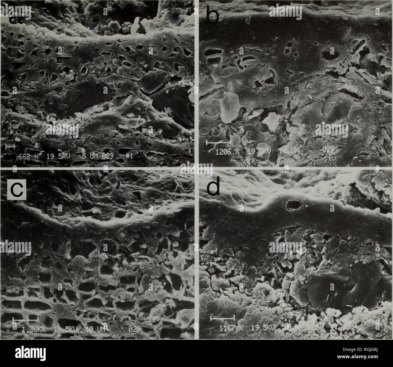
. Bulletin of the British Museum (Natural History) Botany. THELOTREMATACEAE IN SRI LANKA 235. Fig. 3 Cortical structure of the Thelotremataceae viewed with a scanning electron microscope, (a) Loosely organized cellular cortex of Thelotrema imperfectum (Hale 46 338). (b) Dense cellular cortex of Myriotrema album (Hale 4 171). (c) Loosely organized, irregularly pored cortex of Ocellularia chonestoma (Hale 46 344). (d) Dense cellular cortex of O. crassa with large crystal inclusions (Hale 51 210). discovered these peculiar hyphae in some West Indian species (Hale, 1974a: 5, Fig. 3), using the sca
Image details
Contributor:
Book Worm / Alamy Stock PhotoImage ID:
RGJGRJFile size:
7.1 MB (433.9 KB Compressed download)Releases:
Model - no | Property - noDo I need a release?Dimensions:
1700 x 1469 px | 28.8 x 24.9 cm | 11.3 x 9.8 inches | 150dpiMore information:
This image is a public domain image, which means either that copyright has expired in the image or the copyright holder has waived their copyright. Alamy charges you a fee for access to the high resolution copy of the image.
This image could have imperfections as it’s either historical or reportage.
. Bulletin of the British Museum (Natural History) Botany. THELOTREMATACEAE IN SRI LANKA 235. Fig. 3 Cortical structure of the Thelotremataceae viewed with a scanning electron microscope, (a) Loosely organized cellular cortex of Thelotrema imperfectum (Hale 46 338). (b) Dense cellular cortex of Myriotrema album (Hale 4 171). (c) Loosely organized, irregularly pored cortex of Ocellularia chonestoma (Hale 46 344). (d) Dense cellular cortex of O. crassa with large crystal inclusions (Hale 51 210). discovered these peculiar hyphae in some West Indian species (Hale, 1974a: 5, Fig. 3), using the scanning electron microscope, and can now add more details from the larger material in Sri Lanka. Aculeate hyphae are unicellular, non-gelatinized, and erect and seem to fade into the main polysaccharide cortical surface (Fig. 4a). They are far too delicate and translucent to be seen with a light microscope and usually collapse after vacuum treatment needed in electron microscope preparation. There are three major groups of species with aculeate hyphae: (1) species with a loosely organized cortex {Ocellularia chonestoma, O. dolichotata, and O. pertusariiformis already mentioned above); (2) species with a dense, continuous cortex {Myriotrema albocinctum, M. eminens, M. fissurinum, M. hartii, M. olivaceum, M. protoalbum, Ocellularia crassa (Fig. 3d), O. fissa, O. perforata, O. punctulata, and Thelotrema scabiomarginatum); and (3) species with a dense cortex which splits into sheets or layers and may exfoliate at the surface {Myriotrema album (Fig. 3b), M. cinereoglaucescens, M. clandestinum, M. costaricense, M. desquamans, M. fluorescens, M. frondosum, M. glaucophaenum, M. masonhalei, M. microstomum, M. minutum, M. polytretum, M. santessonii, M. subconforme, M. terebrans, M. terebratulum, Ocellularia albomaculata, O. croceopora, O. emersa, O. lankaensis, O.. Please note that these images are extracted from scanned page images that may have been digitally enhanced for readability - c