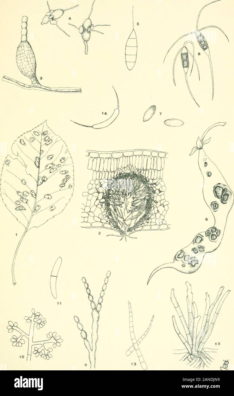
Moulds, mildews, and mushrooms; a guide to the systematic study of the Fungi and Mycetozoa and their literature . he pear. Greatly magnified. (Redrawn from Duggar.) Fig. 5. Diiiemospormm (Sphaeropsidales). Septate appendagedspore. Greatly magnified. Fig. 6. ColletotricJmm (Melanconiales) ; bean pod affected withanthracnose caused by the fungus, y^ natural size. (Redrawn fromCowing); Fig. 7, spores of same, greatly magnified. (Redrawn fromSouth worth.) Fig. 8. Pestalozzia (Melanconiales). Spores showing appendagesand hyaline end-cells. X 400. (Redrawn from Desmazieres.) Fig. 9. Monilia frudigen
Image details
Contributor:
The Reading Room / Alamy Stock PhotoImage ID:
2ANDJN9File size:
7.2 MB (214.7 KB Compressed download)Releases:
Model - no | Property - noDo I need a release?Dimensions:
1259 x 1986 px | 21.3 x 33.6 cm | 8.4 x 13.2 inches | 150dpiMore information:
This image is a public domain image, which means either that copyright has expired in the image or the copyright holder has waived their copyright. Alamy charges you a fee for access to the high resolution copy of the image.
This image could have imperfections as it’s either historical or reportage.
Moulds, mildews, and mushrooms; a guide to the systematic study of the Fungi and Mycetozoa and their literature . he pear. Greatly magnified. (Redrawn from Duggar.) Fig. 5. Diiiemospormm (Sphaeropsidales). Septate appendagedspore. Greatly magnified. Fig. 6. ColletotricJmm (Melanconiales) ; bean pod affected withanthracnose caused by the fungus, y^ natural size. (Redrawn fromCowing); Fig. 7, spores of same, greatly magnified. (Redrawn fromSouth worth.) Fig. 8. Pestalozzia (Melanconiales). Spores showing appendagesand hyaline end-cells. X 400. (Redrawn from Desmazieres.) Fig. 9. Monilia frudigena (ISIoniliales), hypha forming catenulatespores. Greatly magnified. Fig. id. Botrytis vulgaris (Moniliales). End of spore-bearinghypha with clusters of spores. Fig. II. Ramularia (Moniliales). Didymoid spore greatly magni-fied. Fig. 12. Acerailus of Cercospora gossypina (Moniliales) issuingfrom the epidermis of cotton leaf; Fig. 13, spores of same. Both greatlymagnified. ( Redrawn from Southw^orth.) Fig. 14. CeratophoriDii (Moniliales). Spore bearing appendagesat either end. Greatly magnified. (232) PL. 5.. the: MELIOTyPE PRINTING CO., BOSTON. Pl. 6.