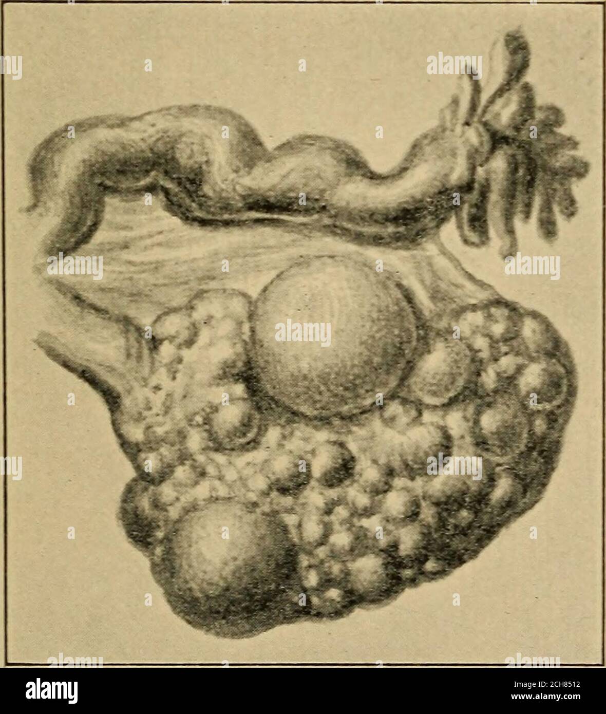
. Atlas and epitome of gynecology . Fig. 34.—Senile cirrhotic atrophy of the ovary.. Fig. 35.—Oligocystic degeneration of the ovary. inflammations of the tubes. (Fig. 36.) This oophoro-salpingitis is combined with perimetrosalpingitis, peri-metro-oophoritis, and pyosalpinx, forming, together withencapsulated ovarian and peritoneal pus sacs, a large ag- 128 CHRONIC OOPHORITIS. PLATE 44. Pelvic Peritonitis, Perioophoritis, Perisalpingitis and Right=sided Pyosalpinx. View of the pouch of Douglas. Pseudoliga-inents fix the uterus aud its aduexa to the sigmoid flexure. The lefttube is beut at an an
Image details
Contributor:
Reading Room 2020 / Alamy Stock PhotoImage ID:
2CH8512File size:
7.2 MB (299.1 KB Compressed download)Releases:
Model - no | Property - noDo I need a release?Dimensions:
1507 x 1659 px | 25.5 x 28.1 cm | 10 x 11.1 inches | 150dpiMore information:
This image is a public domain image, which means either that copyright has expired in the image or the copyright holder has waived their copyright. Alamy charges you a fee for access to the high resolution copy of the image.
This image could have imperfections as it’s either historical or reportage.
. Atlas and epitome of gynecology . Fig. 34.—Senile cirrhotic atrophy of the ovary.. Fig. 35.—Oligocystic degeneration of the ovary. inflammations of the tubes. (Fig. 36.) This oophoro-salpingitis is combined with perimetrosalpingitis, peri-metro-oophoritis, and pyosalpinx, forming, together withencapsulated ovarian and peritoneal pus sacs, a large ag- 128 CHRONIC OOPHORITIS. PLATE 44. Pelvic Peritonitis, Perioophoritis, Perisalpingitis and Right=sided Pyosalpinx. View of the pouch of Douglas. Pseudoliga-inents fix the uterus aud its aduexa to the sigmoid flexure. The lefttube is beut at an angle, the right tube shows inflammatory redness, and is transformed into a pyosalpinx by the agglutination of the ab-dominal ostium. The globular divisions of the tumor are character-istic. (See Plates 40, 42, 59, 74.). (Original water-color.) glutinated tumor (pyo-oophorosalpinx). As in purulentsalpingitis, the cause is to be found in a septic or gonor-rheal mixed infection. Sclerotic oligocystic ovarian degeneration (Plate 45 ;Plate 41, Fig. 3 ; and Fig. 35), may occur alone, l