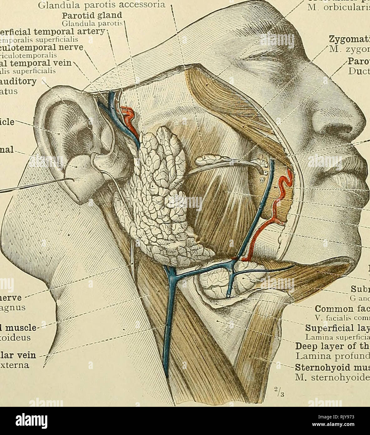
. An atlas of human anatomy for students and physicians. Anatomy. 424 CEPHALIC AND CERVICAL PORTIONS OF THE DIGESTIVE ORGANS Socia parotidis Glandula parotis accessoua Parotid gland Glandula parolisl Superficial temporal artery, A. temporalis superlicialis Auriculotemporal nerve N. aunmlotempor lU-. Superficial temporal vem - V. temporalis superhcialis ^ Cartilage of the external auditory meatusâCartilage meatus auditcrii externi Pinna, or auricle- Auricula Lobe or lobule of the external. ear (turned upwards) Lobulus auriculas. Great auricular nerve -' N. auricularis magnus Sternoc
Image details
Contributor:
Library Book Collection / Alamy Stock PhotoImage ID:
RJY973File size:
7.1 MB (604.9 KB Compressed download)Releases:
Model - no | Property - noDo I need a release?Dimensions:
1507 x 1657 px | 25.5 x 28.1 cm | 10 x 11 inches | 150dpiMore information:
This image is a public domain image, which means either that copyright has expired in the image or the copyright holder has waived their copyright. Alamy charges you a fee for access to the high resolution copy of the image.
This image could have imperfections as it’s either historical or reportage.
. An atlas of human anatomy for students and physicians. Anatomy. 424 CEPHALIC AND CERVICAL PORTIONS OF THE DIGESTIVE ORGANS Socia parotidis Glandula parotis accessoua Parotid gland Glandula parolisl Superficial temporal artery, A. temporalis superlicialis Auriculotemporal nerve N. aunmlotempor lU-. Superficial temporal vem - V. temporalis superhcialis ^ Cartilage of the external auditory meatusâCartilage meatus auditcrii externi Pinna, or auricle- Auricula Lobe or lobule of the external. ear (turned upwards) Lobulus auriculas. Great auricular nerve -' N. auricularis magnus Sternocleidomastoid muscle M. sternocleidomastoideus Kxternal jugular vein V. jugularis externa * See note ^ to p. 413. 2 In the author's nomenclature, the facial vein and the iejnporomaxiUary v posterior/acial, respectively, and the short trunk formed by the union of the facia! 1 â¢I facial An.~1:K. Orbicularis palpebrarum muscle M orbicularis oculi Zygomaticus major muscle ^I z}gomaticus , -Parotid duct, or duct of Stensen / Ductus parotidetis (Stenonis) ^^ Buccal glands (molar '^^ glands)' (ilandulae buccales Buccinator muscle Masseter muscle Facial vein- ~"'. facialis anterior ^Facial artery'' V. maxillaris externa D gastric muscle M digastricus Submaxillary gland *. mdula submaxillaris Common facial veiu'^ f iciahs communis Superficial layer of the deep cer'vical fascia Lamma superhcialis lascije colli Deep layer of the deep cervical fascia Lamina profunda lasci^e colli Sternohyoid muscle M. sternohyoideus ' ximists are termed anterior and [â division of the temporomaxillary Fig. 6bo.âGlandula Parotis, the Parotid Gland ; Glandula Submaxillaris, the Submaxillary Gland. Right Side. Tonsil (amygdala)âTonsilla palatina , Internal pterygoid muscle I[. pterygoideus internus Connective-tissue capsule of the submaxillary gland Facial arterysâA. maxillaris^-, j externa f Submaxillary gland -â f Glandula submaxillaris Digastric muscleâ M. digastricus Stylohyoid muscleâ