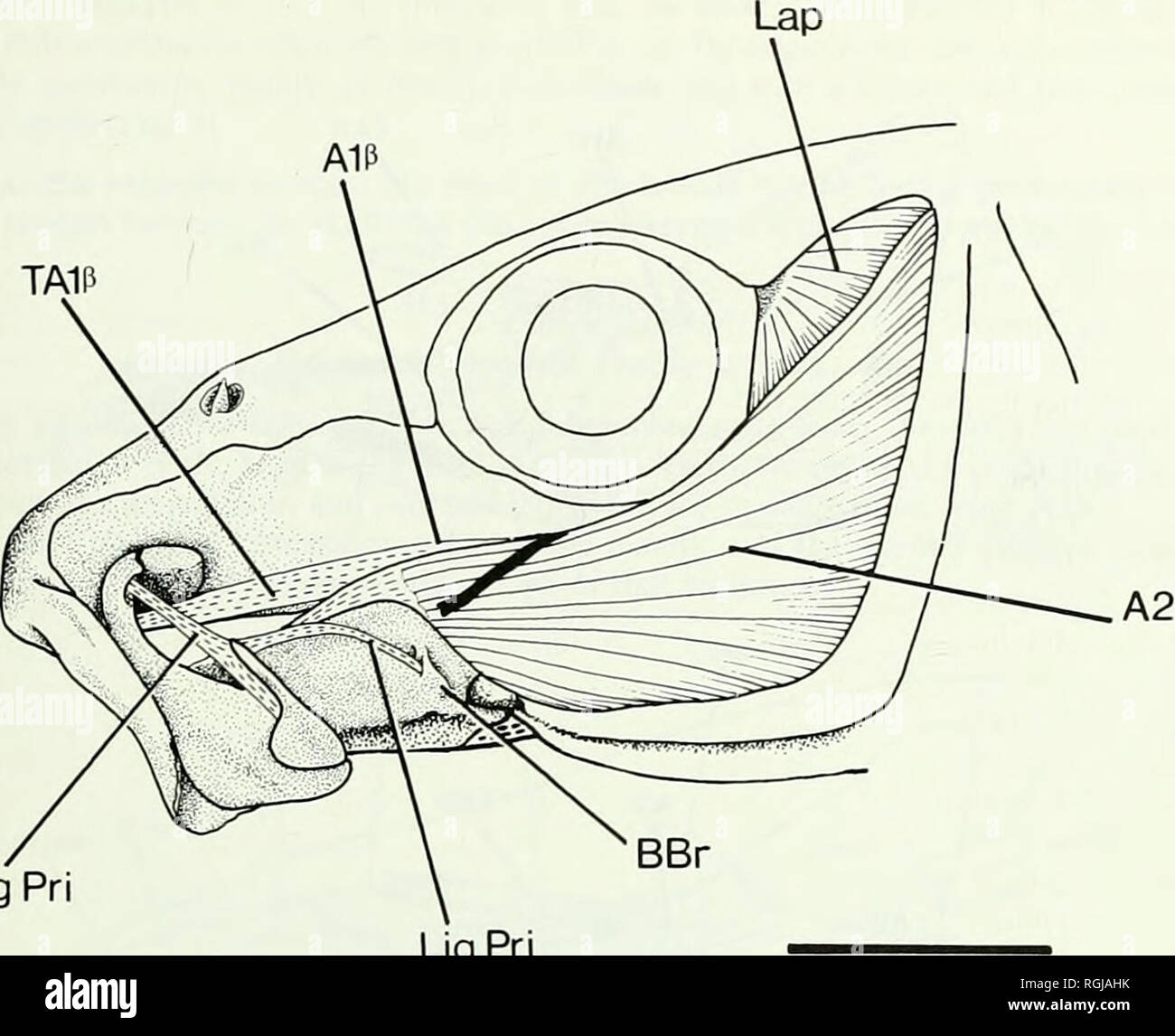
. Bulletin of the British Museum (Natural History). of its ascending process. Another ligament, the palatomaxillary, runs from the head of the auto- palatine bone to the sesamoid cartilage interposed between the palatine and the maxilla; this ligament apparently continues around the posterior face of the cartilage (to which it is very closely applied) and inserts onto the maxilla itself.. Lig Pri Lig Pri 10 mm Fig. 3 Albula vulpes. Jaw musculature and ligaments in left lateral view. Specimen BMNH 1932.2.8 : 5. Adductor mandibulae muscles (Figs 3 & 4). The adductor complex is a rather flat,
Image details
Contributor:
Book Worm / Alamy Stock PhotoImage ID:
RGJAHKFile size:
7.1 MB (247.4 KB Compressed download)Releases:
Model - no | Property - noDo I need a release?Dimensions:
1753 x 1425 px | 29.7 x 24.1 cm | 11.7 x 9.5 inches | 150dpiMore information:
This image is a public domain image, which means either that copyright has expired in the image or the copyright holder has waived their copyright. Alamy charges you a fee for access to the high resolution copy of the image.
This image could have imperfections as it’s either historical or reportage.
. Bulletin of the British Museum (Natural History). of its ascending process. Another ligament, the palatomaxillary, runs from the head of the auto- palatine bone to the sesamoid cartilage interposed between the palatine and the maxilla; this ligament apparently continues around the posterior face of the cartilage (to which it is very closely applied) and inserts onto the maxilla itself.. Lig Pri Lig Pri 10 mm Fig. 3 Albula vulpes. Jaw musculature and ligaments in left lateral view. Specimen BMNH 1932.2.8 : 5. Adductor mandibulae muscles (Figs 3 & 4). The adductor complex is a rather flat, not notice- ably voluminous muscle mass. It shows no clear-cut subdivisions, except for a single tendinous slip which arises dorsomedially near the region of the muscle's insertion onto the dentary. From here it runs forward to insert, tendinously, onto the maxilla a little behind its palatine head (Figs 3 & 4). The few muscle fibres associated with the origin of the tendon are barely separable, as an elongate torpedo-shaped aggregate, from the main body of the muscle. By definition (see Winterbottom, 1974:232) this ill-defined muscle should be identified as section Al of the adductor complex. However, it must be stressed that the muscle in Albula does not appear to have '. . developed from the dorsal enroachment of the fibres of A2 along the primordial ligament. .' (Winterbottom, 1974: 232) because an apparent ligamentum primor- dium, completely unconnected with any part of the adductores, is also present (see above). This dorsal segment of the adductor in Albula seems to be homologous with the AljS division of that muscle in Halosauridae (and Notacanthidae); see below. Hence, it will be given the same designation in this species, viz. Al/3. The remaining and major part of the adductor mass in Albula is identified as an A2 muscle, principally because it has the same relationships with the lower jaw as does the A2 division in other fishes (see Winterbottom, 1974 : 233-234)